Embed Size (px)
Citation preview

http://e-jbm.org/ 1
Copyright © 2015 The Korean Society for Bone and Mineral Research
This is an Open Access article distributed under the terms of the Creative Commons Attribution Non-Commercial Li-cense (http://creativecommons.org/licenses/by-nc/3.0/) which permits unrestricted non-commercial use, distribu-tion, and reproduction in any medium, provided the original work is properly cited.
J Bone Metab 2015;22:1-8http://dx.doi.org/10.11005/jbm.2015.22.1.1pISSN 2287-6375 eISSN 2287-7029
Pathogenesis, Management and Prevention of Atypical Femoral FracturesGun-Il Im, Seung-Hyo JeongDepartment of Orthopaedics, Dongguk University Ilsan Hospital, Goyang, Korea
Much attention has been paid to the relationship between atypical femoral fractures (AFF) and use of bisphosphonates (BPs). While a significant cause-effect relationship was not established in earlier studies, more recent data shows a growing relationship be-tween AFF and BPs use. The definition of an ‘AFF’ has also undergone significant chang-es. This review briefly summarizes the definition, pathogenesis, and management of AFF.
Key Words: Bone remodeling, Diphosphonates, Femoral fractures
INTRODUCTION
Recently, great attention has been paid to the possible relation between the prolonged use of bisphosphonates (BPs) and low-energy femoral subtrochanter and shaft fractures. Those fractures were named “atypical fractures” to distinguish them from “typical fractures” occurring at the femoral neck and trochanteric area from low-energy trauma. Most typical femoral shaft fractures occur from major high- energy trauma, such as traffic accidents or falls from heights. Unlike another complication of BP use, osteonecrosis of the jaw (ONJ), the atypical femoral frac-tures (AFF) is not associated with high doses of BPs.[1]
The first report on a possible relation between prolonged BP use and “atypical fractures” of femoral shaft was from Odvina et al.[2] They described nine patients who had fractures of the femoral shaft, proximal femur, sacrum, ischium, pubis, and ribs. Most of the patients showed delayed healing accompanied by suppress-ed bone turnover. All patients were treated with alendronate, some of them had received estrogen and glucocorticoids: all are known bone turnover-suppressing agents. After their first article, numerous sporadic case reports and case reviews followed and the AFF was characterized by clinical and radiological findings. Sev-eral retrospective case-controlled, and larger epidemiological studies were subse-quently published.[1,3]
As a systemized approach to further define AFF, the American Society for Bone and Mineral Research (ASBMR) organized a multidisciplinary task force in 2009 and published a position paper in 2010. After reviewing published articles on the epidemiology, risk factors, imaging, and clinical managements, the task force de-
Corresponding authorGun-Il ImDepartment of Orthopaedics, Dongguk University Ilsan Hospital, 27 Dongguk-ro, Ilsandong-gu, Goyang 410-773, KoreaTel: +82-31-961-7315Fax: +82-31-961-7314E-mail: [email protected]
Received: September 28, 2014Revised: January 9, 2015Accepted: January 18, 2015
No potential conflict of interest relevant to this article was reported.
Review Article
Gun-Il Im, et al.
2 http://e-jbm.org/ http://dx.doi.org/10.11005/jbm.2015.22.1.1
fined AFF and concluded that the incidence of AFF was very low, particularly considering the number of spine and hip fractures that could be prevented by BP use. They also noted that a causal relationship between BP and AFF had not been established, although statistical power was lack-ing. According to 2010 Task Force report, AFF was defined as atraumatic or low-trauma fractures located in the sub-trochanteric region or the femoral shaft. High-trauma frac-tures, femoral neck fractures, intertrochanteric fractures with spiral subtrochanteric extension, pathological frac-tures associated with primary or metastatic bone tumors, and periprosthetic fractures were excluded from the diag-nosis of AFF. Other major features of AFF included trans-verse or short oblique configuration, non-comminuted in-complete fractures involving only the lateral cortex, while complete fractures extend through both cortices and may have a medial spike. Minor features comprise localized peri-osteal reaction or beaking of the lateral cortex, generalized cortical thickening of the femoral shaft, a history of pro-dromal pain, bilateral fractures and symptoms, and de-layed healing in association with certain medication and medical conditions. All major features are needed to define a fracture as “atypical” while minor features may not be present in some cases.[1] The Food and Drug Administra-tion (FDA) addressed the issue in September 2011. The conclusion of the report was that atypical fractures were very rare (http://www.fda.gov/downloads/AdvisoryCom-mittees/CommitteesMeetingMaterials/drugs/DrugSafety-andRiskManagementAdvisoryCommittee/ucm270958.pdf). The FDA report also discussed the available evidence supporting the relationship between long-term BP treat-ment and the risk of atypical fracture. The mounted data suggest an association between BP use and atypical frac-tures, although causality has not been determined.
Recently, ASBMR published an updated version of previ-ous 2013 report that included revised criteria for AFF.[3] According to the new definition, four of the five major cri-teria (versus all) should be present to define an AFF. The absence of comminution was changed to “non-comminut-ed” or minimally comminuted, and the transverse or short oblique orientation criterion was changed to “the fracture line originates at the lateral cortex and is substantially trans-verse, although it may become oblique as it progresses medially across the femur.” A minor criterion in the 2010 version, “localized periosteal reaction of the lateral cortex”
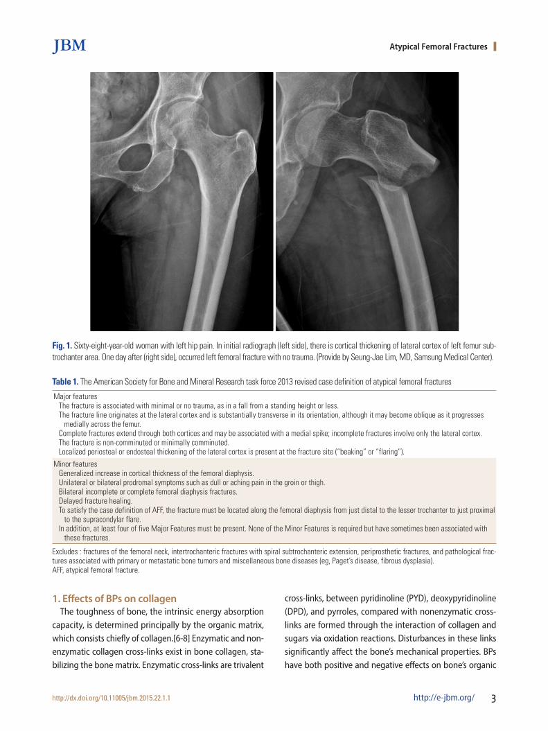
was incorporated as a major criterion: “localized periosteal or endosteal thickening of the lateral cortex is present at the fracture site (beaking or flaring)”. Reasons for changes were a positive correlation between BP use and signs of fa-tigue fractures, including transverse fracture lines on the lateral cortex, periosteal reactions, and a medial spike.[4] “Short oblique fracture line” was also deleted from the new criteria because the definition of short oblique may differ from one physician to another and most orthopedic sur-geons consider a fracture short oblique when the angle between the transverse axis and the fracture is less than 30° (Fig. 1).[4] When “short oblique” is used to define an angle between 30 and 60°, the positive association between BP use and AFF falls dramatically.[5] Compared with the 2010 position, the 2013 ASBMR position statement further clarified the relationship between AFF and BP exposure, reporting a positive correlation between exposure time and the risk of atypical fractures. The absolute risk reported varies between studies and ranges from 3.2 to 50 cases per 100,000 person-years; longer exposure is associated with higher risk (more than 100 cases per 100,000 person-years) (Table 1).[3]
Pathogenesis of AFF
Although the pathogenesis of AFF remains largely un-clear, several pathomechanisms have been proposed. The main radiological features are a transverse orientation and a lack of comminution, which are characteristics of brittle fractures. Localized cortical thickening gives clues as to “fa-tigue fractures.” The term “fatigue fracture” means a frac-ture caused by unusual, excessive repetitive loading on a normal bone while an “insufficiency fracture” indicates nor-mal loading of an abnormal or insufficient bone. In a fa-tigue fracture, fatigue damages, which are microcracks, are not repaired and accumulate, and ultimately coalesce to grow to a critical-size defect, precipitating complete frac-ture.[1,3]
One difference between AFF and exercise-induced fem-oral fatigue fractures is that AFF starts on the lateral aspect of the femur while the other often do on the medial side. Exercise-induced fatigue fractures result in a more oblique fracture surface than do AFF. In contrast, AFF have a smooth transverse fracture surface on lateral side, more character-istic of a brittle material.[3]
Atypical Femoral Fractures
http://dx.doi.org/10.11005/jbm.2015.22.1.1 http://e-jbm.org/ 3
1. Effects of BPs on collagen The toughness of bone, the intrinsic energy absorption
capacity, is determined principally by the organic matrix, which consists chiefly of collagen.[6-8] Enzymatic and non-enzymatic collagen cross-links exist in bone collagen, sta-bilizing the bone matrix. Enzymatic cross-links are trivalent
cross-links, between pyridinoline (PYD), deoxypyridinoline (DPD), and pyrroles, compared with nonenzymatic cross-links are formed through the interaction of collagen and sugars via oxidation reactions. Disturbances in these links significantly affect the bone’s mechanical properties. BPs have both positive and negative effects on bone’s organic
Fig. 1. Sixty-eight-year-old woman with left hip pain. In initial radiograph (left side), there is cortical thickening of lateral cortex of left femur sub-trochanter area. One day after (right side), occurred left femoral fracture with no trauma. (Provide by Seung-Jae Lim, MD, Samsung Medical Center).Figure 1. 68 years old woman with left hip pain. In initial
radiograph(left side), there is cortical thickening of lateral cortex of left femur subtrochanter area. One day after(right side), occurred left femoral fracture with no trauma.(Provide by Seung-Jae Lim, MD, Samsung medical center)
Figure 1. 68 years old woman with left hip pain. In initial radiograph(left side), there is cortical thickening of lateral cortex of left femur subtrochanter area. One day after(right side), occurred left femoral fracture with no trauma.(Provide by Seung-Jae Lim, MD, Samsung medical center)
Table 1. The American Society for Bone and Mineral Research task force 2013 revised case definition of atypical femoral fractures
Major featuresThe fracture is associated with minimal or no trauma, as in a fall from a standing height or less.The fracture line originates at the lateral cortex and is substantially transverse in its orientation, although it may become oblique as it progresses medially across the femur.Complete fractures extend through both cortices and may be associated with a medial spike; incomplete fractures involve only the lateral cortex.The fracture is non-comminuted or minimally comminuted.Localized periosteal or endosteal thickening of the lateral cortex is present at the fracture site (“beaking” or “flaring”).
Minor featuresGeneralized increase in cortical thickness of the femoral diaphysis.Unilateral or bilateral prodromal symptoms such as dull or aching pain in the groin or thigh.Bilateral incomplete or complete femoral diaphysis fractures.Delayed fracture healing.To satisfy the case definition of AFF, the fracture must be located along the femoral diaphysis from just distal to the lesser trochanter to just proximal to the supracondylar flare.In addition, at least four of five Major Features must be present. None of the Minor Features is required but have sometimes been associated with these fractures.
Excludes : fractures of the femoral neck, intertrochanteric fractures with spiral subtrochanteric extension, periprosthetic fractures, and pathological frac-tures associated with primary or metastatic bone tumors and miscellaneous bone diseases (eg, Paget’s disease, fibrous dysplasia).AFF, atypical femoral fracture.
Gun-Il Im, et al.
4 http://e-jbm.org/ http://dx.doi.org/10.11005/jbm.2015.22.1.1
matrix by altering both collagen maturity and cross-link-ing. An increased PYD/DPD ratio is associated with increas-ed strength and stiffness of bone.[9] The PYD/DPD ratio was increased significantly, along with mechanical strength, in vertebral cancellous bone and tibial cortical bone from dogs treated with BPs for 1 year, compared with untreated controls.[6,10] On the other hand, reducing bone turnover also increases pentosidine levels, which are markers for advanced glycation end products (AGEs). AGEs are associ-ated with tissue that is more brittle.[10] Accumulation of pentosidine in bone has been reported to reduce post-yield deformation[9,11] and toughness.[6,12] Tissue from both vertebral[13] and tibial[10] bone from BP-treated animals was less tough than bone from animals not treated with BPs.
There are few data on collagen cross-links in humans treat-ed with BPs, whereas Fourier-transform infrared spectros-copy (FTIR) data show that BP treatment prevented the maturation of collagen and reduced collagen maturity in newly formed bone.[14] Another report[15] reported no change in collagen maturity in women treated with alen-dronate.[16]
2. Effects of BPs on angiogenesisAny agent that suppresses angiogenesis may inhibit the
repair of an impending fatigue fracture. The effects of BPs on the repair of fatigue fracture could be worsened if BPs are antiangiogenic.[17] Several studies in nonskeletal tis-sues have shown that BPs reduce angiogenesis.[18] While direct suppression of angiogenesis by BPs was reported previously,[19] it is difficult to make a distinction between the inhibition of new vessel growth and the suppression of osteoclastic activity because these two phenomena are usually coupled to each other. However, studies of grow-ing animals during skeletal development demonstrated no apparent antiangiogenic effect of clodronate.[20]
3. Effect of BPs on bone material propertiesAnimal studies show that treatment with BPs is associated
with reduced bone toughness.[21-23] Following 1-3 years of BP treatment at doses similar to or greater than those used in postmenopausal women, toughness was 20-30% lower than in control animals.[21,22] Toughness continues to decline in animals with long-term BP treatment without an increase in microdamage accumulation or a further in-crease in secondary mineralization.[13] After feeding using
various doses of alendronate or risedronate for 1 year, there was minimal correspondence between changes in micro-damage accumulation and material-level toughness in ver-tebrae from several groups of BP-treated dogs.[21] Animals not treated with BPs show an age-related three-fold increase in microdamage accumulation without any apparent change in bone toughness.[13] Neither microdamage nor increas-ed secondary mineralization is solely responsible for the change in bone material properties with BP therapy, leav-ing changes in collagen or interactions among all these properties as likely reasons for the progressive decline in toughness.[1,3]
Decreased remodeling is also not solely responsible for reduced toughness, indicating a specific effect of BPs that is independent of reduced turnover. The mechanical effect of BPs in decreasing tissue toughness is countered by their capacity to increase bone mass and mineralization, pro-mote collagen matrix maturation, and prevent microarchi-tectural deterioration of bone.[3] These factors lead to in-creases in bone strength and stiffness that offset reduced toughness and make bone stronger at the structural level. When FTIR was used to compare the physical properties of cortical and cancellous bone of the proximal femur, there was no difference in mineralization, crystallinity, or colla-gen maturity between 19 BP-naïve typical femoral fracture patients and 13 BP-positive femoral fractures patients who had taken BP for an average of 7 years. Those who had tak-en BPs had significantly more homogenous crystallinity and collagen maturity. Greater uniformity of tissue compo-sition was found in those treated with BP. When material stiffness and toughness were measured by micro-indenta-tion techniques, long-term BP users who did not have frac-ture did not show significantly deteriorated properties com-pared with untreated controls. Furthermore, patients with AFF who had been treated with BP for 5.5 years did not have worse properties compared with typical fractures. These results suggest that there is not sufficient evidence that the use of BPs adversely affects the material properties of bone.[3]
4. Effects of BP on healing of fatigue fracturesWhen a complete fracture occurs and is not fixed rigidly,
endochondral ossification is the main healing mechanism of the fracture. An initial inflammatory response is followed by the formation of a chondroid callus. Remodeling that
Atypical Femoral Fractures
http://dx.doi.org/10.11005/jbm.2015.22.1.1 http://e-jbm.org/ 5
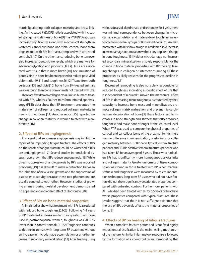
includes resorption of the chondroid callus and replace-ment with lamellar bone then continues over a long peri-od. Osteoclasts play an important role during the remodel-ing phase. BPs do not impair the inflammatory process or the development of a proliferative callus.[24,25] BPs inter-fere with the remodeling phase, delaying the remodeling of the calcified cartilage callus to mature bone. Bone re-modeling is important in the healing of fatigue fractures, and is reduced by BP treatment (Fig. 2).
Animal studies with BP treatment showed that prior alen-dronate eliminated the adaptive remodeling response, sug-gesting that BP treatment impaired the healing response to a fatigue fracture.[26] In a process of developing fatigue fracture, reduction of bone remodeling would prevent or delay the healing of the stress reaction without suppress-ing the appearance of a periosteal callus and eventually result in a complete fracture of the fatigued site.
BPs increase bone strength and decrease fracture risk by suppressing excessive bone remodeling. On the other hand, the reduction of remodeling increases microdamage accu-mulation because microcracks are not removed efficiently. Even in the absence of BP treatment, age-related reduc-tions in bone turnover result in microdamage accumula-tion.[13] Damage accumulates significantly in humans af-ter the age of 70 years,[27,28] although there is broad indi-vidual variability in the amount.
Small decrease in turnover may induce significant accu-mulation of microdamage. Animal studies showed that re-ducing trabecular bone activation frequency by ~40% with risedronate caused a 3-fold increase in microdamage, com-pared with untreated controls, in the canine vertebra.[21]
Suppression by ~20% with raloxifene caused a doubling of microdamage.[29]
There are conflicting data on whether microdamage ac-cumulates with BP treatment in humans. Women treated for an average of 5 years with alendronate showed signifi-cant microcrack accumulation in a subsample.[30] Another study did not find any association between BP treatment and damage accumulation in the iliac crest.[31] It is diffi-cult to confirm whether damage accumulates in the cortex of the femoral diaphysis because neither study evaluated samples from the femoral cortex, and the accumulation of microdamage is site-specific.
The histological appearance was investigated from iliac crest biopsies and biopsies from fracture sites of patients who had been treated with BPs. While bone turnover was suppressed, normal osteoblastic bone formation was not suppressed. BPs do not suppress the formation of the ini-tial callus or affect the formation of woven bone. However, incomplete repair of fractures occurs by the normal cou-pled bone remodeling processes. BP localizes on the site of high turnover, because those sites are associated with in-creased blood flow. With BP suppressing remodeling, in-tracortical repair of developing fatigue fractures is ham-pered and the microcracks can grow to a critical size.[3]
5. Lower limb geometry and AFFThe geometry of the proximal femur partly determines
the stress generated on the lateral aspect of the femoral cortex.[32] The site of AFF along the diaphysis is highly cor-related with the deviation between the anatomical axis (tibiofemoral angle) and the mechanical axis of the lower limb.[33] Patients with more diaphyseal AFF had larger tib-iofemoral angles than those who fractured closer to the lesser trochanter. In a Japanese population, patients who had AFF had significantly greater curvature of the femoral diaphysis than age- and gender matched controls.[33]
Prevention of AFF
Assessment of the benefits and risks before BP treatment is essential to avoid unnecessary complications, such as AFF. Patients at low risk of osteoporosis-related fractures do not need medical treatment. For patients with osteopo-rosis in the spine plus normal or only moderately reduced bone mineral density (BMD) in proximal femur, alternative
InductionHematoma formation on fractured site
InflammationMigration of inflammatory cells
Soft callusFibrocartilaginous callus formation
OssificationCalcified cartilaginous callus
RemodelingMature bone formation
OsteoclastResorption immatured bone
OsteoblastMature bone formation &
mineralization
Bisphosphonate
Inhibit osteoclast formation, migration, and osteolytic activity
Promote apoptosis of osteoclast
Figure 2. Action of BPs in bone healing processFig. 2. Action of bisphosphonates in bone healing process.
Gun-Il Im, et al.
6 http://e-jbm.org/ http://dx.doi.org/10.11005/jbm.2015.22.1.1
treatments for osteoporosis, such as raloxifene or teripara-tide, is indicated depending on the severity of the patient’s condition.[3]
BP therapy is strongly indicated to protect patients from rapid bone loss and increased fracture rates associated with organ transplantation, endocrine disorders, or chemother-apy for breast or prostate cancer, and when aromatase in-hibitors and glucocorticoids are first used. Even in these clinical conditions, long-term BP therapy is not always nec-essary.[34,35]
The optimal duration of BP treatment is still unclear. While studies with alendronate[36] and risedronate[37,38] shows that patients with osteoporosis will have an antifracture benefit for at least 5 years, continued use of BP therapy over 5 years need annual re-evaluation, assessing factors such as BMD, fracture history, newly diagnosed disorders, and other medications known to affect skeletal status, as well as new research findings.
For patients with moderately elevated fracture risk, con-tinuation of BP therapy should be strongly considered. Re-cent or multiple fractures suggest assessment for underly-ing secondary causes and reevaluation of the treatment plan. As these patients are known to be at high risk of fu-ture fracture, discontinuation of BP treatment is not advised. It is not certain whether BP treatment beyond 5 years will reduce the risk. The incidence of clinical (not morphomet-ric) vertebral fractures was significantly lower in those on 10 years of continued alendronate versus those who stopp-ed after 5 years [36] while a reduction in nonvertebral frac-ture incidence was limited to women without a fracture history but with femoral neck T scores that were 2.5 or less.[39] With risedronate, 7 years of therapy did not further re-duce the incidence of vertebral fractures, compared with 3 and 5 years of treatment.[38]
Taking into account the fact that the median BP treat-ment duration in patients with AFF is 7 years, for patients without a recent fracture and with femoral neck T-scores greater than 2.5 after the initial therapeutic course, consid-eration may be given to a “drug holiday’’ from BPs. Contin-ued BP therapy should be reevaluated, particularly in those deemed to be at low or only modestly elevated fracture risk.[1,3]
Whether abandoning BP treatment after 4-5 years in the lower-risk group will lead to fewer atypical subtrochanteric fractures is uncertain. Restarting osteoporosis therapy, with
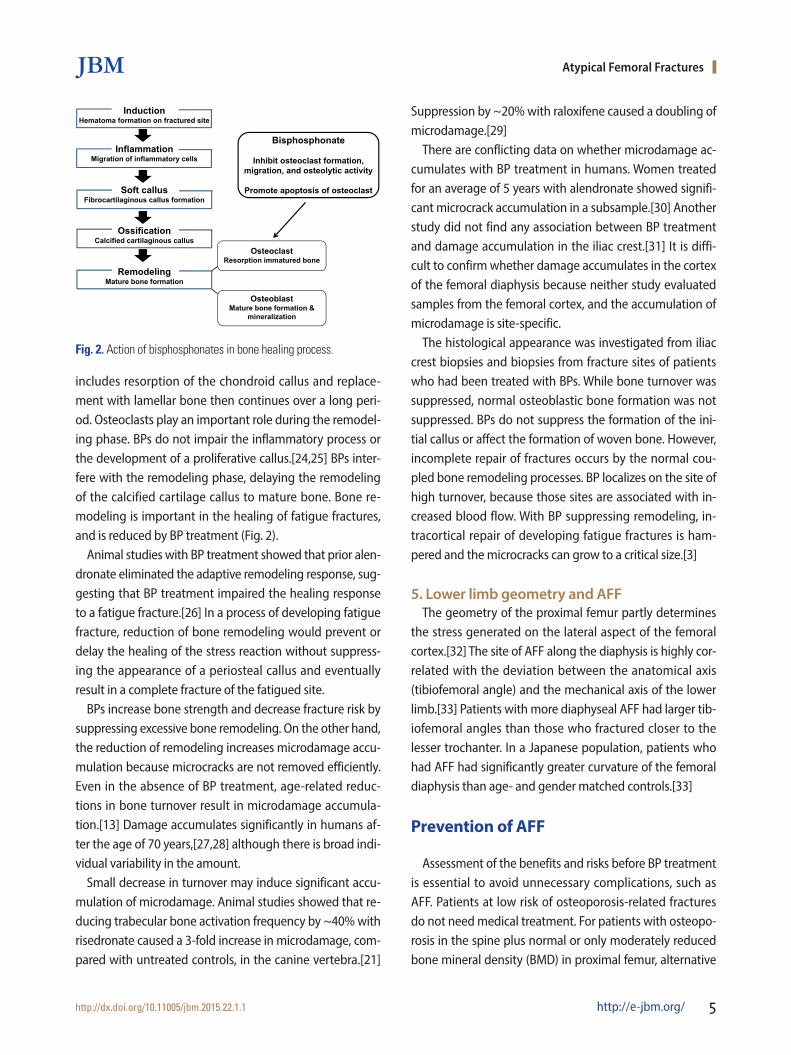
BPs or with a different class of agent, can be considered in patients who appear to be at increasing fracture risk. As there can be no general rule on “BP holidays”, decisions to stop and/or restart therapy must be individualized (Fig. 3).[1,3]
Management of AFF
More than half of the patients who suffer AFF have a pro-dromal thigh or groin pain before frank fracture develops. Thus, it is necessary to educate physicians and patients about this symptom. Physicians should ask patients on BPs and other potent anti-resorptive agents about thigh or groin pain.[1,3]
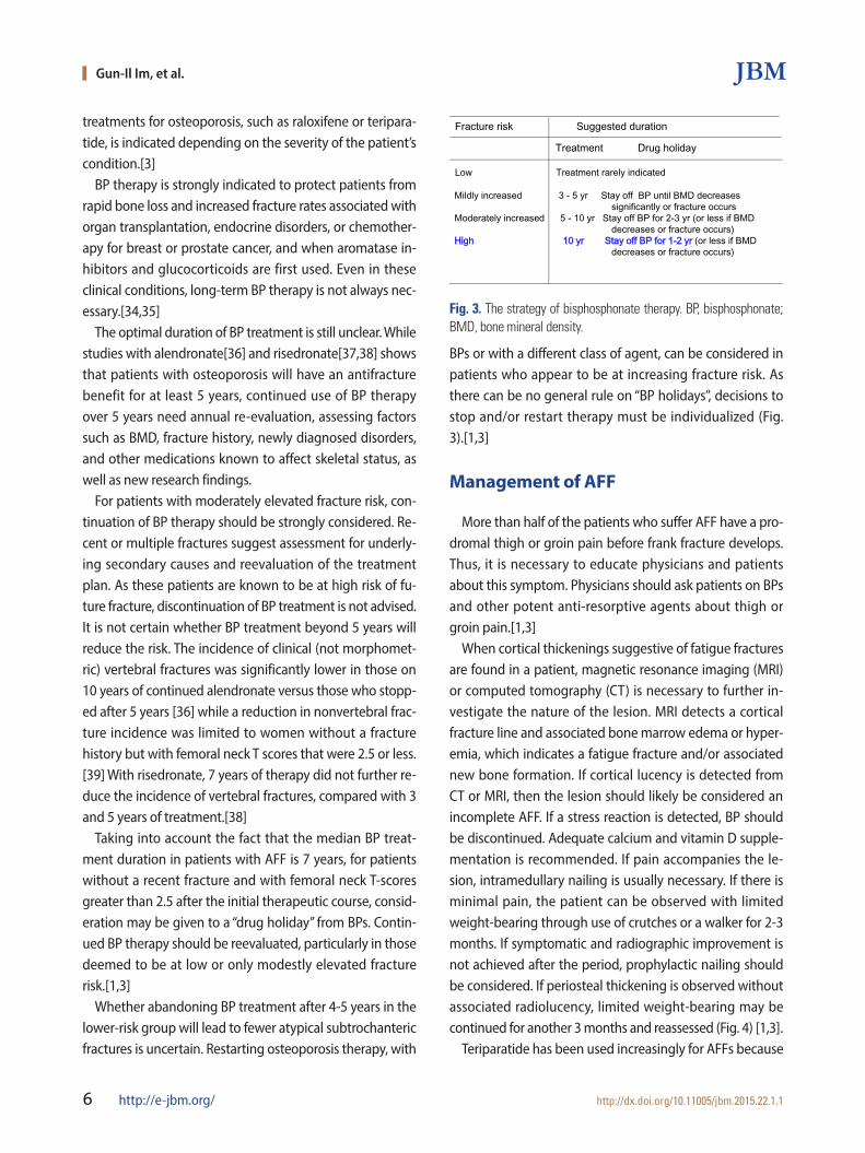
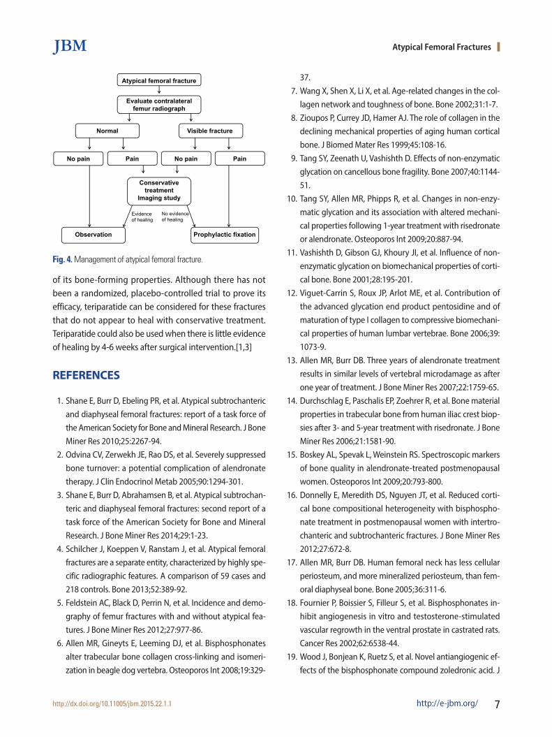
When cortical thickenings suggestive of fatigue fractures are found in a patient, magnetic resonance imaging (MRI) or computed tomography (CT) is necessary to further in-vestigate the nature of the lesion. MRI detects a cortical fracture line and associated bone marrow edema or hyper-emia, which indicates a fatigue fracture and/or associated new bone formation. If cortical lucency is detected from CT or MRI, then the lesion should likely be considered an incomplete AFF. If a stress reaction is detected, BP should be discontinued. Adequate calcium and vitamin D supple-mentation is recommended. If pain accompanies the le-sion, intramedullary nailing is usually necessary. If there is minimal pain, the patient can be observed with limited weight-bearing through use of crutches or a walker for 2-3 months. If symptomatic and radiographic improvement is not achieved after the period, prophylactic nailing should be considered. If periosteal thickening is observed without associated radiolucency, limited weight-bearing may be continued for another 3 months and reassessed (Fig. 4) [1,3].
Teriparatide has been used increasingly for AFFs because
Fracture risk Suggested duration
Treatment Drug holiday
Low Treatment rarely indicated
Mildly increased 3 - 5 yr Stay off BP until BMD decreasessignificantly or fracture occurs
Moderately increased 5 - 10 yr Stay off BP for 2-3 yr (or less if BMDdecreases or fracture occurs)
High 10 yr Stay off BP for 1-2 yr (or less if BMD decreases or fracture occurs)
Figure 3. The strategy of bisphosphonate therapyFig. 3. The strategy of bisphosphonate therapy. BP, bisphosphonate; BMD, bone mineral density.
Atypical Femoral Fractures
http://dx.doi.org/10.11005/jbm.2015.22.1.1 http://e-jbm.org/ 7
of its bone-forming properties. Although there has not been a randomized, placebo-controlled trial to prove its efficacy, teriparatide can be considered for these fractures that do not appear to heal with conservative treatment. Teriparatide could also be used when there is little evidence of healing by 4-6 weeks after surgical intervention.[1,3]
REFERENCES
1. Shane E, Burr D, Ebeling PR, et al. Atypical subtrochanteric and diaphyseal femoral fractures: report of a task force of the American Society for Bone and Mineral Research. J Bone Miner Res 2010;25:2267-94.
2. Odvina CV, Zerwekh JE, Rao DS, et al. Severely suppressed bone turnover: a potential complication of alendronate therapy. J Clin Endocrinol Metab 2005;90:1294-301.
3. Shane E, Burr D, Abrahamsen B, et al. Atypical subtrochan-teric and diaphyseal femoral fractures: second report of a task force of the American Society for Bone and Mineral Research. J Bone Miner Res 2014;29:1-23.
4. Schilcher J, Koeppen V, Ranstam J, et al. Atypical femoral fractures are a separate entity, characterized by highly spe-cific radiographic features. A comparison of 59 cases and 218 controls. Bone 2013;52:389-92.
5. Feldstein AC, Black D, Perrin N, et al. Incidence and demo-graphy of femur fractures with and without atypical fea-tures. J Bone Miner Res 2012;27:977-86.
6. Allen MR, Gineyts E, Leeming DJ, et al. Bisphosphonates alter trabecular bone collagen cross-linking and isomeri-zation in beagle dog vertebra. Osteoporos Int 2008;19:329-
37.7. Wang X, Shen X, Li X, et al. Age-related changes in the col-
lagen network and toughness of bone. Bone 2002;31:1-7.8. Zioupos P, Currey JD, Hamer AJ. The role of collagen in the
declining mechanical properties of aging human cortical bone. J Biomed Mater Res 1999;45:108-16.
9. Tang SY, Zeenath U, Vashishth D. Effects of non-enzymatic glycation on cancellous bone fragility. Bone 2007;40:1144-51.
10. Tang SY, Allen MR, Phipps R, et al. Changes in non-enzy-matic glycation and its association with altered mechani-cal properties following 1-year treatment with risedronate or alendronate. Osteoporos Int 2009;20:887-94.
11. Vashishth D, Gibson GJ, Khoury JI, et al. Influence of non-enzymatic glycation on biomechanical properties of corti-cal bone. Bone 2001;28:195-201.
12. Viguet-Carrin S, Roux JP, Arlot ME, et al. Contribution of the advanced glycation end product pentosidine and of maturation of type I collagen to compressive biomechani-cal properties of human lumbar vertebrae. Bone 2006;39: 1073-9.
13. Allen MR, Burr DB. Three years of alendronate treatment results in similar levels of vertebral microdamage as after one year of treatment. J Bone Miner Res 2007;22:1759-65.
14. Durchschlag E, Paschalis EP, Zoehrer R, et al. Bone material properties in trabecular bone from human iliac crest biop-sies after 3- and 5-year treatment with risedronate. J Bone Miner Res 2006;21:1581-90.
15. Boskey AL, Spevak L, Weinstein RS. Spectroscopic markers of bone quality in alendronate-treated postmenopausal women. Osteoporos Int 2009;20:793-800.
16. Donnelly E, Meredith DS, Nguyen JT, et al. Reduced corti-cal bone compositional heterogeneity with bisphospho-nate treatment in postmenopausal women with intertro-chanteric and subtrochanteric fractures. J Bone Miner Res 2012;27:672-8.
17. Allen MR, Burr DB. Human femoral neck has less cellular periosteum, and more mineralized periosteum, than fem-oral diaphyseal bone. Bone 2005;36:311-6.
18. Fournier P, Boissier S, Filleur S, et al. Bisphosphonates in-hibit angiogenesis in vitro and testosterone-stimulated vascular regrowth in the ventral prostate in castrated rats. Cancer Res 2002;62:6538-44.
19. Wood J, Bonjean K, Ruetz S, et al. Novel antiangiogenic ef-fects of the bisphosphonate compound zoledronic acid. J
Atypical femoral fracture
Evaluate contralateral femur radiograph
Normal Visible fracture
No pain Pain No pain Pain
Observation
Conservativetreatment
Imaging study
Prophylactic fixation
Evidenceof healing
No evidence of healing
Figure 4. Management of atypical femoral fractureFig. 4. Management of atypical femoral fracture.

Gun-Il Im, et al.
8 http://e-jbm.org/ http://dx.doi.org/10.11005/jbm.2015.22.1.1
Pharmacol Exp Ther 2002;302:1055-61.20. Deckers MM, Van Beek ER, Van Der Pluijm G, et al. Dissoci-
ation of angiogenesis and osteoclastogenesis during en-dochondral bone formation in neonatal mice. J Bone Min-er Res 2002;17:998-1007.
21. Allen MR, Iwata K, Phipps R, et al. Alterations in canine ver-tebral bone turnover, microdamage accumulation, and biomechanical properties following 1-year treatment with clinical treatment doses of risedronate or alendronate. Bone 2006;39:872-9.
22. Allen MR, Reinwald S, Burr DB. Alendronate reduces bone toughness of ribs without significantly increasing micro-damage accumulation in dogs following 3 years of daily treatment. Calcif Tissue Int 2008;82:354-60.
23. Mashiba T, Turner CH, Hirano T, et al. Effects of suppressed bone turnover by bisphosphonates on microdamage ac-cumulation and biomechanical properties in clinically rel-evant skeletal sites in beagles. Bone 2001;28:524-31.
24. Cao Y, Mori S, Mashiba T, et al. Raloxifene, estrogen, and alendronate affect the processes of fracture repair differ-ently in ovariectomized rats. J Bone Miner Res 2002;17: 2237-46.
25. Martinez MD, Schmid GJ, McKenzie JA, et al. Healing of non-displaced fractures produced by fatigue loading of the mouse ulna. Bone 2010;46:1604-12.
26. Barrett JG, Sample SJ, McCarthy J, et al. Effect of short-term treatment with alendronate on ulnar bone adaptation to cyclic fatigue loading in rats. J Orthop Res 2007;25:1070-7.
27. Mori S, Harruff R, Ambrosius W, et al. Trabecular bone vol-ume and microdamage accumulation in the femoral heads of women with and without femoral neck fractures. Bone 1997;21:521-6.
28. Schaffler MB, Choi K, Milgrom C. Aging and matrix micro-damage accumulation in human compact bone. Bone 1995; 17:521-25.
29. Allen MR, Iwata K, Sato M, et al. Raloxifene enhances ver-tebral mechanical properties independent of bone densi-ty. Bone 2006;39:1130-5.
30. Stepan JJ, Burr DB, Pavo I, et al. Low bone mineral density is associated with bone microdamage accumulation in post-menopausal women with osteoporosis. Bone 2007;41:378-85.
31. Chapurlat RD, Arlot M, Burt-Pichat B, et al. Microcrack fre-quency and bone remodeling in postmenopausal osteo-porotic women on long-term bisphosphonates: a bone biopsy study. J Bone Miner Res 2007;22:1502-9.
32. Crossley K, Bennell KL, Wrigley T, et al. Ground reaction forc-es, bone characteristics, and tibial stress fracture in male runners. Med Sci Sports Exerc 1999;31:1088-93.
33. Sasaki S, Miyakoshi N, Hongo M, et al. Low-energy diaphy-seal femoral fractures associated with bisphosphonate use and severe curved femur: a case series. J Bone Miner Metab 2012;30:561-7.
34. Cohen A, Addesso V, McMahon DJ, et al. Discontinuing an-tiresorptive therapy one year after cardiac transplantation: effect on bone density and bone turnover. Transplantation 2006;81:686-91.
35. Hershman DL, McMahon DJ, Crew KD, et al. Prevention of bone loss by zoledronic acid in premenopausal women undergoing adjuvant chemotherapy persist up to one year following discontinuing treatment. J Clin Endocrinol Metab 2010;95:559-66.
36. Black DM, Schwartz AV, Ensrud KE, et al. Effects of continu-ing or stopping alendronate after 5 years of treatment: the Fracture Intervention Trial Long-term Extension (FLEX): a randomized trial. JAMA 2006;296:2927-38.
37. Mellström DD, Sörensen OH, Goemaere S, et al. Seven years of treatment with risedronate in women with postmeno-pausal osteoporosis. Calcif Tissue Int 2004;75:462-8.
38. Sorensen OH, Crawford GM, Mulder H, et al. Long-term ef-ficacy of risedronate: a 5-year placebo-controlled clinical experience. Bone 2003;32:120-6.
39. Schwartz AV, Bauer DC, Cummings SR, et al. Efficacy of con-tinued alendronate for fractures in women with and with-out prevalent vertebral fracture: the FLEX trial. J Bone Min-er Res 2010;25:976-82.