Embed Size (px)
Citation preview
PATHOBIOLOGY IN FOCUS
Patient-derived xenografts, the cancer stem cellparadigm, and cancer pathobiology in the 21st centurySamuel A Williams, Wade C Anderson, Marianne T Santaguida and Scott J Dylla
Cancer is a heterogeneous disease manifest in many forms. Tumor histopathology can differ significantly among patientsand cellular heterogeneity within tumors is common. A primary goal of cancer biologists is to better understandtumorigenesis and cancer progression; however, the complex nature of tumors has posed a substantial challenge tounlocking cancer’s secrets. The cancer stem cell (CSC) paradigm for the pathobiology of solid tumors appropriatelyacknowledges phenotypic and functional tumor cell heterogeneity observed in solid tumors and accounts for thedisconnect between drug approval based on response and the general inability of approved therapies to meaningfullyimpact survival due to their failure to eradicate these most important of cellular targets. First proposed to exist decadesago, CSC have only recently begun to be precisely identified due to technical advancements that facilitate identification,isolation, and interrogation of distinct tumor cell subpopulations with differing ability to form and perpetuate tumors.Precise identification of CSC populations and the complete hierarchy of cells within solid tumors will facilitate moreaccurate characterization of patient subtypes and ultimately contribute to more personalized and effective therapies.Rapid advancement in the understanding of tumor biology as it exists in patients requires cooperation amonginstitutions, surgeons, pathologists, cancer biologists and patients alike, primarily because this translational research isbest done with patient-derived tissue grown in the xenograft setting as patient-derived xenografts. This review callsfor a broader change in the approaches taken to study cancer pathobiology, highlights what implications the CSCparadigm has for pathologists and cancer biologists alike, and calls for greater collaboration between institutions,physicians and scientists in order to more rapidly advance our collective understanding of cancer.Laboratory Investigation (2013) 93, 970–982; doi:10.1038/labinvest.2013.92; published online 5 August 2013
KEYWORDS: cancer stem cell; xenograft
INTRODUCTIONDespite significant time, money, and effort expended oncancer research over the last half century, the contribution ofnewly developed therapeutics to improved patient survivalhas been limited (Table 1).1–3 Treatment options for patientswith many solid tumors remains unchanged vs therapiesoffered decades ago. Furthermore, improvements to overallsurvival have resulted from successes associated with earlydetection and cancer prevention initiatives.4,5 Technologicalinnovations such as genetic testing (eg, characterizing BRCA1and BRCA2 mutations), biomarker detection (eg, PSA),and tissue monitoring (eg, colonoscopy) have producedsignificant improvements in the prevention and early detec-tion of breast, prostate, and colorectal cancers, respectively,often permitting preventative or curative localized resectionat early stages of disease. The reduced incidence of smokinghas also significantly contributed to the reduced prevalence
of lung cancer, and consequently decreased the number ofdeaths in the United States as a result of this and othersmoking-related malignancies (though the death rate forwomen with lung cancer rose 6% between 1990–2008 and2012).3,6 Notwithstanding some successes, overall cancermortality continues to rise and is estimated to nearly doubleby 2030 if better early detection technologies and therapiesthat significantly impact survival are not discovered anddeveloped.3
The vast majority of cancer patients continue to be treatedwith untargeted chemotherapeutic agents and/or radiationfollowing surgical resection as primary, and often secondary,courses of action. For many indications, these standard ofcare chemotherapy and/or radiation treatments have changedlittle over the past several decades.1,2,7–9 Although theseinterventions often result in substantial tumor regression,chemo- and radiotherapy regimens are poorly tolerated and
Cancer Biology, Stem CentRx, Inc., South San Francisco, CA, USACorrespondence: Dr SJ Dylla, PhD, Cancer Biology, Stem CentRx, Inc., 450 East Jamie Court, South San Francisco, CA 94080, USA.E-mail: [email protected]
Received 10 February 2013; revised 27 May 2013; accepted 13 June 2013
970 Laboratory Investigation | Volume 93 September 2013 | www.laboratoryinvestigation.org
Laboratory Investigation (2013) 93, 970–982
& 2013 USCAP, Inc All rights reserved 0023-6837/13 $32.00
generally result in the evolution of a more aggressive andrefractory disease that emerges to cause death. As a result,cancer mortality rates remain at levels similar to what wasobserved three decades ago (Table 1; http://seer.cancer.gov/data/citation.html). The discovery and development of suc-cessful targeted therapies has proceeded at an agonizinglyslow pace, possibly because cancer biologists have tradition-ally not had the correct tools, ample access to patient tumorspecimens that provide insight into the full heterogeneity anddiversity of human malignancies, nor employed appropriateexperimental design paradigms to address tumor biology as itexists in patients. Technical and conceptual advancements forhow cancer biologists study and think about cancer calls for achange that should result in greater success in the next severaldecades than what has been achieved over the last century.
ADDRESSING TUMOR HETEROGENEITYEach cancer patient’s tumor is heterogeneous and unique.Diagnosing patients based on histomorphological featuresand an abbreviated panel of immunohistochemistry markersprovides little information that can be considered distinctiveto any given patient. Although these tools have been suc-cessful at segregating tumors into broad subclasses, they havelargely failed to capture or elucidate the enormous variationof disease within indications. For many malignancies, theexistence of additional subtypes has been implied by thedifferential responses of patients to therapeutic regimens. Forothers, molecular markers, gene expression profiling, and themore recent implementation of next-generation DNA-sequencing technologies have helped reveal a broader spec-trum of heterogeneity among patient tumors.10–14 In an agewhere next-generation whole-genome and transcriptomesequencing are not only possible, but increasingly feasibleand rapid, diagnoses and treatment options will increasinglybe tailored to each patient’s tumor using information fromthese and other novel platforms.15–17 For example, genomicDNA sequencing efforts have already begun to identify
existing approved or experimental drugs likely to beefficacious in particular patient subgroups.15,18,19 Despitethese instances, population studies have revealed that mosttumors do not have an isolated driver mutation that can betargeted with an existing therapeutic agent.11,20,21
Consequently, few patients currently benefit from dataobtained using novel platform technologies such as next-generation sequencing, and thus diagnostic and treatmentoption considerations are not widely influenced by thisinformation (reviewed in previous studies18,22–24). Thisshould change as more knowledge is accumulated aroundhow particular genomic mutations impact and alter specificsignaling pathways that can be targeted by small molecule orbiological inhibitors, or combinations thereof. Inherent levelsof complexity need to be better understood for these next-generation platforms to have their utmost utility in realizingthe promise of personalized medicine, and development ofunique treatment regimens for individual patients may notbe practical for some time. Nevertheless, most would agreethat patient diagnosis and treatment considerations shouldtake into account both an individual tumor’s genetics andbiology, determined as close to the initial time of diagnosis aspossible.
As pathologists are all too well aware, malignant tumorsconsist of heterogeneous cellular compositions: an inter-connected mass of dividing, differentiating and dyingcells, supporting stromal cells, infiltrating vasculatureand ensemble of hematopoietic cells. Since Rudolf Virchowfirst proposed that cancer was a cell-based disease in the mid-1850’s,25 the cellular complexity comprising neoplastic malig-nancies has been widely recognized and studied. Underlyingcomplexities are now coming into focus. Specifically, threeinterrelated and increasingly important concepts in cancerbiology are revolutionizing our understanding of tumorpathobiology and should lead to improved therapeuticoptions for cancer patients. First, patient-derived xenograft(PDX) tumor models are increasingly feasible andavailable.26–29 Second, evidence is amassing that supportsthe cancer stem cell (CSC) paradigm: a theoretical model forcancer that accounts for the importance of specific tumor cellsubpopulations to a tumor’s growth and potential for drivingtumor recurrence (reviewed in previous studies30–32). Finally,next-generation DNA and RNA sequencing efforts (eg, TheCancer Genome Atlas) have demonstrated that very fewcommon mutations unite particular solid tumor types;rather, patient tumors have a spectrum of mutations thatcollectively drive tumor growth.11,33,34
In the face of this rapidly emerging and previously un-appreciated complexity, it is not surprising that therapeuticsdeveloped using overly simplistic paradigms and models havebeen only modestly successful at impacting survival.The development of personalized medicine and its efficientimplementation will likely leverage PDX models, the CSCparadigm and next-generation DNA and RNA sequencing toimprove our collective understanding of tumor pathobiology,
Table 1 Five-year survival statistics
% 5-Yearsurvival
% 5-Year survival withdistant disease
USdeaths
’75–’77 ’95–’00 ’01–’07 ’75–’77 ’95–’00 ’01–’07 2012
Lung 12 15 16 ND 2 4 161 000
Colorectal 51 63 65 ND 10 12 52 000
Breast 75 88 90 ND 26 23 39 000
Pancreatic 2 4 6 ND 2 2 38 000
Prostate 68 99 100 ND 33 29 28 000
Liver 3 8 15 ND 3 4 21 000
Ovarian 26 44 44 ND 29 27 16 000
Abbreviation: ND, not determined.
www.laboratoryinvestigation.org | Laboratory Investigation | Volume 93 September 2013 971
PATHOBIOLOGY IN FOCUS PDX, CSC, and cancer biology in the 21st century
S Williams et al
beginning with the more precise identification ofpatient subtypes and the identity of CSC in each of theserespective tumor subtypes. Critical to these analyses is theavailability of tumor models that more accurately reflectpatient tumor biology—and not just several crude models,but many that provide the resolution necessary to representthe diversity of cancer patients encountered in the clinic. Asthe field of cancer biology evolves to recognize and embracethe functional heterogeneity within tumors, so too mustexperimental paradigms evolve, and access to patient tumorspecimens and interdisciplinary collaboration increase, sothat meaningful progress can be made towards significantlyimpacting survival of cancer patients in the 21st Century.
TRADITIONAL MODELS HAVE NOT PREDICTED CLINICALSUCCESSMore than 68 drugs with varying efficacy have been devel-oped and approved for oncology over the last severaldecades,35 yet the unfortunate paucity of success storiesserves as a reminder that the current arsenal of therapies failfar too many patients. It is tempting to speculate that failureof current therapies stems from the limitations of the tissueculture model systems in which they were discovered,validated and/or evaluated for potency during preclinicaldevelopment. In the late 1970’s, colony-forming soft agarassays emerged as a means by which scientists attempted tostudy the nature and potential of human tumor stemcells.36,37 These in vitro colony-forming assays havefacilitated insight into the differentiation potential andrelationship of hematopoietic stem and progenitor cellpopulations in normal hematopoiesis and hematopoieticmalignancies;38 however, little practical insight has beengarnered in solid tissues and tumors.
Traditional cell lines originally derived from patienttumors and adapted to proliferate in in vitro culture condi-tions have been widely established and studied for more thana half century.39 These lines have served as a foundation forcancer research due to their ability to be easily propagatedand studied under defined conditions.39 Unfortunately,continuous passage and culture of cells in vitro tends toselect for cells adapted to thrive in plastic dishes andeliminates variables introduced by tumor resident cellpopulations such as supporting non-tumor stroma,hematopoietic cells and other tumor microenvironmentalfactors such as extracellular matrix proteins. In conjunctionwith the harsh enzymatic manipulations and centrifugationconditions employed during the passage of traditional celllines, in vitro culture conditions have selected for cellularsubsets that flourish in their newfound laboratory setting,and have generally selected for populations that arephenotypically uniform—a gross departure from the naturaltumor state.
Traditional cell lines are maintained in culture conditionsthat depart markedly from the natural setting of humantumors. Specifically, cell lines are cultured in nutrient-rich
media in high oxygen tension as plastic-adherent monolayersor in suspension with a lack of any attachment substrate. Notonly are these cell lines typically clonal, but they are generallyinefficient at initiating tumors when transplanted intocompatible, immunocompromised hosts such as nude, SCID,NOD/SCID, or NOD/SCID/gc-null (ie, NSG) mice.29,40
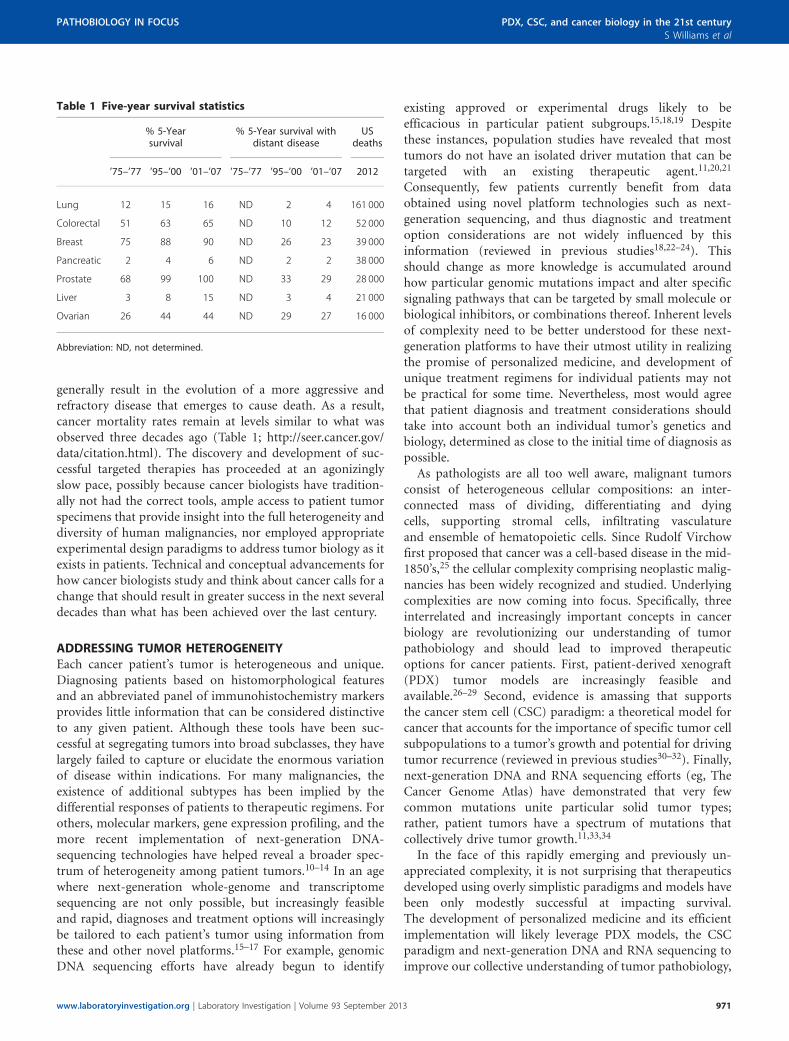
When tumors do arise, these masses are generally homo-geneous in nature and do not reflect tumor biology as itexists in patients (eg, Figures 1a and b). The expansion ofcells in aphysiologic oxygen concentrations (ie, 21% O2:ambient oxygen levels) has contributed to the perpetuationand study of cells that have accrued dozens, if not hundreds,of mutations and chromosomal abnormalities over thecourse of their many passages in vitro.41 Profound karyotypicdysmorphims, including hypotriploid genotypes, are surpri-singly common within the cell lines that have come to definethe standard models studied by many cancer biologists, suchas the NCI60 cell lines (also see http://www.ncbi.nlm.nih.gov/sky/skyweb.cgi). Recent studies have demonstrated thatbrief periods of in vitro culture irreversibly change geneexpression, indicating that even low-passage cell lines may becompromised.40,42 Collectively, these observations call intoquestion the physiological relevance of traditional cell lines assufficient, let alone relevant, models for studying tumorbiology as it exists in cancer patients.
Despite the increasingly apparent limitations of traditionalcell lines, they have made significant contributions to ourunderstanding of tumor biology. The uniformity and controlover experimental conditions afforded by in vitro cell culturehas assisted the development and execution of highlyreproducible studies under defined conditions that enableinsight into drug sensitivity, basic cell biology, and theelucidation of signaling pathways. For example, in vitro cellculture has facilitated the development of high-throughputapproaches leading to the discovery of second and thirdgeneration anti-neoplastic agents (see DeVita and Chu43 forreview). The flexibility and accessibility of traditional celllines perpetuated in vitro has also enabled key mechanisticstudies to shed light on the contribution of specific genes andmutations to cell survival, proliferation, and migration,contributing to the development of, for example, kinase-specific inhibitors such as erlotinib, vemurafenib, andcrizotinib targeting EGFR, BRAF, and ALK/c-MET, respec-tively (reviewed by Sawyers44). Although initially promising,these tailored kinase inhibitors have largely failed to becurative and typically extend life only 3–6 months.15,44
Although chemotherapeutic compounds have also provenhighly effective in vitro, in traditional in vivo tumor modelsand often in the clinic, the untargeted nature of these drugsresults in significant toxicity, and consequently, a narrowtherapeutic window in cancer patients. Moreover, residualtumors generally recur as more aggressive, refractory, andlethal; likely due to the inability of chemotherapy to elimi-nate CSC, and resulting in additional mutations accumulatedin these cells during exposure to genotoxic drug regimens.45
972 Laboratory Investigation | Volume 93 September 2013 | www.laboratoryinvestigation.org
PDX, CSC, and cancer biology in the 21st century
S Williams et al
PATHOBIOLOGY IN FOCUS
Genetically engineered mouse models (GEMMs) are alsopopular models through which tumor biology is studied.Unfortunately, these tumor models also have theirintrinsic shortcomings. It is becoming increasingly clearthat more than one driver mutation is needed to initiatetumorigenesis—both in mice and in men.20,34 Efficienttumorigenesis in GEMMs must often be driven by theintroduction of at least two defined oncogenes and/ormutated tumor suppressors, depending on the mousemodel and/or models needed to be crossed to drivetumorigenesis.46,47 Generating GEMMs with more than onedriver mutation is extremely time-consuming and difficult.Another weakness of these models is that transgeneexpression (eg, mutated KRAS) is commonly driven tosuperphysiological levels, such that protein expression oftenexceeds that ever encountered in patients.46 Moreover, whentumors do arise in these models, they are sporadic in theirgrowth such that it is difficult to design studies powered
by a significant number of animals, while also beingdifficult to monitor without labor-intensive in vivo imagingtechnologies. As previously discussed, the Cancer GenomeAtlas project has demonstrated that the spectrum ofdriver mutations differs significantly among patients, thusthe relevance of particular GEMMs to patients in the clinic isincreasingly questionable.33,34 Notwithstanding the abovecriticisms, GEMMs may have advantages over xenograftmodels in their cellular composition and/or sensitivity totherapeutic agents.48 Specifically, GEMM tumors containstromal and hematopoietic components not possiblein a human tumor xenograft setting, and thus these tumorsmay respond more appropriately to small mole-cules, immunomodulatory, and/or biological agents thatcross-react with mouse antigens. Nevertheless, for thevarious reasons outlined above, GEMMs will likely providelimited insight into oncogenesis and patient tumorheterogeneity.
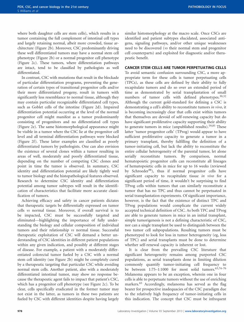
Figure 1 Patient-derived xenograft (PDX) models of colorectal cancer recapitulate primary tumor heterogeneity. Hematoxylin and eosin (H&E) stained
FFPE slides of xenografts generated by traditional HT-29 (a) or SW480 (b) colorectal cancer cell lines, vs a primary colorectal tumor, SCRX-PDX-CR101-p0
(c), and the same patient’s PDX tumor following passaging through NOD/SCID mice, SCRX-PDX-CR101-p1 (d). Note the relative uniformity of the HT-29
and SW480 tumors relative to the primary or PDX tumor following minimal passaging in immunocompromised mice.
www.laboratoryinvestigation.org | Laboratory Investigation | Volume 93 September 2013 973
PATHOBIOLOGY IN FOCUS PDX, CSC, and cancer biology in the 21st century
S Williams et al
Although traditional cell lines have helped gain anunderstanding of the basic biology underlying specific genes,proteins, and/or signaling pathways, cell lines culturedin vitro have not significantly contributed to the discoveryof targets for directed therapies that have meaningfullyimpacted patient survival. Trastuzumab and T-DM1 (anti-Her2biologics) and Imatinib/Gleevec (a BCR/ABL kinase inhibi-tor) are prime examples of cancer drugs that have mean-ingfully impacted patient survival; however, neither wasdiscovered in traditional cell lines or GEMMs.49,50 Her2/Neu(ErbB2) was originally identified as a potentially interestingcancer target based on the observation of elevated expressionin tumor specimens obtained and preserved by patholo-gists.51,52 Moreover, the p210-BCR/ABL fusion protein wasidentified and associated with chronic myelogenous leukemia(CML) upon close examination of chromosomal architectureusing blood smears from CML patients.53,54 Traditional celllines and GEMMs effectively complemented thesediscoveries, but were most effectively utilized as platformsto precisely dissect the molecular and cellular biology ofErbB2 and p210-BCR/ABL, respectively. To understandtumorigenesis and patient heterogeneity observed in theclinic, cancer research, drug discovery, and developmentmust increasingly look beyond in vitro tissue culture modelsand GEMMs and towards experimental systems that betterreplicate human tumor biology and enable the study of manypatient tumors as they are likely to exist in cancer patients.
PDXs: MODELS FOR THE 21ST CENTURYImmunocompromised mouse strains have become morewidely available over the last 20 years and permit theengraftment, passage, and study of human tumor cells in vivoin a xenograft setting.26,27,55–57 As a natural extension ofstandard tissue culture techniques, some researchers havetaken to the xenotransplantation of traditional tumor celllines that have been passaged extensively in vitro. Thesemodels have empowered remarkable advances in the under-standing of angiogenesis and tumor cell invasion, resulting inthe development of therapeutic agents such as bevacizumaband sorafenib;58,59 however, promising preclinical dataobtained using these traditional cell line-initiated xenograftshas not translated into dramatic improvements in overallsurvival in most cancer patients.60
To better preserve the genomic integrity and tumorheterogeneity observed in patients, many researchers areincreasingly turning to PDX models generated using freshlyresected patient tumors immediately transplanted intoimmunocompromised murine hosts without an intermediatein vitro culture step.26,28,42 Serial passage and expansion oftumors through successive generations of murine hostswithout intervening cell culture permits ongoing propaga-tion of tumor lines and the study of tumor biology withoutsubjecting PDX tumor cells to the stressful and compromis-ing conditions encountered in vitro.42 Currently, subcu-taneous PDX models, wherein tumor cells are transplanted
underneath the skin of the hindquarters or in the mammaryfat pad dominate the field of primary xenografts, succeedingintra-ocular, embryonic, and athymic systems.29,61 Manygroups also transplant cells under the kidney capsule ororthotopically; the latter of which may better replicate thetumor microenvironment than subcutaneous models, andthus may be most physiologically relevant. Regardless of thetransplantation site, the cellular complexity and architectureof PDX tumor models remains remarkably faithful to thetumor in its natural state in most cases—complete with in-vading vasculature and supporting stromal cells (Figure 1d).Tumor growth in the xenograft setting ensures that tumorcells are exposed to physiologically relevant oxygen, nutrient,and hormone levels (in cases where there is interspeciescross-reactivity), and provides natural physical substratesfor tumor cell adhesion. In contrast to in vitro propagatedcultures, cytogenetic analyses of PDX models reveals strongpreservation of the chromosomal architecture observed inpatient tumors.28,62 PDX models of melanoma, breast,pancreatic, ovarian, lung, colorectal, and brain-derivedtumors have been successfully established in many labora-tories (reviewed by Tentler et al29), and many have proven toexhibit similar chemoresponsiveness to anti-neoplastic agentsas observed in the same donating patient in the clinic—underscoring the fidelity of these models to the naturaldisease state30,63 (unpublished results).
PDXs faithfully recapitulate much of a tumor’s biology, yetthese models also have their shortcomings. The engraftmentfrequency and growth rate of implanted tumors is highlyvariable by tumor type and subtype, suggesting that sometumors struggle to engraft for reasons that might includea dependence on hematopoietic cells and/or microenviron-mental cues not present in mouse stroma or not compatiblewith human cells. Other factors that likely contribute toinefficient tumor initiation as xenografts include the lengthof time that passes between when the tumor is resected andthe time of transplantation, the absence of an appropriatesupport matrix and/or growth factors, or an inhospitable siteof implantation. Moreover, human tumor stroma andinfiltrating lymphocytes are often lost in the initial passagesin mice if xenograft tumors were initiated with tumor frag-ments containing these human cell populations. The extentto which tumor cells from freshly resected tumors are able towithstand mechanical stress and xenotransplantation barriersis also unclear. For example, breast cancer PDX models ap-pear to be particularly difficult to establish, with a 27% en-graftment rate in the most successful laboratories, comparedwith ovarian (65%), lung (50%), melanoma (59%), andcolorectal (68%) cancer.51,61 Mounting evidence suggests thatthe exact mouse strain (eg, NOD/SCID vs NSG) does notsignificantly impact engraftment efficiency of most solidtumor types, but strain differences can impact the rate atwhich tumors arise.29,64,65
PDX tumor models can be propagated as either discretetumor fragments or as single-cell suspensions. Whereas the
974 Laboratory Investigation | Volume 93 September 2013 | www.laboratoryinvestigation.org
PDX, CSC, and cancer biology in the 21st century
S Williams et al
PATHOBIOLOGY IN FOCUS
former has the advantage of retaining cell–cell interactionsand some tissue architecture during transplantation, thelatter permits single cell-by-cell assessment of phenotype, theinterrogation of differential tumorigenicity by isolated tumorcell subpopulations, and an unbiased sampling of the entiretumor, ensuring that spatially segregated subclones are notinadvertently selected during analysis or passaging. Never-theless the generation of single-cell suspensions from PDXtumors presents unique challenges. Cells in solid tumorsnaturally attach to neighboring cells and the extracellularmatrix, thus requiring tissue and cell disaggregation togenerate single-cell suspensions in order to be analyzedindividually. Physical and enzymatic dissociation techniquesrequired to generate single-cell suspensions can be harsh, andcells that survive the process are often sensitized to detach-ment-induced apoptosis (ie, anoikis).66 In addition, some cellsurface molecules are sensitive to dissociating enzymes suchthat post-dissociation antigen-staining profiles can bedistorted from their natural state in the context of tumors,thus highlighting the importance of keeping tumor dis-sociation times short and remaining cognizant of the impactof particular enzymatic cocktails and dissociation conditionson antigen expression. It is also critically important todiscriminate the live singlet population from cell aggregatesand dead cells when analyzing or isolating cells by flowcytometry and fluorescence-activated cell sorting (FACS) toensure that analyzed, isolated and/or transplanted tumorcell subpopulations comprise single viable cells.67 Therequirement for single cells is critically important whentesting the tumorigenicity of one tumor cell subpopulation vsanother, as it is of utmost importance in protecting againstartifacts associated with cell clumps and impurities that canmire the interpretation of experimental results.
PDX tumor models arguably provide the mostreproducible approximation of tumors in human cancerpatients. Information gathered using these models moreaccurately reflect human tumor biology than other existingmodels, and thus time and money spent studying andunderstanding these, and comparing their pathophysiologyto patient tumors should yield greater insight into cancerpathobiology. Nevertheless, these models are not perfect andefforts to address PDX tumor model deficiencies are ongoing.For example, transplantation of human CD34þ cord bloodcells enriched for human hematopoietic stem and progenitorcells can spur the generation of a human innate andadaptive immune system in mice68—offering the promise ofperforming xenotransplantation experiments in mice with areconstituted human immune system. The combination ofPDX tumor engraftment in these humanized mouse modelsmay facilitate insight into the role of the immune system intumor biology; however, these models will be difficult toemploy broadly owing to the difficulty in efficiently gene-rating these models and need to HLA-match xenograftedtumors with the human immune cell component such thatgraft-vs-tumor, let alone graft-vs-host, responses don’t
complicate the interplay between the human immunesystem, tumor, and mouse microenvironment.
Perhaps the most substantial barrier to PDX tumor modelestablishment and their widespread utilization for researchis time and cost. Unlike traditional tissue culture, whereinexperiments can be rapidly executed using relativelyinexpensive supplies, PDX models involve propagation inexpensive genetically engineered mice requiring equallyexpensive animal husbandry and facility costs. Experimentsmust be planned months in advance, as PDX tumors canoften take up to 24 weeks to arise after transplantation—presenting long experimental cycles. Altogether, the total costof a traditional cell culture-based project might represent afraction of a graduate student’s stipend, whereas PDX modelscan easily rival and exceed the cost of personnel. As a con-sequence, only a minority of the best-funded academic labsare currently equipped to study PDX tumor models, and onlythen in moderation. The substantial time and cost burden ofPDX tumor models will likely require institutional andbroader national support to be more widely embraced on thescale needed to displace traditional cell lines from broaderuse. The long-term benefit of studying more relevant tumorbiology merits these expenditures.
PDX MODELS AND THE RESURRECTION OF TUMOR STEMCELLSFor decades, the field of cancer biology has been dominatedby the stochastic model for tumor evolution. This modeldictates that all tumor cells have equivalent replicativecapacity and that mutations conferring a proliferative orsurvival advantage, for example, result in that cell and itsprogeny eventually becoming the dominant clone in thetumor. Although clonal evolution and competition is cer-tainly a feature of many tumors,69,70 this model alone fails toadequately acknowledge the cellular diversity comprisingmany solid tumors. The stochastic paradigm for cancerremained largely unchallenged as technologies required todemonstrate functional tumor cell heterogeneity (ie,fluorescent tag-labeled monoclonal antibodies, commercialavailability of FACS, and severely immunocompromisedmice) did not became broadly accessible to researchersuntil the late 1990s. These technologies facilitated workdemonstrating the recapitulation of tumor cell heterogeneityin passaged PDX tumor models in a remarkable series ofstudies demonstrating that functional tumor cell hetero-geneity accompanies the phenotypic diversity observedwithin tumors.56,57,71,72 This, in turn, led to reassessmentand reconsideration of the stochastic model for cancerbiology.
The observation that phenotypically distinct tumor cellsubpopulations are uniquely capable of fueling tumor growthin serial transplants, whereas the majority of tumor cellsappear to be bystanders in the process, has led to emergenceof the CSC paradigm for tumorigenesis both in hematologicaland solid tumor malignancies.30,71–73 The CSC paradigm
www.laboratoryinvestigation.org | Laboratory Investigation | Volume 93 September 2013 975
PATHOBIOLOGY IN FOCUS PDX, CSC, and cancer biology in the 21st century
S Williams et al
holds that tumors are comprised of a cellular hierarchy withsome similarities to normal tissue, with a self-renewing,multipotent cell at the apex of the hierarchy that no longerresponds appropriately to environmental cues.30,73 Firstdemonstrated in AML,56,57 data supporting the CSC para-digm have since been extended to a multitude of solid tumors,including breast, glioblastoma, colorectal, ovarian, and pan-creatic cancer.71,72,74–78 Evidence has emerged that only CSCpossess the capacity to generate secondary tumors containingboth CSC and non-tumorigenic (NTG) cell populations(ie, phenotypic and functional heterogeneity), supporting amodel whereby CSC may be rare, but appear solely able todrive fully heterogeneous tumor growth and recurrence.30,73
CSC theoretically retain features of normal stem cells,including their ability to symmetrically or asymmetricallydivide, remain relatively quiescent, express elevated levels ofmultidrug resistance transporters and DNA damage repairenzymes, and better handle oxidative stress. Several of thesecharacteristics are believed to be important in the resistance ofCSC to traditional chemotherapy and radiation.63,73,79–84 Theability of CSC to leverage these characteristics to better handleenvironmental stresses and genotoxic damage potentiallyexplains the disconnect between therapeutic response(ie, tumor burden reduction) and overall survival in theclinic. Tumors can all but be eradicated; however, if CSCpersist, tumors will inevitably recur and are likely to bemore aggressive given the genotoxic insults survived.Diagnostics and/or target discovery approaches that simplyevaluate bulk tumor cells (vs normal tissue), without takingCSC into consideration are unlikely to provide insight onfactors that will impact outcome given the relative infre-quency of CSC in most tumors (often enumerated as o1% ofthe tumor). Such diagnostic and target discovery approacheshave been taken for decades in naivete to the underlyingpresence and biology of CSC.
Since the first functional in vivo demonstration for theexistence of a solid tumor-initiating cell in human breasttumors in 2003,71 the semantics and classification schemessurrounding CSC have remained confusing despite the factthat the CSC paradigm has gained significant traction amongcancer biologists.30,32,85 The exact relationship between CSC,normal stem cell populations, and the ‘cell of origin’ incancer remains something of a mystery.32,86 To be clear,although a tumor’s initial CSC (ie, cell of origin) likelyevolves from normal stem cells as a result of accumulatingmutations that confer oncogenic properties over time,33 thecell of origin in cancer and the predominant CSC present inan evolved tumor may possess differing phenotypes andproperties depending on the point in time the tumor is beingstudied. More specifically, the CSC identity in disease mayevolve an identity different from the cell of origin. CMLserves as an excellent conceptual example for tumorprogression wherein the cell of origin and CSC identitiesdiffer as a result of tumor evolution over the course of diseaseprogression.87–90
In early chronic phase CML, CSC appear to have anormal hematopoietic stem cell (HSC) phenotype, thoughthe CSC behaves differently from normal HSC by skewingcell division towards the production of more abnormal stemcells (via an increased frequency of symmetric, self-renewingcell divisions) and slightly altering the normal course of he-matopoietic differentiation towards certain myeloid linea-ges.90–92 As such, the cell of origin in this disease is thoughtto be the normal HSC. As the disease progresses, additionalmutations accrue that result in, for example, the constitutivenuclear localization of b-catenin, conferring self-renewingproperties with phenotypes akin to hematopoietic progenitorcells, thus converting the tumor progenitor cell to a CSC as aresult of its newfound ability to self-renew indefinitely.89
These abnormal progenitor cells (myeloid blast cells) areunable to differentiate and also proliferate more rapidly thantheir CSC counterparts. Thus, the severe manifestation ofacute phase blast crisis is driven by a more aggressive CSCwith a progenitor cell phenotype. CSC in CML may thus havea stem cell or progenitor cell phenotype, depending on thestage of disease (chronic phase vs blast crisis). In fact, bothlikely co-exist in blast crisis CML patients, but the lessaggressive chronic phase CSC clone(s) are dramaticallyoutnumbered. Similar observations have recently beenmade in both human AML and a mouse model of AML,wherein several tumor-initiating cell populations have beendemonstrated to co-exist.64,93
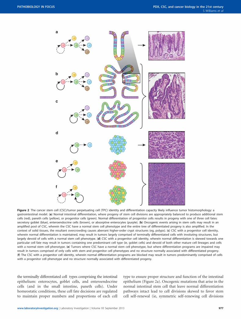
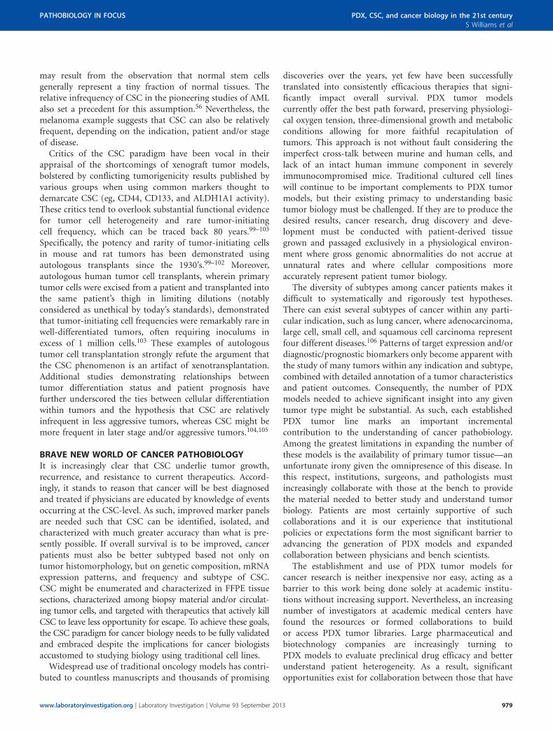
Solid tumors likely also follow this progression from nor-mal stem cell to pre-oncogenic stem cell to CSC. Unlike CML,where pre-oncogenic HSCs are not necessarily retained in aspecific niche and their progeny may disseminate throughoutthe bone marrow and circulation, stem cells for most solidtissues are confined to a defined three-dimensional space, orniche, due to their attachment to neighboring cells. As a resultof these attachments, expansion of the stem cell compartmentand maintenance of the differentiation program might bepredicted to culminate in the formation of a mass of morecrowded cells with some semblance of normal tissue archi-tecture. In cases where there is space available for tissueexpansion, this crowding can cause the formation of involu-tions, or polyps (Figure 2b; vs normal differentiation—Figure 2a). If differentiation is restricted to a particular line-age, the mass of cells might be less organized (Figure 2d).Finally, if all differentiation pathways are blocked, anamorphous mass of cells with no recognizable structure mightresult (Figures 2e and f). The above description for how solidtumors may manifest and evolve at the histomorphologicallevel as a result of CSC identity and differentiation capacityreflects what is observed in the case of gastrointestinal cancerprogression.94–97
Early stages of adenocarcinoma in the intestinal tract, forexample, may produce one of several histomorphologicaloutcomes. In normal intestinal epithelium, stem cells in thecrypt base divide to produce, through a hierarchical series offate-restricted progenitor cells (ie, transit amplifying cells),
976 Laboratory Investigation | Volume 93 September 2013 | www.laboratoryinvestigation.org
PDX, CSC, and cancer biology in the 21st century
S Williams et al
PATHOBIOLOGY IN FOCUS
the terminally differentiated cell types comprising the intestinalepithelium: enterocytes, goblet cells, and enteroendocrinecells (and in the small intestine, paneth cells). Underhomeostatic conditions, these cell fate decisions are regulatedto maintain proper numbers and proportions of each cell
type to ensure proper structure and function of the intestinalepithelium (Figure 2a). Oncogenic mutations that arise in thenormal intestinal stem cell that leave normal differentiationpathways intact lead to cell divisions skewed to favor stemcell self-renewal (ie, symmetric self-renewing cell divisions
Figure 2 The cancer stem cell (CSC)/tumor perpetuating cell (TPC) identity and differentiation capacity likely influence tumor histomorphology: a
gastrointestinal model. (a) Normal intestinal differentiation, where progeny of stem cell divisions are appropriately balanced to produce additional stem
cells (red), paneth cells (yellow), or progenitor cells (green). Normal differentiation of progenitor cells results in progeny with one of three cell fates:
secretory goblet (blue), enteroendocrine cells (brown), or absorptive enterocytes (purple). (b) Oncogenic events arising in stem cells may result in an
amplified pool of CSC, wherein the CSC have a normal stem cell phenotype and the entire tree of differentiated progeny is also amplified. In the
context of solid tissues, the resultant overcrowding causes aberrant higher-order crypt structures (eg, polyps). (c) CSC with a progenitor cell identity,
wherein normal differentiation is maintained, may result in tumors largely comprised of terminally differentiated cells with involuting structures, but
largely devoid of cells with a normal stem cell phenotype. (d) CSC with a progenitor cell identity, wherein normal differentiation is skewed towards one
particular cell fate may result in tumors containing one predominant cell type (ie, goblet cells) and devoid of both other mature cell lineages and cells
with a normal stem cell phenotype. (e) Tumors where CSC have a normal stem cell phenotype, but where differentiation programs are impaired may
result in tumors comprised of only cells with stem and progenitor cell phenotypes and no structure normally associated with differentiated progeny.
(f) The CSC with a progenitor cell identity, wherein normal differentiation programs are blocked may result in tumors predominantly comprised of cells
with a progenitor cell phenotype and no structure normally associated with differentiated progeny.
www.laboratoryinvestigation.org | Laboratory Investigation | Volume 93 September 2013 977
PATHOBIOLOGY IN FOCUS PDX, CSC, and cancer biology in the 21st century
S Williams et al
where both daughter cells are stem cells), which results in atumor containing the full complement of intestinal cell typesand largely retaining normal, albeit more crowded, tissue ar-chitecture (Figure 2b). Moreover, CSC predominantly drivingthese well differentiated tumors may have a normal stem cellphenotype (Figure 2b) or a normal progenitor cell phenotype(Figure 2c). These tumors, where differentiation pathwaysare intact, tend to be classified by pathologists as welldifferentiated.
In contrast, CSC with mutations that result in the blockadeof particular differentiation programs, preventing the gene-ration of certain types of transitional progenitor cells and/ortheir more differentiated progeny, result in tumors withsignificantly less resemblance to normal tissue, although theymay contain particular recognizable differentiated cell types,such as Goblet cells of the intestine (Figure 2d). Impaireddifferentiation potential occurring at the level of the normalprogenitor cell might manifest as a tumor predominantlyconsisting of progenitors and no differentiated cell types(Figure 2e). The same histomorphological appearance mightbe visible in a tumor where the CSC lie at the progenitor celllevel and all terminal differentiation pathways were blocked(Figure 2f). These latter examples are classified as poorlydifferentiated tumors by pathologists. One can also envisionthe existence of several clones within a tumor resulting inareas of well, moderately and poorly differentiated tissue,depending on the number of competing CSC clones andpoint in time the tumor is observed. In summary, CSCidentity and differentiation potential are likely tightly wedto tumor biology and the histopathological features observed.Research to determine CSC identity and differentiationpotential among tumor subtypes will result in the identifi-cation of characteristics that facilitate more accurate classi-fication of tumors.
Achieving efficacy and safety in cancer patients dictatesthat therapeutic targets be differentially expressed on tumorcells vs normal tissue. Moreover, if overall survival is tobe impacted, CSC must be successfully targeted andeliminated—highlighting the importance of fully under-standing the biology and cellular composition of individualtumors and their relationship vs normal tissue. Successfultherapeutic exploitation of CSC will demand a better un-derstanding of CSC identities in different patient populationswithin any given indication, and possibly at different stagesof disease. For example, a patient with a moderately differ-entiated colorectal tumor fueled by a CSC with a normalstem cell identity (see Figure 2b) might be completely curedby a therapeutic targeting that particular CSC while avoidingnormal stem cells. Another patient, also with a moderatelydifferentiated intestinal tumor, may show no response be-cause the therapeutic agent fails to impact that patient’s CSC,which has a progenitor cell phenotype (see Figure 2c). To beclear, cells specifically eradicated in the former tumor maynot exist in the latter, as tumors in these two patients arefueled by CSC with different identities despite having largely
similar histomorphology at the macro scale. Once CSCs areidentified and patient subtypes elucidated, associated anti-gens, signaling pathways, and/or other unique weaknessesneed to be discovered (vs their normal stem and progenitorcell counterparts) and exploited for diagnostic and/or thera-peutic benefit.
CANCER STEM CELLS ARE TUMOR PERPETUATING CELLSTo avoid semantic confusion surrounding CSC, a more ap-propriate term for these cells is tumor perpetuating cells(TPCs), as these cells are defined by their ability to fullyrecapitulate tumors and do so over an extended period oftime as demonstrated by serial transplantation of smallnumbers of tumor cells with defined phenotypes.30,32
Although the current gold-standard for defining a CSC isdemonstrating a cell’s ability to reconstitute tumors in vivo, itis becoming increasingly clear that cells exist within tumorsthat themselves are devoid of self-renewing capacity but dohave significant proliferative capacity supporting their abilityto generate tumors in mice (unpublished results).59,85 Theselatter ‘tumor progenitor cells’ (TProg) would appear to havesufficient proliferative capacity to generate a tumor in aprimary transplant, thereby fulfilling the definition of atumor-initiating cell, but lack the ability to reconstitute theentire cellular heterogeneity of the parental tumor, let aloneserially reconstitute tumors. By comparison, normalhematopoietic progenitor cells can reconstitute all lineagesof hematopoietic cells in mice for up to 16 weeks (reviewedby Schroeder98), thus if normal progenitor cells havesignificant capacity to recapitulate tissue in vivo for asignificant period of time, it wouldn’t be surprising to findTProg cells within tumors that can similarly reconstitute atumor that has no TPC and thus cannot be perpetuated inserial transplantation experiments. Of significant importance,however, is the fact that the existence of distinct TPC andTProg populations would complicate the current widelyaccepted technical definition of CSC. As both TPC and TProgare able to generate tumors in mice in an initial transplant,simple tumorigenesis is not a defining characteristic of CSC,nor can a single transplant be used to distinguish between thetwo tumor cell subpopulations. Resulting tumors must bephenotyped to look for loss in tumor heterogeneity (eg, lossof TPC) and serial transplants must be done to determinewhether self-renewal capacity is inherent or lost.
It is clear from the prevailing CSC literature thatsignificant heterogeneity remains among purported CSCpopulations, as serial transplants done in limiting dilutioncommonly quantify tumor-initiating cell frequency tobe between 1:75–1:1000 for most solid tumors.63,74–76
Melanoma appears to be an exception, wherein one in fourcells is able to perpetuate tumors without the use of enrichingmarkers.65 Accordingly, melanoma has served as the flagbearer for prospective inadequacies of the CSC paradigm dueto the relatively high frequency of tumor-initiating cells inthis indication. The concept that CSC must be infrequent
978 Laboratory Investigation | Volume 93 September 2013 | www.laboratoryinvestigation.org
PDX, CSC, and cancer biology in the 21st century
S Williams et al
PATHOBIOLOGY IN FOCUS
may result from the observation that normal stem cellsgenerally represent a tiny fraction of normal tissues. Therelative infrequency of CSC in the pioneering studies of AMLalso set a precedent for this assumption.56 Nevertheless, themelanoma example suggests that CSC can also be relativelyfrequent, depending on the indication, patient and/or stageof disease.
Critics of the CSC paradigm have been vocal in theirappraisal of the shortcomings of xenograft tumor models,bolstered by conflicting tumorigenicity results published byvarious groups when using common markers thought todemarcate CSC (eg, CD44, CD133, and ALDH1A1 activity).These critics tend to overlook substantial functional evidencefor tumor cell heterogeneity and rare tumor-initiatingcell frequency, which can be traced back 80 years.99–103
Specifically, the potency and rarity of tumor-initiating cellsin mouse and rat tumors has been demonstrated usingautologous transplants since the 1930’s.99–102 Moreover,autologous human tumor cell transplants, wherein primarytumor cells were excised from a patient and transplanted intothe same patient’s thigh in limiting dilutions (notablyconsidered as unethical by today’s standards), demonstratedthat tumor-initiating cell frequencies were remarkably rare inwell-differentiated tumors, often requiring inoculums inexcess of 1 million cells.103 These examples of autologoustumor cell transplantation strongly refute the argument thatthe CSC phenomenon is an artifact of xenotransplantation.Additional studies demonstrating relationships betweentumor differentiation status and patient prognosis havefurther underscored the ties between cellular differentiationwithin tumors and the hypothesis that CSC are relativelyinfrequent in less aggressive tumors, whereas CSC might bemore frequent in later stage and/or aggressive tumors.104,105
BRAVE NEW WORLD OF CANCER PATHOBIOLOGYIt is increasingly clear that CSC underlie tumor growth,recurrence, and resistance to current therapeutics. Accord-ingly, it stands to reason that cancer will be best diagnosedand treated if physicians are educated by knowledge of eventsoccurring at the CSC-level. As such, improved marker panelsare needed such that CSC can be identified, isolated, andcharacterized with much greater accuracy than what is pre-sently possible. If overall survival is to be improved, cancerpatients must also be better subtyped based not only ontumor histomorphology, but on genetic composition, mRNAexpression patterns, and frequency and subtype of CSC.CSC might be enumerated and characterized in FFPE tissuesections, characterized among biopsy material and/or circulat-ing tumor cells, and targeted with therapeutics that actively killCSC to leave less opportunity for escape. To achieve these goals,the CSC paradigm for cancer biology needs to be fully validatedand embraced despite the implications for cancer biologistsaccustomed to studying biology using traditional cell lines.
Widespread use of traditional oncology models has contri-buted to countless manuscripts and thousands of promising
discoveries over the years, yet few have been successfullytranslated into consistently efficacious therapies that signi-ficantly impact overall survival. PDX tumor modelscurrently offer the best path forward, preserving physiologi-cal oxygen tension, three-dimensional growth and metabolicconditions allowing for more faithful recapitulation oftumors. This approach is not without fault considering theimperfect cross-talk between murine and human cells, andlack of an intact human immune component in severelyimmunocompromised mice. Traditional cultured cell lineswill continue to be important complements to PDX tumormodels, but their existing primacy to understanding basictumor biology must be challenged. If they are to produce thedesired results, cancer research, drug discovery and deve-lopment must be conducted with patient-derived tissuegrown and passaged exclusively in a physiological environ-ment where gross genomic abnormalities do not accrue atunnatural rates and where cellular compositions moreaccurately represent patient tumor biology.
The diversity of subtypes among cancer patients makes itdifficult to systematically and rigorously test hypotheses.There can exist several subtypes of cancer within any parti-cular indication, such as lung cancer, where adenocarcinoma,large cell, small cell, and squamous cell carcinoma representfour different diseases.106 Patterns of target expression and/ordiagnostic/prognostic biomarkers only become apparent withthe study of many tumors within any indication and subtype,combined with detailed annotation of a tumor characteristicsand patient outcomes. Consequently, the number of PDXmodels needed to achieve significant insight into any giventumor type might be substantial. As such, each establishedPDX tumor line marks an important incrementalcontribution to the understanding of cancer pathobiology.Among the greatest limitations in expanding the number ofthese models is the availability of primary tumor tissue—anunfortunate irony given the omnipresence of this disease. Inthis respect, institutions, surgeons, and pathologists mustincreasingly collaborate with those at the bench to providethe material needed to better study and understand tumorbiology. Patients are most certainly supportive of suchcollaborations and it is our experience that institutionalpolicies or expectations form the most significant barrier toadvancing the generation of PDX models and expandedcollaboration between physicians and bench scientists.
The establishment and use of PDX tumor models forcancer research is neither inexpensive nor easy, acting as abarrier to this work being done solely at academic institu-tions without increasing support. Nevertheless, an increasingnumber of investigators at academic medical centers havefound the resources or formed collaborations to buildor access PDX tumor libraries. Large pharmaceutical andbiotechnology companies are increasingly turning toPDX models to evaluate preclinical drug efficacy and betterunderstand patient heterogeneity. As a result, significantopportunities exist for collaboration between those that have
www.laboratoryinvestigation.org | Laboratory Investigation | Volume 93 September 2013 979
PATHOBIOLOGY IN FOCUS PDX, CSC, and cancer biology in the 21st century
S Williams et al
access to fresh patient tissue and those who have the re-sources to generate and thoroughly characterize PDX lines.Established and well characterized low-passage PDX tumorlines will serve as the foundation for improved identificationand characterization of tumor cell subpopulations (eg, CSCvs NTG cells) in various tumor subtypes, and as a resultenhance our understanding of cancer biology.
Work with traditional cell lines has borne tens ofthousands of manuscripts at the cost of billions of dollarssince Richard Nixon signed the National Cancer Act in 1971.It is time that cancer biologists abandon what is easy andrelatively inexpensive, increasingly ask pertinent questionswith relevant tumor models, and progressively increase colla-boration with oncologists, pathologists, and other cliniciansregularly seeing patients and/or their tumors in the clinicalsetting. Embracing the CSC paradigm, working with PDXtumor models and maintaining constant awareness of clinicalrelevance are initial and critical steps towards making sig-nificant advances in the understanding of cancer biology inthe next several decades. Such fundamental pursuits as theseshould more efficiently yield the discovery and developmentof novel diagnostics and therapeutics that significantly im-pact overall survival of cancer patients.
ACKNOWLEDGEMENTS
We thank to those who reviewed and provided critical comments to this
manuscript, including Brian Slingerland and Drs Tim Gregory and
Chris Dayton. This work was supported by private financing.
DISCLOSURE/CONFLICT OF INTEREST
All authors are shareholders in Stem CentRx, a privately held and financed
company.
1. Huff CA, Matsui W, Smith BD, et al. The paradox of response andsurvival in cancer therapeutics. Blood 2006;107:431–434.
2. Huff CA, Matsui WH, Douglas Smith B, et al. Strategies to eliminatecancer stem cells: clinical implications. Eur J Cancer 2006;42:1293–1297.
3. Cantley LC, Dalton WS, DuBois RN, et al. AACR Cancer Progress Report2012. Clin Canc Res 2012;18(21 Suppl):S1–S100.
4. Tarver T. Cancer Facts & Figures 2012. American Cancer Society (ACS).Journal of Consumer Health on the Internet 2012;16:366–367.
5. Cancer Prevention and Early Detection Facts and Figures 2012,Atlanta, 2012.
6. Siegel R, Naishadham D, Jemal A. Cancer statistics, 2012. CA Cancer JClin 2012;62:10–29.
7. Lally BE, Urbanic JJ, Blackstock AW, et al. Small cell lung cancer: havewe made any progress over the last 25 years? Oncologist2007;12:1096–1104.
8. Di Marco M, Di Cicilia R, Macchini M, et al. Metastatic pancreaticcancer: is gemcitabine still the best standard treatment? (Review).Oncol Rep 2010;23:1183–1192.
9. Davies JM, Goldberg RM. Treatment of metastatic colorectal cancer.Semin Oncol 2011;38:552–560.
10. Hirsch FR, Wynes MW, Gandara DR, et al. The tissue is the issue:personalized medicine for non-small cell lung cancer. Clin Cancer Res2010;16:4909–4911.
11. Mardis ER. Genome sequencing and cancer. Curr Opin Genet Dev2012;22:245–250.
12. Baron JA. Screening for cancer with molecular markers: progresscomes with potential problems. Nat Rev Cancer 2012;12:368–371.
13. Prat A, Parker JS, Karginova O, et al. Phenotypic and molecularcharacterization of the claudin-low intrinsic subtype of breast cancer.Breast Cancer Res 2010;12:R68.
14. Prat A, Perou CM. Deconstructing the molecular portraits of breastcancer. Mol Oncol. Feb, 5:5–23.
15. Barrett JC, Frigault MM, Hollingsworth S, et al. Are companiondiagnostics useful? Clin Chem 2013;59:198–201.
16. Ross JS. Cancer biomarkers, companion diagnostics and personalizedoncology. Biomark Med 2011;5:277–279.
17. Chin L, Andersen JN, Futreal PA. Cancer genomics: from discoveryscience to personalized medicine. Nat Med 2011;17:297–303.
18. Berman DM, Bosenberg MW, Orwant RL, et al. Investigative pathology:leading the post-genomic revolution. Lab Invest. 2012;92:4–8.
19. Sleijfer S, Bogaerts J, Siu LL. Designing transformative clinical trials inthe cancer genome era. J Clin Oncol 2013;31:1834–1841.
20. Stephens PJ, Tarpey PS, Davies H, et al. The landscape of cancer genesand mutational processes in breast cancer. Nature 2012;486:400–404.
21. Katsios C, Papaloukas C, Tzaphlidou M, et al. Next-generationsequencing-based testing for cancer mutational landscape diversity:clinical implications? Expert Rev Mol Diagn 2012;12:667–670.
22. Strausberg RL, Simpson AJ. Whole-genome cancer analysis as anapproach to deeper understanding of tumour biology. Br J Cancer2010;102:243–248.
23. Meldrum C, Doyle MA, Tothill RW. Next-generation sequencing forcancer diagnostics: a practical perspective. Clin Biochem Rev 2011;32:177–195.
24. Biesecker LG, Burke W, Kohane I, et al. Next-generation sequencing inthe clinic: are we ready? Nat Rev Genet 2012;13:818–824.
25. Virchow R. Editorial. Path Anat Physiol Klin Med 1855;3:23.26. Jin K, Teng L, Shen Y, et al. Patient-derived human tumour tissue
xenografts in immunodeficient mice: a systematic review. Clin TranslOncol 2010;12:473–480.
27. Julien S, Merino-Trigo A, Lacroix L, et al. Characterization of a largepanel of patient-derived tumor xenografts representing the clinicalheterogeneity of human colorectal cancer. Clin Cancer Res2012;18:5314–5328.
28. Reyal F, Guyader C, Decraene C, et al. Molecular profiling of patient-derived breast cancer xenografts. Breast Cancer Res 2012;14:R11.
29. Tentler JJ, Tan AC, Weekes CD, et al. Patient-derived tumourxenografts as models for oncology drug development. Nat Rev ClinOncol 2012;9:338–350.
30. Clarke MF, Dick JE, Dirks PB, et al. Cancer stem cells—perspectives oncurrent status and future directions: AACR Workshop on cancer stemcells. Cancer Res 2006;66:9339–9344.
31. O’Brien CA, Kreso A, Jamieson CH. Cancer stem cells and self-renewal.Clin Cancer Res 2010;16:3113–3120.
32. Valent P, Bonnet D, De Maria R, et al. Cancer stem cell definitionsand terminology: the devil is in the details. Nat Rev Cancer 2012;12:767–775.
33. Tomasetti C, Vogelstein B, Parmigiani G. Half or more of the somaticmutations in cancers of self-renewing tissues originate prior to tumorinitiation. Proc Natl Acad Sci USA. 2013;110:1999–2004.
34. Vandin F, Upfal E, Raphael BJ. De novo discovery of mutated driverpathways in cancer. Genome Res 2012;22:375–385.
35. DiMasi JA, Grabowski HG. Economics of new oncology drugdevelopment. J Clin Oncol 2007;25:209–216.
36. Hamburger AW, Salmon SE. Primary bioassay of human tumor stemcells. Science 1977;197:461–463.
37. Persky B, Thomson SP, Meyskens Jr. FL, et al. Methods for evaluatingthe morphological and immunohistochemical properties of humantumor colonies grown in soft agar. In Vitro 1982;18:929–936.
38. Coulombel L. Identification of hematopoietic stem/progenitor cells:strength and drawbacks of functional assays. Oncogene2004;23:7210–7222.
39. Dulbecco R. Production of Plaques in Monolayer Tissue Cultures bySingle Particles of an Animal Virus. Proc Natl Acad Sci USA. 1952;38:747–752.
40. Tveit KM, Pihl A. Do cell lines in vitro reflect the properties of thetumours of origin? A study of lines derived from human melanomaxenografts. Br J Cancer 1981;44:775–786.
41. Roschke AV, Tonon G, Gehlhaus KS, et al. Karyotypic complexity of theNCI-60 drug-screening panel. Cancer Res 2003;63:8634–8647.
980 Laboratory Investigation | Volume 93 September 2013 | www.laboratoryinvestigation.org
PDX, CSC, and cancer biology in the 21st century
S Williams et al
PATHOBIOLOGY IN FOCUS
42. Daniel VC, Marchionni L, Hierman JS, et al. A primary xenograftmodel of small-cell lung cancer reveals irreversible changes in geneexpression imposed by culture in vitro. Cancer Res 2009;69:3364–3373.
43. DeVita Jr. VT, Chu E. A history of cancer chemotherapy. Cancer Res.2008;68:8643–8653.
44. Sawyers C. Targeted cancer therapy. Nature 2004;432:294–297.45. Yip S, Miao J, Cahill DP, et al. MSH6 mutations arise in glioblastomas
during temozolomide therapy and mediate temozolomide resistance.Clin Cancer Res 2009;15:4622–4629.
46. Lin JH. Applications and limitations of genetically modified mousemodels in drug discovery and development. Curr Drug Metab2008;9:419–438.
47. Dankort D, Curley DP, Cartlidge RA, et al. Braf(V600E) cooperates withPten loss to induce metastatic melanoma. Nat Genet 2009;41:544–552.
48. Combest AJ, Roberts PJ, Dillon PM, et al. Genetically engineeredcancer models, but not xenografts, faithfully predict anticancer drugexposure in melanoma tumors. Oncologist 2012;17:1303–1316.
49. Verma S, Miles D, Gianni L, et al. Trastuzumab emtansine for HER2-positive advanced breast cancer. N Engl J Med 2012;367:1783–1791.
50. Druker BJ, Guilhot F, O’Brien SG, et al. Five-year follow-up of patientsreceiving imatinib for chronic myeloid leukemia. N Engl J Med2006;355:2408–2417.
51. Giovanella BC, Vardeman DM, Williams LJ, et al. Heterotransplantationof human breast carcinomas in nude mice. Correlation betweensuccessful heterotransplants, poor prognosis and amplification of theHER-2/neu oncogene. Int J Cancer 1991;47:66–71.
52. Natali PG, Nicotra MR, Bigotti A, et al. Expression of the p185 encodedby HER2 oncogene in normal and transformed human tissues. Int JCancer 1990;45:457–461.
53. Nowell PC, Hungerford DA. Chromosome studies on normal andleukemic human leukocytes. J Natl Cancer Inst 1960;25:85–109.
54. Rowley JD. Letter: A new consistent chromosomal abnormality inchronic myelogenous leukaemia identified by quinacrine fluorescenceand Giemsa staining. Nature 1973;243:290–293.
55. Dick JE. Immune-deficient mice as models for human hematopoieticdisease. Mol Genet Med 1991;1:77–115.
56. Lapidot T, Sirard C, Vormoor J, et al. A cell initiating human acutemyeloid leukaemia after transplantation into SCID mice. Nature1994;367:645–648.
57. Bonnet D, Dick JE. Human acute myeloid leukemia is organized as ahierarchy that originates from a primitive hematopoietic cell. Nat Med1997;3:730–737.
58. Ferrara N, Hillan KJ, Gerber HP, et al. Discovery and development ofbevacizumab, an anti-VEGF antibody for treating cancer. Nat Rev DrugDiscov 2004;3:391–400.
59. Wilhelm S, Carter C, Lynch M, et al. Discovery and development ofsorafenib: a multikinase inhibitor for treating cancer. Nat Rev DrugDiscov 2006;5:835–844.
60. Rogosin S, Sandler AB. Beyond bevacizumab: antiangiogenic agents.Clin Lung Cancer. 2012;13:326–333.
61. Mattern J, Bak M, Hahn EW, et al. Human tumor xenografts as modelfor drug testing. Cancer Metastasis Rev 1988;7:263–284.
62. Povlsen CO, Visfeldt J, Rygaard J, Jensen G. Growth patterns andchromosome constitutions of human malignant tumours after long-term serial transplantation in nude mice. Acta Pathol Microbiol ScandA. 1975;83:709–716.
63. Dylla SJ, Beviglia L, Park IK, et al. Colorectal cancer stem cells areenriched in xenogeneic tumors following chemotherapy. PLoS One2008;3:e2428.
64. Eppert K, Takenaka K, Lechman ER, et al. Stem cell gene expressionprograms influence clinical outcome in human leukemia. Nat Med2011;17:1086–1093.
65. Quintana E, Shackleton M, Sabel MS, et al. Efficient tumour formationby single human melanoma cells. Nature 2008;456:593–598.
66. Zvibel I, Smets F, Soriano H. Anoikis: roadblock to cell transplantation?Cell Transplant 2002;11:621–630.
67. Alexander CM, Puchalski J, Klos KS, et al. Separating stem cells by flowcytometry: reducing variability for solid tissues. Cell Stem Cell2009;5:579–583.
68. Ishikawa F, Yasukawa M, Lyons B, et al. Development of functionalhuman blood and immune systems in NOD/SCID/IL2 receptor{gamma} chain(null) mice. Blood 2005;106:1565–1573.
69. Kreso A, O’Brien CA, van Galen P, et al. Variable clonal repopulationdynamics influence chemotherapy response in colorectal cancer.Science 2013;339:543–548.
70. Sottoriva A, Spiteri I, Shibata D, et al. Single-molecule genomic datadelineate patient-specific tumor profiles and cancer stem cellorganization. Cancer Res 2013;73:41–49.
71. Al-Hajj M, Wicha MS, Benito-Hernandez A, et al. Prospectiveidentification of tumorigenic breast cancer cells. Proc Natl Acad SciUSA. 2003;100:3983–3988.
72. Singh SK, Hawkins C, Clarke ID, et al. Identification of human braintumour initiating cells. Nature 2004;432:396–401.
73. Reya T, Morrison SJ, Clarke MF, et al. Stem cells, cancer, and cancerstem cells. Nature 2001;414:105–111.
74. Dalerba P, Dylla SJ, Park IK, et al. Phenotypic characterization ofhuman colorectal cancer stem cells. Proc Natl Acad Sci USA.2007;104:10158–10163.
75. O’Brien CA, Pollett A, Gallinger S, et al. A human colon cancer cellcapable of initiating tumour growth in immunodeficient mice. Nature2007;445:106–110.
76. Ricci-Vitiani L, Lombardi DG, Pilozzi E, et al. Identification andexpansion of human colon-cancer-initiating cells. Nature 2007;445:111–115.
77. Hermann PC, Huber SL, Herrler T, et al. Distinct populations of cancerstem cells determine tumor growth and metastatic activity in humanpancreatic cancer. Cell Stem Cell 2007;1:313–323.
78. Zhang S, Balch C, Chan MW, et al. Identification and characterization ofovarian cancer-initiating cells from primary human tumors. Cancer Res2008;68:4311–4320.
79. Bao S, Wu Q, McLendon RE, et al. Glioma stem cells promoteradioresistance by preferential activation of the DNA damageresponse. Nature 2006;444:756–760.
80. Diehn M, Cho RW, Lobo NA, et al. Association of reactive oxygenspecies levels and radioresistance in cancer stem cells. Nature2009;458:780–783.
81. Zhou J, Zhang Y. Cancer stem cells: models, mechanisms andimplications for improved treatment. Cell Cycle 2008;7:1360–1370.
82. Lee CJ, Li C, Simeone DM. Human pancreatic cancer stem cells:implications for how we treat pancreatic cancer. Transl Oncol2008;1:14–18.
83. Eyler CE, Rich JN. Survival of the fittest: cancer stem cells in thera-peutic resistance and angiogenesis. J Clin Oncol 2008;26:2839–2845.
84. Morrison R, Schleicher SM, Sun Y, et al. Targeting the mechanisms ofresistance to chemotherapy and radiotherapy with the cancer stemcell hypothesis. J Oncol 2011;2011:941876.
85. Baccelli I, Trumpp A. The evolving concept of cancer and metastasisstem cells. J Cell Biol 2012;198:281–293.
86. Visvader JE. Cells of origin in cancer. Nature 2011;469:314–322.87. Clarkson B, Strife A, Wisniewski D, et al. Chronic myelogenous
leukemia as a paradigm of early cancer and possible curativestrategies. Leukemia 2003;17:1211–1262.
88. Eaves C, Udomsakdi C, Cashman J, et al. The biology of normal andneoplastic stem cells in CML. Leuk Lymphoma 1993;11(Suppl 1):245–253.
89. Jamieson CH, Ailles LE, Dylla SJ, et al. Granulocyte-macrophageprogenitors as candidate leukemic stem cells in blast-crisis CML.N Engl J Med 2004;351:657–667.
90. Sloma I, Jiang X, Eaves AC, et al. Insights into the stem cells of chronicmyeloid leukemia. Leukemia 2010;24:1823–1833.
91. Dazzi F, Hasserjian R, Gordon MY, et al. Normal and chronic phase CMLhematopoietic cells repopulate NOD/SCID bone marrow with differentkinetics and cell lineage representation. Hematol J. 2000;1:307–315.
92. Eisterer W, Jiang X, Christ O, et al. Different subsets of primary chronicmyeloid leukemia stem cells engraft immunodeficient mice andproduce a model of the human disease. Leukemia 2005;19:435–441.
93. Gibbs Jr KD, Jager A, Crespo O, et al. Decoupling of tumor-initiatingactivity from stable immunophenotype in HoxA9-Meis1-driven AML.Cell Stem Cell 2012;10:210–217.
94. Novelli MR, Williamson JA, Tomlinson IP, et al. Polyclonal origin ofcolonic adenomas in an XO/XY patient with FAP. Science 1996;272:1187–1190.
95. Preston SL, Wong WM, Chan AO, et al. Bottom-up histogenesis ofcolorectal adenomas: origin in the monocryptal adenoma and initialexpansion by crypt fission. Cancer Res 2003;63:3819–3825.
www.laboratoryinvestigation.org | Laboratory Investigation | Volume 93 September 2013 981
PATHOBIOLOGY IN FOCUS PDX, CSC, and cancer biology in the 21st century
S Williams et al
96. Greaves LC, Preston SL, Tadrous PJ, et al. Mitochondrial DNAmutations are established in human colonic stem cells, andmutated clones expand by crypt fission. Proc Natl Acad Sci USA.2006;103:714–719.
97. Baker AM, Graham TA, Wright NA. Pre-tumour clones, periodicselection and clonal interference in the origin and progression ofgastrointestinal cancer: potential for biomarker development. J Pathol2013;229:502–514.
98. Schroeder T. Hematopoietic stem cell heterogeneity: subtypes, notunpredictable behavior. Cell Stem Cell 2010;6:203–207.
99. Furth J, Kahn M. The transmission of leukemia of mice with a singlecell. Am J Cancer 1937;31:276–282.
100. Makino S. Further evidence favoring the concept of the stem cell inascites tumors of rats. Ann N Y Acad Sci 1956;63:818–830.
101. Hewitt H. Studies of the dissemination and quantitative transplantationof a lymphocytic leukemia of CBA mice. Br J Cancer 1958;12:378–401.
102. Bruce W, Van Der Gaag H. A quantitative assay for the number ofmurine lymphoma cells capable of proliferation in vivo. Nature1963;199:79–80.
103. Southam C, Brunschwig A, Dizon Q. Autologous and homologoustransplantation of human cancer. In: Brennan M, Simpson W eds.Biological Interactions in Normal and Neoplastic Growth: A Contributionto the Tumor-Host Problem. 9, Boston, MA, USA, 1962, p723–738.
104. Bailar 3rd JC, Mellinger GT, Gleason DF. Survival rates of patients withprostatic cancer, tumor stage, and differentiation–preliminary report.Cancer Chemother Rep 1966;50:129–136.
105. Li T, Su Y, Mei Y, et al. ALDH1A1 is a marker for malignant prostatestem cells and predictor of prostate cancer patients’ outcome. LabInvest 2010;90:234–244.
106. Thunnissen E, Kerr KM, Herth FJ, et al. The challenge of NSCLCdiagnosis and predictive analysis on small samples. Practicalapproach of a working group. Lung Cancer 2012;76:1–18.
982 Laboratory Investigation | Volume 93 September 2013 | www.laboratoryinvestigation.org
PDX, CSC, and cancer biology in the 21st century
S Williams et al
PATHOBIOLOGY IN FOCUS
![Heme oxygenase-1 in macrophages controls prostate cancer ...€¦ · apoptosis of prostate cancer xenografts [14, 15]. However, the link between regulation of cancer metabolism and](https://img.pdfslide.net/doc/110x75/5fb96cc5a635361b7e48ffde/heme-oxygenase-1-in-macrophages-controls-prostate-cancer-apoptosis-of-prostate.jpg)


![Pancreatic cancer stem cells in patient pancreatic xenografts … · growth in several PDX models of pancreatic cancer [14, 15]. Binding of Apo2L/TRAIL to its receptors results in](https://img.pdfslide.net/doc/110x75/60414d9a3677e1741876c987/pancreatic-cancer-stem-cells-in-patient-pancreatic-xenografts-growth-in-several.jpg)