Embed Size (px)
Citation preview
Periodontics III Summary Notes Enoch Ng
Intro to Perio Surgery
Patient Preparation – Re-evaluation after initial treatment (SRP) - May eliminate some pocket depths
- Can firm the tissue
o Inflamed tissues have capillary loops, are less firm
o Sutures tear more easily through inflamed tissues
- Gives time for patient education/comfort
o Poor homecare is a contraindication for surgery
Indications for Perio Surgery - Improve prognosis of teeth (and their replacements)
o Reduce pocket depths via resection or regeneration
Increased access for hygiene (furcations, decreased pocket depths, etc)
o Reshape hard and soft tissues to more physiologic contours
Furcation involvement – improve or eliminate furcation access
- Improve esthetics
o Mucogingival problems with esthetic concerns/persistent inflammation
Predictors of Disease Etiology Sensitivity (True Positive) Specificity (True Negative)
Plaque 0.4 0.6 Deep pockets are not good predictors for future attachment loss Redness 0.3 0.7
BOP 0.2 0.8 Absence of deep pockets are good predictor for stability Suppuration 0.1 0.9
PD > 6mm 0.1 0.9 CAL is the most important variable in periodontitis AL > 6mm 0.1 0.9
- Critical probing depths
o <3.0mm = lose attachment with SRP
o <4.3mm = lose attachment with surgery
o >6.2mm = SRP not effective
Treatments - Regeneration – improve clinical attachment loss, reduce pocket depth
o Requires healing of PDL, cementum, and bone
- Reparative – reduce pocket depth without new PDL, cementum, and bone healing
- Resective – removal of the pocket wall
o SRP – retraction and shrinkage – sometimes gingiva may heal so well you can’t probe into the full depth
2 weeks = shrinkage
4 weeks = reattachment
6 weeks = re-evaluation
o Gingivectomy – removal of gingiva because it has no chance of healing (ex: beside a filling)
o Apically positioned flap
If patient has poor hygiene, the problem becomes a caries instead of perio problem
- Surgical removal of tooth structure (root amputation, hemisection)
Periodontics III Summary Notes Enoch Ng
Treatment Selection - Overall diagnosis, goals of surgery
- Access
- History of surgery
- Pocket form
- Esthetics
o Anterior teeth – single rooted, patient compliance is huge
o Interproximal bone loss = lose papillae
- Blood supply
Surgical Procedure Method Selection/Considerations Procedure
- Regenerative methods o Papillae preservation o Sulcular flaps o Modified widman flap (to maintain
papillae) – very little tissue loss if done properly
- Resective methods o Gingivectomy o APF
- Smoking considerations - Informed consent - Sedation/anesthesia
o Local anesthesia – keep surgery painless o Inhalation – antianxiety delivery of N2O,
safest method of delivery o Oral sedation – individually variable o Conscious sedation o General anesthesia
- Emergency equipment
- Premedication o Prophylactic antibiotics for surgery o NSAIDS for pain, reduce inflammation o Anti-anxiety medications o Chlorhexidine rinse pre and post-op to
decrease aerosol exposure o Steroids to reduce inflammation
- Tissue Management o Be gentle and careful o Observe patient at all times o Use sharp instruments to avoid
masticating tissue - Surgical Dressings
ZO-Eugenol packs Non-eugenol packs Retention of packs
o Should remain in place for 1 week o Allow Coe-Pak to harden for 3h before
eating o Do not disturb pack (ex: brushing, flossing)
- Post-Op o Printed instructions o Return appointment o Repacking o Tooth mobility o Mouth care between procedures o Probing o Root sensitivity
Desensitizing agents include homecare and in office products
Anterior Mental Nerve Loop - Generally 0.5-3.1mm anterior to mental foramen
- 28% of cases 0.4-2.2mm anterior to mental foramen
- 86-90% Caucasians have anterior loop (mental nerve exiting in posterior direction)
- 45% Blacks have mental nerve exiting at right angle to foramen
Periodontics III Summary Notes Enoch Ng
Intro to Perio Surgery II
Anatomy - Nerves and arteries usually run superior to mylohyoid muscle, but may run inferiorly
- Lingual concavity – estimate how superior it is for implant placement
- An incision following the central grooves to reach the Mn 3rd molars will cut into muscles, nerves, and arteries.
The incision should turn and follow the ramus posterior superiorly
- Lingual nerve position is variable – so long as work is done in the keratinized tissue, should be okay. Outside the
keratinized tissue = greater risk
- It can be difficult to close a flap without creating a lingual flap, but care should be taken because nerve/artery
bundles run on the lingual side of the ramus
o Never do a sharp sectioning on the lingual side, because you don’t know where the danger zone is
o Mylohyoid release –allows for the lingual side to the brought superiorly
Blood Supply and Tissue Survival - Blood Supply
o Full or partial thickness flaps are both useable
o Partial thickness flaps in thin tissue may cause flap death
o Recipient site vascularity affects survival of thin flaps
o Velvet incision = off angled incision by the papilla to allow a large enough piece of tissue for suturing
- Full periosteal horizontal incision
o 24h – disturbance to gingiva coronal to incision
o 48h – local superficial gingival necrosis, overall tissue perfused by perio and intraosseous vessels
o Significance = most of the blood supply comes apical to coronal
Blood supply from the roots/bones = tissue heals like a scrape, top tissue sloughs
In a free gingival graft, lots of keratinized tissue is gained but no height
Height is gained by position tissue coronally (envelop flap with releasing incisions)
- Internal bevel incision between gingiva and periosteum
o 24h – no change
o Significance = healing is fast because of good blood perfusion
- Full thickness flap made, not reflected vs reflected and replaced
o 24h – similar disturbance, reflected flap had 50% greater reduction
o 96h – reflected flap had poorer appearance
o Significance = reflecting flaps decreases blood perfusion to elevated tissue
- FTF reflected, vertical incisions beyond the mucogingival margins, with test group incision length 2x control
o 24h – test group had poorer healing, marginal tissue necrosis
o Significance = critical length:width ratio = 2:1
- Flaps placed over recession areas, test flap 50% narrower than control
o 24h – test flap had 50% reduced circulation
o 7days – cleft-like tissue loss around gingival margin of test flap
Overly long flaps had some ischemic marking at sutures
o Significance = excess tension or excess flexion decreases flap healing
Periodontics III Summary Notes Enoch Ng
Gingival Surgical Procedures
Limited to gingiva, does not involve underlying osseous structures - Gingival curettage – removal of gingival wall of perio pocket to separate out diseased soft tissue
Aka = excisional new attachment procedure, ultrasonic curettage, caustic drugs
o Inadvertent curettage – happens with SRP
o No clinical value (2002) – healing is by long junctional epithelium, no new attachment is gained
- Gingivectomy
o Indications
Elimination of suprabony pockets in firm, fibrous tissue (SRP usually clears edematous pockets)
Elimination of gingival enlargements
Elimination of suprabony abscesses
Before they become infrabony ones
Access for restorative dentistry
Biologic width important in esthetic areas
Body wants around 2mm between bone and gingiva (beware of gingival rebound)
Biologic width = 2.04mm, supra-alveolar tissues (dentogingival junction) = 2.73mm
Esthetics
Ideal width = CI 25% wider than laterals, 10% wider than canines
Ideal height = CI and canine 20% longer than laterals
o Ratio = 1.2/1.0
Surgical stents – want to know if the stent is for what tooth structure is showing, or
where the crown margin is going to be placed (margin can be slightly in pocket)
o Want to preserve some keratinized tissue, do not want to remove all of it
Keratinized gingiva = pocket depth + attached gingiva (mucogingival junction)
o Measure pocket depth, mark it to cause it to bleed. Incision is apical to bleeding point, 45o bevel to root
o Healing
Initially – acute PMN infiltrate and some necrosis, formation of initial protective clot
12-24h – epithelial cells at margin migrate into granulation tissue and beneath the
necrotic tissue
24h – increased CT and angioblasts below surface layer
4-16 days – vasodilation and vascularity start to decrease until normal
o Epithelium grows at 0.5mm/day
5-14days – surface epthelialization complete, keratinization incomplete
7 weeks – complete repair of CT
o Limiting Factors
Amount of keratinized gingiva
Esthetics and esthetic maintenance
Access to osseous defects for correction
Less post-op pain if procedure allows for primary closure
Gingiva may rebound without racial pigmentation – risk for patients with racial pigmentation
Periodontics III Summary Notes Enoch Ng o Electrosurgery
Good hemorrhage control
Bad for patients with poorly shielded cardiac pacemakers
Must be limited to superficial procedures – can cause necrosis if tip touches bone or cementum
Different if only trying to coagulate – can be done at lower temperature
o Chemosurgery
Difficult to control depth
Slower healing
NOT recommended
- Gingivoplasty
o Possible to do APF, gingivectomy, or combined techniques
o Edematous tissue – treat with SRP
o If extremely fibrous and interferes with access – consider treating with gingivectomy
Gingivectomy can recontour at the margin if adequate keratinized gingiva is present
o Apically Positioned Flap – needs firm tissue, conserve keratinized gingiva
Common on palatal side – may end up sitting up in a point during crown lengthening. If it
doesn’t stay down, a blood clot can form there and cause tissue rebound. Tissue must lay down
and be positioned apically to prevent rebound
o Combined technique
Cut border to a regular border first, then place a flap and position apically
If gingival rebound is from drug use for systemic problems, insurance will pay for multiple
gingivectomies
Use packs to prevent hematoma formation to prevent gingival rebound, gives esthetic outcome
- Gingival flap
o The exception from all the others. Full thickness flaps may touch osseous structure but should not
contour it or affect it
Treatment considerations - Functional/esthetic compromise of adjacent teeth
o Opening interdental spaces
o Creating excessively “long” teeth
- Gingival diseases
o Modified by medications
Difedipine
Cyclosporine
Phenytoin (Dilantin)
Drug influenced gingival diseases
Drug influenced gingival enlargements (inflammatory drug induced hyperplasia)
Drug influenced gingivitis (Oral contraceptive associated, other)
Drugs that cause
o Dental plaque induced gingival diseases
Usually seen in young patients rather than older patients
In patient has poor hygiene, tissue will have a greater rebound effect
Beware handicapped patients – their hygiene provider must be informed of importance of care
Periodontics III Summary Notes Enoch Ng
Periodontal Flap Surgery
Surgical Procedures - Gingivectomy - Periodontal flaps - Osseous contouring - Bone grafts - Laterally sliding flaps - Free gingival grafts
Periodontal Flaps - Increase access to root - Reduce Pocket Depth - Expose areas for regeneration - Crown lengthening
Incision Design - Internal bevel scalloped (modified Widman) – most basic horizontal incision
Thins the gingiva, conserves attached gingiva, removes pocket lining epithelium
o Incision to periosteum to detach flap
o Scalloping to loosen pocket epithelium from tooth
o Horizontal incision to remove pocket lining epithlium
- Vertical Incisions
o Placed at line angle
o Should not be at apex of gingival sulcus or interproximally
Loss of papilla or may cause gingival defect
o Usually not placed on palatal side, Mn lingual, or nasopalatine areas (vessels, esthetic zones)
- Blades
o #15 – good for newbs, sharp curved blade
o #11 – sharp straight end with pointed tip
o #12/12B – curved (like a sickle) with tip, good for distal wedge
Flaps - Full thickness – goes through periosteum, might get up to 0.5mm bone loss because bone is thin
- Partial thickness – goes through CT, some CT and all periosteum remains attached to osseous structure
- Conventional flap – removes pocket epithelium
- Sulcular incision – used when you don’t want to lose attached gingiva or in esthetic zones (anterior region)
- Repositioned flap – replacing the flap back to where it was before (modified Widman surgeries)
- Apically positioned flap – used after 4-6week post-op probing after SRP assuming pocket depths don’t improve
Crown Lengthening - Restorative margin cannot be closer than 2mm to crestal bone, or will disrupt osseous structure
- Sounding bone – probing through the biologic width to the bone, gives an idea of what bone contour is like
- Biologic width – expose 3-4mm of tooth coronal to bone during surgery to accommodate 2mm biologic width
- Modified Widman – internal bevel primary incision, then scalloping and removal of desired gingiva
o Buck or orban knives helpful for removing interproximal tissue
- Suturing – slight exposure interproximally is okay. Cortical bone is backed up by cancellous bone
- Coe pack placed to help with healing, prevent rebound
o Left for 3-4 days, up to 7-10 days
o Post-surgical hemorrhage controlled by pressure, sutures, clotting, packs
o Remove pack to ID bleeding source/stop bleeding if necessary
- Chlorhexidine rinses (both pre and post-op)
- Takes 4 weeks to heal
Periodontics III Summary Notes Enoch Ng
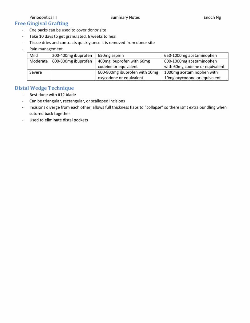
Free Gingival Grafting - Coe packs can be used to cover donor site
- Take 10 days to get granulated, 6 weeks to heal
- Tissue dries and contracts quickly once it is removed from donor site
- Pain management
Mild 200-400mg ibuprofen 650mg aspirin 650-1000mg acetaminophen
Moderate 600-800mg ibuprofen 400mg ibuprofen with 60mg codeine or equivalent
600-1000mg acetaminophen with 60mg codeine or equivalent
Severe 600-800mg ibuprofen with 10mg oxycodone or equivalent
1000mg acetaminophen with 10mg oxycodone or equivalent
Distal Wedge Technique - Best done with #12 blade
- Can be triangular, rectangular, or scalloped incisions
- Incisions diverge from each other, allows full thickness flaps to “collapse” so there isn’t extra bundling when
sutured back together
- Used to eliminate distal pockets

Periodontics III Summary Notes Enoch Ng
Treatment of Osseous Defects - Resection
- Debridement
- Grafting
Types of Defects - Dehiscence – root exposure connected to the rest of the tooth
- Fenestration – window of root exposued
- Positive architecture – interproximal bone more coronal than radicular bone
- Negative architecture – interproximal bone more apical than radicular bone
- Infrabony defect – base of pocket apical to crest of alveolar bone
- Infrabony Pockets (negative architecture)
o 1 osseous wall
o 2 osseous walls – aka crater, most common osseous defect, usually because patient doesn’t floss well
o 3 osseous walls
Furcations CEJ to opening of furcation
Mn molars - Buccal side = 3mm - Lingual side = 4mm
Mx molars - ML side = 3mm - Buccal side = 4mm - DL side = 5mm
- As much as possible, best NOT to open furcation (hard area to clean)
Other Surgical Considerations - Widow’s Peak
o If you leave bone adjacent to tooth surface by line angle (tendency to leave bone by line angle)
o More likely to have pocketing, reduction of bone by line angles to give smooth reduction reduces
pocketing during healing
- Radiographic Limitations – 2D image, can’t diagnose periodontitis or determine number of walls in defect
- Sounding bone (transgingival probing) – probing through the attached gingiva to find out where the bone is
o Used to discover osseous defects and what they are like, can help determine number of walls in defect
Osseous Resection - Often combined with APF – gives a very predictable outcome for reducing/eliminating pocket depths
- Indications
o Wide 3 wall defects
o Interproximal craters (2 wall defects)
o Hemiseptums (1 wall defects)
o Furcations – no blood supply except apically
o Thick alveolar bone
- Ostectomy – shaping to ideal form which may sacrifice some supportive bone (bone adjacent to tooth)
o Crown lengthening – removes some bone from proximal teeth for contouring)
- Osteoplasty – reshaping without sacrificing supporting bone
o Tori removal
Periodontics III Summary Notes Enoch Ng
Regeneration/Repair - Repair – healing of a wound by tissue that does not fully restore architecture or function
o Long junctional epithelium
- Regeneration – reproduction of a lost part resulting in new bone, cementum, and PDL
Bone Grafts - NEED good blood supply
- Fix periodontal defects
o For 1 wall defects, may try osseous resectioning instead of grafting because of low blood supply
- Alveolar ridge augmentation
- Fill extraction sites
- Sinus augmentation
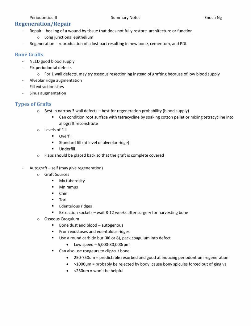
Types of Grafts o Best in narrow 3 wall defects – best for regeneration probability (blood supply)
Can condition root surface with tetracycline by soaking cotton pellet or mixing tetracycline into
allograft reconstitute
o Levels of Fill
Overfill
Standard fill (at level of alveolar ridge)
Underfill
o Flaps should be placed back so that the graft is complete covered
- Autograft – self (may give regeneration)
o Graft Sources
Mx tuberosity
Mn ramus
Chin
Tori
Edentulous ridges
Extraction sockets – wait 8-12 weeks after surgery for harvesting bone
o Osseous Caogulum
Bone dust and blood – autogenous
From exostoses and edentulous ridges
Use a round carbide bur (#6 or 8), pack coagulum into defect
Low speed – 5,000-30,000rpm
Can also use rongeurs to clip/cut bone
250-750um = predictable resorbed and good at inducing periodontium regeneration
>1000um = probably be rejected by body, cause bony spicules forced out of gingiva
<250um = won’t be helpful
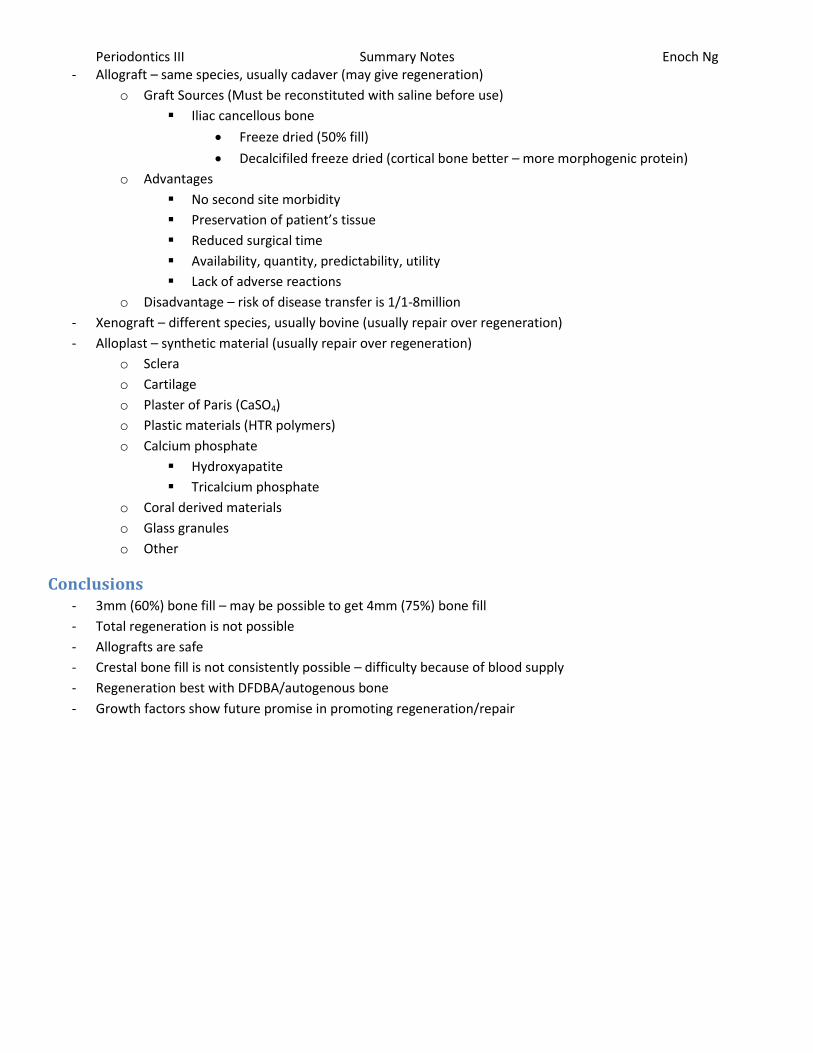
Periodontics III Summary Notes Enoch Ng - Allograft – same species, usually cadaver (may give regeneration)
o Graft Sources (Must be reconstituted with saline before use)
Iliac cancellous bone
Freeze dried (50% fill)
Decalcifiled freeze dried (cortical bone better – more morphogenic protein)
o Advantages
No second site morbidity
Preservation of patient’s tissue
Reduced surgical time
Availability, quantity, predictability, utility
Lack of adverse reactions
o Disadvantage – risk of disease transfer is 1/1-8million
- Xenograft – different species, usually bovine (usually repair over regeneration)
- Alloplast – synthetic material (usually repair over regeneration)
o Sclera
o Cartilage
o Plaster of Paris (CaSO4)
o Plastic materials (HTR polymers)
o Calcium phosphate
Hydroxyapatite
Tricalcium phosphate
o Coral derived materials
o Glass granules
o Other
Conclusions - 3mm (60%) bone fill – may be possible to get 4mm (75%) bone fill
- Total regeneration is not possible
- Allografts are safe
- Crestal bone fill is not consistently possible – difficulty because of blood supply
- Regeneration best with DFDBA/autogenous bone
- Growth factors show future promise in promoting regeneration/repair
Periodontics III Summary Notes Enoch Ng
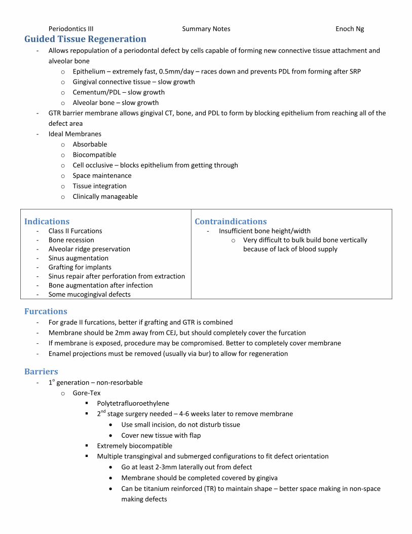
Guided Tissue Regeneration - Allows repopulation of a periodontal defect by cells capable of forming new connective tissue attachment and
alveolar bone
o Epithelium – extremely fast, 0.5mm/day – races down and prevents PDL from forming after SRP
o Gingival connective tissue – slow growth
o Cementum/PDL – slow growth
o Alveolar bone – slow growth
- GTR barrier membrane allows gingival CT, bone, and PDL to form by blocking epithelium from reaching all of the
defect area
- Ideal Membranes
o Absorbable
o Biocompatible
o Cell occlusive – blocks epithelium from getting through
o Space maintenance
o Tissue integration
o Clinically manageable
Indications - Class II Furcations - Bone recession - Alveolar ridge preservation - Sinus augmentation - Grafting for implants - Sinus repair after perforation from extraction - Bone augmentation after infection - Some mucogingival defects
Contraindications - Insufficient bone height/width
o Very difficult to bulk build bone vertically because of lack of blood supply
Furcations - For grade II furcations, better if grafting and GTR is combined
- Membrane should be 2mm away from CEJ, but should completely cover the furcation
- If membrane is exposed, procedure may be compromised. Better to completely cover membrane
- Enamel projections must be removed (usually via bur) to allow for regeneration
Barriers - 1o generation – non-resorbable
o Gore-Tex
Polytetrafluoroethylene
2nd stage surgery needed – 4-6 weeks later to remove membrane
Use small incision, do not disturb tissue
Cover new tissue with flap
Extremely biocompatible
Multiple transgingival and submerged configurations to fit defect orientation
Go at least 2-3mm laterally out from defect
Membrane should be completed covered by gingiva
Can be titanium reinforced (TR) to maintain shape – better space making in non-space
making defects
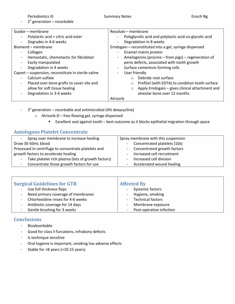
Periodontics III Summary Notes Enoch Ng - 2o generation – resorbable
Guidor – membrane - Polylactic acid + citric acid ester - Degrades in 4-6 weeks
Biomend – membrane - Collagen - Hemostatic, chemotactic for fibroblast - Easily manipulated - Degradation in 4 weeks
Capset – suspension, reconstitute in sterile saline - Calcium sulfate - Placed over bone grafts to cover site and
allow for soft tissue healing - Degradation in 3-4 weeks
Resolute – membrane - Polyglycolic acid and polylactic acid-co-glycolic acid - Degradation in 8 weeks
Emdogain – reconstituted into a gel, syringe dispensed - Enamel matrix protein - Amelogenins (procine – from pigs) – regeneration of
perio defects, associated with tooth growth - Surface cementum forming cells - User friendly
o Debride root surface o PrefGel (with EDTA) to condition tooth surface o Apply Emdogain – gives clinical attachment and
alveolar bone over 12 months Atrisorb
- 3o generation – resorbable and antimicrobial (4% doxycycline)
o Atrisorb-D – free flowing gel, syringe dispensed
Excellent seal against tooth – best outcome as it blocks epithelial migration through space
Autologous Platelet Concentrate - Spray over membrane to increase healing
Draw 30-50mL blood Processed in centrifuge to concentrate platelets and growth factors to accelerate healing
- Take platelet rich plasma (lots of growth factors) - Concentrate those growth factors for use
Spray membrane with this suspension - Concentrated platelets (10x) - Concentrated growth factors - Increased cell recruitment - Increased cell division - Accelerated wound healing
Surgical Guidelines for GTR - Use full thickness flaps - Need primary coverage of membranes - Chlorhexidine rinses for 4-6 weeks - Antibiotic coverage for 14 days - Gentle brushing for 3 weeks
Affected By - Systemic factors - Hygiene, smoking - Technical factors - Membrane exposure - Post-operative infection
Conclusions - Bioabsorbable
- Good for class II furcations, infrabony defects
- Is technique sensitive
- Oral hygiene is important, smoking has adverse effects
- Stable for >8 years (>10-15 years)
Periodontics III Summary Notes Enoch Ng
Multirooted Teeth - Furcation involvement in general = greater chance of tooth loss
- Patients with good hygiene means furcation involvement has no effect on tooth loss
o Hygiene is vital for furcation involved teeth
- Most commonly lost teeth due to periodontitis
o Mx molars – average CEJ to furcation = 4mm
o Mn molars
Enamel projections most common in Mx and Mn 2nd molars
o Mx first premolars
- There are often osseous cavities by furcations
Treatment Options - Odontoplasty – recontour the tooth and cover with a crown. Tooth should already be RCTed or patient will have
lots of sensitivity
- SRP – use a cavitron in furcation; much better than hand instruments or cavitron alone
o Furcations with PD>4mm less favourable than flat surfaces for SRP treatment
o SRP can cause attachment loss (therefore disease progression) if not done properly
- Open debridement
o More calculus is left behind in furcations than non-furcated areas
o True for both open and closed curettage
- APFs
o Increases easier access to furcation areas for hygiene
o Tunneling – creating class III furcation to allow for cleaning
May be problematic – big increased risk of caries
6.7% extracted because of caries
4.7% hemisected because of caries
23.5% developed caries
- Root amputation/hemisection
o Usually failure is from endodontic reasons, not periodontic ones
Periodontic failures usually in Mx molars
Most Mn molars failed due to root fracture
o Most often, hemisected teeth (Mn molars) served as distal bridge abutments
o Topical fluorides and hygiene vital in root hemisections – increased risk of caries
- Regenerative therapy (GTR)
o Class II furcations treated with GTR if there is some infrabony part to lesion
o Clinical/radiographic response generally similar between allogenic bone grafts, GTR, and growth factors
(emdogain)
o Class III furcations do NOT respond well to surgical treatment
Best to maintain non-surgically or have tooth extracted
Periodontics III Summary Notes Enoch Ng
Perio Plastic Surgery - Clinical attachment level = most important parameter for determining perio status, developing treatment plan
- Probing depths help determine what type of treatment to go with
Definitions - Mucogingival defect – departure from normal dimension and morphology of the relationship between gingiva
and alveolar bone
- Mucogingival surgery – perio surgical procedure to correct defects in morphology, position, and/or amount of
gingiva
- Perio plastic surgery – surgical procedure to correct or eliminate anatomic, developmental, or traumatic
deformities of gingiva and alveolar mucosa
Criteria for perio plastic surgery - Surgical site free of plaque, calculus, inflammation
o Good hygiene important to keep perio problem from become a caries problem
- Maintain adequate blood supply
- Be familiar with anatomy of recipient and donor site
- Ensure stable grafting tissue
- Minimize trauma to surgical site
Keratinized Gingiva - 1mm required
- 2mm suggested
- 5mm keratinized gingiva with 3mm attach gingiva for subgingival restorations
Indications - Root Coverage
o Coronally positioned flap
o Semilunar flap
o Laterally positioned flap
o Double papilla flap
o Free Gingival Graft
o GTR
o Connective tissue grafts
- Gingival Augmentation
o Free gingival graft
Increase gingival width
Deepen vestibule
For ridge augmentation
Advantages - Donor material readily available, even with thin
palates - Predictable healing
Disadvantages - 2 surgical sites - Color may not match recipient area - Graft may be overly thick when mature - Donor site sensitivity
o Allow for 25-50% shrinkage of graft
o Can be done to increase the amount of keratinized gingiva around a tooth, also make tissue thicker
Periodontics III Summary Notes Enoch Ng
Millers Classification for Gingival Recession - Class I
o Defect coronal to MGJ
o Interproximal bone and papillae intact, no malpositioning
o 100% coverage
- Class II
o Recession or attachment loss apical to MGJ
o Interproximal bone and papillae intact, no malpositioning
o 100% coverage
- Class III
o Recession or attachment loss at or apical to MGJ
o Some bone loss, papillae loss, or malposition present
o Partial root coverage only expected
- Class IV
o Recession of attachment loss apical to MGJ
o Extreme interproximal bone loss (horizontal bone loss), papillae loss, or extreme malposition present
o No root coverage can be expected
Root Coverage o Considerations
Thin tissue – can be augmented to increase thickness for more stability
Crater defect – 2 osseous walls (one buccal, one lingual)
Cortical bone only, difficult to get bone height because of poor blood supply
- Pedicle Graft
o Laterally positioned flap is the graft
- Free gingival graft
o SRP done, modifiers applied
o Donor tissue (1.5-2.5mm for root coverage) placed between layers of a partial thickness flap
o Sutured into place
- Connective tissue grafts
o Partial thickness flap
o SRP done, tetracycline applied
o Grafted, flap repositioned over graft, sutured
- Connect tissue grafts modified technique
No vertical incisions when preparing recipient site
Excellent blood supply to flap
Avoids scarring in esthetic zones
o Partial thickness flap
o Graft approximated, flap coronally positioned, sutured
o After grafting, will always look bulky but will fade over several months of healing
- Envelop Flap for CT Grafting (Raetzke technique)
o SRP, tetracycline used for surface conditioning
o Papilla undermined to create envelope flap
o Graft placed into envelope, sutured

Periodontics III Summary Notes Enoch Ng
Implants - Impact of Diameter Change – increasing diameter exponentially increases surface area
o 3.7mm = baseline 5.50mm2 seating area
o 4.0mm = 5% more SA 5.50mm2 seating area
o 5.0mm = 35% more SA 12.25mm2 seating area
o 6.0mm = 61% more SA 21.00mm2 seating area
Biologic Width - Bone level
o Vertical bone loss is determined by position of the microgap at implant/abutment interface
o Absent an interface, rough smooth border determines position of alveolar crest
- Probing Depths
o Implants do not have gingival fibers that attach – pocket goes right to bone
Gingvodental and transseptal fibers do not exist in gingiva surround implant abutment
o Some implants (ex: microlock) have tiny microthreads that sometimes may allow CT attachment
Emergence Profile - Coronal aspect of implant should be 2-3mm apical to CEJ of adjacent teeth
o 3mm deep from CEJ of proximal teeth in esthetic zones
- Internal connection gives retention
- Scalloped design sometimes shows 2 rabbit ears over the gingiva – bad esthetics
- Papillae height for contact point to alveolar crest
o <5mm = 100%
o 6mm = 56%
o 7mm = 27%
- Implants should be >3mm apart to preserve the alveolar crest peak interdentally
- Platform switching gives moves the implant/abutment interface away from horizontally, allowing bone to
remain at height instead of receding
Bony Sockets and Implants - Minimum 1mm bone both buccal and lingual to implant
o Prefer 2mm buccal bone for esthetics
o Implant may be placed slightly to the palatal to give buccal bone width
- If the socket is >2mm, it’s worth grafting
- If no grafting is done, lose around 5mm of bone in the anterior
o 2/3 of this bone is lost in the first 3 months
- Immediately post-extraction, place an ovate pontic to help maintain tissue scalloping and papillae form
- Ridge expansion can be done if needed after socket healing
Periodontics III Summary Notes Enoch Ng
Implant Perio/Recall - Use plastic scalers so not to scratch the implant
- Before cementing a crown, feel for what the implant threads feel like
o Sometimes cement can feel like the threads
o In some instances, may want to place a flap to remove the cement to ensure the area is clean
- If using a scaler, cover it with a plastic sleeve or something to prevent scratching/damaging implant
- Prophy paste and a rubber cup on a prophy head/handpiece = good for polishing implant bars when removal of
said bars are not indicated
- Woven nylon floss cords are abrasive enough to remove calculus, good for cleaning supracrevicular
abutments/restorations
- Proxi-floss can be adapted to the abutment surface/ridge bars
o Fins are designed to remove plaque
- G-floss – designed for hygienic restorations
- Sponge tip applicators for Chlorhexidine
- Perio-aids and end-tufted brushes can remove plaque at subgingival margins
- Specific brushes based on
o Final restoration design
o Access to sulcus
o Patient dexterity
Periodontics III Summary Notes Enoch Ng
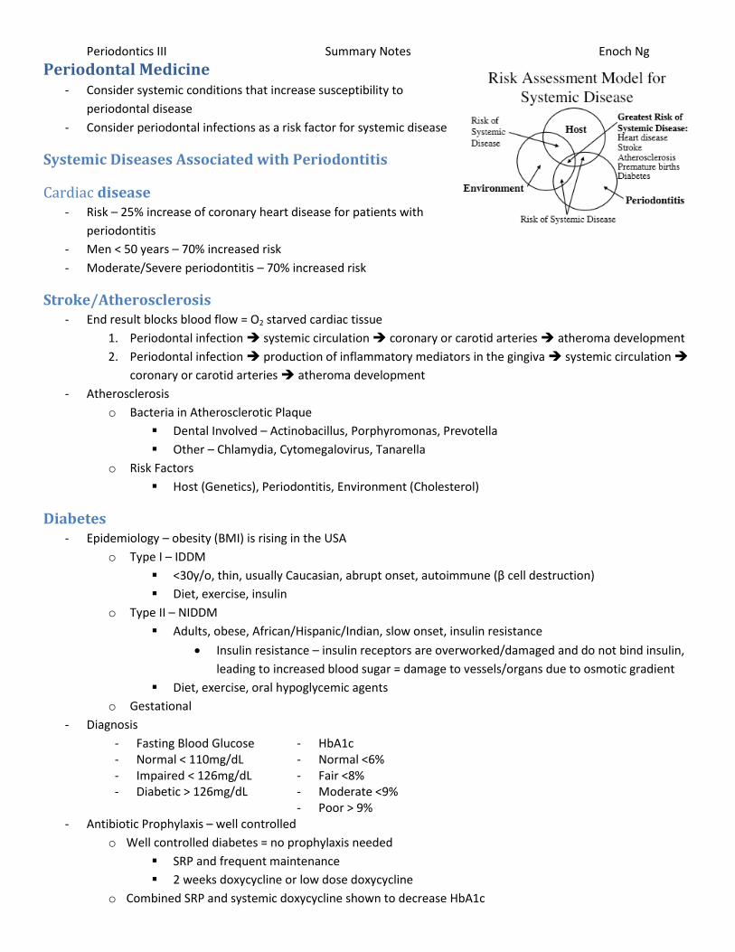
Periodontal Medicine - Consider systemic conditions that increase susceptibility to
periodontal disease
- Consider periodontal infections as a risk factor for systemic disease
Systemic Diseases Associated with Periodontitis
Cardiac disease - Risk – 25% increase of coronary heart disease for patients with
periodontitis
- Men < 50 years – 70% increased risk
- Moderate/Severe periodontitis – 70% increased risk
Stroke/Atherosclerosis - End result blocks blood flow = O2 starved cardiac tissue
1. Periodontal infection systemic circulation coronary or carotid arteries atheroma development
2. Periodontal infection production of inflammatory mediators in the gingiva systemic circulation
coronary or carotid arteries atheroma development
- Atherosclerosis
o Bacteria in Atherosclerotic Plaque
Dental Involved – Actinobacillus, Porphyromonas, Prevotella
Other – Chlamydia, Cytomegalovirus, Tanarella
o Risk Factors
Host (Genetics), Periodontitis, Environment (Cholesterol)
Diabetes - Epidemiology – obesity (BMI) is rising in the USA
o Type I – IDDM
<30y/o, thin, usually Caucasian, abrupt onset, autoimmune (β cell destruction)
Diet, exercise, insulin
o Type II – NIDDM
Adults, obese, African/Hispanic/Indian, slow onset, insulin resistance
Insulin resistance – insulin receptors are overworked/damaged and do not bind insulin,
leading to increased blood sugar = damage to vessels/organs due to osmotic gradient
Diet, exercise, oral hypoglycemic agents
o Gestational
- Diagnosis
- Fasting Blood Glucose - Normal < 110mg/dL - Impaired < 126mg/dL - Diabetic > 126mg/dL
- HbA1c - Normal <6% - Fair <8% - Moderate <9% - Poor > 9%
- Antibiotic Prophylaxis – well controlled
o Well controlled diabetes = no prophylaxis needed
SRP and frequent maintenance
2 weeks doxycycline or low dose doxycycline
o Combined SRP and systemic doxycycline shown to decrease HbA1c

Periodontics III Summary Notes Enoch Ng - Complications
o Blindness
o Kidney disease
Polyuria – frequent urination
Polydipsia – thirsty
Polyphagia – hungry
o Nerve damage
o Heart diseases/stroke
o Periodontal disease
PMN function – decreased phagocytosis and chemotaxis
Increased infections
Collagen metabolism – decreased collagen synthesis by fibroblasts
Increased crosslinking (due to AGE products) decreases cell turnover
o Slower wound healing
- Hyoglycemic Shock – treat with sugar
o Be aware of drug peak activity – greatest risk of hypoglycemia
o Symptoms = Confusion, Shakiness, Agitated, Anxious, Sweating, Dizziness
Low birth weight/premature birth o Pregnancy increases risks
Gingivitis by 60-70%
Heart murmurs by 90%
Cardiac output increased by 50%
o Inform the patient, good oral hygiene is important, best to treat in 2nd trimester
o FDA Pregnancy Drug Classifications
A B C D E Animal No Risk No Risk Toxic Risk Great Risk Human Unknown Unknown
o Local Anesthesias
Class B = Lidocaine, prilocaine, etidocaine (_ocaine, except procaine)
Class C = mepivacaine, bupivacaine, procaine ( _vacaine and procaine)
o Antibiotics
Class B = penicillin, erythromycin, clindamycin, cephalosporin, metronidazole, azithromycin
For erythromycin, avoid Erythromycin Estolate during pregnancy
Class C = Clarithromycin, Ciproflaxin
Class D = tetracycline
o Analgesics
Aspirin – Class C/D (3rd trimester)
Acetaminophen – class B
Can be combined with codeine, hydrocodone, oxycodone
Ibuprofen – class B/D (3rd trimester)
Propoxyphene (opioid analgesic)
Periodontics III Summary Notes Enoch Ng - Pre-Term Low Birth Weight
10% of newborns
<37 weeks, or <5lbs 8oz
Risk increased 7x for pre-term low birth weight for mothers with perio disease
African Americans at greatest risk
Infection increases TNFα and Prostoglandin E2
TNFα and Prostoglandin E2 are childbirth mediators
Excessive secretion results in pre-term low birth weight babies
o SRP on perio active pregnant women may reduce PTLB babies
Untreated = 6.3% PTLB
Prophylaxis + Placebo = 4.9% PTLB
SRP + Placebo = 0.8% PTLB
SRP + Metronidazole = 3.3% PTLB
Osteoporosis - Occurs in women 40-50y/o
- Medications for menopause = hormone replacement therapy, alendronate, calcium
Drugs that interfere with calcium absorption, steroids, diuretics, thyroid medications
o HRT decreases risk of losing teeth, both past and current users
Tooth retention increases with HRT duration
HRT increases breast cancer, cardiovascular disease, stroke
HRT decreases colon cancer, bone fractures
- Oral Consequences
o Low bone mineral density in Mn
o Greater clinical attachment loss
- Medications
- Drug Categories that may cause gingival overgrowth
o Diuretics, Ca++ blockers, ACE inhibitors, α-blockers, immunosuppressant
Organ transplants require immunosuppressant drugs
Cyclosporine may be swapped for Tacrolimus
Alendronate (Fosamax) – bisphosphonate = inhibits bone resorption
o If possible, contact physician and change the drug
Hyperplasia within the first 6 months = switch to another drug
Need good oral hygiene and frequent maintenance
- Drugs that may cause gingival overgrowth
o Dilantin (antiepileptic)
o Cyclosporin (immunosuppressant)
o Nifedipine (Ca++ Channel Blockers)
Smoking

Periodontics III Summary Notes Enoch Ng
Chemotherapeutics
Local vs Systemic Delivery - Can be used to treat periodontitis
- Should be used as an adjunctive treatment to SRP
- Systemic delivery is as good as local delivery
- Local and systemic can be used at the same time if desired
Local Delivery Advantages Local Delivery Disadvantages
- More concentrated - Fewer side effects - Sustained delivery - Patient compliance
- More chair side time - More expensive - No effect on bacterial reservoirs - Do NOT use for pregnant patients
Local Delivery Drugs - Actisite (tetracycline hydrochloride) – still FDA approved, but discontinued. High [tetracycline]
o Perio Fiber Therapy – place fiber for 10days (tissue distension after fiber removal)
- PerioChip (2.5mg Chlorhexidine)
o Gelatin carrier – bioabsorbable, pockets >5mm, no refrigeration
o Good for patients allergic to tetracycline, pockets <5mm cannot be packed into properly
o Patients can brush, but should NOT floss in site for 10 days, regular diet okay
o Site moderately sensitive for first week
o >2mm reduction – 30.3% vs 13.5% SRP alone after 9 months
- Atridox (8.5% Doxycycline)
o Only drug approved to increase CAL (probably through long junctional epithelium)
o Bioabsorbable, controlled release for 7days for pockets >5mm
Can be packed TOO deeply into pocket, creating discomfort
o 2 syringe mixing – 450mg atrigel to 50mg doxycycline
o Liquid solidifies upon contact with crevicular fluid – sets into wax-like consistency in 1-2min
o Significantly reduces anaerobic bacteria, but doesn’t develop antibiotic resistance
- Arestin (Minocycline)
o Sustained release as a powder, no reconstitution or refrigeration needed, 2 year stability
o 25% more patients went from >6mm PD to <5mm PD over 9 months compared to SRP alone
- Metronidazole (25% metronidazole)
o NOT approved in USA (degrades within 24h)
o Biodegradable, anaerobic bactericidal, >5mm pockets
o NOT shown to be more effective with SRP than SRP alone
Actisite Atridox Periochip Arestin
Ease of Use Moderate Easy Easy Easy
# of Sites Multi (1-2 fibers/tooth) 8-15 (sites/syringe) 8 (1 chip/site) 1 (1 site/carp)
Dressing/Glue Y Y/N N N
GCF Conc ug/mL 1300 1000 125 1000
Release (days) 10 7 10 10
Removal Y N N N

Periodontics III Summary Notes Enoch Ng
Systemic Delivery Drugs - Periostat – only FDA approved oral systemic treatment for chronic periodontitis – suppresses tissue destroying
enzyme activity
o 20mg = no antimicrobial action, no bacterial flora changes and no resistance after 18 months
o Acts as an enzyme suppressor
- Adjunctive to SRP, promote attachment level and decrease pocket depth
- Indications – maintenance patients, refractory/recurrent periodontitis, smokers trying to quit
- Prescription:
o 20mg doxycycline 2x daily, duration up to clinician o 1h before meals, take with adequate fluids, do not double up dosage o Efficacy – minimum = 3months, max = 9months o Safety – 12months, follow traditional tetracycline contraindications
Rx: Periostat Disp: 180 capsules Sig : 1 cap BID Refills: 2
Indications for Controlled Delivery - Pockets >5mm
- BOP
- Not responsive to SRP
- Esthetic concerns (surgery contraindicated)
- Refractory periodontitis
- Medically compromised patients (surgery contraindicated)
- Recent oral cancer
- Uncontrolled diabetes
- Smoking patients
- Dental phobic patients
Oral Manifestations of Uncontrolled Diabetes - Severe gingival inflammation
- Acute gingival or periodontal abscesses
- Rapidly advancing perio disease
Systemic Antibiotics for Perio Therapy - Not necessary most of the time – offer little/no advantage as adjunct to conventional therapy
- Refractory disease – progressive destruction of perio attachment in spite of good conventional mechanical
therapy
o Doxycycline 100mg (21 tablets)
q12h for day, then qd until gone
Take 1hr before or 2hr after meals
Decreased absorption by – antacids, NaHCO3, Al, Mg, Ca, antidiarrheals, Fe, Zn, food/dairy
Bacteriostatis, acts on protein synthesis (affects G+ and G-)
Side effect – tooth discoloration
o Amox 500mg and metronidazole 250mg (22 tablets each)
2 tabs each, then 1 tab q8h until gone
Metronidazole – may take with or w/o food
Bactericidal (DNA synthesis), obligate anaerobes only
Side effects – GI tract, anticoagulant, disulfiram-like reaction, don’t mix with ^OH
Periodontics III Summary Notes Enoch Ng o Clindamycin 150mg (30 tablets)
2 tabs immediately, then 1 tab q6h until gone
May take with or w/o food
Bacteriostatic (affects G+ and G-)
Side effects – pseudomembranous colitis (toxin from C. difficile
o Treat with oral vanco if necessary
o Ciprofloxacin 250mg and metronidazole 250mg
2 tablets each q12h for 5 days
- Aggressive periodontitis
o Juvenile perio – incisors and first molars affected severely
Actinobacillus actinomycetemcomitans – resists removal by mechanical debridement, even
when surgically accessed
o Use doxocycline or amox/metronidazole combination for drug therapy
- Systemic Conditions
o Chediak-higashi syndrome
o Down’s syndrome
o Diabetes
o AIDS – AIDS associated necrotizing ulcerative periodontitis
Gingivitis – chlorhexidine rinse
Periodontitis (NUG, NUP) – chlorhexidine rinse
Metronidazole therapy (250mg, 1 tab qid for 22tabs)
o Cancer
Routine Care (17-20days after chemotherapy)
WBC > 2000/mm3
Platelet > 50,000/mm3
If catheter present – Amox 2g or clinda 600mg 1h pre-op
o Papillon-LeFevre syndrome
Rx – augmentin (amox 500mg, clavulanic acid 125mg)
Clavulanic acid – increases amox effectiveness be inactivating beta lactamases
Don’t want to give tetracyclines to younger patients – may stain permanent dentition
o Leukemia
o Neutropenia
o Hypophosphatasia
o Leukocyte adhesion deficiency
Uncontrolled Diabetes HbA1c Fasting Blood Glucose
<6% = normal <8% = fair <9% = moderate >9% = poor
< 110mg/dL = normal < 126mg/dL = impaired >125mg/dL = diabetic >200mg/dL = defer elective treatment, use antibiotic if must treat
- Periostat – no antibacterial activity, inhibits collagenases
o Rx: doxycycline 100mg
o Disp: 21tablets
o Sig: 1 tablet q12h first day, qd after until done
Periodontics III Summary Notes Enoch Ng
Pregnancy - Antibiotics may decrease efficacy of birth control pills
Signs of Infection - SHaRP
- Pain, redness, edema, pus, fistula
- Fever, increased vitals, lymphadenopathy, malaise, increased WBC count
- Penicillin VK 500mg (28tablets)
o 1 tab q6h
Pen VK take 1hr before or 2hr after meals
Amox or augmentin take with or w/o food
Bactericidal (cell wall synthesis)
Side effects – allergies, pseudomembranous colitis
Allergies – 3-10% population
Anaphylactic – 1/7K-25K
Cross-reactivity (cephalosporins) – 3-5%
o If pen/amox ineffective within 48-72hr, add in augmentin or metronidazole
Amox > Pen VK – better absorbed, longer serum half life, may take with food
Peaks at 2h, half life 0.7-1.4hr instead of 30min
- Erythromycin
o Azirthro and erythro – 1hr before or 2hr after meals
o Clarithro – with or w/o food
o Bactericidal – protein synthesis
o Azithro better for perio disease
Better anaerobic coverage, long serum ½ life, qd only, category B pregnancy
250mg BID first day, then qd for 5 days
o Side effects
Erythro – GI tract and nausea
Antibiotic Principles - Switch if no response in 48-72h
- Continue 2-3 days after symptoms gone
- Loading dose = double maintenance dose
- If using oral contraceptive, use an additional form of birth control
Periodontics III Summary Notes Enoch Ng
Advancements in Surgical Techniques
LASER – light amplification by stimulated emission of radiation - Wavelength determines character of laser
o Soft Tissue Lasers – Argon, CO2, Nd:YAG, diode
May target pigment, water, bone, etc
May cause necrosis of hard tissue if used improperly
- Irbium laser can be used for SRP (calculus and endotoxin removal), but has great potential to damage hard tissue
and is NOT shown to be greater benefit for clinical attachment loss than conventional SRP
Advantage Disadvantage
- Hemostasis - Bactericidal - Minimal wound contraction
- Precautions for eyes, other tissues - Reflected beam - CO2 can cut tissue, others may cause damage
- Laser oscillation decreases intensity (chance of damaging stuff)
Diode Laser - Affected by:
o Type of lesion
o Wavelength used
o Vascularity
o Selected mode of operation
o Speed of cutting desired
o Exposure time of tissue to laser
- Operations
o Tissue management/gingivectomy
o Gingival troughing (for impression taking around a crown prep, etc)
o Hemostasis
o Gingival sculpting/gingivectomy
o Crown exposure for orthodontics
o Biopsy (fibromas, etc)
o Frenectomy
Laser Evidence - Conflicting results, comparisons between relative effectiveness of lasers vs conventional is impossible
- Nd:YAG or Er:YAG lasers may be useful in SRP adjunctive to conventional or stand alone, but no advantage in
gaining clinical attachment loss
iCAT – radiographic stents - The image on its own is not enough – shows thickness, but doesn’t show if its in the needed position for good
implant placement
Piezoelectic Surgery - Works like a jackhammer – hits on a hard surface
- Soft tissue has give, so it doesn’t cut soft tissue
- Very useful for sinus lifts, hard tissue surgeries
Periodontics III Summary Notes Enoch Ng
Occlusion and Orthodontics
Occlusion o Physiologic – no signs of dysfunction or disease
o Traumatic – occlusion associated with dysfunction or disease
More rapid progression of periodontally involved teeth
Primary – excessive occlusal force on normal dentition
Secondary – normal force on a periodontally compromised tooth
o Therapeutic – specific interventions designed to treat dysfunction
Stable endpoint of Mn closure
Bilateral distribution of occlusal forces
Axial loading of teeth
- Therapeutic priority – control inflammation
o After inflammation, THEN address residual mobility
- Clinical Features of Occlusal Trauma
o Tooth mobility, increased displacement, stable pattern adaptation
o Tooth migration, pain on percussion, radiographic changes (widened PDL, apical resorption, etc)
o TMJ dysfunction
o Excessive wave facets, fractures
o Fremitus – vibration of palpation
- Treatment
o Evaluate vitality and parafunctional habits
o Occlusal adjustments – prophylactic equilibration is contraindicated
o Splitting
o Orthodontic tooth movement
o Occlusal reconstruction
o Extraction
- Outcome Assessment
o Decreased mobility or stable pattern
o Decreased migration of teeth
o Stable of decreased radiographic changes
o Relief of pain, fremitus, occlusal interferences
o Stable, functional, physiologic and esthetically acceptable occlusion

Periodontics III Summary Notes Enoch Ng
Perio-Ortho - Orthodontic Extrusion
Bracket placement
Extrusive ortho forces
Circumcrestal fiberotomy
Prevents rotational relapse, funneling or lipping of alveolar crest bone
Done every 1-2 weeks while tooth is being extruded, supracrestal gingival fibers are
severed under LA
Speed of extrusion can allow for increased/decreased bone movement
Fast extrusion – 2-3mm/week, soft tissue but not bone movement
Slow extrusion – 2-3mm/month, bone and soft tissue moves with the tooth
Possible APF
o Advantages
May eliminate need for osseous resection
No loss of interproximal papillae
No post-op sensitivity
o Concerns
Hard to achieve good emergence profile in final prosthesis
Complicated hygiene
Tooth may be difficult to restore/maintain due to root form
- The Fiber Groups and Modeling
Classical – interradicular, apical, oblique, horizontal, alveolar crest, trans-septal
Buccal/Lingual – alveolar, alveologingival, dentoperiosteal, cementogingival, circular
Interproximal – alveologingival, dentoperiosteal, circular, dentogingival
o Fibroblasts – both creates and destroys
Secretes collagen, elastic, GS
Destroys collagen, elastic, GC intracellularly and extracellukarky
o Osteoclasts:osteoblasts
Coupled process
Systemic factors – hormones, Vit D, etc
Local factors - GFs, cytokines, etc
- Goals of Ortho Movement
o Force to maximize movement
Leading side – pressure, resorption, collagen fiber compression
Trailing side – tension, deposition, collagen fibers stretch
o No pain
o Without root resorption
o Maintains healthy PDL throughout movement
Periodontics III Summary Notes Enoch Ng - Anchorage
o Active element – part that’s moving
o Resistance lement – anchorage
o Molars as anchor teeth, move single tooth and a time, consider large vs small roots
Usually take 3 teeth to anchor single tooth
Implants cannot move (no PDL), so can be used to move molars or entire arches
Absolute anchorage
Movement simultaneously (not segments, single teeth)
ANY tooth movement (including absolute intrusion
- Surgical Considerations
o Access
o Angle of implant
o Midline suture
o Avoid nasal floor perforation
o Explanation of the implant
o Diagnostic waxup – level the occlusal plane
Wilckodontics - Corticotomies
o Cut cortical bone, elevate flap
o Lines and dots (marrow space)
o Healing phenomenon
o Less bone mass
o Stimulate osteoblasts and osteoclasts surgically
o Can perform locally (not generally done)
Just boring into the bone, to stimulate osteoblastic/clastic activity, is fine. Don’t need to remove
cortical bone around the anchorage site
o Makes for much faster tooth movement
Periodontics III Summary Notes Enoch Ng
Perio-Restorative - Minimal risk = sealed margins, good proximal contacts, no overhangs
o If any of these are present, this increases risk factors for perio
- Interproximal caries and dental restorations = local risk factors for localized perio attachment loss
o Monitor these sites, take appropriate steps to minimize the risk
o Open contact = food impaction, harder to keep area clean, bacterial trap/retention area
- Widened PDL = should consider occlusal trauma
- Retraction cord = causes REVERSIBLE trauma – heals in about 12 days (2 weeks)
Surgical Instruments - Blades = good, if you nick the bone it doesn’t cause any problems
- Electrosurg and laser = must be 100% sure you are NOT contacting hard tissue, or it will cause localized necrosis
Contouring - Over contoured teeth create a bacterial trap, makes it difficult to probe properly to get good measurements
- Under contoured teeth cause food impaction trauma and provide a bacterial trap
- Gingival overgrowth/swelling may be caused from drugs, be aware before offering surgical services
- Infrabony pockets are a big problem = harder to clean, harder to access, etc
Patient Expectations - Patient esthetic expectations are primarily culturally based
- Metal allergies – use high noble metals to avoid allergic reactions
o Take a good medical history
o Use all ceramics/porcelains to avoid this problem
o Most common metal allergy is nickel
- When splinting teeth, check the occlusion
o When splinted, the teeth may not be mobile. However, a widened PDL may appear for the entire area
This is a good indication of occlusal trauma/disequilibration
Periodontics III Summary Notes Enoch Ng
Perio Supportive Therapy - Purpose of Perio Therapy – increase longevity of the person’s natural dentition by preserving supporting
structures of the teeth
- Maintenance and Supportive Therapy – act of continually caring for and preserving the dentition in health and
function
Objectives - Early recognition of the disease
- Prevention of disease recurrence
- Prevention of further disease advancement
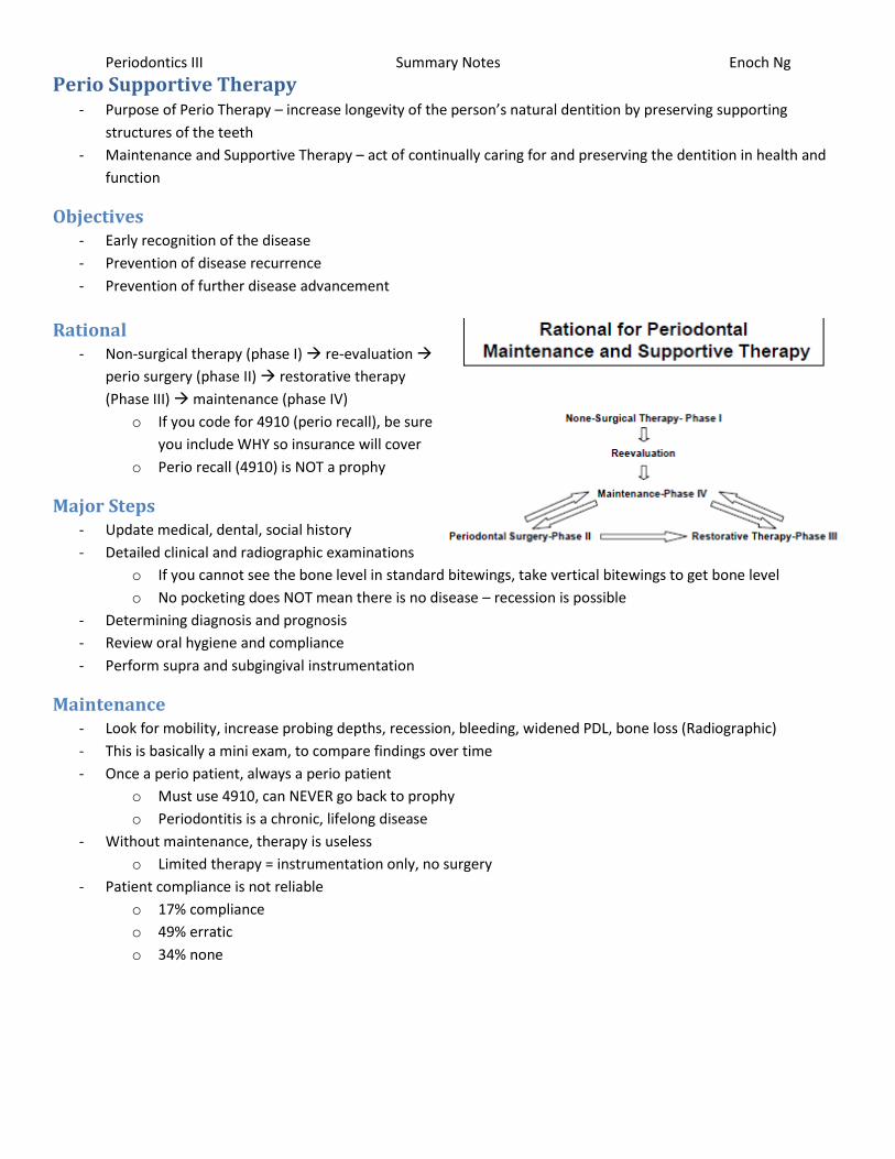
Rational - Non-surgical therapy (phase I) re-evaluation
perio surgery (phase II) restorative therapy
(Phase III) maintenance (phase IV)
o If you code for 4910 (perio recall), be sure
you include WHY so insurance will cover
o Perio recall (4910) is NOT a prophy
Major Steps - Update medical, dental, social history
- Detailed clinical and radiographic examinations
o If you cannot see the bone level in standard bitewings, take vertical bitewings to get bone level
o No pocketing does NOT mean there is no disease – recession is possible
- Determining diagnosis and prognosis
- Review oral hygiene and compliance
- Perform supra and subgingival instrumentation
Maintenance - Look for mobility, increase probing depths, recession, bleeding, widened PDL, bone loss (Radiographic)
- This is basically a mini exam, to compare findings over time
- Once a perio patient, always a perio patient
o Must use 4910, can NEVER go back to prophy
o Periodontitis is a chronic, lifelong disease
- Without maintenance, therapy is useless
o Limited therapy = instrumentation only, no surgery
- Patient compliance is not reliable
o 17% compliance
o 49% erratic
o 34% none
Periodontics III Summary Notes Enoch Ng
Perio Therapy Results - Establish health
- Restore function
- Preserve health
Prevent and Treat Gingivitis - Usually, no significant calculus builds up in 2 weeks
- Plaque accumulation for 2 weeks is completely reversible
- Health is associated with wealth/country
o Norway = better hygiene and care
o Sri Lanka = worse hygiene/care/condition
Studies done at age 40, where CAL begins to appear
Age isn’t an issue, more the accumulation of the disease
As you get older, the more you accumulate so the greater the progression
Prevent and Treat Periodontitis - BOP over prolonged period = approx. 70% greater chance for attachment loss/periodontitis
- Gingival recession predominant lesion before age 40
- Highest rate of periodontal disease occurs between ages 50-60
o Pocket periodontology principal mode of destruction between ages 50-60
- ¼ of the population has stable healthy periodontal condition throughout life
- ¾ of the population has slight/moderate perio disease, from 0.02-0.2mm/year
o Cumulative average of 2.44mm as patient approaches 60y/o
o Mean annual risk of initial attachment loss highest between 16-34y/o
Only 20% of sites continued to lose more attachment
<1% had substantial loss (>4mm)
Teeth Mortality - Teeth w/o inflamed gingiva maintained average of 51 years
- Teeth w/ inflamed gingiva 46x more likely to be lost
- SRP/Prophy is better than no treatment
o People who receive perio therapy lose much less dentition than those who do not receive perio therapy
Chance is roughly 5x greater for loss
- Most patients don’t lose teeth, but a few patients lose lots of teeth
o This can skew the data
- Perio therapy (surgically positioning apically) may change the problem from a perio to caries/sensitivity
Summary - Best single action to prevent gingivitis = oral hygiene
- Best ways to treat gingivitis = oral hygiene, calculus removal
- Gingival bleeding/attachment loss = more likely to lose more attachment if there is bleeding
- 5x more tooth loss in untreated population compared to treated population
- The ultimate measure to determine efficacy of perio therapy = tooth retention
Periodontics III Summary Notes Enoch Ng
Perio for the GP - Curret perio therapy is provided mostly by specialists
- Practices that employ hygiene:
No Hygienists 1 Hygienist 2 Hygienists 3+ Hygienists
33.8% 24.8% 24.0% 17.4%
- Most patients treated for gum disease are <45y/o
o 29% are 25-35y/o
o 26% are 35-45y/o
Dental Team - Analysis of the Dental Team
o Desire and motivation to make transformation
o Role of each member in the team
This isn’t just the DDS and DH, it includes assistants, receptionists, etc
- Analysis of Practice Style
o Willingness to critically analyze existing practice
o Commitment to invest time, energy, funds required
Educational and Clinical Considerations o Didactic and clinical education and updates
o Record keeping and quality assurance
o Instruments and equipment needed
o Recall system
o Referral philosophy and process
o Dealing with leaving and new patients
- The standard of care does NOT vary per location
Periodontal Disease - Gingival disease
- Chronic periodontitis
- Aggressive periodontitis
- Periodontitis as a manifestation of systemic diseases
- Necrotizing periodontal diseases
- Abscesses of the periodontium
- Periodontitis associated with endodontic lesions
- Developmental or acquired deformities and conditions
Periodontics III Summary Notes Enoch Ng
Referrals - Having a good relationship with specialists and general practitioners
o Some of the worst experiences for periodontists are receiving referrals from other providers
- Developing the dental hygienist as a referral source
o Hygienists collect a TON of info, so they’re a good source for suggesting when to refer out a patient
- There are 4 variables that influence number of referrals per month from a GP to a specialist
o Female gender
o Practicing with one other dentist
o Employing 2 or more hygienists
o Being more than 5 miles away from the nearest specialist
- When in doubt, seek periodontal consultation and/or a second opinion
- Provide information relevant to:
o Chief concerns expressed by patient and therapist
o Health status and need for special consideration
o Any past experience with perio treatment
o Anticipated dental therapeutic procedures
o Quality FMX
- Expect to receive back from the specialist:
o Periodontal diagnosis and prognosis
o Proposed perio treatment plan and possible alternatives
o Needs for coordinated treatments including but not limited to: endodontics, TMD, ortho, OMS,
restorative, prosthetic reconstruction, implants, and recommended schedule for perio maintenance
o Patient interest and willingness to accept and follow-up on the proposed perio treatment
- Majority of patients have:
o Early (1-2mm CAL)
o Moderate (3-4mm CAL)
Chronic periodontitis that are diagnosed early can be typically treated/maintained in by GP
- Aggressive
o Localized or generalized periodontitis in kids or adolescents
o Individuals 30y/o or older with significant bone loss (>4mm or 1/3 of root length)
- Advanced (>5mm CAL) chronc periodontitis with sites depict
o Progressive deepening of pockets with CAL and/or bone loss as seen on radiographs
o Infrabony defects that have the potential for regenerative therapy
o Multiple rooted teeth with more than 4mm CAL with furcation involvement that will complicate therapy
o Mucogingival defects that exhibit progressive recession and/or are an esthetic concern
- Other clinical situations
o Comprehensive perio appraisal prior to initiating ortho or extensive restorative therapy on adults
o Atypical perio diseases associated with immune response or systemic health
o Excision of proliferative or excessive gingival tissues
o Crown lengthening surgery as indicated for restorative and/or esthetic purposes
o Mucogingival defects that exhibit progressive recession and/or are of an esthetic concern
o Tooth/teeth extraction in conjunction with ridge preservation/augmentation
o Exposure of impacted teeth in conjunction with ortho treatment
o Surgical placement of root formed dental implants
Periodontics III Summary Notes Enoch Ng
Conclusion - Main reason for non-compliance is patients don’t attend their own dentist exclusively for maintenance therapy
- Tooth loss and perio deterioration was more marked in this group that patients who in addition attended
specialist office for maintenance therapy
- More is spent on oral health ($71Billion) than cancer ($62Billion)
o Heart conditions have the most ($90Billion)
- Average total expenditure for insurance covered patients is ~ $400
o Only 6-7% of all insured patients reach their yearly maximum
- Around half of all perio procedures are done by non-specialists
o Some preventative procedures are also perio procedures
Background Information and Dental Codes - Dental benefits plans vary greatly between providers
- Not all carriers legally required to adhere to ADA standard
o Many states designate the ADA as standard for dental carriers, not-for-profit carries (Blue Cross, Delta),
are not governed by such provisions
- Many carriers created contracts based on old CDT codes – until these expire, they will not change over to new
CDT codes
o Insurance carriers typically have large in-house computer systems that are complex and may take time
to re-program
- Dental vs dental hygiene provided services
- Determine overall medical, oral, dental, and perio status
- Use appropriate CDT code regardless of patient insurance
o Clinical evaluation codes
o Preventative codes
o Non-surgical codes
o Surgical codes
o Implant codes
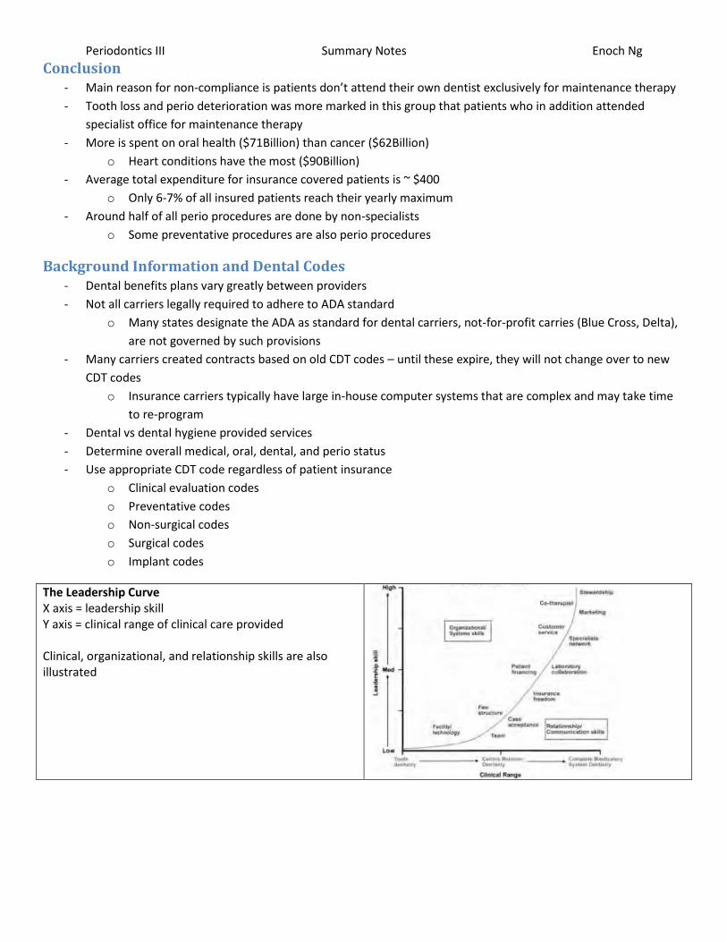
The Leadership Curve X axis = leadership skill Y axis = clinical range of clinical care provided Clinical, organizational, and relationship skills are also illustrated