Embed Size (px)
Citation preview
Doctoral dissertation
To be presented by permission of the Faculty of Medicine of the University of Kuopio
for public examination in Auditorium, Kuopio University Hospital ,
on Friday 15th January 2010, at 12 noon
Institute of Clinical MedicineDepartment of Clinical Radiology,
Department of NeurosurgeryKuopio University Hospital
University of Kuopio
PAULA BENDEL
Imaging of the Brain After AneurysmalSubarachnoid Hemorrhage
One-year MRI Outcome of Surgical and EndovascularTreatment
JOKAKUOPIO 2009
KUOPION YLIOPISTON JULKAISUJA D. LÄÄKETIEDE 473KUOPIO UNIVERSITY PUBLICATIONS D. MEDICAL SCIENCES 473
Distributor : Kuopio University Library P.O. Box 1627 FI-70211 KUOPIO FINLAND Tel. +358 40 355 3430 Fax +358 17 163 410 www.uku.fi/kirjasto/julkaisutoiminta/julkmyyn.shtml
Series Editors: Professor Raimo Sulkava, M.D., Ph.D. School of Public Health and Clinical Nutrition Professor Markku Tammi, M.D., Ph.D. Institute of Biomedicine, Department of Anatomy
Author´s address: Department of Clinical Radiology Kuopio University Hospital P.O.Box 1777 FI-70211 KUOPIO FINLAND Tel. +358 17 173 907 Fax +358 17 173 342 Supervisors: Professor Ritva Vanninen, M.D., Ph.D. Institute of Clinical Medicine Department of Clinical Radiology University of Kuopio
Docent Timo Koivisto, M.D., Ph.D. Institute of Clinical Medicine Department of Neurosurgery University of Kuopio
Reviewers: Docent Veikko Kähärä, M.D., Ph.D. Department of Radiology Tampere University Hospital
Docent Ari Karttunen, M.D., Ph.D. Department of Diagnostic Radiology Oulu University Hospital
Opponent: Docent Leena Valanne, M.D., Ph.D. Helsinki Medical Imaging Center Helsinki University Hospital
ISBN 978-951-27-1373-8ISBN 978-951-27-1390-5 (PDF)ISSN 1235-0303
KopijyväKuopio 2009Finland
Bendel, Paula. Imaging of the Brain after Aneurysmal Subarachnoid Hemorrhage- OneyearMRI Outcome of Surgical and Endovascular Treatment. Kuopio University Publications D.Medical Sciences 473. 2009. 124p.
ISBN 9789512713738ISBN (PDF)9789512713905ISSN 1235-0303
ABSTRACT
Background: Aneurysmal subarachnoid hemorrhage (aSAH) is a devastating disease withan overall casefatality of 4050%. Most of the surviving patients are left with neurologicaldeficits and cognitive impairments. Brain MRI could possibly provide valuable information onpatients' recovery potential together with detailed neurological and neuropsychologicalexamination after aSAH.
Patients and Methods: Onehundred and sixtyeight consecutive aSAH patients wererandomly assigned to either endovascular (n=82) or surgical (n=86) treatment. Altogether138 conventional and 77 volumetric MRI examinations were available one year after aSAH.T2PD and 3DT1weighted images were analyzed in detail. Volumetric analyses focused totemporomesial structures; hippocampus (HC) and amygdala (AM), and gray matter (GM)volume loss in patients with ruptured anterior cerebral artery (ACA) aneurysm. The atrophicenlargement of the cerebrospinal fluid (CSF) volumes was quantified. MRI findings werecorrelated with neuropsychological test scores.
Results: Fortyfour (31.9%) of 138 patients had no lesions associated with aSAH. Accordingto intention to treat, lesions were more frequent after surgical treatment, predominating infrontal and temporal lobes. Ischemic lesions of the parental artery territory of the rupturedaneurysm were more frequent after surgery. Ischemic lesion volumes correlated withneuropsychological test scores. After aSAH, the HC and AM volumes were found to besmaller than in the matched control individuals. After treatment of an ACA aneurysm, GMatrophy was detected on frontobasal cortical areas and HC ipsilateral to the surgicalapproach. Enlarged ventricular and sulcal CSF volumes were detected after aSAH. HigherFisher scores, preoperative hydrocephalus and older age were found to associate withventricular and sulcal enlargement, both correlating with neuropsychological defects.
Conclusion: aSAH is frequently followed by brain parenchymal changes, especially infrontotemporal areas. Lesions seem to be more frequent after surgical than endovasculartreatment. Temporomesial volume loss is a common finding after aSAH in general. GMatrophy, principally involving the frontobasal cortical areas and hippocampus ipsilateral to thesurgical approach is detected after aSAH and treatment of the ruptured ACA aneurysm.Atrophic CSFenlargement together with reduced GM volumes is a common sequela afteraSAH. Both enlarged CSFspaces and brain parenchymal lesion volumes correlate withneuropsychological test performance after aSAH.
National Library of Medicine Classification: WG 580, WL 355, WN 185
Medical Subject Headings: Amygdala; Aneurysm; Atrophy; Embolization,Therapeutic/methods; FollowUp Studies; Hippocampus; Humans; Imaging, ThreeDimensional; Intracranial Aneurysm /surgery; Magnetic Resonance Imaging;Neuropsychological Tests; Randomized Controlled Trial; Rupture; SubarachnoidHemorrhage; Treatment Outcome

ACKNOWLEDGEMENTS
This work was carried out in the Department of Clinical Radiology, University of Kuopio, in
Collaboration with Departments of Neurosurgery and Neurology, Kuopio University Hospital
during the years 20022009.
I owe my deepest feeling of gratitude to my principal supervisor Professor Ritva Vanninen,
M.D., who kindly suggested this most interesting topic. It has been a true privilege to work in
her excellent, enthusiastic, energetic, warm and professional guidance. This thesis would
never have been finished without her encouragement. She has given her essential support in
all phases of this study. Besides her excellent scientific and psychological skills, I also
admire her expertise as a clinical neuroradiologist.
I owe my deepest gratitude to Docent Timo Koivisto, M.D., my other supervisor. He has
originally collected the patients who were included in this study. It was a priority to deepen
the radiological aspect of his originally unique, prospective, randomized Outcome Study of
Aneurysmal Subarachnoid Hemorrhage. He has always found time to discuss this study with
me. I have also enjoyed our friendship and the time we have spent together with our families.
I wish to express my warm gratitude to Head of The Department of Clinical Radiology,
Professor Hannu Manninen, M.D. His favourable attitude towards research projects has
provided excellent atmosphere and realistic possibilities to combine clinical work and
research.
I am greatly indebted for the physicists Mervi Könönen, M.Sc. and Eini Niskanen M.Sc., who
have been extremely essential persons to resolve all the technical and mathematical
problems we have faced during this study. I highly appreciate their positive and constructive
attitude and skills to communicate.
I want to express my gratitude to Professors and neurosurgeons Matti Vapalahti, M.D. and
Juha Hernesniemi, M.D., who were essential persons to build up the original randomized
study protocol and who have also performed most of the surgical operations during this
study. I am also grateful for the possibility to begin my medical career on 1997 by working as
a resident in the Department of Neurosurgery.
My warmest thanks belong to my coworkers; deceased neuropsychologist Heleena
Hurskainen, M.Sc., who performed all the neuropsychological tests; Docent Tuomo
Hänninen, M.S.c. and Marja Äikiä, M.S.c., Ph.D. who have further interpreted the
neuropsychological data and essentially helped with the neuropsychological aspects in the
manuscripts; neurointerventionist Tapani Saari, M.D., who has performed all the
embolization procedures together with Professor Ritva Vanninen. I want to specially thank
my colleque Päivi Koskenkorva, M.D., for sharing the VBMmysteries with me. I owe my
warm thanks to Anni Kolehmainen, M. D., and Corina Pennanen, M.D, Ph.D for their help in
the volumetric measurements.
I am most grateful to all my collegues in the Department of Neuroradiology; especially Teppo
Mäkelä for always having “a minute”. I also warmly want to thank Aki Ikonen, Anna Sutela,
Antti Lehtimäki, Juhana Hakumäki and Sanna Keränen for sharing their knowledge and
expertise in Neuroradiology.
I thank the official referees of this study, Docent Veikko Kähärä, M.D., and Docent Ari
Karttunen, M.D., for their time, effort and valuable comments.
I wish to warmly thank Docent David Laaksonen, M.D., and Juulia Vanninen, B. A. for
linguistic editing of the manuscripts and thesis.
I owe my sincerest thanks to the skilful personnel of our department; especially people
working on the MRIunit on that time the imaging studies were performed. I also want to
especially thank Jari Räisänen for essential help he had provided me with numerous
technical problems.
I owe my sincere thanks to Tuula Bruun, Eija Hassinen and Taina Airola for their invaluable
secretarial assistance.
The collaboration and positive attitude to work, sports and moments after work has been very
special and important in the Department of Clinical Radiology and I want to thank all my
collegues for providing the most positive atmosphere to work at.
I want to warmly thank my father and motherin law, Marja and Burghardt Bendel for their
patience and support. I cordially want to thank my brothers; Kuku, Juhana and Janne
together with their families for their help and stimulating company. I want to owe my sincerest
gratitude to my cousin and friend Veera for everything.
There is (or hopefully will be) life beyond the work and research. I want to thank all my dear
friends; especially Annika, Adrienn, Ulla, Petri, Mikko and Otto for sharing the most enjoyable
moments and also sorrows of life.
I dedicate my dearest thanks to my parents Leena and Matti Puranen for their love and
everlasting patience and unselfish, loving support with our children during and beyond this
work. This thesis would not exist without your invaluable help and encouragement.
I want to thank all the seriously ill patients who participated in this study.
Finally, I want to express my deepest love to my family; to my dear, loving, patient, equal and
practical husband Stepani and our dearest children Jaakko, Venla and the Baby One urging
me to finish up with this thesis, for teaching and reminding me what is essential in this life.
This study was finally supported Kuopio University EVO funding (Grants 5063515 and
5063501), Natalia and Frederik Trube Foundation from the Finnish Cultural Foundation
PohjoisSavo Regional Fund, Instrumentarium Foundation, The Finnish Society of Radiology,
Maire Taponen Foundation and The Finnish Medical Society Duodecim.
Kuopio, December 2009 Paula Bendel

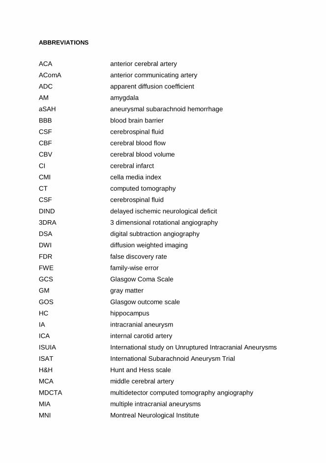
ABBREVIATIONS
ACA anterior cerebral artery
AComA anterior communicating artery
ADC apparent diffusion coefficient
AM amygdala
aSAH aneurysmal subarachnoid hemorrhage
BBB blood brain barrier
CSF cerebrospinal fluid
CBF cerebral blood flow
CBV cerebral blood volume
CI cerebral infarct
CMI cella media index
CT computed tomography
CSF cerebrospinal fluid
DIND delayed ischemic neurological deficit
3DRA 3 dimensional rotational angiography
DSA digital subtraction angiography
DWI diffusion weighted imaging
FDR false discovery rate
FWE familywise error
GCS Glasgow Coma Scale
GM gray matter
GOS Glasgow outcome scale
HC hippocampus
IA intracranial aneurysm
ICA internal carotid artery
ISUIA International study on Unruptured Intracranial Aneurysms
ISAT International Subarachnoid Aneurysm Trial
H&H Hunt and Hess scale
MCA middle cerebral artery
MDCTA multidetector computed tomography angiography
MIA multiple intracranial aneurysms
MNI Montreal Neurological Institute
MPR multiplanar reformat
MPRAGE magnetization prepared rapid acquisition gradient echo
MRA magnetic resonance angiography
MRI magnetic resonance imaging
MTT mean transit time
PCA posterior cerebral artery
PComA posterior communicating artery
PCT perfusion computed tomography
PD proton density
QOL quality of life
ROI region of interest
SAH subarachnoid hemorrhage
SD standard deviation
SI signal intensity
SPET single photon emission computed tomography
SPM statistical parametrical mapping
TCD transcranial Doppler
VBA vertebrobasilar artery
VBM voxelbased morphometry
WM white matter
WMH white matter hyperintensity
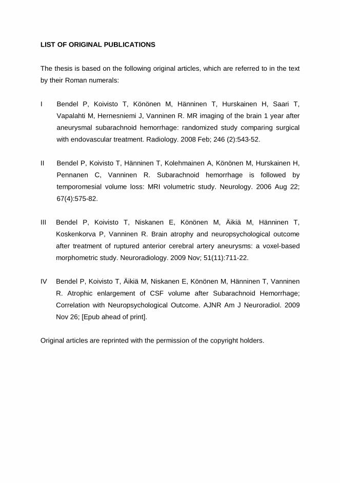
LIST OF ORIGINAL PUBLICATIONS
The thesis is based on the following original articles, which are referred to in the text
by their Roman numerals:
I Bendel P, Koivisto T, Könönen M, Hänninen T, Hurskainen H, Saari T,
Vapalahti M, Hernesniemi J, Vanninen R. MR imaging of the brain 1 year after
aneurysmal subarachnoid hemorrhage: randomized study comparing surgical
with endovascular treatment. Radiology. 2008 Feb; 246 (2):54352.
II Bendel P, Koivisto T, Hänninen T, Kolehmainen A, Könönen M, Hurskainen H,
Pennanen C, Vanninen R. Subarachnoid hemorrhage is followed by
temporomesial volume loss: MRI volumetric study. Neurology. 2006 Aug 22;
67(4):57582.
III Bendel P, Koivisto T, Niskanen E, Könönen M, Äikiä M, Hänninen T,
Koskenkorva P, Vanninen R. Brain atrophy and neuropsychological outcome
after treatment of ruptured anterior cerebral artery aneurysms: a voxelbased
morphometric study. Neuroradiology. 2009 Nov; 51(11):71122.
IV Bendel P, Koivisto T, Äikiä M, Niskanen E, Könönen M, Hänninen T, Vanninen
R. Atrophic enlargement of CSF volume after Subarachnoid Hemorrhage;
Correlation with Neuropsychological Outcome. AJNR Am J Neuroradiol. 2009
Nov 26; [Epub ahead of print].
Original articles are reprinted with the permission of the copyright holders.

CONTENTS
1. INTRODUCTION 17
2. REVIEW OF THE LITERATURE 18
2.1. Subarachnoid hemorrhage (SAH) 18
2.1.1. Incidence, etiology and risk factors 18
2.1.2. Cerebral aneurysms 18
2.1.3. Nonaneurysmal subarachnoid hemorrhage 19
2.2. Vascular anatomy of the brain, circle of Willis and typical aneurysms 20
2.2.1. Components of the circle of Willis 20
2.2.2. Typical sites for cerebral saccular aneurysms and aneurysm
detection 21
2.2.3. Cortical branches of the supratentorial cerebral arteries and their
vascular territories 23
2.2.4. Perforating branches of the circle of Willis and middle cerebral
artery 24
2.2.5. Circulation of the brainstem and cerebellum 24
2.3. Treatment 24
2.3.1. Natural history of ruptured cerebral aneurysms 24
2.3.2. Clinical assessment of an aSAH patient 25
2.3.3. Surgical treatment of ruptured aneurysms 25
2.3.4. Endovascular treatment of ruptured aneurysms 26
2.3.5. Combination treatment of ruptured aneurysms 27
2.3.6. Treatment for associating nonruptured aneurysms 27
2.3.7. Consequences and complications of aSAH (Acute impact to
brain, hematomas, vasospasm, microcirculatory dysfunction,
delayed ischemic neurological deficit and hydrocephalus) 28
2.4. Diagnostic Imaging in acute SAH 31
2.4.1. Computed tomography (CT) and CT angiography (CTA) 31
2.4.2. Digital subtraction angiography (DSA) 32
2.4.3. MRI and MR angiography (MRA) 33
2.5. Imaging in subacute aSAH 34
2.5.1. CT and MR perfusion imaging 35
2.5.2. Diffusion weighted imaging (DWI) 35
2.5.3. Positron emission tomography (PET), single photon emission
computed tomography (SPECT), XenonCT and Transcranial
Doppler (TCD) 36
2.6. Late Imaging after aSAH 37
2.6.1. Computed tomography, CT angiography 37
2.6.2. Digital subtraction angiography 37
2.6.3. Magnetic resonance imaging and MRA 38
2.7. Lesions detected on late MR Imaging after aSAH 41
2.7.1. Ischemic lesions in the vascular territory of the ruptured
aneurysm 42
2.7.2. Ischemic lesions in the vascular territory other than the
ruptured aneurysm 42
2.7.3. Focal laminar cortical infarcts following aSAH 43
2.7.4. Lesions related to hematoma and superficial siderosis 43
2.7.5. Retraction injury due to surgical manipulation 43
2.7.6. Ventricular dilation, permanent shunts 44
2.7.7. Presence of lacunar infarctions and leukoaraiosis 44
2.8. Volumetric Magnetic Resonance Imaging 45
2.8.1. Manual volumetry 45
2.8.2. Voxelbased morphometry 46
2.9. Methods for outcome assessment 48
2.9.1. Clinical outcome scales and prognostic factors 48
2.9.2. Neuropsychological outcome 48
2.9.3. Quality of life 49
3. AIMS OF THE STUDY 50
4. PATIENTS AND METHODS
4.1. Study design and study inclusion criteria and patients 51
4.2. Fisher and Hunt and Hess Grades and clinical vasospasm 52
4.3. Diagnostic angiography and embolization procedure 52
4.4. Surgical treatment 53
4.5. Patient care after treatment 54
4.6. Followup protocol: MR Imaging 54
4.6.1. Conventional MRI protocol 54
4.6.2. MRI Analysis 54
4.6.3. Volumetric MRI 56
4.6.4. Semiautomatic volumetry (Study II) 56
4.6.5. Voxelbased morphometry (Studies IIIIV) 58
4.7. Followup protocol: Clinical outcome and neuropsychological evaluation 59
4.8. Study populations 61
4.8.1. Original study population and MRI study population (Study I) 61
4.8.2. Combination treatment of the ruptured aneurysm and
treatment of additional associating nonruptured aneurysms 63
4.8.3. Volumetric study populations (Studies II, III and IV) 63
4.8.4. Neuropsychological study population (Studies IIV) 65
4.8.5. Control individuals (Studies II, III and IV) 65
4.9. Statistics 65
5. RESULTS 67
5.1. Comparability of study groups 67
5.2. Brain parenchymal lesions detected on late MR imaging and
comparison of surgical vs. endovascular treatment modalities (Study I) 68
5.2.1. Ischemic lesions in the parental artery territory 69
5.2.2. Ischemic lesions in other vascular territories 70
5.2.3. Residual signs of hematoma and superficial siderosis 70
5.2.4. Retraction injury due to surgical manipulation and
instrumentation 70
5.2.5. Lesions caused by shunt device 71
5.2.6. Previous infarctions, brain atrophy, lacunar infarctions and
leukoaraiosis 71
5.2.7. Demonstrative images of the typical findings detected on MRI
one year after aSAH 71
5.3. Temporomesial structures: Hippocampus and Amygdala (Study II) 73
5.3.1. Temporomesial volumes; aSAH patients vs. control individuals 73
5.3.2. Temporomesial volumes in endovascular and surgical
subgroups vs. controls 74
5.3.3. Temporomesial volumes and the side of the treatment 74
5.4. VBM analysis of patients after treatment of ruptured anterior cerebral
artery aneurysm (Study III) 74
5.4.1. Clinical outcome and conventional MRI findings 74
5.4.2. VBM results: All ACA patients vs. controls 75
5.4.3. VBM results: Surgical ACA patients vs. controls 79
5.4.4. VBM results: Endovascular ACA patients vs. controls 79
5.4.5. VBM results: Endovascular ACA patients vs. surgical ACA
patients 81
5.5. Ventriculomegaly and enlargement of CSF volumes after aSAH
(Study IV) 81
5.5.1. Patients and controls, preoperative hydrocephalus and
permanent shuntdevice 81
5.5.2. Degree of ventriculomegaly and enlarged CSFspaces 81
5.5.3. Associations between ventricular and sulcal enlargement
and clinical and other radiological features 83
5.6. Correlations of MRIdetectable findings and neuropsychological
outcome (Studies IIV) 84
5.6.1. Neuropsychological analysis of the MRI study population
(Study I) 84
5.6.2. Neuropsychological results and temporomesial
volume correlation (Study II) 86
5.6.3. Neuropsychological test results in volumetric ACA population
and their correlation to GM/ICV and CSF/ICV ratios (Study III) 86
5.6.4. Clinical outcome, neuropsychological results and CSF volume
correlation (Study IV) 87
5.7. The effect of age and MRI and neuropsychological outcome
(Studies I, II and IV) 89
5.7.1. Age and MRI and neuropsychological deficits 89
5.7.2. Age and temporomesial structures 89
5.7.3. Age and ventricular and sulcal CSF spaces 90
6. DISCUSSION 91
6.1. Oneyear MRI Outcome after aSAH: Surgical versus endovascular
treatment (Study I) 91
6.2. Temporomesial volume loss after aSAH (Study II) 93
6.3. Brain atrophy after ruptured ACA aneurysm and treatment (Study III) 95
6.4. Atrophic enlargement of CSF volumes after aSAH (Study IV) 97
6.5. General considerations 99
7. CONCLUSIONS 100
8. REFERENCES 101
APPENDIX: ORIGINAL PUBLICATIONS IIV 125
17
1. INTRODUCTION
Aneurysmal subarachnoidal hemorrhage (aSAH) is a devastating disease with an
overall casefatality of 40 50% (van Gijn et al. 2001). The incidence of aSAH is
increasing concistently with age (de Rooij et al. 2007, Fogelholm 1981). However,
the median age of death caused by aSAH is significantly lower being 59 years,
compared to 73 years for intracerebral hemorrhage and 81 years for ischemic stroke.
It has been estimated that aSAH covers over 25% of all strokerelated years of
potential life lost before of age 65 (Johnston et al. 1998). Moreover, most of the
surviving patients are left with neurological deficits and neuropsychological and
cognitive impairments (Molyneux et al. 2002, Molyneux et al. 2005, Vapalahti et al.
1984, Vilkki et al. 1989). The factors that influence longterm cognitive outcome after
aSAH are clinically important as such factors may be potentially modifiable. This
study was carried out to describe in detail the magnetic resonance imaging (MRI)
outcome one year after aSAH in patients who were randomly assigned to surgical
clipping versus endovascular coil treatment of the ruptured aneurysm. The aim was
to discover, if MRI could provide valuable information on patients' recovery potential
together with detailed neurological and neuropsychological examination after aSAH.
18
2. REVIEW OF THE LITERATURE
2.1. Subarachnoid hemorrhage (SAH)
2.1.1. Incidence, etiology and risk factors
Aneurysmal subarachnoid hemorrhage is a serious condition: global mortality
ranges from 32 to 67% (Hop et al. 1997). Twenty to 30% of the survivors are left with
disabling sequelae. Half of the patients who were previously employed cannot return
to their previous work after aSAH (Buchanan et al. 2000). Another study has showed
that 60% of aSAH victims referred to neurosurgical unit can be saved and can
recover to normal (Saveland et al. 1992). A recent review including 51 studies (33
new) reported an overall incidence of aSAH being approximately 9 per 100 000 (de
Rooij et al. 2007). However, aSAH is significantly more common disease in Finland
and Japan compared to other countries (de Rooij et al. 2007, Fogelholm 1981,
Inagawa et al. 1995, Sarti et al. 1991).
The cause for nontraumatic SAH is rupture of an intracranial aneurysm (IA) in
more than 80% of cases (van Gijn et al. 2001). Risk factors for aSAH are advanced
age (Rinkel 2008, Rinkel et al. 1998 ), female gender (Linn et al. 1996, Morita et al.
2005, Rinkel 2008, Rinkel et al. 1998), smoking (Feigin et al. 2005, Isaksen et al.
2002, Juvela et al. 1993) , hypertension (Feigin et al. 2005, Feigin et al. 2005,
Sandvei et al. 2009), excessive use of alcohol (Feigin et al. 2005), having one or
more affected close relatives with aSAH (Rinkel et al. 1998, Ronkainen et al. 1997)
and autosomal dominant polycystic kidney disease (Gieteling et al. 2003, Rinkel et al.
1998, Ronkainen et al. 1997). Furthermore, the larger size and irregular shape and
certain locations (vertebrobasilar, posterior and anterior communicating arteries) of
the aneurysm have been listed to increase the risk of rupture in asymptomatic
patients with incidental cerebral aneurysm (Wiebers et al. 2003).
2.1.2. Cerebral aneurysms
Intracranial arterial aneurysms (IA) are not congenital, but develop in the course
of life. An estimated frequency of cerebral aneurysm for an average adult without
specific risk factors is 23% (Rinkel et al. 1998) and the percentage increases with
19
age. Most cerebral aneurysm are saccular and arise at sites of arterial branchings,
usually at the base of the brain, either on the circle of Willis itself or at nearby
branching points. The risk of rupture increases along the size of an aneurysm, but
most ruptured aneurysms are small, less than 10 mm (Rinkel et al. 1998). Reports of
IA in some patients with rare mendelian disorders such as autosomal dominant
polycystic kidney disease, EhlersDanlos syndrome type IV, Marfan syndrome,
neurofibromatosis type 1 and fibromuscular dysplasia suggest that IAs are more
common in subjects with these diseases than in the general population (Schievink
1997, Schievink et al. 2005, Wills et al. 2003). There are different basic types of
intracranial aneurysms: saccular or “berry” aneurysms, fusiform aneurysms, blood
blisterlike aneurysms and dissecting aneurysms. Saccular aneurysms are the most
common type and they are true aneurysms, i.e., they are dilations of vascular lumen
due to weakness of all vessel wall layers. The aneurysmal sac itself is usually
composed only of intima and adventitia. The adventitia may be infiltrated by
lymphocytes and phagocytes. Trombotic debris is often present in the lumen of
aneurysmal sac (Okazaki 1989). The occurrence, growth, thrombosis, and even
rupture of intracranial saccular aneurysms can be explained by abnormal
hemodynamic shear stresses on the walls of large cerebral arteries, particularly at
the branching points of the arteries (Strother et al. 1992). It has been recently
suggested that before rupture, the wall of saccular cerebral artery aneurysm
undergoes morphological changes associated with remodeling of the aneurysm wall.
Some of these changes, e.g. smooth muscle cell proliferation and macrophage
infiltration, may reflect ongoing repair attempts that thus could be enhanced with
pharmacological therapy (Frosen et al. 2004, Frosen et al. 2006).
2.1.3. Nonaneurysmal subarachnoid hemorrhage
SAH of unknown origin, usually a perimesencephalic type of SAH (pmSAH),
represents 9% to 15% of cases of SAH patients (Kassell et al. 1990). In pmSAH, the
origin of the bleeding remains unclear and various pathogenetic mechanisms, such
as small cerebellar or pontine venous angiomas, capillary telangiectasias, intramural
hematomas of the basilar artery or specific anatomic variations of the
perimesencephalic venous drainage system have been discussed as possible
explanations for nonaneurysmal pmSAH (Ikeda et al. 1998). Patients with pmSAH
20
have been shown to regain independence for activities of daily life and have a normal
life expectancy, and they are not at risk for rebleeding. However, up to 25% of these
patients are left with symptoms including headache or dizziness, fatigue,
forgetfulness and irritability (Greebe et al. 2007).
2.2. Vascular anatomy of the brain, circle of Willis and typical aneurysms
2.2.1. Components of circle of Willis
The knowledge of normal vascular anatomy of the brain and angiographic
findings of each individual patient are important in analyzing MR images after aSAH.
With the familiarity of these factors, it is possible to determine the most probable
etiology of the lesions detected on late CT or MR imaging after aSAH (e.g. ischemic
lesions in the vascular territory of the ruptured aneurysm and ischemic lesions in the
other, remote vascular territories and to differentiate the ischemic lesions from the
lesions induced by surgical manipulation) (Hadjivassiliou et al. 2001, Kivisaari et al.
2001).
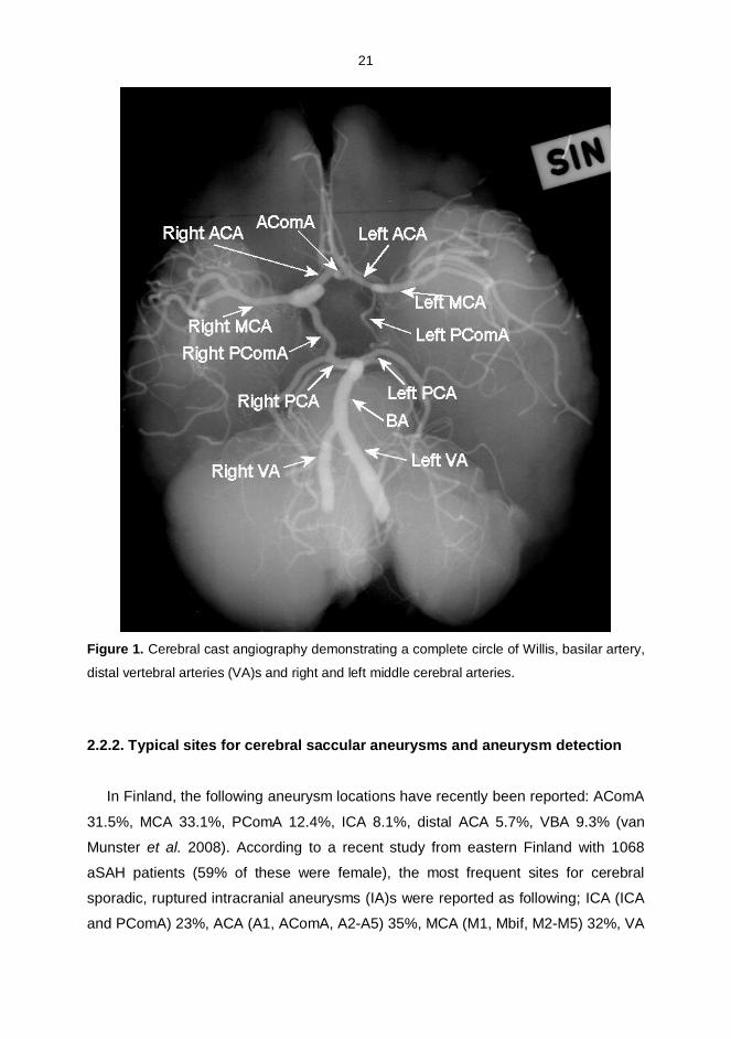
The anastomotic ring that connects both halves of the “anterior” circulation with
each other and with the vertebrobasilar system is called the circle of Willis. The circle
of Willis lies above the sella turcica within the interpeduncular and suprasellar
cisterns. A complete circle of Willis is an arterial polygon; a nonagon or a ninesided
structure. The complete circle of Willis consists of two internal carotid arteries (ICAs),
two anterior cerebral arteries (ACAs), the anterior communicating artery (AComA),
two posterior communicating arteries (PComAs), the basilar artery (BA) and two
posterior cerebral arteries (PCAs). Moreover, the middle cerebral arteries (MCA) are
a continuum of the ICA distal to branching point of the ACAs. The different variations
and incompleteness of circle of Willis are common. Most of the “berry” or saccular
aneurysms arise from the branching points of these vessels (Osborn 1999). Figure 1shows the complete circle of Willis.
21
Figure 1. Cerebral cast angiography demonstrating a complete circle of Willis, basilar artery,
distal vertebral arteries (VA)s and right and left middle cerebral arteries.
2.2.2. Typical sites for cerebral saccular aneurysms and aneurysm detection
In Finland, the following aneurysm locations have recently been reported: AComA
31.5%, MCA 33.1%, PComA 12.4%, ICA 8.1%, distal ACA 5.7%, VBA 9.3% (van
Munster et al. 2008). According to a recent study from eastern Finland with 1068
aSAH patients (59% of these were female), the most frequent sites for cerebral
sporadic, ruptured intracranial aneurysms (IA)s were reported as following; ICA (ICA
and PComA) 23%, ACA (A1, AComA, A2A5) 35%, MCA (M1, Mbif, M2M5) 32%, VA
22
(VA and posterior inferior cerebellar artery (PICA)) 3%, BA (BA bifurcation) 6% and
PCA 1% (Huttunen et al. 2009). However, the most frequent site for unruptures sIAs
was the MCA bifurcation (3944% of the unruptured aneurysms). In Finland, the
predominance of MCA aneurysms has also been reported in other series (Fogelholm
1981, Pakarinen 1967), while forensic reviews report slightly different locations for
ruptured cerebral aneurysms; 2027% ACA, 2634% MCA, 3746 % ICA and 813%
VBA (Rinkel et al. 1998).
Noninvasive imaging studies such as spiral computed tomographic angiography
(CTA) or highresolution magnetic resonance angiography (MRA) imaging can
provide an overview of the carotid siphon and major arteries at the base of the brain.
Detailed examination of the circle Willis, especially visualizing its' small but important
perforating branches still relies on the high resolution conventional digital subtraction
angiography (DSA). Because the circle of Willis constitutes the best potential source
of collateral blood in occlusive vascular disorders, awareness of its normal anatomy
is essential, even though the vascular supply can widely vary in an individual patient.
Accurate angiographic evaluation is also an integral part of preoperative planning
before surgery or endovascular treatment of the ruptured aneurysm.
2.2.3. Cortical branches of the supratentorial cerebral arteries and theirvascular territories
The distal segment of the ACA, the pericallosal arteries, gives rise to the cortical
and callosal branches. The callosal branches supply the rostrum, genu and the body
of the corpus callosum. These branches are joined posteriorly by the splenial
branches of the PCA. In the most frequent disposition, the cortical area of supply of
the ACA is the medial surface of the hemisphere extending to the superior frontal
sulcus and the parietooccipital sulcus. On the orbitofrontal surface, the arterial
territory includes the medial orbital gyri. At the most, the cortical ACA territory
reaches the inferior frontal sulcus, and at the least, it includes only the anterior part of
the frontal lobe (Osborn 1999, Tatu et al. 1998).
The MCA is divided anatomically into four segments: horizontal segments (M1),
insular segments (M2), opercular segments (M3; the first ascending branch is
sometimes historically called the “candelabra” because the angiographic appearance
resembles a branched candlestick) and cortical branches (M4).The MCA begins its
23
division into cortical branches at the base of the Sylvian fissure, extends over the
surface of the hemisphere and forms M4 segments. The most frequent area of
supply of the MCA extends from the lateral surface of the hemisphere to the superior
frontal sulcus, the intraparietal sulcus, and the inferior temporal gyrus. On the
orbitofrontal surface, the arterial territory includes the lateral orbital gyri. The
maximum area covers the whole lateral surface of the hemisphere, reaching the
interhemispheric fissure. The minimum territory is confined between the inferior
frontal and the superior temporal sulci (Osborn 1999, Tatu et al. 1998).
As the PCA approaches the dorsal surface of the midbrain, it gives rise to cortical
branches. The branches include the hippocampal arteries and the splenial artery that
anastomose with the distal part of the pericallosal artery to supply the splenium of the
corpus callosum. The most frequent cortical distribution of the PCA includes the
inferomedial surfaces of the temporal and occipital lobes extended to the parieto
occipital fissure. In general, the parietooccipital and calcarine arteries supply the
posterior one third of the brain, along with the interhemispheric fissure including the
primary visual cortex. At the most, the cortical supply can extend as far as the
superior temporal sulcus and the upper part of the precentral sulcus, and at the least,
the supply can extend only as far as the medial face of the occipital lobe, limited by
the parietooccipital fissure (Osborn 1994, Osborn 1999, Tatu et al. 1998).
2.2.4. Perforating branches of the circle of Willis and Middle Cerebral artery
Important perforating branches arise from every part of the circle of Willis and
middle cerebral artery: 1. From anterior cerebral arteries arise the medial
lenticostriatae arteries and quite often the recurrent artery of Heubner, which can
supply the caudate nucleus head, anterior limb of the internal capsule and part of the
basal ganglia. 2. From anterior communicating artery arise perforating brances to
supply the superior surface of the optic chiasm and anterior hypothalamus. These
branches may have a significant vascular territory that includes part of the corpus
callosum, columns of the fornix, parolfactory areas, lamina terminalis and
hypothalamus. 3. From posterior communicating arteries arise number of small, but
important perforating branches, the anterior thalamoperforating arteries to supply part
of the thalamus, the infralenticular limb of the internal capsule and optic tracts. 4.
From distal basilar artery and proximal posterior cerebral arteries arise numerous
24
small perforating arteries. These branches, the posterior thalamoperforating arteries
and the thalamogeniculate arteries supply the midbrain and thalamus. 5. From the
middle cerebral artery arise the lateral lenticulostriatal arteries, which supply the
substantia innominata, lateral aspect of the anterior commissure, most of the
putamen and lateral globus pallidus, the superior half of the internal capsule and
adjacent corona radiate, and the body and the head (except the anterior inferior
portion) of the caudate nucleus. Portions of the optic radiations and arcuate
fasciculus are also supplied by these branches (Osborn 1999).
2.2.5. Circulation of the brainstem and cerebellum
The cerebellar arterial supply depends on three long arteries. The posterior
inferior cerebellar artery (PICA) gives rise to two branches and vascularises the
inferior vermis and the inferior and posterior surfaces of the cerebellar hemispheres.
The anterior inferior cerebellar artery (AICA) supplies the anterior surface of the
simple, superior, and inferior semilunar lobules as well as the flocculus and the
middle cerebellar peduncle. The superior cerebellar artery (SCA) divides into medial
and lateral branches and vascularises the superior half of the cerebellar
hemispheres, vermis and the dentate nucleus. These three cerebellar arteries also
take part in the vascularisation of the brainstem. The territory of the SCA often
includes the upper part of the pontine tegmentum. The PICA takes part in the lateral
and posterior arterial groups of the medulla. The AICA supplies the middle cerebellar
peduncle and often the lower part of the pontine tegmentum (Duvernoy 1999, Osborn
1999, Tatu et al. 1996). However, the vascular anatomy of the posterior fossa can be
extremely variable in different individuals.
2.3. Treatment
2.3.1. Natural history of ruptured cerebral aneurysms
Once an aneurysm has ruptured, rebleeding is the most feared complication.
Rebleeding peaks in the first few days after the initial bleeding (Kassell et al. 1983,
Pakarinen 1967). Rebleeding is more frequent in patients with poor clinical condition
and those with large aneurysms. If an aneurysm is not treated, the risk of rebleeding
25
within 4 weeks is estimated to be 3540% (Hijdra et al. 1987). The outcome after
aSAH is very poor without treatment. In the historical Finnish series by Pakarinen,
the mortality at first recurrence was 64% and at second recurrence it had risen to
86% (Pakarinen 1967).
2.3.2. Clinical assessment of an aSAH patient
The clinical condition of a patient needs to be evaluated acutely after aSAH,
since severity of bleeding is the most important prognostic factor for clinical outcome
(Koivisto et al. 2000). The Hunt & Hess grading scale (Hunt et al. 1966) is the most
common system for grading the clinical condition of the patient and it is widely used
for aSAH patients as following: Grade 0= Asymptomatic, no bleeding, I=
Asymptomatic, or minimal headache and/or slight nuchal rigidity, II= Moderate to
severe headache, nuchal rigidity, no neurological deficit other than cranial nerve
palsy, III= Drowsiness, confusion, or mild local deficit, IV= Stupor, moderate to
severe hemiplegia, possibly early decerebrate rigidity, and vegetative disturbances,
V= Deep coma, moribund appearance. Also the Glasgow Coma Scale (GCS) is
commonly used in clinical practise, (Teasdale et al. 1976) and it has further been
modified in the World Federation of Neurosurgical Societies (WFNS) classification
(Report of Word Federation Of Neurological Surgeons Committee, 1988).
Since the natural history of the ruptured aneurysm is poor, an early treatment
should be performed (Fogelholm 1981). The treatment options for excluding a
ruptured aneurysm from the circulation are microneurosurgical clipping (Krayenbuhl
et al. 1970) and endovascular procedures, e.g. coiling (Molyneux et al. 2005).
2.3.3. Surgical Treatment of ruptured aneurysms
The goal of surgical treatment of IAs is to isolate the aneurysm from the
circulation, while preserving the normal blood flow through parent artery and branch
vessels. Surgical treatment is best performed in a microsurgical operation, where a
neurosurgeon places a clip across the aneurysm neck (Yasargil et al. 1975). This
was first done by W. Dandy in 1938 (Dandy 1938), but only in 1960s, after the
introduction of operation microscope and improved neurointensivecare, the results
of surgery for ruptured aneurysms reached an acceptable level of morbidity and
26
mortality in goodgrade patients (Dandy 1938, Kassell et al. 1982, Langmoen et al.
1999, Vapalahti et al. 1984). Most of the ruptured aneurysms are surgically
operable. However the location, size and the configuration of the ruptured aneurysm
may sometimes complicate the operation (Hernesniemi et al. 1993). Posterior
circulation aneurysms can be difficult lesions to be treated by surgery, and they have
potential for high morbidity and mortality (Rosengart et al. 2007), particularly in
elderly patients or patients in poor neurological condition (Schievink et al. 1995).
Anecdotal clinical series have reported other surgical techniques, such as external
wrapping, coating of IAs and the use of the excimer laserassisted nonocclusive
anastomosis technique (ELANA) for surgical treatment of ruptured aneurysms not
suitable for conventional surgery (Bederson et al. 2009, Muench et al. 2007). Modern
surgical clips are nonferromagnetic and safe for MR imaging (Kanal et al. 1999).
2.3.4. Endovascular treatment of ruptured aneurysms
Endovascular techniques were first used for aneurysms that were considered
inoperable or in patients whose previous surgical treatment had failed (Guglielmi et
al. 1992, Malisch et al. 1997). In 1991 Guglielmi and his coworkers introduced an
electrically detachable coil system (GDC) which started the endovascular treatment
era of ruptured IAs (Guglielmi et al. 1991, Guglielmi et al. 1991). GDCs are pushed
into the aneurysm sac through a microcatheter, and they can be repositioned,
retrieved, or replaced by a coil of different size until the situation is considered
satisfactory. GDCs are detached from the deploying wire by electrolysis. The
standard coil embolization techniques have developed further since GDCs and other
detachable platinium coils. For example soft coils, 2D and 3D shaped coils (Lubicz et
al. 2005, Slob et al. 2005), liquid embolic materials (Molyneux et al. 2004),
remodelling techniques (Moret et al. 1997) and endovascular stents have been
introduced (Lylyk et al. 2005, Tahtinen et al. 2009). Embolization with coils has
recently been used increasingly for treatment of IAs (Brilstra et al. 1999, Molyneux et
al. 2002, Molyneux et al. 2005, Renowden et al. 2009, Vinuela et al. 1997). A
majority of small and moderate sized aneurysms with reasonable neckaspect ratio
are treatable by endovascular means (Vinuela et al. 1997). Common reasons for
relative unsuitability for endovascular treatment are: aneurysmal size too small/too
large, unsuitable neckaspect ratio, aberrations in intracranial vasculature,
27
incorporation of distal branches in the aneurysmal neck, aneurysm symptomatic due
to mass effect, acute recurrence/rebleed that follows initial endovascular treatment,
and attempted, but unsuccessful prior endovascular treatment. Ruptured cerebral
aneurysms deemed unsuitable for endovascular intervention are often difficult to treat
surgically (Choudhari et al. 2007). Longterm studies evaluating the experience with
aneurysm coil embolization during the past decade indicate that embolization of the
ruptured aneurysm is a safe and durable treatment method (Koebbe et al. 2006, van
der Schaaf et al. 2005). However, as a result of the lower rates of complete occlusion
of the aneurysm after endovascular than surgical treatment (Gerlach et al. 2007,
Murphy et al. 2005, Ogilvy et al. 2002), the questions regarding the longterm efficacy
of coiling and the possibility of higher rates of rebleedings and recurrence remain
(Molyneux et al. 2009, Raftopoulos 2005). A recent study shows that virtually all
aneurysm reopenings develop within the first 6 months after coiling. Thus no
prolonged imaging followup is routinely suggested for those aneurysms that are
adequately occluded during this time (Rooij et al. 2009). Endovascular coils are
nonferromagnetic and thus safe for MR imaging (Shellock et al. 1997).
2.3.5. Combination treatment of ruptured aneurysms
Although most aneurysms can be clipped microsurgically or coiled endovascularly,
a subset of patients may require a combined (crossover) approach. Multimodality
approaches are best used with complex aneurysms in which conventional therapy
with a single modality has failed (Lawton et al. 2008, Thielen et al. 1997).
2.3.6. Treatment for associating nonruptured aneurysms
Unruptured, associated aneurysms are found in up to 30% of patients with aSAH
(Huttunen et al. 2009, Investigators 1998, Juvela et al. 2000, Rinne et al. 1994).
Different national cohorts of aSAH patients differ with respect of age of the patient
and the number and sites of associated aneurysms. When Dutch and Finnish
population were compared, multiple aneurysms were more frequent in Kuopio
(27.8%) than in Utrecht (14.8%) (van Munster et al. 2008). According to local study,
multiple MCAAs, found in 20% of the patients with aneurysms, were common in this
Finnish population (Rinne et al. 1996). A large multicenter study, ISUIA Unruptured
28
Intracranial Aneurysm: Risk of Rupture and Risks of Surgical Intervention Study,
showed that the likelihood of rupture of (unruptured) IAs that were less than 7 mm in
diameter was exceedingly low in group of patients who had no history of aSAH from
a different aneurysm and was substantially higher among those patients with a
history of aSAH from a different aneurysm (Wiebers et al. 2003). A coexisting or
associated nonruptured aneurysm of all sizes in patients with aSAH due to another
treated aneurysm carry a higher risk for future hemorrhage than similar sized
aneurysms without a prior SAH history. Other factors that favour treatment for
unruptured aneurysms include several factors: a young patient with a long life
expectancy, a family history of aneurysm rupture, large aneurysms, symptomatic
aneurysms, observed aneurysm growth, and established low treatment risks
(Bederson et al. 2000).
2.3.7. Consequences and complications of aSAH (Acute impact to brain,hematomas, vasospasm, microcirculatory dysfunction, delayed ischemicneurological deficit and hydrocephalus)
19 22% of patients with aSAH presents with an intracerebral hematoma (ICH)
(Kivisaari et al. 2001, Tokuda et al. 1995). The clinical outcome of aSAH patients with
ICH is worse than that of aSAH patients without ICH (Bailes et al. 1990, Hauerberg et
al. 1994).
Vasospasm following cerebral aneurysm rupture is one of the most devastating
sequelae and is traditionally thought to present the most common cause of delayed
ischemic neurological deficits (DIND) (Fisher et al. 1977) and a common cause of
morbidity and mortality in the patients who survive the initial bleeding. Vasospasm is
a frequent complication in the early clinical course after aSAH, occurring in 40 to 70%
of patients (Schuknecht et al. 1999).
The peak frequency of cerebral ischemia is from 5 to 14 days after aSAH.
Although DIND has traditionally been interpreted to be caused by vasospasm
(Fergusen et al. 2007, Fisher et al. 1980) and arterial narrowing, recent studies show
that arterial narrowing is neither a necessary nor a sufficient condition (Rabinstein et
al. 2004, Rabinstein et al. 2005, Weidauer et al. 2007). Not all patients with DIND
have macrovascular vasospasm and there are other mechanisms, such as
hypovolemia, hypotension (Chang et al. 2000, Wijdicks et al. 1985), impaired
29
fibrinolytic activity, inflammatory and endotheliumrelated processes leading to the
activation mechanisms of the coagulation cascades and formation of microtrombosis
and thus leading to development of DIND (Vergouwen et al. 2008). Predictors for
ischemic lesions in the vascular territory other than the ruptured aneurysm are the
total amount of subarachnoid blood (Brouwers et al. 1992, Rabinstein et al. 2004)
and loss of consciousness at the time of hemorrhage (Brouwers et al. 1992, Hop et
al. 1999), which suggests that the global ischemia during the initial event could well
be the key factor (van der Schaaf et al. 2006). The triple Htreatment including
induced hypertension, hypervolemia and hemodilution, is usually the intervention
strategy, although controlled trials are missing (Bederson et al. 2009). Oral
nimodipine have been indicated to reduce poor outcome related to aSAH (Allen et al.
1983). The value of other calcium antagonists, whether administrated orally or
intravenously, remains still uncertain (Bederson et al. 2009). Neurointerventional
treatments such as transluminar balloon angioplasties and infusions of intraarterial
vasodilating agents such as nicardipine or nimodipine are also widely used in many
units treating patients after aSAH (Hoh et al. 2005, Rabinstein et al. 2004, Tejada et
al. 2007), but the effectiveness of these therapies is not well established (Bederson
et al. 2009). Treatment of aSAH patients with several pharmacological agents such
as aspirin, enoxaparin, tirilizad, magnesium sulphate, ebselen, endothelin1a
antagonists and the role of statins have been recently published, but the benefits of
these therapies still remain unclear and further studies are needed (Bederson et al.
2009). Systemic hypothermia has been used in several clinical settings to protect the
brain against ischemic injuries, but its role in treating aSAH patients has not been
established (Bederson et al. 2009, Todd et al. 2005).
The concept of early brain injury (EBI) has been introduced only recently and
refers to the immediate injury to the brain as a whole, occurring within the first 72
hours of the ictus, secondary to aSAH. Therefore EBI refers to the events that occur
in the brain before the development of vasospasm, including elevation of intracranial
pressure, reduction of cerebral blood flow, suppression of cerebral perfusion
pressure, fall in brain oxygenation and neuronal cell death. It can be suggested that
the etiology of vasospasm may be linked to that of EBI, because they share many of
the same characteristics (Kusaka et al. 2004). Major early injuries after the initial
bleeding are breakdown of blood– brain barrier (BBB) and formation of brain edema
(Doczi 1985). There are currently a number of pathways that have been implicated to
30
EBI and breakdown of BBB including the apoptotic cell death occurring in neurons
and in cerebral endothelial cells, inflammatory and ischemic pathways (Cahill et al.
2006, Doczi 2001). MRI has been described as a powerful tool for the noninvasive
detection of EBI. In experimental models, the use of apparent diffusion gradients
demonstrates cellular swelling after a propagating wave of ischemia, which could be
seen spreading throughout the ipsilateral and contralateral hemispheres (Busch et al.
1998). Animal studies using SAH models have demonstrated profound hippocampal
neuronal loss (up to 30%) within relatively short period of time. It is believed that the
loss of hippocampal neurons occur secondary to the global ischemia, which occurs at
the time of SAH (Park et al. 2004). EBI has been speculated to present a
combination of physiological insults to the brain, resulting in global ischemia, BBB
breakdown, edema and cellular death signalling. These changes occur acutely and
chronically although after 72 hours vasospasm becomes the main protagonist. The
consequences of EBI can be seen in immediately and in the long term (Cahill et al.
2006, Cahill et al. 2009).
Chronic hydrocephalus is a wellknown complication following aSAH occurring in
1030% of patients (Dehdashti et al. 2004, Gruber et al. 1999, Jartti et al. 2008,
Tapaninaho et al. 1993). Well established risk factors for chronic hydrocephalus
requiring a shunt placement include: advanced age, Hunt and Hess Grades IVV,
Fisher Grades 34, intraventricular bleeding and acute hydrocepahalus on admission
(Dehdashti et al. 2004, Dorai et al. 2003, Tapaninaho et al. 1993). Additionally,
vertebrobasilar origin of the ruptured aneurysm, postoperative complications
(Tapaninaho et al. 1993) and clinical vasospasm and endovascular treatment (Dorai
et al. 2003) have also been reported as risk factors for shuntdependent
hydrocephalus. A recent study reported a relative risk reduction for hydrocephalus in
aSAH patients, in whom the fenestration of lamina terminalis had been performed in
the surgical operation (Komotar et al. 2009).
31
2. 4. Diagnostic Imaging in acute SAH
2.4.1. Computed tomography (CT) and CT angiography (CTA)
Computed tomography scanning is the first imaging procedure when SAH is
suspected. Blood detected in the basal cisterns on CT highly suggests a ruptured
aneurysm (van Gijn et al. 1980) in patients without trauma. CT scanning has a high
ability to detect the extravasated blood in the basal cisterns and the local maxima of
the blood may suggest the most probable location of the ruptured aneurysm (van der
Jagt et al. 1999). Timing of the CT scan after bleeding is associated with the
probability of recognizing a hemorrhage on CT scans, which is 85% after 5 days,
50% after 1 week, 30% after 2 weeks (mostly patients with hematomas), and almost
nil after 3 weeks (van Gijn et al. 1982). Fisher grading has been widely applied to
quantify the amount of blood in cisterns: Grade 1= no blood detected, Grade 2=
diffuse blood, vertical blood layers (interhemispheric, ambient, lateral sylvian
cisterns), <1mm thick, Grade 3= localized clot and/or vertical blood layers > 1mm
thick, Grade 4= intracerebral or intraventricular clot with diffuse or no subarachnoid
blood. Because Fisher developed the grading system only for his own research
purposes, the actual blood clot thicknesses referred in his scale are not directly
applicable in clinical use. Consequently, Grades 12 are usually referred as mild and
moderate bleeding and Grades 34 are referred as severe bleeding (Fisher et al.
1980, Friedman et al. 2002). The amount of extravasated blood on the initial CT scan
is related with subsequent vasospasm, delayed cerebral ischemia and clinical
outcome (Bell et al. 1980, Fisher et al. 1980, Kistler et al. 1983, Suzuki et al. 1980).
After SAH has been diagnosed, the multidetector computed tomography angiography
(MDCTA) is usually immediately performed to detect the ruptured aneurysm. Sixty
fourrow MDCTA can provide prompt and accurate diagnostic and anatomic
information in the setting of SAH with an excellent detection rate in acute ruptured
aneurysms (Kangasniemi et al. 2004, Nijjar et al. 2007, Pechlivanis et al. 2005).
Recently, subtraction 3D CTA has been compared favourably with DSA for detection
and characterization of IAs (Li et al. 2009, Sakamoto et al. 2006). When 64slice CTA
is used in the evaluation of aSAH, the information obtained is usually adequate to
determine treatment modality allocation in twothirds of the cases (Agid et al. 2006,
32
Miley et al. 2008 ) and the possibility of coil embolization can be reliably determined
with multidetector CTA (Papke et al. 2007, Westerlaan et al. 2007).
2.4.2. Digital subtraction angiography (DSA)
Conventional cerebral angiography is still the gold standard for aneurysm
detection (van Gijn et al. 2001). Cerebral DSA is relatively safe and the rate of
neurologic complications seems to have decreased in the modern era with smaller
angiographic catheters, new contrast materials and DSA allowing smaller volumes of
radiographic contrast per injection. The combination of DSA with 3D rotational
angiography (3DRA) is currently the most sensitive technique to detect untreated
aneurysms and should be performed in suspicious cases of SAH where the
aneurysm is not depicted by 64 MDCTA, because 64 MDCTA may occasionally miss
aneurysms less than 34 mm in size (McKinney et al. 2008). With intraarterial DSA,
the prevalence of permanent neurologic deficit has been reported to be 0.3% in 1992
(Waugh et al. 1992). Heiserman et al. (1994) prospectively evaluated one thousand
consecutive cerebral angiographic procedures performed using transfemoral
catheterization and filmscreen methods. The overall incidence of neurologic deficits
was 1.0 % and the incidence of persistent deficit was 0.5 % (Heiserman et al. 1994).
All complications occurred in patients presenting with a history of stroke, TIA or
carotid bruit, a patient population known to be at risk for atherosclerotic changes.
However, the risk of permanent neurological complication associated with cerebral
angiography in patients with SAH is significantly lower, 0.070.9% (Cloft et al. 1999,
Dion et al. 1987). In addition to neurological complications other angiographic
complications are possible following DSA including hematomas and
pseudoaneurysms of the puncture site, local or systemic infections, thrombosis of the
femoral artery, renal damage and adverse reactions with the use of contrast.
Angiographic studies serve not only to identify one or more aneurysms as
potential causes in a patient with aSAH, but also to study the anatomical
configuration of the aneurysm in relation to adjoining arteries, which allows optimum
selection of treatment (coiling or clipping) (van Gijn et al. 2007). The 3DRA
furthermore improves the detection of small aneurysms, especially locating on the
anterior communicating artery (van Rooij et al. 2008, van Rooij et al. 2008). The
3DRA with modern software helps to reconstruct the surface luminar anatomical
33
conditions of the aneurysm and it has been demonstrated to have a good correlation
to surgical anatomy (Tanoue et al. 2000).
2.4.3. MRI and MR angiography (MRA)
Because of the greater availability and feasibility of CT imaging in patients with
suspected subarachnoid hemorrhage, only a few studies of MRI in the acute phase
after aSAH have been reported. These suggest that during the first few hours and
days, MR with proton density and fluid attenuation recovery (FLAIR) images are as
sensitive as CT imaging (Fiebach et al. 2004). After the initial days, when
hyperdensity on CT scans decreases, MR is better for detecting blood, with FLAIR
and T2* and SWI images being most sensitive techniques (Mitchell et al. 2001,
Thomas et al. 2008, van Gijn et al. 2007). Although conventional angiography
remains the gold standard for aneurysm detection, sometimes noninvasive
techniques are used for detection of ruptured aneurysm. 3D timeof flight (TOF)
magnetic resonance angiography (MRA) does not require contrast material and it is
the most convenient diagnostic study not carrying essentially any risks. MRATOF
has been mainly used for aneurysmal screening of people at high risk of aneurysms
(Ronkainen et al. 1997, van Gijn et al. 2007), but the procedure is less feasible for
patients who are restless or need mechanical ventilation, and therefore less suitable
in acute subarachnoid hemorrhage. Moreover, MR angiography is rarely sufficient for
treatment planning. However, standard crosssectional MR is the best method to
demonstrate the presence of thrombus within the aneurysmal sac (Curnes et al.
1993). MRTOF angiography can detect IAs as small as 23 mm in diameter,
although the detectability is lower for small aneurysms (Okahara et al. 2002).
Compared to DSA, the sensitivity of 1.5T MRA in detection of IAs has been
demonstrated to be between 79% and 97% and the specificity of 91100% has been
reported (Okahara et al. 2002, White et al. 2001). Its sensitivity is equivalent to that of
CTA and is dependent of aneurysm size with one study suggesting a minimum size
of 5 mm for a clinically useful degree of sensitivity for both techniques (White et al.
2001). ThreeTesla (3T) MRA is becoming more widely available in clinical practise
and it has been shown to be superior to 1.5 T in depiction of IAs, especially small
aneurysms (Gibbs et al. 2004). For imaging IAs, 3.0T TOF MR angiography also
offers better image quality than 3.0T CE MR angiography using the ellipticalcentric
34
technique (Gibbs et al. 2005). It is essential to distinguish between intradural and
extradural location of the aneurysm, because extradural aneurysms that involve the
cavernous segment of the ICA have little or no risk for the hemorrhage. Therefore
there is no need to preventively treat for SAH and they are commonly just followed
(Kupersmith et al. 1992). However, the ICA is surrounded by bone, the double dural
ring (proximal and distal), the optic nerve, the third cranial nerve and the cavernous
sinus, which contribute to its complex anatomical structure. Contrastenhanced (CE)
MRA and multiplanar reformat (MPR) are very useful techniques for determining the
location of juxtadural ring aneurysms (Tsuboi et al. 2007) and CE 3D constructive
interference in steady state (CISS) MR imaging is useful for the differentiation
between paraclinoid and cavernous sinus aneurysms (Hirai et al. 2008).
2.5. Imaging in subacute aSAH
In the subacute stage (days 520 after aSAH), the diagnosis of delayed cerebral
ischemia most often requires repeat CT scans and laboratory tests to rule out
hydrocephalus and other systemic reasons (e.g. abnormal electrolytes, infection) as
a cause of the deterioratation of a aSAH patient. Various methods have been used to
measure cerebral blood flow and cerebral perfusion including CT, MRI, positron
emission tomography (PET), single photon emission computed tomography
(SPECT), xenon CT, and transcranial Doppler sonography (TCD).
Because of the reported morbidity associated with transporting critically ill
patients outside of the intensive care unit (ICU), the portable CT scanners for the ICU
have recently been introduced and found to be feasible and costeffective (Masaryk
et al. 2008). Radiographic assessment of vasospasm after aSAH by quantitative
techniques such as PET and xenon CT, offer tools to identify areas at increased risk
for infarction. MRI is more sensitive in detecting early changes in the brain, especially
with diffusion weighted imaging. Transcranial Doppler sonography, perfusionCT,
perfusionMRI and even DSA are used to detect early cerebral ischemia by
increased blood flow velocity, prolonged mean transit time (MTT) or arterial
narrowing (Bederson et al. 2009).
35
2.5.1. CT and MR perfusion imaging
Perfusion computed tomography (PCT) is a relatively new technique that allows
rapid qualitative and quantitative evaluation of cerebral perfusion by generating maps
of cerebral blood flow (CBF), cerebral blood volume (CBV) and mean transit time
(MTT). The technique is based on the central volume principle (CBF = CBV/ MTT)
and requires the use of commercially available software employing complex
deconvolution algorithms to produce the perfusion maps (Hoeffner et al. 2004).
MDCTA with PCT can assess the location and severity of cerebrovascular
vasospasm and its related perfusion abnormalities. It can identify severe vasospasm
with risk of delayed ischemia and can thus guide to the invasive treatment (Binaghi et
al. 2007, Sviri et al. 2006, Sviri et al. 2006, Yoon et al. 2006). Cerebral blood flow
alterations are common after aSAH, although it has also been reported that
decreased cerebral perfusion by itself may not be sufficient to cause DIND
(Dankbaar et al. 2009). A recent study suggested that there may be perfusion
abnormality also without macrovascular vasospasm in the watershed areas or in the
vicinity of sulcal cloth. Also cerebral hyperperfusion alterations have been described
following vasospasminduced infarctions (Aralasmak et al. 2009). Another recent
study, performed on stroke patients suggested, that increased contrast concentration
improves peak opacification of tissue, indicating that CTP evaluation is better
performed with the highest available concentration contrast agent (Silvennoinen et al.
2007).
Review of the literature yielded only a small number of perfusionweighted MRI
(PWMRI) studies in patients with aSAH analysing haemodynamic changes in regard
to the presence of vasospasm (Leclerc et al. 2002, Shimoda et al. 2001), but a recent
study shows that PWMRI might also reveal early impairment of cerebral
autoregulation in patients after aSAH by means of regional CBF (rCBF) and regional
CBV (rCBV) (Hattingen et al. 2008).
2.5.2. Diffusion weighted imaging (DWI)
Even in the first few minutes after acute arterial occlusion, DWI trace sequences
change into a hyperintense signal and there is a fall in the apparent diffusion
coefficient (ADC) values (Beauchamp et al. 1998). The main pathophysiological
36
explanation is the existence of cytotoxic edema due to proton pump failure. As a
result, water moves from the extracellular spaces into the cells. The resultant cellular
swelling leads to a drop in ADC. With a combination of PWI and DWI imaging it is
possible to detect areas with a perfusion–diffusion mismatch which represents areas
of misery perfusion where the tissue is ischaemic but not yet infarcted. This ‘‘tissue at
risk’’ concept derives from studies of ischaemic stroke, grossly identifies the
ischaemic penumbra (Wu et al. 2005). Quantitative measurements of ADC are more
sensitive than DWI for detection of even mild changes in water diffusivity. It has been
shown that patients with vasospasm, including the patients without symptoms,
presented abnormalities on DWI with a reduction of the ADC prevalently in the white
matter (CondetteAuliac et al. 2001). Being able to detect the early abnormalities on
DWI with parenchymal involvement in asymptomatic patients prior to symptomatic
vasospasm might help in preventing the DIND (CondetteAuliac et al. 2001). A recent
study also reported a finding of global vasogenic edema in subacute stage after
aSAH, with elevated ADC values in normal appearing T2weighted MR images (Liu
et al. 2007).
2.5.3. Positron emission tomography (PET), single photon emission computedtomography (SPECT), XenonCT and transcranial Doppler (TCD)
Positron emission tomography (PET) is the oldest standard for quantitative
evaluation of rCBF and cerebral autoregulation after aSAH (Kawai et al. 2008,
Powers et al. 1985). Naderi et al. demonstrated in 1994 that brain perfusion SPECT
is a nontraumatic, noninvasive, nonallergic, and inexpensive method for predicting
cerebral vasospasm (Naderi et al. 1994). XenonCT measures CBF directly and low
CBF values have been found to be associated with the development of ischemic
areas (Yonas et al. 1989). However, PET and SPECT require more time compared to
PCT and thus, they may be unsuitable for restless patients with unstable conditions
in clinical practise. Xenon CT (XeCT) is a practical technique which may be
performed bedside and may be used to assess cerebral blood flow response to a
changing variable e.g. vasoactive drug treatment (Carlson et al. 2009). Transcranial
Doppler ultrasonography (TCD) is based on the alterations of blood flow velocities
which correlate to arterial narrowing and thus, in theory, also to CBF and clinical
deterioration (Aaslid et al. 1984). TCD is an old, inexpensive, noninvasive, easily
37
repeatable bedside method used to assess indirectly the cerebral perfusion.
However, it is an operatordependent technique (Horn et al. 2001) and a recent study
concluded that its overall sensitivity for identifying patients at high risk for DIND is
limited (Carrera et al. 2009).
2.6. Late Imaging after aSAH
2.6.1. Computed tomography, CT angiography
CT scanning is widely available, easily repeatable, has short study time, and is
sensitive to detect ischemic lesions and hydrocepahalus. Moreover, CT is less
sensitive to motion artefacts compared to MRI due to considerably shorter scanning
time. CT imaging results correlate strongly with clinical outcome (Juvela et al. 2005,
Naidech et al. 2009, Vilkki et al. 2004) and most imaging studies after aSAH have
been thus far made by using CT. Clip and coil artefacts cause a problem in
determining the exact occlusion rates after both endovascular and surgical treatment
of aSAH, and there are only a few studies using postoperative CTA for controlling the
occlusion rate of the aneurysm. With CTA, the streak artefacts from the coils harass
inevitably the perianeurysmal area and aneurysmal occlusion rates cannot be
satisfactorily evaluated by CTA (Masaryk et al. 2000). However, recent studies
reporting the role of CTA for postoperative evaluation of aneurysmal occlusion rates
after surgical clipping suggest that CTA might be considered valuable in patients
treated with titanium clips (Chen et al. 2009, Uysal et al. 2009). However, for
evaluating the aneurysmal occlusion rates CTA is heavily debated and not commonly
used in clinical practise.
2.6.2. Digital subtraction angiography
Conventional digital subtraction angiography (DSA), especially with 3D rotational
angiography (van Rooij et al. 2008), remains as the gold standard for postoperative
imaging of IAs. A literature review from 19791999 with more than 1500 clipped
aneurysms reported a 5.2 % incomplete occlusion after surgical clipping (Thornton et
al. 2000). According to a recent study, postoperative DSA detects unplanned vessel
occlusions and findings of incomplete occlusions of the aneurysm in one sixth of the
38
patients, and posterior circulation aneurysms and large or giantsize aneurysms
seem to be more prone to incomplete clipping (Kivisaari et al. 2004). Significantly
better primary angiographic results have been reported after surgical than
endovascular treatment with anterior circulation aneurysm (Murphy et al. 2005,
Vanninen et al. 1999). The ISAT study reported complete occlusion rates of 66%;
neck remnants or subtotal occlusion rates of 26% and incomplete occlusion rates
were detected in 8% of endovascularly treated patients at one year after aSAH
(Molyneux et al. 2005).
2.6.3. Magnetic resonance imaging and MRA
Magnetic resonance imaging is a powerful tool for diagnosing central nervous
system disorders. There are a few contraindications to MRI. These include the
presence of cardiac pacemakers, implanted neurostimulators, cochlear implants,
metal in the eye, and older ferromagnetic IA clips, which might be displaced by the
magnetic field (Shellock et al. 1991). MRI of the nervous system offers the following
advantages over CT: superior tissue contrast, the ability to obtain images in multiple
planes, the absence of artefacts caused by bone, vascular imaging capability and the
absence of ionizing radiation. Patients with chronic kidney disease, who need
contrastenhanced (CE) imaging studies, have been traditionally selected to MR
imaging with gadoliniumbased contrastmedia instead of using CT with iodinated
contrast media in order to avoid the development of contrastinduced nephropathy
(CIN). However, recently the administration of Gdbased contrast agents has been
associated with a severe, potentially fatal, adverse reaction, termed nephrogenic
systemic fibrosis (NSF), in patients with moderate to severe renal insufficiency
(Sadowski et al. 2007, Thomsen 2007) and thus the safety questions of the contrast
media still remain partly unsolved. However, MR imaging with CE is rarely needed to
obtain the diagnostic information of the brain after aSAH.
The disadvantage of MRI is a longer scanning time compared to CT, which
makes MRI more sensitive to motion artefacts and less practical for patients whose
condition is unstable or who are restless. MRI involves imaging of the proton, the
positively charged spinning nucleus of hydrogen atoms that are abundant in tissues
containing water, proteins, lipids, and other macromolecules. A MR image represents
a display of spatially localized signal intensities. These signal intensities, represented
39
on the final image as points of relative brightness or darkness, depend on the
strength of the magnetic field and the imaging technique, called the pulse sequence,
as well as on tissue characteristics, including the T1 and T2 relaxation times, the
density of mobile protons, and other factors, such as magnetic susceptibility, chemical
shift, and blood flow. Tissues with a large amount of freely mobile water usually
appear dark in T1weighted images but bright in protondensityweighted and T2
weighted images. Fluids such as cerebrospinal fluid appear very dark in T1weighted
images, intermediate in protondensityweighted images, and very bright in T2
weighted images. For most clinical studies, all three types of images are used
because each contributes to the diagnosis of normal and abnormal structures. On the
other hand, T2weighted images may be better for detecting necrosis and cyst
formation in a tumor, and T1weighted images may be best for detecting subacute
hemorrhage. T1weighted images can be obtained quickly, typically in just a few
minutes. The use of the fast spinecho imaging techniques has shortened the
scanning times. For cerebral ischemia, MR Imaging is traditionally considered
superior to CT scanning in specificity and sensitivity and the advantages of MRI
compared to CT become more pronounced when brain stem or cerebellum has to be
evaluated (Awad et al. 1986, Edelman et al. 1993, Gilman 1998). The appearance of
hemorrhage on MRI is heterogeneous and varies according to the age hemorrhage,
reflecting the mix of magnetically active products of hemoglobin breakdown and the
physical state of the clot (Gomori et al. 1988). Hematomas that are three or more
days old or older usually have bright components in T1weighted images at 1.5 T
representing extracellular methemoglobin, whereas chronic lesions (over three weeks
old) contain dark areas in images because of hemosiderin and ferritin. These features
make MRI more sensitive than CT for the detection of older collections of blood;
intracerebral, sulcal and extraaxial (Zyed et al. 1991). Superficial siderosis of the
central nervous system results from hemosiderin deposition in the subpial layers of
the brain and spinal cord. A clinical history of subarachnoid hemorrhage is often
absent. Patients present with slowly progressive gait ataxia and sensorineural
hearing impairment. Superficial siderosis is detected on MR images as a rim of
hypointensity on T2images, enveloping the cortical fissures, the surface of
cerebellum and brainstem (Kumar 2007). Moreover, T2*weighted images and
susceptibilityweighted images (SWI) are very powerful to detect superficial areas of
low intensity of the hemosiderin in the chronic phase after aSAH (Imaizumi et al.
40
2003, Thomas et al. 2008). Nonheme iron is abundantly present in the brain in three
different forms: "low molecular weight" complexes, iron bound to "medium molecular
weight complexes" metalloproteins such as transferrin, and "high molecular weight"
complexes as ferritin and hemosiderin. Ferritinbound iron is the main storage form of
iron and is present predominantly in the extrapyramidal nuclei where its amounts
normally increase as a function of age. Ferritin is water soluble and shortens both T1
and T2 relaxation, with results as a signal change on the MR images. Hemosiderin, a
degradation product of ferritin, is waterinsoluble with a stronger T2 shortening effect
than ferritin. The larger cluster size of hemosiderin and its waterinsolubility explain a
lack of significant T1shortening effect on T1weighted images. Thus, in MRI, typical
hypointense rims can be observed under the brain surface on T2weighted images.
In contrast, on T1weighted images, the findings are less obvious and if present,
would be shown as hyperintense rims over the brain (Vymazal et al. 2000).
Small, bright foci in the white matter are common incidental findings in T2
weighted images, especially in elderly patients. Vascular white matter
hyperintensities (WMH) represent one of the main neuroimaging findings in
individuals older than 65 years and their clinical significance is still poorly understood
(Schmidt et al. 1992). Cognitive performance, especially executive functions seem to
associate with WMHs (Jokinen et al. 2006). Periventricular caps and small, bright
subcortical dots correlate with and are risk factors for cerebrovascular disease and
cognitive decline (Awad et al. 1986, Kertesz et al. 1988). Excessive leukoaraiosis has
been associated with e.g. cognitive impairment, difficulties in walking and urinary
incontinence (Kuo et al. 2004). Leukoaraiosis is defined as small vessel white matter
ischemic changes on T2weighted images with no signal changes on T1weighted
images, and it can be scored threepoint score as following: absent, punctuate and/or
early confluent and confluent (Schmidt et al. 1999). These lesions correspond
histopathologically with small areas of minor perivascular damage, reduced
myelination, and thinning of the neuropil (Fazekas et al. 1991). Dilated perivascular
(VirchowRobin) spaces not associated with lesions in the neuropil are preferentially
located in the basal ganglia and midbrain (Heier et al. 1989). They do not appear
bright in protondensityweighted images (as opposed to true whitematter lesions)
and increase in frequency with age, but after adjustment for the effect of age, they
have no known clinical correlation.
41
Nonferromagnetic platinum coils are inert and induce practically no susceptibility
artefact on MRI when a short echo time is utilized (Walker et al. 2005). MRA has
been used in followup studies of previously coiled aneurysms. A vast majority of
embolized patients are eligible to be monitored solely by noncontrast three
dimensonal TOFtechnique (Kahara 2006). The sensitivity of 3D TOF MRA compared
with DSA for detection of residual aneurysm has ranged between 71% and 97% and
the specificity in ruling out residual filling in aneurysms from 89% to 100%,
respectively (Okahara et al. 2004, Westerlaan et al. 2005). Because of the typical
susceptibility artefact (diameter typically 2030mm) a surgical clip causes (Romner et
al. 1989) MRA TOF cannot be applied for detection of residual aneurysm after
surgical clipping.
2.7. Lesions detected on late MR Imaging after aSAH
In analyzing MR images after aSAH, it is essential to be familiar with the vascular
anatomy and arterial territories of the brain. On the basis of knowledge of normal
vascular anatomy and angiographic findings of each patient, it is possible to
determine the most probable etiology of the lesions detected on CT or MR imaging
(Hadjivassiliou et al. 2001, Kivisaari et al. 2001). Ischemic lesions in parental artery
territory (vascular territory of the ruptured aneurysm) and other vascular territories (a
damage often caused by vasospasm), can thus be differentiated. Late structural
brain damage seen on MR imaging can also be associated with the primary
hemorrhage itself: superficial siderosis and residual signs of hematoma. Knowledge
of surgical techniques used in each case is important when lesions in MR images are
interpreted to be caused by surgery. A majority of the retraction injury lesions are
located in the basal aspect of the frontotemporal lobes (Hadjivassiliou et al. 2001,
Kivisaari et al. 2001). Patients suffering from aSAH have more lesions in the brain
tissue detected by MRI at 2 to 6 years from ictus than might be suspected on the
basis of early CT studies. A Finnish study concluded that 81% of surgically treated
aSAH patients presented with increased signal intensity on T2weighted images,
consistent with infarction; 48% of the patients had lesions in the frontal lobes
(Kivisaari et al. 2001). However, CT performed 3 months postoperatively revealed
hypodense areas on the scans in only 57% of these patients and showed lesions in
the frontal lobes in only 16% of the patients (Kivisaari et al. 2001).
42
2.7.1. Ischemic lesions in the vascular territory of the ruptured aneurysm
Different patterns of cerebral infarcts (CI) can have different pathophysiology.
Single cortical or lenticulostriatal infarction in the vascular territory of the ruptured
aneurysm can be related to primary bleeding or complications of surgery or
angiography and embolization, such as largevessel occlusion, perforator vessel
occlusion, arterial rupture or dissection (Rabinstein et al. 2005). Recent studies,
particularly those using MRI, report the frequent occurrence of subcortical, often
apparently asymptomatic ischemic areas (Kivisaari et al. 2001, Shimoda et al. 2001).
Rabinstein et al. have introduced the two most common patterns of ischemic lesions
after aSAH; 1. single cortical infarction in the area of the ruptured aneurysm and; 2.
multiple infarctions, often including bilateral and subcortical lesions. It is still unclear,
if these two patterns represent different pathophysiological mechanisms or different
degrees of severity of the same vascular process (Rabinstein et al. 2005). Simple
classification for CI after aSAH (single versus multiple, cortical versus deep versus
combined) has been shown to give useful prognostic information and a recent study
showed that aSAH patients with combined deep and cortical CI was associated with
the worst prognosis (Naidech et al. 2009). According to the vascular anatomy of each
aSAH patient, it can be evaluated if the detected ischemic lesion locates in the
vascular territory of the ruptured aneurysm (Tatu et al. 1996, Tatu et al. 1998).
2.7.2. Ischemic lesions in the vascular territory other than the rupturedaneurysm
Unlike the tromboembolic stroke or sudden arterial occlusion, cerebral ischemia
after aSAH usually has a gradual onset and it often involves more than the territory of
the parent or a single cerebral artery or one of its branches (Rabinstein et al. 2005).
The clinical manifestations evolve gradually and consist of hemispheric focal deficits
in a quarter of patients, a reduction in the level of consciousness in another quarter
and both signs in the remaining half (Hijdra et al. 1986). According to the vascular
anatomy of each aSAH patient, it can be determined if the detected ischemic lesion
locates in the vascular territory other than that of a ruptured aneurysm (Tatu et al.
1996, Tatu et al. 1998).
43
2.7.3. Focal laminar cortical infarcts following aSAH
The recently described infarct pattern after aneurysmal aSAH includes cortical
bandlike lesions. In contrast to territorial infarcts or lacunar infarcts in the white
matter which develop as a result of moderate or severe proximal and/or distal
vasospasm visible on angiography, the cortical bandlike lesions adjacent to sulcal
clots may also develop without evidence of vessel occlusion or macroscopic
vasospasm, implying a vasospastic reaction of the most distal superficial and
intraparenchymal vessels (Dreier et al. 2002, Naidech et al. 2006, Rabinstein et al.
2004, Weidauer et al. 2008).
2.7.4. Lesions related to hematoma and superficial siderosis
Residual signs of hematoma on late MRI after aSAH can be easily detected with
the help of admission CT scan or control scans after treatment; highsignalintensity
(SI) lesion on T2images correlating the primary CT finding. Local or more
widespread sulcal siderosis is often present and 8 to 72% incidence of hemosiderin
after aSAH has been previously published (Imaizumi et al. 2003, Kivisaari et al.
2001). A recent study showed that the extent of hemosiderin deposition was
significantly associated with several factors, including age, CT findings and poor
prognosis. Furthermore, T2*weighted MRI demonstrated more hemosiderin deposits
in the subarachnoid spaces and cisterns with aneurysms treated with coil
embolization than aneurysms treated with clipping and craniotomy (Imaizumi et al.
2003).
2.7.5. Retraction injury due to surgical manipulation
Knowledge of the surgical techniques used in each individual case is important
when highSI lesions on T2weighted MR images are interpreted to be caused by
surgery. A typical retraction injury does not follow the vascular anatomy of the brain.
A majority of the retraction injury lesions are located in the basal and superficial
aspects of the frontotemporal lobes. The volumes of the focal encephalomalasia due
to retraction injury typically vary from large areas adjacent to sylvian fissure to small
areas of resected gyrus rectus (Hadjivassiliou et al. 2001, Kivisaari et al. 2001). The
44
orbital surface of the frontal lobe can show individual anatomical variations (Duvernoy
1999, Tamraz 2006). Nonetheless, the gyrus rectus is easily identified and often it
shows focal atrophy due to surgical resection. A wide variety of surgical approaches
has been reported for clipping the ruptured anterior circulation aneurysms, e.g. the
pterional approach (Solomon 2001), the lateral supraorbital approach (Hernesniemi
et al. 2005), the orbitozygomatic approach (Zabramski et al. 1998) and the anterior
interhemispheric approach (Yeh et al. 1985). In the ACA area, the most important
factor in selecting the side of approach is the dominance of A1s in preoperative
imaging; the side of the dominant A1 is often preferred. If a large ICH is present, the
side of ICH is used to prevent bilateral gyrus rectus resection, regardless of the A1
dominancy or other anatomical features. In case of the posterior circulation
aneurysms, e.g. the pterional route, subtemporal (Hernesniemi et al. 2005) lateral
suboccipital (Orakcioglu et al. 2005) and anterior petrosectomy (Figueiredo et al.
2006) approaches have been used. Presence of other aneurysms may influence
selection as clipping of all available aneurysms should be attempted through the
same exposure (Hernesniemi et al. 2008).
2.7.6. Ventricular dilation, permanent shunts
Ventricular dilation after aSAH is usually related to hydrocephalus, however,
ventricular dilation is often combined with sulcal dilation (Vilkki et al. 1989), a finding
similar to the diffuse brain atrophy detected in patients who survive a severe
traumatic brain injury (Poca et al. 2005). As a rough planimetric estimate of
ventricular size, the cella media index (CMI) and the width of the third ventricle have
been used (Jartti et al. 2008). CMI can be calculated when the width of the lateral
ventricles is divided by the width of the outer layer of the skull. Evans index, where
maximal frontal horn width diameter is divided by the maximal width of the frontal
skull has also been used to assess ventricular dilation (Zhang et al. 2008).
2.7.7. Presence of lacunar infarctions and leukoaraiosis
MR is superior to CT to detect small lacunar lesions (lesions in the basal ganglia,
thalamus and white matter), particularly those located deep in the perforator areas
and in the brain stem and cerebellum (Awad et al. 1986, Edelman et al. 1993, Gilman
45
1998). Some of these lesions, especially those located in putamen and claustrum
can be asymptomatic (Shimoda et al. 2001). In the recent CT and MRI based
analyses 2039% of all infarcts after aSAH are lacunar type (Rabinstein et al. 2005,
Shimoda et al. 2001, Weidauer et al. 2007). Many older people have agerelated
changes in white matter on brain MRIs (Breteler et al. 1994, Pantoni et al. 1995).
Incidences of leukoaraiosis have been previously reported in 3260% of aSAH
patients (Hadjivassiliou et al. 2001, Kivisaari et al. 2001).
2.8. Volumetric Magnetic Resonance Imaging
Many MRIbased clinical trials have used quantitative measurements of the brain
tissue volumes. These volumetric studies have been concentrated mainly to
neurodegenerative processes, such as Alzheimer's disease, stroke and multiple
sclerosis, where recent or emerging therapies have been evaluated with volumetric
MRI (Ciumas et al. 2008, Teipel et al. 2008). Various methods are currently available
to study volumetric changes in the brain. Most of these structural analyses are
performed using highresolution threedimensional T1weighted images. Volumetric
analyses can be performed by manually or semimanually delineating regions of
interest (ROI), however, manual methods are timeconsuming and raterdependent.
Moreover, sometimes the focus of interest is in the structural changes occuring in the
whole brain instead in one specific structure and thus several methods that enable
the assessment of the whole brain have been developed. These include
deformationbased morphometry (DBM) (Ashburner et al. 1998, Gaser et al. 2001 ),
which provides information about global differences in brain shape, tensorbased
morphometry (TBM) (Good et al. 2001), which provides information about local shape
differences in the brain and voxel based morphometry (VBM) (Ashburner et al. 2000,
Good et al. 2001), which compares neuroanatomical differences on a voxel by voxel
basis, and methods for measuring the cortical thickness (MacDonald et al. 2000).
2.8.1. Manual Volumetry
Manual volumetric methods are currently the gold standard to determine the
brain structural volumes and they have been widely used in the clinical studies of
cognitive disorders (AD and MCI), often concentrating to temporomesial structures;
46
entorhinal cortex (Insausti et al. 1998), hippocampus and amygdala (Soininen et al.
1994). In the manual measurements the ROI is manually defined in consecutive
slices covering the specific anatomical structure, (e.g. hippocampus) or a specific
detected lesion (e.g. ischemic lesion) on brain MRI. The volume then is determined
from summing all voxels contained in the ROI across all slices and multiplying the
structure or lesion areas by the sum of the section and gap thicknesses. Manual
measurements have further been developed to semiautomatic, commercial methods
such as EasyMeasure (Bendel et al. 2006).
2.8.2. Voxelbased morphometry
The most widely published automated postprocessing method to date is voxel
based morphometry (VBM). The method is based on a lowdimensional spatial
transformation of brain scans into a common reference space to get rid of global
differences in brain size and shape. After segmentation, differences in grey matter
(GM) volumes remaining on a local scale after accounting for global differences are
the parameters of interest that drive a voxelbased univariate statistic (Ashburner et
al. 2000). In a modified form of VBM, the normalization process is iterated such that
the final normalization parameters are driven by brain grey matter only. This
modification aims to increase the validity of the results by reducing the influence of
nonbrain tissue (Good et al. 2001). It is also possible to assess changes in white
matter (WM) and cerebrospinal fluid (CSF), and VBM can be used for longitudinal
evaluations of brain volume changes. The method of VBM is demonstrated on
Figure 2. To determine the anatomical locations of reduced GM, the Montreal
neurological Institute (MNI) coordinates obtained from the peak voxels are
transferred into Talairach space using the mni2tal routine (http://imaging.mrc
cbu.cam.ac.uk/imaging/MniTalairach). The anatomical locations of the peak voxels in
the significant clusters are determined by using the Talairach Daemon Applet
(Lancaster et al. 2000) and further verified from anatomical brain atlases (Duvernoy
1999, Tamraz 2006).
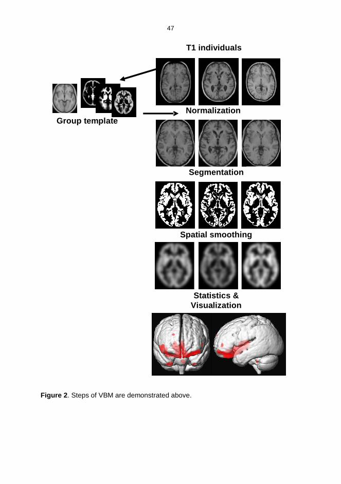
47
Figure 2. Steps of VBM are demonstrated above.
T1 individuals
Normalization
Segmentation
Spatial smoothing
Statistics &Visualization
Group template
48
2.9. Methods for outcome assessment
2.9.1. Clinical outcome scales and prognostic factors
The clinical outcome is usually evaluated at the outpatient department by a
neurosurgeon, usually two to three months after aSAH and treatment of the ruptured
aneurysm. The Glasgow Outcome Scale (GOS) (Jennett et al. 1975) is often used as
a rough outcome measure. GOS scores are graded as following: GOS V= Good
recovery, GOS IV= Moderate disability; disabled, but independent, GOS III= Severe
disability; conscious but disabled, GOS II= Persistent vegetative state, GOS I=
Death. There are other rough categorical outcome grading systems such as old
Rankin Outcome scale (Rankin 1957) which have also been used for assessment of
the clinical outcome.
The main determinant of outcome is the severity of the initial bleed (Broderick et
al. 1994, Hop et al. 1997, Koivisto et al. 2000). The clinical and cognitive outcome is
related to the several wellestablished pre and postreatment clinical factors: higher
Hunt and Hess grade, advanced age, a high Fisher score, preoperative
hydrocephalus, clinical vasospasm, and larger aneurysm, all favoring the poorer
outcome (Hernesniemi et al. 1993, Hoh et al. 2004, Kassell et al. 1990, Koivisto et al.
2000 , Molyneux et al. 2005, Salary et al. 2007). Massachusetts General Hospital
grade incorporates prognostic factors such as age, Hunt and Hess grade, size and
location of the aneurysm, and Fisher grade which alone may not demonstrate
statistical association, but may add incrementally to the risk of poor outcome of a
patient (Hoh et al. 2004). Clipping of an anterior cerebral artery aneurysm has been
associated with a higher impairment rating (Bornstein et al. 1987). Furthermore, high
blood glucose levels (Kruyt et al. 2009), occurrence of hypovolemia and hypotension
(Chang et al. 2000) and elevated blood Ddimer (Juvela et al. 2006) have been
correlated with worse outcome.
2.9.2. Neuropsychological outcome
The longterm neuropsychosocial effects of aSAH are considerable, even in
patients who regain functional independence (Wermer et al. 2007). Many patients
surviving from aSAH are left with persisting neurological and cognitive impairments
49
(Bellebaum et al. 2004, Hackett et al. 2000, Kreiter et al. 2002, Mayer et al. 2002,
Ogden et al. 1997) and half of the surviving patients cannot return to their previous
work (Powell et al. 2002). During the past decade, there have been many studies
reporting functional outcome; cognitive function and quality of life (QOL).
Neuropsychological testing is a feasible outcome measure (Anderson et al. 2006,
Egge et al. 2005) in patients after aSAH, especially with good clinical outcome.
However, good neurological outcome (GOS V) score does not exclude persisting
neuropsychological deficits, and even 62% of GOS V patients suffer cognitive
impairments (Hutter et al. 1993). Global cerebral edema and diffuse brain damage
and leftsided infarction or frontal medial infarctions are important risk factors for
cognitive dysfunction after aSAH (Kreiter et al. 2002, Vilkki et al. 1989).
2.9.3. Quality of life
Quality of life (QOL) constitutes a criterion of evaluation that describes the daily
life tasks in physical, physiological, and social fields of health and has been
measured in outcome studies after aSAH. The information has been collected with
regard to the QOL by semistructured interview and different quality of life scores, for
example EURO QOL (EQ5D) (Brooks 1996, Brooks 1996, Hutter et al. 1993, Ogden
et al. 1997) or validated instruments (Hop et al. 1998, Hop et al. 2001, Hutter et al.
2001).
50
3. AIMS OF THE STUDY
The general aim of the study was to determine the MRI outcome and to compare
it in aneurysmal subarachnoid hemorrhage (aSAH) patients who were randomly
assigned to endovascular versus surgical treatment of acutely ruptured intracranial
aneurysms.
The more specific fourfold aims were:
I: To prospectively evaluate, with MR imaging, the longterm outcome of the brain
after endovascular versus neurosurgical treatment for aSAH.
II: To evaluate and compare the volumes of temporomesial structures (amygdala and
hippocampus) after endovascular and surgical treatment of aSAH.
III: To assess the regional atrophic patterns of the brain and their clinical and
neuropsychological relevance after aSAH due to a ruptured anterior cerebral artery
aneurysm by employing voxelbased morphometry (VBM).
IV: To quantify the possible late (atrophic) ventricular and other cerebrospinal fluid
(CSF) space dilation in patients with aSAH and to correlate the cognitive outcome
with the degree of enlarged CSF spaces.
51
4. PATIENTS AND METHODS
4.1. Study design and study inclusion criteria and patients
The study design was approved by the Ethical Committee of the University of
Kuopio and Kuopio University Hospital. During the original study period between
February 1, 1995 and December, 1999, all patients who were admitted to Kuopio
University Hospital because of SAH were assessed as candidates for the prospective
randomized study.
CT was performed to all candidates to confirm the diagnosis of SAH. After CT an
informed consent from the patient or from the patients' closest relative were obtained.
The final recruitment into the study was made after the diagnostic angiography,
where the diagnosis of aneurysmal SAH was confirmed. All patients with ruptured IAs
suitable for both endovascular and surgical treatment were consecutively included.
The exclusion criteria for enrollment into the study were the following: 1. Age> 75
years; 2. Bleeding > 3 days previously; 3. Large hematoma requiring urgent surgery;
4. Mass effect of the aneurysm causing neurological deficit; 5. A previous
aneurysmal surgery.
Based on the diagnostic angiography, the aneurysm was not considered as
suitable for endovascular treatment and thus not randomized if: 1. the neck of the
aneurysm was wider than the fundus; 2. the aneurysm was fusiform; 3. the neck and
its' relationship to parent vessel and adjacent branches were not distinguishable; 4.
the size of the aneurysm was < 2 mm (less than the smallest coil available). The
patients' suitability for endovascular treatment and randomization was assessed
according to the morphology of the aneurysm that had most probably ruptured (size
and the irregularity of the aneurysm and the localization of hemorrhage detected on
CT). Randomization procedure was performed in three different categories according
to the pretreatment clinical condition of the patient (Hunt & Hess Grade III, Grade III,
and Grade IVV) in order to avoid the selection bias. Sealed envelopes were
allocated for each group of patients in randomization procedure.
52
4.2. Fisher and Hunt and Hess Grades and clinical vasospasm
Every patient had preoperative CT scans, which were evaluated in consensus by
a neurosurgeon (T.K) and a neuroradiologist (R.V) for assessment of the Fisher
grade (Fisher et al. 1980) and possible hydrocephalus. The Fisher grades were
further dichotomized into grades 02 (minor/moderate bleeding) and grades 34
(severe bleeding). The Hunt and Hess grades were assessed by one of six resident
neurosurgeons (with 16 years of experience) and confirmed by two senior
neurosurgeons. Clinical criteria for symptomatic vasospasm (assessed by one of 7
senior neurosurgeons) were a decrease of Glasgow Coma Scale by 2 scores or
appearance of new localizing symptoms (dysphasia or hemiparesis); other reasons
for deterioration, e.g. hydrocephalus, metabolic disorders, postoperative bleeding
and infections being ruled out.
4.3. Diagnostic angiography and embolization procedure
The diagnostic angiography was performed via the femoral artery through a 6F
introducer. Digital subtraction angiography equipment (Polytron; Siemens Medical
Engineering Group, Erlangen, Germany) with a 1024 x 1024 matrix was used. The
internal carotid and vertebral arteries were selectively catherized. Angiography was
followed by the immediate random assignment and if indicated, endovascular
treatment during the same session. At angiography, the site, orientation and
morphology of the aneurysm were carefully evaluated and the size of the aneurysm
and the width of the neck were measured with a digital caliber using two coins as a
reference. In all grade IVV patients and majority of grade III patients, embolization
was performed with use of general anesthesia. In patients with grade III and in some
of the grade III patents, no method of sedation was used. During the procedure,
systemic administration of heparin (initial bolus of 5000 IU in the first 5 patients and
2500 IU in the remaining patients, followed by 1000 IU after an hour and according to
the measured activated clotting time thereafter). The endovascular procedures were
performed by two interventional neuroradiologists (1 and 3 years of experience in
neurointerventions in the beginning of the study). A 6 F guiding cathether was
advanced to ICA near the level of skull base or to the upper cervical portion of the
vertebral artery. A microcatheter (Tracker10 or Tracker18; Target Therapeutics,
53
Fremont, California, USA) with two tip markers and a Dasher14, usually followed by
a Dasher10 guidewire (Target Therapeutics, Fremont, California, USA) were used
for hyperselective catheterization of the aneurysmal sac. By using a distal roadmap
control, care was taken not to touch the aneurysmal wall with the guidewire or the
catheter. A continuous, pressurized flush of heparinized saline solution was
maintained in both the guiding catheter and the microcathetheter. Once
catheterization had been achieved, the aneurysmal sac was filled with Guglielmi
detachable coils (GDCs; Target Therapeutics, Fremont, California, USA), which can
be electrically detached. Complete occlusion of the aneurysmal sac and neck was
always attempted. The largest coil, selected according the measured aneurysm
diameter, was positioned first to form a basketlike frame in the aneurysm. The
smaller coils were then sequentially delivered into the aneurysm until the lumen was
completely occluded and flow inside the aneurysm as well the secundary pouch was
arrested. If the size of selected coil proved to be unsuitable, the GDC system allowed
removal of the coil and repositioning of the mesh to an optimal position. Oral
administration of aspirin (250 mg daily) was continued for 3 months after
embolization. After endovascular treatment, the followup DS angiographs were
performed 3, 12 and 36 months after aSAH.
4.4. Surgical treatment
Surgical procedures were performed under balanced anesthesia with
hyperventilation by a team consisting of 7 senior neurosurgeons (1030 years of
experience in neurosurgery) with a collective experience of approximately 2000
aneurysm operations. All patients received corticosteroids and mannitol. Pterional,
subtemporal, frontal interhemispherical or lateral suboccipital approaches were used
depending on the location of the aneurysm. A standard microsurgical method was
used for clipping the aneurysmal neck with Sugita (Mizuho Medical Co., Tokyo,
Japan) or Yasargil (Aesculap AG & Co. KG, Tuttlingen, Germany) aneurysm clip. If
feasible, the aneurysm was opened, coagulated or both. Followup angiography was
scheduled after surgical clipping during the primary hospitalization, and in case of
neckremnant, 12 months after clipping.
54
4.5. Patient care after treatment
After the randomization procedure and treatment, both endovascular and surgical
patients were treated in the intensive care unit and neurosurgical ward in a similar
manner. The hypervolemic therapy, which was ascertained with SwanGanz catheter
placement and measurements of the pulmonary capillary wedge pressure was used.
Regardless of the treatment modality, prolonged bedrest (10 days), corticosteroids
and intravenous nimodipine were used in every patient. Symptoms of vasospasm
were recorded on daily basis.
4.6. Followup protocol: MR Imaging
4.6.1. Conventional MRI protocol
Patients underwent 1.5 Tesla MRI (Magnetom Vision; Siemens Medical Systems,
Erlangen, Germany) with a circularpolarized head coil. MRI protocol consisted of
axial T2 and intermediateweighted (TR/TE 2625/9816ms, matrix 260x512, slice
thickness 5mm) images, magnetic resonance time of flight angiography for the
embolized patients and a threedimensional T1weightedsequence: magnetisation
prepared rapid gradient echo (MPRAGE) sequence (TR/TE/TI 9.7/4/20ms, flip angle
12º, FOV 250mm, matrix 256x 256). All coils and surgical clips were non
ferromagnetic and MRIsafe.
4.6.2. MRI Analysis
All MRI examinations were analyzed in a consensus by a neuroradiologist (R.V.),
a neurosurgeon (T.K.) and a resident (P.B.) with15, 13 and 2 years of experience in
interpretation of MRI images on the time. Blinded reading was not possible because
of characteristic clip and coil artefacts. The presence of any highSI lesions on T2
and intermediateweighted images (lowsignalintensity on 3D T1weighted images)
was visually evaluated and lesion volumes were measured. The most probable
etiology of the highSI lesion was determined and classified in consensus with
knowledge of all clinical, radiological and surgical details of the patient. CTscans and
angiographic images were available during the MRI evaluation.
55
The following factors were analyzed and quantified according to anatomical
locations (Tatu et al. 1996, Tatu et al. 1998). Ischemic lesions in 1. parental artery
territory (vascular territory of the ruptured aneurysm) and 2. other vascular territories,
based on knowledge of normal vascular anatomy and angiographic findings of the
individual patient; 3. Superficial siderosis and residual signs of hematoma; 4. Signs of
retraction injury due to surgical manipulation and instrumentation; 5. Ventricular size
and possible hydrocephalus; 6. Parenchymal lesions associated with intraventricular
catheter and permanent shunt placement and 7. Previous infarctions, degree of
possible previous atrophy, and presence of lacunar infarctions and leukoaraiosis.
The parental artery of the ruptured aneurysm was classified as: 1. anterior
cerebral artery (ACA), 2. middle cerebral artery (MCA), 3. internal carotid artery
(ICA), or 4. vertebrobasilar artery (VBA). If the ruptured aneurysm was located in the
AComA or the basilar tip, the bilateral vascular territories of frontopolar,
callosomarginal, and pericallosal arteries and posterior cerebral arteries were
considered as parental arteries. Ischemic lesions in vascular territories other than the
parental arteries were considered to represent general vasospastic etiology. Signs of
parenchymal or Sylvian hematoma were evaluated with help of primary CTscans.
The convexity of the brain, basal cisterns and Sylvian region were evaluated for
possible signs of superficial siderosis. Approximate adjustment for ventricular size
was made visually on consensus reading. As a rough measure of ventricular size, the
modified cella media index (mCMI) was calculated as the maximum diameter of the
lateral ventricles/ intraparenchymal diameter at the same level. Degree of
leukoaraiosis was scored as: absent, punctuate/ early confluent or confluent
(Schmidt et al. 1999). Volumes of ischemic and retraction lesions were measured by
a single interpreter (P.B., based on the initial concensus reading) on a PACS
workstation (SECTRA EE, version 10.2.P4; Linköping, Sweden) by drawing a region
of interest according to the lesion margin on each T2 weighted image and by
multiplying the lesion areas by the sum of the slice (5 mm) and gap (1.5 mm)
thickness.
56
4.6.3. Volumetric MRI
The sequence for the volumetric analyses was scheduled as an additional
sequence at the end of the imaging, if the patient was cooperative and there were no
movement artefacts in the basic images and the patient was willing to stay extra time
in the MR scanner.
The 3D T1 sequence needed for volumetry was obtained using the
magnetisationprepared rapid gradient echo (MPRAGE) sequence (TR/TE/TI
9.7/4/20ms, flip angle 12º, FOV 250mm, matrix 256 x256). The coronal slices
(thickness 2.0 mm) included the whole cerebrum.
4.6.4. Semiautomatic volumetry (Study II)
Standard neuroanatomical landmarks (such as ACPC line, hemispheres and
orbits) were used to reconstruct coronal slices (thickness 2 mm) perpendicular to the
long axis of the left hippocampus (HC). These sixteenbit Siemens format images
were converted to the ANALYZE format (MAYO Foundation, Minnesota, USA). In
study II, the volumes of the amygdalae (AM) and HC were manually measured by a
single observer. To maintain blind assessment and confidentiality, data was
registered and coded by subject numbers only. The intraclass correlation coefficients
for intrarater reliability (bilateral measurements of 10 patients) were 0.94 for the AM
and 0.88 for the HC volumes. The point counting method (EasyMeasure, version 1.0,
MariArc, Liverpool, UK) was used to calculate the volumes of the hippocampus and
the amygdala. The pointcounting method consisted of overlaying a systematic array
of the test points (one point per 9 pixels) completely over each slice. Instances in
which a point, in fact a cross, lies within the area of interest, are recorded by clicking
a computer mouse. Subsequently, the total number of the test points is multiplied by
the volume of the test point (17.17 mm3). Boundaries of the HC and AM were
identified with the use of the neuroanatomy atlas (Martin 1996) and previously
published research (Watson et al. 1992). The point counting of the structures
proceeded from anterior to posterior.
The amygdaloid volume consisted of the volumes of the central nucleus, the
corticomedial nucleus and the basolateral nucleus. Hippocampal volumetry included
the dentate gyrus, the cornus ammonis (hippocampus proper) and the subiculum.
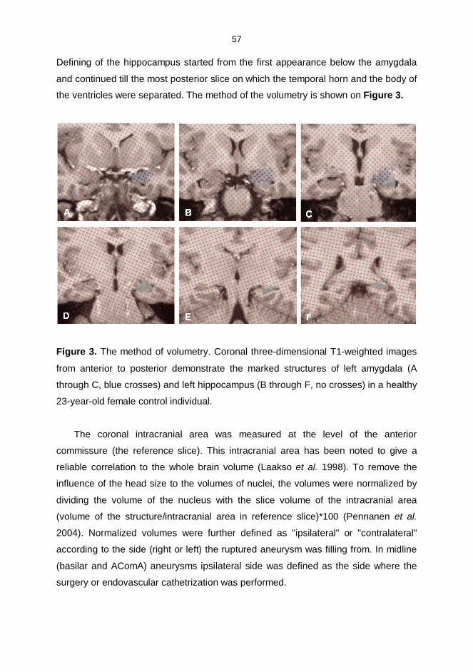
57
Defining of the hippocampus started from the first appearance below the amygdala
and continued till the most posterior slice on which the temporal horn and the body of
the ventricles were separated. The method of the volumetry is shown on Figure 3.
Figure 3. The method of volumetry. Coronal threedimensional T1weighted images
from anterior to posterior demonstrate the marked structures of left amygdala (A
through C, blue crosses) and left hippocampus (B through F, no crosses) in a healthy
23yearold female control individual.
The coronal intracranial area was measured at the level of the anterior
commissure (the reference slice). This intracranial area has been noted to give a
reliable correlation to the whole brain volume (Laakso et al. 1998). To remove the
influence of the head size to the volumes of nuclei, the volumes were normalized by
dividing the volume of the nucleus with the slice volume of the intracranial area
(volume of the structure/intracranial area in reference slice)*100 (Pennanen et al.
2004). Normalized volumes were further defined as "ipsilateral" or "contralateral"
according to the side (right or left) the ruptured aneurysm was filling from. In midline
(basilar and AComA) aneurysms ipsilateral side was defined as the side where the
surgery or endovascular cathetrization was performed.
58
4.6.5. Voxelbased morphometry (studies III IV)
Optimized VBM analysis (Ashburner et al. 2000, Good et al. 2001) was
performed using the VBM2 (study III) toolbox (http://dbm.neuro.unijena/vbm/) under
SPM2 (Wellcome Department of Imaging Neuroscience, London, UK;
www.fil.ion.ac.uk/spm). The VBM procedure started with creating a customized
template and customized prior probability maps using the standard procedure of
VBMtoolbox: normalization to the MNI T1weighted template provided by SPM,
resampling the voxel size to 2 x 2 x 2 mm, segmenting the images into gray matter
(GM), white matter (WM), and cerebrospinal fluid (CSF), smoothing both the
normalized images and the segments with 8 mm Gaussian kernel, averaging the
smoothed images and segments.
In order to define the normalization parameters for spatial normalization and
absolute volumes of different tissue types, the original images were segmented into
GM, WM and CSF using the customized prior probability maps. The absolute
volumes of GM, WM, and CSF were calculated from the segments, and they were
further summarized to obtain the total intracranial volume (ICV) and the GM/ICV and
CSF/ICV ratios were calculated from these values. The normalization parameters
were estimated using these GM segments and the customized GM template. The
original images were then normalized using the estimated normalization parameters,
and were resampled to 2 x 2 x 2 mm. The normalized images were further
segmented into GM, WM, and CSF using the customized prior probability maps.
Finally, the GMsegments were modulated and smoothed with a 12 mm Gaussian
kernel.
VBM Group analyses (Study III) were performed in SPM2 including age, sex and
intracranial area as nuisance covariates in order to compare the GM volumes
between the patients and controls. Between group differences were assessed using
a ttest with a height threshold of P<0.05, corrected for multiple comparisons by the
familywise error (FWE) or by the false discovery rate (FDRmethod) (Genovese et al.
2002) method. When surgical and endovascular patients were compared in VBM
analysis, the group differences were assessed using a ttest with a height threshold
of P<0.05, corrected for multiple comparisons by the false discovery rate (FDR)
method, because no voxels survived the conservative FWEmethod. Only those
clusters exceeding a size of 50 voxels were included in the analysis. To determine
59
the anatomical locations of reduced GM, the MNI coordinates obtained from the peak
voxels were transferred into Talairach space using the mni2tal routine
(http://imaging.mrccbu.cam.ac.uk/imaging/MniTalairach). The anatomical locations of
the peak voxels in the significant clusters were found using the Talairach Daemon
Applet (Lancaster et al. 2000) and further verified from an anatomical brain atlas
(Duvernoy 1999).
In study III, the aim was to examine the differences in grey matter volumes
between the patients and controls with respect to the side of the treatment
(craniotomy or endovascular cathetherization) and the contralateral side. Therefore,
the MR images of the patients in whom the approach of the treatment was on the left
hemisphere (17 out of 37) were flipped around the anteriorposterior axis using
MRIcro (www.sph.sc.edu/comd/rorden/mricro.html) prior to any data analysis.
Furthermore, same proportion of controls (12 out of 30), matching with the patients'
age in decades, had their images similarly flipped.
In study IV, for quantitation of the ventricular size at the grouplevel, an averaged
normalized CSF segment image was calculated using the segmentation routine
provided by VBM5 toolbox in SPM5 for both patients and control individuals. Using
these averaged CSFimages, a ventricular ROI was manually traced (by P.B) for both
groups with MriCro (http://www.sph.sc.edu/comd/rorden/mricro.html) and the average
volumes of the ventricles were calculated for patient and control individual groups.
4.7. Followup protocol: Clinical Outcome, Neuropsychological evaluation
Clinical outcome and GOS one year after aSAH were assessed by a single
neurosurgeon (T.K.). Detailed neuropsychological examination was performed by a
single neuropsychologist (H.H.) one year after aSAH. Two experienced
neuropsychologists (M.Ä. and T.H.) further interpreted the neuropsychological test
results using the normative data.
The comprehensive evaluation included tests of general intelligence, memory
and selected language abilities and assessment of attention and flexibility of mental
processing as explained in the following: General intellectual ability: On the basis of
subtests of the Wechsler Adult Intelligence ScaleRevised (WAISR) (Wechsler 1981)
the scores for general verbal and nonverbal ability and total intelligence quotients
60
were calculated. The Modified Boston Naming test (Lezak MD 2004) was used to
examine naming ability and the Finnish version of the Verbal Fluency Test on letters
(Borkowski et al. 1967) was used to evaluate word fluency. Learning and memory:
Memory was tested by the Wechsler Memory Scale (WMS) (Wechsler 1974). The
Memory Quotient was calculated to assess shortterm memory performance.
Delayed recall of Logical Memory Subtest (Wechsler 1974) and the Visual
Reproduction Subtest (Wechsler 1974) was asked 45 minutes later. Nonverbal
memory was also assessed by the Rey Complex Figure Test (Lezak MD 2004).
Attention and flexibility of mental processing and psychomotor speed: The Stroop
Test (Golden 1978) (form A, B and C) and the Trail Making Test (Reitan 1958) (part
A and B) were used to evaluate sustained attention and resistance to interference.
The Finger Tapping test was used to assess simple psychomotor speed.
Neuropsychological examination was divided into four different cognitive domains
(general intellectual functioning, memory, language and executive functions).
Impairment in any of the four cognitive domains was defined on the basis of
normative data, where the scores fell below the normal range in either measure of
the domain. General intellectual functioning was estimated on the basis of five
subtests of the Wechsler Adult Intelligence ScaleRevised (WAISR) (Wechsler
1981). Language was evaluated with a shortened version of the Boston Naming Test
and Verbal Fluency test (Lezak MD 2004). Memory was assessed with the Logical
Memory and Visual Reproduction subtests (immediate and delayed recall) from
Wechsler Memory Scale (WMS) (Wechsler 1974). Percent retention scores
(calculated by dividing the delayed recall score by immediate recall score) were the
measures for verbal and visual memory. Executive functions were assessed with the
Trail Making Test and the Stroop Test (Golden 1978, Reitan 1958). In order to extract
the executive component in these tests, a difference score was calculated by
subtracting the time taken in Trail Making A from the time taken in Trail Making B. A
similar calculation was performed for recorded times for colour naming and the
interference part of the Stroop Test. The neuropsychological test results were then
further dichotomized.
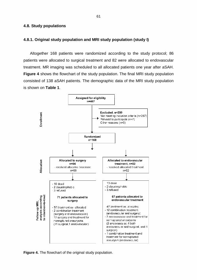
61
4.8. Study populations
4.8.1. Original study population and MRI study population (study I)
Altogether 168 patients were randomized according to the study protocol; 86
patients were allocated to surgical treatment and 82 were allocated to endovascular
treatment. MR imaging was scheduled to all allocated patients one year after aSAH.
Figure 4 shows the flowchart of the study population. The final MRI study population
consisted of 138 aSAH patients. The demographic data of the MRI study population
is shown on Table 1.
Figure 4. The flowchart of the original study population.
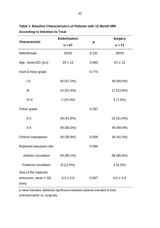
62
Table 1. Baseline Characteristics of Patients with 12 Month MRI
According to Intention to Treat
CharacteristicEmbolization
n = 67p
Surgery
n = 71
Male/female 35/32 0.131 28/43
Age, mean±SD (yrs) 50 ± 13 0.485 51 ± 13
Hunt & Hess grade 0.774
III 45 (67.2%) 49 (69.0%)
III 15 (22.4%) 17 (23.9%)
IVV 7 (10.4%) 5 (7.0%)
Fisher grade 0.187
02 28 (41.8%) 22 (31.0%)
34 39 (58.2%) 49 (69.0%)
Clinical Vasospasm 18 (26.9%) 0.058 30 (42.3%)
Ruptured aneurysm site 0.094
Anterior circulation 59 (88.1%) 68 (95.8%)
Posterior circulation 8 (11.9%) 3 (4.2%)
Size of the ruptured
aneurysm, mean ± SD
(mm)
6.3 ± 2.9 0.697 6.5 ± 2.9
pvalue indicates statistical significance between patients intended to treat
endovascularily vs. surgically.
63
4.8.2. Combination treatment of the ruptured aneurysm and treatment of
additional associated nonruptured aneurysms
Altogether 15 of the 138 (10.9%) patients who underwent followup MRI, have had
combination treatment (both endovascular and surgical) of the ruptured aneurysm.
The indications for combination treatment were failure in the primary treatment
procedure or revascularisation of the aneurysm in followup DS angiography.
Combination treatment was required more frequently in the endovascular group
(n=13, 19.4%) than in the surgical group (n=2, 2.8%, p=0.002). Thirtyfive of the138
(25.4%) patients had 54 nonruptured aneurysms, which were treated using the same
criteria currently recommended (Bederson et al. 2000): embolization in 6, surgery in
23, combination treatment in 3 and angiographic followup in 22 cases. All analyses
where treatment groups (surgical. vs. endovascular) were compared with each other
were performed according to intentiontotreat and also repeated after excluding
patients with combination treatment or additional treatment for nonruptured
aneurysms.
4.8.3. Volumetric study populations (studies II, III and IV)
Altogether 77 patients from the randomized population (33 endovascular, 36
surgical and 8 combination treatment patients) underwent threedimensional T1
weighted examinations. In study II, 77 volumetric measurements were available. In
study IV 76 volumetric measurements were available; one volumetric study had to be
excluded, because the scanning area on T1weighted volumetric sequence did not
cover the whole cerebrum. Study III consisted of 37 ACA volumetric patients (after
exclusion of 5 ACA patients whose associated unruptured aneurysms had been
treated before MRI). The demographic data of the volumetric study population is
shown on Table 2.
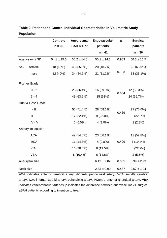
64
Table 2. Patient and Control Individual Characteristics in Volumetric StudyPopulation
Controls
n = 30
Aneurysmal
SAH n = 77
Endovascular
patients
n = 41
p Surgical
patients
n = 36
Age, years ± SD 54.1 ± 15.5 50.2 ± 14.8 50.1 ± 14.3 0.963 50.3 ± 15.5
Sex female 18 (60%) 43 (55.8%) 20 (48.7%) 23 (63.9%)
male 12 (40%) 34 (44.2%) 21 (51.2%) 0.183 13 (36.1%)
Fischer Grade
0 2
3 4
28 (36.4%)
49 (63.6%)
16 (39.0%)
25 (61%)0.604
12 (33.3%)
24 (66.7%)
Hunt & Hess Grade
I II
III
IV V
55 (71.4%)
17 (22.1%)
5 (6.5%)
28 (68.3%)
9 (22.0%)
4 (9.8%)
0.45927 (75.0%)
8 (22.2%)
1 (2.8%)
Aneurysm location
ACA
MCA
ICA
VBA
42 (54.5%)
11 (14.3%)
16 (20.8%)
8 (10.4%)
23 (56.1%)
4 (9.8%)
8 (19.5%)
6 (14.6%)
0.409
19 (52.8%)
7 (19.4%)
8 (22.2%)
2 (5.6%)
Aneurysm size 6.12 ± 2.82 0.685 6.39 ± 2.93
Neck size 2.83 ± 0.99 0.487 2.67 ± 1.04
ACA indicates anterior cerebral artery, AComA, pericallosal artery. MCA; middle cerebral
artery. ICA; internal carotid artery, ophthalmic artery, PComA, anterior choroidal artery. VBA
indicates vertebrobasilar arteries. p indicates the difference between endovascular vs. surgical
aSAH patients according to intention to treat.
65
4.8.4. Neuropsychological study population (studies IIV)
The neuropsychological examination was available from 116 out of 138 (84.1%)
patients, who underwent the one year MRI. In the volumetric study populations, 76
out of 77 (98.7%, study II), all 37 ACA patients (study III) and 75 out of 76 (98.7%,
study IV) underwent the neuropsychological examination.
4.8.5. Control individuals (Studies II, III and IV)
30 healthy control individuals matching their age and gender to the volumetric
study population (studies II IV) were also imaged for volumetric purposes with the
same MRI equipment than the aSAH patients. The demographic data of the control
individuals is also shown on Table 2.
4.9. Statistics
All analyses were performed with the statistical package for Windows (SPSS, Inc.,
Chicago, IL, USA, versions 11.5 16). The intentiontotreat analysis was used in all
comparisons of endovascular and surgical patients. In addition to intentiontotreat
analyses, the censored analyses were also performed after excluding patients having
combination treatment for ruptured aneurysm or treatment for additional, nonruptured
aneurysms.
In general, all continuous variables were tested for normal distribution with the
KolmogorovSmirnov 1sample test (level of statistical significance difference at
p<0.1). The ChiSquare test was used for categorical data and dichotomized
variables. The Student ttest was used to compare normally distributed data and
MannWhitneyU test was used to compare nonparametric, continuousscale data for
nonnormally distributed variables. Spearman and Pearson correlation coefficients
were used to assess correlation between nonparametric continuousscale variables.
Differences were considered to be statistically significant, if the twotailed p value
was <0.05. The multiple comparisons were corrected with Bonferroni adjustment
(Norman G 1994) and the significant pvalue in these analyses using the same
parameter twice was 0.05/2 =0.025. The group differences in volumetric analyses are

66
based on ttest statistics with a height threshold of P<0.05 and explained in more
detail in paragraph 4.6.3.
67
5. RESULTS
5.1. Comparability of the study groups
According to the intention to treat analysis, in the MRIstudy population with 138
patients, the endovascular and surgical treatment groups did not show any
statistically significant differences in their age or sex distribution, nor in their Hunt &
Hess Grades, Fisher grades, size or location of the ruptured aneurysm or
preoperative hydrocephalus (study I). The endovascular and surgical treatment
groups remained also balanced in a censored analysis after excluding the patients
who had undergone combination treatment or treatment for additional nonruptured
aneurysms. Moreover, the different treatment groups did not show statistically
significant differences in abovementioned parameters (studies II, III and IV) in
volumetric studies. The control individual group was used in volumetric comparisons
(studies IIIV) and the control individuals were statistically balanced in terms of age
and gender. In study III, all aSAH patients (n=37: 20 endovascular, 17 surgical) and
controls (n=30) were balanced for age and sex in comparison between aSAH
patients and controls. The final group analyses in study III were performed after
excluding patients with crossover treatment (5 endovascular patients) and after this
exclusion there was a male dominance in the endovascular subgroup (12 of 15, 80%)
vs. controls (13 of 30, 43.3%; p=0.020). The surgical group and controls remained
gender balanced.
The volumetric study population mostly consisted of patients with good recovery;
66 patients had good outcome and ten patients (7 surgical, 3 endovascular) had
moderate disability. Moreover, the clinical outcome was better in the volumetric
subgroup compared to the original study population.
The mean interval between aSAH and MRI was 15.8 ± 8.5 months (range 964
months). The diagnostic quality of the MRI examinations was excellent in 134
(97.1%) and suboptimal, but diagnostic in 4 (2.9%) examinations.
68
5.2. Brain parenchymal lesions detected on late MR imaging and comparison of
surgical vs. endovascular treatment modalities (Study I)
One year after aSAH only 44 (31.9%) patients (29, 43.3% endovascular, 15,
21.2% surgical) had normal MRI of the brain or the finding was analogous to the
admission CTscan. Parenchymal high SIintensity lesions on T2 and intermediate
weighted images of different etiology were seen in 38 (56.7%) endovascular and in
56 (78.9%) surgical patients (p=0.005). In patients with higher H & H and Fisher
grades, parenchymal lesions were seen significantly more often than in patients with
lower grades. MRIdetectable parenchymal lesions of different etiologies are
demonstrated on Tables 3 and 4.
Table 3. Number of Parenchymal MRI lesions One Year After aSAH inPatients Intended to Treat Endovascularily vs. Surgically
Embolizationn = 67
P Surgeryn = 71
Parenchymal lesionsaltogether 38 (56.7%) 0.005 56 (78.9%)
H & H
I III 31/60 (52.7%) 0.003 51/66 (77.3%)
IV V 7/7 (100%) 5/5 (100%)
Fisher
0 2 12/28 (42.9%) 0.144 14/22 (63.6%)
3 4 26/39 (66.7%) 0.034 42/49 (85.7%)
Anatomical locations:
Frontal lobe 34 (50.7%) 0.018 50 (70.4%)
Temporal lobe 15 (22.4%) 0.002 34 (47.9%)
Parietal lobe 8 (11.9%) 0.212 14 (19.7%)
Occipital lobe 2 (3.0%) 0.953 2 (2.8%)
Thalamus and basalganglia 7 (10.4%) 0.057 16 (22.5%)
Brain stem andcerebellum 4 (6.0%) 0.037 0 (0%)
P value indicates statistical significance between patients intended to treat endovascularily vs.surgically.
69
Table 4. Brain MRI Findings According to the Etiology of the Lesion 1 year AfteraSAH in Patients Intended to Treat Endovascularily vs. Surgically
Embolization, n=67 P Surgery, n=71
MRI findings n (%) n (%)
Any Ischemic deficits 19 (28.4%) 0.005 37 (52.1%)
Parental artery territory 15 (22.4%) 0.003 33 (46.5%)
Cortical involvement 13 (19.4%) 29 (40.8%)
Perforator artery territory 6 (9.0%) 15 (21.1%)
More than one lesion 4 (6.0%) 0.178 9 (12.7%)
Other vascular territory 7 (10.4%) 0.379 11 (15.5%)
Cortical involvement 8 (11.9%) 16 (22.5%)
Perforator artery territory 4 (6.0%) 3 (4.2%)
More than one lesion 3 (4.5%) 0.757 4 (5.6%)
Superficial brain retraction deficits 10 (14.9%) <0.001 40 (56.3%)
Deficit caused by preoperative ICH 15 (22.4%) 0.683 18 (25.4%)
Superficial siderosis 28 (41.8%) 0.778 28 (39.4%)
Note. P value indicates statistical significance between patients intended to treat endovascularily vs. surgically.Perforator artery territory includes the branches of the thalamoperforating arteries, thalamogeniculate arteries (fromposterior communicating arteries, basilar artery and posterior cerebral arteries), medial lenticulostriatae arteries andrecurrent artery of Heubner (from anterior cerebral arteries and anterior communicating arteries) and laterallenticulostriatae arteries (from middle cerebral artery).
5.2.1. Ischemic lesions in the parental artery territory
Highsignalintensity lesions on T2 and intermediateweighted images indicating
infarction in the vascular territory of the ruptured aneurysm were seen in 15/67
(22.4%) endovascular and in 33/71 (46.5%) surgical patients (p=0.003). Four
endovascular and 7 surgical patients had two separate infarctions and 2 surgical
patients had three separate infarctions in the parental artery territory. In patients who
had infarctions, the mean volume of the infarcted tissue was 17.6cm3 (range 0.14
121.5cm3) in the endovascular group, and 20.9cm3 (range 0.09261.5cm3) in the
surgical group (p=0.209). In all patients included, the mean volume of the infarcted
tissue in the parental artery territory was 3.9cm3 (range 0.0121.5 cm3) after
70
endovascular treatment and 9.7cm3 (range 0.0261.5cm3) after surgical treatment
(p=0.002).
5.2.2. Ischemic lesions in other vascular territories
Eighteen out of 67 (26.9%) endovascular and 30/71 (42.3%) surgical patients had
symptoms of clinical vasospasm (p=0.058). Brain infarctions in vascular territories
other than that of the ruptured aneurysm were seen in 7/67 (10.4%) endovascular
and in 11/71 (15.5%) surgical patients (p=0.379). In patients who had remote
infarctions, the mean volume of the infarcted tissue was 27.6cm3 (range 0.17
58.5cm3) in endovascular and 43.3cm3 (range 1.29247.5cm3) in surgical patients
(p=0.821). One endovascular patient had two and 2 patients had three infarctions in
remote territories. In the surgical group, one patient had two, 2 patients had three,
and one patient had four separate infarctions in remote territories. In all patients, the
mean volume of the ischemic lesions in remote vascular territories was 2.9cm3 (range
0.058.5cm3) after endovascular treatment and 6.7cm3 (range 0.0247.5cm3) after
surgical treatment (p=0.373).
5.2.3. Residual signs of hematoma and superficial siderosis
Highsignalintensity lesions on T2 and intermediateweighted images due to
previous intracerebral or sylvian hematoma were detected in 15 (22.4%)
endovascular and 18 (25.4%) surgical patients (p=0.683). Signs of superficial
hemosiderosis were present in 28 (41.8%) endovascular and 28 (39.4%) surgical
patients (p=0.778).
5.2.4. Retraction injury due to surgical manipulation and instrumentation
Lesions in the brain parenchyma due to mechanical surgical trauma were clearly
more common after surgical (n=40, 56.3%) than endovascular (n=10, 14.9%)
treatment (p<0.001). Retraction injuries were also detected after endovascular
treatment because 13 endovascular patients needed additional surgery or
nonruptured aneurysms were surgically ligated before MRI. The mean volumes of the
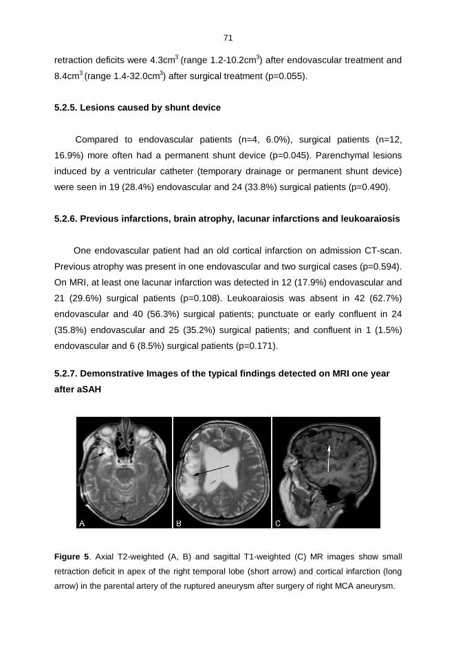
71
retraction deficits were 4.3cm3 (range 1.210.2cm3) after endovascular treatment and
8.4cm3 (range 1.432.0cm3) after surgical treatment (p=0.055).
5.2.5. Lesions caused by shunt device
Compared to endovascular patients (n=4, 6.0%), surgical patients (n=12,
16.9%) more often had a permanent shunt device (p=0.045). Parenchymal lesions
induced by a ventricular catheter (temporary drainage or permanent shunt device)
were seen in 19 (28.4%) endovascular and 24 (33.8%) surgical patients (p=0.490).
5.2.6. Previous infarctions, brain atrophy, lacunar infarctions and leukoaraiosis
One endovascular patient had an old cortical infarction on admission CTscan.
Previous atrophy was present in one endovascular and two surgical cases (p=0.594).
On MRI, at least one lacunar infarction was detected in 12 (17.9%) endovascular and
21 (29.6%) surgical patients (p=0.108). Leukoaraiosis was absent in 42 (62.7%)
endovascular and 40 (56.3%) surgical patients; punctuate or early confluent in 24
(35.8%) endovascular and 25 (35.2%) surgical patients; and confluent in 1 (1.5%)
endovascular and 6 (8.5%) surgical patients (p=0.171).
5.2.7. Demonstrative Images of the typical findings detected on MRI one yearafter aSAH
Figure 5. Axial T2weighted (A, B) and sagittal T1weighted (C) MR images show small
retraction deficit in apex of the right temporal lobe (short arrow) and cortical infarction (long
arrow) in the parental artery of the ruptured aneurysm after surgery of right MCA aneurysm.
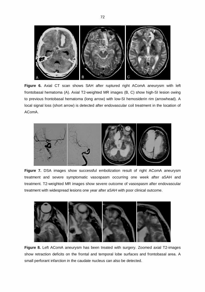
72
Figure 6. Axial CT scan shows SAH after ruptured right AComA aneurysm with left
frontobasal hematoma (A). Axial T2weighted MR images (B, C) show highSI lesion owing
to previous frontobasal hematoma (long arrow) with lowSI hemosiderin rim (arrowhead). A
local signal loss (short arrow) is detected after endovascular coil treatment in the location of
AComA.
Figure 7. DSA images show successful embolization result of right AComA aneurysm
treatment and severe symptomatic vasospasm occurring one week after aSAH and
treatment. T2weighted MR images show severe outcome of vasospasm after endovascular
treatment with widespread lesions one year after aSAH with poor clinical outcome.
Figure 8. Left AComA aneurysm has been treated with surgery. Zoomed axial T2images
show retraction deficits on the frontal and temporal lobe surfaces and frontobasal area. A
small perforant infarction in the caudate nucleus can also be detected.
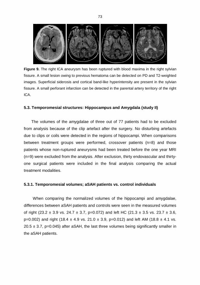
73
Figure 9. The right ICA aneurysm has been ruptured with blood maxima in the right sylvian
fissure. A small lesion owing to previous hematoma can be detected on PD and T2weighted
images. Superficial siderosis and cortical bandlike hyperintensity are present in the sylvian
fissure. A small perforant infarction can be detected in the parental artery territory of the right
ICA.
5.3. Temporomesial structures: Hippocampus and Amygdala (study II)
The volumes of the amygdalae of three out of 77 patients had to be excluded
from analysis because of the clip artefact after the surgery. No disturbing artefacts
due to clips or coils were detected in the regions of hippocampi. When comparisons
between treatment groups were performed, crossover patients (n=8) and those
patients whose nonruptured aneurysms had been treated before the one year MRI
(n=9) were excluded from the analysis. After exclusion, thirty endovascular and thirty
one surgical patients were included in the final analysis comparing the actual
treatment modalities.
5.3.1. Temporomesial volumes; aSAH patients vs. control individuals
When comparing the normalized volumes of the hippocampi and amygdalae,
differences between aSAH patients and controls were seen in the measured volumes
of right (23.2 ± 3.9 vs. 24.7 ± 3.7, p=0.072) and left HC (21.3 ± 3.5 vs. 23.7 ± 3.6,
p=0.002) and right (18.4 ± 4.9 vs. 21.0 ± 3.9, p=0.012) and left AM (18.8 ± 4.1 vs.
20.5 ± 3.7, p=0.045) after aSAH, the last three volumes being significantly smaller in
the aSAH patients.
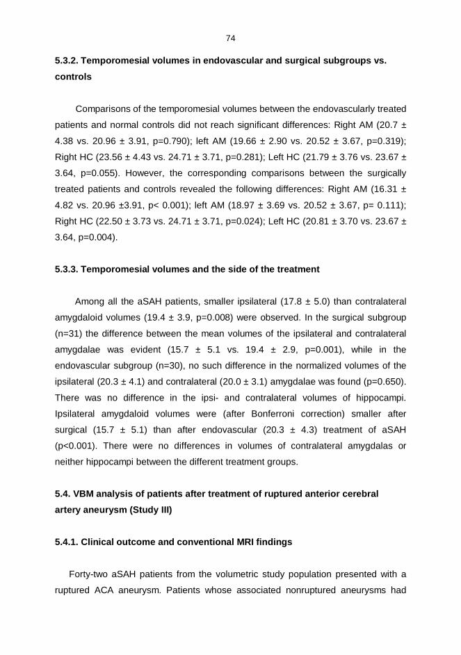
74
5.3.2. Temporomesial volumes in endovascular and surgical subgroups vs.
controls
Comparisons of the temporomesial volumes between the endovascularly treated
patients and normal controls did not reach significant differences: Right AM (20.7 ±
4.38 vs. 20.96 ± 3.91, p=0.790); left AM (19.66 ± 2.90 vs. 20.52 ± 3.67, p=0.319);
Right HC (23.56 ± 4.43 vs. 24.71 ± 3.71, p=0.281); Left HC (21.79 ± 3.76 vs. 23.67 ±
3.64, p=0.055). However, the corresponding comparisons between the surgically
treated patients and controls revealed the following differences: Right AM (16.31 ±
4.82 vs. 20.96 ±3.91, p< 0.001); left AM (18.97 ± 3.69 vs. 20.52 ± 3.67, p= 0.111);
Right HC (22.50 ± 3.73 vs. 24.71 ± 3.71, p=0.024); Left HC (20.81 ± 3.70 vs. 23.67 ±
3.64, p=0.004).
5.3.3. Temporomesial volumes and the side of the treatment
Among all the aSAH patients, smaller ipsilateral (17.8 ± 5.0) than contralateral
amygdaloid volumes (19.4 ± 3.9, p=0.008) were observed. In the surgical subgroup
(n=31) the difference between the mean volumes of the ipsilateral and contralateral
amygdalae was evident (15.7 ± 5.1 vs. 19.4 ± 2.9, p=0.001), while in the
endovascular subgroup (n=30), no such difference in the normalized volumes of the
ipsilateral (20.3 ± 4.1) and contralateral (20.0 ± 3.1) amygdalae was found (p=0.650).
There was no difference in the ipsi and contralateral volumes of hippocampi.
Ipsilateral amygdaloid volumes were (after Bonferroni correction) smaller after
surgical (15.7 ± 5.1) than after endovascular (20.3 ± 4.3) treatment of aSAH
(p<0.001). There were no differences in volumes of contralateral amygdalas or
neither hippocampi between the different treatment groups.
5.4. VBM analysis of patients after treatment of ruptured anterior cerebralartery aneurysm (Study III)
5.4.1. Clinical outcome and conventional MRI findings
Fortytwo aSAH patients from the volumetric study population presented with a
ruptured ACA aneurysm. Patients whose associated nonruptured aneurysms had
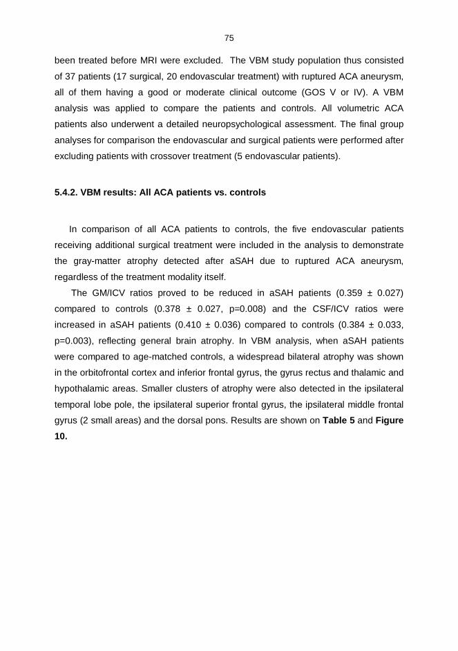
75
been treated before MRI were excluded. The VBM study population thus consisted
of 37 patients (17 surgical, 20 endovascular treatment) with ruptured ACA aneurysm,
all of them having a good or moderate clinical outcome (GOS V or IV). A VBM
analysis was applied to compare the patients and controls. All volumetric ACA
patients also underwent a detailed neuropsychological assessment. The final group
analyses for comparison the endovascular and surgical patients were performed after
excluding patients with crossover treatment (5 endovascular patients).
5.4.2. VBM results: All ACA patients vs. controls
In comparison of all ACA patients to controls, the five endovascular patients
receiving additional surgical treatment were included in the analysis to demonstrate
the graymatter atrophy detected after aSAH due to ruptured ACA aneurysm,
regardless of the treatment modality itself.
The GM/ICV ratios proved to be reduced in aSAH patients (0.359 ± 0.027)
compared to controls (0.378 ± 0.027, p=0.008) and the CSF/ICV ratios were
increased in aSAH patients (0.410 ± 0.036) compared to controls (0.384 ± 0.033,
p=0.003), reflecting general brain atrophy. In VBM analysis, when aSAH patients
were compared to agematched controls, a widespread bilateral atrophy was shown
in the orbitofrontal cortex and inferior frontal gyrus, the gyrus rectus and thalamic and
hypothalamic areas. Smaller clusters of atrophy were also detected in the ipsilateral
temporal lobe pole, the ipsilateral superior frontal gyrus, the ipsilateral middle frontal
gyrus (2 small areas) and the dorsal pons. Results are shown on Table 5 and Figure10.
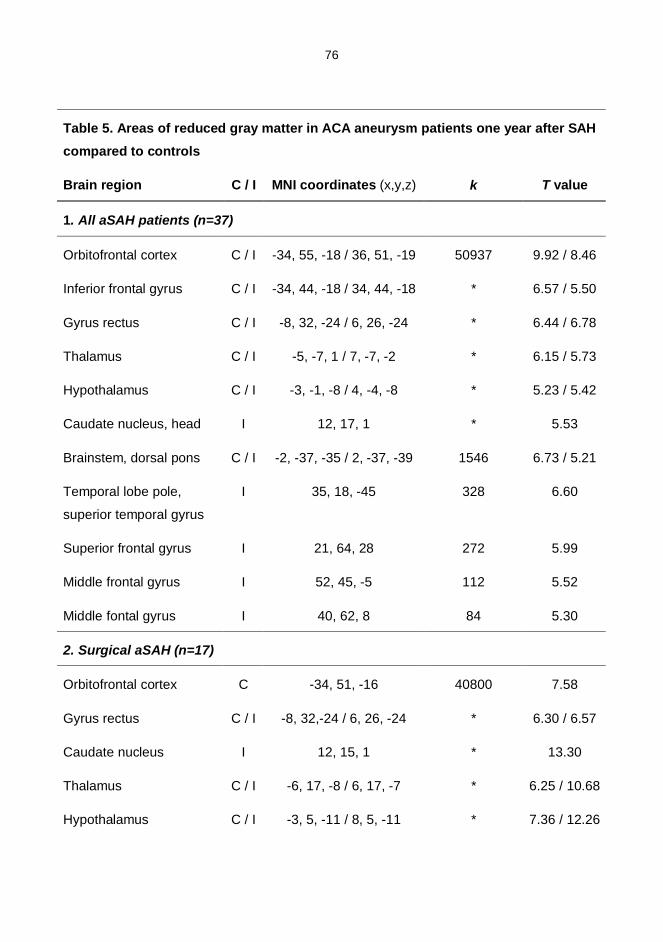
76
Table 5. Areas of reduced gray matter in ACA aneurysm patients one year after SAHcompared to controls
Brain region C / I MNI coordinates (x,y,z) k T value
1. All aSAH patients (n=37)
Orbitofrontal cortex C / I 34, 55, 18 / 36, 51, 19 50937 9.92 / 8.46
Inferior frontal gyrus C / I 34, 44, 18 / 34, 44, 18 * 6.57 / 5.50
Gyrus rectus C / I 8, 32, 24 / 6, 26, 24 * 6.44 / 6.78
Thalamus C / I 5, 7, 1 / 7, 7, 2 * 6.15 / 5.73
Hypothalamus C / I 3, 1, 8 / 4, 4, 8 * 5.23 / 5.42
Caudate nucleus, head I 12, 17, 1 * 5.53
Brainstem, dorsal pons C / I 2, 37, 35 / 2, 37, 39 1546 6.73 / 5.21
Temporal lobe pole,
superior temporal gyrus
I 35, 18, 45 328 6.60
Superior frontal gyrus I 21, 64, 28 272 5.99
Middle frontal gyrus I 52, 45, 5 112 5.52
Middle fontal gyrus I 40, 62, 8 84 5.30
2. Surgical aSAH (n=17)
Orbitofrontal cortex C 34, 51, 16 40800 7.58
Gyrus rectus C / I 8, 32,24 / 6, 26, 24 * 6.30 / 6.57
Caudate nucleus I 12, 15, 1 * 13.30
Thalamus C / I 6, 17, 8 / 6, 17, 7 * 6.25 / 10.68
Hypothalamus C / I 3, 5, 11 / 8, 5, 11 * 7.36 / 12.26
77
Hippocampus I 33, 12, 16 11437 8.52
Parahippocampal gyrus I 21, 35, 6 * 8.12
Superior temporal gyrus,
temporal lobe pole
C 30, 18, 46 496 8.01
Middle frontal gyrus I 50, 49, 4 1639 7.79
Orbitofrontal cortex I 39, 53, 17 * 7.53
Superior temporal gyrus,
temporal lobe pole
I 28, 17, 45 648 7.78
Brainstem, dorsal pons C 2, 37, 35 235 6.03
Posterior orbital gyrus I 36, 29, 21 239 5.97
Inferior frontal gyrus C 50, 48, 5 106 5.83
Anterior insula I 38, 13, 5 197 5.72
Superior frontal gyrus I 21, 63, 29 74 5.71
3. Embolized aSAH (n=15)
Orbitofrontal cortex C 34, 54, 19 793 7.60
Orbitofrontal cortex I 32, 51, 22 1049 8.43
Thalamus C 5, 7, 1 1616 6.27
C= contralateral, I=ipsilateral to ruptured aneurysm treatment side, MNI coordinates x,y,z of
anatomical structures in each cluster refer to Montreal Neurological Institute (MNI) space. k=
cluster extent in voxels. * =cluster extent in voxels is included in the number reported above. T
value = statistical significance of GM volume loss of the corresponding MNI coordinate, assessed
using a ttest (Tvalue) with a height threshold of P < 0.05, corrected for multiple comparisons by
the familywise error (FWE) method. In larger clusters, several coordinates of local maximas and
their corresponding Tvalues are reported.
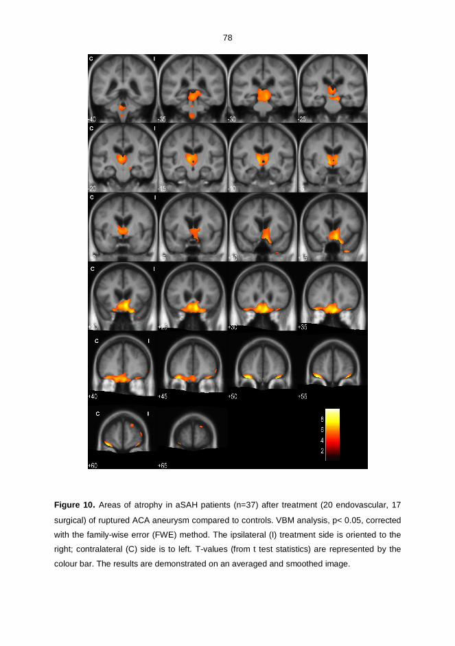
78
Figure 10. Areas of atrophy in aSAH patients (n=37) after treatment (20 endovascular, 17
surgical) of ruptured ACA aneurysm compared to controls. VBM analysis, p< 0.05, corrected
with the familywise error (FWE) method. The ipsilateral (I) treatment side is oriented to the
right; contralateral (C) side is to left. Tvalues (from t test statistics) are represented by the
colour bar. The results are demonstrated on an averaged and smoothed image.
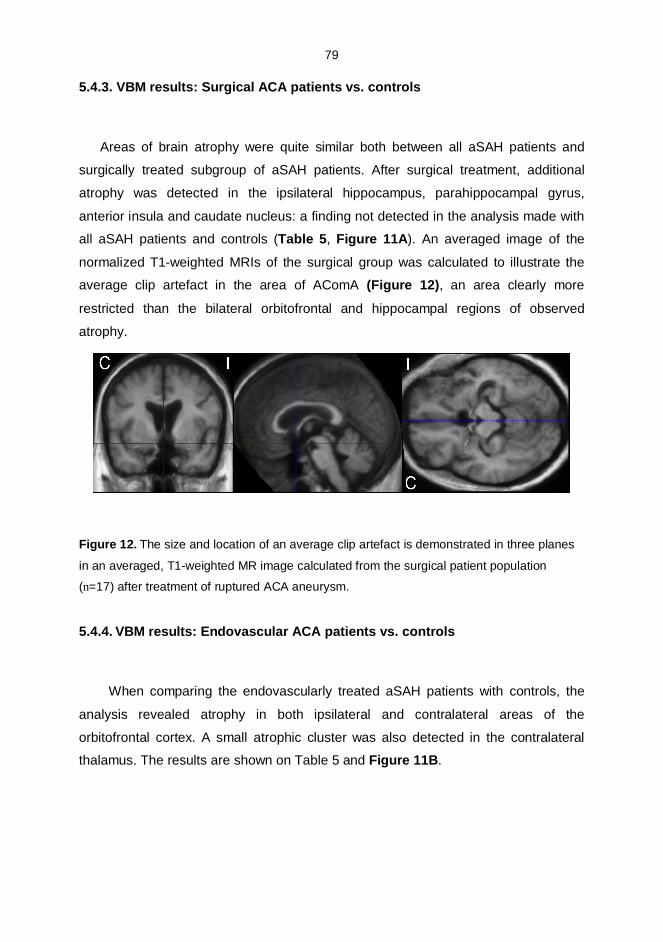
79
5.4.3. VBM results: Surgical ACA patients vs. controls
Areas of brain atrophy were quite similar both between all aSAH patients and
surgically treated subgroup of aSAH patients. After surgical treatment, additional
atrophy was detected in the ipsilateral hippocampus, parahippocampal gyrus,
anterior insula and caudate nucleus: a finding not detected in the analysis made with
all aSAH patients and controls (Table 5, Figure 11A). An averaged image of the
normalized T1weighted MRIs of the surgical group was calculated to illustrate the
average clip artefact in the area of AComA (Figure 12), an area clearly more
restricted than the bilateral orbitofrontal and hippocampal regions of observed
atrophy.
Figure 12. The size and location of an average clip artefact is demonstrated in three planes
in an averaged, T1weighted MR image calculated from the surgical patient population
(n=17) after treatment of ruptured ACA aneurysm.
5.4.4. VBM results: Endovascular ACA patients vs. controls
When comparing the endovascularly treated aSAH patients with controls, the
analysis revealed atrophy in both ipsilateral and contralateral areas of the
orbitofrontal cortex. A small atrophic cluster was also detected in the contralateral
thalamus. The results are shown on Table 5 and Figure 11B.
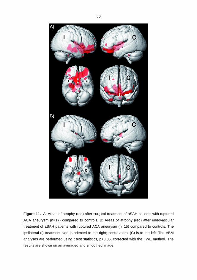
80
Figure 11. A: Areas of atrophy (red) after surgical treatment of aSAH patients with ruptured
ACA aneurysm (n=17) compared to controls. B: Areas of atrophy (red) after endovascular
treatment of aSAH patients with ruptured ACA aneurysm (n=15) compared to controls. The
ipsilateral (I) treatment side is oriented to the right; contralateral (C) is to the left. The VBM
analyses are performed using t test statistics, p<0.05, corrected with the FWE method. The
results are shown on an averaged and smoothed image.
81
5.4.5. VBM results: Endovascular ACA patients vs. surgical ACA patients
When comparing the endovascularly treated patients to the surgically treated
patients, the GM/ICV ratios did not significantly differ between treatment groups,
p=0.193, nor did the CSF/ICV ratios significantly differ between treatment groups,
p=0.249. When compared to endovascularly treated patients, VBM analysis revealed
statistically significant atrophic gray matter areas in surgically treated patients only in
the hemisphere ipsilateral to surgical approach. Areas of atrophy were detected in
the ipsilateral hippocampus and amygdala, in ipsilateral thalamic and hypothalamic
regions, and in small areas in ipsilateral insula, temporal lobe pole, middle temporal
gyrus and frontal subcentral gyrus.
5.5. Ventriculomegaly and enlargement of CSF volumes after aSAH (Study IV)
5.5.1. Patients and controls, preoperative hydrocephalus and permanent shuntdevice
The study IV was performed to compare the CSF volumes between 76
volumetric aSAH patients and 30 age and gender matched control individuals.
Preoperative hydrocephalus was detected in 16 (40.0%) out of 40 endovascular and
11 (30.6%) out of 36 surgical patients, p=0.390. A permanent shunt device was
present in four (1 endovascular, 3 surgical, p=0.255) patients. The mean interval
between aSAH and shunt operation was 4.75 ± 2.87 weeks (range 18 weeks).
5.5.2. Degree of ventriculomegaly and enlarged CSFspaces
The mCMI measurements were available in all patients from original MRI
population (n=138) and CSF/ICVs from all volumetric patients (n=76). The mCMI
proved to be higher in patients with aSAH when compared to the age and sex
matched control individuals. The more detailed analyses of the CSF volumes in 76
patients were in line with the rough planimetric measurements: the mean volume of
the ventricular system (obtained from manually drawn ROIanalysis) was 64.14 cm3
in the aSAH patients, being 31.4 % larger compared to the volume in control
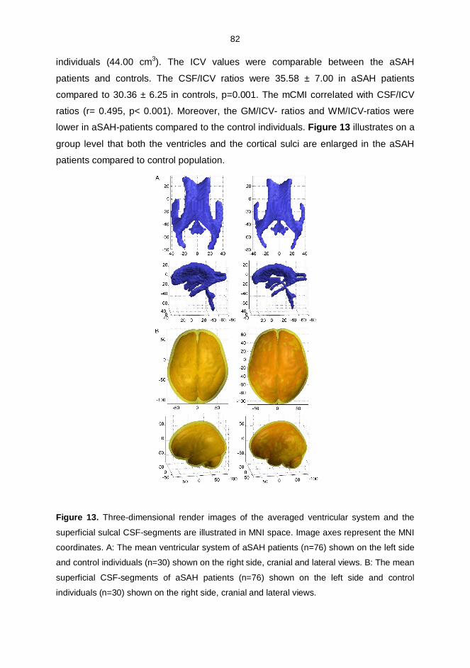
82
individuals (44.00 cm3). The ICV values were comparable between the aSAH
patients and controls. The CSF/ICV ratios were 35.58 ± 7.00 in aSAH patients
compared to 30.36 ± 6.25 in controls, p=0.001. The mCMI correlated with CSF/ICV
ratios (r= 0.495, p< 0.001). Moreover, the GM/ICV ratios and WM/ICVratios were
lower in aSAHpatients compared to the control individuals. Figure 13 illustrates on a
group level that both the ventricles and the cortical sulci are enlarged in the aSAH
patients compared to control population.
Figure 13. Threedimensional render images of the averaged ventricular system and the
superficial sulcal CSFsegments are illustrated in MNI space. Image axes represent the MNI
coordinates. A: The mean ventricular system of aSAH patients (n=76) shown on the left side
and control individuals (n=30) shown on the right side, cranial and lateral views. B: The mean
superficial CSFsegments of aSAH patients (n=76) shown on the left side and control
individuals (n=30) shown on the right side, cranial and lateral views.
83
5.5.3. Associations between ventricular and sulcal enlargement and clinicaland other radiological features
The mCMIs (n=138 aSAH patients) were comparable between the treatment
groups: 0.23 ± 0.1 after endovascular and 0.23 ± 0.1 after surgical treatment. The
treatment method did not significantly affect the measured CSF/ICV ratios (n=76);
CSF/ICV was 34.49 ± 7.7 after endovascular treatment and 36.78 ± 6.0 after surgical
treatment (p= 0.289).
At the time of MRI clinical or radiological evidence of raised intracranial pressure
could not be detected among any of the patients studied. There were no differences
in mCMI or CSF/ICV ratios when the patients with or without the permanent shunt
device were compared.
Patients with originally higher Hunt & Hess and Fisher grades showed more
pronounced mCMIs than did patients with lower Hunt & Hess and Fisher grades.
Higher Fisher grade was also associated with higher CSF/ICV ratios and patients
with preoperative hydrocephalus tended to have higher CSF/ICV ratios. Associations
between enlarged CSF spaces and contributing factors (such as preoperative
hydrocephalus, Hunt & Hess and Fisher grades) are demonstrated on Table 6.
84
Table 6. Associations of Clinical and Radiological findings with enlargement ofventricles and other CSF volumes after aSAH
Descriptive Mean mCMI p Mean CSF/ICV p
Preoperative hydrocephalus < 0.001 0.051
no (n=27) 0.21 ± 0.06 34.44 ± 7.57
yes (n=49) 0.28 ± 0.06 37.64 ± 5.35
Hunt & Hess grade < 0.001 0.293
III (n=55) 0.21 ± 0.06 34.98 ± 7.54
IIIV (n=21) 0.29 ± 0.05 37.15 ± 5.18
Fisher grade 0.001 0.014
02 (n=27) 0.20 ± 0.06 32.75 ± 7.75
34 (n=49) 0.25 ± 0.06 37.13 ± 6.10
Note. For calculation mCMI indexes and the ttest was used. mCMI refers to maximalventricular width/ maximal intracranial width ratio measured from the same axial T2weighted slice. CSF refers to CSF volume determined by SPM5. ICV= Total intracranialvolume (contains gray matter, white matter and CSF segments) measured by SPM5. ForCSF/ICV indexes the MannWhitneyUtest was used. For calculations of mCMI, n=138,and for calculations of CSF/ICVs, n=76.
5.6. Correlations of MRIdetectable findings and neuropsychological outcome(Studies IIV)
5.6.1. Neuropsychological analysis of the MRI study population (Study I)
The neuropsychological examination one year after aSAH was available in
116/138 (84.1%) patients with MRI. 17/127 (13.4%) patients with good or moderate
clinical outcome refused the neuropsychological examination and in 5/11(45.5%)
patients, who had severe neurological deficits or who were in vegetative state the
neuropsychological examination was not possible to perform. Clinical and
neuropsychological outcome between patients intended to treat endovascularly vs.
surgically (n=116) are shown in Table 7 (containing previously unpublished data).
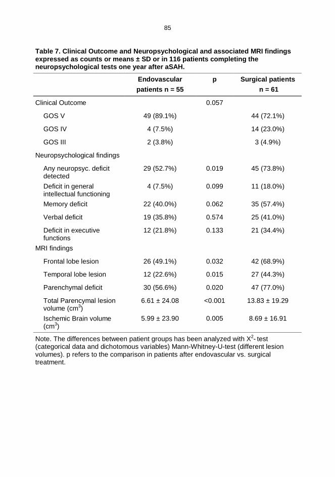
85
Table 7. Clinical Outcome and Neuropsychological and associated MRI findingsexpressed as counts or means ± SD or in 116 patients completing theneuropsychological tests one year after aSAH.
Endovascularpatients n = 55
p Surgical patients n = 61
Clinical Outcome 0.057
GOS V 49 (89.1%) 44 (72.1%)
GOS IV 4 (7.5%) 14 (23.0%)
GOS III 2 (3.8%) 3 (4.9%)
Neuropsychological findings
Any neuropsyc. deficitdetected
29 (52.7%) 0.019 45 (73.8%)
Deficit in generalintellectual functioning
4 (7.5%) 0.099 11 (18.0%)
Memory deficit 22 (40.0%) 0.062 35 (57.4%)
Verbal deficit 19 (35.8%) 0.574 25 (41.0%)
Deficit in executivefunctions
12 (21.8%) 0.133 21 (34.4%)
MRI findings
Frontal lobe lesion 26 (49.1%) 0.032 42 (68.9%)
Temporal lobe lesion 12 (22.6%) 0.015 27 (44.3%)
Parenchymal deficit 30 (56.6%) 0.020 47 (77.0%)
Total Parencymal lesionvolume (cm3)
6.61 ± 24.08 <0.001 13.83 ± 19.29
Ischemic Brain volume(cm3)
5.99 ± 23.90 0.005 8.69 ± 16.91
Note. The differences between patient groups has been analyzed with X2 test(categorical data and dichotomous variables) MannWhitneyUtest (different lesionvolumes). p refers to the comparison in patients after endovascular vs. surgicaltreatment.
86
5.6.2. Neuropsychological results and temporomesial volume correlation
(Study II)
Among the aSAH patients, age of the patient correlated with all
neuropsychological parameters. The hippocampal volumes correlated with a visual
memory test (Visual reproduction subtest (Wechsler 1974) ) and several tests of
attention, flexibility of mental processing, intellectual ability and psychomotor speed.
A test of verbal episodic memory (Wechsler 1974) and another visual memory test
(Rey) (Lezak 1995) did not correlate with hippocampal volumes. The volumes of the
amygdalae did not significantly correlate with neuropsychological test results.
Since the neurocognitive deficits noted among the volumetric aSAH patients
were quite similar to those expected in patients with frontal lobe lesions, the
correlations between the neuropsychological test results and hippocampal volumes
were analyzed separately in patients with and without a frontal lobe lesion.
Furthermore, significant correlations were found between the impaired
neuropsychological test results and reduced hippocampal volumes in both subgroups
of the patients with aSAH.
5.6.3. Neuropsychological test results in volumetric ACA population and theircorrelation to GM/ICV and CSF/ICV ratios (Study III)
Analysis of neuropsychological data revealed cognitive deficit in at least in one of
the four cognitive domains in 20 (54.1%) patients (10 endovascular, 10 surgical,
p=0.591). Impairment in executive functions was detected more often after surgical
treatment (n=8, 72.7%) than after endovascular treatment (n=3, 27.3%, p=0.033) of a
ruptured ACA aneurysm. Ten out of the 11 patients with executive deficits also had a
frontal lobe lesion that was detectable by MRI. Memory deficits were detected in
45.0% of the endovascular and 47.0% of the surgical patients. Verbal disturbances
were found in 25% of the endovascular and 29.4% of the surgical patients. No
differences among treatment groups were observed in cognitive domains other than
the executive functions (intelligence, memory, and verbal skills). The VBMbased
covariate analyses did not reveal any statistically significant specific areas of atrophy,
when the dichotomized neuropsychological deficits in general, or a dichotomized
87
executive deficits, or a dichotomized GOS, were used as a covariate. The analyses
in endovascular and surgical subgroups also failed to reveal any statistically
significant specific areas of GM atrophy that could be straightly related to these
cognitive deficits.
In patients with at least one neuropsychological deficit, the GM/ICVratios (0.354 ±
0.025) were slightly lower and the CSF/ICV ratios (0.420 ± 0.036) were slightly higher
when compared to patients with no neuropsychological deficits (GM/ ICV= 0.367 ±
0.029, ns, and CSF/ICV= 0.404 ± 0.035, ns). The patients with a deficit in executive
functions (n=11) showed lower GM/ICVratios (0.342 ± 0.015) and higher CSF/ ICV
ratios (0.433 ± 0.032) compared to patients without an executive defict (GM/ICV=
0.366 ± 0.028, p=0.014 and CSF/ICV= 0.403 ± 0.033, p=0.013).
5.6.4. Clinical outcome, neuropsychological results and CSF volume
correlation (Study IV)
GOS II IV patients had larger ventricles and CSFspaces (mCMI = 0.29 ± 0.08,
CSF/ICV= 38.19 ± 5.81) compared to GOS V patients (mCMI = 0.23 ± 0.06, p<0.001
and CSF/ICV= 35.19 ± 7.12, p=0.237).
In the group of patients with at least one neuropsychological deficit detected, the
mCMIs (0.249 ± 0.07) and CSF/ICV ratios (37.45 ± 6.13) were higher compared to
those patients without any neuropsychological deficit (mCMI=0.211 ± 0.05, p=0.003
and CSF/ICV= 32.16 ± 7.21, p=0.001). In line with the higher CSF/ICV ratios, the
aSAH patients with neuropsychological deficits showed lower GM/ICV ratios than the
aSAHpatients without neuropsychological deficits. In separate analyses of each
cognitive domain, higher mCMIs and CSF/ICV ratios were detected in the patients
with a deficit (Table 8).
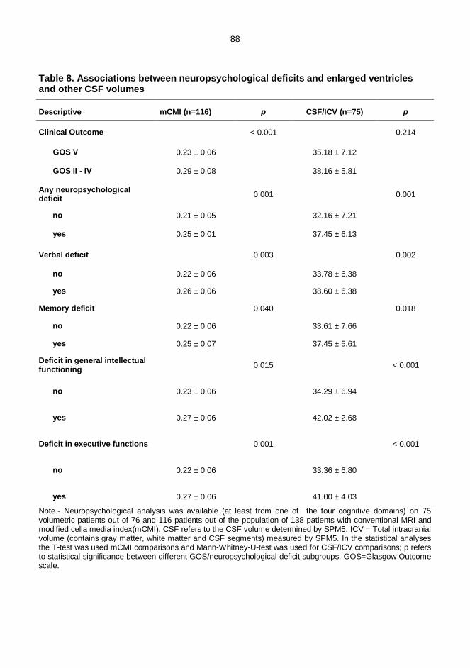
88
Table 8. Associations between neuropsychological deficits and enlarged ventriclesand other CSF volumes
Descriptive mCMI (n=116) p CSF/ICV (n=75) p
Clinical Outcome < 0.001 0.214
GOS V 0.23 ± 0.06 35.18 ± 7.12
GOS II IV 0.29 ± 0.08 38.16 ± 5.81
Any neuropsychologicaldeficit 0.001 0.001
no 0.21 ± 0.05 32.16 ± 7.21
yes 0.25 ± 0.01 37.45 ± 6.13
Verbal deficit 0.003 0.002
no 0.22 ± 0.06 33.78 ± 6.38
yes 0.26 ± 0.06 38.60 ± 6.38
Memory deficit 0.040 0.018
no 0.22 ± 0.06 33.61 ± 7.66
yes 0.25 ± 0.07 37.45 ± 5.61
Deficit in general intellectualfunctioning 0.015 < 0.001
no 0.23 ± 0.06 34.29 ± 6.94
yes 0.27 ± 0.06 42.02 ± 2.68
Deficit in executive functions 0.001 < 0.001
no 0.22 ± 0.06 33.36 ± 6.80
yes 0.27 ± 0.06 41.00 ± 4.03
Note. Neuropsychological analysis was available (at least from one of the four cognitive domains) on 75volumetric patients out of 76 and 116 patients out of the population of 138 patients with conventional MRI andmodified cella media index(mCMI). CSF refers to the CSF volume determined by SPM5. ICV = Total intracranialvolume (contains gray matter, white matter and CSF segments) measured by SPM5. In the statistical analysesthe Ttest was used mCMI comparisons and MannWhitneyUtest was used for CSF/ICV comparisons; p refersto statistical significance between different GOS/neuropsychological deficit subgroups. GOS=Glasgow Outcomescale.
89
5.7. The effect of age and MRI and neuropsychological outcome (studies I, II
and IV)
5.7.1. Age and MRI and neuropsychological deficits
There were no statistically significant correlations between the MRI detectable
lesion volumes (total parenchymal lesion volume, retraction volume or ischemic
lesion volume) and age of the patient. However, in our study population, the 44 (21
male, 23 female) patients without focal parenchymal lesions were significantly
younger (mean age 44.8 ± 2.4 years) compared to the control individuals (54.1 ± 5.5
years, p=0.006). To compare the late atrophy in aSAH patients without focal MRI
abnormalities with age and gender matched control individuals, we selected a
balanced subgroup of younger controls (n=18; 9 male, 9 female, mean age 44.0 ±
11.3), and found out that significantly higher CSF/ICV ratios were detected in aSAH
patients (31.39 ± 7.52) compared to CSF/ICV ratios in controls (26.56 ± 4.05,
p=0.027). There was no significant difference in mCMIs, when comparing the aSAH
patients without focal parenchymal deficit (mCMI=0.21 ± 0.06) and the subgroup of
younger controls (mCMI=0.19 ± 0.03, p=0.353). The age of the patient correlated
significantly with all separate neuropsychological parameters with a single exception:
the correlation coefficient between the age of a patient and memory quotient was not
found, r= 0.090, p=0.338. Patients with dichotomized neuropsychological deficit
were older (mean age 55.6 ± 13.1 years) than patients with normal cognitive profile
(mean age 42.3 ± 14.1 years, p<0.001). Furthermore, in a separate analysis age of
the patient was found to strongly associate with all of the four cognitive domains
(general intellectual functioning, memory, language and executive functions).
5.7.2. Age and temporomesial structures
In the matched control population, the following correlation coefficients between
the age of the individual and the measured temporomesial volumes were obtained:
AM right (r=0.41, p=0.03), AM left (r=0.52, p=0.004), HC right (r=0.60, p=0.001),
HC left (r=0.60, p=0.002).
In the aSAH population correlation between age and both ipsi and contralateral
hippocampal volumes was somewhat lower: HC ipsilateral (r=0.31, p=0.006) and HC
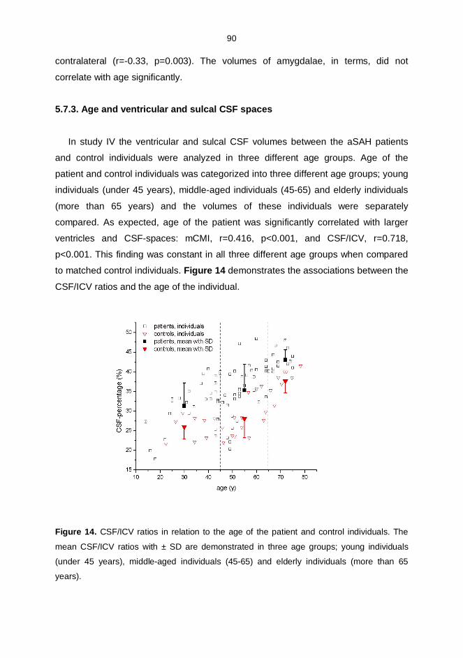
90
contralateral (r=0.33, p=0.003). The volumes of amygdalae, in terms, did not
correlate with age significantly.
5.7.3. Age and ventricular and sulcal CSF spaces
In study IV the ventricular and sulcal CSF volumes between the aSAH patients
and control individuals were analyzed in three different age groups. Age of the
patient and control individuals was categorized into three different age groups; young
individuals (under 45 years), middleaged individuals (4565) and elderly individuals
(more than 65 years) and the volumes of these individuals were separately
compared. As expected, age of the patient was significantly correlated with larger
ventricles and CSFspaces: mCMI, r=0.416, p<0.001, and CSF/ICV, r=0.718,
p<0.001. This finding was constant in all three different age groups when compared
to matched control individuals. Figure 14 demonstrates the associations between the
CSF/ICV ratios and the age of the individual.
Figure 14. CSF/ICV ratios in relation to the age of the patient and control individuals. The
mean CSF/ICV ratios with ± SD are demonstrated in three age groups; young individuals
(under 45 years), middleaged individuals (4565) and elderly individuals (more than 65
years).
91
6. DISCUSSION
6.1. Oneyear MRI Outcome after aSAH: Surgical versus Endovasculartreatment (Study I)
The past 15 years have seen a revolution in the treatment of IAs. The
development of the Guglielmi detachable coils introduced a potential alternative
treatment for IAs in certain patient populations. Although not replacing open surgery,
the continued improvements have allowed aneurysms that previously were amenable
only to open clip ligation to be safely treated with durable longterm outcomes
(Koebbe et al. 2006). The main advantage of endovascular coiling is that the
craniotomy is avoided and recovery after treatment is more rapid. The major
disadvantage is that the recanalizing of the aneurysm may occur as observed at
longterm followups (Molyneux et al. 2009, Nieuwkamp et al. 2009).
Most of the long term radiological outcome studies of the brain parenchyma after
aSAH have been performed after surgical clipping (Kivisaari et al. 2001, Romner et
al. 1989) and by using CT (Hoh et al. 2004, Orbo et al. 2008, Rabinstein et al. 2005).
The present study is the first prospective, randomized study focusing to late MRI
detectable structural brain damage after endovascular or surgical treatment of aSAH.
The study suggests a higher incidence of brain parenchymal lesions in surgically
treated patients than in endovascularly treated patients with aSAH, especially in the
frontotemporal areas. These parenchymal lesions are of clinical importance, because
their volumes clearly correlated with neuropsychological test performance. Many
lesions were surgeryrelated brain retraction injuries, but in the surgical group there
was also a significantly higher incidence of ischemic lesions in the parental artery
territory of the ruptured aneurysm than in the endovascular group. Probably the main
parental and small perforating arteries are more prone to spasm, occlusions or
thromboembolism during the surgical procedure.
In remote vascular territories, probably representing more generalized form of
global vasospasm, no difference in the incidence of ischemic lesions was found
between the two treatment modalities. It has been suggested that endovascular
treatment could carry a higher risk for vasospasm, because the blood degradation
products in the sulci might cause chemical irritation to the leptomeningeal arteries,
compared to open surgery where some of the subarachnoidal blood may be removed
(Gruber et al. 1998). In the present study, no beneficial effect of surgical clot removal
92
was observed, but instead surgical patients tended to have a higher incidence of
clinical vasospasm. However, equal frequency and size of ischemic lesions were
detected in remote vascular territories, both in cortical branch and perforator artery
territories.
Knowledge of the surgical techniques in each individual case was important
when etiologies of the parenchymal lesions were evaluated. A majority of the
retraction injury lesions were located in the basal aspect of the frontotemporal lobes.
A typical retraction injury does not follow the vascular anatomy of the brain. In the
acute phase of aSAH, the brain is generally swollen with edema, in addition to the
mass effect of the possible hematoma and hydrocephalus, making the surface of the
brain especially vulnerable.
The clinical significance of the persisting blood degradation deposits on brain
surface remains unclear, but clinical manifests such as cerebellar ataxia,
sensorineural hearing loss and myelopathy have been suggested (McCarron et al.
2003). Surprisingly, in this study signs of hematomas and superficial siderosis were
significantly more frequent than reported earlier (Kivisaari et al. 2001) and almost
equal in both treatment groups, suggesting no obvious beneficial effect of surgical
cisternal rinsing.
A study by Hadjivassilou et al. suggested that coiling might cause less structural
damage to brain parenchyma (Hadjivassiliou et al. 2001) than surgical clipping. The
International Subarachnoid Aneurysm Trial (ISAT) demonstrated that endovascular
coiling is more likely to result in independent survival than neurosurgical clipping in
good grade patients with anterior circulation aneurysms suitable for both treatment
modalities at one year after aSAH. According to ISAT study, the risk of death at 5
years was still significantly lower in coiling group than in the clipping group, but the
proportion of the survivors who were independent did not differ between the two
groups any more (83% endovascular and 82% neurosurgical) (Molyneux et al. 2009).
In the present study, the results are reported primarily according to intentionto
treat analysis. However, repeating all the analyses after censoring the patients with
combination treatment or additional treatment for nonruptured aneurysms did not
produce notable changes in the statistically significant differences between the
treatment groups. Thus, in the light of the long term MRIfindings, these results may
provide further support to other studies; showing less structural damage on the brain
parenchyma (Hadjivassiliou et al. 2001) and reduced mortality after endovascular
93
treatment compared to surgical treatment of the ruptured aneurysm (Molyneux et al.
2009). In a certain group of patients, a finding of fewer parenchymal lesions could be
a factor, in combination with other criteria, when choosing an optimal treatment
method for patients with aSAH, although complete occlusion of the aneurysm may
require additional surgical treatment in some patients.
6.2. Temporomesial volume loss after aSAH (Study II)
Hippocampus and amygdale volumes have been widely studied in various
neuropsychiatric disorders, since these structures play a vital role in processing
memory formation and stress and emotional regulation (Geuze et al. 2005). Stress,
hypoxia and increased glutamate have been associated with damage to the
hippocampus (Fujioka et al. 2000, Kalviainen et al. 1998, Villarreal et al. 2002).
Animal studies using SAH models have demonstrated profound hippocampal
neuronal loss (up to 30%) within relatively short period of time. It is believed that the
loss of hippocampal neurons occur secondary to the global ischemia, which occurs at
the time of SAH (Park et al. 2004). This is probably exacerbated by BBB breakdown
and brain edema. Hippocampal structures seem to have a more pronounced
vulnerability to global brain ischemia than the temporal lobe in general (Fujioka et al.
2000). Significantly reduced hippocampal volumes have been reported after
traumatic brain injury (TBI) and this volume reduction is not always related to the
severity of the injury (Bigler et al. 2002). In our clinical work, we have observed by
visual estimation that the temporomesial volumes seem sometimes atrophic after
aSAH, even if there are no focal parenchymal lesions on MRI.
In the present study, focusing in temporomesial volumes, it was found that aSAH
and its treatment were followed by atrophy in temporomesial structures. The AM
ipsilateral to the ruptured aneurysm was smaller in patients after surgical than
endovascular treatment. The treatment modality did not significantly affect the
measured HC volumes. Furthermore, a clear correlation was demonstrated between
neuropsychological performance and reduced temporomesial volumes. This is
interesting since, in the MRI protocol used, the volumetric sequence was scheduled
to the end of the imaging as an extra sequence, if the patient was cooperative and
willing to stay extra time in the scanner, a fact favouring the good grade patients to
be included to this volumetric study. The patients with suboptimal recovery and
94
severe neurological deficits could not participate in the longer MRI examination
protocol necessary to volumetric analysis. Thus the clinical outcome was better in
this volumetric population than in most of the aSAH populations (Hackett et al. 2000).
Therefore, the results cannot be generalized in a straightforward manner to all aSAH
patients. The damage that aSAH causes to the temporomesial structures is probably
as severe or even more pronounced in patients with poorer outcomes. However, in
the patients with poor outcome, the clinical consequences of temporomesial volume
loss would probably vanish among the major neurologic deficits, making the possible
volumetric measurements irrelevant.
In spite of the seemingly good clinical outcome in this volumetric study sample,
the temporomesial volume study clearly demonstrated neuropsychological
impairments correlating with reduced temporomesial volumes one year after aSAH.
Although the volumetric study focused on the reduced temporomesial volumes after
aSAH, two thirds of the patients also have other radiologically documented lesions on
brain MRI, most of these locating in the frontal lobes. However, the presence of a
frontal lesion was not significantly associated with the hippocampal volumes
measured. As expected, the presence of a frontal lesion was significantly associated
with worse results on the neuropsychological tests. A further analysis was made
regarding the correlations between the neuropsychological test results and
hippocampal volumes separately in patients with and without a frontal lesion.
Significant correlations were found between impaired neuropsychological test results
and reduced hippocampal volumes in both subgroups of the patients with or without
a frontal lobe lesion after aSAH, suggesting that the detected hippocampal volume
loss seems to be associated with impaired neuropsychological performance
regardless of a possible coexisting MRIdetectable lesion in frontal lobes. Those
correlations were seen in a larger number of the tests applied in patients with a
frontal lobe lesion, indicating that the effect of the hippocampal volume loss is
probably more pronounced in patients with a documented frontal lobe lesion. A
possible explanation is that the changes in hippocampal volumes are also associated
with more widespread brain changes that affect several aspects of cognition and are
not specific to memory. The relation between hippocampus and memory
performance is well established (Squire et al. 2004). In our study, the correlations
between neuropsychological test results and hippocampal volumes were somewhat
unexpected. Namely, significant correlations were consistently found between
95
hippocampal volumes and several tests of attention, flexibility of mental processing,
intellectual ability and psychomotor speed. On the other hand only one out of three
memory tests correlated with hippocampal volumes. It can be assumed assumed that
both the temporomesial volume loss and other local parenchymal lesions together
contribute to the development of neurocognitive deficits which are often detected in
patients after aSAH.
6.3. Brain atrophy after ruptured ACA aneurysm and treatment (Study III)
The ACA is a common location for cerebral aneurysms and accounts 35% of
ruptured aneurysms in recent report of Kuopio database of 1068 aSAH patients
(Huttunen et al. 2009). According to Proust et al. 80% of ACA aneurysm patients
admitted to hospital in good clinical status attain favourable clinical recovery (Proust
et al. 2003). Nevertheless, more than 60% of ruptured ACA aneurysm patients with
GOS 1 score may suffer cognitive impairment (Hutter et al. 1993). For ACA location
of the ruptured aneurysm, most studies report deficits in verbal memory, executive
functions and QOL (Bottger et al. 1998). In order to optimize the homogeneity of the
study population, the VBM study focused on patients with a good or moderate clinical
outcome and ruptured ACA aneurysms. Based on the clinical experience and visual
and quantitative analyses in conventional MRI, it was noted that especially frontal
lobe lesions are frequent in both treatment groups (Bendel et al. 2008) and therefore
it was hypothetized that on a group level a similar atrophic pattern would be observed
due to homogenous location of the ruptured aneurysm, and primary bleeding and
treatment approach (both surgical and endovascular). The VBM study elucidates the
pathophysiology behind the common cognitive disorders associated with the sequela
due to a ruptured ACA aneurysm. It was decided that analyses would be performed
separately to all patients, surgical and endovascular subgroups and their ageand
gendermatched control individuals. When patients with ruptured ACA aneurysms
were compared to agematched controls, they showed bilateral widespread atrophy
in the orbitofrontal cortex and inferior frontal gyrus, gyrus rectus and ipsilateral
caudate nucleus. The analyses also revealed atrophy in the thalamic regions and
dorsal brainstem when comparing the GM volumes between aSAH patients and
controls. Lateral ventricles are principally surrounded by white matter, however there
are also areas of central GM structures (the thalamus, caudate and brainstem nuclei)
96
around the ventricles, and thus the detected thalamic and brainstem atrophy
probably represents the more widespread central brain atrophy after aSAH, which
has been reported in previous studies (Bendel et al. 2009, Mayer et al. 2002, Vilkki et
al. 1989). In line with this finding, the aSAH patients showed significantly higher
mean relative CSF volumes and lower relative GM volumes than control individuals.
An additional finding after the surgical treatment was hippocampal atrophy ipsilateral
to treatment side, as expected according to the former volumetric study focusing to
temporomesial structures after aSAH. Atrophy could not be detected in other
temporomesial structures, perhaps due to smaller sample size in this study limited to
only volumetric ACA patients.
Detailed neuropsychological examination of the ACA study population revealed
deficits in several cognitive domains. Impaired general intellectual functions, verbal
deficits and memory disturbances were equally common after both treatment
modalities, but problems in executive functions were more frequently detected after
surgical treatment. In accordance with this finding, the MRIdetectable frontal lobe
lesions were more frequent after surgical than endovascular treatment. In a recent
study by Proust et al. neuropsychological outcome after treatment of an ACA
aneurysm was studied and an increased risk for focal encephalomalacia in the
surgical group compared to endovascular one was reported, but with no significant
difference in executive functions (Proust et al. 2009). Executive functions have long
been suspected to depend on the prefrontal cortex, the neocortex at the anterior end
of the brain (Miller 2000). Interestingly, in this study, the areas of reduced GM were
more pronounced after surgical than endovascular treatment (Figure 11). In keeping
with these neuropsychological and VBM results, recent studies suggest comparable
or even better neuropsychological outcomes (Chan et al. 2002, Fontanella et al.
2003) after endovascular than surgical treatment of the ruptured ACA aneurysms.
Although brain parenchymal lesions seen on MR images are frequent after aSAH
(Bendel et al. 2008, Kivisaari et al. 2001), especially in frontotemporal areas, it is well
known that absence of pathological findings in individuals' MR images does not
exclude the possibility of cognitive malfunctioning (Romner et al. 1989). Therefore we
suggest that these VBMassisted group comparisons might enlighten clinically
significant atrophic patterns detected in the bilateral frontobasal cortical areas and
hippocampus ipsilateral to the surgical approach after treatment of ruptured ACA
aneurysm. Perhaps these atrophic findings may be also associated with the
97
observed impaired cognitive outcome. It is notable that the atrophic areas after
treatment of ACA aneurysm were found in patients who achieved a good or at least
moderate outcome according to GOS, and thus cannot be generalized to aSAH
patients with worse clinical outcomes. However, the atrophic findings would most
likely be even more pronounced in those patients who were recovering poorly.
6.4. Atrophic enlargement of CSF volumes after aSAH (Study IV)
A recent study concluded that posttraumatic ventriculomegaly is frequently
observed in clinical practise in patients surviving from traumatic brain injury (Poca et
al. 2005). We share this clinical experience and have also detected ventricular
dilation in patients after aSAH, as has been reported in the literature (Dehdashti et al.
2004, Jartti et al. 2008, Sethi et al. 2000). Diffuse atrophic brain damage after aSAH
has been recognized in clinical practice and it was mentioned in the literature already
in 1928 (Bagley 1928).
In study IV, the ventricular and sulcal enlargement after aSAH was quantified and
related it to patients neuropsychological outcome. It is assumed that this is the first
time modern threedimensional image analysis methods were used in patients who
have recovered from aSAH. In the late MRI study, performed one year after aSAH,
all patients with clinical and radiological findings suggesting active hydrocephalus
had already been treated with a permanent shuntdevice. Moreover, there were no
differences in ventricular enlargement and sulcal dilation of CSF spaces when the
patients with or without the permanent shunt device were compared, suggesting that
patients with symptomatic hydrocephalus component had been previously
successfully treated. In the aSAH patients, the cortical sulci were also wider than
those in the control individuals (Figure 13). Therefore it can be suggested that the
ventricular dilation together with detected reduced gray and white matter volumes of
the brain, observed in the chronic phase after aSAH should be interpreted to
represent diffuse brain atrophy rather than chronic hydrocephalus.
The mechanism behind the diffuse brain atrophy after aSAH remains unclear and
it can be speculated that it cannot be ischemia only. It has been well established that
even in good grade aSAH patients, who do not exhibit clinical or radiological
vasospasm and who do not have any obvious complications with regard to surgery or
postoperative course, still show longterm psychosocial difficulties (Hutter et al. 1999,
98
Kreiter et al. 2002). Up to 50% of the survivors after aSAH never return to their
previous employment, which further indicates underdiagnosed injuries sustained at
the time of initial bleed (Hutter et al. 1999, Kreiter et al. 2002). Moreover, absence of
focal parenchymal lesions in individuals MR images does not exclude the possibility
of cognitive malfunctioning (Romner et al. 1989). Diffuse brain atrophy together with
reduced temporomesial volumes and specific atrophic patterns after treatment of
ruptured aneurysms of certain locations may partly explain this phenomenon (Bendel
et al. 2009, Bendel et al. 2006, Bendel et al. 2009). General atrophy may also
enhance the neuropsychological deficits caused by focal lesions on brain MRI.
The recent concept of early brain injury (EBI) has been suggested as the primary
cause of mortality in aSAH patients (Broderick et al. 1994) and EBI has also been
suggested to play an important role in subtle changes in behaviour, memory and
psychosocial difficulties detected in patients after aSAH (Cahill et al. 2006, Cahill et
al. 2009). EBI and a global ischemic injury at the moment of acute SAH results to
raised ICP and decreased CBF and trigger a number of critical pathways initiating
acutely after bleeding such as inflammation, hypoxia, apoptosis, oxidative stress and
excitotoxicity which all are interrelated with a similar end result, cell death (Park et al.
2004). Consequently, a number of mechanisms have been proposed to explain
global edema after aSAH. Those mechanisms include diffuse ischemic injury due to
transient ictal cerebral circulation arrest (Shigeno et al. 1982), diffuse inflammatory or
neurotoxic effects of widespread subarachnoid blood and its degradation products on
brain tissue (Germano et al. 1994), or abnormal autoregulation (Handa et al. 1992). A
recent study concluded that mild vasogenic global edema with increased ADCvalues
is a relatively common finding after aSAH in the normal appearing brain tissue (Liu et
al. 2007). It can be suggested that the diffuse brain atrophy after aSAH detected in
this study may be related to these mechanisms of EBI. In this study, higher Fischer
grades and preoperative hydrocephalus, factors also reflecting the severity of EBI,
were found to associate with increased atrophy. As expected, patients with focal
lesions detected on MR imaging showed more pronounced atrophy than those
patients without these lesions.
The current study shows that diffuse atrophic ventricular and sulcal enlargement
are common sequelae after aneurysmal subarachnoid hemorrhage. Furthermore,
enlarged CSFspaces strongly correlate with neuropsychological test performance. It
is probable that diffuse brain atrophy together with focal parenchymal lesions such as
99
cortical infarctions and retraction lesions all contribute to the development of
neurocognitive deficits in patients recovering from aSAH.
6.5. General considerations
The psychological problems faced by aSAH survivors have well been described
in the past. Even patients with less severe aSAH often have longterm problems with
memory, mood and concentration. Many aSAH survivors never return to their
previous employment, which further indicates underdiagnosed injuries probably
sustained at the time of initial bleed. The aetiology behind this phenomenon remains
partly unclear.
On a group level among aSAH patients, these results have now shown a
significant general and temporomesial atrophy, both correlating with
neuropsychological outcome of the patients. General atrophy was also detected in a
patient group without any obvious MRIdetectable lesions in the brain parenchyma.
These diffuse atrophic findings may partly be explained by an early brain injury, EBI,
a result of a number of critical, molecular pathways with a similar end result, cell
death.
MR imaging and careful analysis of the images after aSAH may help the clinician
to predict the clinical recovery potential of the individual patient together with
neurological assessment and detailed neuropsychological analysis. Focal MRI
detectable brain parenchymal lesions are common in this patient group and the
detected lesion volumes significantly correlate with the cognitive outcome.
Although the case fatality of aneurysmal aSAH has decreased with introduction of
recent improved techniques in diagnostic and management strategies, severity of the
initial bleeding remains still the most important prognostic factor for clinical outcome
of an individual aSAH patient. The only way to significantly improve the outcome of
aSAH would be preventing aSAH by diagnosing and treating aneurysms before they
rupture.
100
7. CONCLUSIONS
1. Aneurysmal subrachnoid hemorrhage (aSAH) is frequently followed by permanent
lesions in brain parenchyma, a majority of which are related to the primary
bleeding, but some are adverse effects of the treatment procedures. Parental
artery territory ischemic lesions seem to be more frequent after surgical than after
endovascular treatment of the ruptured aneurysm, especially in frontotemporal
areas. Lesions associated with surgical retraction occur in more than half of the
patients treated surgically.
2. aSAH and its treatment is followed by temporomesial volume loss that can be
detected in magnetic resonance imaging one year after aSAH. Amygdaloid
volume loss ipsilateral to the ruptured cerebral aneurysm seems to be more
pronounced after surgical than endovascular treatment of the aneurysm. The
scores of several neuropsychological measurements are correlated with the
hippocampal, but not amygdaloid volumes.
3. Gray matter atrophy, principally involving the frontobasal cortical areas and
hippocampus ipsilateral to the surgical approach is detected after aSAH and
treatment of the ruptured ACA aneurysm. Areas of reduced gray matter are more
pronounced after surgical than endovascular treatment. Together with possible
focal cortical infarctions and brain retraction deficits in individual patients, this
finding may explain the neuropsychological disturbances commonly detected after
treatment of ruptured ACA aneurysms.
4. Ventricular and sulcal enlargement together with reduced brain parenchymal
volumes are common sequelae after aSAH and indicate general atrophy rather
than hydrocephalus. Higher Fisher scores, preoperative hydrocephalus and older
age were found to associate with ventricular and sulcal enlargement. Enlarged
CSF spaces correlate with cognitive deficits after aSAH. A simple measure:
modified cella media index proved to be a feasible tool to assess the diffuse
atrophic brain damage after aSAH.
101
8. REFERENCES
Aaslid, R., Huber, P., and Nornes, H., 1984. Evaluation of cerebrovascular spasmwith transcranial Doppler ultrasound. J Neurosurg, 60(1), pp.3741.
Agid, R., Lee, S.K., Willinsky, R.A., Farb, R.I., and terBrugge, K.G., 2006. Acutesubarachnoid hemorrhage: using 64slice multidetector CT angiography to "triage"patients' treatment. Neuroradiology, 48(11), pp.787794.
Allen, G.S., Ahn, H.S., Preziosi, T.J., Battye, R., Boone, S.C., Boone, S.C., Chou,S.N., Kelly, D.L., Weir, B.K., Crabbe, R.A., Lavik, P.J., Rosenbloom, S.B., Dorsey,F.C., Ingram, C.R., Mellits, D.E., Bertsch, L.A., Boisvert, D.P., Hundley, M.B.,Johnson, R.K., Strom, J.A., and Transou, C.R., 1983. Cerebral arterial spasmacontrolled trial of nimodipine in patients with subarachnoid hemorrhage. N Engl JMed, 308(11), pp.619624.
Anderson, S.W., Todd, M.M., Hindman, B.J., Clarke, W.R., Torner, J.C., Tranel, D.,Yoo, B., Weeks, J., Manzel, K.W., and Samra, S., 2006. Effects of intraoperativehypothermia on neuropsychological outcomes after intracranial aneurysm surgery.Ann Neurol, 60(5), pp.518527.
Aralasmak, A., Akyuz, M., Ozkaynak, C., Sindel, T., and Tuncer, R., 2009. CTangiography and perfusion imaging in patients with subarachnoid hemorrhage:correlation of vasospasm to perfusion abnormality. Neuroradiology, 51(2), pp.8593.
Ashburner, J. and Friston, K.J., 2000. Voxelbased morphometrythe methods.Neuroimage, 11(6 Pt 1), pp.805821.
Ashburner, J., Hutton, C., Frackowiak, R., Johnsrude, I., Price, C., and Friston, K.,1998. Identifying global anatomical differences: deformationbased morphometry.Hum Brain Mapp, 6(56), pp.348357.
Awad, I., Modic, M., Little, J.R., Furlan, A.J., and Weinstein, M., 1986. Focalparenchymal lesions in transient ischemic attacks: correlation of computedtomography and magnetic resonance imaging. Stroke, 17(3), pp.399403.
Awad, I.A., Spetzler, R.F., Hodak, J.A., Awad, C.A., and Carey, R., 1986. Incidentalsubcortical lesions identified on magnetic resonance imaging in the elderly. I.Correlation with age and cerebrovascular risk factors. Stroke, 17(6), pp.10841089.
Bagley, 1928. Blood in the cerebrospinal fluid. resultant functional and organicalterations in the central nervous system. A. Experimental data. Arch Surg, 17, pp.1838.
Bailes, J.E., Spetzler, R.F., Hadley, M.N., and Baldwin, H.Z., 1990. Managementmorbidity and mortality of poorgrade aneurysm patients. J Neurosurg, 72(4), pp.559566.
Beauchamp, N.J., Jr., Ulug, A.M., Passe, T.J., and van Zijl, P.C., 1998. MR diffusionimaging in stroke: review and controversies. Radiographics, 18(5), pp.12691283;discussion 12831265.
102
Bederson, J.B., Awad, I.A., Wiebers, D.O., Piepgras, D., Haley, E.C., Jr., Brott, T.,Hademenos, G., Chyatte, D., Rosenwasser, R., and Caroselli, C., 2000.Recommendations for the management of patients with unruptured intracranialaneurysms: A Statement for healthcare professionals from the Stroke Council of theAmerican Heart Association. Stroke, 31(11), pp.27422750.
Bederson, J.B., Connolly, E.S., Jr., Batjer, H.H., Dacey, R.G., Dion, J.E., Diringer,M.N., Duldner, J.E., Jr., Harbaugh, R.E., Patel, A.B., and Rosenwasser, R.H., 2009.Guidelines for the management of aneurysmal subarachnoid hemorrhage: astatement for healthcare professionals from a special writing group of the StrokeCouncil, American Heart Association. Stroke, 40(3), pp.9941025.
Bell, B.A., Kendall, B.E., and Symon, L., 1980. Computed tomography in aneurysmalsubarachnoid haemorrhage. J Neurol Neurosurg Psychiatry, 43(6), pp.522524.
Bellebaum, C., Schafers, L., Schoch, B., Wanke, I., Stolke, D., Forsting, M., andDaum, I., 2004. Clipping versus coiling: neuropsychological follow up afteraneurysmal subarachnoid haemorrhage (SAH). J Clin Exp Neuropsychol, 26(8),pp.10811092.
Bendel, P., Koivisto, T., Aikia, M., Niskanen, E., Kononen, M., Hanninen, T., andVanninen, R., 2009. Atrophic Enlargement of CSF Volume after SubarachnoidHemorrhage: Correlation with Neuropsychological Outcome. AJNR Am J Neuroradiol,Epub ahead of print, Nov 26.
Bendel, P., Koivisto, T., Hanninen, T., Kolehmainen, A., Kononen, M., Hurskainen,H., Pennanen, C., and Vanninen, R., 2006. Subarachnoid hemorrhage is followed bytemporomesial volume loss: MRI volumetric study. Neurology, 67(4), pp.575582.
Bendel, P., Koivisto, T., Kononen, M., Hanninen, T., Hurskainen, H., Saari, T.,Vapalahti, M., Hernesniemi, J., and Vanninen, R., 2008. MR imaging of the brain 1year after aneurysmal subarachnoid hemorrhage: randomized study comparingsurgical with endovascular treatment. Radiology, 246(2), pp.543552.
Bendel, P., Koivisto, T., Niskanen, E., Kononen, M., Aikia, M., Hanninen, T.,Koskenkorva, P., and Vanninen, R., 2009. Brain atrophy and neuropsychologicaloutcome after treatment of ruptured anterior cerebral artery aneurysms: a voxelbased morphometric study. Neuroradiology, 51(11), pp.711722.
Bigler, E.D., Anderson, C.V., Blatter, D.D., and Andersob, C.V., 2002. Temporal lobemorphology in normal aging and traumatic brain injury. AJNR Am J Neuroradiol,23(2), pp.255266.
Binaghi, S., Colleoni, M.L., Maeder, P., Uske, A., Regli, L., Dehdashti, A.R.,Schnyder, P., and Meuli, R., 2007. CT angiography and perfusion CT in cerebralvasospasm after subarachnoid hemorrhage. AJNR Am J Neuroradiol, 28(4), pp.750758.
Borkowski, J., Benton, A., and Spreen, O., 1967. Word fluency and brain damage.Neuropsychologia, 5, pp.135140.
Bornstein, R.A., Weir, B.K., Petruk, K.C., and Disney, L.B., 1987. Neuropsychologicalfunction in patients after subarachnoid hemorrhage. Neurosurgery, 21(5), pp.651654.
103
Bottger, S., Prosiegel, M., Steiger, H.J., and Yassouridis, A., 1998. Neurobehaviouraldisturbances, rehabilitation outcome, and lesion site in patients after rupture andrepair of anterior communicating artery aneurysm. J Neurol Neurosurg Psychiatry,65(1), pp.93102.
Breteler, M.M., van Swieten, J.C., Bots, M.L., Grobbee, D.E., Claus, J.J., van denHout, J.H., van Harskamp, F., Tanghe, H.L., de Jong, P.T., van Gijn, J., and et al.,1994. Cerebral white matter lesions, vascular risk factors, and cognitive function in apopulationbased study: the Rotterdam Study. Neurology, 44(7), pp.12461252.
Brilstra, E.H., Rinkel, G.J., van der Graaf, Y., van Rooij, W.J., and Algra, A., 1999.Treatment of intracranial aneurysms by embolization with coils: a systematic review.Stroke, 30(2), pp.470476.
Broderick, J.P., Brott, T.G., Duldner, J.E., Tomsick, T., and Leach, A., 1994. Initialand recurrent bleeding are the major causes of death following subarachnoidhemorrhage. Stroke, 25(7), pp.13421347.
Brooks, R., 1996. EuroQol: the current state of play. Health Policy, 37(1), pp.5372.
Brooks, R., 1996. Quality of life measures. Crit Care Med, 24(10), pp.1769.
Brouwers, P.J., Wijdicks, E.F., and Van Gijn, J., 1992. Infarction after aneurysmrupture does not depend on distribution or clearance rate of blood. Stroke, 23(3),pp.374379.
Buchanan, K.M., Elias, L.J., and Goplen, G.B., 2000. Differing perspectives onoutcome after subarachnoid hemorrhage: the patient, the relative, the neurosurgeon.Neurosurgery, 46(4), pp.831838; discussion 838840.
Busch, E., Beaulieu, C., de Crespigny, A., and Moseley, M.E., 1998. Diffusion MRimaging during acute subarachnoid hemorrhage in rats. Stroke, 29(10), pp.21552161.
Cahill, J., Calvert, J.W., Solaroglu, I., and Zhang, J.H., 2006. Vasospasm and p53induced apoptosis in an experimental model of subarachnoid hemorrhage. Stroke,37(7), pp.18681874.
Cahill, J., Calvert, J.W., and Zhang, J.H., 2006. Mechanisms of early brain injury aftersubarachnoid hemorrhage. J Cereb Blood Flow Metab, 26(11), pp.13411353.
Cahill, J. and Zhang, J.H., 2009. Subarachnoid hemorrhage: is it time for a newdirection? Stroke, 40(3 Suppl), pp.S8687.
Carlson, A.P. and Yonas, H., 2009. Radiographic assessment of vasospasm afteraneurysmal subarachnoid hemorrhage: the physiological perspective. Neurol Res,31(6), pp.593604.
Carrera, E., Schmidt, J.M., Oddo, M., Fernandez, L., Claassen, J., Seder, D., Lee, K.,Badjatia, N., Connolly, E.S., Jr., and Mayer, S.A., 2009. Transcranial Doppler forpredicting delayed cerebral ischemia after subarachnoid hemorrhage. Neurosurgery,65(2), pp.316323; discussion 323314.
104
Chan, A., Ho, S., and Poon, W.S., 2002. Neuropsychological sequelae of patientstreated with microsurgical clipping or endovascular embolization for anteriorcommunicating artery aneurysm. Eur Neurol, 47(1), pp.3744.
Chang, H.S., Hongo, K., and Nakagawa, H., 2000. Adverse effects of limitedhypotensive anesthesia on the outcome of patients with subarachnoid hemorrhage. JNeurosurg, 92(6), pp.971975.
Chen, W., Yang, Y., Qiu, J., Peng, Y., and Xing, W., 2009. Clinical application of 16row multislice computed tomographic angiography in the preoperative andpostoperative evaluation of intracranial aneurysms for surgical clipping. Surg Neurol,71(5), pp.559565.
Choudhari, K.A., Ramachandran, M.S., McCarron, M.O., and Kaliaperumal, C., 2007.Aneurysms unsuitable for endovascular intervention: surgical outcome andmanagement challenges over a 5year period following International SubarachnoidHaemorrhage Trial (ISAT). Clin Neurol Neurosurg, 109(10), pp.868875.
Ciumas, C., Montavont, A., and Ryvlin, P., 2008. Magnetic resonance imaging inclinical trials. Curr Opin Neurol, 21(4), pp.431436.
Cloft, H.J., Joseph, G.J., and Dion, J.E., 1999. Risk of cerebral angiography inpatients with subarachnoid hemorrhage, cerebral aneurysm, and arteriovenousmalformation: a metaanalysis. Stroke, 30(2), pp.317320.
CondetteAuliac, S., Bracard, S., Anxionnat, R., Schmitt, E., Lacour, J.C., Braun, M.,Meloneto, J., Cordebar, A., Yin, L., and Picard, L., 2001. Vasospasm aftersubarachnoid hemorrhage: interest in diffusionweighted MR imaging. Stroke, 32(8),pp.18181824.
Curnes, J.T., Shogry, M.E., Clark, D.C., and Elsner, H.J., 1993. MR angiographicdemonstration of an intracranial aneurysm not seen on conventional angiography.AJNR Am J Neuroradiol, 14(4), pp.971973.
Dandy, W.E., 1938. Intracranial Aneurysm of the Internal Carotid Artery: Cured byOperation. Ann Surg, 107(5), pp.654659.
Dankbaar, J.W., Rijsdijk, M., van der Schaaf, I.C., Velthuis, B.K., Wermer, M.J., andRinkel, G.J., 2009. Relationship between vasospasm, cerebral perfusion, anddelayed cerebral ischemia after aneurysmal subarachnoid hemorrhage.Neuroradiology, 51(12), pp.813819.
de Rooij, N.K., Linn, F.H., van der Plas, J.A., Algra, A., and Rinkel, G.J., 2007.Incidence of subarachnoid haemorrhage: a systematic review with emphasis onregion, age, gender and time trends. J Neurol Neurosurg Psychiatry, 78(12), pp.13651372.
Dehdashti, A.R., Rilliet, B., Rufenacht, D.A., and de Tribolet, N., 2004. Shuntdependent hydrocephalus after rupture of intracranial aneurysms: a prospective studyof the influence of treatment modality. J Neurosurg, 101(3), pp.402407.
Dion, J.E., Gates, P.C., Fox, A.J., Barnett, H.J., and Blom, R.J., 1987. Clinical eventsfollowing neuroangiography: a prospective study. Stroke, 18(6), pp.9971004.
105
Doczi, T., 1985. The pathogenetic and prognostic significance of bloodbrain barrierdamage at the acute stage of aneurysmal subarachnoid haemorrhage. Clinical andexperimental studies. Acta Neurochir (Wien), 77(34), pp.110132.
Doczi, T.P., 2001. Impact of cerebral microcirculatory changes on cerebral blood flowduring cerebral vasospasm after aneurysmal subarachnoid hemorrhage. Stroke,32(3), pp.817.
Dorai, Z., Hynan, L.S., Kopitnik, T.A., and Samson, D., 2003. Factors related tohydrocephalus after aneurysmal subarachnoid hemorrhage. Neurosurgery, 52(4),pp.763769; discussion 769771.
Dreier, J.P., Sakowitz, O.W., Harder, A., Zimmer, C., Dirnagl, U., Valdueza, J.M., andUnterberg, A.W., 2002. Focal laminar cortical MR signal abnormalities aftersubarachnoid hemorrhage. Ann Neurol, 52(6), pp.825829.
Duvernoy, H., 1999. The Human Brain Surface, Blood Supply, and Threedimensional Sectional Anatomy. 2 nd edition, SpringerVerlag Wien New York.
Edelman, R.R. and Warach, S., 1993. Magnetic resonance imaging (1). N Engl JMed, 328(10), pp.708716.
Egge, A., Waterloo, K., Sjoholm, H., Ingebrigtsen, T., Forsdahl, S., Jacobsen, E.A.,and Romner, B., 2005. Outcome 1 year after aneurysmal subarachnoid hemorrhage:relation between cognitive performance and neuroimaging. Acta Neurol Scand,112(2), pp.7680.
Fazekas, F., Kleinert, R., Offenbacher, H., Payer, F., Schmidt, R., Kleinert, G.,Radner, H., and Lechner, H., 1991. The morphologic correlate of incidental punctatewhite matter hyperintensities on MR images. AJNR Am J Neuroradiol, 12(5), pp.915921.
Feigin, V., Parag, V., Lawes, C.M., Rodgers, A., Suh, I., Woodward, M., Jamrozik, K.,and Ueshima, H., 2005. Smoking and elevated blood pressure are the most importantrisk factors for subarachnoid hemorrhage in the AsiaPacific region: an overview of 26cohorts involving 306,620 participants. Stroke, 36(7), pp.13601365.
Feigin, V.L., Rinkel, G.J., Lawes, C.M., Algra, A., Bennett, D.A., van Gijn, J., andAnderson, C.S., 2005. Risk factors for subarachnoid hemorrhage: an updatedsystematic review of epidemiological studies. Stroke, 36(12), pp.27732780.
Fergusen, S. and Macdonald, R.L., 2007. Predictors of cerebral infarction in patientswith aneurysmal subarachnoid hemorrhage. Neurosurgery, 60(4), pp.658667;discussion 667.
Fiebach, J.B., Schellinger, P.D., Gass, A., Kucinski, T., Siebler, M., Villringer, A.,Olkers, P., Hirsch, J.G., Heiland, S., Wilde, P., Jansen, O., Rother, J., Hacke, W., andSartor, K., 2004. Stroke magnetic resonance imaging is accurate in hyperacuteintracerebral hemorrhage: a multicenter study on the validity of stroke imaging.Stroke, 35(2), pp.502506.
Figueiredo, E.G., Zabramski, J.M., Deshmukh, P., Crawford, N.R., Spetzler, R.F., andPreul, M.C., 2006. Comparative analysis of anterior petrosectomy andtranscavernous approaches to retrosellar and upper clival basilar artery aneurysms.Neurosurgery, 58(1 Suppl), pp.ONS1321; discussion ONS1321.
106
Fisher, C.M., Kistler, J.P., and Davis, J.M., 1980. Relation of cerebral vasospasm tosubarachnoid hemorrhage visualized by computerized tomographic scanning.Neurosurgery, 6(1), pp.19.
Fisher, C.M., Roberson, G.H., and Ojemann, R.G., 1977. Cerebral vasospasm withruptured saccular aneurysmthe clinical manifestations. Neurosurgery, 1(3), pp.245248.
Fogelholm, R., 1981. Subarachnoid hemorrhage in middleFinland: incidence, earlyprognosis and indications for neurosurgical treatment. Stroke, 12(3), pp.296301.
Fontanella, M., Perozzo, P., Ursone, R., Garbossa, D., and Bergui, M., 2003.Neuropsychological assessment after microsurgical clipping or endovasculartreatment for anterior communicating artery aneurysm. Acta Neurochir (Wien),145(10), pp.867872; discussion 872.
Friedman, J.A., Goerss, S.J., Meyer, F.B., Piepgras, D.G., Pichelmann, M.A., McIver,J.I., Toussaint, L.G., 3rd, McClelland, R.L., Nichols, D.A., Atkinson, J.L., and Wijdicks,E.F., 2002. Volumetric quantification of Fisher Grade 3 aneurysmal subarachnoidhemorrhage: a novel method to predict symptomatic vasospasm on admissioncomputerized tomography scans. J Neurosurg, 97(2), pp.401407.
Frosen, J., Piippo, A., Paetau, A., Kangasniemi, M., Niemela, M., Hernesniemi, J.,and Jaaskelainen, J., 2004. Remodeling of saccular cerebral artery aneurysm wall isassociated with rupture: histological analysis of 24 unruptured and 42 ruptured cases.Stroke, 35(10), pp.22872293.
Frosen, J., Piippo, A., Paetau, A., Kangasniemi, M., Niemela, M., Hernesniemi, J.,and Jaaskelainen, J., 2006. Growth factor receptor expression and remodeling ofsaccular cerebral artery aneurysm walls: implications for biological therapy preventingrupture. Neurosurgery, 58(3), pp.534541; discussion 534541.
Fujioka, M., Nishio, K., Miyamoto, S., Hiramatsu, K.I., Sakaki, T., Okuchi, K., Taoka,T., and Fujioka, S., 2000. Hippocampal damage in the human brain after cardiacarrest. Cerebrovasc Dis, 10(1), pp.27.
Gaser, C., Nenadic, I., Buchsbaum, B.R., Hazlett, E.A., and Buchsbaum, M.S., 2001.Deformationbased morphometry and its relation to conventional volumetry of brainlateral ventricles in MRI. Neuroimage, 13(6 Pt 1), pp.11401145.
Genovese, C.R., Lazar, N.A., and Nichols, T., 2002. Thresholding of statistical mapsin functional neuroimaging using the false discovery rate. Neuroimage, 15(4), pp.870878.
Gerlach, R., Beck, J., Setzer, M., Vatter, H., Berkefeld, J., Du Mesnil de Rochemont,R., Raabe, A., and Seifert, V., 2007. Treatment related morbidity of unrupturedintracranial aneurysms: results of a prospective single centre series with aninterdisciplinary approach over a 6 year period (19992005). J Neurol NeurosurgPsychiatry, 78(8), pp.864871.
Germano, A.F., Dixon, C.E., d'Avella, D., Hayes, R.L., and Tomasello, F., 1994.Behavioral deficits following experimental subarachnoid hemorrhage in the rat. JNeurotrauma, 11(3), pp.345353.
107
Geuze, E., Vermetten, E., and Bremner, J.D., 2005. MRbased in vivo hippocampalvolumetrics: 2. Findings in neuropsychiatric disorders. Mol Psychiatry, 10(2), pp.160184.
Gibbs, G.F., Huston, J., 3rd, Bernstein, M.A., Riederer, S.J., and Brown, R.D., Jr.,2004. Improved image quality of intracranial aneurysms: 3.0T versus 1.5T timeofflight MR angiography. AJNR Am J Neuroradiol, 25(1), pp.8487.
Gibbs, G.F., Huston, J., 3rd, Bernstein, M.A., Riederer, S.J., and Brown, R.D., Jr.,2005. 3.0Tesla MR angiography of intracranial aneurysms: comparison of timeofflight and contrastenhanced techniques. J Magn Reson Imaging, 21(2), pp.97102.
Gieteling, E.W. and Rinkel, G.J., 2003. Characteristics of intracranial aneurysms andsubarachnoid haemorrhage in patients with polycystic kidney disease. J Neurol,250(4), pp.418423.
Gilman, S., 1998. Imaging the brain. First of two parts. N Engl J Med, 338(12),pp.812820.
Golden, C., 1978. Stroop Color and Word Tests. Chigaco, Ill.: Stoerting.
Gomori, J.M., Grossman, R.I., and Steiner, I., 1988. Highfield magnetic resonanceimaging of intracranial hematomas. Isr J Med Sci, 24(45), pp.218223.
Good, C.D., Ashburner, J., and Frackowiak, R.S., 2001. Computationalneuroanatomy: new perspectives for neuroradiology. Rev Neurol (Paris), 157(89 Pt1), pp.797806.
Good, C.D., Johnsrude, I.S., Ashburner, J., Henson, R.N., Friston, K.J., andFrackowiak, R.S., 2001. A voxelbased morphometric study of ageing in 465 normaladult human brains. Neuroimage, 14(1 Pt 1), pp.2136.
Greebe, P. and Rinkel, G.J., 2007. Life expectancy after perimesencephalicsubarachnoid hemorrhage. Stroke, 38(4), pp.12221224.
Gruber, A., Reinprecht, A., Bavinzski, G., Czech, T., and Richling, B., 1999. Chronicshuntdependent hydrocephalus after early surgical and early endovascular treatmentof ruptured intracranial aneurysms. Neurosurgery, 44(3), pp.503509; discussion 509512.
Gruber, A., Ungersbock, K., Reinprecht, A., Czech, T., Gross, C., Bednar, M., andRichling, B., 1998. Evaluation of cerebral vasospasm after early surgical andendovascular treatment of ruptured intracranial aneurysms. Neurosurgery, 42(2),pp.258267; discussion 267258.
Guglielmi, G., Vinuela, F., Dion, J., and Duckwiler, G., 1991. Electrothrombosis ofsaccular aneurysms via endovascular approach. Part 2: Preliminary clinicalexperience. J Neurosurg, 75(1), pp.814.
Guglielmi, G., Vinuela, F., Duckwiler, G., Dion, J., Lylyk, P., Berenstein, A., Strother,C., Graves, V., Halbach, V., Nichols, D., and et al., 1992. Endovascular treatment ofposterior circulation aneurysms by electrothrombosis using electrically detachablecoils. J Neurosurg, 77(4), pp.515524.
108
Guglielmi, G., Vinuela, F., Sepetka, I., and Macellari, V., 1991. Electrothrombosis ofsaccular aneurysms via endovascular approach. Part 1: Electrochemical basis,technique, and experimental results. J Neurosurg, 75(1), pp.17.
Hackett, M.L. and Anderson, C.S., 2000. Health outcomes 1 year after subarachnoidhemorrhage: An international populationbased study. The Australian CooperativeResearch on Subarachnoid Hemorrhage Study Group. Neurology, 55(5), pp.658662.
Hadjivassiliou, M., Tooth, C.L., Romanowski, C.A., Byrne, J., Battersby, R.D.,Oxbury, S., Crewswell, C.S., Burkitt, E., Stokes, N.A., Paul, C., Mayes, A.R., andSagar, H.J., 2001. Aneurysmal SAH: cognitive outcome and structural damage afterclipping or coiling. Neurology, 56(12), pp.16721677.
Handa, Y., Hayashi, M., Takeuchi, H., Kubota, T., Kobayashi, H., and Kawano, H.,1992. Time course of the impairment of cerebral autoregulation during chroniccerebral vasospasm after subarachnoid hemorrhage in primates. J Neurosurg, 76(3),pp.493501.
Hattingen, E., Blasel, S., Dettmann, E., Vatter, H., Pilatus, U., Seifert, V., Zanella,F.E., and Weidauer, S., 2008. Perfusionweighted MRI to evaluate cerebralautoregulation in aneurysmal subarachnoid haemorrhage. Neuroradiology, 50(11),pp.929938.
Hauerberg, J., Eskesen, V., and Rosenorn, J., 1994. The prognostic significance ofintracerebral haematoma as shown on CT scanning after aneurysmal subarachnoidhaemorrhage. Br J Neurosurg, 8(3), pp.333339.
Heier, L.A., Bauer, C.J., Schwartz, L., Zimmerman, R.D., Morgello, S., and Deck,M.D., 1989. Large VirchowRobin spaces: MRclinical correlation. AJNR Am JNeuroradiol, 10(5), pp.929936.
Heiserman, J.E., Dean, B.L., Hodak, J.A., Flom, R.A., Bird, C.R., Drayer, B.P., andFram, E.K., 1994. Neurologic complications of cerebral angiography. AJNR Am JNeuroradiol, 15(8), pp.14011407; discussion 14081411.
Hernesniemi, J., Dashti, R., Lehecka, M., Niemela, M., Rinne, J., Lehto, H.,Ronkainen, A., Koivisto, T., and Jaaskelainen, J.E., 2008. Microneurosurgicalmanagement of anterior communicating artery aneurysms. Surg Neurol, 70(1), pp.828; discussion 29.
Hernesniemi, J., Ishii, K., Niemela, M., Kivipelto, L., Fujiki, M., and Shen, H., 2005.Subtemporal approach to basilar bifurcation aneurysms: advanced technique andclinical experience. Acta Neurochir Suppl, 94, pp.3138.
Hernesniemi, J., Ishii, K., Niemela, M., Smrcka, M., Kivipelto, L., Fujiki, M., and Shen,H., 2005. Lateral supraorbital approach as an alternative to the classical pterionalapproach. Acta Neurochir Suppl, 94. pp.1721.
Hernesniemi, J., Vapalahti, M., Niskanen, M., Tapaninaho, A., Kari, A., Luukkonen,M., Puranen, M., Saari, T., and Rajpar, M., 1993. Oneyear outcome in earlyaneurysm surgery: a 14 years experience. Acta Neurochir (Wien), 122(12), pp.110.
Hijdra, A., Braakman, R., van Gijn, J., Vermeulen, M., and van Crevel, H., 1987.Aneurysmal subarachnoid hemorrhage. Complications and outcome in a hospitalpopulation. Stroke, 18(6), pp.10611067.
109
Hijdra, A., Van Gijn, J., Stefanko, S., Van Dongen, K.J., Vermeulen, M., and VanCrevel, H., 1986. Delayed cerebral ischemia after aneurysmal subarachnoidhemorrhage: clinicoanatomic correlations. Neurology, 36(3), pp.329333.
Hirai, T., Kai, Y., Morioka, M., Yano, S., Kitajima, M., Fukuoka, H., Sasao, A.,Murakami, R., Nakayama, Y., Awai, K., Toya, R., Akter, M., Korogi, Y., Kuratsu, J.,and Yamashita, Y., 2008. Differentiation between paraclinoid and cavernous sinusaneurysms with contrastenhanced 3D constructive interference in steady state MRimaging. AJNR Am J Neuroradiol, 29(1), pp.130133.
Hoeffner, E.G., Case, I., Jain, R., Gujar, S.K., Shah, G.V., Deveikis, J.P., Carlos,R.C., Thompson, B.G., Harrigan, M.R., and Mukherji, S.K., 2004. Cerebral perfusionCT: technique and clinical applications. Radiology, 231(3), pp.632644.
Hoh, B.L., Curry, W.T., Jr., Carter, B.S., and Ogilvy, C.S., 2004. Computedtomographic demonstrated infarcts after surgical and endovascular treatment ofaneurysmal subarachnoid hemorrhage. Acta Neurochir (Wien), 146(11), pp.11771183.
Hoh, B.L. and Ogilvy, C.S., 2005. Endovascular treatment of cerebral vasospasm:transluminal balloon angioplasty, intraarterial papaverine, and intraarterialnicardipine. Neurosurg Clin N Am, 16(3), pp.501516, vi.
Hop, J.W., Rinkel, G.J., Algra, A., and van Gijn, J., 1997. Casefatality rates andfunctional outcome after subarachnoid hemorrhage: a systematic review. Stroke,28(3), pp.660664.
Hop, J.W., Rinkel, G.J., Algra, A., and van Gijn, J., 1998. Quality of life in patients andpartners after aneurysmal subarachnoid hemorrhage. Stroke, 29(4), pp.798804.
Hop, J.W., Rinkel, G.J., Algra, A., and van Gijn, J., 1999. Initial loss of consciousnessand risk of delayed cerebral ischemia after aneurysmal subarachnoid hemorrhage.Stroke, 30(11), pp.22682271.
Hop, J.W., Rinkel, G.J., Algra, A., and van Gijn, J., 2001. Changes in functionaloutcome and quality of life in patients and caregivers after aneurysmal subarachnoidhemorrhage. J Neurosurg, 95(6), pp.957963.
Horn, P., Vajkoczy, P., Bauhuf, C., Munch, E., PoecklerSchoeniger, C., andSchmiedek, P., 2001. Quantitative regional cerebral blood flow measurementtechniques improve noninvasive detection of cerebrovascular vasospasm afteraneurysmal subarachnoid hemorrhage. Cerebrovasc Dis, 12(3), pp.197202.
Hunt, W.E., Meagher, J.N., and Hess, R.M., 1966. Intracranial aneurysm. A nineyearstudy. Ohio State Med J, 62(11), pp.11681171.
Hutter, B.O. and Gilsbach, J.M., 1993. Which neuropsychological deficits are hiddenbehind a good outcome (Glasgow = I) after aneurysmal subarachnoid hemorrhage?Neurosurgery, 33(6), pp.9991005; discussion 10051006.
Hutter, B.O., KreitschmannAndermahr, I., and Gilsbach, J.M., 2001. Healthrelatedquality of life after aneurysmal subarachnoid hemorrhage: impacts of bleedingseverity, computerized tomography findings, surgery, vasospasm, and neurologicalgrade. J Neurosurg, 94(2), pp.241251.
110
Hutter, B.O., KreitschmannAndermahr, I., Mayfrank, L., Rohde, V., Spetzger, U., andGilsbach, J.M., 1999. Functional outcome after aneurysmal subarachnoidhemorrhage. Acta Neurochir Suppl, 72, pp.157174.
Huttunen, T., von und zu Franunberg, M., Frösen, J., Lehecka, M., Tromp, G., Helin,K., Koivisto,T., Rinne, J., Ronkainen, A., Hernesniemi, J., Jääskeläinen, J., 2009.Saccular intracranial aneurysm disease – distribution of site, size and age suggestdifferent etiologies for aneurysm formation and rupture in 316 familial and 1454sporadic Eastern Finnish patients. In press.
Ikeda, K., Iwasaki, Y., Hatanaka, N., and Kinoshita, M., 1998. Causes ofperimesencephalic nonaneurysmal subarachnoid hemorrhage. Neurology, 50(5),pp.1518.
Imaizumi, T., Chiba, M., Honma, T., and Niwa, J., 2003. Detection of hemosiderindeposition by T2*weighted MRI after subarachnoid hemorrhage. Stroke, 34(7),pp.16931698.
Inagawa, T., Tokuda, Y., Ohbayashi, N., Takaya, M., and Moritake, K., 1995. Study ofaneurysmal subarachnoid hemorrhage in Izumo City, Japan. Stroke, 26(5), pp.761766.
Insausti, R., Juottonen, K., Soininen, H., Insausti, A.M., Partanen, K., Vainio, P.,Laakso, M.P., and Pitkanen, A., 1998. MR volumetric analysis of the humanentorhinal, perirhinal, and temporopolar cortices. AJNR Am J Neuroradiol, 19(4),pp.659671.
Investigators, 1998. Unruptured intracranial aneurysmsrisk of rupture and risks ofsurgical intervention. International Study of Unruptured Intracranial AneurysmsInvestigators. N Engl J Med, 339(24), pp.17251733.
Isaksen, J., Egge, A., Waterloo, K., Romner, B., and Ingebrigtsen, T., 2002. Riskfactors for aneurysmal subarachnoid haemorrhage: the Tromso study. J NeurolNeurosurg Psychiatry, 73(2), pp.185187.
Jartti, P., Karttunen, A., Isokangas, J.M., Jartti, A., Koskelainen, T., and Tervonen, O.,2008. Chronic hydrocephalus after neurosurgical and endovascular treatment ofruptured intracranial aneurysms. Acta Radiol, 49(6), pp.680686.
Jennett, B. and Bond, M., 1975. Assessment of outcome after severe brain damage.Lancet, 1(7905), pp.480484.
Johnston, S.C., Selvin, S., and Gress, D.R., 1998. The burden, trends, anddemographics of mortality from subarachnoid hemorrhage. Neurology, 50(5),pp.14131418.
Jokinen, H., Kalska, H., Mantyla, R., Pohjasvaara, T., Ylikoski, R., Hietanen, M.,Salonen, O., Kaste, M., and Erkinjuntti, T., 2006. Cognitive profile of subcorticalischaemic vascular disease. J Neurol Neurosurg Psychiatry, 77(1), pp.2833.
Juvela, S., Hillbom, M., Numminen, H., and Koskinen, P., 1993. Cigarette smokingand alcohol consumption as risk factors for aneurysmal subarachnoid hemorrhage.Stroke, 24(5), pp.639646.
111
Juvela, S., Porras, M., and Poussa, K., 2000. Natural history of unrupturedintracranial aneurysms: probability of and risk factors for aneurysm rupture. JNeurosurg, 93(3), pp.379387.
Juvela, S. and Siironen, J., 2006. Ddimer as an independent predictor for pooroutcome after aneurysmal subarachnoid hemorrhage. Stroke, 37(6), pp.14511456.
Juvela, S., Siironen, J., Varis, J., Poussa, K., and Porras, M., 2005. Risk factors forischemic lesions following aneurysmal subarachnoid hemorrhage. J Neurosurg,102(2), pp.194201.
Kahara, V., 2006. Postprocedural monitoring of cerebral aneurysms. Acta Radiol,47(3), pp.320327.
Kalviainen, R., Salmenpera, T., Partanen, K., Vainio, P., Riekkinen, P., and Pitkanen,A., 1998. Recurrent seizures may cause hippocampal damage in temporal lobeepilepsy. Neurology, 50(5), pp.13771382.
Kanal, E. and Shellock, F.G., 1999. Aneurysm clips: effects of longterm and multipleexposures to a 1.5T MR system. Radiology, 210(2), pp.563565.
Kangasniemi, M., Makela, T., Koskinen, S., Porras, M., Poussa, K., and Hernesniemi,J., 2004. Detection of intracranial aneurysms with twodimensional and threedimensional multislice helical computed tomographic angiography. Neurosurgery,54(2), pp.336340; discussion 340331.
Kassell, N.F. and Torner, J.C., 1982. The International Cooperative study on timing ofaneurysm surgery. Acta Neurochir (Wien), 63(14), pp.119123.
Kassell, N.F. and Torner, J.C., 1983. Aneurysmal rebleeding: a preliminary reportfrom the Cooperative Aneurysm Study. Neurosurgery, 13(5), pp.479481.
Kassell, N.F., Torner, J.C., Haley, E.C., Jr., Jane, J.A., Adams, H.P., and Kongable,G.L., 1990. The International Cooperative Study on the Timing of Aneurysm Surgery.Part 1: Overall management results. J Neurosurg, 73(1), pp.1836.
Kassell, N.F., Torner, J.C., Jane, J.A., Haley, E.C., Jr., and Adams, H.P., 1990. TheInternational Cooperative Study on the Timing of Aneurysm Surgery. Part 2: Surgicalresults. J Neurosurg, 73(1), pp.3747.
Kawai, N., Nakamura, T., Tamiya, T., and Nagao, S., 2008. Regional cerebral bloodflow and oxygen metabolism in aneurysmal subarachnoid hemorrhage: positronemission tomography evaluation of clipping versus coiling. Acta Neurochir Suppl,105), pp.211215.
Kertesz, A., Black, S.E., Tokar, G., Benke, T., Carr, T., and Nicholson, L., 1988.Periventricular and subcortical hyperintensities on magnetic resonance imaging.'Rims, caps, and unidentified bright objects'. Arch Neurol, 45(4), pp.404408.
Kistler, J.P., Crowell, R.M., Davis, K.R., Heros, R., Ojemann, R.G., Zervas, T., andFisher, C.M., 1983. The relation of cerebral vasospasm to the extent and location ofsubarachnoid blood visualized by CT scan: a prospective study. Neurology, 33(4),pp.424436.
112
Kivisaari, R.P., Porras, M., Ohman, J., Siironen, J., Ishii, K., and Hernesniemi, J.,2004. Routine cerebral angiography after surgery for saccular aneurysms: is it worthit? Neurosurgery, 55(5), pp.10151024.
Kivisaari, R.P., Salonen, O., Servo, A., Autti, T., Hernesniemi, J., and Ohman, J.,2001. MR imaging after aneurysmal subarachnoid hemorrhage and surgery: a longterm followup study. AJNR Am J Neuroradiol, 22(6), pp.11431148.
Koebbe, C.J., Veznedaroglu, E., Jabbour, P., and Rosenwasser, R.H., 2006.Endovascular management of intracranial aneurysms: current experience and futureadvances. Neurosurgery, 59(5 Suppl 3), pp.S93102; discussion S103113.
Koivisto, T., Vanninen, R., Hurskainen, H., Saari, T., Hernesniemi, J., and Vapalahti,M., 2000. Outcomes of early endovascular versus surgical treatment of rupturedcerebral aneurysms. A prospective randomized study. Stroke, 31(10), pp.23692377.
Komotar, R.J., Hahn, D.K., Kim, G.H., Starke, R.M., Garrett, M.C., Merkow, M.B.,Otten, M.L., Sciacca, R.R., and Connolly, E.S., Jr., 2009. Efficacy of lamina terminalisfenestration in reducing shuntdependent hydrocephalus following aneurysmalsubarachnoid hemorrhage: a systematic review. Clinical article. J Neurosurg, 111(1),pp.147154.
Krayenbuhl, H. and Yasargil, M.G., 1970. Diagnosis and therapy of intracranialaneurysms. Surg Annu, 2(0), pp.327343.
Kreiter, K.T., Copeland, D., Bernardini, G.L., Bates, J.E., Peery, S., Claassen, J., Du,Y.E., Stern, Y., Connolly, E.S., and Mayer, S.A., 2002. Predictors of cognitivedysfunction after subarachnoid hemorrhage. Stroke, 33(1), pp.200208.
Kruyt, N.D., Biessels, G.J., de Haan, R.J., Vermeulen, M., Rinkel, G.J., Coert, B., andRoos, Y.B., 2009. Hyperglycemia and clinical outcome in aneurysmal subarachnoidhemorrhage: a metaanalysis. Stroke, 40(6), pp.e424430.
Kumar, N., 2007. Superficial siderosis: associations and therapeutic implications.Arch Neurol, 64(4), pp.491496.
Kuo, H.K. and Lipsitz, L.A., 2004. Cerebral white matter changes and geriatricsyndromes: is there a link? J Gerontol A Biol Sci Med Sci, 59(8), pp.818826.
Kupersmith, M.J., Hurst, R., Berenstein, A., Choi, I.S., Jafar, J., and Ransohoff, J.,1992. The benign course of cavernous carotid artery aneurysms. J Neurosurg, 77(5),pp.690693.
Kusaka, G., Ishikawa, M., Nanda, A., Granger, D.N., and Zhang, J.H., 2004.Signaling pathways for early brain injury after subarachnoid hemorrhage. J CerebBlood Flow Metab, 24(8), pp.916925.
Laakso, M.P., Soininen, H., Partanen, K., Lehtovirta, M., Hallikainen, M., Hanninen,T., Helkala, E.L., Vainio, P., and Riekkinen, P.J., Sr., 1998. MRI of the hippocampusin Alzheimer's disease: sensitivity, specificity, and analysis of the incorrectly classifiedsubjects. Neurobiol Aging, 19(1), pp.2331.
113
Lancaster, J.L., Woldorff, M.G., Parsons, L.M., Liotti, M., Freitas, C.S., Rainey, L.,Kochunov, P.V., Nickerson, D., Mikiten, S.A., and Fox, P.T., 2000. AutomatedTalairach atlas labels for functional brain mapping. Hum Brain Mapp, 10(3), pp.120131.
Langmoen, I.A., Ekseth, K., HauglieHanssen, E., and Nornes, H., 1999. Surgicaltreatment of anterior circulation aneurysms. Acta Neurochir Suppl, 72), pp.107121.
Lawton, M.T., QuinonesHinojosa, A., Sanai, N., Malek, J.Y., and Dowd, C.F., 2008.Combined microsurgical and endovascular management of complex intracranialaneurysms. Neurosurgery, 62(6 Suppl 3), pp.15031515.
Leclerc, X., Fichten, A., Gauvrit, J.Y., Riegel, B., Steinling, M., Lejeune, J.P., andPruvo, J.P., 2002. Symptomatic vasospasm after subarachnoid haemorrhage:assessment of brain damage by diffusion and perfusionweighted MRI and singlephoton emission computed tomography. Neuroradiology, 44(7), pp.610616.
Lezak, M., 1995. Neuropsychological Assessment. New York, NY: Oxford UniversityPress.
Lezak MD, H.D., Loring DW, 2004. Neuropsychological assessment. New York, NY.4 edition, ed. Oxford University. Press.
Li, Q., Lv, F., Li, Y., Li, K., Luo, T., and Xie, P., 2009. Subtraction CT angiography forevaluation of intracranial aneurysms: comparison with conventional CT angiography.Eur Radiol, 19(9), pp.22612267.
Linn, F.H., Rinkel, G.J., Algra, A., and van Gijn, J., 1996. Incidence of subarachnoidhemorrhage: role of region, year, and rate of computed tomography: a metaanalysis.Stroke, 27(4), pp.625629.
Liu, Y., Soppi, V., Mustonen, T., Kononen, M., Koivisto, T., Koskela, A., Rinne, J., andVanninen, R.L., 2007. Subarachnoid hemorrhage in the subacute stage: elevatedapparent diffusion coefficient in normalappearing brain tissue after treatment.Radiology, 242(2), pp.518525.
Lubicz, B., Leclerc, X., Gauvrit, J.Y., Lejeune, J.P., and Pruvo, J.P., 2005. Threedimensional packing with complex orbit coils for the endovascular treatment ofintracranial aneurysms. AJNR Am J Neuroradiol, 26(6), pp.13421348.
Lylyk, P., Ferrario, A., Pasbon, B., Miranda, C., and Doroszuk, G., 2005. BuenosAires experience with the Neuroform selfexpanding stent for the treatment ofintracranial aneurysms. J Neurosurg, 102(2), pp.235241.
MacDonald, D., Kabani, N., Avis, D., and Evans, A.C., 2000. Automated 3Dextraction of inner and outer surfaces of cerebral cortex from MRI. Neuroimage,12(3), pp.340356.
Malisch, T.W., Guglielmi, G., Vinuela, F., Duckwiler, G., Gobin, Y.P., Martin, N.A.,and Frazee, J.G., 1997. Intracranial aneurysms treated with the Guglielmi detachablecoil: midterm clinical results in a consecutive series of 100 patients. J Neurosurg,87(2), pp.176183.
Martin, J., 1996. Neuroanatomy text and atlas. 2nd ed. Stamford: Appleton & Lange.
114
Masaryk, A.M., Frayne, R., Unal, O., Rappe, A.H., and Strother, C.M., 2000. Utility ofCT angiography and MR angiography for the followup of experimental aneurysmstreated with stents or Guglielmi detachable coils. AJNR Am J Neuroradiol, 21(8),pp.15231531.
Masaryk, T., Kolonick, R., Painter, T., and Weinreb, D.B., 2008. The economic andclinical benefits of portable head/neck CT imaging in the intensive care unit. RadiolManage, 30(2), pp.5054.
Mayer, S.A., Kreiter, K.T., Copeland, D., Bernardini, G.L., Bates, J.E., Peery, S.,Claassen, J., Du, Y.E., and Connolly, E.S., Jr., 2002. Global and domainspecificcognitive impairment and outcome after subarachnoid hemorrhage. Neurology,59(11), pp.17501758.
McCarron, M.O., Flynn, P.A., Owens, C., Wallace, I., Mirakhur, M., Gibson, J.M., andPatterson, V.H., 2003. Superficial siderosis of the central nervous system many yearsafter neurosurgical procedures. J Neurol Neurosurg Psychiatry, 74(9), pp.13261328.
McKinney, A.M., Palmer, C.S., Truwit, C.L., Karagulle, A., and Teksam, M., 2008.Detection of aneurysms by 64section multidetector CT angiography in patientsacutely suspected of having an intracranial aneurysm and comparison with digitalsubtraction and 3D rotational angiography. AJNR Am J Neuroradiol, 29(3), pp.594602.
Miley, J.T., Taylor, R.A., Janardhan, V., Tummala, R., Lanzino, G., and Qureshi, A.I.,2008. The value of computed tomography angiography in determining treatmentallocation for aneurysmal subarachnoid hemorrhage. Neurocrit Care, 9(3), pp.300306.
Miller, E.K., 2000. The prefrontal cortex and cognitive control. Nat Rev Neurosci, 1(1),pp.5965.
Mitchell, P., Wilkinson, I.D., Hoggard, N., Paley, M.N., Jellinek, D.A., Powell, T.,Romanowski, C., Hodgson, T., and Griffiths, P.D., 2001. Detection of subarachnoidhaemorrhage with magnetic resonance imaging. J Neurol Neurosurg Psychiatry,70(2), pp.205211.
Molyneux, A., Kerr, R., Stratton, I., Sandercock, P., Clarke, M., Shrimpton, J., andHolman, R., 2002. International Subarachnoid Aneurysm Trial (ISAT) of neurosurgicalclipping versus endovascular coiling in 2143 patients with ruptured intracranialaneurysms: a randomised trial. Lancet, 360(9342), pp.12671274.
Molyneux, A.J., Cekirge, S., Saatci, I., and Gal, G., 2004. Cerebral AneurysmMulticenter European Onyx (CAMEO) trial: results of a prospective observationalstudy in 20 European centers. AJNR Am J Neuroradiol, 25(1), pp.3951.
Molyneux, A.J., Kerr, R.S., Birks, J., Ramzi, N., Yarnold, J., Sneade, M., andRischmiller, J., 2009. Risk of recurrent subarachnoid haemorrhage, death, ordependence and standardised mortality ratios after clipping or coiling of anintracranial aneurysm in the International Subarachnoid Aneurysm Trial (ISAT): longterm followup. Lancet Neurol, 8(5), pp.427433.
115
Molyneux, A.J., Kerr, R.S., Yu, L.M., Clarke, M., Sneade, M., Yarnold, J.A., andSandercock, P., 2005. International subarachnoid aneurysm trial (ISAT) ofneurosurgical clipping versus endovascular coiling in 2143 patients with rupturedintracranial aneurysms: a randomised comparison of effects on survival, dependency,seizures, rebleeding, subgroups, and aneurysm occlusion. Lancet, 366(9488),pp.809817.
Moret, J., Cognard, C., Weill, A., Castaings, L., and Rey, A., 1997. Reconstructiontechnic in the treatment of wideneck intracranial aneurysms. Longterm angiographicand clinical results. Apropos of 56 cases. J Neuroradiol, 24(1), pp.3044.
Morita, A., Fujiwara, S., Hashi, K., Ohtsu, H., and Kirino, T., 2005. Risk of ruptureassociated with intact cerebral aneurysms in the Japanese population: a systematicreview of the literature from Japan. J Neurosurg, 102(4), pp.601606.
Muench, E., Meinhardt, J., Schaeffer, M., Schneider, U.C., Czabanka, M., Luecke, T.,Schmiedek, P., and Vajkoczy, P., 2007. The use of the excimer laserassistedanastomosis technique alleviates neuroanesthesia during cerebral highflowrevascularization. J Neurosurg Anesthesiol, 19(4), pp.273279.
Murphy, M., Bell, D., Worth, R.D., Jehle, K.S., Critchley, G.R., and Norris, J.S., 2005.Angiography postclipping and coiling of cerebral aneurysms. Br J Neurosurg, 19(3),pp.225228.
Naderi, S., Ozguven, M.A., Bayhan, H., Gokalp, H., Erdogan, A., and Egemen, N.,1994. Evaluation of cerebral vasospasm in patients with subarachnoid hemorrhageusing single photon emission computed tomography. Neurosurg Rev, 17(4), pp.261265.
Naidech, A.M., Bendok, B.R., Bassin, S.L., Bernstein, R.A., Batjer, H.H., and Bleck,T.P., 2009. Classification of cerebral infarction after subarachnoid hemorrhageimpacts outcome. Neurosurgery, 64(6), pp.10521057; discussion 10571058.
Naidech, A.M., Drescher, J., Tamul, P., Shaibani, A., Batjer, H.H., and Alberts, M.J.,2006. Acute physiological derangement is associated with early radiographic cerebralinfarction after subarachnoid haemorrhage. J Neurol Neurosurg Psychiatry, 77(12),pp.13401344.
Nieuwkamp, D.J., Setz, L.E., Algra, A., Linn, F.H., de Rooij, N.K., and Rinkel, G.J.,2009. Changes in case fatality of aneurysmal subarachnoid haemorrhage over time,according to age, sex, and region: a metaanalysis. Lancet Neurol, 8(7), pp.635642.
Nijjar, S., Patel, B., McGinn, G., and West, M., 2007. Computed tomographicangiography as the primary diagnostic study in spontaneous subarachnoidhemorrhage. J Neuroimaging, 17(4), pp.295299.
Norman G, S.D., 1994. Biostatistics The bare essentials. St. Louis.
Ogden, J.A., Utley, T., and Mee, E.W., 1997. Neurological and psychosocial outcome4 to 7 years after subarachnoid hemorrhage. Neurosurgery, 41(1), pp.2534.
Ogilvy, C.S., Hoh, B.L., Singer, R.J., and Putman, C.M., 2002. Clinical andradiographic outcome in the management of posterior circulation aneurysms by useof direct surgical or endovascular techniques. Neurosurgery, 51(1), pp.1421;discussion 2112.
116
Okahara, M., Kiyosue, H., Hori, Y., Yamashita, M., Nagatomi, H., and Mori, H., 2004.Threedimensional timeofflight MR angiography for evaluation of intracranialaneurysms after endosaccular packing with Guglielmi detachable coils: comparisonwith 3D digital subtraction angiography. Eur Radiol, 14(7), pp.11621168.
Okahara, M., Kiyosue, H., Yamashita, M., Nagatomi, H., Hata, H., Saginoya, T.,Sagara, Y., and Mori, H., 2002. Diagnostic accuracy of magnetic resonanceangiography for cerebral aneurysms in correlation with 3Ddigital subtractionangiographic images: a study of 133 aneurysms. Stroke, 33(7), pp.18031808.
Okazaki, H., 1989. Malformative vascular lesions. in Fundamentals ofNeuropathology. 2. edition, IgakuSchoin.
Orakcioglu, B., Schuknecht, B., Otani, N., Khan, N., Imhof, H.G., and Yonekawa, Y.,2005. Distal posterior inferior cerebellar artery aneurysms: clinical characteristics andsurgical management. Acta Neurochir (Wien), 147(11), pp.11311139; discussion1139.
Orbo, M., Waterloo, K., Egge, A., Isaksen, J., Ingebrigtsen, T., and Romner, B., 2008.Predictors for cognitive impairment one year after surgery for aneurysmalsubarachnoid hemorrhage. J Neurol, 255(11), pp.17701776.
Osborn, A.G., 1994. Normal Vascular Anatomy. In Osborn AG. DiagnosticNeuroradiology. St Louis: Mosby.
Osborn, A.G., 1999. Diagnostic Cerebral Angiography. Vol. Second edition: LippincottWilliams&Wilkins.
Pakarinen, S., 1967. Incidence, aetiology, and prognosis of primary subarachnoidhaemorrhage. A study based on 589 cases diagnosed in a defined urban populationduring a defined period. Acta Neurol Scand, 43, pp.Suppl 29:2128.
Pantoni, L. and Garcia, J.H., 1995. The significance of cerebral white matterabnormalities 100 years after Binswanger's report. A review. Stroke, 26(7), pp.12931301.
Papke, K., Kuhl, C.K., Fruth, M., Haupt, C., SchlunzHendann, M., Sauner, D.,Fiebich, M., Bani, A., and Brassel, F., 2007. Intracranial aneurysms: role ofmultidetector CT angiography in diagnosis and endovascular therapy planning.Radiology, 244(2), pp.532540.
Park, S., Yamaguchi, M., Zhou, C., Calvert, J.W., Tang, J., and Zhang, J.H., 2004.Neurovascular protection reduces early brain injury after subarachnoid hemorrhage.Stroke, 35(10), pp.24122417.
Pechlivanis, I., Schmieder, K., Scholz, M., Konig, M., Heuser, L., and Harders, A.,2005. 3Dimensional computed tomographic angiography for use of surgery planningin patients with intracranial aneurysms. Acta Neurochir (Wien), 147(10), pp.10451053; discussion 1053.
Pennanen, C., Kivipelto, M., Tuomainen, S., Hartikainen, P., Hanninen, T., Laakso,M.P., Hallikainen, M., Vanhanen, M., Nissinen, A., Helkala, E.L., Vainio, P.,Vanninen, R., Partanen, K., and Soininen, H., 2004. Hippocampus and entorhinalcortex in mild cognitive impairment and early AD. Neurobiol Aging, 25(3), pp.303310.
117
Poca, M.A., Sahuquillo, J., Mataro, M., Benejam, B., Arikan, F., and Baguena, M.,2005. Ventricular enlargement after moderate or severe head injury: a frequent andneglected problem. J Neurotrauma, 22(11), pp.13031310.
Powell, J., Kitchen, N., Heslin, J., and Greenwood, R., 2002. Psychosocial outcomesat three and nine months after good neurological recovery from aneurysmalsubarachnoid haemorrhage: predictors and prognosis. J Neurol NeurosurgPsychiatry, 72(6), pp.772781.
Powers, W.J., Grubb, R.L., Jr., Baker, R.P., Mintun, M.A., and Raichle, M.E., 1985.Regional cerebral blood flow and metabolism in reversible ischemia due tovasospasm. Determination by positron emission tomography. J Neurosurg, 62(4),pp.539546.
Proust, F., Debono, B., Hannequin, D., Gerardin, E., Clavier, E., Langlois, O., andFreger, P., 2003. Treatment of anterior communicating artery aneurysms:complementary aspects of microsurgical and endovascular procedures. J Neurosurg,99(1), pp.314.
Proust, F., Martinaud, O., Gerardin, E., Derrey, S., Leveque, S., Bioux, S., Tollard, E.,Clavier, E., Langlois, O., Godefroy, O., Hannequin, D., and Freger, P., 2009. Quoalityof life and brain damage after microsurgical clip occlusion or endovascular coilembolization for ruptured anterior communicating artery aneurysms:neuropsychological assessment. J Neurosurg, 110 (1), pp.1929.
Rabinstein, A.A., Friedman, J.A., Nichols, D.A., Pichelmann, M.A., McClelland, R.L.,Manno, E.M., Atkinson, J.L., and Wijdicks, E.F., 2004. Predictors of outcome afterendovascular treatment of cerebral vasospasm. AJNR Am J Neuroradiol, 25(10),pp.17781782.
Rabinstein, A.A., Friedman, J.A., Weigand, S.D., McClelland, R.L., Fulgham, J.R.,Manno, E.M., Atkinson, J.L., and Wijdicks, E.F., 2004. Predictors of cerebralinfarction in aneurysmal subarachnoid hemorrhage. Stroke, 35(8), pp.18621866.
Rabinstein, A.A., Weigand, S., Atkinson, J.L., and Wijdicks, E.F., 2005. Patterns ofcerebral infarction in aneurysmal subarachnoid hemorrhage. Stroke, 36(5), pp.992997.
Raftopoulos, C., 2005. Is surgical clipping becoming underused? Acta Neurochir(Wien), 147(2), pp.117123; discussion 123114.
Rankin, J., 1957. Cerebral vascular accidents in patients over the age of 60. I.General considerations. Scott Med J, 2(4), pp.127136.
Reitan, R., 1958. Validity of the Trail making Tests as an indicator of organic braindamage. Percept mot Skills., 8, pp.271276.
Renowden, S.A., Benes, V., Bradley, M., and Molyneux, A.J., 2009. Detachable coilembolisation of ruptured intracranial aneurysms: a single center study, a decadeexperience. Clin Neurol Neurosurg, 111(2), pp.179188.
Report of World Federation of Neurological Surgeons Committee on a UniversalSubarachnoid Hemorrhage Grading Scale. 1988. Journal of Neurosurgery (June,68(6)), pp.985986.
118
Rinkel, G.J., 2008. Natural history, epidemiology and screening of unrupturedintracranial aneurysms. J Neuroradiol, 35(2), pp.99103.
Rinkel, G.J., Djibuti, M., Algra, A., and van Gijn, J., 1998. Prevalence and risk ofrupture of intracranial aneurysms: a systematic review. Stroke, 29(1), pp.251256.
Rinne, J., Hernesniemi, J., Niskanen, M., and Vapalahti, M., 1996. Analysis of 561patients with 690 middle cerebral artery aneurysms: anatomic and clinical features ascorrelated to management outcome. Neurosurgery, 38(1), pp.211.
Rinne, J., Hernesniemi, J., Puranen, M., and Saari, T., 1994. Multiple intracranialaneurysms in a defined population: prospective angiographic and clinical study.Neurosurgery, 35(5), pp.803808.
Romner, B., Olsson, M., Ljunggren, B., Holtas, S., Saveland, H., Brandt, L., andPersson, B., 1989. Magnetic resonance imaging and aneurysm clips. Magneticproperties and image artifacts. J Neurosurg, 70(3), pp.426431.
Romner, B., Sonesson, B., Ljunggren, B., Brandt, L., Saveland, H., and Holtas, S.,1989. Late magnetic resonance imaging related to neurobehavioral functioning afteraneurysmal subarachnoid hemorrhage. Neurosurgery, 25(3), pp.390396; discussion396397.
Ronkainen, A., Hernesniemi, J., Puranen, M., Niemitukia, L., Vanninen, R.,Ryynanen, M., Kuivaniemi, H., and Tromp, G., 1997. Familial intracranial aneurysms.Lancet, 349(9049), pp.380384.
Rooij, W.J.v. and Sluzewski, M., 2009. Opinion: Imaging FollowUp after Coiling ofIntracranial Aneurysms. AJNR Am J Neuroradiol, 30(9), pp.16461648.
Rosengart, A.J., Schultheiss, K.E., Tolentino, J., and Macdonald, R.L., 2007.Prognostic factors for outcome in patients with aneurysmal subarachnoidhemorrhage. Stroke, 38(8), pp.23152321.
Sadowski, E.A., Bennett, L.K., Chan, M.R., Wentland, A.L., Garrett, A.L., Garrett,R.W., and Djamali, A., 2007. Nephrogenic systemic fibrosis: risk factors andincidence estimation. Radiology, 243(1), pp.148157.
Sakamoto, S., Kiura, Y., Shibukawa, M., Ohba, S., Arita, K., and Kurisu, K., 2006.Subtracted 3D CT angiography for evaluation of internal carotid artery aneurysms:comparison with conventional digital subtraction angiography. AJNR Am JNeuroradiol, 27(6), pp.13321337.
Salary, M., Quigley, M.R., and Wilberger, J.E., Jr., 2007. Relation among aneurysmsize, amount of subarachnoid blood, and clinical outcome. J Neurosurg, 107(1),pp.1317.
Sandvei, M.S., Romundstad, P.R., Muller, T.B., Vatten, L., and Vik, A., 2009. Riskfactors for aneurysmal subarachnoid hemorrhage in a prospective population study:the HUNT study in Norway. Stroke, 40(6), pp.19581962.
Sarti, C., Tuomilehto, J., Salomaa, V., Sivenius, J., Kaarsalo, E., Narva, E.V., Salmi,K., and Torppa, J., 1991. Epidemiology of subarachnoid hemorrhage in Finland from1983 to 1985. Stroke, 22(7), pp.848853.
119
Saveland, H., Hillman, J., Brandt, L., Edner, G., Jakobsson, K.E., and Algers, G.,1992. Overall outcome in aneurysmal subarachnoid hemorrhage. A prospective studyfrom neurosurgical units in Sweden during a 1year period. J Neurosurg, 76(5),pp.729734.
Schievink, W.I., 1997. Genetics of intracranial aneurysms. Neurosurgery, 40(4),pp.651662; discussion 662653.
Schievink, W.I., Riedinger, M., and Maya, M.M., 2005. Frequency of incidentalintracranial aneurysms in neurofibromatosis type 1. Am J Med Genet A, 134A(1),pp.4548.
Schievink, W.I., Wijdicks, E.F., Piepgras, D.G., Chu, C.P., O'Fallon, W.M., andWhisnant, J.P., 1995. The poor prognosis of ruptured intracranial aneurysms of theposterior circulation. J Neurosurg, 82(5), pp.791795.
Schmidt, R., Fazekas, F., Kapeller, P., Schmidt, H., and Hartung, H.P., 1999. MRIwhite matter hyperintensities: threeyear followup of the Austrian Stroke PreventionStudy. Neurology, 53(1), pp.132139.
Schmidt, R., Fazekas, F., Kleinert, G., Offenbacher, H., Gindl, K., Payer, F., Freidl,W., Niederkorn, K., and Lechner, H., 1992. Magnetic resonance imaging signalhyperintensities in the deep and subcortical white matter. A comparative studybetween stroke patients and normal volunteers. Arch Neurol, 49(8), pp.825827.
Schuknecht, B., Fandino, J., Yuksel, C., Yonekawa, Y., and Valavanis, A., 1999.Endovascular treatment of cerebral vasospasm: assessment of treatment effect bycerebral angiography and transcranial colour Doppler sonography. Neuroradiology,41(6), pp.453462.
Sethi, H., Moore, A., Dervin, J., Clifton, A., and MacSweeney, J.E., 2000.Hydrocephalus: comparison of clipping and embolization in aneurysm treatment. JNeurosurg, 92(6), pp.991994.
Shellock, F.G. and Curtis, J.S., 1991. MR imaging and biomedical implants,materials, and devices: an updated review. Radiology, 180(2), pp.541550.
Shellock, F.G., Detrick, M.S., and BrantZawadski, M.N., 1997. MR compatibility ofGuglielmi detachable coils. Radiology, 203(2), pp.568570.
Shigeno, T., Fritschka, E., Brock, M., Schramm, J., Shigeno, S., and CervosNavarro,J., 1982. Cerebral edema following experimental subarachnoid hemorrhage. Stroke,13(3), pp.368379.
Shimoda, M., Takeuchi, M., Tominaga, J., Oda, S., Kumasaka, A., and Tsugane, R.,2001. Asymptomatic versus symptomatic infarcts from vasospasm in patients withsubarachnoid hemorrhage: serial magnetic resonance imaging. Neurosurgery, 49(6),pp.13411348; discussion 13481350.
Silvennoinen, H.M., Hamberg, L.M., Valanne, L., and Hunter, G.J., 2007. Increasingcontrast agent concentration improves enhancement in firstpass CT perfusion. AJNRAm J Neuroradiol, 28(7), pp.12991303.
120
Slob, M.J., van Rooij, W.J., and Sluzewski, M., 2005. Coil thickness and packing ofcerebral aneurysms: a comparative study of two types of coils. AJNR Am JNeuroradiol, 26(4), pp.901903.
Soininen, H.S., Partanen, K., Pitkanen, A., Vainio, P., Hanninen, T., Hallikainen, M.,Koivisto, K., and Riekkinen, P.J., Sr., 1994. Volumetric MRI analysis of the amygdalaand the hippocampus in subjects with ageassociated memory impairment:correlation to visual and verbal memory. Neurology, 44(9), pp.16601668.
Solomon, R.A., 2001. Anterior communicating artery aneurysms. Neurosurgery,48(1), pp.119123.
Squire, L.R., Stark, C.E., and Clark, R.E., 2004. The medial temporal lobe. Annu RevNeurosci, 27, pp.279306.
Strother, C.M., Graves, V.B., and Rappe, A., 1992. Aneurysm hemodynamics: anexperimental study. AJNR Am J Neuroradiol, 13(4), pp.10891095.
Suzuki, J., Komatsu, S., Sato, T., and Sakurai, Y., 1980. Correlation between CTfindings and subsequent development of cerebral infarction due to vasospasm insubarachnoid haemorrhage. Acta Neurochir (Wien), 55(12), pp.6370.
Sviri, G.E., Britz, G.W., Lewis, D.H., Newell, D.W., Zaaroor, M., and Cohen, W., 2006.Dynamic perfusion computed tomography in the diagnosis of cerebral vasospasm.Neurosurgery, 59(2), pp.319325; discussion 319325.
Sviri, G.E., Mesiwala, A.H., Lewis, D.H., Britz, G.W., Nemecek, A., Newell, D.W.,Lam, A., and Cohen, W., 2006. Dynamic perfusion computerized tomography incerebral vasospasm following aneurysmal subarachnoid hemorrhage: a comparisonwith technetium99mlabeled ethyl cysteinate dimersinglephoton emissioncomputerized tomography. J Neurosurg, 104(3), pp.404410.
Tahtinen, O.I., Vanninen, R.L., Manninen, H.I., Rautio, R., Haapanen, A.,Niskakangas, T., Rinne, J., and KeskiNisula, L., 2009. Widenecked intracranialaneurysms: treatment with stentassisted coil embolization during acute (<72 hours)subarachnoid hemorrhageexperience in 61 consecutive patients. Radiology, 253(1),pp.199208.
Tamraz, J.C., Comair, Y.G, 2006. Atlas of Regional Anatomy of the Brain Using MRIWith Functional Correlations: SpringerVerlag Berlin Heidelberg New York.
Tanoue, S., Kiyosue, H., Kenai, H., Nakamura, T., Yamashita, M., and Mori, H., 2000.Threedimensional reconstructed images after rotational angiography in theevaluation of intracranial aneurysms: surgical correlation. Neurosurgery, 47(4),pp.866871.
Tapaninaho, A., Hernesniemi, J., Vapalahti, M., Niskanen, M., Kari, A., Luukkonen,M., and Puranen, M., 1993. Shuntdependent hydrocephalus after subarachnoidhaemorrhage and aneurysm surgery: timing of surgery is not a risk factor. ActaNeurochir (Wien), 123(34), pp.118124.
Tatu, L., Moulin, T., Bogousslavsky, J., and Duvernoy, H., 1996. Arterial territories ofhuman brain: brainstem and cerebellum. Neurology, 47(5), pp.11251135.
121
Tatu, L., Moulin, T., Bogousslavsky, J., and Duvernoy, H., 1998. Arterial territories ofthe human brain: cerebral hemispheres. Neurology, 50(6), pp.16991708.
Teasdale, G. and Jennett, B., 1976. Assessment and prognosis of coma after headinjury. Acta Neurochir (Wien), 34(14), pp.4555.
Teipel, S.J., Meindl, T., Grinberg, L., Heinsen, H., and Hampel, H., 2008. Novel MRItechniques in the assessment of dementia. Eur J Nucl Med Mol Imaging, 35 Suppl 1,pp.S5869.
Tejada, J.G., Taylor, R.A., Ugurel, M.S., Hayakawa, M., Lee, S.K., and Chaloupka,J.C., 2007. Safety and feasibility of intraarterial nicardipine for the treatment ofsubarachnoid hemorrhageassociated vasospasm: initial clinical experience withhighdose infusions. AJNR Am J Neuroradiol, 28(5), pp.844848.
Thielen, K.R., Nichols, D.A., Fulgham, J.R., and Piepgras, D.G., 1997. Endovasculartreatment of cerebral aneurysms following incomplete clipping. J Neurosurg, 87(2),pp.184189.
Thomas, B., Somasundaram, S., Thamburaj, K., Kesavadas, C., Gupta, A.K.,Bodhey, N.K., and Kapilamoorthy, T.R., 2008. Clinical applications of susceptibilityweighted MR imaging of the brain a pictorial review. Neuroradiology, 50(2), pp.105116.
Thomsen, H.S., 2007. Current evidence on prevention and management of contrastinduced nephropathy. Eur Radiol, 17 Suppl 6, pp.F3337.
Thornton, J., Bashir, Q., Aletich, V.A., Debrun, G.M., Ausman, J.I., and Charbel, F.T.,2000. What percentage of surgically clipped intracranial aneurysms have residualnecks? Neurosurgery, 46(6), pp.12941298; discussion 12981300.
Todd, M.M., Hindman, B.J., Clarke, W.R., and Torner, J.C., 2005. Mild intraoperativehypothermia during surgery for intracranial aneurysm. N Engl J Med, 352(2), pp.135145.
Tokuda, Y., Inagawa, T., Katoh, Y., Kumano, K., Ohbayashi, N., and Yoshioka, H.,1995. Intracerebral hematoma in patients with ruptured cerebral aneurysms. SurgNeurol, 43(3), pp.272277.
Tsuboi, T., Tokunaga, K., Shingo, T., Itoh, T., Mandai, S., Kinugasa, K., and Date, I.,2007. Differentiation between intradural and extradural locations of juxtadural ringaneurysms by using contrastenhanced 3dimensional timeofflight magneticresonance angiography. Surg Neurol, 67(4), pp.381387.
Uysal, E., Ozel, A., Erturk, S.M., Kirdar, O., and Basak, M., 2009. Comparison ofmultislice computed tomography angiography and digital subtraction angiography inthe detection of residual or recurrent aneurysm after surgical clipping with titaniumclips. Acta Neurochir (Wien), 151(2), pp.131135.
Walker, M.T., Tsai, J., Parish, T., Tzung, B., Shaibani, A., Krupinski, E., and Russell,E.J., 2005. MR angiographic evaluation of platinum coil packs at 1.5T and 3T: an invitro assessment of artifact production: technical note. AJNR Am J Neuroradiol, 26(4),pp.848853.
122
van der Jagt, M., Hasan, D., Bijvoet, H.W., Pieterman, H., Dippel, D.W., Vermeij,F.H., and Avezaat, C.J., 1999. Validity of prediction of the site of ruptured intracranialaneurysms with CT. Neurology, 52(1), pp.3439.
van der Schaaf, I., Algra, A., Wermer, M., Molyneux, A., Clarke, M., van Gijn, J., andRinkel, G., 2005. Endovascular coiling versus neurosurgical clipping for patients withaneurysmal subarachnoid haemorrhage. Cochrane Database Syst Rev, (4),pp.CD003085.
van der Schaaf, I., Wermer, M.J., van der Graaf, Y., Velthuis, B.K., van de Kraats,C.I., and Rinkel, G.J., 2006. Prognostic value of cerebral perfusioncomputedtomography in the acute stage after subarachnoid hemorrhage for the development ofdelayed cerebral ischemia. Stroke, 37(2), pp.409413.
van Gijn, J., Kerr, R.S., and Rinkel, G.J., 2007. Subarachnoid haemorrhage. Lancet,369(9558), pp.306318.
van Gijn, J. and Rinkel, G.J., 2001. Subarachnoid haemorrhage: diagnosis, causesand management. Brain, 124(Pt 2), pp.249278.
van Gijn, J. and van Dongen, K.J., 1980. Computerized tomography in subarachnoidhemorrhage: difference between patients with and without an aneurysm onangiography. Neurology, 30(5), pp.538539.
van Gijn, J. and van Dongen, K.J., 1982. The time course of aneurysmalhaemorrhage on computed tomograms. Neuroradiology, 23(3), pp.153156.
van Munster, C.E., von und zu Fraunberg, M., Rinkel, G.J., Rinne, J., Koivisto, T.,and Ronkainen, A., 2008. Differences in aneurysm and patient characteristicsbetween cohorts of Finnish and Dutch patients with subarachnoid hemorrhage: timetrends between 1986 and 2005. Stroke, 39(12), pp.31663171.
van Rooij, W.J., Peluso, J.P., Sluzewski, M., and Beute, G.N., 2008. Additional valueof 3D rotational angiography in angiographically negative aneurysmal subarachnoidhemorrhage: how negative is negative? AJNR Am J Neuroradiol, 29(5), pp.962966.
van Rooij, W.J., Sprengers, M.E., de Gast, A.N., Peluso, J.P., and Sluzewski, M.,2008. 3D rotational angiography: the new gold standard in the detection of additionalintracranial aneurysms. AJNR Am J Neuroradiol, 29(5), pp.976979.
Vanninen, R., Koivisto, T., Saari, T., Hernesniemi, J., and Vapalahti, M., 1999.Ruptured intracranial aneurysms: acute endovascular treatment with electrolyticallydetachable coilsa prospective randomized study. Radiology, 211(2), pp.325336.
Vapalahti, M., Ljunggren, B., Saveland, H., Hernesniemi, J., Brandt, L., andTapaninaho, A., 1984. Early aneurysm operation and outcome in two remoteScandinavian populations. J Neurosurg, 60(6), pp.11601162.
Watson, C., Andermann, F., Gloor, P., JonesGotman, M., Peters, T., Evans, A.,Olivier, A., Melanson, D., and Leroux, G., 1992. Anatomic basis of amygdaloid andhippocampal volume measurement by magnetic resonance imaging. Neurology,42(9), pp.17431750.
Waugh, J.R. and Sacharias, N., 1992. Arteriographic complications in the DSA era.Radiology, 182(1), pp.243246.
123
Wechsler, D., 1974. Wechsler Memory Scale Manual. San Antonio: ThePsychological Corporation.
Wechsler, D., 1981. Wechsler Adult Intelligence ScaleRevised. New York: Thepsychological corporation.
Weidauer, S., Lanfermann, H., Raabe, A., Zanella, F., Seifert, V., and Beck, J., 2007.Impairment of cerebral perfusion and infarct patterns attributable to vasospasm afteraneurysmal subarachnoid hemorrhage: a prospective MRI and DSA study. Stroke,38(6), pp.18311836.
Weidauer, S., Vatter, H., Beck, J., Raabe, A., Lanfermann, H., Seifert, V., andZanella, F., 2008. Focal laminar cortical infarcts following aneurysmal subarachnoidhaemorrhage. Neuroradiology, 50(1), pp.18.
Vergouwen, M.D., Vermeulen, M., Coert, B.A., Stroes, E.S., and Roos, Y.B., 2008.Microthrombosis after aneurysmal subarachnoid hemorrhage: an additionalexplanation for delayed cerebral ischemia. J Cereb Blood Flow Metab, 28(11),pp.17611770.
Wermer, M.J., Kool, H., Albrecht, K.W., and Rinkel, G.J., 2007. Subarachnoidhemorrhage treated with clipping: longterm effects on employment, relationships,personality, and mood. Neurosurgery, 60(1), pp.9197; discussion 9798.
Westerlaan, H.E., Gravendeel, J., Fiore, D., Metzemaekers, J.D., Groen, R.J., Mooij,J.J., and Oudkerk, M., 2007. Multislice CT angiography in the selection of patientswith ruptured intracranial aneurysms suitable for clipping or coiling. Neuroradiology,49(12), pp.9971007.
Westerlaan, H.E., van der Vliet, A.M., Hew, J.M., Meiners, L.C., Metzemaekers, J.D.,Mooij, J.J., and Oudkerk, M., 2005. Timeofflight magnetic resonance angiography inthe followup of intracranial aneurysms treated with Guglielmi detachable coils.Neuroradiology, 47(8), pp.622629.
White, P.M., Teasdale, E.M., Wardlaw, J.M., and Easton, V., 2001. Intracranialaneurysms: CT angiography and MR angiography for detection prospective blindedcomparison in a large patient cohort. Radiology, 219(3), pp.739749.
Wiebers, D.O., Whisnant, J.P., Huston, J., 3rd, Meissner, I., Brown, R.D., Jr.,Piepgras, D.G., Forbes, G.S., Thielen, K., Nichols, D., O'Fallon, W.M., Peacock, J.,Jaeger, L., Kassell, N.F., KongableBeckman, G.L., and Torner, J.C., 2003.Unruptured intracranial aneurysms: natural history, clinical outcome, and risks ofsurgical and endovascular treatment. Lancet, 362(9378), pp.103110.
Wijdicks, E.F., Vermeulen, M., ten Haaf, J.A., Hijdra, A., Bakker, W.H., and van Gijn,J., 1985. Volume depletion and natriuresis in patients with a ruptured intracranialaneurysm. Ann Neurol, 18(2), pp.211216.
Vilkki, J., Holst, P., Ohman, J., Servo, A., and Heiskanen, O., 1989. Cognitive deficitsrelated to computed tomographic findings after surgery for a ruptured intracranialaneurysm. Neurosurgery, 25(2), pp.166172.
124
Vilkki, J.S., Juvela, S., Siironen, J., Ilvonen, T., Varis, J., and Porras, M., 2004.Relationship of local infarctions to cognitive and psychosocial impairments afteraneurysmal subarachnoid hemorrhage. Neurosurgery, 55(4), pp.790802; discussion802793.
Villarreal, G., Hamilton, D.A., Petropoulos, H., Driscoll, I., Rowland, L.M., Griego,J.A., Kodituwakku, P.W., Hart, B.L., Escalona, R., and Brooks, W.M., 2002. Reducedhippocampal volume and total white matter volume in posttraumatic stress disorder.Biol Psychiatry, 52(2), pp.119125.
Wills, S., Ronkainen, A., van der Voet, M., Kuivaniemi, H., Helin, K., Leinonen, E.,Frosen, J., Niemela, M., Jaaskelainen, J., Hernesniemi, J., and Tromp, G., 2003.Familial intracranial aneurysms: an analysis of 346 multiplex Finnish families. Stroke,34(6), pp.13701374.
Vinuela, F., Duckwiler, G., and Mawad, M., 1997. Guglielmi detachable coilembolization of acute intracranial aneurysm: perioperative anatomical and clinicaloutcome in 403 patients. J Neurosurg, 86(3), pp.475482.
Wu, O., Ostergaard, L., and Sorensen, A.G., 2005. Technical aspects of perfusionweighted imaging. Neuroimaging Clin N Am, 15(3), pp.623637, xi.
Vymazal, J., Urgosik, D., and Bulte, J.W., 2000. Differentiation between hemosiderinand ferritinbound brain iron using nuclear magnetic resonance and magneticresonance imaging. Cell Mol Biol (Noisylegrand), 46(4), pp.835842.
Yasargil, M.G. and Fox, J.L., 1975. The microsurgical approach to intracranialaneurysms. Surg Neurol, 3(1), pp.714.
Yeh, H. and Tew, J.M., Jr., 1985. Anterior interhemispheric approach to aneurysms ofthe anterior communicating artery. Surg Neurol, 23(2), pp.98100.
Yonas, H., Sekhar, L., Johnson, D.W., and Gur, D., 1989. Determination ofirreversible ischemia by xenonenhanced computed tomographic monitoring ofcerebral blood flow in patients with symptomatic vasospasm. Neurosurgery, 24(3),pp.368372.
Yoon, D.Y., Choi, C.S., Kim, K.H., and Cho, B.M., 2006. Multidetectorrow CTangiography of cerebral vasospasm after aneurysmal subarachnoid hemorrhage:comparison of volumerendered images and digital subtraction angiography. AJNRAm J Neuroradiol, 27(2), pp.370377.
Zabramski, J.M., Kiris, T., Sankhla, S.K., Cabiol, J., and Spetzler, R.F., 1998.Orbitozygomatic craniotomy. Technical note. J Neurosurg, 89(2), pp.336341.
Zhang, Y., Londos, E., Minthon, L., Wattmo, C., Liu, H., Aspelin, P., and Wahlund,L.O., 2008. Usefulness of computed tomography linear measurements in diagnosingAlzheimer's disease. Acta Radiol, 49(1), pp.9197.
Zyed, A., Hayman, L.A., and Bryan, R.N., 1991. MR imaging of intracerebral blood:diversity in the temporal pattern at 0.5 and 1.0 T. AJNR Am J Neuroradiol, 12(3),pp.469474.
Kuopio University Publications D. Medical Sciences D 434. Hassinen, Maija. Predictors and consequences of the metabolic syndrome: population-based studies in aging men and women. 2008. Acad. Diss. D 435. Saltevo, Juha. Low-grade inflammation and adiponectin in the metabolic syndrome. 2008. 109 p. Acad. Diss. D 436. Ervasti, Mari. Evaluation of Iron Status Using Methods Based on the Features of Red Blood Cells and Reticulocytes. 2008. 104 p. Acad. Diss. D 437. Muukka, Eija. Luomun tie päiväkotiin: luomuruokailun toteutettavuus ja ravitsemuksellinen merkitys päiväkotilapsille. 2008. 168 p. Acad. Diss. D 438. Sörensen, Lars. Work ability and health-related quality of life in middle-aged men: the role of physical activity and fitness. 2008. 83 p. Acad. Diss. D 439. Maaranen, Päivi. Dissociation in the finnish general population. 2008. 97 p. Acad. Diss. D 440. Hyvönen, Juha. Suomen psykiatrinen hoitojärjestelmä 1990-luvulla historian jatkumon näkökulmasta. 2008. 279 p. Acad. Diss. D 441. Mäkinen, Heidi. Disease activity and remission in rheumatoid arthritis: comparison of available disease activity measures and development of a novel disease sctivity indes: the mean overall index for rheumatoid arthritis (MOI-RA). 2008. 129 p. Acad. Diss. D 442. Kousa, Anne. The regional association of the hardness in well waters and the incidence of acute myocardial infarction in rural Finland. 2008. 92 p. Acad. Diss. D 443. Olkku, Anu. Glucocorticoid-induced changes in osteoblastic cells: cross-talk with wnt and glutamate signalling pathways. 2009. 118 p. Acad. Diss. D 444. Mattila, Riikka. Effectiveness of a multidisciplinary lifestyle intervention on hypertension, cardiovascular risk factors and musculoskeletal symptoms. 2009. 92 p. Acad. Diss. D 445. Hartmann-Petersen, Susanna. Hyaluronan and CD44 in epidermis with special reference to growth factors and malignant transformation. 2009. 103 p. Acad. Diss. D 446. Tolppanen, Anna-Maija. Genetic association of the tenomodulin gene (TNMD) with obesity- and inflammation-related phenotypes. 2009. 111 p. Acad. Diss. D 447. Lehto, Soili Marianne. Biological findings in major depressive disorder with special reference to the atypical features subtype. 2009. 115 p. Acad. Diss. D 448. Nieminen, Jyrki. Effect of functional loading on remodelling in canine, and normal and collagen type II transgenic murine bone. 2009. 107 p. Acad. Diss. D 449. Torpström, Jaana. Yliopistokoulutus ravitsemusasiantuntijuuden kehittäjänä. 2009. 164 p. Acad. Diss.