Embed Size (px)
Citation preview

1194
[Frontiers in Bioscience, Landmark, 21, 1194-1210, June 1, 2016]
1. ABSTRACT
Heart failure (HF) is one of the main causes for cardiovascular morbidity and mortality. This study was designed to examine the effect of PDE-5 inhibition on cardiac geometry, function and apoptosis in post-infarct HF. Our data revealed that treatment of the PDE-5 inhibitor sildenafil, beginning 3 days after left anterior descending coronary artery ligation, attenuated LV remodeling, cardiac dysfunction, cardiomyocyte apoptosis and mitochondrial anomalies including ATP production, mitochondrial respiratory defects, decline of mitochondrial membrane potential (MMP) and compromised mitochondrial ultrastructure. Sildenafil partially ameliorated the downregulation of Sirt3 protein and acetylation of PGC-1α in peri-infarct myocardial regions. In cultured neonatal mouse ventricular myocytes subjected to hypoxia for 24 hrs, sildenafil suppressed
PDE5 inhibitors protect against post-infarction heart failure
Na Li1, Yuan Yuan1, Shuang Li1, Cao Zeng1, Wenjun Yu1, Mingzhi Shen1,2, Rongqing Zhang1, Congye Li1, Yingmei Zhang1, Haichang Wang1
1Department of Cardiology, Xijing Hospital, Fourth Military Medical University, Xi’an 710032, China, 2Department of Cardiology, Hainan Branch of PLA General Hospital, Sanya, Hainan, 572013, China
TABLE OF CONTENTS
1. Abstract2. Introduction3. Methods and materials
3.1. Animals and materials3.2. Experimental groups3.3. Cardiomyocyte preparation and transfection3.4. Transmission electron microscopy (TEM)3.5. Cardiac mitochondria isolation3.6. Oxygen consumption rate (OCR) and ATP content3.7. Histological and morphometric analyses3.8. Mitochondrial membrane potential3.9. Myocardial apoptosis3.10. Protein kinase G (PKG) activity3.11. Western blotting3.12. Statistical analyses
4. Results4.1. Sildenafil ameliorated cardiac dysfunction and LV remodeling during post-infarction HF4.2. Sildenafil attenuated alterations in mitochondrial ultrastructure and improved mitochondrial
function during post-infarction HF4.3. Sildenafil prevented post-infarction-induced apoptosis associated with mitochondrial dysfunction4.4. Sildenafil treatment enhanced Sirt1 and Sirt3 expression along with PGC-1α activity in post-
infarction HF4.5. Sirt3/PGC-1α pathway was directly involved in sildenafil-mediated cardioprotection against
hypoxia5. Discussion6. Acknowlegement7. References
apoptosis, promoted ATP production and elevated MMP, along with the increased Sirt3 protein expression and decreased PGC-1α acetylation. Interestingly, knock down of Sirt3 attenuated or nullified sildenafil-offered beneficial effects. Our findings demonstrated that sildenafil exerts its cardioprotective effect against post-infarction injury by improving mitochondrial ultrastructure and function via the Sirt3/PGC-1α pathway. This observation should shed some lights towards application of sildenafil in energy-related cardiovascular diseases.
2. INTRODUCTION
Coronary artery diseases, in particular myocardial ischemia, represent the most common forms of cardiovascular diseases worldwide. Although
PDE5 inhibitors preserve mitochondrial dysfunction in HF
1195 © 1996-2016
considerable progress has been made in the clinical therapeutics recently to drastically improve the overall survival in patients with various cardiovascular diseases, post-infarction heart failure (HF) remains a major cause of cardiovascular mortality (1, 2). Among the plethora of mechanisms postulated for the onset and progression of HF, impaired mitochondrial energy metabolism has gained much attention in the etiology of HF (3, 4). Mitochondria are cellular organelles mainly responsible for cellular respiration and energy production, thus mitochondrial dysfunction is expected to lead to energy derangement and cell death, en route to the pathogenesis of HF (5-7). Therefore, a better understanding the complex mechanisms responsible for mitochondrial dysfunction should be pertinent to the identification of novel therapeutic strategies in the preservation of mitochondrial integrity and optimization of HF management.
The cGMP-specific phosphodiesterase-5 (PDE-5) inhibitors have been approved clinically for erectile dysfunction and pulmonary arterial hypertension. The PDE-5 inhibitor sildenafil, in particular, has been extensively investigated and documented to ameliorate the development of cardiac hypertrophy in the pressure-overloaded HF (8) and the progression of myocardial ischemia injury (9, 10). PDE-5 inhibition enhances the accumulation of cGMP, which leads to cardioprotective effects due to the cGMP-dependent activation of protein kinase G (PKG). PKG phosphorylates various effectors that are important in cellular survival and vascular relaxation, such as endothelial and inducible nitric oxide synthase (11, 12). In addition, sildenafil protects against ischemic-reperfusion injury through opening ATP-sensitive mitochondrial K+ channels (13-15). Sildenafil has also been demonstrated to increase the pro-survival Bcl-2/Bax ratio and to preserve mitochondrial membrane potential (MMP) in cardiomyocytes (11). These actions are believed to be possibly mediated through a cGMP-dependent regulation of mitochondrial biogenesis and function in cultured myocytes (16-18). These findings suggest that sildenafil may improve cardiac mitochondrial function in pathological conditions. Nonetheless, the impact of sildenafil treatment on mitochondrial function, especially in the setting of post-infarction HF, remains essentially elusive.
PGC-1α (peroxisome proliferator-activated receptor coactivator) is a key regulator of myocardial energy metabolism through governance of mitochondrial biogenesis and function (19-21). Decreased PGC-1α expression has been reported in HF, while PGC-1α downregulation is consistent with metabolic derangements of mitochondrial energy found in failing hearts (22, 23). PGC-1α activity is known to be regulated by a wide variety of post-translational modifications, including phosphorylation, acetylation, methylation, ubiquitination, and O-linked N-acetylglucosylation (24). PGC-1α acetylation affects
its nuclear localization, resulting in inhibition of its transcriptional activity. A better understanding of PGC-1α acetylation and associated regulatory mechanisms in post-infarction HF is pertinent to the understanding of mitochondrial injury in post-infarction HF.
Sirtuins are classified as highly conserved NAD+-dependent protein deacetylases and/or ADP-ribosyltransferases (25-27). NAD+ levels serve as an indicator of cellular metabolism. To this end, sirtuins are heavily involved in metabolic regulation. Among the sirtuins family, Sirt1 and Sirt3 have been extensively examined. However, earlier finding suggested that Sirt3, rather than Sirt1, may be involved in the development of cardiac hypertrophy and subsequently HF (28). Sirt3 knockout murine hearts exhibited robust cardiac hypertrophy in response to pro-hypertrophy stimuli (29), in conjunction with lowered ATP production in the hypertrophied hearts (30). Sirt3 has been implicated in ATP synthesis and energy regulation in various tissues, including the heart, liver, and kidney (31). Although Sirt3 is believed to be involved in cardiac hypertrophy and mitochondrial energy metabolism, the interplay between cardiac hypertrophy and mitochondrial function through Sirt3 regulation under post-infarction HF has not been explored. Given that Sirt3 functions as a deacetylase, we hypothesized that it may play a role in regulating the acetylation and deacetylation status of PGC-1α and, therefore, PGC-1α activity.
Here, we sought to determine the effect of sildenafil on post-infarction remodeling through regulation of mitochondrial structure and, subsequently, mitochondria oxidative respiration. We also evaluated whether the protective effect of sildenafil on mitochondrial function was mediated via a Sirt3/PGC-1α-dependent mechanism.
3. METHODS AND MATERIALS
3.1. Animals and materialsMale C57BL/6 mice (8 weeks old) were
purchased from the Experimental Animal Center of the Fourth Military Medical University (Xi’an, China). All experiments were performed in accordance with the National Institutes of Health Guidelines on the Use of Laboratory Animals and were approved by the Fourth Military Medical University Committee on Animal Care. Sildenafil was purchased from Pfizer (USA). All other chemicals and reagents were obtained from Sigma-Aldrich (USA) unless otherwise noted.
3.2. Experimental groupsMice were anesthetized with 2% isoflurane,
and myocardial infarction (MI) was induced as previously described (32). Because of coronary variation, the cardiac function of each animal was assessed 3 days after MI to ensure similarities between groups. The
PDE5 inhibitors preserve mitochondrial dysfunction in HF
1196 © 1996-2016
mice were randomized into two groups that received a 4-week treatment of either 0.2. ml saline (control; ip, bid) or 21 mg/kg sildenafil in 0.2. ml saline (ip, bid). A third group of sham-operated mice were subjected to a left thoracotomy without ligation of the coronary artery as a surgical control; these mice received no treatment. Cardiac function was assessed at 7 and 28 days using M-mode echocardiography (Visual Sonics VeVo770). At 28 days, the peri-infarct region (1mm border the white and thin infarct area) of the heart was collected.
3.3. Cardiomyocyte preparation and transfection
Cardiomyocytes were isolated from 1-day-old neonatal C57BL/6/6J mice as previously described but with some modifications (33). Briefly, cultures were supplemented with bromodeoxyuridine to prevent fibroblast proliferation, and the enzyme digestion period was shortened to guarantee cell viability. Negative control shRNA and Sirt3 shRNA plasmids were purchased from Santa Cruz (USA). Transfection was using Effectene (Qiagen), according to manufacturer’s instructions. After 48 hours of transfection, cells were exposed to hypoxia for 24 hours. In the sildenafil group, sildenafil was added at a concentration of 1μM.
3.4. Transmission electron microscopy (TEM)Mitochondrial ultrastructure was examined
using TEM. Cardiac specimens from the peri-infarct region were prepared as previously described (34). Semi-thin sections of random regions and regions with abnormal ultrastructure (identified using light microscopy) were imaged using TEM. Ultrathin sections were cut at 50-80 nm thick, stained with uranium acetate and lead citrate, and observed with a JEM-1400 transmission electron microscope (Japan) at 60 kV. Mitochondria were imaged at 10,000× and 40,000× magnifications. The photomicrographs from each animal were evaluated separately and then assessed by group.
3.5. Cardiac mitochondria isolationMitochondria were isolated from the hearts
of the mice as previously described but with some modification (35). Still-beating hearts were removed from mice anesthetized with 1% pentobarbital sodium solution. The hearts were then pooled and rapidly minced in ice-cold MSE buffer (10 mM Tris base, 250 mM sucrose, and 1 mM EDTA-Na2) to separate mitochondria. Heart tissue was homogenized in MSE buffer 8-10 times with a tissue grinder at 500 RPM, followed by filtration with a gauze filter to remove the unbroken tissue and fiber. The homogenate was centrifuged twice at 1,000 g for 10 min, and the supernatant was saved. Mitochondria were pelleted from the supernatant by centrifuging it once at 3,000 g and once at 10,000 g. The pellet was then rinsed with MSE buffer, while the supernatant was saved as crude cytosol. The final pellet was rinsed and resuspended in 50 μl of incubation medium (10 mM Tris base, 250 mM sucrose,
1 mM EDTA-Na2, and 0.1%. BSA). Mitochondria were incubated for 15 min on wet ice, and protein concentrations were determined by a BCA assay with BSA as a control. All work was performed on wet ice at 0°C.
3.6. Oxygen consumption rate (OCR) and ATP content
The OCR of mitochondria isolated from the border zones of the hearts were measured using the Seahorse XF24 analyzer (Seahorse Biosciences, USA) as previously described (37). Mitochondria were resuspended at 4°C in 1X mitochondrial assay solution (MAS; 70 mM sucrose, 220 mM mannitol, 10 mM KH2PO4, 5 mM MgCl2, 2 mM HEPES, 1 mM EGTA, and 0.2.% (w/v) fatty acid-free BSA, pH 7.2.). Next, 5 μg of mitochondria in 50-μl 1X MAS was added to each well of the V7 XF24 plate while on ice. The plate was then centrifuged at 2,000x g for 20 minutes at 4°C, and 450 μl of 1.1.x MAS solution (10 mM pyruvate and 2mM malate in 1X MAS) was added to each well. The plate was transferred to the XF24 instrument and allowed to equilibrate for 3 min prior to measuring. Next, 4 mM ADP, 20 μM oligomycin, 10 μM FCCP, 40 μM antimycin A were sequentially injected, and basal (substrate but no ADP), stage 3 (+ADP), stage 4 (with ADP exhaustion), stage 3u (+FCCP), and stage 4o (+oligomycin) respiration rates were measured after each injection. The respiratory control ratio (RCR) was obtained by dividing the stage 3 respiration rate by the stage 4o respiration rate.
The ATP content of the myocardium and the ventricular myocytes of the neonatal mice were measured using an ATP bioluminescent assay kit (Promega, USA).
3.7. Histological and morphometric analysesAfter heart function studies, animals were
euthanized and their hearts were removed. To measure fibrosis, the heart was arrested in diastole by an intravenous injection of 10% potassium chloride, and then post-fixed overnight in 4% paraformaldehyde. The heart was then embedded in paraffin and cut transversely for histological studies. Tissues were cut into 4-μm thick sections for hematoxylin-eosin and Masson’s trichrome staining. In addition, Sirius Red staining was performed to stain for collagen as previously described (37), and images were obtained under polarized light using a Nikon Digital Sight color camera and Nikon Element software. High resolution images were obtained using a whole slider scanner (NanoZoomer Digital Pathology System, Hamamatsu, Herrsching, Japan), and cardiac fibrosis was quantified using Image Pro-Plus, version 6.0. The collagen-positive areas were then calculated as the ratio of the collagen-stained area divided by the area of the entire left ventricle (LV).
3.8. Mitochondrial membrane potentialMitochondria (50 μg) isolated from the peri-
infarct zone was incubated with JC-1 (Molecular
PDE5 inhibitors preserve mitochondrial dysfunction in HF
1197 © 1996-2016
Probes, Beyotime) at 37°C for 20 min. Fluorescence was measured at 490 nm (green) and 525 nm (red) with excitation at 530 nm and 590 nm using a fluorescence microplate reader. MMP was calculated using the JC-1 red/green ratio. Primary cardiomyocytes were grown on six-well plates, washed with PBS and incubated with 5 mM JC-1 dye at 37°C for20 min. Cells were then washed three times with PBS, and 5,000 cells/sample were analyzed for red and green fluorescence using flow cytometry (BD FACSCalibur). Mitochondria with a normal MMP have red fluorescence, while those with abnormally collapsed MMP have green fluorescence.
3.9. Myocardial apoptosisMyocardial apoptosis was measured using
terminal deoxynucleotidyl transferase-mediated dUTP-Xnick end labeling (TUNEL) staining as previously described (38). Color video images of 10 separate fields were captured randomly and digitized using an Olympus confocal microscopy for each slide. Apoptotic cells were calculated as the percentage of TUNEL-positive cells among total cell nuclei. Cardiomyocyte apoptosis was measured using flow cytometry (BD FACSCalibur).
3.10. Protein kinase G (PKG) activityCardiac protein kinase G activity was examined
using a commercially available PKG activity kit (GMS, USA). Activity was measured per the manufacturer’s instructions.
3.11. Western blottingProteins were separated using SDS-PAGE gels,
transferred to PVDF membranes (polyvinylidenedifluoride, Millipore), and then incubated overnight at 4°C with antibodies against PGC-1α (1:1000, Abcam), Sirt1 (1:1000, Abcam), Sirt3 (1:1000, Cell Signaling Technology), Bcl-2 (1:1000, Abcam), Bax (1:1000, Abcam), PDE5a (1:1000, Abcam) and β-actin (1:1,000, ZhongShan GordenBridge Biotechnology). PGC-1α acetylation was further assessed using anti-acetylated-Lysine (1:1000, Cell Signaling Technology). Blots were then washed to remove excessive primary antibody, and incubated for 1 h with horseradish peroxidase-conjugated secondary antibody (1:5000). Blots were visualized using enhanced chemiluminescence (Millipore), and the films were scanned with ChemiDocXRS (Bio-Rad Laboratory, Hercules, CA, USA). Band intensity was analyzed with Lab Image software.
3.12. Statistical analysesAll values are presented as mean ± standard
deviation. Statistical significance was evaluated using ANOVA, and a P value <0.0.5 was considered statistically significant.
4. RESULTS
4.1. Sildenafil ameliorated cardiac dysfunction and LV remodeling during post-infarction HF
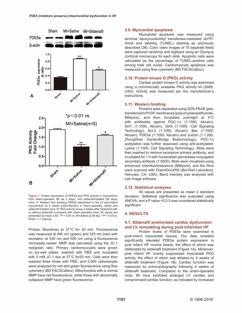
Protein levels of PDE5a were examined in post-infarct myocardial tissues. Our data revealed significantly elevated PDE5a protein expression in post infarct HF murine hearts, the effect of which was obliterated by sildenafil treatment (Figure 1A). Moreover, post infarct HF overtly suppressed myocardial PKG activity, the effect of which was ablated by 4 weeks of sildenafil treatment (Figure 1B). Cardiac function was measured by echocardiography following 4 weeks of sildenafil treatment. Compared to the sham-operated mice, MI mice exhibited enlarged LV cavities and compromised cardiac function, as indicated by increased
Figure 1. Protein expression of PDE5a and PKG activity in myocardium from sham-operated, MI (at 3 days), and sildenafil-treated (28 days) mice. A: Western blot showing PDE5a expression in the LV peri-infarct myocardium at 4 weeks post-infarction in sham-operated, saline and sildenafil-treated mice. B: PKG activity assay 4 weeks after treatment with or without sildenafil compared with sham-operated mice. All values are presented as mean ± SD. *P < 0.05 vs. MI+Saline at 28 day; **P < 0.05 vs. Sham. n = 5/group.
PDE5 inhibitors preserve mitochondrial dysfunction in HF
1198 © 1996-2016
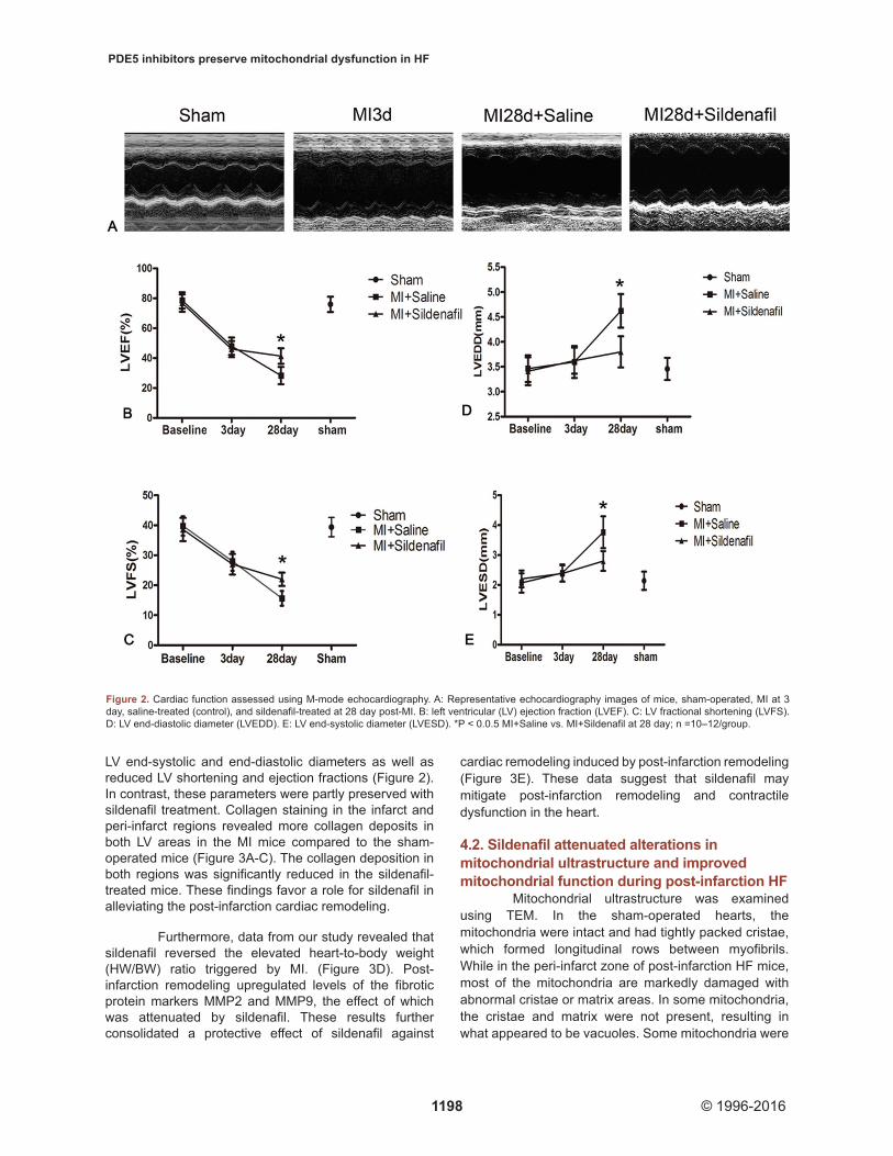
LV end-systolic and end-diastolic diameters as well as reduced LV shortening and ejection fractions (Figure 2). In contrast, these parameters were partly preserved with sildenafil treatment. Collagen staining in the infarct and peri-infarct regions revealed more collagen deposits in both LV areas in the MI mice compared to the sham-operated mice (Figure 3A-C). The collagen deposition in both regions was significantly reduced in the sildenafil-treated mice. These findings favor a role for sildenafil in alleviating the post-infarction cardiac remodeling.
Furthermore, data from our study revealed that sildenafil reversed the elevated heart-to-body weight (HW/BW) ratio triggered by MI. (Figure 3D). Post-infarction remodeling upregulated levels of the fibrotic protein markers MMP2 and MMP9, the effect of which was attenuated by sildenafil. These results further consolidated a protective effect of sildenafil against
cardiac remodeling induced by post-infarction remodeling (Figure 3E). These data suggest that sildenafil may mitigate post-infarction remodeling and contractile dysfunction in the heart.
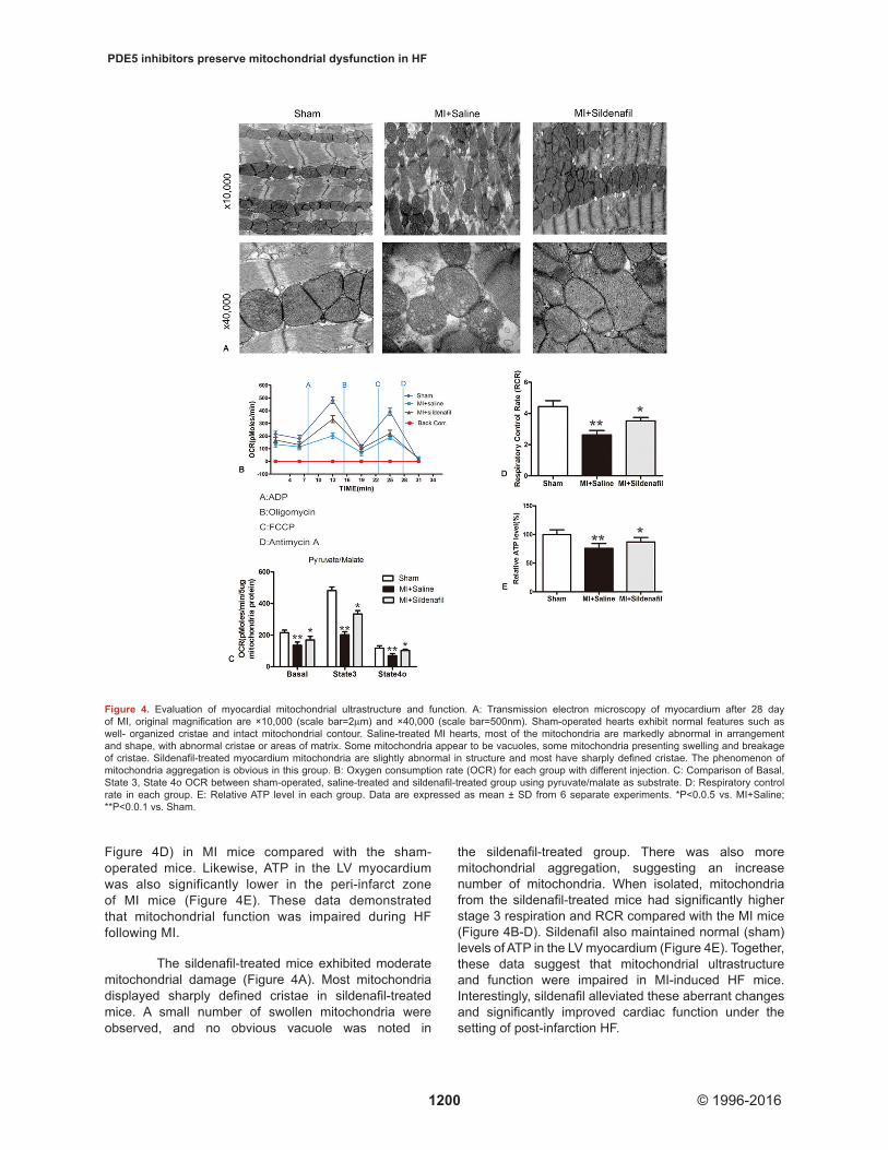
4.2. Sildenafil attenuated alterations in mitochondrial ultrastructure and improved mitochondrial function during post-infarction HF
Mitochondrial ultrastructure was examined using TEM. In the sham-operated hearts, the mitochondria were intact and had tightly packed cristae, which formed longitudinal rows between myofibrils. While in the peri-infarct zone of post-infarction HF mice, most of the mitochondria are markedly damaged with abnormal cristae or matrix areas. In some mitochondria, the cristae and matrix were not present, resulting in what appeared to be vacuoles. Some mitochondria were
Figure 2. Cardiac function assessed using M-mode echocardiography. A: Representative echocardiography images of mice, sham-operated, MI at 3 day, saline-treated (control), and sildenafil-treated at 28 day post-MI. B: left ventricular (LV) ejection fraction (LVEF). C: LV fractional shortening (LVFS). D: LV end-diastolic diameter (LVEDD). E: LV end-systolic diameter (LVESD). *P < 0.0.5 MI+Saline vs. MI+Sildenafil at 28 day; n =10–12/group.

PDE5 inhibitors preserve mitochondrial dysfunction in HF
1199 © 1996-2016
also swollen and had disoriented cristae, breakage, and mitochondrial disarrangement. Sildenafil reversed the mitochondrial ultrastructural disorder in MI hearts. (Figure 4A)
Mitochondrial respiration function was also scrutinized (Figure 4B-D). Our data revealed that mitochondria isolated from the peri-infarct zones of HF
mice had significantly lower basal OCRs with pyruvate and malate compared to mitochondria from heart of the sham-operated group (Figure 4C). The maximal ADP respiration rate (stage 3) was 50% less in these MI mice compared to the sham-operated mice. The RCR, an indicator for the efficiency of electron movement through the electron transport chain and during respiratory coupling, was decreased by 40% (P<0.0.1;
Figure 3. Gross morphology and histology of hearts in myocardium from sham-operated, MI (at 3 days), and sildenafil-treated (28 days) mice. A: Representative images of Masson’s trichrome-stained transverse ventricular sections of mice hearts depicting myocardial fibrosis at 28 day in sham-operated, saline and sildenafil-treated mice. B: Representative Sirius red and methyl green stained cross-sections of sham-operated mice, saline and sildenafil -treated MI mice at day 28 after MI. Sirius red stained collagen viewed under polarized light (magnification 20X). Scale bar represent 60μm. C: Myocardial fibrosis (percentage of LV). *P <0.0.5 vs. saline at 28 day; N= 4–6/group. D: The ratio of heart weight/body weight in sham-operated, saline and sildenafil-treated mice. E: Western blot and quantitation of MMP2 and MMP9 expression in sham-operated, saline-treated and sildenafil-treated mice. All values are presented as mean ± SD. *P<0.0.5 vs. MI+Saline at 28 day; **P<0.0.5 vs. Sham. n = 4–6/group.
PDE5 inhibitors preserve mitochondrial dysfunction in HF
1200 © 1996-2016
Figure 4D) in MI mice compared with the sham-operated mice. Likewise, ATP in the LV myocardium was also significantly lower in the peri-infarct zone of MI mice (Figure 4E). These data demonstrated that mitochondrial function was impaired during HF following MI.
The sildenafil-treated mice exhibited moderate mitochondrial damage (Figure 4A). Most mitochondria displayed sharply defined cristae in sildenafil-treated mice. A small number of swollen mitochondria were observed, and no obvious vacuole was noted in
the sildenafil-treated group. There was also more mitochondrial aggregation, suggesting an increase number of mitochondria. When isolated, mitochondria from the sildenafil-treated mice had significantly higher stage 3 respiration and RCR compared with the MI mice (Figure 4B-D). Sildenafil also maintained normal (sham) levels of ATP in the LV myocardium (Figure 4E). Together, these data suggest that mitochondrial ultrastructure and function were impaired in MI-induced HF mice. Interestingly, sildenafil alleviated these aberrant changes and significantly improved cardiac function under the setting of post-infarction HF.
Figure 4. Evaluation of myocardial mitochondrial ultrastructure and function. A: Transmission electron microscopy of myocardium after 28 day of MI, original magnification are ×10,000 (scale bar=2μm) and ×40,000 (scale bar=500nm). Sham-operated hearts exhibit normal features such as well- organized cristae and intact mitochondrial contour. Saline-treated MI hearts, most of the mitochondria are markedly abnormal in arrangement and shape, with abnormal cristae or areas of matrix. Some mitochondria appear to be vacuoles, some mitochondria presenting swelling and breakage of cristae. Sildenafil-treated myocardium mitochondria are slightly abnormal in structure and most have sharply defined cristae. The phenomenon of mitochondria aggregation is obvious in this group. B: Oxygen consumption rate (OCR) for each group with different injection. C: Comparison of Basal, State 3, State 4o OCR between sham-operated, saline-treated and sildenafil-treated group using pyruvate/malate as substrate. D: Respiratory control rate in each group. E: Relative ATP level in each group. Data are expressed as mean ± SD from 6 separate experiments. *P<0.0.5 vs. MI+Saline; **P<0.0.1 vs. Sham.
PDE5 inhibitors preserve mitochondrial dysfunction in HF
1201 © 1996-2016
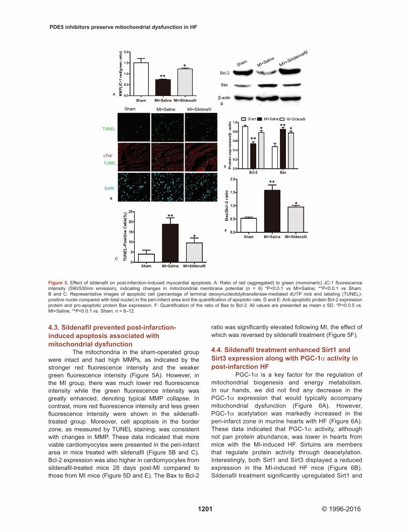
4.3. Sildenafil prevented post-infarction-induced apoptosis associated with mitochondrial dysfunction
The mitochondria in the sham-operated group were intact and had high MMPs, as indicated by the stronger red fluorescence intensity and the weaker green fluorescence intensity (Figure 5A). However, in the MI group, there was much lower red fluorescence intensity while the green fluorescence intensity was greatly enhanced, denoting typical MMP collapse. In contrast, more red fluorescence intensity and less green fluorescence intensity were shown in the sildenafil-treated group. Moreover, cell apoptosis in the border zone, as measured by TUNEL staining, was consistent with changes in MMP. These data indicated that more viable cardiomyocytes were presented in the peri-infarct area in mice treated with sildenafil (Figure 5B and C). Bcl-2 expression was also higher in cardiomyocytes from sildenafil-treated mice 28 days post-MI compared to those from MI mice (Figure 5D and E). The Bax to Bcl-2
ratio was significantly elevated following MI, the effect of which was reversed by sildenafil treatment (Figure 5F).
4.4. Sildenafil treatment enhanced Sirt1 and Sirt3 expression along with PGC-1α activity in post-infarction HF
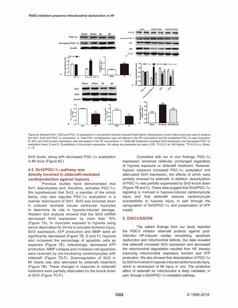
PGC-1α is a key factor for the regulation of mitochondrial biogenesis and energy metabolism. In our hands, we did not find any decrease in the PGC-1α expression that would typically accompany mitochondrial dysfunction (Figure 6A). However, PGC-1α acetylation was markedly increased in the peri-infarct zone in murine hearts with HF (Figure 6A). These data indicated that PGC-1α activity, although not pan protein abundance, was lower in hearts from mice with the MI-induced HF. Sirtuins are members that regulate protein activity through deacetylation. Interestingly, both Sirt1 and Sirt3 displayed a reduced expression in the MI-induced HF mice (Figure 6B). Sildenafil treatment significantly upregulated Sirt1 and
Figure 5. Effect of sildenafil on post-infarction-induced myocardial apoptosis. A: Ratio of red (aggregated) to green (monomeric) JC-1 fluorescence intensity (590/530nm emission), indicating changes in mitochondrial membrane potential (n = 6) *P<0.0.1 vs MI+Saline; **P<0.0.1 vs Sham. B and C: Representative images of apoptotic cell (percentage of terminal deoxynucleotidyltransferase-mediated dUTP nick end labeling (TUNEL)-positive nuclei compared with total nuclei) in the peri-infarct area and the quantification of apoptotic rate. D and E: Anti-apoptotic protein Bcl-2 expression protein and pro-apoptotic protein Bax expression. F: Quantification of the ratio of Bax to Bcl-2. All values are presented as mean ± SD. *P<0.0.5 vs. MI+Saline; **P<0.0.1 vs. Sham. n = 8–12.
PDE5 inhibitors preserve mitochondrial dysfunction in HF
1202 © 1996-2016
Sirt3 levels, along with decreased PGC-1α acetylation in MI mice (Figure 6C)
4.5. Sirt3/PGC-1α pathway was directly involved in sildenafil-mediated cardioprotection against hypoxia
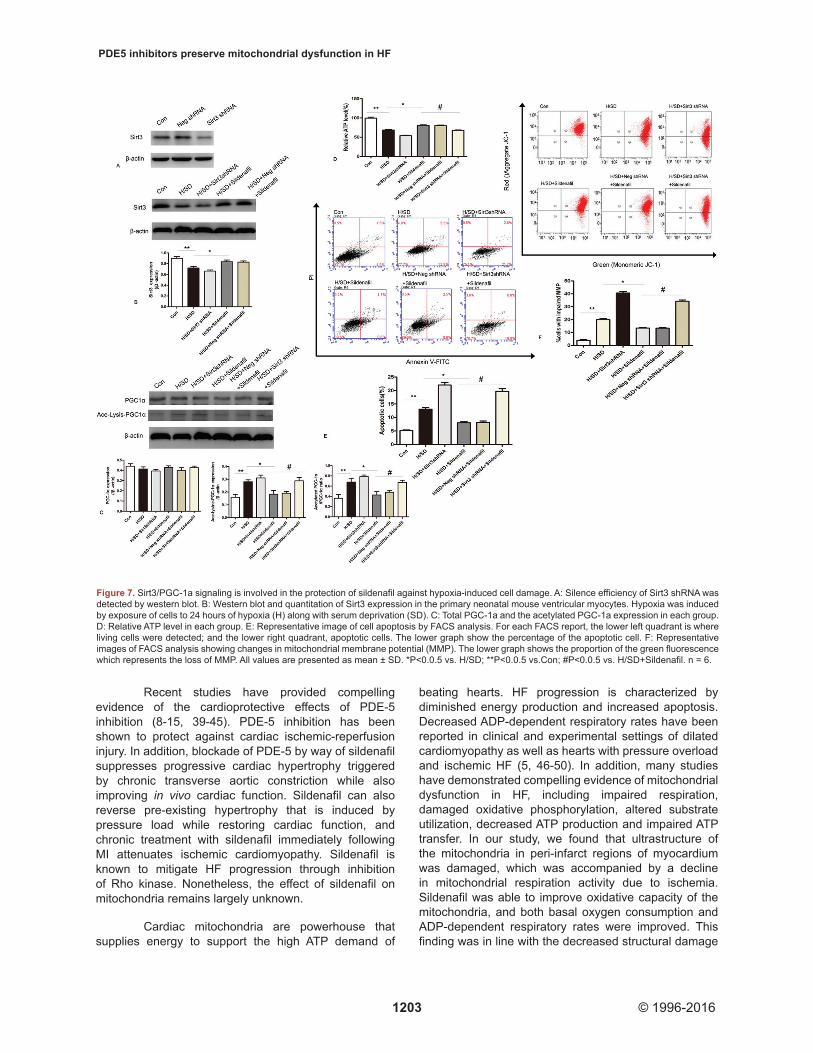
Previous studies have demonstrated that Sirt1 deacetylates and, therefore, activates PGC-1α. We hypothesized that Sirt3, a member of the sirtuin family, may also regulate PGC-1α acetylation in a manner reminiscent of Sirt1. Sirt3 was knocked down in cultured neonatal mouse ventricular myocytes to determine its role in hypoxia-induced damage. Western blot analysis showed that the Sirt3 shRNA decreased Sirt3 expression by more than 70% (Figure 7A). In myocytes exposed to hypoxia using serum deprivation for 24 hrs to simulate ischemic injury, Sirt3 expression, ATP production and MMP were all significantly decreased (Figure 7B, D and F), Hypoxia also increased the percentage of apoptotic cells as expected (Figure 7E). Interestingly, decreased ATP production, MMP collapse and increased cell apoptosis were reversed by pre-incubating cardiomyocytes with sildenafil (Figure 7D-F). Downregulation of Sirt3 in MI hearts was also alleviated by sildenafil treatment (Figure 7B). These changes in response to sildenafil treatment were partially attenuated by the knock-down of Sirt3 (Figure 7C-F).
Consistent with our in vivo findings, PGC-1α expression remained relatively unchanged regardless of hypoxia exposure or sildenafil treatment. However, hypoxic exposure increased PGC-1α acetylation and attenuated Sirt3 expression, the effects of which were partially reversed by sildenafil. In addition, deacetylation of PGC-1α was partially suppressed by Sirt3 knock down (Figure 7B and C). These data suggest that Sirt3/PGC-1α signaling is involved in hypoxia-induced cardiomyocyte injury and that sildenafil reduces cardiomyocyte susceptibility to hypoxia injury, in part through the upregulation of Sirt3/PGC-1α and preservation of ATP supply.
5. DISCUSSION
The salient findings from our study depicted the PDE-5 inhibitor sildenafil protects against post-infarction HF-induced cardiac remodeling, apoptosis dysfunction and mitochondrial defects. Our data revealed that sildenafil increased Sirt3 expression and decreased the mitochondrial degradation resulted from HF, thereby improving mitochondrial respiratory function and ATP production. We also showed that deacetylation of PGC-1α by Sirt3 is involved in hypoxia-induced cardiomyocyte injury, which is reminiscent of MI injury in vivo. The protective effect of sildenafil for mitochondria is likely mediated, in part, through a Sirt3/PGC-1α-mediated pathway.
Figure 6. Aberrant Sirt1, Sirt3 and PGC-1a expression in myocardial infarction induced heart failure. Myocardium of peri-infarct area was used to analyze the Sirt1, Sirt3 and PGC-1α expression. A: Total PGC-1a expression was not altered in the HF myocardium but the acetylated PGC-1a was increased. B: Sirt1 and Sirt3 protein expression was decreased in the HF myocardium. C: Sildenafil treatment increased Sirt3 expression and decreased PGC-1a acetylation level. D and E: Quantitation of the protein expression. All values are presented as mean ± SD. *P<0.0.5 vs. MI+Saline; **P<0.0.5 vs. Sham. n = 6.
PDE5 inhibitors preserve mitochondrial dysfunction in HF
1203 © 1996-2016
Recent studies have provided compelling evidence of the cardioprotective effects of PDE-5 inhibition (8-15, 39-45). PDE-5 inhibition has been shown to protect against cardiac ischemic-reperfusion injury. In addition, blockade of PDE-5 by way of sildenafil suppresses progressive cardiac hypertrophy triggered by chronic transverse aortic constriction while also improving in vivo cardiac function. Sildenafil can also reverse pre-existing hypertrophy that is induced by pressure load while restoring cardiac function, and chronic treatment with sildenafil immediately following MI attenuates ischemic cardiomyopathy. Sildenafil is known to mitigate HF progression through inhibition of Rho kinase. Nonetheless, the effect of sildenafil on mitochondria remains largely unknown.
Cardiac mitochondria are powerhouse that supplies energy to support the high ATP demand of
beating hearts. HF progression is characterized by diminished energy production and increased apoptosis. Decreased ADP-dependent respiratory rates have been reported in clinical and experimental settings of dilated cardiomyopathy as well as hearts with pressure overload and ischemic HF (5, 46-50). In addition, many studies have demonstrated compelling evidence of mitochondrial dysfunction in HF, including impaired respiration, damaged oxidative phosphorylation, altered substrate utilization, decreased ATP production and impaired ATP transfer. In our study, we found that ultrastructure of the mitochondria in peri-infarct regions of myocardium was damaged, which was accompanied by a decline in mitochondrial respiration activity due to ischemia. Sildenafil was able to improve oxidative capacity of the mitochondria, and both basal oxygen consumption and ADP-dependent respiratory rates were improved. This finding was in line with the decreased structural damage
Figure 7. Sirt3/PGC-1a signaling is involved in the protection of sildenafil against hypoxia-induced cell damage. A: Silence efficiency of Sirt3 shRNA was detected by western blot. B: Western blot and quantitation of Sirt3 expression in the primary neonatal mouse ventricular myocytes. Hypoxia was induced by exposure of cells to 24 hours of hypoxia (H) along with serum deprivation (SD). C: Total PGC-1a and the acetylated PGC-1a expression in each group. D: Relative ATP level in each group. E: Representative image of cell apoptosis by FACS analysis. For each FACS report, the lower left quadrant is where living cells were detected; and the lower right quadrant, apoptotic cells. The lower graph show the percentage of the apoptotic cell. F: Representative images of FACS analysis showing changes in mitochondrial membrane potential (MMP). The lower graph shows the proportion of the green fluorescence which represents the loss of MMP. All values are presented as mean ± SD. *P<0.0.5 vs. H/SD; **P<0.0.5 vs.Con; #P<0.0.5 vs. H/SD+Sildenafil. n = 6.
PDE5 inhibitors preserve mitochondrial dysfunction in HF
1204 © 1996-2016
observed in the mitochondria of the peri-infarct area in sildenafil-treated mice.
Angiogenesis is a compensatory response to tissue ischemia, and sildenafil has been reported to promote angiogenesis and lessen ischemic injury (41, 51). The protective role of sildenafil for mitochondria can be attributed to its role of promoting angiogenesis. Sildenafil has been shown to ameliorate the long term progression of cardiac remodeling and function in advanced stages of pressure-overload hypertrophy by restoring mitochondria biogenesis and respiration (52). In our hands, isolated mitochondrial function was also assessed to reveal a protective role of sildenafil for mitochondrial function during post-infarction HF. However, we failed to examine the effect of sildenafil in sham-operated mice. Sildenafil citrate has been shown to depress H2O2 generation in wild-type rats by mimicking superoxide dismutase without overtly affecting mitochondrial respiration rates (53). These discrepancies could be explained by the approaches employed to assess mitochondrial function and different physiological and/or pathological conditions.
Cardiomyocyte apoptosis contributes to the progression of HF after MI (54). In addition, chronic cardiac remodeling with chamber dilation and impaired systolic function are associated with increased myocyte apoptosis in the infarct border zone after MI (55). Loss of cardiomyocytes is accompanied by increased extracellular matrix deposition and eventually results into cardiac dysfunction. Damaged mitochondria are unable to provide the high energy amounts required for cardiomyocyte survival, thereby leading to pronounced apoptosis (56, 57). In our study, we observed a lower Bax/Bcl-2 ratio and an elevated MMP in sildenafil-treated murine hearts compared with control ones. These results were further consolidated in murine cardiomyocytes subjected to hypoxia. Thus, our data suggested that sildenafil counters cardiomyocyte apoptosis possibly through protection against mitochondrial dysfunction in post-infraction HF.
It is unclear how the beneficial effects of sildenafil affected mitochondria. In our study, we found that the protective effect of sildenafil was likely to be mediated through Sirt3. Sirt3 is a member of the sirtuin family of proteins and is involved in cardiac hypertrophy. Sirt3-deficient mice are more likely to develop cardiac hypertrophy and interstitial fibrosis by 8 weeks of age. Moreover, these mice display severe hypertrophic responses when faced with hypertrophic stimuli. In contrast, transgenic mice that overexpress cardiac Sirt3 are effectively protected against agonist-mediated cardiac hypertrophy. The protective role of Sirt3 is likely to be mediated through its activation of Foxo3a-dependent antioxidant mechanisms (29). Nonetheless, Sirt3 possesses multiple properties related to mitochondria
energy metabolism. Hearts of Sirt3-deficient mice exhibits decreased ATP production compared with the wild-type counterparts (31). Furthermore, in stress conditions where ATP demand is high, Sirt3 expression increases (58). These studies demonstrate that Sirt3 is an important regulator of cellular ATP levels. Sirt3 expression significantly decreased in the control mice with MI in our study. However, in mice treated with sildenafil, Sirt3 expression was preserved.
Recent studies have also shown that PGC-1α acetylation serves as a post-translational modification inactivation process (59). PGC-1α is a master regulator of myocardial energy metabolism and has been studied in many diverse physiological and pathophysiological conditions. The heart has a very large energy demand that is satisfied by a high capacity mitochondrial oxidation system; thus, it is not surprising that PGC-1α expression is high in the heart. Several studies have found that PGC-1α is important for controlling of metabolic pathways in the heart during development as well as in response to physiological stressors and pathological stimuli (22, 24). PGC-1α, a positive regulator of mitochondrial biogenesis and respiration, plays an essential role in neurodegenerative disorders and heart failure, as well as other pathological conditions associated with mitochondrial defects (22). PGC-1α is highly inducible in response to physiological stimuli such as exercise to cope with increased myocardial ATP demand (60). In addition, the expression of PGC-1α and its targets, including PPARs and ERRs, is decreased in cardiac hypertrophy and heart failure (22). PGC-1α downregulation during pathological cardiac hypertrophy seems to be consistent with the derangements in mitochondrial metabolism that occur in hypertrophied and failing hearts. However, the alteration occurred mainly in the face of pressure overload-induced HF. Mice lacking PGC-1α develop signatures of HF as well as a marked drop in cellular ATP levels in the pressure overloaded heart (61). Mitochondria isolated from muscles of transgenic mice that ectopically express PGC-1α also have a higher substrate oxidation capacity than those isolated from wild-type controls (62).
In our study, we demonstrated that isolated mitochondria from the sildenafil-treated post-infarction mice had a higher respiratory capacity than those from the sham-operated mice. Interestingly, a significant decrease in PGC-1α protein expression was not observed in the peri-infarct area, despite previous reports that PGC-1α protein expression was decreased following post-infarction (63). Nonetheless, this experiment was performed on day 14 following acute MI, and PGC-1α mRNA expression was only examined within the remote viable myocardial area. A previous study suggested that myocardium from different regions, including the infarct core, peri-infarct region and remote area (unaffected by infarct), presented distinct apoptosis rates after early and
PDE5 inhibitors preserve mitochondrial dysfunction in HF
1205 © 1996-2016
late acute MI, indicating involvement of multiple cellular mechanisms (64). Therefore, the temporal and spatial differences in PGC-1α protein expression and activity in response to myocardial infarction may underscore the discrepancy. In fact, our current experimental findings were consistent with an earlier report where PGC-1α levels were examined at 4 and 12 weeks following acute MI (65). In contrast, changes in PGC-1α activity, as assessed using PGC-1α acetylation, were noted in the ischemic area. PGC-1α is the target of extensive post-translational modifications, including phosphorylation, acetylation, methylation, ubiquitination, and O-linked N-acetylglycosylation (24). These types of modifications allow the fine tuning of PGC-1α in response to various energy stresses. PGC-1α acetylation is known to occur at several lysine residues and is catalyzed by the acetyl transferase GCN5 (general control of amino acid synthesis 5). With acetylation, nuclear localization of PGC-1α is altered, leading to inhibition of transcriptional activity. Therefore, deacetylation of PGC-1α is needed to restore its activity. Many studies have shown that Sirt1 deacetylates PGC-1α and protects against metabolic diseases (66). The Sirt1-regulated changes in PGC-1α activity may trigger energy substrate drift through increasing fatty acid oxidation for energy production (67). To this end, Sirt3 modulates energy metabolism through a more direct mitochondrial function regulation. However, whether Sirt3 modulates mitochondrial function en route to the regulation of PGC-1α activity through ATP modulation (e.g. increased mitochondrial biogenesis and ATP synthesis) remains elusive. Sirt3 was initially thought to be localized in mitochondria although recent studies suggest expression in the nucleus and the cytoplasm (68). Our data confirmed that reduced energy supply during post-infarction HF is associated with high levels of PGC-1α acetylation and decreased Sirt3 expression.
The relationship between Sirt3 and PGC-1α is not well understood. Previous studies have reported that Sirt3 increases in response to cold exposure and caloric restriction. In addition, Sirt3 stimulates downstream CREB-mediated PGC-1α expression in brown pre-adipocytes. In skeletal muscle, a lack of Sirt3 inactivates AMPK and pCREB, leading to the inhibition of PGC-1α activity (69). In hepatocytes and C2C12 myotubes, Sirt3 functions as a downstream target of PGC-1α and mediates the effects of PGC-1α on cellular ROS production and mitochondrial biogenesis (70). PGC-1α has also been shown to control Sirt3 expression during brown adipocyte differentiation (71). Here, we found that decreased Sirt3 expression in HF is accompanied by an increase in PGC-1α acetylation. In addition, sildenafil promoted Sirt3 expression and suppressed PGC-1α acetylation. The knock-down of Sirt3 in cultured cardiomyocytes mitigated the beneficial effect of sildenafil in PGC-1α acetylation under hypoxic conditions.
In summary, the present study demonstrated that the PDE-5 inhibitor sildenafil exerts cardioprotective effects against MI-induced HF. These effects are mediated, in part, by improved mitochondrial function and preserved Sirt3 levels. Sirt3 is the only sirtuin reported to display an apparent role with the increased life span in humans (29). We have identified Sirt3 as a new target for maintaining energy balance in the heart. Furthermore, we demonstrated that sildenafil can upregulate Sirt3 expression, which suggests that sildenafil could be used broadly to treat other energy related diseases. However, further studies are needed to delineate the mechanism behind sildenafil and other PDE-5 inhibitors-induced regulation on Sirt3 function.
6. ACKNOWLEGEMENT
NL, YY, and SL contributed equally to this work. This work was supported partly by National Science Foundation of China (#31171090, #81400198 and #81370195).
7. REFERENCES
1. J. V. Tu, L. Nardi, J. Fang, J. Liu, L. Khalid and H. Johansen: National trends in rates of death and hospital admissions related to acute myocardial infarction, heart failure and stroke, 1994-2004. CMAJ, 180(13), E118-25 (2009)DOI: 10.1503/cmaj.081197
2. P. S. Jhund and J. J. McMurray: Heart failure after acute myocardial infarction: a lost battle in the war on heart failure? Circulation, 118(20), 2019-21 (2008)DOI: 10.1161/CIRCULATIONAHA.108. 813493
3. R. Ventura-Clapier, A. Garnier, V. Veksler and F. Joubert: Bioenergetics of the failing heart. Biochim Biophys Acta, 1813(7), 1360-72 (2011)DOI: 10.1016/j.bbamcr.2010.09.006
4. S. Neubauer: The failing heart--an engine out of fuel. N Engl J Med, 356(11), 1140-51 (2007)DOI: 10.1056/NEJMra063052
5. M. G. Rosca and C. L. Hoppel: Mitochondria in heart failure. Cardiovasc Res, 88(1), 40-50 (2010)DOI: 10.1093/cvr/cvq240
6. H. Lemieux, S. Semsroth, H. Antretter, D. Hofer and E. Gnaiger: Mitochondrial respiratory control and early defects of oxidative phosphorylation in the failing human heart. Int J Biochem Cell Biol, 43(12), 1729-38 (2011)
PDE5 inhibitors preserve mitochondrial dysfunction in HF
1206 © 1996-2016
DOI: 10.1016/j.biocel.2011.08.0087. M. G. Rosca and C. L. Hoppel: New aspects
of impaired mitochondrial function in heart failure. J Bioenerg Biomembr, 41(2), 107-12 (2009)DOI: 10.1007/s10863-009-9215-9
8. E. Takimoto, H. C. Champion, M. Li, D. Belardi, S. Ren, E. R. Rodriguez, D. Bedja, K. L. Gabrielson, Y. Wang and D. A. Kass: Chronic inhibition of cyclic GMP phosphodiesterase 5A prevents and reverses cardiac hypertrophy. Nat Med, 11(2), 214-22 (2005)DOI: 10.1038/nm1175
9. V. Q. Chau, F. N. Salloum, N. N. Hoke, A. Abbate and R. C. Kukreja: Mitigation of the progression of heart failure with sildenafil involves inhibition of RhoA/Rho-kinase pathway. Am J Physiol Heart Circ Physiol, 300(6), H2272-9 (2011)DOI: 10.1152/ajpheart.00654.2010
10. F. N. Salloum, A. Abbate, A. Das, J. E. Houser, C. A. Mudrick, I. Z. Qureshi, N. N. Hoke, S. K. Roy, W. R. Brown, S. Prabhakar and R. C. Kukreja: Sildenafil (Viagra) attenuates ischemic cardiomyopathy and improves left ventricular function in mice. Am J Physiol Heart Circ Physiol, 294(3), H1398-406 (2008)DOI: 10.1152/ajpheart.91438.2007
11. A. Das, L. Xi and R. C. Kukreja: Phosphodiesterase-5 inhibitor sildenafil preconditions adult cardiac myocytes against necrosis and apoptosis. Essential role of nitric oxide signaling. J Biol Chem, 280(13), 12944-55 (2005)DOI: 10.1074/jbc.M404706200
12. F. Salloum, C. Yin, L. Xi and R. C. Kukreja: Sildenafil induces delayed preconditioning through inducible nitric oxide synthase-dependent pathway in mouse heart. Circ Res, 92(6), 595-7 (2003)DOI: 10.1161/01.RES.0000066853.09821.98
13. F. N. Salloum, Y. Takenoshita, R. A. Ockaili, V. P. Daoud, E. Chou, K. Yoshida and R. C. Kukreja: Sildenafil and vardenafil but not nitroglycerin limit myocardial infarction through opening of mitochondrial K(ATP) channels when administered at reperfusion following ischemia in rabbits. J Mol Cell Cardiol, 42(2), 453-8 (2007)DOI: 10.1016/j.yjmcc.2006.10.015
14. R. Ockaili, F. Salloum, J. Hawkins and R. C. Kukreja: Sildenafil (Viagra) induces powerful cardioprotective effect via opening of mitochondrial K(ATP) channels in rabbits. Am J Physiol Heart Circ Physiol, 283(3), H1263-9 (2002)DOI: 10.1152/ajpheart.00324.2002
15. T. Gori, S. Sicuro, S. Dragoni, G. Donati, S. Forconi and J. D. Parker: Sildenafil prevents endothelial dysfunction induced by ischemia and reperfusion via opening of adenosine triphosphate-sensitive potassium channels: a human in vivo study. Circulation, 111(6), 742-6 (2005)DOI: 10.1161/01.CIR.0000155252.23933.2D
16. A. D. Costa, S. V. Pierre, M. V. Cohen, J. M. Downey and K. D. Garlid: cGMP signalling in pre- and post-conditioning: the role of mitochondria. Cardiovasc Res, 77(2), 344-52 (2008)DOI: 10.1093/cvr/cvm050
17. K. Miyashita, H. Itoh, H. Tsujimoto, N. Tamura, Y. Fukunaga, M. Sone, K. Yamahara, D. Taura, M. Inuzuka, T. Sonoyama and K. Nakao: Natriuretic peptides/cGMP/cGMP-dependent protein kinase cascades promote muscle mitochondrial biogenesis and prevent obesity. Diabetes, 58(12), 2880-92 (2009)DOI: 10.2337/db09-0393
18. E. Nisoli, E. Clementi, C. Paolucci, V. Cozzi, C. Tonello, C. Sciorati, R. Bracale, A. Valerio, M. Francolini, S. Moncada and M. O. Carruba: Mitochondrial biogenesis in mammals: the role of endogenous nitric oxide. Science, 299(5608), 896-9 (2003)DOI: 10.1126/science.1079368
19. J. Lin, C. Handschin and B. M. Spiegelman: Metabolic control through the PGC-1 family of transcription coactivators. Cell Metab, 1(6), 361-70 (2005)DOI: 10.1016/j.cmet.2005.05.004
20. C. Handschin and B. M. Spiegelman: Peroxisome proliferator-activated receptor gamma coactivator 1 coactivators, energy homeostasis, and metabolism. Endocr Rev, 27(7), 728-35 (2006)DOI: 10.1210/er.2006-0037
21. H. Liang and W. F. Ward: PGC-1alpha: a key regulator of energy metabolism. Adv Physiol Educ, 30(4), 145-51 (2006)DOI: 10.1152/advan.00052.2006
PDE5 inhibitors preserve mitochondrial dysfunction in HF
1207 © 1996-2016
22. B. N. Finck and D. P. Kelly: Peroxisome proliferator-activated receptor gamma coactivator-1 (PGC-1) regulatory cascade in cardiac physiology and disease. Circulation, 115(19), 2540-8 (2007)DOI: 10.1161/CIRCULATIONAHA.107 .670588
23. R. Ventura-Clapier, A. Garnier and V. Veksler: Transcriptional control of mitochondrial biogenesis: the central role of PGC-1alpha. Cardiovasc Res, 79(2), 208-17 (2008)DOI: 10.1093/cvr/cvn098
24. P. J. Fernandez-Marcos and J. Auwerx: Regulation of PGC-1alpha, a nodal regulator of mitochondrial biogenesis. Am J Clin Nutr, 93(4), 884S-90 (2011)DOI: 10.3945/ajcn.110.001917
25. S. Imai, C. M. Armstrong, M. Kaeberlein and L. Guarente: Transcriptional silencing and longevity protein Sir2 is an NAD-dependent histone deacetylase. Nature, 403(6771), 795-800 (2000)DOI: 10.1038/35001622
26. J. Landry, A. Sutton, S. T. Tafrov, R. C. Heller, J. Stebbins, L. Pillus and R. Sternglanz: The silencing protein SIR2 and its homologs are NAD-dependent protein deacetylases. Proc Natl Acad Sci U S A, 97(11), 5807-11 (2000)DOI: 10.1073/pnas.110148297
27. J. S. Smith, C. B. Brachmann, I. Celic, M. A. Kenna, S. Muhammad, V. J. Starai, J. L. Avalos, J. C. Escalante-Semerena, C. Grubmeyer, C. Wolberger and J. D. Boeke: A phylogenetically conserved NAD+-dependent protein deacetylase activity in the Sir2 protein family. Proc Natl Acad Sci U S A, 97(12), 6658-63 (2000)DOI: 10.1073/pnas.97.12.6658
28. V. B. Pillai, N. R. Sundaresan and M. P. Gupta: Regulation of Akt signaling by sirtuins: its implication in cardiac hypertrophy and aging. Circ Res, 114(2), 368-78 (2014)DOI: 10.1161/CIRCRESAHA.113.300536
29. N. R. Sundaresan, M. Gupta, G. Kim, S. B. Rajamohan, A. Isbatan and M. P. Gupta: Sirt3 blocks the cardiac hypertrophic response by augmenting Foxo3a-dependent antioxidant defense mechanisms in mice. J Clin Invest, 119(9), 2758-71 (2009)DOI: 10.1172/jci39162
30. T. Shi, F. Wang, E. Stieren and Q. Tong: SIRT3, a mitochondrial sirtuin deacetylase, regulates mitochondrial function and thermogenesis in brown adipocytes. J Biol Chem, 280(14), 13560-7 (2005)DOI: 10.1074/jbc.M414670200
31. B. H. Ahn, H. S. Kim, S. Song, I. H. Lee, J. Liu, A. Vassilopoulos, C. X. Deng and T. Finkel: A role for the mitochondrial deacetylase Sirt3 in regulating energy homeostasis. Proc Natl Acad Sci U S A, 105(38), 14447-52 (2008)DOI: 10.1073/pnas.0803790105
32. E. Gao, Y. H. Lei, X. Shang, Z. M. Huang, L. Zuo, M. Boucher, Q. Fan, J. K. Chuprun, X. L. Ma and W. J. Koch: A novel and efficient model of coronary artery ligation and myocardial infarction in the mouse. Circ Res, 107(12), 1445-53 (2013)DOI: 10.1161/CIRCRESAHA.110.223925
33. P. Sreejit, S. Kumar and R. S. Verma: An improved protocol for primary culture of cardiomyocyte from neonatal mice. In vitro Cell Dev Biol Anim, 44(3-4), 45-50 (2008)DOI: 10.1007/s11626-007-9079-4
34. W. Yan, H. Zhang, P. Liu, H. Wang, J. Liu, C. Gao, Y. Liu, K. Lian, L. Yang, L. Sun, Y. Guo, L. Zhang, L. Dong, W. B. Lau, E. Gao, F. Gao, L. Xiong, Y. Qu and L. Tao: Impaired mitochondrial biogenesis due to dysfunctional adiponectin-AMPK-PGC-1alpha signaling contributing to increased vulnerability in diabetic heart. Basic Res Cardiol, 108(3), 329 (2013)DOI: 10.1007/s00395-013-0329-1
35. M. R. Sayen, A. B. Gustafsson, M. A. Sussman, J. D. Molkentin and R. A. Gottlieb: Calcineurin transgenic mice have mitochondrial dysfunction and elevated superoxide production. Am J Physiol Cell Physiol, 284(2), C562-70 (2003)DOI: 10.1152/ajpcell.00336.2002
36. G. W. Rogers, M. D. Brand, S. Petrosyan, D. Ashok, A. A. Elorza, D. A. Ferrick and A. N. Murphy: High throughput microplate respiratory measurements using minimal quantities of isolated mitochondria. PLoS One, 6(7), e21746 (2013)DOI: 10.1371/journal.pone.0021746
37. L. C. Junqueira, G. Bignolas and R. R. Brentani: Picrosirius staining plus polarization microscopy, a specific method for collagen
PDE5 inhibitors preserve mitochondrial dysfunction in HF
1208 © 1996-2016
detection in tissue sections. Histochem J, 11(4), 447-55 (1979)DOI: 10.1007/BF01002772
38. L. Tao, Y. Wang, E. Gao, H. Zhang, Y. Yuan, W. B. Lau, L. Chan, W. J. Koch and X. L. Ma: Adiponectin: an indispensable molecule in rosiglitazone cardioprotection following myocardial infarction. Circ Res, 106(2), 409-17 (2010)DOI: 10.1161/CIRCRESAHA.109.211797
39. A. Das, F. N. Salloum, L. Xi, Y. J. Rao and R. C. Kukreja: ERK phosphorylation mediates sildenafil-induced myocardial protection against ischemia-reperfusion injury in mice. Am J Physiol Heart Circ Physiol, 296(5), H1236-43 (2009)DOI: 10.1152/ajpheart.00100.2009
40. Y. A. Bremer, F. Salloum, R. Ockaili, E. Chou, W. B. Moskowitz and R. C. Kukreja: Sildenafil citrate (viagra) induces cardioprotective effects after ischemia/reperfusion injury in infant rabbits. Pediatr Res, 57(1), 22-7 (2005)DOI: 10.1203/01.PDR.0000147736.27672.15
41. A. Senthilkumar, R. D. Smith, J. Khitha, N. Arora, S. Veerareddy, W. Langston, J. H. Chidlow, Jr., S. C. Barlow, X. Teng, R. P. Patel, D. J. Lefer and C. G. Kevil: Sildenafil promotes ischemia-induced angiogenesis through a PKG-dependent pathway. Arterioscler Thromb Vasc Biol, 27(9), 1947-54 (2007)DOI: 10.1161/ATVBAHA.107.147421
42. D. Westermann, P. M. Becher, D. Lindner, K. Savvatis, Y. Xia, M. Frohlich, S. Hoffmann, H. P. Schultheiss and C. Tschope: Selective PDE5A inhibition with sildenafil rescues left ventricular dysfunction, inflammatory immune response and cardiac remodeling in angiotensin II-induced heart failure in vivo. Basic Res Cardiol, 107(6), 308 (2012)DOI: 10.1007/s00395-012-0308-y
43. E. Takimoto, N. Koitabashi, S. Hsu, E. A. Ketner, M. Zhang, T. Nagayama, D. Bedja, K. L. Gabrielson, R. Blanton, D. P. Siderovski, M. E. Mendelsohn and D. A. Kass: Regulator of G protein signaling 2 mediates cardiac compensation to pressure overload and antihypertrophic effects of PDE5 inhibition in mice. J Clin Invest, 119(2), 408-20 (2009)DOI: 10.1172/jci35620
44. S. Hsu, T. Nagayama, N. Koitabashi, M. Zhang, L. Zhou, D. Bedja, K. L. Gabrielson,
J. D. Molkentin, D. A. Kass and E. Takimoto: Phosphodiesterase 5 inhibition blocks pressure overload-induced cardiac hypertrophy independent of the calcineurin pathway. Cardiovasc Res, 81(2), 301-9 (2009)DOI: 10.1093/cvr/cvn324
45. T. Nagayama, S. Hsu, M. Zhang, N. Koitabashi, D. Bedja, K. L. Gabrielson, E. Takimoto and D. A. Kass: Sildenafil stops progressive chamber, cellular, and molecular remodeling and improves calcium handling and function in hearts with pre-existing advanced hypertrophy caused by pressure overload. J Am Coll Cardiol, 53(2), 207-15 (2009)DOI: 10.1016/j.jacc.2008.08.069
46. M. G. Rosca, E. J. Vazquez, J. Kerner, W. Parland, M. P. Chandler, W. Stanley, H. N. Sabbah and C. L. Hoppel: Cardiac mitochondria in heart failure: decrease in respirasomes and oxidative phosphorylation. Cardiovasc Res, 80(1), 30-9 (2008)DOI: 10.1093/cvr/cvn184
47. D. F. Dai, S. C. Johnson, J. J. Villarin, M. T. Chin, M. Nieves-Cintron, T. Chen, D. J. Marcinek, G. W. Dorn, 2nd, Y. J. Kang, T. A. Prolla, L. F. Santana and P. S. Rabinovitch: Mitochondrial oxidative stress mediates angiotensin II-induced cardiac hypertrophy and Galphaq overexpression-induced heart failure. Circ Res, 108(7), 837-46 (2011)DOI: 10.1161/CIRCRESAHA.110.232306
48. V. G. Sharov, A. V. Todor, N. Silverman, S. Goldstein and H. N. Sabbah: Abnormal mitochondrial respiration in failed human myocardium. J Mol Cell Cardiol, 32(12), 2361-7 (2000)DOI: 10.1006/jmcc.2000.1266
49. H. Bugger, M. Schwarzer, D. Chen, A. Schrepper, P. A. Amorim, M. Schoepe, T. D. Nguyen, F. W. Mohr, O. Khalimonchuk, B. C. Weimer and T. Doenst: Proteomic remodelling of mitochondrial oxidative pathways in pressure overload-induced heart failure. Cardiovasc Res, 85(2), 376-84 (2010)DOI: 10.1093/cvr/cvp344
50. A. Garnier, D. Fortin, C. Delomenie, I. Momken, V. Veksler and R. Ventura-Clapier: Depressed mitochondrial transcription factors and oxidative capacity in rat failing cardiac and skeletal muscles. J Physiol, 551(Pt 2), 491-501 (2003)
PDE5 inhibitors preserve mitochondrial dysfunction in HF
1209 © 1996-2016
DOI: 10.1113/jphysiol.2003.04510451. A. Pyriochou, Z. Zhou, V. Koika, C.
Petrou, P. Cordopatis, W. C. Sessa and A. Papapetropoulos: The phosphodiesterase 5 inhibitor sildenafil stimulates angiogenesis through a protein kinase G/MAPK pathway. J Cell Physiol, 211(1), 197-204 (2007)DOI: 10.1002/jcp.20929
52. E. Bretes, A. M. Wojdyla-Mamon, J. Kowalska, J. Jemielity, R. Kaczmarek, J. Baraniak and A. Guranowski: Hint2, the mitochondrial nucleoside 5’-phosphoramidate hydrolase; properties of the homogeneous protein from sheep (Ovis aries) liver. Acta Biochim Pol, 60(2), 249-54 (2013)Doi not found
53. M. A. Fernandes, R. J. Marques, J. A. Vicente, M. S. Santos, P. Monteiro, A. J. Moreno and J. B. Custodio: Sildenafil citrate concentrations not affecting oxidative phosphorylation depress H2O2 generation by rat heart mitochondria. Mol Cell Biochem, 309(1-2), 77-85 (2008)DOI: 10.1007/s11010-007-9645-9
54. Y. Hojo, T. Saito and H. Kondo: Role of apoptosis in left ventricular remodeling after acute myocardial infarction. J Cardiol, 60(2), 91-2 (2012)DOI: 10.1016/j.jjcc.2012.05.014
55. F. Sam, D. B. Sawyer, D. L. Chang, F. R. Eberli, S. Ngoy, M. Jain, J. Amin, C. S. Apstein and W. S. Colucci: Progressive left ventricular remodeling and apoptosis late after myocardial infarction in mouse heart. Am J Physiol Heart Circ Physiol, 279(1), H422-8 (2000)
56. S. Desagher and J. C. Martinou: Mitochondria as the central control point of apoptosis. Trends Cell Biol, 10(9), 369-77 (2000)DOI: 10.1016/S0962-8924(00)01803-1
57. X. Wang: The expanding role of mitochondria in apoptosis. Genes Dev, 15(22), 2922-33 (2001)Doi not found
58. B. Paital and L. Samanta: A comparative study of hepatic mitochondrial oxygen consumption in four vertebrates by using Clark-type electrode. Acta Biol Hung, 64(2), 152-60 (2013)DOI: 10.1556/ABiol.64.2013.2.2
59. E. H. Jeninga, K. Schoonjans and J. Auwerx:
Reversible acetylation of PGC-1: connecting energy sensors and effectors to guarantee metabolic flexibility. Oncogene, 29(33), 4617-24 (2010)DOI: 10.1038/onc.2010.206
60. K. Baar, A. R. Wende, T. E. Jones, M. Marison, L. A. Nolte, M. Chen, D. P. Kelly and J. O. Holloszy: Adaptations of skeletal muscle to exercise: rapid increase in the transcriptional coactivator PGC-1. FASEB J, 16(14), 1879-86 (2002)DOI: 10.1096/fj.02-0367com
61. Z. Arany, M. Novikov, S. Chin, Y. Ma, A. Rosenzweig and B. M. Spiegelman: Transverse aortic constriction leads to accelerated heart failure in mice lacking PPAR-gamma coactivator 1alpha. Proc Natl Acad Sci U S A, 103(26), 10086-91 (2006)DOI: 10.1073/pnas.0603615103
62. J. St-Pierre, J. Lin, S. Krauss, P. T. Tarr, R. Yang, C. B. Newgard and B. M. Spiegelman: Bioenergetic analysis of peroxisome proliferator-activated receptor gamma coactivators 1alpha and 1beta (PGC-1alpha and PGC-1beta) in muscle cells. J Biol Chem, 278(29), 26597-603 (2003)DOI: 10.1074/jbc.M301850200
63. C. K. Sun, L. T. Chang, J. J. Sheu, C. Y. Wang, A. A. Youssef, C. J. Wu, S. Chua and H. K. Yip: Losartan preserves integrity of cardiac gap junctions and PGC-1 alpha gene expression and prevents cellular apoptosis in remote area of left ventricular myocardium following acute myocardial infarction. Int Heart J, 48(4), 533-46 (2007)DOI: 10.1536/ihj.48.533
64. A. Abbate, R. Bussani, M. S. Amin, G. W. Vetrovec and A. Baldi: Acute myocardial infarction and heart failure: role of apoptosis. Int J Biochem Cell Biol, 38(11), 1834-40 (2006)DOI: 10.1016/j.biocel.2006.04.010
65. J. Kraljevic, J. Marinovic, D. Pravdic, P. Zubin, Z. Dujic, U. Wisloff and M. Ljubkovic: Aerobic interval training attenuates remodelling and mitochondrial dysfunction in the post-infarction failing rat heart. Cardiovasc Res, 99(1), 55-64 (2013)DOI: 10.1093/cvr/cvt080
66. M. Lagouge, C. Argmann, Z. Gerhart-Hines, H. Meziane, C. Lerin, F. Daussin, N. Messadeq, J. Milne, P. Lambert, P. Elliott,
PDE5 inhibitors preserve mitochondrial dysfunction in HF
1210 © 1996-2016
B. Geny, M. Laakso, P. Puigserver and J. Auwerx: Resveratrol improves mitochondrial function and protects against metabolic disease by activating SIRT1 and PGC-1alpha. Cell, 127(6), 1109-22 (2006)DOI: 10.1016/j.cell.2006.11.013
67. Z. Gerhart-Hines, J. T. Rodgers, O. Bare, C. Lerin, S. H. Kim, R. Mostoslavsky, F. W. Alt, Z. Wu and P. Puigserver: Metabolic control of muscle mitochondrial function and fatty acid oxidation through SIRT1/PGC-1alpha. EMBO J, 26(7), 1913-23 (2007)DOI: 10.1038/sj.emboj.7601633
68. M. B. Scher, A. Vaquero and D. Reinberg: SirT3 is a nuclear NAD+-dependent histone deacetylase that translocates to the mitochondria upon cellular stress. Genes Dev, 21(8), 920-8 (2007)DOI: 10.1101/gad.1527307
69. O. M. Palacios, J. J. Carmona, S. Michan, K. Y. Chen, Y. Manabe, J. L. Ward, 3rd, L. J. Goodyear and Q. Tong: Diet and exercise signals regulate SIRT3 and activate AMPK and PGC-1alpha in skeletal muscle. Aging (Albany NY), 1(9), 771-83 (2009)Doi not found
70. X. Kong, R. Wang, Y. Xue, X. Liu, H. Zhang, Y. Chen, F. Fang and Y. Chang: Sirtuin 3, a new target of PGC-1alpha, plays an important role in the suppression of ROS and mitochondrial biogenesis. PLoS One, 5(7), e11707 (2010)DOI: 10.1371/journal.pone.0011707
71. A. Giralt, E. Hondares, J. A. Villena, F. Ribas, J. Diaz-Delfin, M. Giralt, R. Iglesias and F. Villarroya: Peroxisome proliferator-activated receptor-gamma coactivator-1alpha controls transcription of the Sirt3 gene, an essential component of the thermogenic brown adipocyte phenotype. J Biol Chem, 286(19), 16958-66 (2011)DOI: 10.1074/jbc.M110.202390
Key Words: Phosphodiesterase Inhibitors, Mitochondrial Dysfunction, Heart Failure, Remodeling, Sirt3, PGC-1 alpha deacetylation
Send correspondence to: Haichang Wang, Department of Cardiology, Xijing Hospital, Fourth Military Medical University, Xi’an 710032, China, Tel: 8684775183, Fax: 8684771170, E-mail: [email protected]