Embed Size (px)
Citation preview

Siemens Healthcare Headquarters Siemens Healthcare GmbH Henkestraße 127 91052 Erlangen Germany Phone: +49 9131 84-0 siemens.com/healthcare
siemens.com/medhistory
Printed in Germany | CG CT 2336 06152.3 | © Siemens Healthcare GmbH 2015

Answers for life.
MedHistory Milestones
The History of Computed Tomography at Siemens A retrospective


The History of Computed Tomography at SiemensA retrospective

Contents
Foreword
“Röntgen must be crazy”
What is computed tomography, and what are its strengths?
From idea to SIRETOM
New insights into the brain
From head to toe
03
04
06
08
12
14
20
22
30
32
40
42
Changing times
A seemingly very peculiar idea
A paradigm shift in computed tomography
Small dose, big progress
Twice the scanning power
Improved detail for big impact
40 years of experience in a single unit
From small factory to global player
CEOs of the Computed Tomography Business Unit
50
52
55
2

Foreword
Viewed in evolutionary terms, humanity’s most important trait is curiosity. The curiosity to learn how the world works. The curiosity to experience what happens when new things are combined together. The curiosity to see what lies beyond the horizon. Without curiosity, Wilhelm Conrad Röntgen might have simply shrugged and moved on in 1895, when he discovered X-rays more or less by accident and X-rayed first a thick book and then his wife’s hand. Luckily, he didn’t do that, but instead pressed on to find out more, with his curiosity about these extra-ordinary insights into the human body laying the cornerstone for many of the most important medical imaging methods of our time. In the late 1960s and early 1970s, Sir Godfrey Hounsfield worked based on Röntgen’s discoveries, developing a method of producing axial images of “slices” of the human body. Today’s computed tomography systems still work on the same general principle – although there is a world of difference between today’s technology and the technology available back then. Siemens Healthcare has been a major influence throughout the develop-ment of this technology, from its infancy in the form
of grainy black-and-white images to today’s high-resolution 3D and 4D data sets, which are navigable in infinite degrees. As far back as 1975 – just three years after Hounsfield had brought the EMI Mark 1 up to market readiness – Siemens launched the SIRETOM, the first CT head scanner. Together with our many longstanding medical cooperation partners, we have worked ceaselessly since then to advance computed tomography into an ever-broader range of fields. The curiosity to see how we can improve diagnosis and treatment guidance, boost patient comfort, and reduce radiation doses has spurred us onward throughout the past 40 years. Year after year, innovation after innovation. Right up to the present day, and our versatile portfolio encompassing made-to-measure solutions for any issue, from the SOMATOM Force, the current most powerful CT scanner in the world, to the extremely robust systems used for basic care, the SOMATOM Scope and SOMATOM Spirit.
But our curiosity is not yet satisfied – far from it. “There are probably many discoveries right around the corner, just waiting for someone to bring them to
Walter Märzendorfer
CEO of the Computed Tomography & Radiation Oncology Business Unit
Enjoy reading!
Walter Märzendorfer
life,” Hounsfield said. We are excited to see what else we will be able to discover and bring to life in the decades to come, since we firmly believe that we at Siemens Healthcare, driven by our passion for medical innovation, will continue to make history in the field of computed tomography for a long and dynamic time to come. With this in mind, we hope you will view this publication as a kind of serial novel. It is a chance to pause for a moment and look back on what has been achieved so far. I invite you to share this moment with us, and would also like to take this opportunity to thank all of our cooperation partners, users, and employees for the trust they have placed in us and their contributions to our CT innovations, both for myself personally and on behalf of the entire Siemens Healthcare team. Our shared passion for advances in medical technology and our close working relation-ships – all of them geared toward the long term – have been one of the key factors in the tremendous success of Siemens CT over the past 40 years.
3

Modern medical technology unlocks fascinating views of the inside of the human body. Medical imaging makes it possible for physicians to visualize both the morphology and the function of the human body in detail. The clear images of pathological changes or injuries that this makes possible represent a huge contribution to diagnosis and treatment guidance today. Depending on the requirements in the specific case, high-resolution images of any part of the body – from the crown of the head to the soles of the feet – are available. These images can then be used to filter out the information that is of medical interest and present it optimally using sophisticated data processing algorithms. But 120 years ago, physicians still had to rely on external signs and symptoms – or on a surgical scalpel – to identify injuries and certain diseases. That changed on November 8, 1895, with what is still the most important discovery ever in the history of medical technology: Wilhelm Conrad Röntgen, a physicist, discovered X-rays.
Late that evening, Röntgen was working at his lab in the city of Würzburg, experimenting with a vacuum tube made of glass, which he was using to generate beams of electrons. He wrapped the tube with black paper so that he would not be disturbed by the light generated by the electric discharge occurring in the gas inside it. But when Röntgen started his experiment in the darkened lab, a coated paper that happened to be lying near the tube began to glow brightly. Röntgen was astonished – it definitely
couldn’t be ordinary light. He placed a thick book between the tube and the paper, but the rays simply passed through it. Röntgen then held his hand up to the strange rays and made the most exciting discovery of his life: The coated paper showed the shadowy outline of the bones in his hand!
Röntgen was not the first scientist to observe these rays – but he was the first to recognize their importance and study the phenomenon scientifically. But at the very start, he kept his observations to himself, spending several weeks on further research on his own. “I didn’t tell anyone anything about my work; I told my wife that I was doing something that, if people heard about it, would make them say, ‘Röntgen must be crazy.’”
Röntgen’s wife was named Bertha, and part of her body has become world-famous. To be able to offer proof of his discovery, Röntgen X-rayed her hand and captured the image on a photographic plate. On January 1, 1896, he published his work with a few photos as evidence in an insert in the report on the meeting of the Würzburg Physikalisch-Medizinische Gesellschaft. The title of his treatise: “On a New Kind of Rays.” Not long afterward, as he himself had expected, “chaos broke out.” The news of his sensational discovery spread around the world in just a few days.
Scientists were thrilled, and even laypeople celebrated the discovery. As “X-ray fever” caught on, anything
and everything was X-rayed: coin purses, doors, furniture – and most of all, human bodies. Unlike in conventional photography – from the Greek photos, for “light,” and graphos, for “drawing” – early X-ray images were more like shadow images. Röntgen’s name became a household word, and the rays he had discovered even came to be called after him in German. Particular acclaim came from Sweden: Röntgen was awarded the first Nobel Prize in Physics in 1901.
X-rays had already become an integral part of modern medicine by then. Around 1900, X-ray technology had advanced beyond merely providing images of the skeleton; it could also be used to see inflammations, gallstones, and foreign bodies. For the first time ever, doctors had a way to detect the early stages of what was then the most common cause of death in the Western world: pulmonary tuberculosis. At the same time, numerous X-ray pioneers were at work on new examination methods and improved equipment. Over the years, X-ray images have become so clear that they also show soft tissue. Special contrast agents injected into a patient’s bloodstream ultimately even made it possible to visualize the vascular system.
Right from the start, the predecessors of Siemens Healthcare played a major role in these ongoing
“Röntgen must be crazy”
Bertha Röntgen’s hand, 1895
4

developments and improvements. For example, Siemens & Halske, which was based in Berlin, launched the first complete X-ray system on the market already in March 1896. Max Gebbert, the owner of the Erlangen-based firm Reiniger, Gebbert & Schall, also recognized the potential of X-ray tech-nology right away. Just three days after hearing of Röntgen’s discovery, he sent an engineer to Würzburg to learn more about the new rays.
Just under 80 years later, a new technology sparked great fascination in the medical community, much as the first X-ray images had done before. Computed tomography (CT) is also based on X-ray technology, but it visualizes the inside of the body onscreen, one slice at a time. This method can be used to locate tumors, hematomas, and internal injuries with great accuracy. In conventional X-ray images, different structures are superimposed on top of each other; images of the lungs, for example, are affected by the structures of the bones. This means that X-ray images depict minor differences in density between different kinds of tissue poorly, if at all. The “slice images” produced by computed tomography, by contrast, present slices of the body without superimposition, as if individual sections had been taken out of the body. In advanced systems, these slices are just 0.5 to 1 millimeter thick, allowing doctors to see even the tiniest changes in tissue.
Siemens launched its first CT system on the market in 1975. The SIRETOM cranium scanner generated tomographic images of the brain, taking just under five minutes per scan. Development advanced rapidly. Just two years later, a head scan using the Siemens SOMATOM whole-body scanner took only five seconds. Today’s high-performance systems are even faster, and they also offer incomparably better image quality. A lot has happened over the 40-year history of computed tomography at Siemens. It is a history packed with discoveries, inventions, and innovations.
Wilhelm Conrad Röntgen, 1900
5

What is computed tomography, and what are its strengths?
6

Conventional X-ray systems beam rays through the body, visualizing bones and tissue on special X-ray film. In the process, structures in the path of radiation are shown superimposed. Computed tomography, by contrast, measures the weakening of X-rays within the tissue, visualizing the inside of the body as tomograms – slice images – on a screen. The CT scanner “slices” the body into thin sections. In principle, this is much like slicing a marble cake, which provides a detailed picture of just where the dark and light batter are distributed inside the cake. The medical images offer a detailed, high-contrast view of the tissue inside the body, with significant advantages for many medical issues: they are free of superimpositions, so the image is not affected by other bodily structures; the body is shown with spatial depth and can be viewed as a 3D model on the monitor; and the very high image resolution makes it possible to see even tiny blood vessels, such as those in and around the heart muscle or in the brain. Computed tomography is especially suitable for visualizing hairline fractures, changes in the organs, tumor search and heart examinations. It is also used in emergency rooms to rapidly diagnose internal injuries, to plan surgical interventions, and to check the progress of treatments. A CT scan is completely painless and generally takes less than ten minutes from preparation to the final image.
A brief introduction to present-day CT technology
The ring-shaped opening of the CT scanner, which laypeople often call the “donut,” is known in the medical community as the gantry. The gantry houses the measurement system, consisting of the X-ray tube and the opposite detector. The measurement system, which typically weighs between 400 and 1,600 kilograms, circles the patient several times per second. During this process, the tube transmits a fan-shaped X-ray beam, which is weakened less by soft tissue than by firmer tissue as it passes through the body. When they reach the detector, the rays hit a “scintillator” – Siemens uses a highly specialized ceramic mixture – that converts the detected X-rays into light. Photodiodes then convert the light into electricity, and a converter produces digital data from the analog signals and transmits them to the
computer for analysis. The computer translates the measurements into individual section images or even a three-dimensional model of the entire body, all without a noticeable delay. The physician can turn the body on the monitor, zoom in and out, and even do a virtual “fly-through” to examine structures such as the intestine.
Today’s CT systems analyze the individual physical anatomy and calculate the optimum dose of radiation for each scan. Radiation exposure is measured in millisieverts (mSv). Annual exposure to naturally occurring X-ray radiation for a person in Germany averages 2.4 millisieverts. The minimum dose for a lung scan with a present-day CT scanner can be 0.1 mSv, and typical doses range from 2 to 3 mSv.
Rotation of the X-ray unit and detector
X-ray unit = tube
Gantry
Patient table
Detector
7

The discovery of X-rays was a dream come true for many doctors. Being able to see inside the body facilitates diagnosis of certain diseases, injuries, and medical conditions – and, in many cases, makes a detailed and reliable examination possible in the first place. Technological progress in the years after the discovery unlocked a steady stream of new uses, as sharper images made even softer tissue within the body visible. But conventional X-ray images did not offer conclusive findings for many medical questions, since all of the structures in the area of the body that had been scanned appeared superimposed one over the other in the final picture. And so physicians soon had a new dream – the dream of slice images, free of superimposition, that could be localized accurately in all three dimensions.
One of the first ideas that scientists came up with toward this aim was stereoscopy. This technology is similar to the 3D glasses you might find in a movie theater today, giving a film spatial depth. Both eyes see the same image, but from different perspectives. The brain merges these images, thereby perceiving the image as having depth. Physicians can use this trick with X-ray images to do things such as identify the site and extent of inflammations. Stereoscopic images are fuzzy, however, and they require a lot of
From idea to SIRETOM
Pre-production model, 1975
Bef
ore
197
5
8

practice to interpret. In addition, stereoscopy is not truly a tomography-based method; here, as elsewhere, the structures of the body are superimposed in the images.
Conventional tomography was developed in the early 1930s, producing the first slice images of the human body to be free of superimposition. The Siemens-Introskop, for example, worked according to the following principle: The X-ray film and tube moved around the part of the body that was to be scanned. At the center of the rotation, where the rays were focused, a sharp picture of the structures of the body emerged, but outside this area, the image was fuzzy and blurred. The results were impressive at the time, with images depicting individual slices of the body, just a few millimeters thick. This enabled findings that were not possible with conventional X-ray images.
The first steps had been taken, but tomography still had a long way to go before making a breakthrough. Two prerequisites were especially important in terms of achieving accurate, fast tomography methods: computers with sufficient processing speed, and the mathematical bases for calculating the images. Computer technology had advanced to this point in the 1960s; as for the math, various researchers were at work independently of each other, and even
without knowing about the work being done by the others. Allan M. Cormack, an American physicist, and British engineer Godfrey Hounsfield made particularly noteworthy contributions. Between 1957 and 1963, Cormack developed a method to calculate the behavior of X-ray radiation in the human body. He suspected that even the tiniest differences in soft tissue could be depicted using X-ray technology, but he did not have the opportunity to put his ideas into practice. The first functional computed tomography unit, a system used for soft tissue diagnosis of the head, was built in London some years later. It was a surprise in two ways: First, the medical community was astonished by the images it produced, and second, it came from a surprising source – EMI, a record company. Hounsfield, viewed today as the father of computed tomography, worked in the research department there. Together with his colleagues, he made it possible for the first patient to undergo a scan using the new method, which took place on October 1, 1971. Computed tomography received an enthusiastic response from medical professionals, and many people called it the most important invention since the discovery of X-rays. Cormack and Hounsfield became famous and were jointly awarded the Nobel Prize in Medicine in 1979 for their pioneering work in this field.
A Siemens-Introskop conventional tomograph, 1934 SIRETOM prototype, 1974 A head scan using the prototype, 1974
The impressive results brought an outbreak of “CT fever.” More than 15 other companies joined EMI in developing CT scanners. The head of development in the medical technology arm at Siemens, Oskar Dünisch, and the head of Siemens X-ray development, Friedrich Gudden, visited EMI in London. The visit “was highly informative,” Gudden wrote in his memoirs. “Excellent food and Godfrey Hounsfield, the inventor of computed tomography, joined in. He made an excellent impression on me, calm and unpretentious, a real British gentleman. And what he explained was fascinating – for example, that collecting the measurements for an image took nine days at the start.”
A development department dedicated specifically to CT was established in the fundamental research unit at Siemens in Erlangen not long afterward, in 1972. The goal was to come up with a powerful prototype optimized for workflows in hospitals and medical practices. The pioneering figures were Friedrich Gudden, Gerhard Linke, Karlheinz Pauli, Benedikt Steinle, and Reiner Liebetruth. Steinle, for example, developed a method of reconstructing images that was later used by all other companies as well; Liebetruth introduced flicker-free image display on TV screens. The team grew and received support from
9

other Siemens teams as well. “Unforgettable” is how Gudden describes “the tremendous enthusiasm of our […] significantly larger development team.” Work continued until late night every day. Gudden often drove employees who relied on public transit home personally after midnight. The excitement even caught on among employees of DEC, an American computer manufacturer that provided the computer used for the SIRETOM. Specialists from the service team helped Siemens technicians eliminate artifacts and errors in the images and “were pleased with us at the ongoing improvement in the images.”
When they first started work, the technicians and engineers at Siemens were able to build on their experiences with X-ray technology. Many components had already been developed and merely needed to be adapted to their new purpose. For example, a therapeutic X-ray tube turned out to be especially suitable for use as a radiation source in computed tomography. The technicians modified the tube and designed a high-voltage generator that kept the power supply especially stable in order to prevent measurement errors. There were also aspects that were newly developed from scratch, including the detector and a new system that converted the computer’s calculations into digital images and
displayed them on a 44-centi-meter monitor. A second screen built into the control console made it possible to take Polaroid pictures with a built-in camera. Scan results could also be recorded on tape if desired. Alongside the fundamental technology for taking measure-ments and producing images, the engineers also paid attention to seemingly minor details. The scanning table, for example, was designed with patient comfort in
mind. This pleased patients and doctors alike, since if the patient does not lie still during the scan, the image will contain visual artifacts, and the results will be fuzzy and difficult to interpret. The team also aimed to make using the system as simple and secure for personnel as possible. With this in mind, all of the controls needed during a scan were built into a single control console. A safety system with automatic locking mechanisms made it practically impossible for operators to make mistakes.
In the first half of 1974, the preliminary work was completed, initial trial runs were possible, and the prototype had been given a name: The first computed tomography system from Siemens was named SIRETOM. Even back then, the head scanner was already able to produce images of double slices of the brain by using two detectors placed side by side. After initial test images were produced at the Siemens research lab, trial runs were to take place in clinical settings as soon as possible. To this end, Siemens formed a close partnership with Professor Hans Hacker and his team at the neuroradiology depart-ment at the Goethe University medical center in Frankfurt. The SIRETOM prototype arrived in Frankfurt on June 19, 1974, and was commissioned exactly eight days later, when the first patient scan was performed.
The SIRETOM measurement system, 1974
The system was put to heavy use right away, scanning 1,750 patients until mid-February 1975. At an average of four scans per patient, that works out to about 7,000 scans in all, delivering about 14,000 images. The trial run was followed with great interest by a large number of medical professionals and engineers. “Legions of visitors were brought to Frankfurt, including competitors, who admired the processing time, convenient use, and image reproduction alike,” Gudden explained some years later. He also pointed out that the unit was far superior to all others on the market at the time. But it was still the only proto-type of its kind, and it was a long way from series production. “If we had been able to deliver at the time, any number of them would have been sold. When American doctors asked about the delivery time and heard our answer, they either laughed or cried, depending on their nature.”
Hans Hacker was another firm believer in the new technology. In a report, he wrote in summary that computed tomography would be “one of the most important methods used to investigate diseases and disorders of the brain in the future, and the SIRETOM can be viewed as a reliable and easy-to-operate system for this kind of scan.” With the experience gained in Frankfurt and the findings generated by other trial runs, the engineers at Siemens went to work on the finishing touches, getting the SIRETOM ready for series production. They also boosted the resolution from 80 x 80 to 128 x 128 pixels. In 1975, Siemens presented the scanner to the medical community at the European Congress of Radiology (ECR), in Edinburgh, and at the annual meeting of the Radiological Society of North America (RSNA), in Chicago – and then, on December 1, the time had finally come: Professor Hacker’s prototype was dismantled, and he was the first to receive a series-produced model of the Siemens SIRETOM cranium scanner.
The SIRETOM control center at Frankfurt University Hospital, 1974
10

SIRETOM 1974SIRETOM at the European Congress
of Radiology, Edinburgh, 1975
11

1975
12

The SIRETOM cranium scanner passed the clinical validation tests performed in 1975. The system was already being used at several university medical centers to perform neurological scans, for example where there was a suspicion of brain disease or to plan an operation. SIRETOM proved especially useful in the field of trauma care, allowing physicians to identify and locate possible brain injuries in just a short time without placing additional strain on the patient. That represented a major step forward at the time: Conventional X-ray images of the head showed bleeding or tumors barely, if at all, since the bones of the skull overlay the soft tissue of the brain, causing an overshadowing effect. The process required laborious, time-consuming preparations that were uncomfortable for patients. The only way to see the
vessels of the brain using the methods commonly available back then was using contrast agents, and visualizing the chambers of the brain required the displacement of cerebrospinal fluid with air. Patients generally needed to spend several days in the hospital following the lengthy procedure.
Computed tomography was significantly simpler, faster, and less invasive even as far back as 1975, and it was considerably more accurate than any other method previously used to examine the brain. With the SIRETOM, the patient could be scanned on an outpatient basis and completely without pain. The system depicted tumors, cysts, hemorrhages, and even tiny areas of calcification without contrast media. A SIRETOM scan took 30 minutes at most, including positioning the patient, and scanning a
New insights into the brain
double slice itself was completed in just four to five minutes. During the examination, a fine X-ray beam scanned the brain, point by point. The detector located opposite the beam registered several hundred values and forwarded them to the computer. After every scan, the SIRETOM unit turned the X-ray tube and detector by one degree. After an additional 179 turns, the system had measured two neighboring slices, each one centimeter thick. If necessary, four double slices could be scanned, providing a picture of the entire brain. The computer processed the measurements so quickly that the doctor could call up the resulting image just two seconds after the final measurement was taken. A life-size image of the brain was then shown onscreen and could be printed as a Polaroid picture or stored on tape as desired.
A head scan using a series- produced model, 1975
Series-produced model of the SIRETOM, the first Siemens CT scanner, 1975
13

The technology was ready to be launched on the mar-ket, and the medical community was impressed by the new possibilities – within just a short time, computed tomography became the preferred method for exam-ining the tissue of the brain. The high-contrast images it produced sparked a logical desire among many physicians: They wanted overlay-free CT images of the entire body to enable them to examine other areas, such as the liver, intestines, and joints. Even before work on the SIRETOM unit was completed, Siemens began studying the technical fundamentals that would be needed for whole-body CT scans. The engi-neers faced two problems in particular: The gantry needed to be much larger, and the scan time had to be shorter. The latter point was a necessity because patient movements, such as breathing, would lead to artifacts – images that were blurry and difficult to interpret – if the scan time was too long. This meant that image quality depended on scanning time, and so the stated goal of the CT developers in the mid-1970s
was to reduce the time from just under five minutes to 20 seconds instead. In many cases, this would make it possible to produce images during a pause in the patient’s breathing.
The SIRETOM scanning method had mechanical limi-tations that meant that the scan time could not be shortened to any meaningful degree. A new tech-nology had to be developed to accelerate the entire measurement system. The time-consuming, detailed process of scanning used for the SIRETOM, with the measurement system turning by one degree after every step and then performing another scan, was replaced by a system that rotated 360 degrees around the patient in a single pass. This was made possible by a special tube and a new configuration: A fan beam X-ray tube generated a broad fan of X-rays covering the entire patient. Instead of a single detector ele-ment, the opposite side of the unit now housed a larger arc-shaped detection device with numerous in-dividual detectors to capture the entire fan-shaped
beam. This structure was a major challenge to the engineers, since the components weighed several hundred kilograms and built up a huge amount of centrifugal force during the rapid rotation, and yet the system also had to be designed to run extremely quietly and evenly.
After a three-year development period, Siemens presented its SOMATOM whole-body CT system in September 1977. The system was even faster than the requisite 20 seconds per section: In normal operation, the SOMATOM could scan a slice either eight or four millimeters thick in just four seconds. In quick scan mode, in which only the data from two-thirds of a revolution were collected and used to reconstruct an image, the unit needed just two and a half seconds per section. The detector system consisted of 256 discrete measurement elements. On each rotation, the SOMATOM collected more than 92,000 measure-ments, which were converted by a computer and presented on a monitor as a grayscale image without any delay. For archiving purposes, scan results could be photographed or stored on videotape or magnetic disks.
The gantry opening of the SIRETOM was 23.5 centi-meters across, while that of the SOMATOM measured 54 centimeters. The patient was positioned on a remote-controlled conveyor and moved into the opening for the scan. A light-beam localizer helped position the patient optimally. Before the scan, the doctor selected from among specific measurement programs for the various types of tissue in the body in order to adjust settings such as the power of the X-ray beam accordingly. With these adjustments, the first SOMATOM unit was already able to visualize various parts of the body, including the kidneys, abdominal aorta, and numerous details of the musculature with-out the need to administer a contrast medium to the patient. However, the system was still much too slow to produce sharp images of the beating heart.
From head to toe
SIRETOM 2000 head scanner, 1977 SOMATOM abdominal scan image produced without contrast media, 1977
14

SOMATOM whole-body scanner, 1977
1976
–198
7
15

1974 1983
16

But computed tomography was advancing rapidly. Shortly after the SOMATOM was introduced, Siemens presented an improved version of its SIRETOM head scanner. The SIRETOM 2000 delivered significantly im-proved performance and greater convenience than its predecessor. Instead of an image matrix of 128 x 128, the SIRETOM 2000 now had 256 x 256 pixels, so it offered four times higher resolution per image. And even with the considerable gains that had been made in image quality, the engineers had also markedly reduced the scanning time. What had taken five minutes with the previous model now took just 60 seconds with the new one – a long time compared to the SOMATOM, but it made no difference for scans of the brain and neck, since these areas of the body are not subject to fuzzy images as a result of patient movements. Patients and medical staff benefited from the revised configuration of the SIRETOM 2000. Low gantry depth and free access from the back made preparations easier and more convenient, and the 29-centimeter opening made it more comfortable for patients to undergo a scan, since they had more room and a clearer view. This made the system a much better fit than the previous model for small children and in emergencies in particular.
For the new SOMATOM produced in 1979, Siemens
refined the detector system, an especially important element in terms of image quality. With 512 detectors instead of the previous 256, the SOMATOM 2 system now offered twice the spatial resolution. During a scan, there were now twice as many measurements to be processed – 368,640 instead of 184,230. To keep the scan time from doubling, too, the engineers developed a faster processor. The duration increased slightly, from four to five seconds in normal operation and from two and a half to three seconds in quick scan mode, but the advantages of the new configura-tion more than made up for the difference. For exam-ple, it was now possible, for the first time, to visualize the beating heart using a cardio CT addition. This was achieved by a method called “triggering”: An ECG measured the heart function and synchronized the SOMATOM 2 with the patient’s heartbeat. The unit then emitted an X-ray pulse only at certain points in the cardiac cycle, meaning that it did not measure the heart when it was pumping, but instead during the brief resting phases in between. This kept the CT im-age largely free of disruptions due to the movement of the heart.
The two images of the brain on page 16 show how quickly computed tomography developed within a decade. The image on the left was produced in
1974, the one on the right is an image taken with the current SOMATOM model in 1983. While a doctor can already identify and localize tumors or hemorrhages in the older image, even details of the brain and the optic nerves are clearly visible ten years later. Such detailed images are made up of a huge volume of data, meaning the measurements that need to be calculated and converted by the computer. To be able to deliver an immediate image even before the scan is complete, the SOMATOM was equipped with what was then the fastest mass-produced image pro-cessor in the world. It was able to complete about 25,000,000 calculations per second. In 2015, any smartphone is a lot faster than that, but back in 1983, this processing speed was highly impressive.
Also impressive were the weight and dimensions of a new CT unit introduced by Siemens in 1984: It weighed approximately 25 tons and was about 15 meters long. The new unit was a semi-trailer truck housing an entire CT department, with a radiation-protected, air-conditioned examining room, interpre-tation room and technical section. This computed tomography unit on wheels offered numerous advan-tages. In rural areas, where patient density is lower, operating a stationary CT unit can often be prohibi-tively costly. With the SOMATOM trucks, several small
hospitals or medical practices could share the investment costs and allow their patients to undergo CT scans. The mobile CT technology could also help large medical centers that faced a temporary need to perform more scans than usual. Operating these kinds of systems is more expen-sive and laborious, and they do face challenges such as inclement weather and poor transportation routes.
Preparing for a scan with the SOMATOM 2, 1979
Lung cancer image, 1980 A full CT system in a semi-trailer truck, 1984
17

But the SOMATOM unit in a trailer or bus met the same quality standards as a stationary system. More than 15 SOMATOM systems literally hit the road in the spring of 1984, with plans to boost that number to around 30 by the end of the year. The engineers also had smaller hospitals and radiologists in private practice in mind when making the plans for the SOMATOM DR 1 entry-level model. As a result, the major goals for the development of this system were to achieve low purchase and operating costs and limited space needs without having to compromise on quality and comfort. This was achieved by heavily revising the system components. For example, a new and significantly smaller X-ray generator made it possible to install an entire CT unit in less than 40 square meters of space. The high-performance X-ray tube could absorb more heat and cooled off faster, so it could be operated with a less extensive cooling system. The SOMATOM DR 1 was part of the new SOMATOM DR family and could easily be retooled or expanded as needed, using components from other models to adjust it to different tasks.
Another “family member” underwent a detailed comparison at the same time. At the 1984 RSNA, American physicist Thomas Payne, of the independent
institution Midwest Radiation Consultants, Inc., in St. Paul, Minnesota, presented the results of his tests of state-of-the-art CT systems from various manufacturers. In the process, Payne used special measurement phantoms to compare detail resolution and artifacts in the CT image. The Siemens SOMATOM DRH, a high-end model with 704 detector elements, performed the best in all the measurements. The images from the Siemens system offered the highest geometric resolution, displayed the least image artifacts, and were also superior to those produced by competing devices in the posterior cranial fossa, an area especially prone to artifacts.
By the mid-1980s, CT images had already become so detailed and expressive that they not only helped physicians make diagnoses; scientists from other fields had begun to use the technology for their research as well. Egyptologists, cultural historians, and anthropologists were interested in ancient Egyptian mummies, a rich trove of information on their living conditions and other aspects of their times. Computed tomography was able to bring the insides of these mummies to light without damaging their valuable exteriors. This allowed researchers to identify things like changes in the skeleton and teeth
and see signs of operations performed during lifetime and possible causes of death. Until not long before then, this kind of CT scan would have been impossible because the gantry was too small, but the 70-centimeter gantry of most of the new SOMATOM models from 1984 onward could fit even a large, bulky sarcophagus. This large opening not only made it possible to scan 4,000-year-old mummies, but in radiological practice it also meant added comfort and convenience for doctors – and above all, for overweight and obese patients.
Another of the many interesting examples of the use of computed tomography in research concerns an animal even older than any mummy. The field of paleo-ornithology, the study of fossilized birds, faced a hotly debated question at the time: Could an archaeopteryx fly, or not? About 145 million years after the pigeon-sized archosaur died out, CT images taken with a Siemens SOMATOM system provided new impetus for the debate without damaging the few fossilized specimens that had been found.
In 1987, computed tomography had reached the point where scanner performance could hardly be enhanced at all anymore – at least not with the basic
A scan performed with the SOMATOM DR, 1984 SOMATOM DR with accessories, 1984 Functionality check of the SOMATOM DR, 1986Osteoporosis check, 1986
18

technical framework that had been used so far. The main limiting factors were the supply of energy to the gantry and the transmission of measurement data from the gantry to the image processor. Up until then, X-ray tube and detector system had been connected to the power supply via cords. This meant that the gantry could not rotate continuously, but instead had to be accelerated in one direction, stopped after a 360-degree rotation, and then accelerated in the opposite direction. To further reduce scan times and thus improve image quality, engineers went to work on a wide range of different solutions in the 1980s. The technology that would ultimately catch on is still used for the power supply in most CT scanners today: Instead of cords, the rotating components get their electricity via slip rings. It is no longer necessary to stop the system after every rotation, so it can rotate continuously and collect data without interruption.
Slip ring technology accelerates the entire scanning process while at the same time laying the foundation for one of the most important innovations in the history of computed tomography. But we’ll come back to that later. First, let’s take a look at what was then the fastest CT scanner ever: the Siemens SOMATOM Plus.
CT scan of an archaeopteryx, 1986
A cast of Archaeopteryx siemensii, donated to the Berlin Museum für Naturkunde
by Werner von Siemens in 1880
19

1987
20

In the first ten years after the launch of the SOMATOM, there had been no changes in the fundamental technology used for CT scanners. Engineers expanded the possible applications, improved the components, and thus pushed the existing technical framework to its limit. One key aspect of that limit was the way the measurement system, which weighed several hundred kilograms, worked: acceleration, 360-degree rotation, deceleration, stop, rotation in the other direction, stop – with umpteen repetitions per scan, an extreme level of mechanical strain with no further room for improvement. Shortening scan times further, a crucial point in achieving better image quality, would only be possible if the measurement system could rotate continuously.
In late 1987, Siemens shortened the time needed for a 360-degree scan with the world’s first fully rotating – and thus fastest – CT system to just one second. The SOMATOM Plus was the first unit in a new CT generation. Continuous rotation was made possible in particular by a new kind of energy supply. Up until then, the gantry had received its electricity via cords, but now the energy was transmitted using slip rings. The entire measurement system ran on a newly developed bearing designed for ongoing high-speed rotation. Alongside the higher system speed, this technology had the advantage that operation was significantly quieter and involved less wear than with the previous start-stop method.
Significantly higher speed also meant significantly larger data volumes. To transmit the information, Siemens used an optoelectronic system, converting
Changing times
the electronic data from the measurement system into light and transmitting it in that form. The other components of the SOMATOM Plus had also been adapted to the potential offered by the higher system speed. The DURA X-ray tube had twice the power of previous tubes and cooled down considerably faster. This made it possible to take more than 100 individual images in a twelve-second scan, all without pausing. A technology known as MULTIFAN scanned the patient’s tissue from different angles, making it possible to visualize the most detailed structures of the bones and soft parts in a single image.
With the SOMATOM Plus, Siemens solidified its leadership in the CT market, securing a technological lead over competitors for several years and at the same time laying the cornerstone for the next revolution to come in high-power systems: spiral CT.
Abdominal scan, 1988The measurement system of the SOMATOM Plus, ca. 1988
Rendering of space requirements for the SOMATOM Plus, 1988
21

How much can power and quality in computed tomography still be improved? Is there a fixed limit? And if so, can that limit be transcended with a new approach? These and similar questions were increasingly making the rounds among specialists in the mid-1980s. In fact, computed tomography was reaching the point at which major gains were no longer possible with the existing technology. With slip rings, and the continuous rotation they enabled in the SOMATOM Plus, Siemens overcame this barrier and laid the groundwork for one of the biggest innovations in the history of computed tomography: spiral CT.
At first, the idea used to do this sounded quite peculiar indeed, since spiral CT does exactly what engineers and designers had been supposed to avoid in computed tomography until then: It moves the patient inside the scanner. Conventional CT systems work sequentially, meaning that the tube and detector circle the patient while the table stays in a fixed position and produce a scan of one slice of the patient’s body. After each scan sequence, the table moves a few millimeters along the body’s longitudinal axis, and then the scan of the next section is produced. If the patient moves during the scan, the measurement data are no longer consistent. This leads to motion artifacts, which can even render the image unusable for diagnostic purposes in extreme cases. The basic idea behind spiral CT is to move the table holding the patient through the measurement field continuously in order to leverage the benefits of even faster scanning. The X-rays then scan the body in a spiral path. The medical community’s response to this suggestion was skeptical at first. Critics even called spiral CT “a method of producing artifacts in CT.”
But spiral CT promised to bring a huge leap in performance if the issue of blurring caused by movement could be resolved. Various researchers working independently of each other and – as is often the case with new scientific methods – without knowing of each other’s work developed the initial
concepts and conducted experiments. Many of them scrapped the idea at first, though, or saw it as merely a theory without practical benefit. At Siemens, a team headed by physicist Willi A. Kalender started researching spiral CT in 1988, and about a year later the group presented physical tests and clinical studies of the method’s performance. The solution to the problem of motion artifacts lay in mathematics. Complicated algorithms needed to be added to the software used to reconstruct the images in order to factor the movement of the table into the measure-ments. The other components were largely similar to those of conventional systems, but they needed to be adapted to the specific requirements of spiral CT. Some of them needed significantly more power, and controlling the processes within the system was much more complicated.
Later that year, Kalender and his team built the first spiral CT prototype, but the technical limitations that applied in 1989 were still too extensive for the system to be used in clinical settings. A year later, after extensive experimentation and clinical tests, the time had finally come, and Siemens launched the SOMATOM Plus-S on the market as the world’s first spiral volume scanner. A “volume scan” is an image of an entire area of the body, such as a whole organ. With sequential scans, offset images can be the result: Movements between the individual sectional scans, such as the natural contraction of the intestines or
A seemingly very peculiar idea
Direction of continuous patient transport
Path of the rotating gantry (tube and detector)
22

1989
–199
8
The SOMATOM Plus-S, the world’s first spiral CT system, 1991 23

The 3000th SOMATOM leaves the test facility, 1991
Control and analysis console, 1989
Bone mineral content determination via spiral CT, 1991
breathing, lead to individual slices being positioned differently. If these individual images are then lined up with each other, they can be so far out of alignment in extreme cases that the final result shows double slices or even incomplete ones. Through spiral scanning, the SOMATOM Plus-S set new standards in volume imaging, scanning a volume of as much as 30 centimeters within 30 seconds in a single pass, without any gaps. With spiral CT, the movements taking place inside the patient’s body were no longer an issue.
The level of detail in the images produced by the SOMATOM Plus-S was already so high that it was even possible to determine the mineral content of a patient’s bones. This meant that the system could be used together with the OSTEO CT software to diagnose and monitor the progression of osteoporosis. The SOMATOM Plus-S automatically located the contours of the vertebrae, determined the slices to be scanned, and then presented the results in a clearly laid out, easy-to-understand diagram. The crucial factor in this process was that the scans could be reproduced accurately in order to see the progress of the disease during regular-check-ups.
It was already clear in 1990 that the future belonged to spiral CT. Still, for more than two years the SOMATOM Plus-S remained the only system on the market to use this scanning technique. The other major CT manufacturers announced their own systems using slip ring and spiral technology at the RSNA in the fall of 1992. At that time, many experts assumed that spiral CT would only be used in high-end systems also in the future. This forecast later turned out to be mistaken, but a few more years would pass before the first spiral CT system for the lower segments of the market was presented.
If you look closely at the pictures of the control and analysis console, you can see the user interface. The monitor on the left shows a result image, and
on the right, the operator controls the scan using special commands. One command, such as “TOMO/2/20/120/50,” sets parameters such as the slice thickness, X-ray power, and number of slices. From today’s standpoint, this method of operation seems very old-fashioned, but it did not take long to learn, so customers accepted it for a long time, especially since the entry options were expanded to include an electric pen that converted writing into graphics – truly state-of-the-art technology at the time.
Other features of the CT systems from around 1990 also seem antiquated today. Installing the systems was much more labor-intensive back then, so it was also more time-consuming. Merely setting up the mechanical parts of the gantry took a technician several days. Replacing parts was also complicated and sometimes required two service technicians. Computed tomography units needed much more energy and more space, at least 36 square meters. They were also very sensitive to electromagnetic pulses. If an error occurred in the system, the screen would merely show a cryptic number without further explanation.
Siemens launched its “Project 47” with the goal of significantly improving these and other points. A team composed of former ultrasound engineers and “old” CT engineers was tasked with developing an unprecedented CT system: a system that could be installed within two days and took up a maximum of 20 square meters of space, was operated by a user interface like that of a PC and a computer mouse, cost just one-third of the price of previous entry-level models, and required significantly less energy. The final product of Project 47 was an extremely unusual CT system: the SOMATOM AR. In technical terms, quite a few major new developments had been realized for the first time in this entry-level unit. The unusual thing about it was that new developments
24
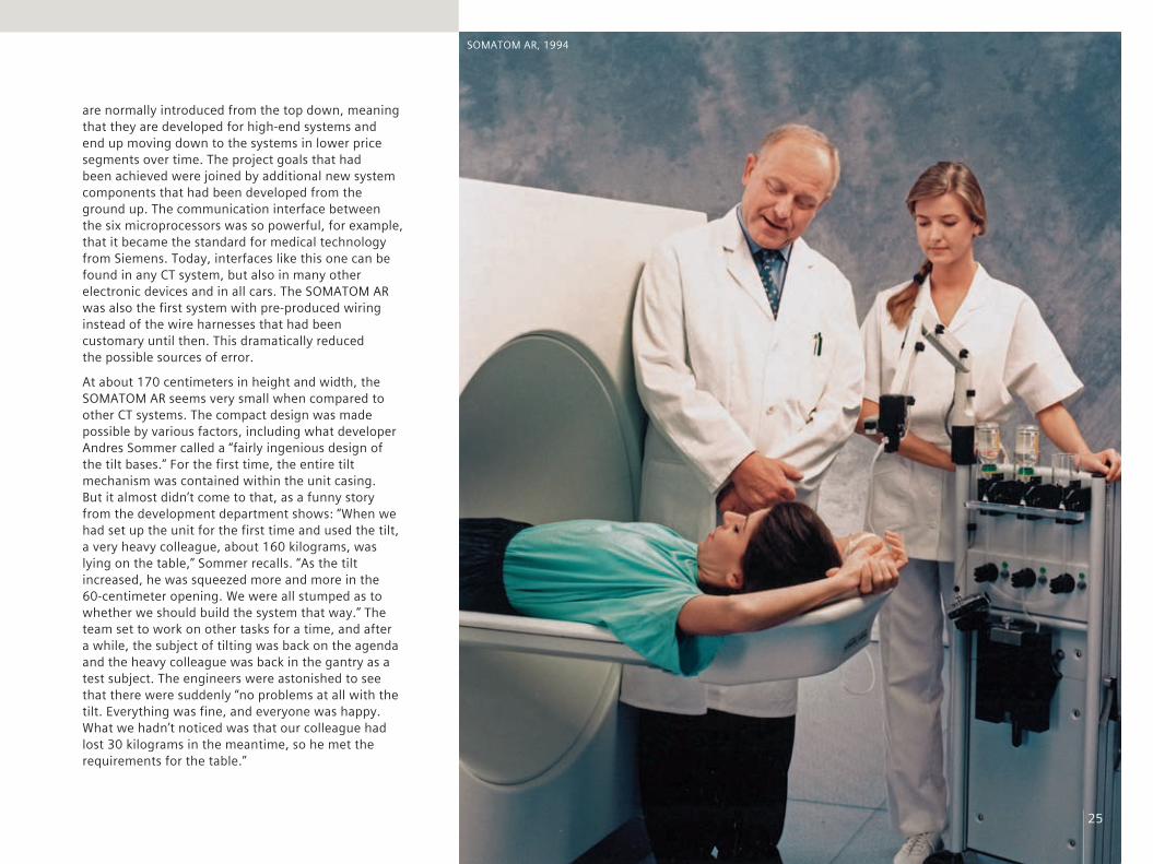
SOMATOM AR, 1994
are normally introduced from the top down, meaning that they are developed for high-end systems and end up moving down to the systems in lower price segments over time. The project goals that had been achieved were joined by additional new system components that had been developed from the ground up. The communication interface between the six microprocessors was so powerful, for example, that it became the standard for medical technology from Siemens. Today, interfaces like this one can be found in any CT system, but also in many other electronic devices and in all cars. The SOMATOM AR was also the first system with pre-produced wiring instead of the wire harnesses that had been customary until then. This dramatically reduced the possible sources of error.
At about 170 centimeters in height and width, the SOMATOM AR seems very small when compared to other CT systems. The compact design was made possible by various factors, including what developer Andres Sommer called a “fairly ingenious design of the tilt bases.” For the first time, the entire tilt mechanism was contained within the unit casing. But it almost didn’t come to that, as a funny story from the development department shows: “When we had set up the unit for the first time and used the tilt, a very heavy colleague, about 160 kilograms, was lying on the table,” Sommer recalls. “As the tilt increased, he was squeezed more and more in the 60-centimeter opening. We were all stumped as to whether we should build the system that way.” The team set to work on other tasks for a time, and after a while, the subject of tilting was back on the agenda and the heavy colleague was back in the gantry as a test subject. The engineers were astonished to see that there were suddenly “no problems at all with the tilt. Everything was fine, and everyone was happy. What we hadn’t noticed was that our colleague had lost 30 kilograms in the meantime, so he met the requirements for the table.”
25

SOMATOM Plus4, 1994
26
The SOMATOM AR came out in 1991. It was aimed at customers who wanted to shift from a conventional X-ray system to CT. To ensure that it was also available to customers in more remote areas, such as in Africa or India, the entire system was designed to be transported with just one truck, and, even more importantly, the SOMATOM AR needed so little energy that a standard electrical outlet was enough to supply it with power. The system was a complete success. Siemens produced almost three times more SOMATOM AR units than had been planned. Over the years, new models of the AR family were launched after being upgraded with new technologies and adapted to the various markets. In 1994, Siemens presented the SOMATOM AR.SP with spiral CT. Two years later, the AR family was reissued in a fresh, contemporary design. The technology inside stayed the same, since it had proven to be highly reliable; nearly a quarter-century later, in 2014, Siemens employees discovered a SOMATOM AR built in 1992 in China, still running perfectly and scanning 15 to 20 patients per day.
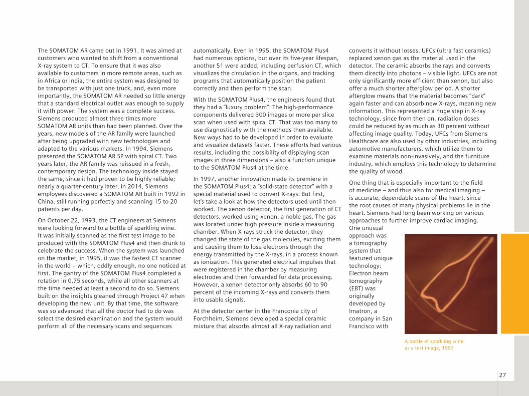
On October 22, 1993, the CT engineers at Siemens were looking forward to a bottle of sparkling wine. It was initially scanned as the first test image to be produced with the SOMATOM Plus4 and then drunk to celebrate the success. When the system was launched on the market, in 1995, it was the fastest CT scanner in the world – which, oddly enough, no one noticed at first. The gantry of the SOMATOM Plus4 completed a rotation in 0.75 seconds, while all other scanners at the time needed at least a second to do so. Siemens built on the insights gleaned through Project 47 when developing the new unit. By that time, the software was so advanced that all the doctor had to do was select the desired examination and the system would perform all of the necessary scans and sequences
automatically. Even in 1995, the SOMATOM Plus4 had numerous options, but over its five-year lifespan, another 51 were added, including perfusion CT, which visualizes the circulation in the organs, and tracking programs that automatically position the patient correctly and then perform the scan.
With the SOMATOM Plus4, the engineers found that they had a “luxury problem”: The high-performance components delivered 300 images or more per slice scan when used with spiral CT. That was too many to use diagnostically with the methods then available. New ways had to be developed in order to evaluate and visualize datasets faster. These efforts had various results, including the possibility of displaying scan images in three dimensions – also a function unique to the SOMATOM Plus4 at the time.
In 1997, another innovation made its premiere in the SOMATOM Plus4: a “solid-state detector” with a special material used to convert X-rays. But first, let’s take a look at how the detectors used until then worked. The xenon detector, the first generation of CT detectors, worked using xenon, a noble gas. The gas was located under high pressure inside a measuring chamber. When X-rays struck the detector, they changed the state of the gas molecules, exciting them and causing them to lose electrons through the energy transmitted by the X-rays, in a process known as ionization. This generated electrical impulses that were registered in the chamber by measuring electrodes and then forwarded for data processing. However, a xenon detector only absorbs 60 to 90 percent of the incoming X-rays and converts them into usable signals.
At the detector center in the Franconia city of Forchheim, Siemens developed a special ceramic mixture that absorbs almost all X-ray radiation and
converts it without losses. UFCs (ultra fast ceramics) replaced xenon gas as the material used in the detector. The ceramic absorbs the rays and converts them directly into photons – visible light. UFCs are not only significantly more efficient than xenon, but also offer a much shorter afterglow period. A shorter afterglow means that the material becomes “dark” again faster and can absorb new X-rays, meaning new information. This represented a huge step in X-ray technology, since from then on, radiation doses could be reduced by as much as 30 percent without affecting image quality. Today, UFCs from Siemens Healthcare are also used by other industries, including automotive manufacturers, which utilize them to examine materials non-invasively, and the furniture industry, which employs this technology to determine the quality of wood.
One thing that is especially important to the field of medicine – and thus also for medical imaging – is accurate, dependable scans of the heart, since the root causes of many physical problems lie in the heart. Siemens had long been working on various approaches to further improve cardiac imaging. One unusual approach was a tomography system that featured unique technology: Electron beam tomography (EBT) was originally developed by Imatron, a company in San Francisco with
A bottle of sparkling wine as a test image, 1993
27

which Siemens worked in the 1990s. Unlike in conventional CT systems, in which the X-ray tube assembly and detector system rotated around the patient, the gantry of an EBT scanner did not contain any moving mechanical parts. The X-rays were generated in a three-meter-long vacuum tube behind the gantry. Electrons moving at high speed struck anode ring segments arranged in a semicircle in the lower part of the gantry, where they generated the X-rays that struck the detector semicircle in the upper part of the gantry.
The advantage of EBT technology is its extremely rapid scanning time, at just 50 milliseconds per slice –
Miyabi Angio-CT, 1998An image of the lungs produced with the SOMATOM Plus4, 1997
SOMATOM Plus4, 1994
perfect for cardiac CT. But the method also had serious disadvantages, especially when it came to visualizing other areas of the body. The quality of the images was not even close to that of images produced by conventional CT scanners, and further development activities also did not promise any significant improvements. With this in mind, Siemens decided in the mid-1990s to halt development of electron beam tomography and focus its resources on other approaches – about which the rest of this section will tell more.
After the innovations of the previous ten years, there was no comparison between early systems and the
diagnostic quality and user and patient friendliness offered by the new units. Along with the SOMATOM Plus and SOMATOM AR product families, Siemens offered numerous other systems tailored to the various needs of its customers, such as the Miyabi Angio-CT system, a combination of a full angiography workstation and a CT system that slides on a track. Many limits had been overcome, but in the mid-1990s, the engineers were faced with a new one: The power of X-ray tubes could not be further increased at will without affecting the tube lifespan. But there was a way to put the existing power to better use while at the same time making a huge leap in cardiac CT: multislice CT.
28

The UFC detector for the SOMATOM Plus4, 1997
29

1998
30

Up until this point, physicians had to decide between volume size and sharp detail: Was it necessary to visualize the entire organ, or would it be enough to produce images of thin slices, but at a higher resolution? With the SOMATOM Volume Zoom, the question became moot. This was due to two factors. First, the rotation time was just 0.5 seconds per rotation, and second – and more importantly – the machine featured the new multislice technology. Conventional detectors scan one slice per revolution. In multislice technology, the photodiode is spread among the detector elements on separate rows that process the signals transmitted by the X-ray tube independently of each other, thereby recording several slices per rotation – in the SOMATOM Volume Zoom, four at once. This multiple-row detector utilizes the X-ray output significantly more efficiently, enables image resolution that is as much as eight times higher in a longitudinal direction relative to the patient, and considerably reduces scanning time for large areas of
the body. Siemens achieved this high resolution by arranging the individual slices in a certain way. In an “adaptive array detector,” the slices are very narrow, widening toward the outer edges. Because variable settings are available for the X-ray tube collimator, resolutions of between 0.5 and 5 millimeters per slice can be selected, producing much thinner slices than previously.
To Siemens, the main goal of developing multislice CT technology was to further reduce scan times and further enlarge the possible scan volume. But the SOMATOM Volume Zoom was a milestone in many ways, representing a paradigm shift in computed tomography. Up until then, vascular examinations had been performed invasively, generally using catheters. Multislice technology ushered in the era of routine vascular imaging using CT scanners. It also brought a fundamental shift in how scan results were viewed: Because the slices were much thinner, significantly larger volume data sets were now available. This
A paradigm shift in computed tomography
meant that it became less and less common to visualize individual slices, and the grand era of three-dimensional imaging began. The SOMATOM Volume Zoom was also the first unit to have an automatic operating concept. Before then, users had to think about the right scan parameters before every scan, but now the SureView software handled this aspect, determining the optimum settings for the scanner. The SOMATOM Volume Zoom marked an especially important point in the history of cardiac CT. The first CT image of the coronary vessels was produced at the Klinikum Grosshadern facility, in Munich, in 1999. It took about 40 seconds, but Siemens recognized that there was further potential and began devoting great efforts to pushing development forward, in cooperation with clinical partners. Over the next few years, this would turn out to be a major contribution to the field of cardiac imaging – without Siemens, there would probably be no such thing as routine cardiac CT today.
Multislice spiral CTSingle-slice spiral CT Presentation at the RSNA, 1998 Image of coronary arteries at Klinikum Grosshadern, Munich, 1999
31

1998
–200
5 Three-dimensional image of the interior walls of the intestine in syngo, 2003
32

In the 1970s, computed tomography was a revolution. In the 1980s, there was still something special, something exclusive, about it. By the early 1990s, it had become a matter of course, an established technology and a crucial part of everyday clinical practice. Entry-level models put CT within reach even for hospitals and radiology practices with smaller budgets. Several major innovative leaps, especially continuous rotation and spiral CT, improved image quality and significantly expanded the range of applications. On the eve of the new millennium, the dose per scan was just a fraction of the X-ray power that was needed in older CT scanners. This was due to two factors: significantly more efficient hardware, such as the UFC detector, and software specially developed for this purpose, such as Combined Applications to Reduce Exposure (CARE). Among other things, CARE technology calculated the smallest possible dose that would deliver the best possible
image quality for each patient individually. Depending on the patient’s anatomy, CARE was able to reduce the dose of radiation by as much as 56 percent.
Siemens unveiled another groundbreaking software innovation in 1999. With syngo, the company became the first manufacturer of medical technology to craft a standardized user interface for all of its systems. CT scanners, magnetic resonance imaging (MRI) systems and other imaging systems from the same manufacturer had previously had different software interfaces – and operators had to learn how to use each and every one of them separately. syngo allowed Siemens to standardize the operation of its equipment. When a hospital or medical practice purchases a new system from Siemens, the learning curve for staff is much shorter. The graphical user interface consists entirely of simple, self-explanatory symbols. Behind the syngo interface are numerous
functions that have been optimized for workflows in clinical settings and medical practices. For example, all of the data on a patient can be compiled in the electronic patient file, so the physician can always keep track of past scans and tests, including CT findings, lab results, and operative reports. Cross-department networking speeds workflows, allowing doctors to focus more on patients. Siemens, too, benefits directly from syngo. The interface makes integrating new developments into the existing system much easier. In February 2000, the company received the iF Interaction Design Award from the international judging panel of Industrie Forum Design Hannover for syngo. The jury’s statement: “In short, the epitome of a user interface, which is already clear from the fact that work steps and connections were readily apparent even to us as laypeople without background knowledge of the subject.”
Ultra-fast, smooth-running medical care is a must in hospital emergency rooms. Trauma rooms are where initial care for patients with serious injuries or trauma starts. Doctors there maintain the patient’s vital functions and diagnose the injuries or the physical cause of the emergency. CT is excellently suited to providing rapid diagnoses. But at this point, it was still necessary for patients who were to undergo a CT scan to be moved, first from the operating table to the CT table and then, at most hospitals, also from the trauma room to the scanner room. The optimum solution for emergency rooms would be if the patient could lie motionless on a freestanding operating table and the CT scanner could be moved to the patient for diagnoses instead of the other way around. With this
Small dose, big progress
SOMATOM Plus 4 CARE ad, 1994 Preparing for a scan in a trauma room, 2001
33

in mind, Siemens developed the Sliding Gantry – a SOMATOM on rails – in 1998. In the new system, the first of its kind in the world, the patient lay completely still on the table and received all of the necessary care from medical staff without any space restrictions. If necessary, a fully functional SOMATOM Plus4 could be moved into the scanning position in less than one minute. The system could also be combined optionally with the Siemens MULTISTAR Plus angiography C-arm and various ultrasound diagnostic systems.
Not long after the development of the SOMATOM Plus4 Sliding Gantry, Siemens received an inquiry from James Wong, Chair of the Department of Radiation Oncology at Morristown Memorial Hospital in Morristown, New Jersey. Wong was using linear accelerators to fight cancer. A linear accelerator is a radiation treatment device that accelerates electrons almost to the speed of light and beams them at cancer cells. Up until 1999 radiation therapy had involved “flying blind.” This meant that when treatment plans were drawn up, the tumor’s position was determined one time and it was assumed that it was always in the same location, even if the patient was positioned
slightly differently during different treatment sessions. A simple detector was used to check the location of the bones, but it was usually not possible to see the tumor in the process. On top of that, organs do move inside the body. These factors meant that radiation fields were expanded, in some cases significantly, in order to be sure that all of the cells of the tumor would be covered. What Wong wanted to do was check the tumor’s position with a combination of a linear accelerator and CT scanner before every radiation treatment.
The team headed by engineer Andres Sommer at Siemens in Erlangen began looking for solutions to the mechanical issues and concluded that „this kind of solution was easy to build“. They relied on the principle of the Sliding Gantry to develop the PRIMATOM, the world’s first 3D image-guided radiation therapy system.
The team installed this cross between a CT scanner and a linear accelerator in Morristown in April 1999. Several months later, Sommer visited the site for a closer look at the workflows involved. He discovered that analyzing and interpreting the images and resetting the system for the next patient took a very long time. Conveniently, the team was working on developing the first version of an image reconstruction software program based on syngo at the time. Specifically for the PRIMATOM, they added a new software function that showed the necessary shifting of the patient in 3D images. The visit had been worthwhile. The system now worked ten times faster than before.
In addition to significantly greater accuracy, the PRIMATOM also brought other advantages to radiation therapy, including this one: “Integrating imaging into therapy unlocks a new dimension for us in treating cancer patients, namely the ability to consider how the tumor changes over time during the course of treatment,” as Dr. Jürgen Debus, a professor and Medical Director of Clinical Radiology at the Heidelberg University Hospital, said in 2004. At the time, more than 500 patients had been treated with the first PRIMATOM in Europe, which was being operated at the German Cancer Research Center (DKFZ) in cooperation with the University of Heidelberg. The PRIMATOM with syngo and the additional software function represented a milestone in radiation therapy, and it is still recognized as the gold standard for image-guided systems to this day.
In the fall of 1999, the field of CT at Siemens entered uncharted territory. The UK National Maritime Museum in London, the Royal London Hospital, and the medical technology arm at Siemens teamed up to learn more about the craftsmanship of British shipbuilders in the 17th and 18th centuries. Endoscopic scans performed in the past had yielded only modest insight, and so the plan now was to use a CT scanner to generate cross-sectional images of the ships. Since the 70-centimeter gantry of a SOMATOM Plus4 was a bit too small to fit a life-size sailing ship, the researchers scanned historical model ships, which had been built at the time by British carpenters to serve as exact prototypes for shipbuilding. The engineers at Siemens adjusted the image processing algorithms to accommodate the particular characteristics of the materials used in the ships. Naturally, the research took place in the evening so that it would not impede routine operations at the hospital.
The length of the gantry is irrelevant when scanning model ships – but when dealing with patients, it’s a different story. Some people feel cramped inside the
PRIMATOM, 1999 Image of model ships, 1999
34

SOMATOM Emotion, 2001
“tube,” and some get an uneasy feeling just looking at it. This means that one important goal of any development in this area of medical technology is to boost patient comfort and make the scan as pleasant as possible. As a result, the new SOMATOM models launched in 1999, Emotion and Balance, featured a gantry that was just 56 centimeters long – at that time, the shortest on the market. The compact design not only made undergoing the scan easier for many patients, but also benefited operators, who had better access to the patient. For these and other aspects of their design, such as improved service and maintenance friendliness, the SOMATOM Emotion and SOMATOM Balance received the iF Product Design Award for the year 2000.
35

A few weeks after the awards ceremony, Siemens launched the SOMATOM Esprit. The new model also had a gantry just 56 centi-meters long, but it was also even more compact overall. The entry-level SOMATOM Esprit did not have an additional cooling
system, which meant that it needed just 17 square meters of space. At the same time, the system came equipped with features otherwise found only in larger CT scanners. The standard features included a UFC detector and spiral CT, and functions such as CT angiography or CARE Bolus, a program to reduce the amount of contrast agents, could also be added upon request. The system could be installed and ready for the first scan in just one day.
The computer used for CT systems is based on microprocessors, just like in a cell phone or PC. In 1965, Gordon Moore, one of the co-founders of Intel, observed a correlation in the development of electronic components that, in a slightly modified form, still applies to microprocessors to this day: “Moore’s law” holds that the power of microprocessors doubles every 24 months. This means that if a hospital buys a CT scanner today, that same system, equipped with a new microprocessor, would work even faster in a few years, and it would even be possible to add new functions and applications. For customers who wanted to keep their system up-to-date, Siemens began offering its Evolve service package, later marketed under the name syngo Evolve Package, for all imaging systems. The SOMATOM Volume Zoom and Volume Access systems installed between August 1999 and October 2000 were the first to benefit from this program, receiving hardware
that was four times faster in July 2002. The package included the syngo VA40 software upgrade, which brought various features with it, including new applications for pediatrics and cardiology. Customers also had the option to add new functions to their systems, such as the ability to do a virtual “fly-through” of a patient’s intestine. Users were able to adjust the scope of the service package on an individual basis to suit their needs. More than 75 percent of all customers purchasing a new SOMATOM in 2002 chose to receive this service.
Each of the established imaging methods has its own major strengths, meaning that it is especially suitable for certain types of scans. Ultrasound diagnosis is the first choice for many routine examinations, such as for preventive care during pregnancy; magnetic resonance imaging (MRI) delivers extremely sharp, detailed images of areas of soft tissue, such as the brain; and computed tomography offers high-resolution images of the skeleton and precision results when time is of the essence, such as when a stroke is suspected. Two other important methods used for clinical imaging are positron emission tomography (PET) and single-photon emission computed tomo-graphy, or SPECT. These nuclear medicine methods can be used to obtain a detailed picture of bodily functions and metabolic processes. They are used primarily in diagnosis and treatment of cancer, heart disease and neurological disorders. But because they are geared specifically toward biochemical processes, PET and SPECT are of limited utility in visualizing the anatomical details of the body. In many scans, however, simultaneously visualizing metabolic processes and anatomical structures with accuracy down to the sub-millimeter level is crucial in terms of optimum treatment planning – for example, being able to determine quickly and accurately where and to what extent a patient’s heart muscle has sustained damage due to an inadequate supply of blood after a heart attack. Up until then, this kind of diagnostic
procedure required two separate scans, one with a PET or SPECT scanner and one by CT, and then the result images were superimposed over each other. This method was time-consuming and meant a lot of work for medical staff, and it was also cumbersome for patients.
Hybrid systems could combine the particular strengths of these methods of nuclear medicine with those of CT in order to detect certain diseases and disorders even faster, earlier, and with greater reliability. The same idea had also occurred to David Townsend, of the University of Pittsburgh, and Ron Nutt, of CTI PET Systems, in Knoxville, Tennessee, a joint venture between Siemens and CTI. They applied for a patent on the idea of combining PET and CT technology, with plans to build the first hybrid system with support from the CT team at Siemens. To this end, Siemens delivered a SOMATOM AR with spiral CT capability from Forchheim to Pittsburgh in 1997. Thomas Beyer, then a research associate at the university, built a prototype there by combining the SOMATOM AR with a PET system from Knoxville. The first scans, performed on more than 300 cancer patients, already showed impressive results. Building on this, a Siemens team was tasked with getting the combination system ready for the market. They produced a special CT scanner based on the SOMATOM Emotion, sent it to Knoxville, and built a combined PET/CT scanner there, launching it on the market in 2001 as the Siemens Biograph. The path they had taken – opening up the possibility of producing simultaneous CT and PET images – soon turned out to be the right one. By about five years later, individual PET scanners were no longer being sold. The Biograph was built in nine versions, with multislice technology and numerous new functions and improvements.
In the early 2000s, Siemens began considering whether a hybrid of SPECT and CT was also possible without lowering quality standards, and if so, how. A team of engineers working in nuclear medicine and
SPECT-CT Symbia, 2004
36

Presenting the first PET/CT combination scanner at the RSNA, 2000
computed tomography planned the unusual architecture, decided on the SOMATOM Emotion as the CT component of the system, and combined it with the latest SPECT technology, the e.cam. Siemens presented the results of this development, the TruePoint SPECT-CT, in mid-2004, giving the new product family the name Symbia. The system was extremely versatile. In addition to combined SPECT-CT scans, Symbia systems could also be used for purely SPECT or CT scanning, depending on the doctor’s needs. The engineers’ decision to use the SOMATOM Emotion turned out to be just right. The SOMATOM Emotion is still on the market to this day, and upgrades and improvements for this scanner can be imported seamlessly into the Symbia family.
In December 2001, a scant three years after com puted tomography had first been used to visualize the coronary arteries, Siemens took the next step by introducing the world’s first 16-slice multislice CT scanner. The SOMATOM Sensation 16 now made even the surrounding segments of the coronary arteries and their delicate side branches visible. The leap from four to 16 slices and the even faster rotation time – just 0.4 seconds – brought numerous advantages. For example, it now took only about ten seconds to perform a lung scan. But there was one area where the SOMATOM Sensation 16 really excelled: cardiac imaging in conjunction with the “HeartView CT” software. This program even allowed doctors to see areas of narrowing and deposits in the coronary arteries, a hugely important factor in terms of early detection in particular. For their work on HeartView CT, the development team headed by Bernd Ohnesorge and Thomas Flohr was nominated for the Deutscher Zukunftspreis, the highest award for innovation in science and technology given in Germany, in 2002. That same year, the Design Award of the Federal Republic of Germany went to the CT development unit at Siemens in recognition of a very special system: the SOMATOM Smile.
37

An entry-level model, the SOMATOM Smile was designed for the needs of private radiology practices and smaller medical facilities in China, Southeast Asia, and Brazil. The system was simply plugged into a normal electrical outlet and was ready to go in just three hours. If technical problems cropped up, an intelligent self-test function helped to identify the
malfunctioning or defective parts and then displayed the correct order number onscreen. Customers could order the replace-ment component from Siemens and replace it them-selves based on convenient color-coding. The concept used to operate the system was also revolutionary: The unit came with a CD-ROM con taining training software that taught users – step by step and in clear, easily understood terms – how to use all of the system’s
functions, from turning on the scanner to patient positioning and preparing images that could be used for diagnosis. For its ease of use, appealing design, and intelligent overall concept, the SOMATOM Smile not only won the Design Award of the Federal Republic of Germany, but also brought Siemens the red dot award for 2001 and the iF Design Award for 2002.
Computed tomography places an immense strain on the X-ray tube, especially since the introduction of spiral CT and the high speeds that come with it. Up until then, tubes had worked according to the following principle: The airless X-ray tube contained a cathode and a rotating anode that were linked to each other through the application of high voltage. The cathode was heated up, causing it to discharge electrons. The electrons were accelerated and collided with the anode. This generated X-ray radiation, which was then beamed out of the tube. The weak points of this design are the development of heat and the position of the rotating anode. The longer a CT scan lasts, the hotter the anode gets. It is cooled by discharging the heat through the vacuum to the cooling oil surrounding the tube. This cooling process is not very efficient, necessitating longer pauses to cool down between scans. Two major design changes significantly boosted X-ray tube performance: pivot bearings outside the tube, for greater stability, and direct cooling of the anode.
Back in 1993, Willi A. Kalender and Wolfgang Knüpfer started working at Siemens on what they called a rotating envelope tube, an approach later continued by a team of developers headed by Peter Schardt and Erich Hell. In this design, it is not the anode that rotates, but the entire vacuum tube instead. This makes the mechanical construct significantly more robust and compact, yielding further benefits. The most important factor is the direct cooling of the anode: Compared with conventional CT tubes, the rotating envelope tube discharges about ten times more heat. The rapid cooling meant that the engineers could install anodes that were significantly smaller, and thus also weighed less, which further enhanced mechanical stability and made it possible to achieve even greater rotation speeds. Another prominent new development lay in the details: The “z-spring focus” recorded every projection from two to four different perspectives during the scan, significantly improving image clarity. In 2003,
Siemens presented this unique development to the public under the name STRATON X-ray tube – an innovation that brought the team headed by Peter Schardt and Erich Hell a nomination for the Deutscher Zukunftspreis award for innovation in science and technology in 2005.
The ability to bring CT systems fully up to date without completely redesigning the underlying framework was a major goal for the engineers with each development. The SOMATOM Sensation family offers a good example of the benefits of this approach. In 2001, the SOMATOM Sensation 16 was the first system in the world with a 16-slice detector. With z-spring focus technology, the engineers at Siemens quadrupled the number of slices that could be scanned at once, making the latest model introduced in 2003, the SOMATOM Sensation 64, the world’s first 64-slice CT system. Special optimizations for specific requirements could also be realized within a system family. Siemens launched the SOMATOM Sensation Cardiac 64, a system specifically optimized for cardiovascular imaging, in 2004. The rotation time was faster than the base system, at 0.37 seconds to the base system’s 0.33, and additional hardware and software designed specifically for cardiac scans came pre-installed. At the same time, the SOMATOM Sensation Open made it easier to scan overweight and obese patients. This scanner built on the SOMATOM Sensation 16, but with upgrades including an 82- centimeter gantry and a STRATON tube. This shows that technological evolution, with all the innovations, improvements, and new application options that come with it, increasingly boosts performance across a system family over the years. A technological revolution generally depends on fully redesigning the basic framework and then introducing the new design in a new system family – just like with the next groundbreaking CT system from Siemens: the SOMATOM Definition, the first dual-source CT scanner.
A conventional CT tube versus STRATON, 2005
SOMATOM Smile, 2000
38

SOMATOM Sensation 16, 2002
Heart scan with SOMATOM Sensation Cardiac 64, 2004
39

2005
Trial run with test body, 2006
40
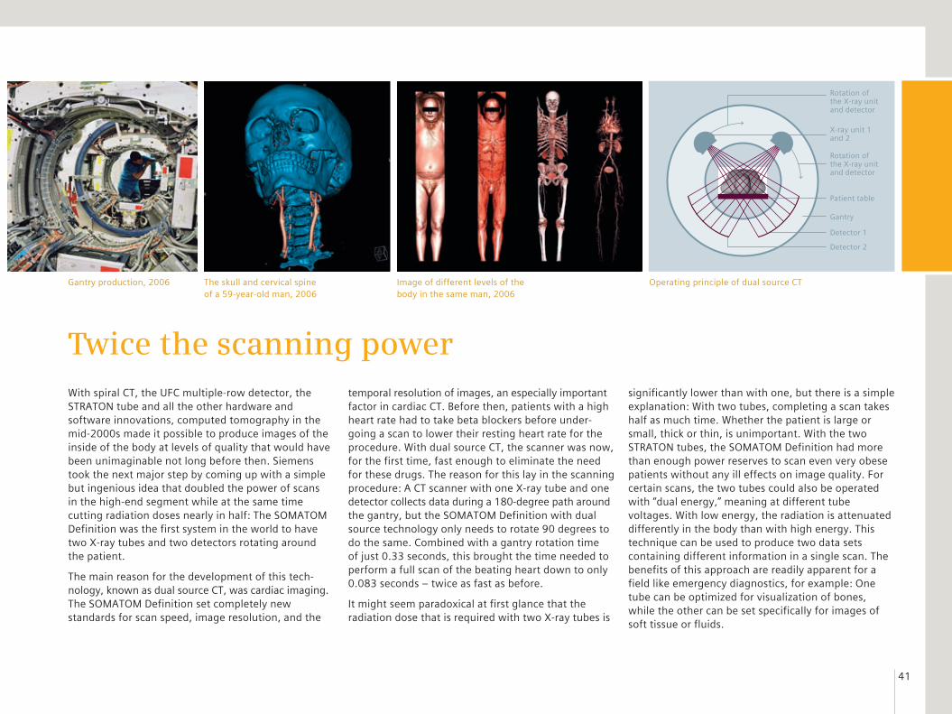
With spiral CT, the UFC multiple-row detector, the STRATON tube and all the other hardware and software innovations, computed tomography in the mid-2000s made it possible to produce images of the inside of the body at levels of quality that would have been unimaginable not long before then. Siemens took the next major step by coming up with a simple but ingenious idea that doubled the power of scans in the high-end segment while at the same time cutting radiation doses nearly in half: The SOMATOM Definition was the first system in the world to have two X-ray tubes and two detectors rotating around the patient.
The main reason for the development of this tech-nology, known as dual source CT, was cardiac imaging. The SOMATOM Definition set completely new standards for scan speed, image resolution, and the
temporal resolution of images, an especially important factor in cardiac CT. Before then, patients with a high heart rate had to take beta blockers before under-going a scan to lower their resting heart rate for the procedure. With dual source CT, the scanner was now, for the first time, fast enough to eliminate the need for these drugs. The reason for this lay in the scanning procedure: A CT scanner with one X-ray tube and one detector collects data during a 180-degree path around the gantry, but the SOMATOM Definition with dual source technology only needs to rotate 90 degrees to do the same. Combined with a gantry rotation time of just 0.33 seconds, this brought the time needed to perform a full scan of the beating heart down to only 0.083 seconds – twice as fast as before.
It might seem paradoxical at first glance that the radiation dose that is required with two X-ray tubes is
Twice the scanning powersignificantly lower than with one, but there is a simple explanation: With two tubes, completing a scan takes half as much time. Whether the patient is large or small, thick or thin, is unimportant. With the two STRATON tubes, the SOMATOM Definition had more than enough power reserves to scan even very obese patients without any ill effects on image quality. For certain scans, the two tubes could also be operated with “dual energy,” meaning at different tube voltages. With low energy, the radiation is attenuated differently in the body than with high energy. This technique can be used to produce two data sets containing different information in a single scan. The benefits of this approach are readily apparent for a field like emergency diagnostics, for example: One tube can be optimized for visualization of bones, while the other can be set specifically for images of soft tissue or fluids.
Image of different levels of the body in the same man, 2006
Operating principle of dual source CTGantry production, 2006 The skull and cervical spine of a 59-year-old man, 2006
Trial run with test body, 2006
Rotation of the X-ray unit and detector
X-ray unit 1 and 2
Rotation of the X-ray unit and detector
Gantry
Patient table
Detector 1
Detector 2
41
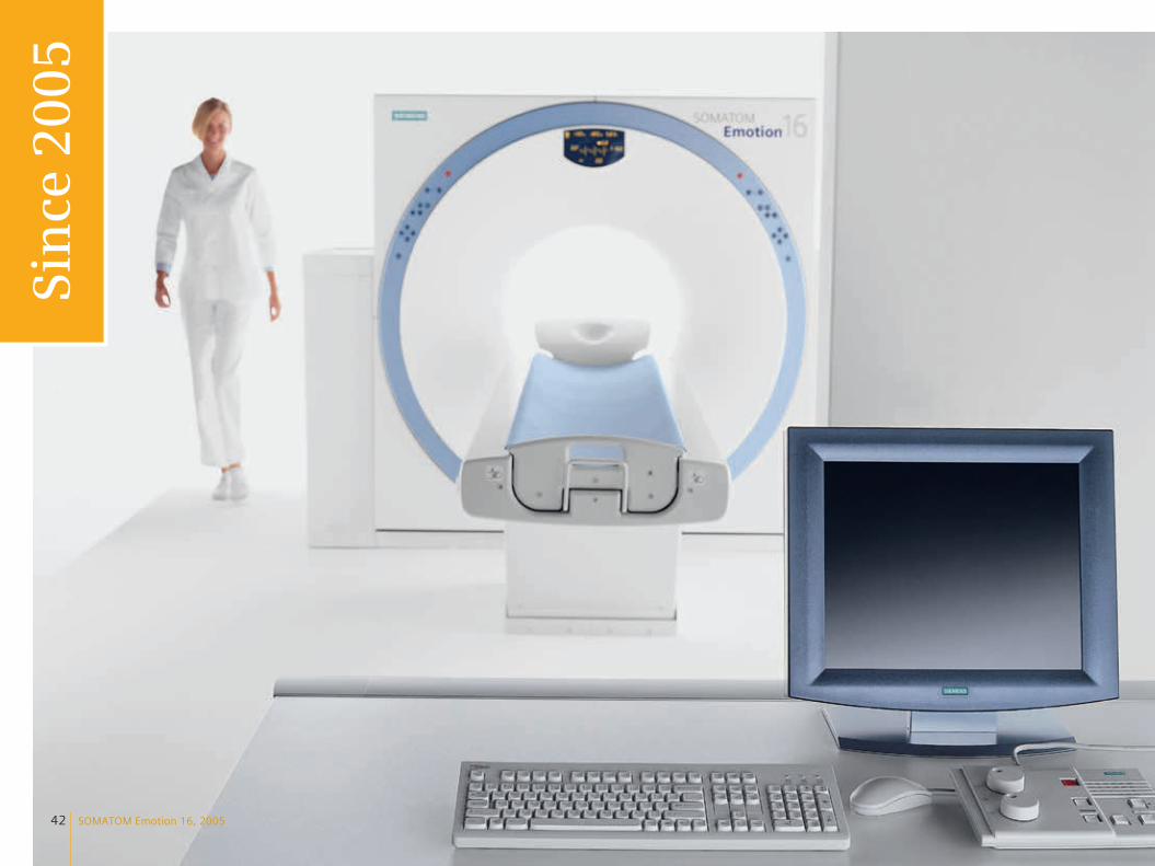
Sin
ce 2
005
SOMATOM Emotion 16, 200542

The long chain of individual technical improvements that scientists and engineers all over the world have worked on has made computed tomography into an essential tool for everyday clinical use over the years. Comparing older images with those produced today paints an especially vivid picture of how far this technology has come, but the figures are also impressive in their own right: Back in the 1970s, the first CT systems took about five minutes to scan one slice, a time that had been reduced to just 4 to
10 seconds by 1980 and 1 to 2 seconds ten years later on. The fastest scanners in 2005 needed just 0.33 seconds to do the same. Advances in CT technology are similarly apparent if we look at the volume of data collected for each 360-degree rotation, which rose from less than 0.06 megabytes (MB) in a prototype from 1972 to 1 MB in the early 1980s, 2 MB in 1990, and finally as much as 100 MB in 2005. Each spiral scan produced in 1990 generated between 24 and 48 megabytes of data, while a scanner from 2005 collected up to 4000 MB in just a few seconds. Over the years, innovations from Siemens have brought fresh drive to the field of computed tomography time and again, even spurring advances in whole new directions and setting trends in many cases.
The latest Siemens development in 2005, dual source CT, sounds like an easily implemented idea: Just integrate a second tube, put in a second detector, and there you have it – a dual source scanner. In reality, the technical side of implementing this idea was a huge challenge for the engineers, because the gantry was already very tightly packed in conventional systems with single source CT. Without the compact STRATON X-ray tube, dual source CT would not have been, at least not without changing the structure and making the scanner significantly larger. But the new tube technology alone was not enough to make the leap. The engineers had to optimize almost all of the other components as well and make them more compact. This included the entire cooling system and
the way that the electronics were arranged in the gantry. It was just this task – developing even more powerful, more efficient components – that would keep the engineers busy over the years to come as well.
It all started with the optimization of the SOMATOM Emotion, which had become one of the most commercially successful CT scanners in the world since the first model was launched. The new model introduced in 2005 turned the system from a low-cost entry-level model with a single-slice detector to a low-cost entry-level model with a 16-slice detector. The fully overhauled system retained all of the strengths of the predecessor model, including the small installation space of just 18 square meters, and low power consumption, but it also offered numerous new functions. The new unit had an Internet connection, adding flexibility for operators. Optional syngo WebSpace software allowed doctors to access and work with scan results in encrypted form via the internet. The Guardian service program was installed to monitor the scanner’s functions online and flag any abnormalities before disruptions could arise.
A similar system from the SOMATOM Emotion family helped clear up a suspected murder. The question of whether famed pharaoh Tutankhamen had been killed by a blow to the head more than 3,000 years before was a source of considerable debate among Egyptologists. In January 2005, Egyptian
Improved detail for big impact
syngo WebSpace, 2006
43

The 3,300-year-old mummy of the pharaoh Tutankhamen before the CT scan, 2005
44

researchers cracked the case with help from a SOMATOM Emotion 6. The 1,700 tomographic images taken of the mummy showed no signs of murder; instead, the results pointed to the effects of a hunting accident. At the same time, the CT images also showed that Tutankhamen was between the ages of 18 and 20 at the time of his death, not between 23 and 27, as some Egyptologists had estimated. As part of the research project, the Egyptian Council of Antiquities scanned numerous other mummies and historical finds, some of them about 5,000 years old. The SOMATOM Emotion 6 was loaded on a tractor-trailer and driven to wherever it was needed. This meant that the delicate ancient Egyptian remains were hardly moved at all, treating them as carefully as possible.
A bust of the Egyptian ruler Nefertiti had already been scanned using CT technology back in 1992, a process that yielded a startling secret: Inside the sculpture, there was a second portrait of Nefertiti in limestone that looked different from the outer depiction. CT had made such tremendous advances since the time of this first scan that the hidden portrait could now be shown in full detail. With this in mind, the National Geographic Channel decided to re-scan the queen in 2007 for a TV documentary, with help from Siemens. The new scan image, produced with a SOMATOM Sensation 64 and showing structures as tiny as 0.3 millimeters, even reveales clear lines around the mouth. The nose is less harmonious, the shoulders stocky and asymmetrical. Nefertiti seems older and
The hidden portrait becomes visible, 2007The bust of Nefertiti, 2007
less characteristic. The researchers suspect that the limestone core inside the bust is closer to the queen’s real image than the plaster exterior.
Solving archaeological mysteries was an exciting and interesting sideline, but computed tomography was naturally geared mainly toward use in medicine. In the mid-2000s, the technology was so accurate and advanced that it was hard to imagine that there was any further room for improvement. But work on technical details, patient comfort and user friendliness would continue over the next few years and bring astonishing further progress and new developments. At that time, Siemens started development of various features, including a new generation of detectors, and optimized the hardware and software used for the product families.
Siemens introduced a number of single source systems with flexible configuration options to join its top-of-the-line SOMATOM Definition model with dual source CT technology. In 2007, the high-end system with one X-ray tube was the SOMATOM Definition AS. The “AS” stands for “adaptive scanner,” meaning that the system adjusted to all patients and medical requirements. The Siemens engineers achieved this
flexibility by combining components such as a 78-centimeter gantry and a scan length of up to 200 centimeters for the first time in this system. This made the SOMATOM Definition AS suitable for a wide range of different patient groups, from overweight people to those with claustrophobia or children, and at the same time, it could perform complex neurology or cardiology scans with no restrictions, along with fast scans in emergency situations, such as trauma, stroke, or heart attack patients. These kinds of emergency examinations were also the main area of use for the first SOMATOM Definition AS ever delivered, which was installed at the trauma center at the Erlangen University Hospital in late 2007.
The CT systems from Siemens did not start being adaptable only once they were in clinical use; their flexibility started even before the purchase. All of the current systems offered by the company at the time could be configured by customers according to their requirements. From the number of detector rows to software and service packages, there were numerous options available. The systems could be upgraded with new hardware and software innovations as needed in the years to come. This was true of both
45

pure CT systems in the SOMATOM family and hybrid systems like the Miyabi, Symbia, and Biograph mCT families. In 2008, all of these systems were equipped with multislice detectors, and the Biograph mCT and many SOMATOM systems could even offer as many as 128 slices upon request.
In 2005, when Siemens became the only CT manufacturer to turn its back on the race for ever-greater numbers of detector rows and instead focus on the completely new dual source technology, the move was viewed as a risky one. But it soon became apparent that the venture had been worthwhile:
A few weeks after the first SOMATOM Definition was installed, experts estimated that a large portion of the approximately 600,000 catheter examinations performed each year could be replaced by cardiac CT. Clinical studies documented this technique’s benefits, especially for cardiac imaging, the place where computed tomography really excelled. Among other findings, researchers from Zurich University Hospital showed that with the SOMATOM Definition, it was possible to significantly lower the dose used for a cardiac scan in comparison to conventional computed
tomography. Siemens built on this success and on its experiences with dual source technology, and in 2009 launched a new model that was once again the fastest CT scanner in the world at the time: the SOMATOM Definition Flash.
An example from cardiac imaging provides a vivid picture of the advances that had been made with the SOMATOM Definition Flash. Previously, producing detailed images of the heart with as few artifacts as possible had required doses averaging between 8 and 30 millisieverts. The SOMATOM Definition Flash needed less than one millisievert to do the job. This
was made possible by a number of factors, including further improved and specialized syngo programs and, most especially, the premium scanner’s outstanding speed. The gantry rotated around the patient in 0.28 seconds, almost four times per second. At the same time, the patient was moved through the scanner twice as fast as in conventional systems in use at the time. This meant that a person two meters tall could be scanned from head to toe in less than 5 seconds, while a chest scan took 0.6 seconds and a cardiac scan just 0.25 seconds – less than half a heartbeat.
Biograph mCT, 2008
To further reduce the radiation dose, Siemens developed an innovative mathematical method of processing images. The IRIS (Iterative Reconstruction in Image Space) algorithm ran much faster than previous methods, despite the additional calculations involved, and made it possible to reduce the dose by a further 60 percent. The first systems were equipped with the new method starting in the spring of 2010. A year later, Fast Care software helped clinical personnel optimize the dose while also streamlining workflows. Reducing the dose any more would lead to “noise” in the image, meaning artifacts and
diminished image quality – unless an even more efficient detector was developed, that is.
The leap from xenon to solid-state detectors had already yielded significant improve-ments in converting X-ray signals. But this second generation of detectors had a weak point that affected image quality: After the X-rays were converted to light
signals, the photodiode transmitted them to a converter that transformed the analog electrical impulses into digital ones. This took place via several hundred wires. The longer these wires were, the greater the electronic noise – and the greater the electronic noise, the worse the image quality. Siemens launched the first detector that eliminated nearly all of these wires in 2012: In the Stellar detector, all of the electronics used for signal conversion are combined in a single chip located directly under the photodiode.
syngo.CT Cardiac Function software, 2013A foot scan with dual energy, Grosshadern University Hospital, Munich, 2007
46

Dual Source CT SOMATOM Definition Flash, 2009
47

During the early stages of the six-year process of developing the Stellar detector, it was not yet clear whether it would even be possible to combine the photodiode and converter in the first place. To replace the wiring, about 90 contacts per square centimeter, each just 0.1 millimeters in diameter, had to be etched through a silicon plate. There were also several layers of oxide isolating the tiny contact holes from the silicon. In cooperation with semiconductor manu-facturer ams, the engineers at Siemens were able to realize this completely new electronic design. The extremely short transmission routes significantly boosted signal quality. Stellar reduced electronic noise by as much as 30 percent, which brought a comparable dose reduction – with improved image quality at the same time.
The outer structure of the Stellar detector was the same as in conventional Siemens detectors. The rest of the system architecture remained unchanged, and current scanners such as the SOMATOM Definition Flash could easily be upgraded with Stellar. The Siemens SOMATOM Definition Edge, the new high-end model with single source CT in 2012, came standard with the new detector technology. With
Stellar, the system made structures as small as 0.30 millimeters visible. This system also marked the introduction of a new dual energy technology in single source CT that needed significantly less radiation than was customary for scans performed with this method using one X-ray tube.
Reducing the radiation dose also played an important role in the development of the Vectron X-ray tube, in 2013. The operating voltage of X-ray tubes can be set to between 70 and 150 kilovolts (kV). To keep the radiation dose low, especially for CT scans performed using contrast media, many radiologists prefer 70 kV or 80 kV as the voltage. Until then, however, only children and slender people could be examined using this method, known as “low-kV” scanning, since the X-ray tubes simply did not have enough power at low voltage. The Vectron tube eliminated this technical limitation. It had more than enough power reserves – over twice the amount of other CT tubes – to scan even people with thicker bodies using the low-kV method. In addition, the contrast-to-noise ratio was so high that it was even possible to reduce the amount of contrast medium used in CT angiography.
Spiral CT, multislice, UFC, the Straton and Vectron tube, Sliding Gantry, dual source, dual energy, the Stellar detector – these and many other hardware and software developments over the last 40 years have made Siemens the leading innovator in the field of computed tomography. The current system families for the year 2015 are the product of years of ex-perience and cooperation with scientists, researchers, and medical professionals all over the world. The portfolio today ranges from energy-efficient entry-level models to high-end scanners with two X-ray tubes and from systems for routine clinical appli-cations to highly specialized trauma room CT units on rails. Scanner families that have stood the test of time for many years, like the Biograph and Symbia, are always at the forefront of advances in technology, and they are joined by new product classes such as the SOMATOM Perspective or the SOMATOM Scope, introduced in mid-2014, an all-around CT for routine clinical use with ultra-low operating costs that needs just eight square meters of space. And the develop-ment process is far from over – as shown by the current highlight in the history of computed tomo-graphy at Siemens, the SOMATOM Force.
A dual energy scan with the SOMATOM Definition Edge, University of Erlangen-Nuremberg, 2014
A conventional detector element (top) and Stellar (bottom), 2012
The SOMATOM Scope, which needs just eight square meters of space, 2014
48

SOMATOM on rails, Frankfurt University Hospital, 2014
SOMATOM Perspective with up to 128 slices, 2014
49

2013
50

The prototype of what was once again the fastest CT scanner in the world was set up at the University Medical Centre Mannheim in late 2013. The gantry of the SOMATOM Force held a child carrier with a small, wiggly patient – a one-year-old boy who had been admitted to the hospital with a suspected case of pneumonia. An X-ray image had been taken, but the results were inconclusive. But then things moved very quickly. The SOMATOM Force scanned the infant’s chest in just 0.3 seconds – without any artifacts from his movements or breathing getting in the way of the final image. The image showed tiny areas of inflammation in the lung tissue, and it did so at a dose of radiation no higher than with a conventional X-ray. This finding allowed the doctors to initiate suitable treatment for the little patient right away.
The SOMATOM Force is the world’s most powerful CT scanner, pushing the limits of present-day
technological feasibility. The system took a 600-person team five years to develop. It brings together all of the high-end components from Siemens and pushes them even further. The 1.6-ton gantry rotates around the patient four times a second. That’s like a Mercedes E-Class vehicle tracing a circular path on a small round coffee table with five times the accelerative force of a fighter jet. At the same time, the two Stellar detectors and the Vectron X-ray tubes have to be kept in position with the utmost accuracy, down to not only the millimeter, but even the micrometer. Together with the table speed, which has been increased from 45 to 73.7 centimeters per second – the fastest on the market – this means the system can now scan an adult’s entire upper body in under one second. The achievable resolution is 0.24 millimeters, as compared with the 0.33 millimeters possible with the previous model. Without any compromises in image quality,
40 years of experience in a single unitthe SOMATOM Force scans a lung at a dose of just 0.1 millisieverts – about the same as the natural level of radiation exposure occurring on a flight from Germany to Argentina.
Over the last 40 years, new areas of application for CT have been unlocked on an ongoing basis. The tech-nology is now essential in many disciplines, such as cardiac imaging. Current systems have changed clinical practice: The necessary dose is now so low that CT scanners are used for regular checks such as to monitor the progress of treatment and for early detection of diseases, such as for lung scans in smokers. On average, eight people are scanned in Siemens CT systems worldwide every second, and CT is growing ever more important as it is developing more and more into a comfortable, convenient, and uncomplicated procedure – in short, CT scans will become the new X-rays.
Upper body scan, Mannheim University Medical Centre, 2013
SOMATOM Force in Mannheim, 2013SOMATOM Force, 2013
51

Loca
tio
ns
Detector production at the main plant in Erlangen, 198152

In 1975, Siemens built the first SIRETOM in a relatively small production hall in Erlangen and delivered it to Munich. Today, the company produces more than one thousand systems a year at three sites spread around the globe and ships them all over the world. The Siemens plant in Forchheim is home to the largest and most advanced computed tomography production facility worldwide. Siemens produces CT scanners for the Asian market in Shanghai, and in August 2012, a 6,000-square-meter Siemens factory in Joinville, Brazil, began supporting worldwide sales activities involving the SOMATOM family. The sites are seamlessly connected with each other. In close cooperation with other business units at Siemens, teams work on hardware innovations and develop new software applications. All of the X-ray tubes used in Siemens CT scanners come from the company’s own X-ray plants, which are located in Rudolstadt, Erlangen, and the Chinese city of Wuxi.
Forchheim, Germany Siemens Healthcare opened a small branch location in Forchheim, about 20 kilometers away from the company’s headquarters in Erlangen, in 1986. At first, about 280 employees worked here, producing sheet metal housings for medical equipment, but now, the site has become one of the most important locations for medical technology at Siemens, with a staff of about 1,800. Siemens began expanding the site at regular intervals right from the very start, and finally moved the company’s growing CT manufacturing activities from Erlangen to Forchheim. In November 1994, after 13 months of construction, the company began operating one of the world’s most advanced factories for medical technology: the only CT produc-tion site in Europe and an expanded plant for angio-graphy systems. The two units were closely inter-connected in terms of development, manu facturing, quality management, international logistics, and marketing. The site has received numerous awards and distinctions over the years, including the title of “Factory of the Year” in 1998. The plant is still growing today. A 25,000-square-meter office and development building is scheduled to be dedicated in early 2016, offering space for 750 employees.
Shanghai, China The history of Siemens in China starts back in the era of the Qing dynasty. In 1872, the company supplied China’s first dial telegraph, and seven years later, Siemens installed a lighting system, including power plant, at the Shanghai harbor. The bustling city in eastern China also became the headquarters of Siemens Shanghai Medical Equipment Ltd. (SSME), the first Chinese production site for Siemens medical technology, which was founded in 1992. In two decades, the plant has grown from a simple assembly factory to a major site with activities spanning development, production, customer service and training for engineers and specialized workers in the Asia-Pacific area. The computed tomography business unit alone has about 420 employees here. Alongside CT scanners, SSME also produces ultrasound systems, X-ray units, and medical components. The plant, which is located in the heart of the Shanghai International Medical Zone, grew by more than 32,000 square meters in 2013 and now occupies over 100,000 square meters in all.
From small factory to global player
Production in Forchheim, 2015 Production in Shanghai, 2014
53

Forchheim, 201554

CEOs of the Computed Tomography Business Unit
1972–1984 Friedrich Gudden (CT development)
1985–1986 Walter Schwarze
1986–1990 Wolfgang Feindor
1991–1995 Peter Bertsch
1996–2000 Klaus Hambüchen
2000–2004 Richard Hausmann
2004–2008 Bernd Montag
2008–2011 Sami Atiya
Seit 2011 Walter Märzendorfer
55

Publisher Siemens Healthcare GmbH Henkestraße 127 91052 Erlangen Germany
Author Ingo Zenger
Editorial cooperation Siemens MedArchiv Erlangen Siemens Computed Tomography & Radiation Oncology
Layout Andrea tom Felde, Siemens Healthcare GmbH, Henkestr. 127, 91052 Erlangen, Germany
Production Norbert Moser, Siemens Healthcare GmbH, Henkestr. 127, 91052 Erlangen, Germany
© Siemens Healthcare GmbH, 2015 All rights reserved.
Print run 2.300
Sources This publication is based primarily on sources held in the collections of the Siemens MedArchiv (SMA), in Erlangen, and contemporary reports.
Images © page 4: Deutsches Röntgen-Museum
© All other images: Siemens MedArchiv, Erlangen Siemens Computed Tomography & Radiation Oncology
Publishing details

![ORGANISMS AND MANUFACTURES: ON THE HISTORY OF PLANT INVENTIONS · 2007] Organisms and Manufactures: On the History of Plant Inventions 541 complex product. An elite variety produced](https://img.pdfslide.net/doc/110x75/5f0c1abc7e708231d433c303/organisms-and-manufactures-on-the-history-of-plant-inventions-2007-organisms-and.jpg)