Embed Size (px)
Citation preview
262 WMJ • DECEMBER 2013
• • •
Author Affiliations: Children’s Hospital of Wisconsin and Medical College of Wisconsin, Milwaukee, Department of Radiology (Subramanian, Chandra, Maheshwari); Department of Surgery (Whitehouse, Arca); Department of Pathology (Suchi).
Corresponding Author: Subramanian Subramanian, MD, Pediatric Radiology Fellow, Children’s Hospital of Wisconsin, 9000 W Wisconsin Ave, Milwaukee, WI 53226; phone 414.266.2523; fax 414.266.8666; e-mail [email protected].
CASE REPORT
diopulmonary or chest wall abnormality was evident on clinical examination. Due to persistent symptoms for 1 year and no relief with physical therapy and chiroprac-tor treatments, cross-sectional imaging was ordered by his primary care physician. A computed tomography (CT) of the chest abdomen was performed that showed a well-defined ovoid hypoattenuating lesion (30-40 HU) centered in the left crus of the diaphragm adjacent to the descending thoracic aorta and fundus of the stomach
(Figure 1). There was no calcification or post-contrast enhance-ment within this lesion. The differential diagnosis included sequestration, ganglioneuroma, or foregut duplication cyst. The patient underwent magnetic resonance imaging (MRI) of the spine for further characterization and evaluation of intraspinal extension of this lesion. MRI revealed mild scoliosis in the dor-solumbar vertebrae with normal appearance of the spinal cord. In addition, there was a well-defined, homogeneous 4.8 x 3 cm, non-enhancing T1 hypointense lesion (Figure 2a & 2b) and T2 hyperintense lesion (Figure 2c) centered in the left crus of the diaphragm. The lesion appeared cystic with a thin wall and small posterior septation. Based on these findings, a foregut duplication cyst was considered the most likely possibility.
The patient underwent a thorocoscopic excision of the cys-tic lesion (Figure 3). It was covered by diaphragmatic muscle fibers. The pleural surface of the diaphragm was scored and the muscle was cut to expose the cyst, which was filled with mucus. The diaphragmatic defect was closed with interrupted sutures. Notably, the inferior aspect (abdominal surface) of the diaphragm remained intact. The excised cyst was collapsed, measuring approximately 2.4 x 2.0 cm. Microscopically, the cyst was lined by ciliated pseudostratified columnar epithelium (Figure 4). The cyst wall contained lobules of seromucous secretory units, thin layers of smooth muscle, and islands of cartilage. These findings confirmed the diagnosis of bronchogenic cyst. The post-operative course was uneventful and the patient was discharged on postop-erative day 2. He was doing well on his postoperative clinic visit, with resolution of his preoperative back pain.
INTRODUCTIONBronchogenic cysts are developmental foregut malformations and most commonly are found in the mediastinum close to the tra-cheobronchial tree. Intradiaphragmatic bronchogenic cyst rarely has been reported. We report an unusual case of bronchogenic cyst in a pediatric patient who presented with left-sided chest pain.
CASE PRESENTATIONA 13-year-old boy was referred to our hospital with persistent left lower back pain. His history was significant for having been struck by a motor vehicle approximately 1 year earlier. He sus-tained bruising on the right chest wall due to the impact, and landed on his left side. Imaging at the time of his initial trauma included radiographs of the chest and cervical, thoracic, and lum-bar spine. These were all normal. His pain had been constant and was aggravated by deep inspiration. On examination, he was noted to have mild scoliosis of the dorsolumbar spine with ten-derness in the lower thoracic area along the left back. No car-
ABSTRACTBronchogenic cysts are congenital foregut malformations thought to develop due to abnormal budding of tracheal diverticulum and proximal bronchial structures during embryologic develop-ment. The cyst is lined by ciliated pseudostratified columnar epithelium and the wall contains cartilage and layers of smooth muscle. These lesions most commonly are seen in the mediasti-num, lung, or pleural spaces. The intradiaphragmatic location of the bronchogenic cyst rarely has been reported in the literature. We report the clinical presentation and computed tomography and magnetic resonance imaging findings in a pediatric patient who presented with left-sided chest pain and was found to have a mass in the region of the diaphragm.
Subramanian Subramanian, MD; Tushar Chandra, MD; Jill Whitehouse, MD; Mariko Suchi, MD, PhD; Marjorie Arca, MD; Mohit Maheshwari, MD
Bronchogenic Cyst in the Intradiaphragmatic Location

263VOLUME 112 • NO. 6 263
accounting for less than 30 cases in the English literature. Almost all of these were adults at presentation.4-6 In a review of 68 patients with bronchogenic cysts, McAdams, et al1 found only 2 patients with a cyst in the intradiaphragmatic location, both of whom were adults. The only pediatric patient with an intra-diaphragmatic bronchogenic cyst was reported by Elemen, et al.7
They reported a 19-month-old girl who presented with fevers. A chest CT showed a cystic lesion that appeared to be located on segment VIII of the liver. Surgical excision revealed a broncho-genic cyst of the right diaphragm.
Pain was the most common presenting symptom in patients with bronchogenic cyst in most series.1 However, other present-ing symptoms may include fever (due to infection) and symptoms ascribable to pressure on adjacent structures. We believe that the
DISCUSSIONBrochogenic cysts arise from abnormal budding of the tracheo-bronchial tree during the 26th to 40th day of gestation.1 They are lined by respiratory epithelium that enlarges due to accumulation of mucus. These cysts also may contain air if they have commu-nication with the tracheobronchial tree.1 They usually are found in the mediastinum in 85% patients, and 79% occur in middle mediastinum.1 Mediastinal bronchogenic cysts in newborns and infants can cause respiratory distress due to compression of the airway, and may require surgical resection.2 Bronchogenic cysts can be associated with other congenital pulmonary malforma-tions like sequestration or lobar emphysema.3 They also may be found in the lung, pleura, retroperitoneum, and neck.
Bronchogenic cysts located within the diaphragm are rare,
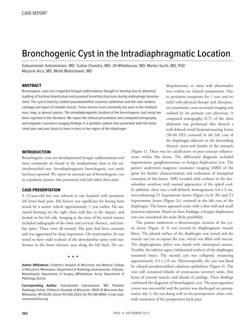
Figure 1. Contrast Enhanced CT of Abdomen Figure 2. Lesions in the Left Crus of the Diaphragm
1a
1b
Axial image (1a) and coronal image (1b) show an ovoid hypoattenuating non-enhancing lesion (arrow) splitting the left crus of the diaphragm and abutting the stomach.
T1 axial image (2a) and post contrast T1 axial image (2b) show non-enhancing isointense lesion splitting the leaves of left crus diaphragm (arrow). The cyst is hyperintense (arrow head) on the T2 image (2c) and demonstrates a small posterior septation.
2a
2b
2c
264 WMJ • DECEMBER 2013
nosis for cystic lesions of the diaphragm include gastrointestinal duplication cyst, cystic pulmonary sequestration, cystic teratoma, mesothelium lined cyst, posttraumatic cyst, or hydatid cyst.5
The management of diaphragmatic cyst is surgical excision,3-6
which establishes the diagnosis and relieves any associated symp-toms. Malignant transformation of bronchogenic cysts has been reported.8 Our patient underwent a thoracoscopic excision of the cyst, which considerably shortened his hospital stay and acceler-ated his return to full function.
CONCLUSIONIntradiaphragmatic bronchogenic cyst presenting in pediatric patients with low back pain is rare. Cross-section imaging (CT/MRI) is required when clinical examination and radiographs are unremarkable and the patient’s symptoms persist. It should be considered in the differential diagnosis for any cystic lesion of the diaphragm. In this case, the presence of splitting of the leaves of crura by the lesion helped to localize it to diaphragm. Absence of post-contrast enhancement on CT and MRI and lack of restricted diffusion suggested the cystic nature of the lesion. MRI helps to evaluate for the presence of intra spinal extension. Surgical resec-tion is the treatment of choice.
Funding/Support: None declared.
Financial Disclosures: None declared.
REFERENCES1. McAdams HP, Kirejczyk WM, Rosado-de-Christenson ML, Matsumoto S. Bronchogenic cyst: imaging features with clinical and histopathological correlation. Radiology. 2000;217(2):441-446.2. Eraklis AJ, Griscom NT, McGovern JB. Bronchogenic cyst of mediastinum in infancy. N Engl J Med. 1969; 281(21):1150-1155.3. Larson D. Bronchogenic cyst. In: Donnelly LF, ed. Diagnostic Imaging: Pediatrics. 2nd ed. Salt Lake City, UT: Amirsys Publishing Inc; 2012; 2:16-19.4. Chang YC, Chen JS, Chang YL, Lee YC. Video-assisted thoracoscopic excision of intradiaphragmatic bronchogenic cyst: two cases. J Laparoendosc Adv Surg Tech A. 2006;16(5):489-492.5. Anile M, Stasio MD, Vitolo D, Venuta F. Intradiaphragmatic bronchogenic cyst. Eur J Cardiothorac Surg. 2006; 29(5):839.6. Liou CH, Hsu HH, Hsueh CJ, Juan CJ, Chen CY. Imaging findings of intradiaphrag-matic bronchogenic cyst: a case report. J Formos Med Assoc. 2001;100(10):712-714.7. Elemen L, Tugay M, Tugay S, Gürcan NI, Erkus B, Gurbuz Y. Bronchogenic cyst of the right hemidiaphragm mimicking a hydatid cyst of the liver: report of the first pediatric case. Pediatr Surg Int. 2008;24(8): 957-959.8. Okada Y, Mori H, Maeda T, Obashi A, Itoh Y, Doi K. Congenital mediastinal bronchogenic cyst with malignant transformation: an autopsy report. Pathol Int. 1996;46(8):594–600.
chronic back pain in our patient was at least in part related to the bronchogenic cyst. It was brought to clinical attention due to trauma. However, trauma did not have any causal relationship to the development of the bronchogenic cyst.
The CT and MRI findings of bronchogenic cyst have been well described in the literature.1 They are usually sharply margin-ated with soft tissue or water attenuation, with cystic character-istics. Some bronchogenic cysts may have soft tissue attenuation, and contrast enhanced CT or MRI may help in distinguishing cystic from solid lesion.1 Ten percent of bronchogenic cysts can have calcification. In our patient, the lesion had attenuation of 30-40 HU, and showed no enhancement or calcification. On MRI, the lesion was hyper-intense on T2W imaging and did not show any restricted diffusion or post contrast enhancement consistent with cystic lesion. Both CT and MRI demonstrated that the lesion was centered in the diaphragm and appeared to split the crus. Apart from bronchogenic cyst, the differential diag-
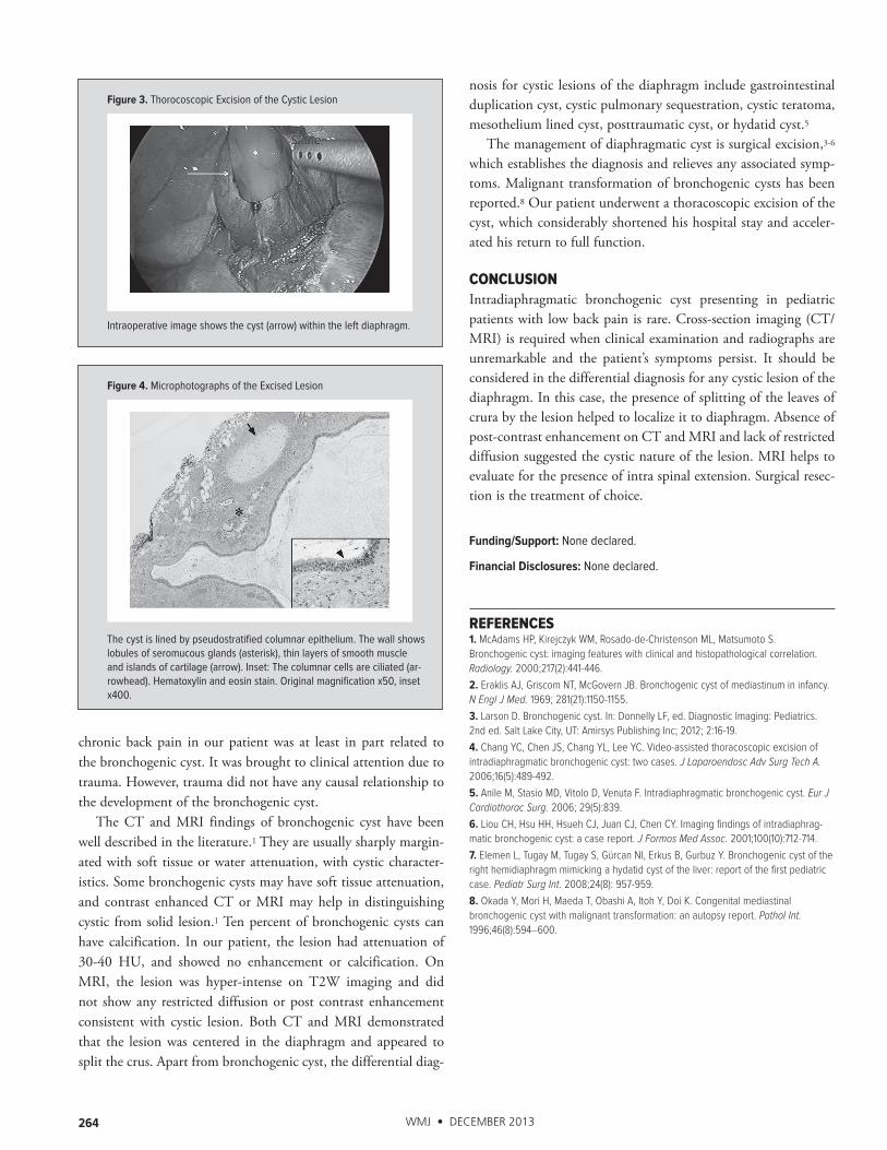
Figure 3. Thorocoscopic Excision of the Cystic Lesion
Figure 4. Microphotographs of the Excised Lesion
Intraoperative image shows the cyst (arrow) within the left diaphragm.
The cyst is lined by pseudostratified columnar epithelium. The wall shows lobules of seromucous glands (asterisk), thin layers of smooth muscle and islands of cartilage (arrow). Inset: The columnar cells are ciliated (ar-rowhead). Hematoxylin and eosin stain. Original magnification x50, inset x400.

The mission of WMJ is to provide a vehicle for professional communication and continuing education for Midwest physicians and other health professionals.
WMJ (ISSN 1098-1861) is published by the Wisconsin Medical Society and is devoted to the interests of
the medical profession and health care in the Midwest. The managing editor is responsible for oversee-
ing the production, business operation and contents of the WMJ. The editorial board, chaired by the
medical editor, solicits and peer reviews all scientific articles; it does not screen public health, socioeco-
nomic, or organizational articles. Although letters to the editor are reviewed by the medical editor, all
signed expressions of opinion belong to the author(s) for which neither WMJ nor the Wisconsin Medical
Society take responsibility. WMJ is indexed in Index Medicus, Hospital Literature Index, and Cambridge
Scientific Abstracts.
For reprints of this article, contact the WMJ at 866.442.3800 or e-mail [email protected].
© 2013 Wisconsin Medical Society


![Case Report Epidermoid Cyst of Orbit in a Newborn · 2019. 7. 31. · Several orbital cystic lesions may occur in the childhood [ ]. Cystic lesions of the orbit include cysts of the](https://img.pdfslide.net/doc/110x75/60c2c7fbda131303c22e5ef2/case-report-epidermoid-cyst-of-orbit-in-a-newborn-2019-7-31-several-orbital.jpg)