Embed Size (px)
Citation preview
Pediatric Respiratory Distress
Theresa Guins, MD
Division of Emergency Medicine Children’s Hospital of The King’s Daughters
Assistant Professor of Pediatrics Eastern Virginia Medical School

Objectives Assessment Croup Bronchiolitis Asthma Foreign Bodies Pertussis
Respiratory Distress
Frequent reason for call to EMS/ED visit Due to higher metabolic demands, less
reserve, anatomic and physiologic differences, children decompensate more quickly.
How is Pediatric Airway Management Different?
Anatomy/Physiology Etiology of airway problems Equipment Frequency with which many practitioners
encounter true pediatric airway problems means less experience available
Why be aggressive in Pediatric Airway Management?
Children have limited ability to compensate for respiratory compromise.
Early recognition of any dysfunction and anticipation of respiratory failure is essential.

Pediatric Assessment Triangle
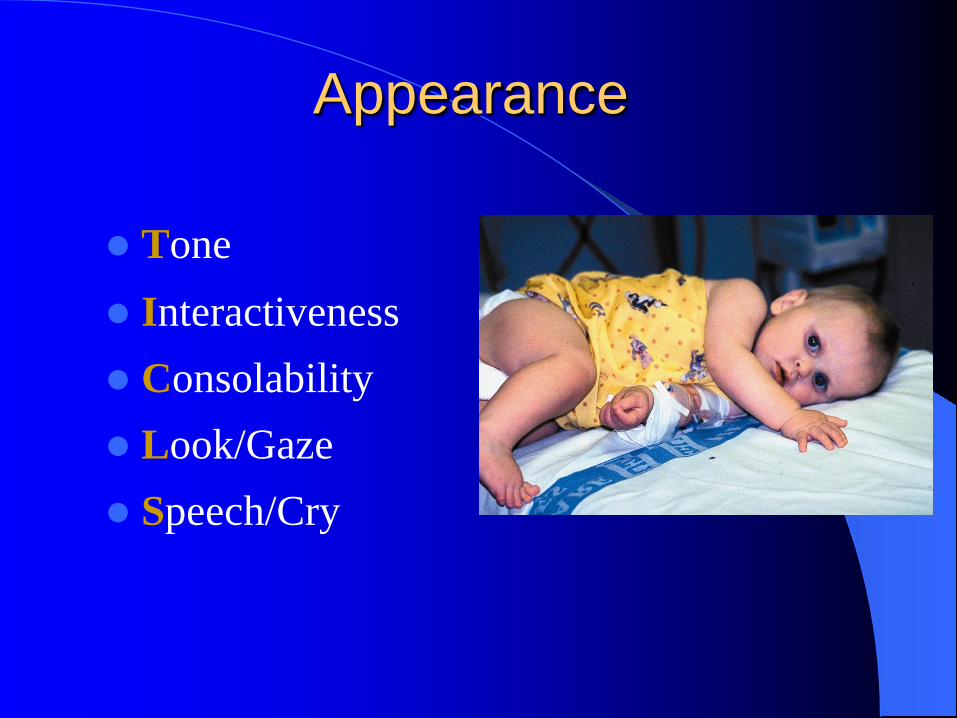
Appearance
Tone Interactiveness Consolability Look/Gaze Speech/Cry
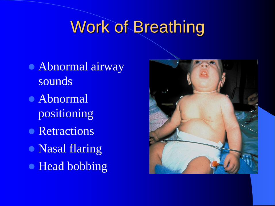
Work of Breathing
Abnormal airway sounds
Abnormal positioning
Retractions Nasal flaring Head bobbing

Normal Respiratory Rates
Infant 30-60 Toddler 24-40 Preschool 20-30 School-age 12-25 Adolescent 12-16
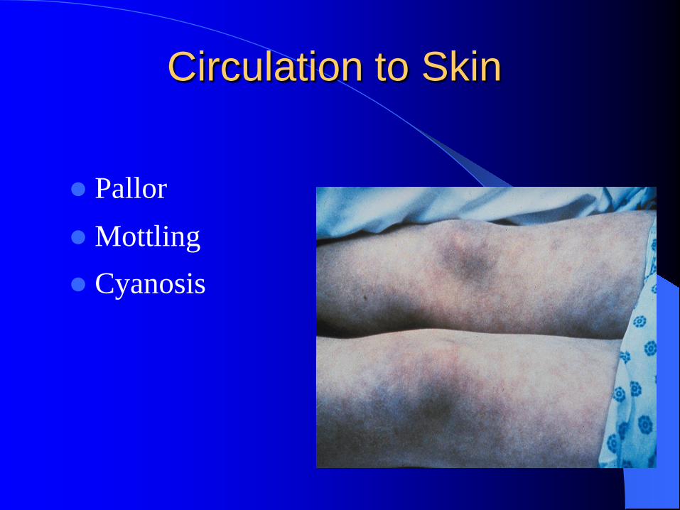
Circulation to Skin
Pallor Mottling Cyanosis
Assessment
Now officially CAB per AHA guidelines! However, A is still extremely important in a
peds patient. Do them simultaneously! A: Assess patency and ability to maintain the
airway, intervene if necessary. Level of consciousness is very important to note.

Assessment B: Breathing
– Count respiratory rate, know normal ranges – Assess for increased work of breathing, retracting,
flaring, grunting, head bobbing. – Listen to evaluate aeration and breath sounds
C. Circulation – Assess color ( lips, mucous membranes, nail beds) – Central and peripheral pulses – Capillary refill and peripheral perfusion
Physical Exam—Observation
Respiratory rate – Periodic breathing in infants – Inaccurate while crying
Respiratory pattern – Inspiratory to expiratory ratio – Normal is less than 1 to 1
Retractions – Subcostal or “belly breathing” – Intercostal – Suprasternal
Physical Exam—Auscultation
Wheezing Stridor Crackles Rhonchi Snot
Wheezing Whistling noise thru constricted bronchioles KEY: More time spent in expiration Noise typically is heard more in expiration Varying degrees of respiratory distress Frequently confused for transmitted upper
airway noises
Examples: Asthma, Bronchiolitis

Stridor
High-pitched noise Usually heard in inspiration Typically with suprasternal retractions Often anxious appearing
Examples: Croup, Bacterial Tracheitis,
Epiglottitis
Crackles
Sounds like velcro Difficult to hear in noisy environment Characterize as focal or diffuse Varying degrees of respiratory distress
Examples: Pneumonia, Congestive Heart
Failure

Rhonchi
Coarser than crackles Finer than transmitted upper airway noise If pathologic should not change with:
– Coughing – Time
Example: Pneumonia, Bronchiolitis
Transmitted Upper Airway Noise Sources include:
– Snot – Relaxed hypo pharyngeal tissues
Changes with respiration May require prolonged listening to distinguish
from other sounds Frequently mistaken for wheezing
Examples: URI, neurologically impaired
persons

Tricks to Improving Your Exam
Changing position of patient Distract the patient Have patient cough Have patient breathe through mouth Have patient blow nose Decrease ambient noise Listen for a little longer

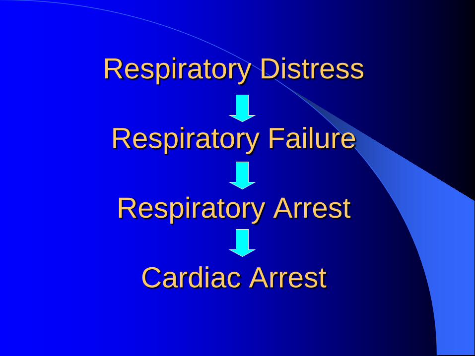
Respiratory Distress
Respiratory Failure
Respiratory Arrest
Cardiac Arrest
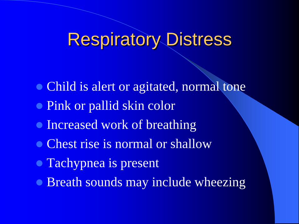
Respiratory Distress
Child is alert or agitated, normal tone Pink or pallid skin color Increased work of breathing Chest rise is normal or shallow Tachypnea is present Breath sounds may include wheezing
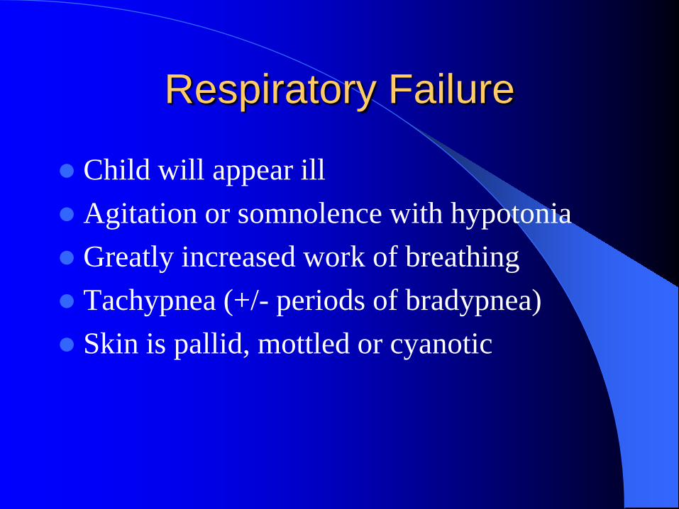
Respiratory Failure
Child will appear ill Agitation or somnolence with hypotonia Greatly increased work of breathing Tachypnea (+/- periods of bradypnea) Skin is pallid, mottled or cyanotic
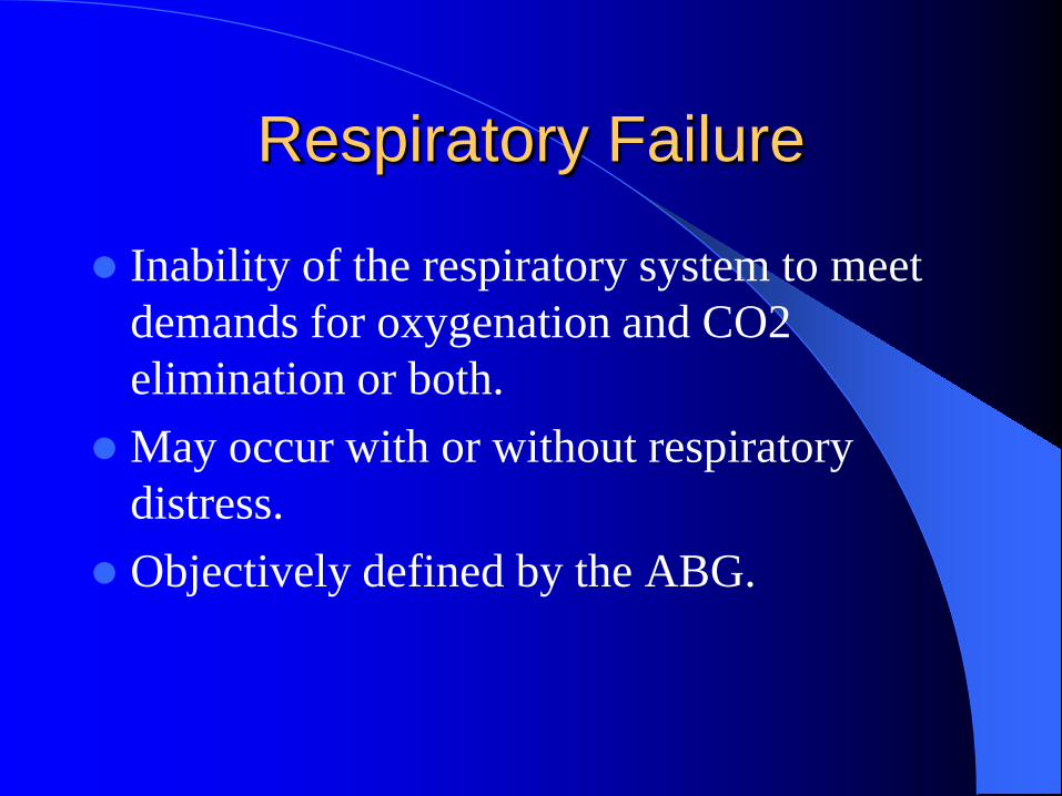
Respiratory Failure
Inability of the respiratory system to meet demands for oxygenation and CO2 elimination or both.
May occur with or without respiratory distress.
Objectively defined by the ABG.
Respiratory Arrest
Unresponsive, no muscle tone No visible chest rise Absent work of breathing Cyanosis
Cardiac arrest will follow quickly!!

What Can We Do?

Goals of Airway Therapy
Recognize respiratory distress and failure before they progress to arrest.
Anticipate respiratory problems. Support those functions that are lost or
compromised. Start with least invasive methods.
Interventions
Open and Position the Airway Oxygen Nasopharyngeal Airway Oropharyngeal Airway Bag-Valve-Mask Ventilation
Interventions
Administer Oxygen – Nasal cannula – Simple face mask – Non-rebreather mask
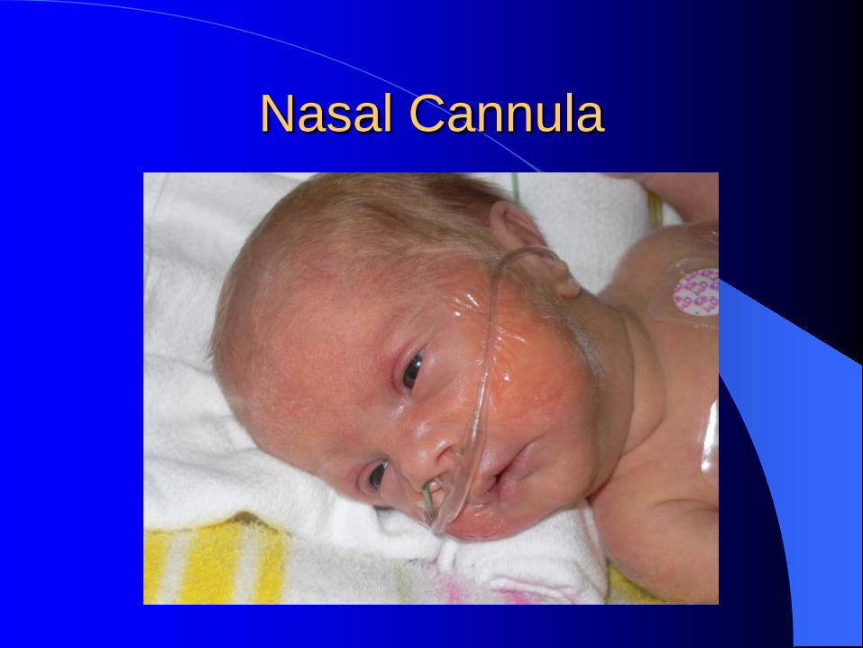
Nasal Cannula
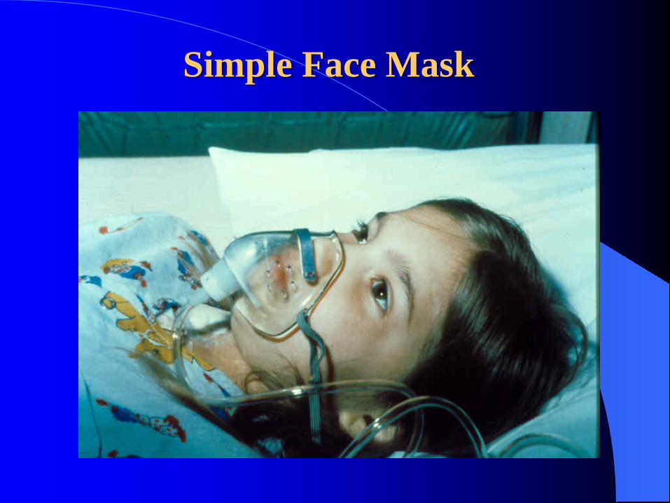
Simple Face Mask
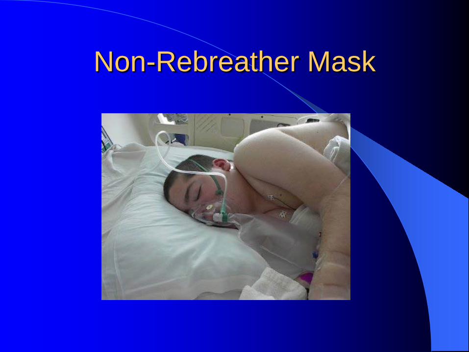
Non-Rebreather Mask
Nasopharyngeal Airway
Soft rubber or plastic tube in many sizes Used to bypass upper airway obstruction Well tolerated in semiconscious or
conscious patients Easily obstructed with secretions in small
children
Nasopharyngeal Airway
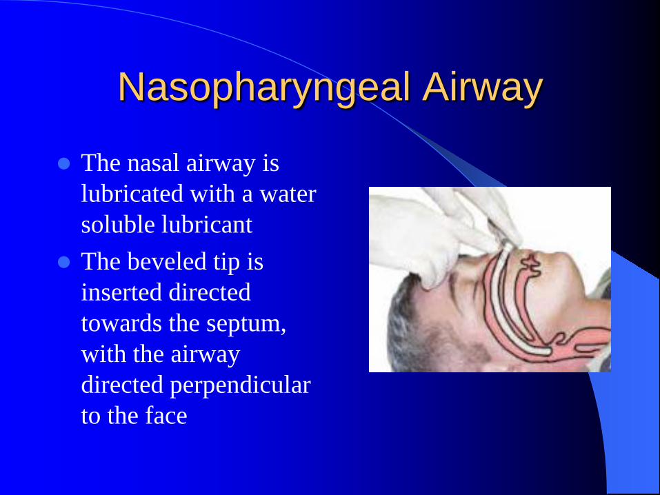
The nasal airway is lubricated with a water soluble lubricant
The beveled tip is inserted directed towards the septum, with the airway directed perpendicular to the face
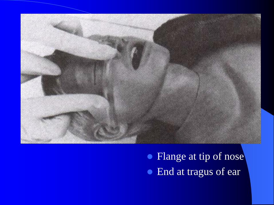
Flange at tip of nose End at tragus of ear

Oropharyngeal Airway
Holds tongue away from the posterior pharyngeal wall.
Only in unconscious patients. Measure from the corner of the mouth to the angle of the jaw.
Anyone who provides critical care to children needs to be
expert in managing the unprotected airway with a
bag-valve-mask.

“When bag-valve-mask is done appropriately, it can be
every bit as effective as endotracheal intubation.”
Jim Seidel, MD
Airway Management Maxim
Airway Management DOES NOT Mean Intubation!
Airway management means just that!
Patients will not die because you do not or cannot intubate them. They will die if you do
not ventilate and/or oxygenate them.

Croup
Also known as laryngotracheobronchitis.
Infection causes inflammation of the larynx and subglottic airway
Typical age is 6 months to 3 years.
– Rare beyond age 6 years.
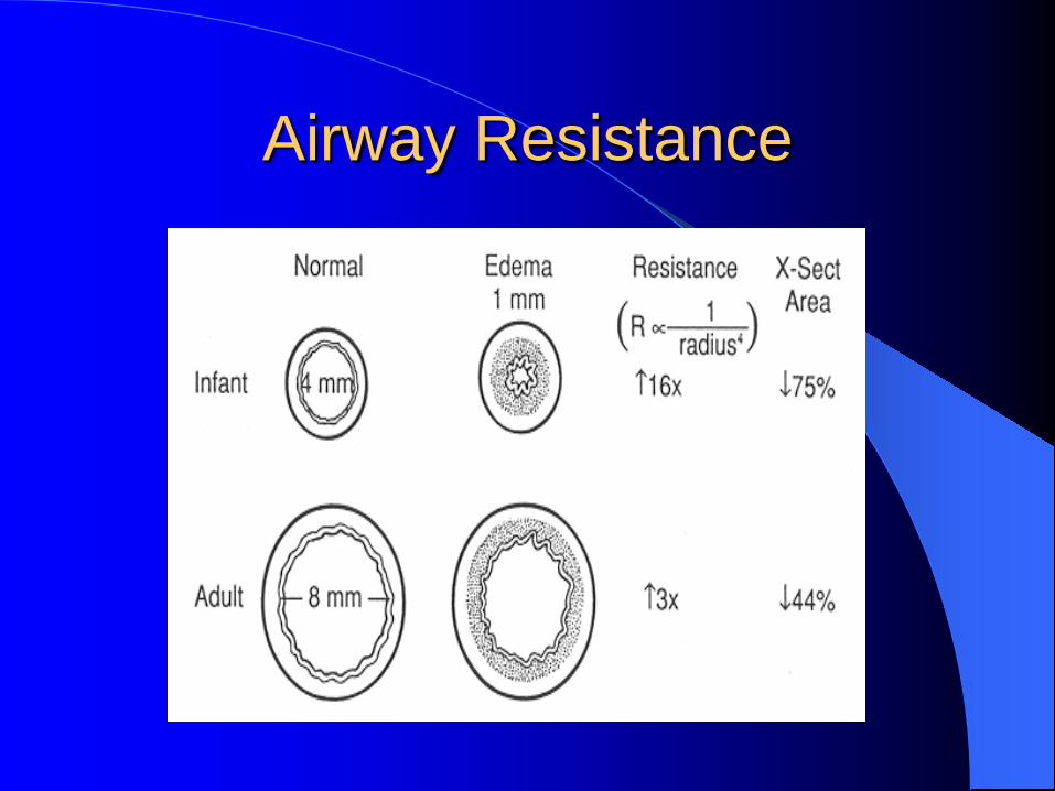
Airway Resistance
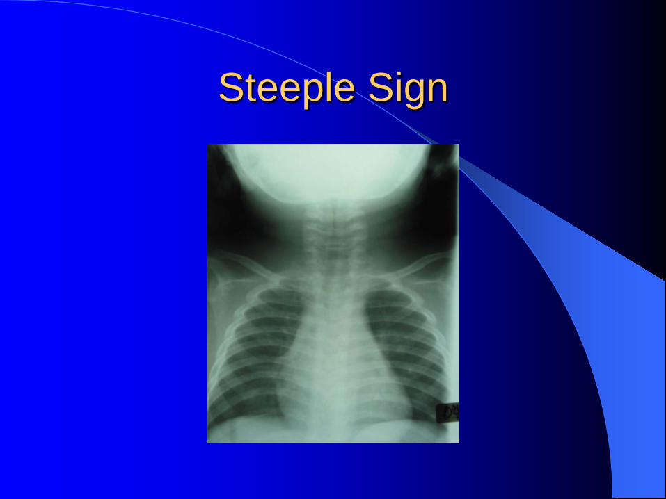
Steeple Sign
Croup
Cause is primarily viral – Parainfluenza virus type 1 and 2 – RSV, Influenza and Adenovirus less often
• Outbreaks most common in fall/winter • Most EMS calls/ED visits 10pm-4am.
Clinical Presentation
Many have 12 to 48 hours of URI symptoms prior to onset of upper airway obstruction.
Spasmodic croup- abrupt onset of stridor and barky cough, typically while asleep.
Fever is common, ranges from 38 to 40.5.

Clinical Presentation
Mild cases: Hoarse with a barky cough More severe:
– Stridor – Retractions – Diminished breath sounds – Agitation

Croup Hypoxia and cyanosis can develop, but rare. Prolonged respiratory distress can lead to
fatigue and respiratory failure. Need for intubation or death from croup are
also rare occurrences.
History
Factors in the history that may suggest significant worsening: – Rapidly progressing symptoms – History of prior episodes of croup – Underlying airway abnormalities
Differential Diagnosis
Bacterial Tracheitis Acute Epiglottitis Peritonsillar or Retropharyngeal Abscess Airway Foreign Body Congenital Airway Anomalies
Evaluation
CAB’s – Assess the child for signs of obstruction or impending failure/arrest.
Keep the child as comfortable as possible.
– agitation and fear may make the airway narrowing worse.
Evaluation
Before even touching the child, assess: – Overall appearance – Work of breathing – Chest expansion – Quality of the cry/voice – Abnormal sounds, stridor vs. wheezing?
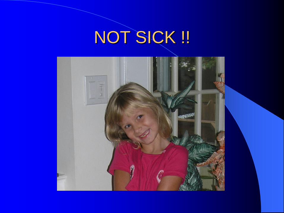
NOT SICK !!

SICK !!!
Physical Findings
Increased work of breathing – Tachypnea, retractions, nasal flaring
Inspiratory stridor – At rest, or only heard when agitated/crying?
Breath sounds are usually clear!! – Wheezing is a sign of lower airway obstruction
Treatment of Croup
Mist Oxygen Steroids Nebulized epinephrine
Treatment
Mist/Humidified air – no proven benefit – May help prevent inspissation of secretions
Oxygen: If the child is in moderate to
severe distress or hypoxic. – Use judgment in the non-hypoxic child that is
more agitated by the therapy.
Treatment
Corticosteroids – mainstay of therapy – Decreases the edema in the airway – Requires several hours for onset of effect – Not recommended in the pre-hospital setting – Dexamethasone is the steroid of choice.
Treatment
Nebulized Epinephrine – Rapid improvement due to decrease in the
airway edema.
– Racemic vs. L-epinephrine: Equally effective with the same incidence of side effects.
(most common is tachycardia)

Nebulized Epi
Indications: – Children with croup who are in severe
respiratory distress – Stridor at rest
Dose: 0.5 mL/kg per dose of the 1:1000
preparation. Max dose: 5 mL.
Treatment
Intubation: VERY rarely necessary! Less than 1% of patients seen in the ED for
croup require intubation. If deemed necessary, use an ETT that is ½ to 1 size smaller to account for the
edema in the airway.
Disposition
Most children who present to the ED for croup are discharged to home.
If a nebulized epi treatment was given, the child is observed for 2 to 3 hours for recurrence of stridor. – Return of stridor requiring further nebulized epi
treatment meets criteria for admission.
Bronchiolitis
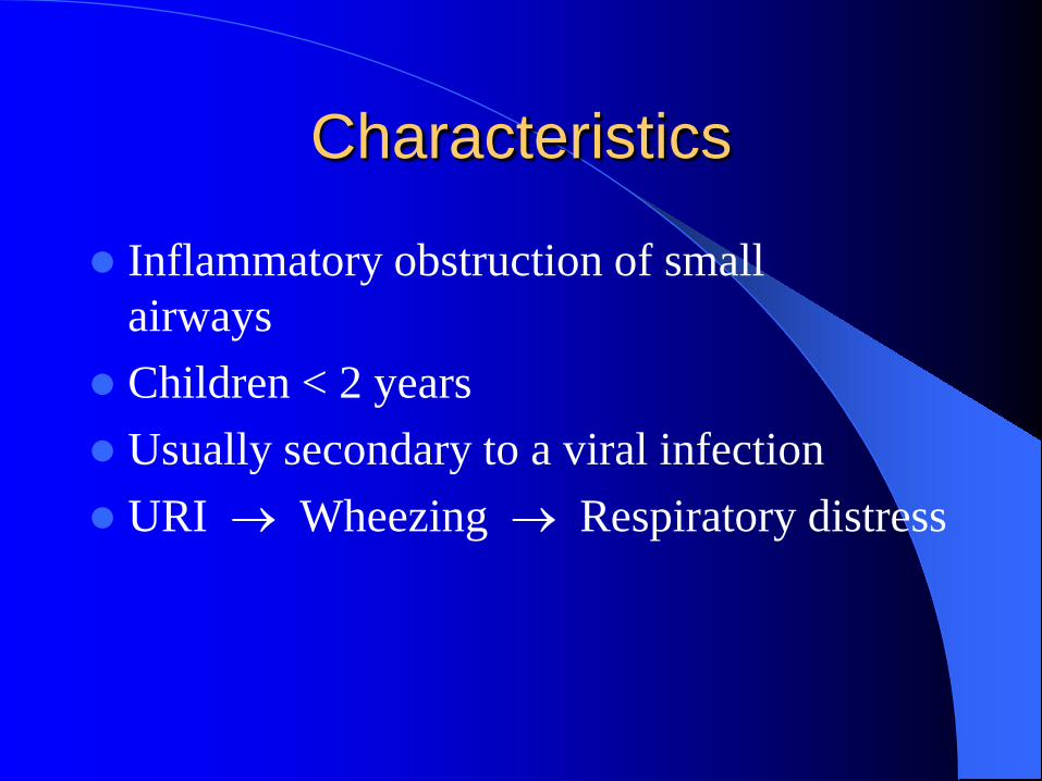
Characteristics
Inflammatory obstruction of small airways
Children < 2 years Usually secondary to a viral infection URI → Wheezing → Respiratory distress
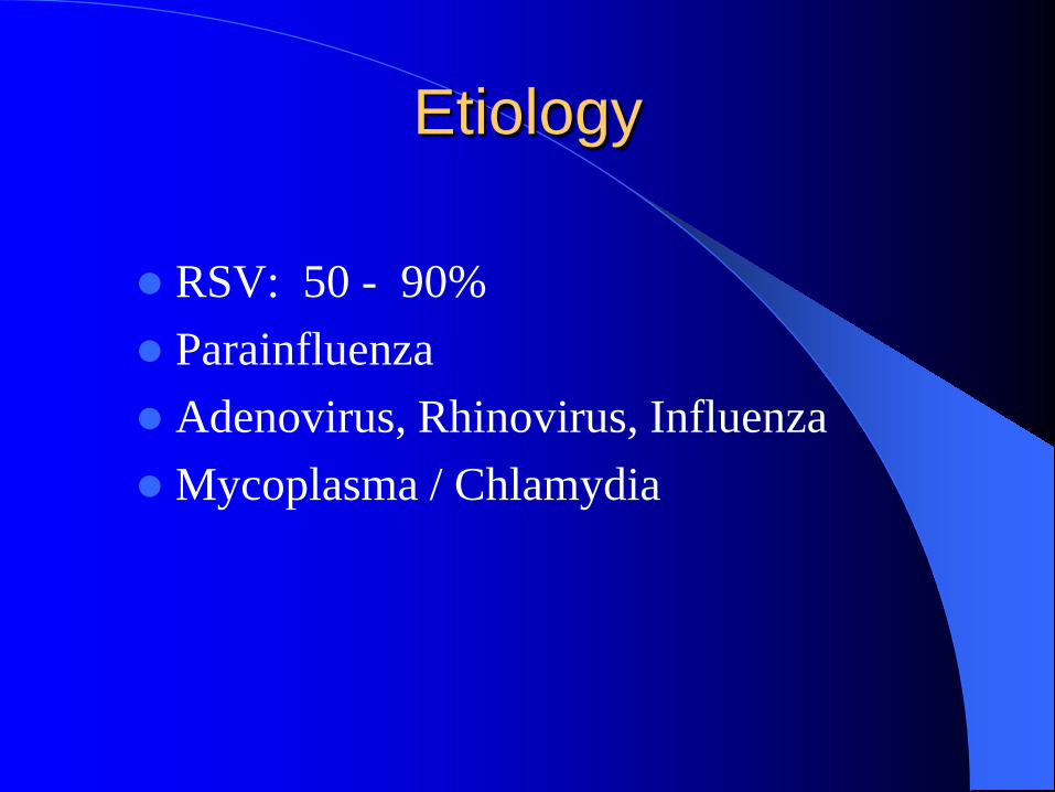
Etiology
RSV: 50 - 90% Parainfluenza Adenovirus, Rhinovirus, Influenza Mycoplasma / Chlamydia
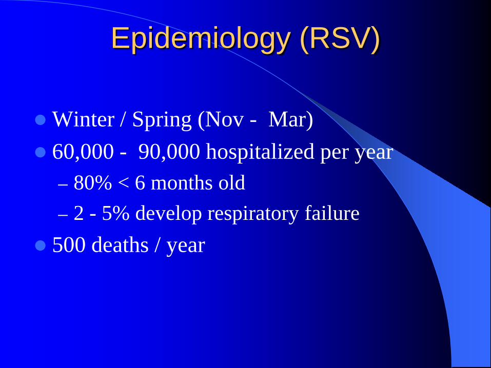
Epidemiology (RSV)
Winter / Spring (Nov - Mar) 60,000 - 90,000 hospitalized per year
– 80% < 6 months old – 2 - 5% develop respiratory failure
500 deaths / year
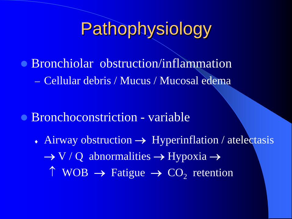
Pathophysiology
Bronchiolar obstruction/inflammation – Cellular debris / Mucus / Mucosal edema
Bronchoconstriction - variable
♦ Airway obstruction → Hyperinflation / atelectasis → V / Q abnormalities → Hypoxia → ↑ WOB → Fatigue → CO2 retention
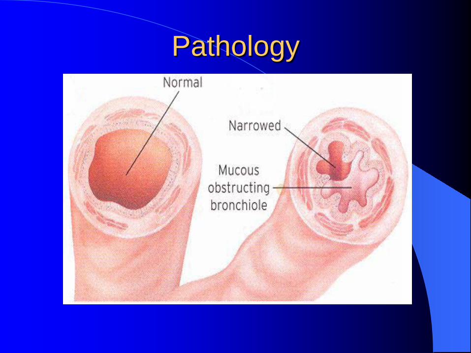
Pathology
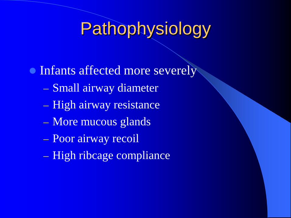
Pathophysiology
Infants affected more severely – Small airway diameter – High airway resistance – More mucous glands – Poor airway recoil – High ribcage compliance
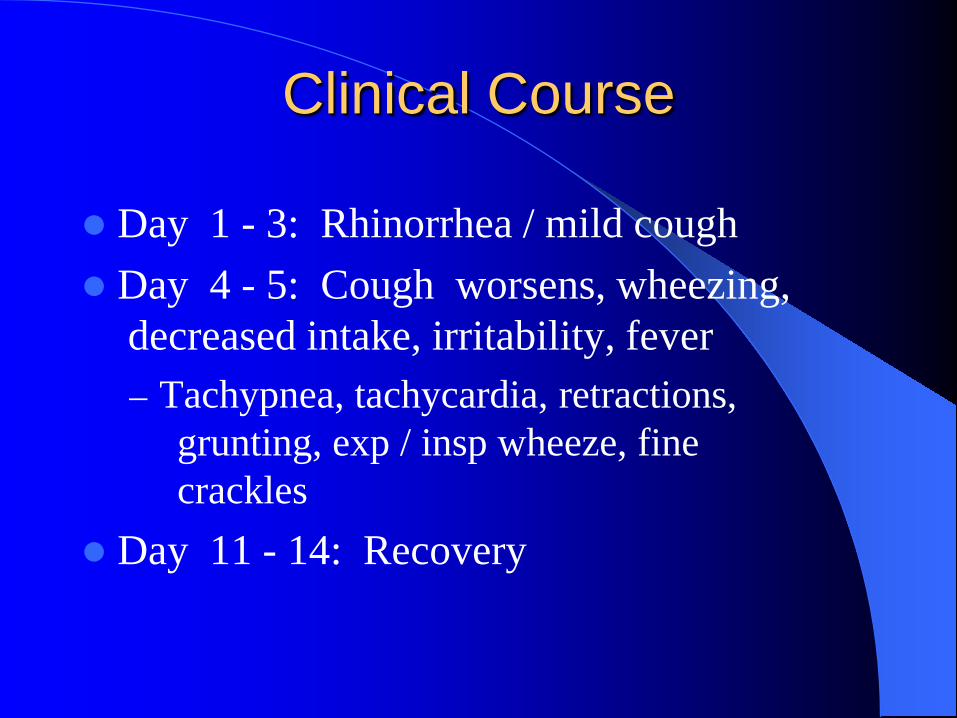
Clinical Course
Day 1 - 3: Rhinorrhea / mild cough Day 4 - 5: Cough worsens, wheezing,
decreased intake, irritability, fever – Tachypnea, tachycardia, retractions,
grunting, exp / insp wheeze, fine crackles
Day 11 - 14: Recovery
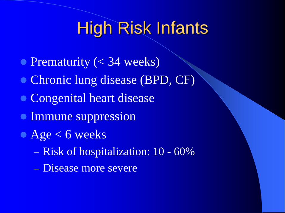
High Risk Infants
Prematurity (< 34 weeks) Chronic lung disease (BPD, CF) Congenital heart disease Immune suppression Age < 6 weeks
– Risk of hospitalization: 10 - 60% – Disease more severe

Complications
Severe respiratory distress Respiratory failure Apnea (15 - 20% of hospitalized infants) Dehydration Bacterial superinfection (1%) Pneumothorax / pneumomediastinum (rare)
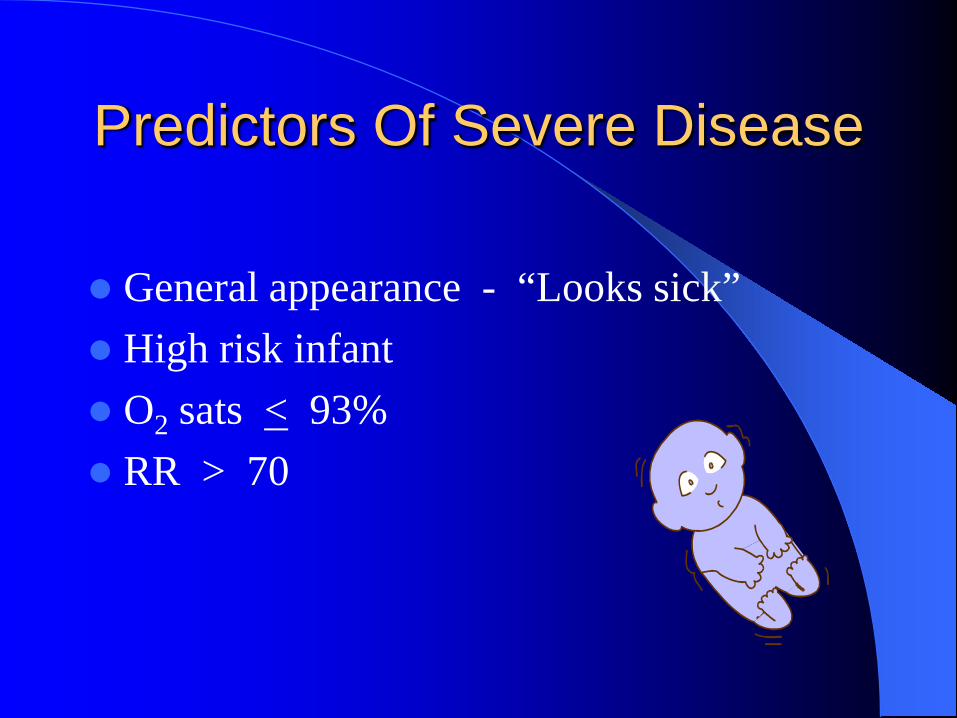
Predictors Of Severe Disease
General appearance - “Looks sick” High risk infant O2 sats < 93% RR > 70
Differential Diagnosis Asthma Congestive heart failure Foreign body aspiration Pertussis Cystic Fibrosis Bacterial pneumonia / sepsis
What To Do ?
EMS Priorities
Depends on severity of presentation: – None/Minimal distress, not hypoxic: transport – Moderate/Severe distress: provide oxygen and
consider Albuterol, 2.5 mg HHN. – Apnea or Poor respiratory effort: BVM
– Frequently reassess these children as their
condition can change very rapidly.
Beta2 Agonists
Subgroup of patients may respond to
β2 agonists (Albuterol) Non-responders more likely to be admitted Adverse affects:
– Tachycardia, irritability, hypoxemia, exacerbation of airway obstruction

Epinephrine
Epinephrine is more effective than albuterol or placebo
The preferred bronchodilator in the treatment of bronchiolitis
Has not been evaluated and is currently not recommended in the pre-hospital setting.
Steroids
For previously healthy infants with bronchiolitis, corticosteroids are not effective
May consider if previous episodes of wheezing / underlying cardiopulmonary disease / ? severe illness
Antibiotics
65 - 70% of hospitalized infants with bronchiolitis are febrile
Incidence of SBI : 0 - 1.9 %
Antibiotics are not routinely indicated
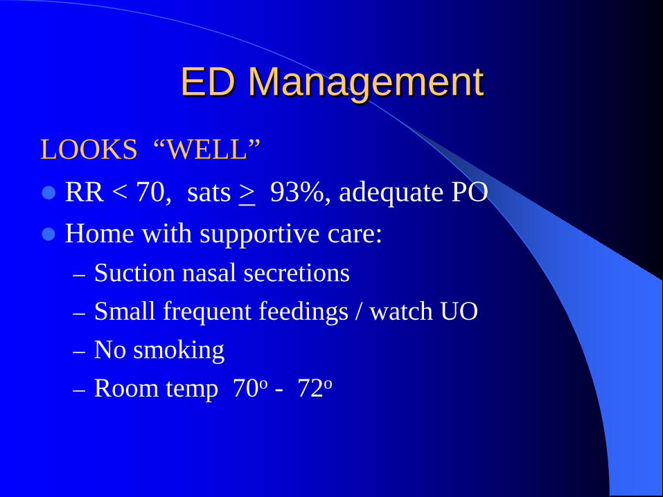
ED Management LOOKS “WELL” RR < 70, sats > 93%, adequate PO Home with supportive care:
– Suction nasal secretions – Small frequent feedings / watch UO – No smoking – Room temp 70o - 72o
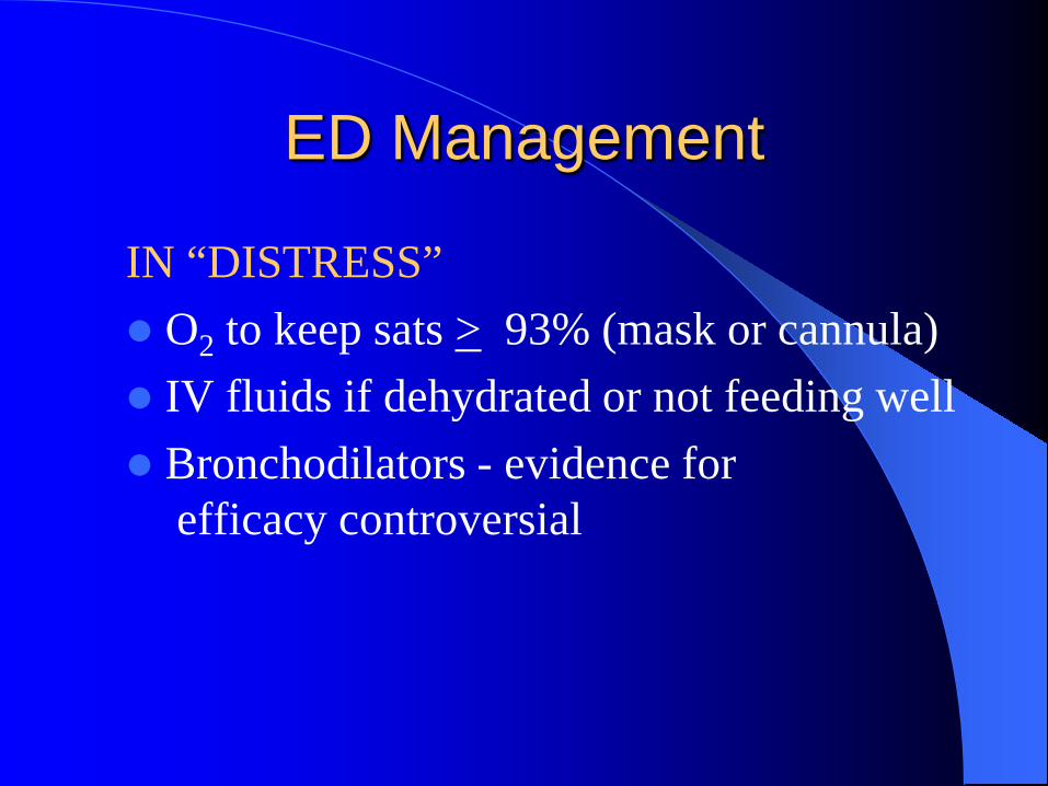
ED Management
IN “DISTRESS” O2 to keep sats > 93% (mask or cannula) IV fluids if dehydrated or not feeding well Bronchodilators - evidence for
efficacy controversial
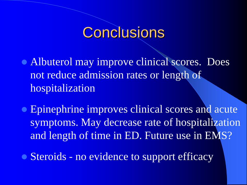
Conclusions
Albuterol may improve clinical scores. Does not reduce admission rates or length of hospitalization
Epinephrine improves clinical scores and acute symptoms. May decrease rate of hospitalization and length of time in ED. Future use in EMS?
Steroids - no evidence to support efficacy
Asthma
Definition Asthma Chronic disease of the airways
– Inflammation – Obstruction – Hyperresponsiveness
Clinically: Recurrent episodes of wheezing,
breathlessness, chest tightness and cough
Definition
Status Asthmaticus Persistent airflow obstruction that fails to
improve or worsens despite appropriate therapy
Assessment
Physical Examination General appearance / level of consciousness
- most useful Wheezing correlates poorly with severity Tachypnea / use of accessory muscles /
ability to speak
Management
Oxygen Mechanisms of action
– Improves tissue oxygenation – Facilitates bronchodilation – Reduces pulmonary vasoconstriction
Tight fitting or non-rebreather face mask / high flow oxygen
Keep O2 sats ≥ 95%
EMS Treatment Options
Quick relief medications – Beta-adrenergic agonists (Albuterol) – Anticholinergics (Atrovent)
Medications to reverse inflammation
– Corticosteroids (Solumedrol)
β-Adrenergic Agonists
Albuterol is the treatment of choice – 2.5 mg for <20 kg, and 5 mg for >20 kg.
Nebulizer or MDI
– Depends on child’s coordination, technique, cooperation, degree of airflow obstruction
– 6-10 puffs from MDI = 2.5 mg nebulized
β-Adrenergic Agonists
Quick relief drugs of choice Mechanisms of action
– Smooth muscle relaxation – Decrease airway edema – Enhance mucociliary clearance – Inhibit inflammatory mediator release

β-Adrenergic Agonists
Adverse effects
Dose and route related Tachycardia, tremors, agitation, vomiting,
arrhythmias ↓ in O2 sats due to worsening V/Q ratio
BUT if patient looks better and is moving air better PATIENCE – Treat the patient and not the
oximeter
β-Adrenergic Agonists
Levalbuterol No conclusive evidence that levalb safer or
more effective than racemic albuterol In acute asthma standard dose of levalb
(0.625 or 1.25 mg) may be too low More expensive
β-Adrenergic Agonists
Intramuscular Epinephrine
Indications – Severe bronchospasm – Patient unable to cooperate with or not
responding to inhaled therapy
Anticholinergics
Mechanisms of action: Atrovent
Inhibit parasympathetic mediated bronchoconstriction
Decrease mucosal edema and secretions Weak bronchodilators when used alone Augment bronchodilating effects of
Albuterol
Anticholinergics
Indications
Moderate / severe asthma exacerbations Patient not responding appropriately to
initial Albuterol treatment.
• Dose: 0.5 mg, may use up to 3 doses in ED.
• Combine with albuterol

Other Options ?
Corticosteroids (Solumedrol)
Mechanisms of action
Suppress mediators of inflammation → decrease airway edema and secretions
Reverse down regulation of β-agonist receptors thus potentiating their effectiveness → increase bronchodilation
Corticosteroids
Not recommended in areas with rapid transport times: takes hours to work.
Discretion of medical control in more remote areas.
Requires placement of IV, which very few pediatric asthma patients require
Varicella issue: be very careful!!!
Magnesium Sulfate
Mechanisms of action
Blocks calcium mediated smooth muscle contraction → bronchodilation
Potentiates effect of β-adrenergic agonists Decreases inflammatory response
Magnesium Sulfate
Indications – Severe bronchospasm unresponsive to initial β-agonist treatment – Currently not recommended for pre-hospital
use in pediatric patients. Must carefully monitor HR / BP High levels cause CNS depression, muscle
weakness, nausea, flushing
Summary
Rapid assessment β-agonists - frequently and appropriate dose Ipratropium - enhances bronchodilation Corticosteroids – consider Maintain oxygenation - children die from
hypoxia, not hypercarbia
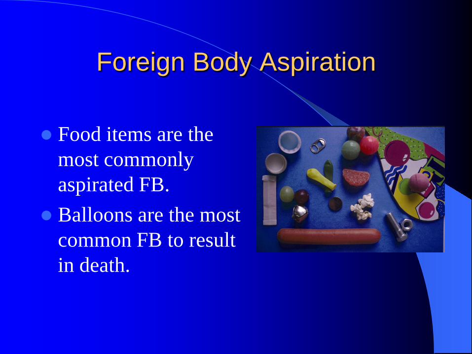
Foreign Body Aspiration
Food items are the most commonly aspirated FB.
Balloons are the most common FB to result in death.
Airway Foreign Bodies
80% of episodes occur in children < 3yo, peak age between 1-2 yo Food most commonly aspirated by infants and toddlers (peanuts) Toy balloons most common object involved in fatal childhood foreign body aspiration
Your First Clue: Foreign Body Aspiration
A history of choking is the most reliable predictor of FB aspiration.
Other signs and symptoms include: – Upper airway: Stridor, respiratory or
cardiopulmonary arrest. – Lower airway: Coughing, wheezing,
retractions, decreased breath sounds, cyanosis.
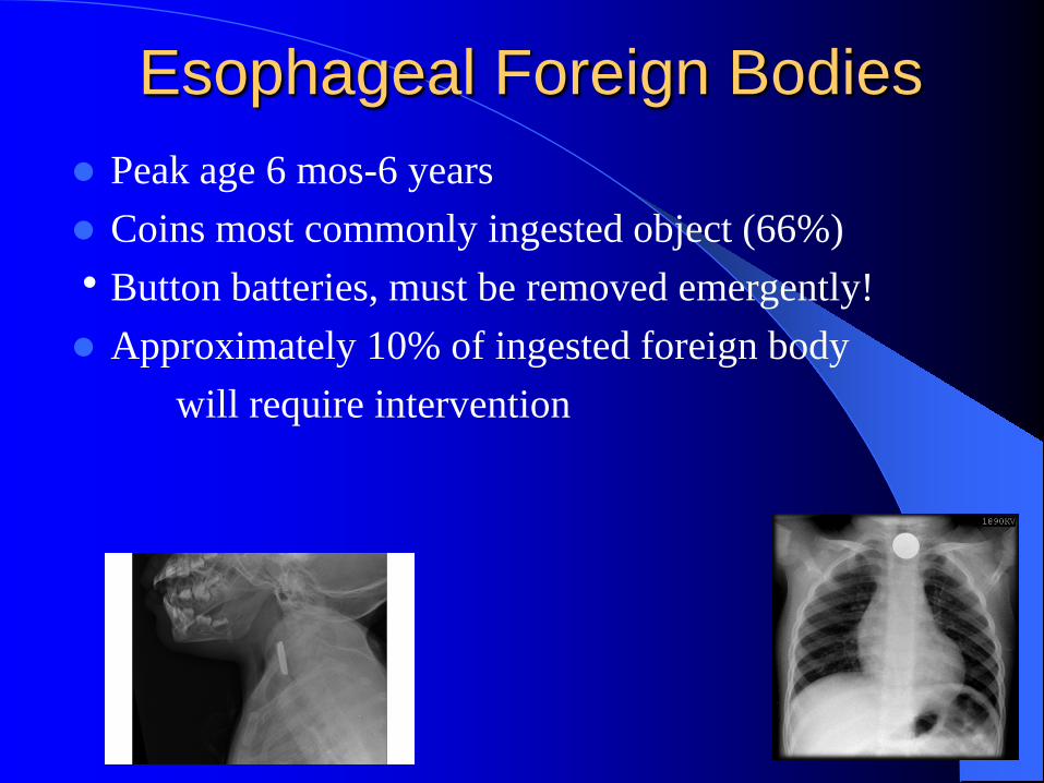
Esophageal Foreign Bodies Peak age 6 mos-6 years
Coins most commonly ingested object (66%) Button batteries, must be removed emergently! Approximately 10% of ingested foreign body will require intervention
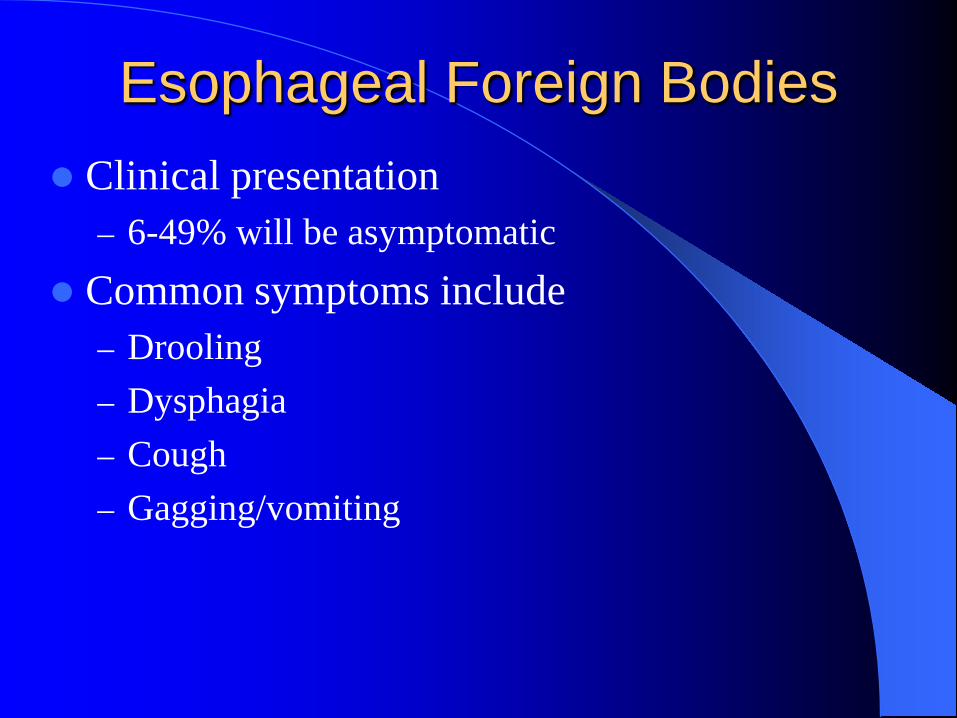
Esophageal Foreign Bodies Clinical presentation
– 6-49% will be asymptomatic Common symptoms include
– Drooling – Dysphagia – Cough – Gagging/vomiting
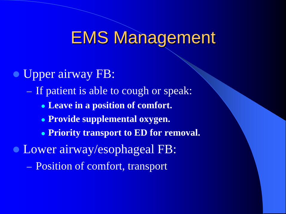
EMS Management
Upper airway FB: – If patient is able to cough or speak:
Leave in a position of comfort. Provide supplemental oxygen. Priority transport to ED for removal.
Lower airway/esophageal FB: – Position of comfort, transport

Definitive Management Laryngoscopy and
removal with pediatric Magill forceps
If unable to grasp FB at or near the vocal cords, OK to push it in to secure the airway!

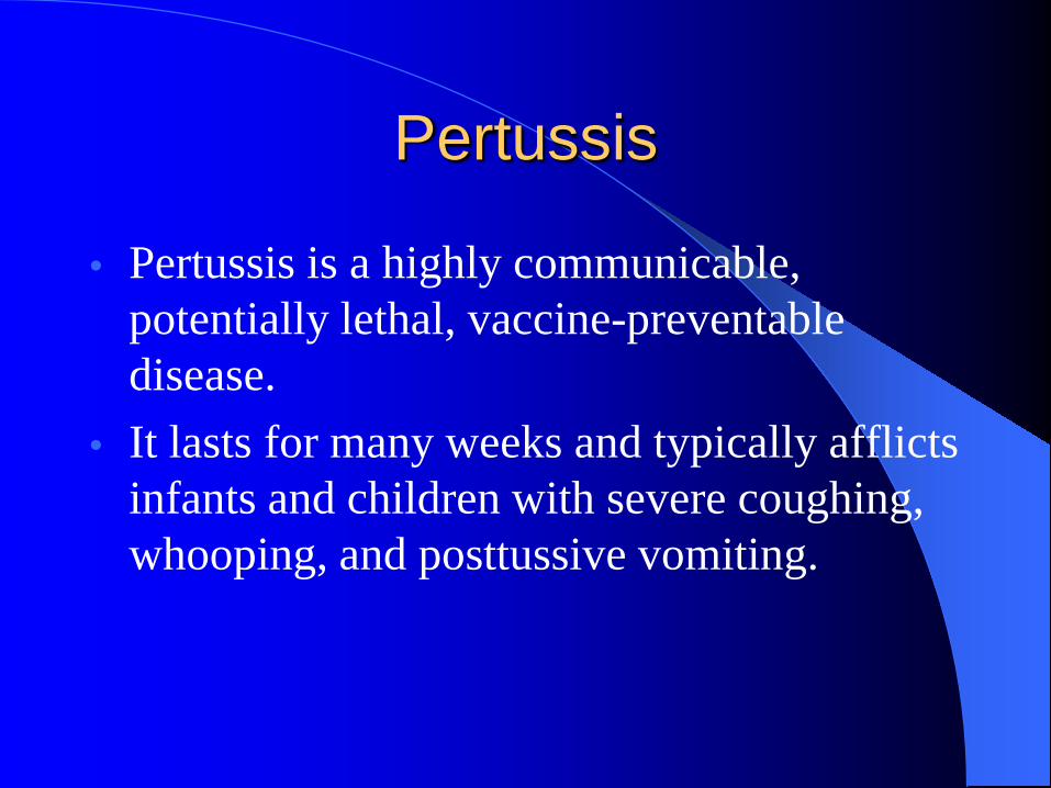
Pertussis
• Pertussis is a highly communicable, potentially lethal, vaccine-preventable disease.
• It lasts for many weeks and typically afflicts infants and children with severe coughing, whooping, and posttussive vomiting.

• Pertussis is caused by a bacterium named Bordetella pertussis.
• Transmission: by close contact with cases via aerosolized droplets.
• Neither infection nor immunization provides lifelong immunity
• In 2013, 28,000 cases of pertussis were reported in the U.S.
Facts
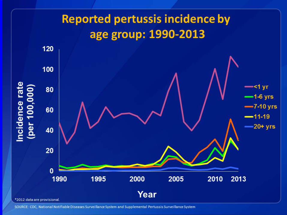
• Lack of natural booster events and waning immunity since childhood immunization were responsible for the increase in cases of pertussis in people older than 10 years of age noted before use of the adolescent booster immunization.
• The incubation period is 7 to 10 days, with a range of 5 to 21 days
Epidemiology
• Pertussis begins with mild upper respiratory tract symptoms similar to the common cold (catarrhal stage).
• It then progresses to cough and then usually to paroxysms of cough (paroxysmal stage) characterized by inspiratory whoop and commonly followed by vomiting.
• Fever is absent or minimal.
Clinical Manifestations
• Symptoms wane gradually over weeks to months (convalescent stage).
• Sudden unexpected death can be caused by pertussis, especially in the young infants.
• The duration of classic pertussis is 6 to 10 weeks.
Clinical Manifestations

• Antimicrobial agents administered during the catarrhal stage may ameliorate the disease.
• After the cough is established, antimicrobial agents have no discernible effect on the course of illness but are recommended to limit the spread of organisms to others.
• Azithromycin, erythromycin, or clarithromycin are appropriate first-line agents for treatment and prophylaxis
Treatment
EMS Priorities
Pediatric Assessment Triangle Provide Oxygen if sats <95% Supportive care, no IV needed No medications have any effect Proper isolation measures
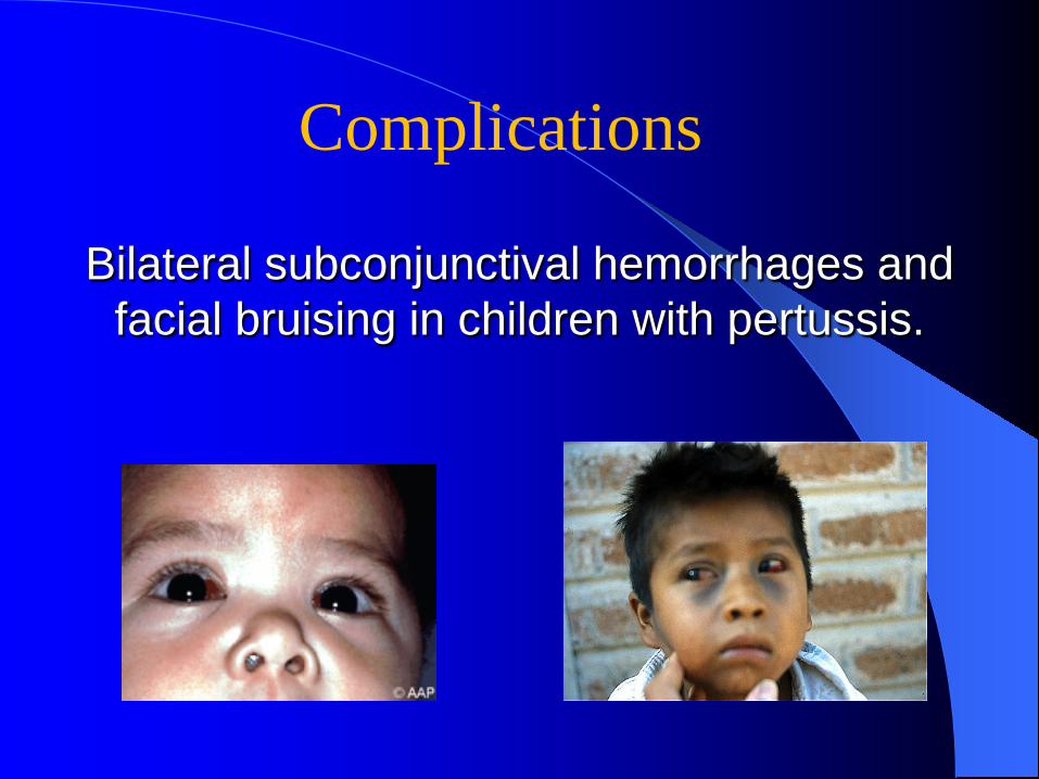
Bilateral subconjunctival hemorrhages and facial bruising in children with pertussis.
Complications
• All health care professionals should observe standard
precautions and wear a respiratory mask when examining a patient with a cough illness suspected or confirmed to be pertussis.
• Exposed, unprotected people should be given prophylaxis promptly.
Control Measures
Preexposure immunization of health care personnel with tetanus toxoid, reduced diphtheria toxoid, and acellular pertussis
(Tda.p) vaccine is recommended
Control Measures