Embed Size (px)
Citation preview
Pediatrics for Midwifery (Midw3132)
By: Chalachew Adugna and Amare Wondim(BSc, MSc)
Department of pediatric and child health nursing
University of Gondar, Ethiopia 2020
1
OVERVIEW OF PEDIATRICS
2
Learning Objectives:
After completion of this unit, the students will
be able to describe:
Definition of pediatrics
Origin and history of pediatrics
Difference between adult and pediatric medicine
Pediatrics history taking and physical
examination
3
Introduction
Definition:
•Child Health is the concern of pediatrics.
• I.e. Pediatrics ↔ Child Health.
•Pediatrics: the term pediatrics is derived from Greek
words:
• “pedia” meaning a child
• “iatrike” meaning treatment (Rx.)
• “ics” meaning a branch of science.4
Definition…
•Thus, pediatrics is a branch of medical science
that deals with the care of childhood from
conception to adolescent in health and illness.
•It concern with prevention, promotion, curative
and rehabilitative care of children
5
Definition…
•Pediatrics is concerned with the health of
infants, children and adolescents, their growth
and development, and their opportunity to
achieve full potential as adults (Richard E. Behrman in
Nelson's Textbook of Pediatrics)
6
Pediatric Nursing
•Pediatric nursing is the specialized area of the
nursing practice concerning the care of children
during wellness and illness, which includes
preventive, promotive, curative and
rehabilitative care of children.
7
Importance of Pediatrics
•Major consumers of health care
•35 – 40 of total population are children below the age
of 15
•More vulnerable to various health problems
•Majority of Child’s morbidity & mortality are
preventable
•Needs special care to survive & thrive
•Wealth of tomorrow society and nation8
Origin and history of pediatrics
• The importance of childhood as a unique period of
development was understood more fully in the 17th and
18th centuries by John Locke (English philosopher)- the
newborn infant comes into the world with no inherited
predispositions, but rather with a mind as a tabula rasa
(Latin for “blank slate”) that is gradually filled with ideas,
concepts, and knowledge from experiences in the world.
• He concluded that the quality of early experiences,
particularly how children are raised and educated, shapes
the direction of a child‛s life9
Origin and history of pediatrics…..
• Jean Jacques Rousseau (French philosopher)- children at
birth are innately good, not evil, and that their natural
tendencies should be protected against the corrupting
influences of society
• The sympathetic, romantic attitude toward children inspired by
Rousseau had an important influence on society
• Arthur Jacobi- has been recognized as the father of pediatrics.
Under his direction, several hospitals opened pediatric units.
He helped to found the American Pediatric Society in 1888.
10
Origin and history of pediatrics…..
•Pediatrics became a medical specialty in the mid 19th
century
•Before that time the care and treatment of childhood
diseases was included with in general medicine and
obstetrics
•Virtually all nations have practicing departments of
pediatrics or child health
11
Origin and history of pediatrics…..
Pediatrics become an independent medical
specialty:
•The health problems of children differ from those of
adults
•Children response to an illness is influenced by age
•Management of childhood illness is significantly
different with that of adults
•Worldwide, children represent a higher proportion of
total population12
Origin and history of pediatrics…..
•The health problems of children vary widely within
populations
•Economic consideration, educational, social, and
cultural considerations, prevalence and ecology of
infectious agents, climate and geography, agricultural
resources and practices, industrialization and
urbanization; gene frequencies for some disorders;
and health and social welfare infrastructure.
13
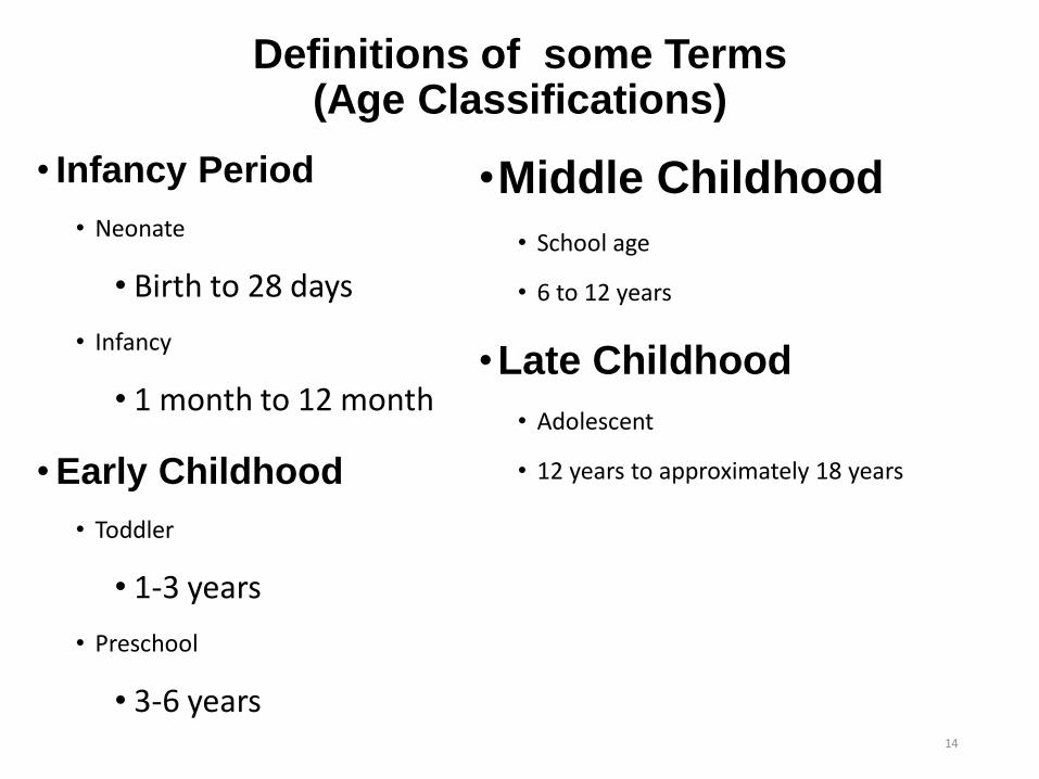
Definitions of some Terms (Age Classifications)
• Infancy Period
• Neonate
• Birth to 28 days
• Infancy
• 1 month to 12 month
• Early Childhood
• Toddler
• 1-3 years
• Preschool
• 3-6 years
•Middle Childhood
• School age
• 6 to 12 years
•Late Childhood
• Adolescent
• 12 years to approximately 18 years
14
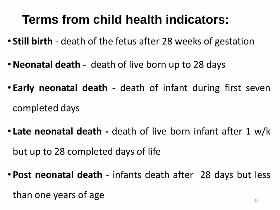
Terms from child health indicators:
• Still birth - death of the fetus after 28 weeks of gestation
• Neonatal death - death of live born up to 28 days
• Early neonatal death - death of infant during first seven
completed days
• Late neonatal death - death of live born infant after 1 w/k
but up to 28 completed days of life
• Post neonatal death - infants death after 28 days but less
than one years of age15
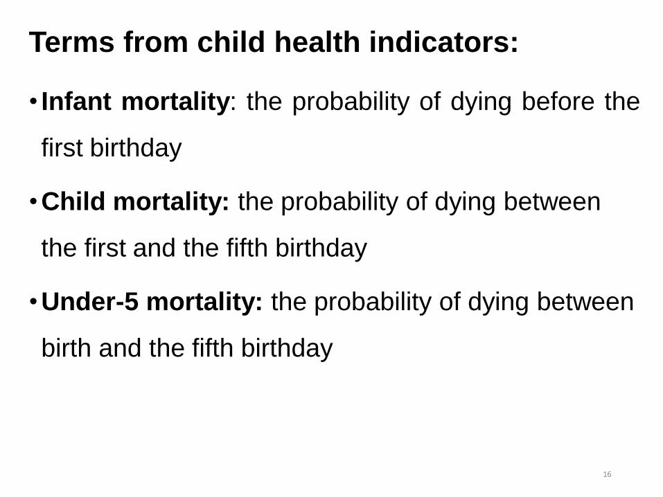
Terms from child health indicators:
• Infant mortality: the probability of dying before the
first birthday
•Child mortality: the probability of dying between
the first and the fifth birthday
•Under-5 mortality: the probability of dying between
birth and the fifth birthday
16
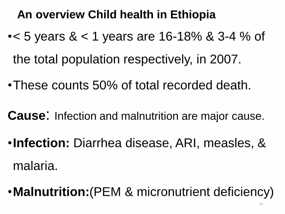
An overview Child health in Ethiopia
•< 5 years & < 1 years are 16-18% & 3-4 % of
the total population respectively, in 2007.
•These counts 50% of total recorded death.
Cause: Infection and malnutrition are major cause.
•Infection: Diarrhea disease, ARI, measles, &
malaria.
•Malnutrition:(PEM & micronutrient deficiency) 17
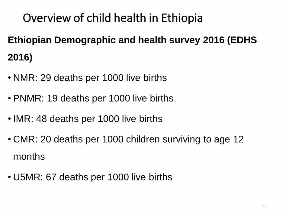
Overview of child health in Ethiopia
Ethiopian Demographic and health survey 2016 (EDHS
2016)
• NMR: 29 deaths per 1000 live births
• PNMR: 19 deaths per 1000 live births
• IMR: 48 deaths per 1000 live births
• CMR: 20 deaths per 1000 children surviving to age 12
months
• U5MR: 67 deaths per 1000 live births
18
Differences between a Child and an Adult
•Size - newborn to adolescent
•Physical - anatomy, growth & development
•Cognitive - communication & understanding
•Needs - emotional, psychological & social
•Rights - even a CHILD of all ages has rights!
19
United Nations convention on the Rights of The Child(1989)
All children need:
To be free from discrimination
To develop physically and mentally in freedom and
dignity
To have a name and nationality
To have adequate nutrition, housing, recreation, and
medical services
20
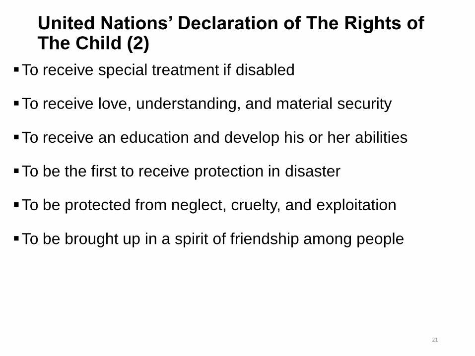
United Nations’ Declaration of The Rights of The Child (2)
To receive special treatment if disabled
To receive love, understanding, and material security
To receive an education and develop his or her abilities
To be the first to receive protection in disaster
To be protected from neglect, cruelty, and exploitation
To be brought up in a spirit of friendship among people
21
The Art of Pediatric NursingPhilosophy of Care
•Family centered care
•Family oriented yet individualized and coordinated.
•Recognizes the family as the constant in a child’s
life
•Considers the needs of all family members in
relation to the care of the child.
22
The Art of Pediatric Nursing…
•Atraumatic Care
•Therapeutic care in settings, by personnel, and
through the use of interventions that eliminate or
minimize the psychologic and physical distress
experienced by children and their families in the
health care system.
23
Atraumatic Care
•The overriding goal in providing atraumatic care is
first, do no harm.
•Three principles:
1) Prevent or minimize the child’s separation from the
family
2) Promote a sense of control
3) Prevent or minimize bodily injury and pain
24

Role of the pediatric nurse
•Primary care giver
•Advocate
•Health educator
•Consultant
•Support and counseling
•Coordination and collaboration
•Social worker
•Researcher 25
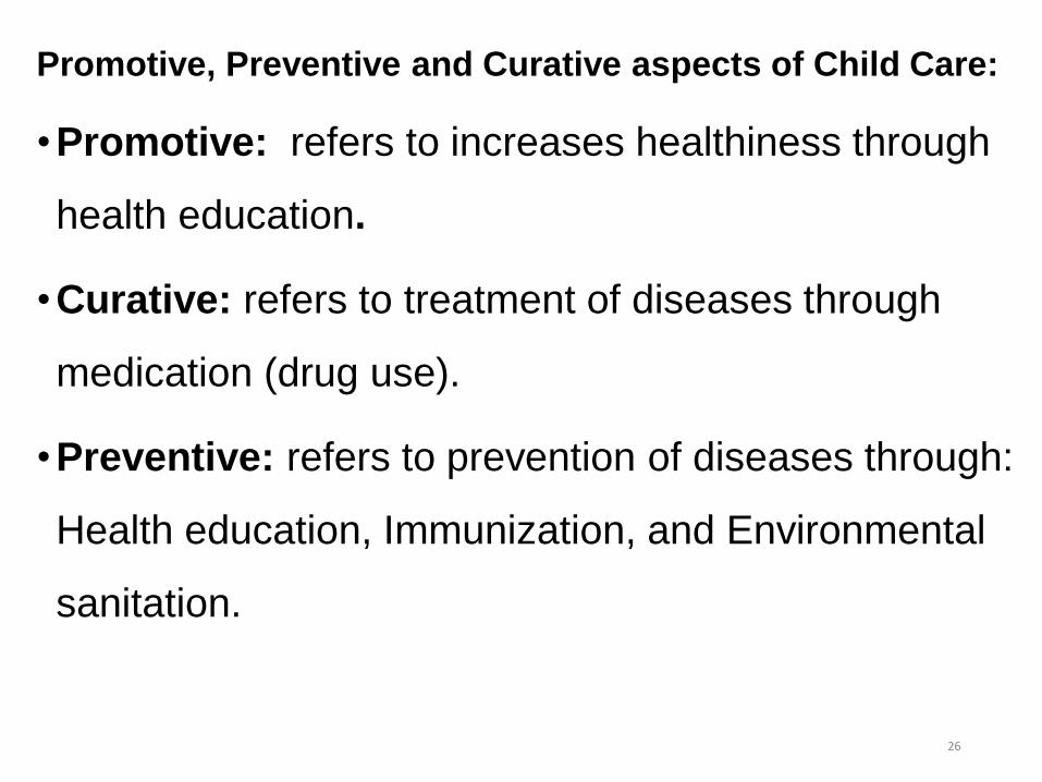
Promotive, Preventive and Curative aspects of Child Care:
•Promotive: refers to increases healthiness through
health education.
•Curative: refers to treatment of diseases through
medication (drug use).
•Preventive: refers to prevention of diseases through:
Health education, Immunization, and Environmental
sanitation.
26
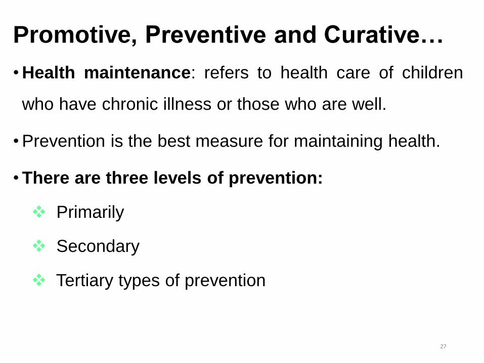
Promotive, Preventive and Curative…
• Health maintenance: refers to health care of children
who have chronic illness or those who are well.
• Prevention is the best measure for maintaining health.
• There are three levels of prevention:
Primarily
Secondary
Tertiary types of prevention
27
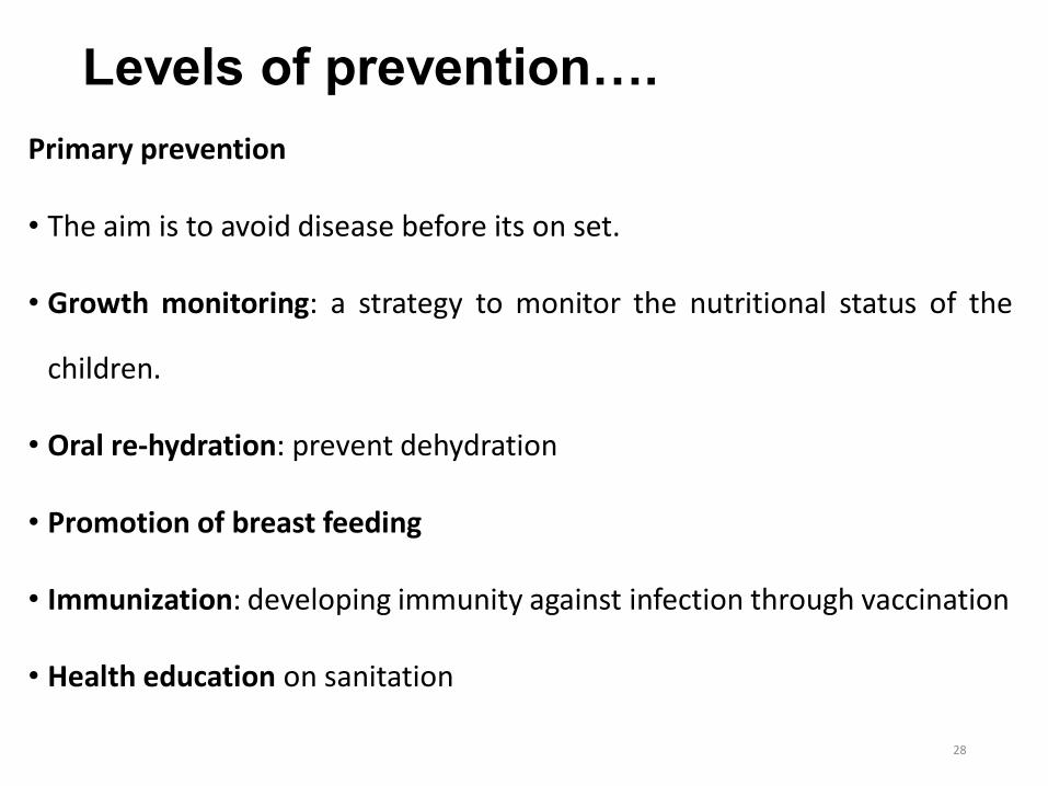
Levels of prevention….
Primary prevention
• The aim is to avoid disease before its on set.
• Growth monitoring: a strategy to monitor the nutritional status of the
children.
• Oral re-hydration: prevent dehydration
• Promotion of breast feeding
• Immunization: developing immunity against infection through vaccination
• Health education on sanitation
28
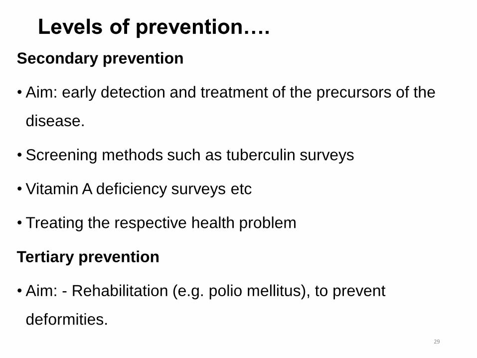
Levels of prevention….
Secondary prevention
• Aim: early detection and treatment of the precursors of the
disease.
• Screening methods such as tuberculin surveys
• Vitamin A deficiency surveys etc
• Treating the respective health problem
Tertiary prevention
• Aim: - Rehabilitation (e.g. polio mellitus), to prevent
deformities.
29
Pediatric History and Physical Examination
•Key elements in the history taking process
include; establishing a warm, caring
atmosphere and asking questions in a no
confrontational, unhurried manner
•Use simple language
•Good eye contact and a sense of undivided
attention should be maintained.
30
Pediatric History and Physical Examination…
•Sit opposite the caregiver and/or patient at a
comfortable distance
•Outside interruption should be kept to a
minimum
•Greet in a friendly manner and introduce your self
•Ascertain who is with the child, It may not be the
mother but another family member
•Older child should be involved in the history 31

Pediatric History and Physical Examination…
• Even younger children should be asked simple things in words they
can understand
• Remember that the mother is giving you her version of the problem,
not the child's.
• Always take notice of what the mother is saying, and listen to her
complaints.
• The mother will know what is worrying her about the child, and any
interruptions should be to guide her rather than try and impose your
diagnosis on her.
32
Pediatric History and Physical Examination…
• There are so many differences
between pediatric and adult
history
1. Content difference
2. Approach difference
3. Parents as primary informant
• Content difference
•Perinatal history
•Developmental history
•Social history
• Immunization history
33
History
• Identifying data
•Name, age, sex, name of parents (informant), date
of examination, date of admission, source of data
•Chief complaint (CC):
•What is the reason for the health visit
•Must be informant’s own word and must include the
duration.
34
History…
• History of present illness (HPI):
• Onset of symptoms, duration, timing (intermittent vs.
constant)
• Progression of illness over time, Associated symptoms
• Aggravating or alleviating factors;
• Similar episodes in the past, treatment, outcome,
complications
• What the family has done so far to manage
• Past medical history (PMH):
• Previous admission, surgery, trauma, asthma, diabetes 35
History…
Perinatal history:
• Antenatal follow up of the mother
• Any illness during pregnancy like hypertension, diabetes
mellitus
• Immunization for tetanus
• The onset, duration of labor or ROM, the mode of delivery,
place of delivery, who conducted the delivery, the birth
weight, APGAR score or did the new born cried immediately
after birth, any procedure immediately after birth ?
• Jaundice, cyanosis, convulsion during neonatal period 36
History…
Nutritional(dietary)history:
• Type of feeding
• Duration of exclusive breast feeding, time of initiation, frequency, total
duration of BF.
• Formula feeding
• Animal milk, commercial infant formula, amount
• Complementary feeding
• Start at 6 months with liquid and semisolid foods
• Current diet
37
History...Immunization history:
• Up-to-date?
• Is the child/infant being immunized?
•Was he immunized only during National polio
campaigns?
• When was the last vaccination?
• Route of vaccine?
Allergies: Drug, food ?
38
History...
Developmental history:
•Age at attainment of important milestones (walking,
talking, self-care).
•Relationships with siblings, peers, adults.
•School grade and performance, behavioral problems.
39

History...
• Family history:
• Medical problems in family, including the patient's disorder;
diabetes, seizures, asthma, allergies, cancer, cardiac, renal
or GI disease, tuberculosis, smoking.
•Genetic diseases, infant deaths
• Social history:
• Family income, occupation of the parents, housing, school
and play facilities available for the child.
40
History...
• Review of systems:
•Check list of symptoms
•Almost similar with adults
41
Pediatrics Physical Examination
IMPORTANT HINTS
• Avoid irritating the child and prevent him from crying (if
possible)
• Examine the child in the most comfortable way according to
his age (exam table, mother’s hands, mother’s lap, while
playing with a toy…)
• Postpone the painful and/or irritating examination
(temp/throat/ears)
42
Pediatrics Physical Examination….
•Specific techniques similar to adult
• Inspection
•Palpation
•Percussion
•Auscultation
•Approach and order different
43

Pediatrics Physical Examination….
General appearance
Facial expression (pain)
Cry (high pitched, weak cry)
Grunting (respiratory distress)
Does the child appear to be:
• Well, acutely ill/toxic, chronically ill, wasted, or
malnourished
• Alert and active or lethargic/fatigued?
• Well hydrated or dehydrated?
• Unusual body odors?44
Vital signs
Temperature
Heart Rate
Respiratory Rate
Blood Pressure
Pulse Oximetry
45
Pediatrics Physical Examination….
•Vital signs vary based on the age of the patient
•For newborns and infants take apical heart rate
•Take respiratory rate for full minute
•For measuring BP make sure the cuff covers no
less than ½ and no more than 2/3 the length of
the arm
46
Anthropometric Measurements
Height, weight; head circumference in children < 2 years;
Always use (plot on) growth charts and determine the
percentiles.
Use appropriate scale for age to measure the weight.
Naked weight (when possible)
Measure recumbent length till 2 years of age and then
standing length (height) after that
47
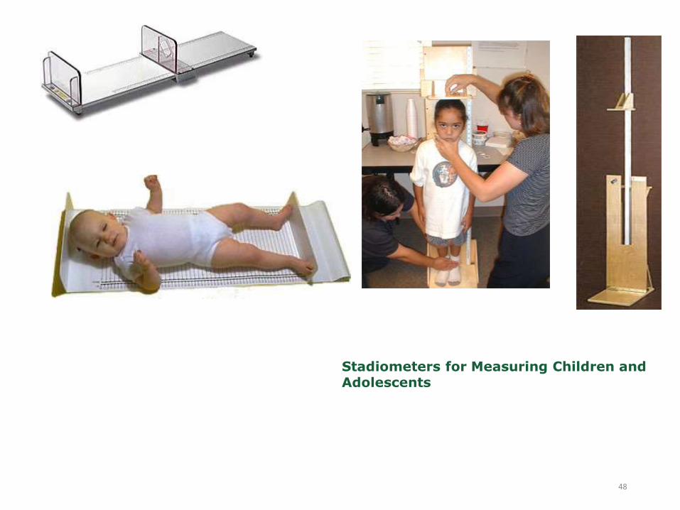
Stadiometers for Measuring Children and Adolescents
48

Skin, Hair, and nails
•Skin: Color, skin turgor, edema, texture, rash
•Hair: Texture, color, distribution, areas of hair loss.
•Nails: Color, texture, shape, capillary refill time (in
seconds).
49
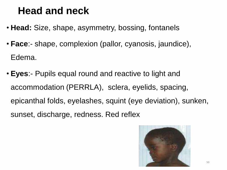
Head and neck
• Head: Size, shape, asymmetry, bossing, fontanels
• Face:- shape, complexion (pallor, cyanosis, jaundice),
Edema.
• Eyes:- Pupils equal round and reactive to light and
accommodation (PERRLA), sclera, eyelids, spacing,
epicanthal folds, eyelashes, squint (eye deviation), sunken,
sunset, discharge, redness. Red reflex
50
Pediatrics Physical Examination….
Ears: Pinnas (size, position), deformity, discharge, ext. canal & Tympanic
membranes (shape, color, position, light reflex)
Nose: Shape, discharge, deviated septum, patency, polyp, flaring
Mouth: Lips (fissures, cleft lip, herpes lesion), teeth, mucus membrane
color and moisture, tongue, cleft palate
Throat: Tonsils (erythema, exudate),hoarseness, stridor
Lymph node: Location, size, tenderness, mobility and consistency of
cervical, axillary, supraclavicular, and inguinal nodes
Neck: Stiffness, torticollis, lymphadenopathy, thyroid nodules, position of
trachea
51
Lungs/Thorax
• Inspection
• Pattern of breathing
• Abdominal breathing is normal in infants
• Period breathing is normal in infants (pause < 15 seconds)
• Respiratory rate
• Use of accessory muscles: retraction location, degree/flaring
• Chest wall configuration
52
Lungs/Thorax…
•Auscultation
•Equality of breath sounds
•Crackles, wheezes, rhonchi
•Upper airway noise
•Percussion and palpation often not possible and
rarely helpful
53
Cardiovascular
•Pulses
•Apical pulse - varies with age
•Rate and rhythm
•S3 common
54
Abdomen
Inspection
• look for scars, masses, skin lesions, asymmetry, visible
pulsations
• Shape
• Infants usually have protuberant abdomens, Becomes
more scaphoid as child matures
• Umbilicus (infection, hernias)
• Auscultation: listen in all quadrants or until bowel sounds are
heard as they will transmit throughout the entire abdomen
55
Abdomen…
Palpation
•Tenderness - avoid tender area until end of exam
•Liver, spleen, kidneys - May be palpable in normal
newborn
•Rebound, guarding - Have child blow up belly to
touch your hand
56
Genitalia
• Inspect external genitalia for abnormality, signs of
abuse, or infection.
•Male Genitalia: Circumcision, hypospadias,
phimosis, size of testes, cryptorchidism, hydrocele,
hernia, inguinal masses.
•Female Genitalia: Imperforate hymen, discharge,
labial adhesions, clitoral hypertrophy, pubertal
changes.
57
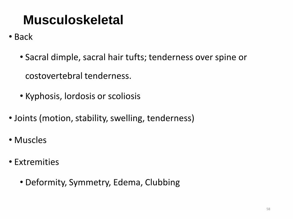
Musculoskeletal
• Back
• Sacral dimple, sacral hair tufts; tenderness over spine or
costovertebral tenderness.
• Kyphosis, lordosis or scoliosis
• Joints (motion, stability, swelling, tenderness)
• Muscles
• Extremities
• Deformity, Symmetry, Edema, Clubbing
58
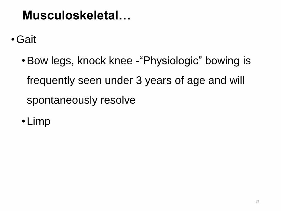
Musculoskeletal…
•Gait
•Bow legs, knock knee -“Physiologic” bowing is
frequently seen under 3 years of age and will
spontaneously resolve
•Limp
59
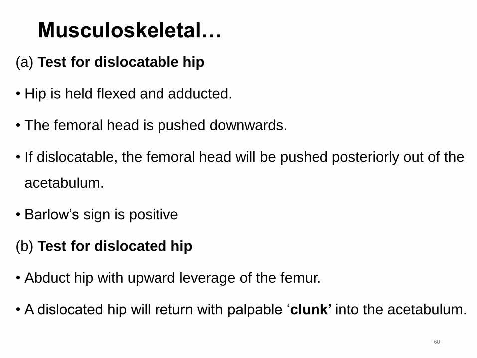
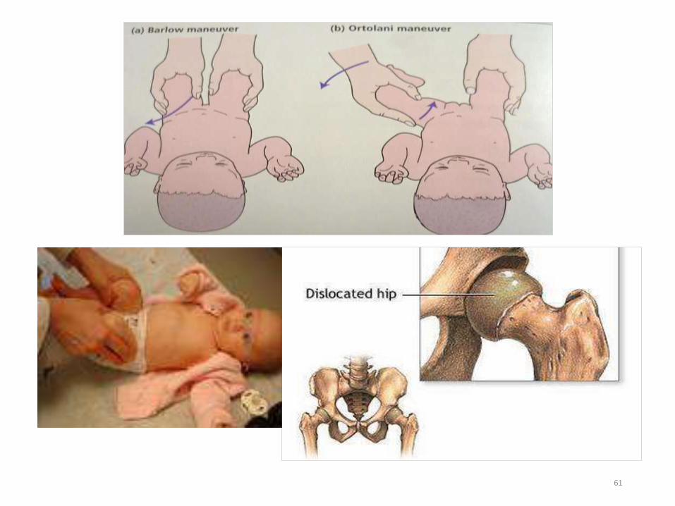
Musculoskeletal…
(a) Test for dislocatable hip
• Hip is held flexed and adducted.
• The femoral head is pushed downwards.
• If dislocatable, the femoral head will be pushed posteriorly out of the
acetabulum.
• Barlow’s sign is positive
(b) Test for dislocated hip
• Abduct hip with upward leverage of the femur.
• A dislocated hip will return with palpable ‘clunk’ into the acetabulum.
60
61
Neurologic
•Similar to adult
•Extent of neurologic exam dictated by history and
index of suspicion
•Much of the usual neurologic examination is done by
observation with respect to age
62
Neurologic…
•Level of consciousness
•Cranial nerve examination
•Sensory examination
•Motor examination
•Deep tendon reflexes
63

CHILD GROWTH AND DEVELOPMENT
64
Learning Objective
After studying the material in this unit, the student will be
able to:
Identify the difference between growth and development
Describe milestones of normal growth and development
Detect deviation from normal growth and development
Use growth-monitoring chart to assess nutritional status of
under 5 children
Recognize needs of the growing child
65
Growth and Development
•What is Growth ?
•What is Development ?
•What is the difference and similarity in growth
and development ?
66
Growth and Development
Growth:
• An increase in size of a living organism from a
simple to a complex form.
Development:
• An increase in skill and complexity of function
•Growth and development go together but at
different speeds.
67

Growth and Development…
•Growth is attributable to increased number of
cells
•Development is intimately related to
maturation of the nervous system, but may
be profoundly modified by environment &
experience
68
Principles of Growth & Development
•Continuous Process
•Predictable Sequence
•Don’t progress at the same rate (↑ periods of GR in
early childhood and adolescents & ↓ periods of GR in
middle childhood)
•Not all body parts grow in the same rate at the same
time
•Each child grows in his/her own unique way 69
Principles of Growth & Development…
•Each stage of growth & development is affected by
the preceding types of development
•Cephalocaudal (head down to toes)
•Proximodistal (center of the body to the peripheral)
•General to specific
70

Principles of Growth & Development…
71

Factors Affecting growth and development
• What are the Factors Affecting growth and development?
72
Factors Affecting growth and development
•Genetic factors
• The growth of a child is a result of complex interactions of
genetic and environmental factors.
•Neural control of growth
• Growth center in brain that keeps the child in his or her
genetically determined growth curve:
• If a child deviates from a growth pattern for a period of time
because of malnutrition or illness, a period of accelerated or
’catch-up‘ growth brings him or her back to the ’predetermined‘
curve, implying some sort of central control mechanism. 73
Factors Affecting growth…
• Hormonal influence
• Most of the endocrine glands influence growth
significantly either by promoting protein synthesis,
regulating substrate supply, or enhancing the effect of
other hormones on specific organs.
• E.g growth hormone, insulin, thyroid hormone, and sex
hormones.
• Nutrition
• Adequate food to provide substrate for energy and
synthesis of proteins is essential for normal growth.
74
Factors Affecting growth…
•Environmental and social factors
•The home, neighborhood, and school environments
have profound effect on the child’s psychosocial
development.
•Socio-economic status also significantly affects the
growth of a child: thus, children in top
socioeconomic groups are taller than children of
lower socioeconomic class.
75
Types of growth and development
Types of growth:
Physical growth (Ht., Wt., head & chest circumference)
Physiological growth (vital signs …)
Types of development:
Motor development
Cognitive development
Emotional development
Social development
76
Weight:
•Normal birth weight is 3400gms (normal range :
2500 –4000gms).
•Double birth Wt. at 4–7 months of age.
•Triple birth Wt. at 10-12 months of age.
•Quadruple birth Wt. at 2-2.5 years of age.
•Newborns lose 5-10% of body Wt. immediately
after birth
•Newborns gain 30gm/day during the 1st 5-6 months
and 2-3 kg/yearly after 1st year.77
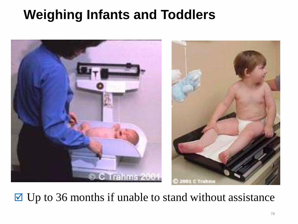
Weighing Infants and Toddlers
78
Up to 36 months if unable to stand without assistance
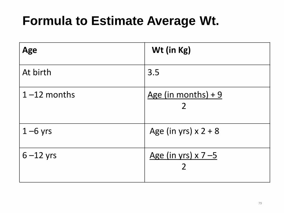
Formula to Estimate Average Wt.
Age Wt (in Kg)
At birth 3.5
1 –12 months Age (in months) + 92
1 –6 yrs Age (in yrs) x 2 + 8
6 –12 yrs Age (in yrs) x 7 –5 2
79
Height/Length
• Growth in stature progress less rapidly than Wt.
• Normal newborn length is 50 cm (range 46 –56cms).
• Increase 25 –30cms in first year of life.
• After first year, gain 6 – 8cms yearly.
• Birth length doubles by 3 –4 years.
• Eventual adult Ht can be approximated by doubling child’s Ht
by 2 years of age (i.e. Ht at 2 yrs of age half adult Ht).
80
Height/Length ….
•The required principles to measure the Height or
Length include:
•Length
•Measure length of children up to 2 years
• Use supine position, which requires 2 people
•Straight knees and keep ankles in neutral position
•Record measurement to the nearest 0.1 cm
81
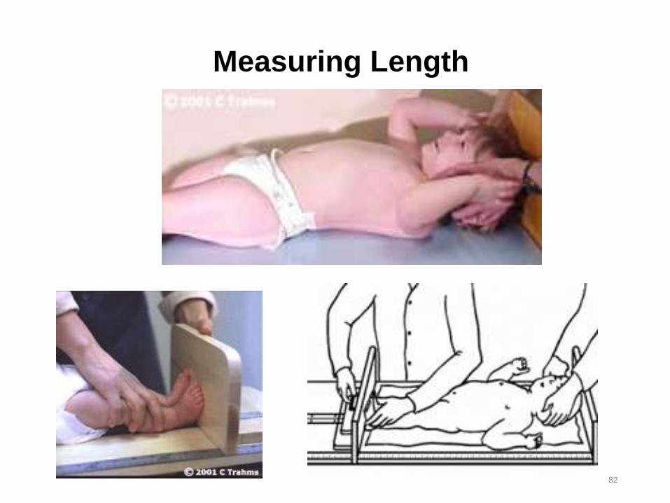
82
Measuring Length
Height/Length ….Height
• Measure Ht for children > 2 years old
• Use a tape meter or a measuring tape plastered on a wall
• Remove shoes
• Make sure the legs are close to each other and the heels, the
buttock and the shoulder blade touch the wall and the head looks
straight ahead
• Place a ruler or a hard paper on top of the head to perpendicular to
the wall to take the measurement.
• If there is large hair, press gently83
84
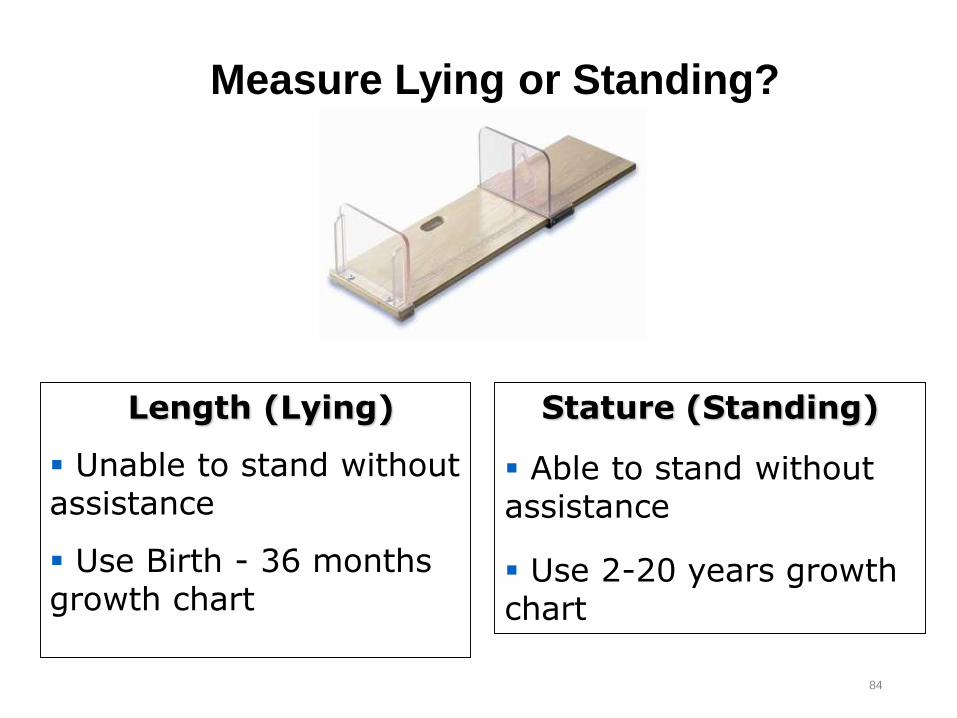
Measure Lying or Standing?
Length (Lying)
Unable to stand without assistance
Use Birth - 36 months growth chart
Stature (Standing)
Able to stand without assistance
Use 2-20 years growth chart
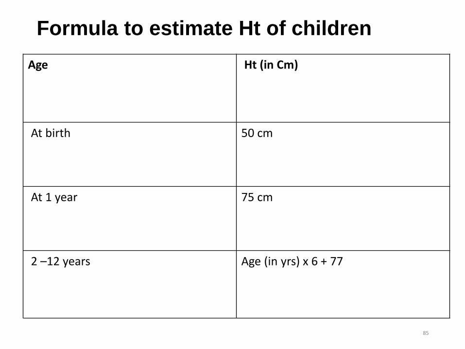
Formula to estimate Ht of children
Age Ht (in Cm)
At birth 50 cm
At 1 year 75 cm
2 –12 years Age (in yrs) x 6 + 77
85
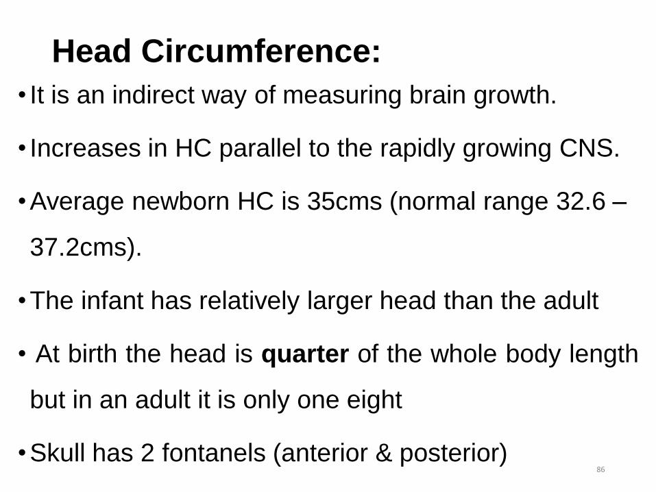
Head Circumference:
• It is an indirect way of measuring brain growth.
• Increases in HC parallel to the rapidly growing CNS.
•Average newborn HC is 35cms (normal range 32.6 –
37.2cms).
•The infant has relatively larger head than the adult
• At birth the head is quarter of the whole body length
but in an adult it is only one eight
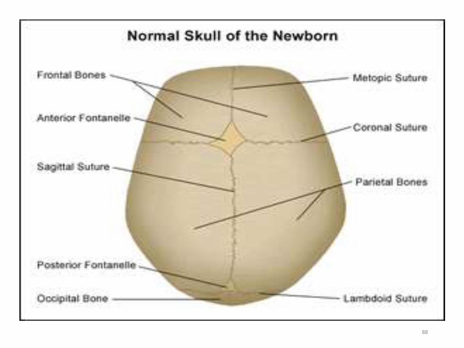
•Skull has 2 fontanels (anterior & posterior)86
Anterior fontanel
•Diamond in shape
•The junction of the sagittal, corneal and frontal
sutures forms it
•Between 2 frontal & 2 parietal bones
•3-4 cm in length and 2-3 cm width
• It closes at 12-18 months of age
87
88
Posterior fontanel
•Triangular
•Located between occipital & 2 parietal bones
•Closes by 6-8 w of age.
89
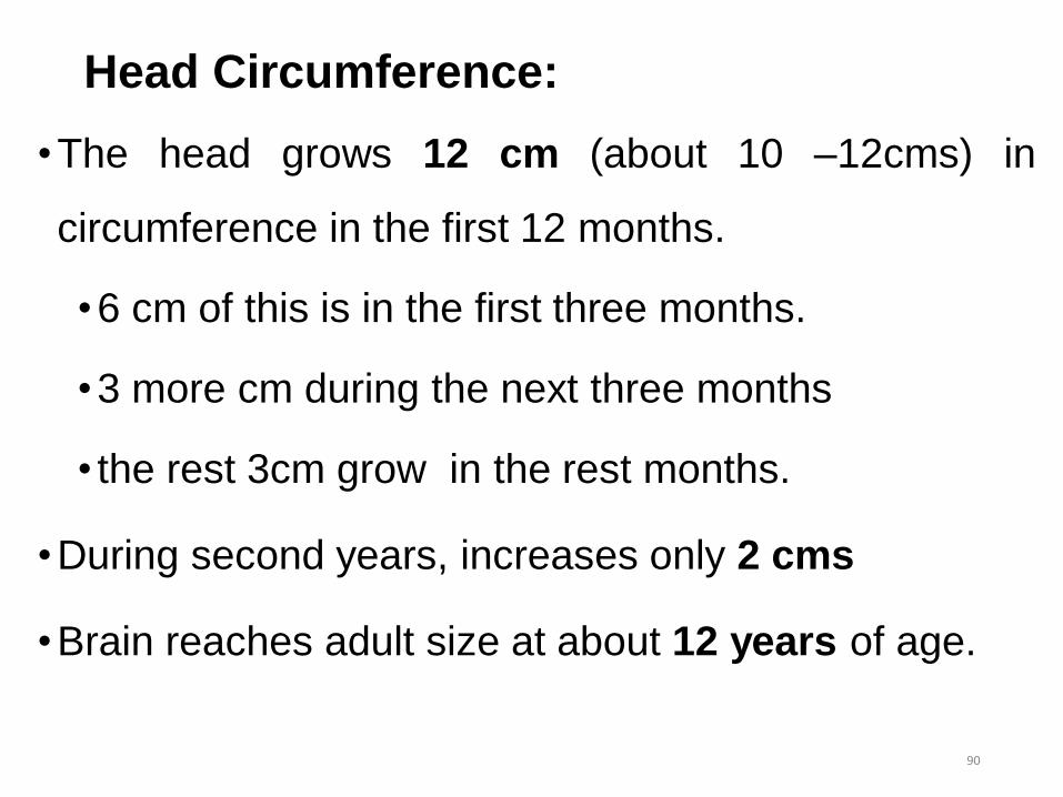
Head Circumference:
•The head grows 12 cm (about 10 –12cms) in
circumference in the first 12 months.
•6 cm of this is in the first three months.
•3 more cm during the next three months
• the rest 3cm grow in the rest months.
•During second years, increases only 2 cms
•Brain reaches adult size at about 12 years of age.
90
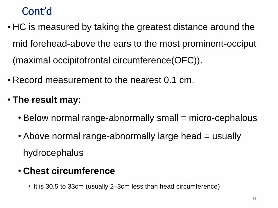
Cont’d
• HC is measured by taking the greatest distance around the
mid forehead-above the ears to the most prominent-occiput
(maximal occipitofrontal circumference(OFC)).
• Record measurement to the nearest 0.1 cm.
• The result may:
• Below normal range-abnormally small = micro-cephalous
• Above normal range-abnormally large head = usually
hydrocephalus
• Chest circumference
• It is 30.5 to 33cm (usually 2–3cm less than head circumference)
91

OFC
92
Other indices of Growth:
•Body Proportions
•Skeletal maturation
•Dental eruption
93
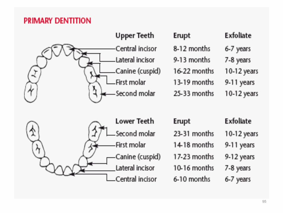
Dental Eruption
•Eruption of teeth starts by 5–6 months of age.
It is called "Milky teeth" or "Deciduous teeth"
or "Temporary teeth".
94
95
Developmental milestones
Age periods and developmental milestones:
• The development of a child can be accessed from different points of
view.
• What he can do in the way of moving around?
• (motor development)
• How he talks and makes his wants known?
• (language)
• How he fits into his family and community?
• (social behavior)
96
Developmental milestones…..
•The various skills the baby learns are called
Millstones.
• In watching development we notice at what age the
child learns to do certain things, such as smiling at
his mother, sitting without support, grasping objects
with his hands, walking and talking
97
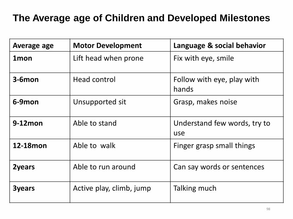
The Average age of Children and Developed Milestones
98
Average age Motor Development Language & social behavior
1mon Lift head when prone Fix with eye, smile
3-6mon Head control Follow with eye, play with hands
6-9mon Unsupported sit Grasp, makes noise
9-12mon Able to stand Understand few words, try to use
12-18mon Able to walk Finger grasp small things
2years Able to run around Can say words or sentences
3years Active play, climb, jump Talking much
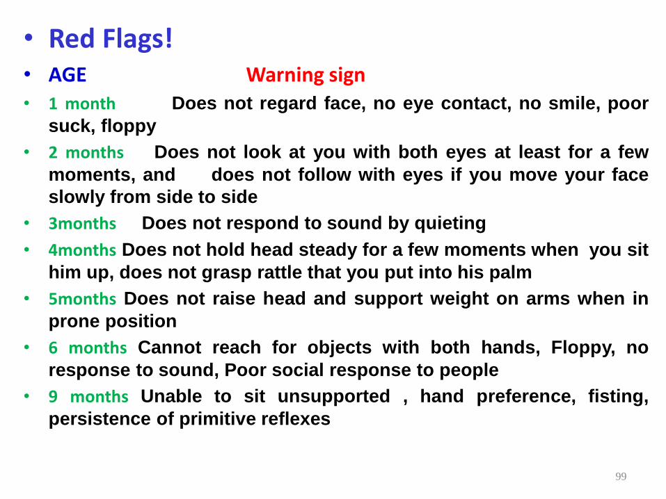
• Red Flags!• AGE Warning sign• 1 month Does not regard face, no eye contact, no smile, poor
suck, floppy
• 2 months Does not look at you with both eyes at least for a few
moments, and does not follow with eyes if you move your face
slowly from side to side
• 3months Does not respond to sound by quieting
• 4months Does not hold head steady for a few moments when you sit
him up, does not grasp rattle that you put into his palm
• 5months Does not raise head and support weight on arms when in
prone position
• 6 months Cannot reach for objects with both hands, Floppy, no
response to sound, Poor social response to people
• 9 months Unable to sit unsupported , hand preference, fisting,
persistence of primitive reflexes
99
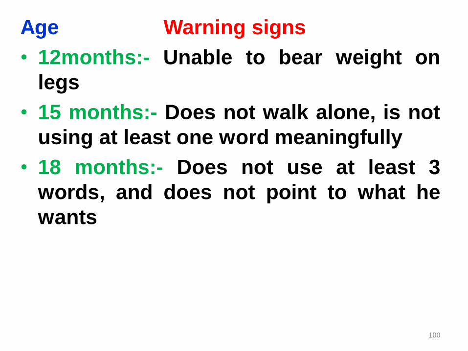
Age Warning signs
• 12months:- Unable to bear weight on
legs
• 15 months:- Does not walk alone, is not
using at least one word meaningfully
• 18 months:- Does not use at least 3
words, and does not point to what he
wants
100
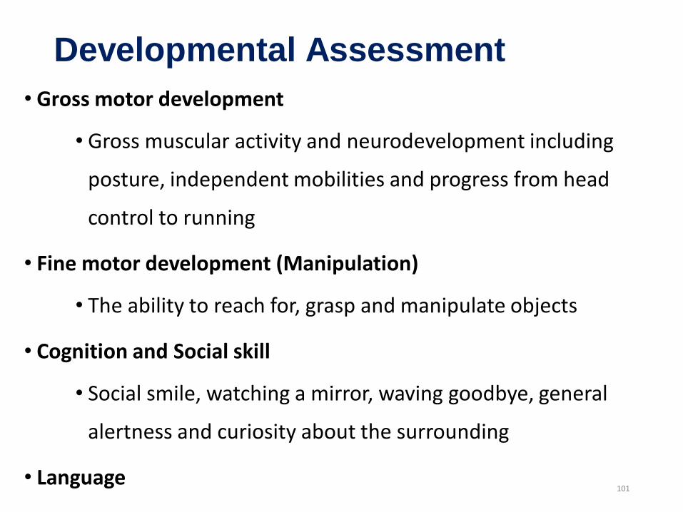
Developmental Assessment
• Gross motor development
• Gross muscular activity and neurodevelopment including
posture, independent mobilities and progress from head
control to running
• Fine motor development (Manipulation)
• The ability to reach for, grasp and manipulate objects
• Cognition and Social skill
• Social smile, watching a mirror, waving goodbye, general
alertness and curiosity about the surrounding
• Language 101
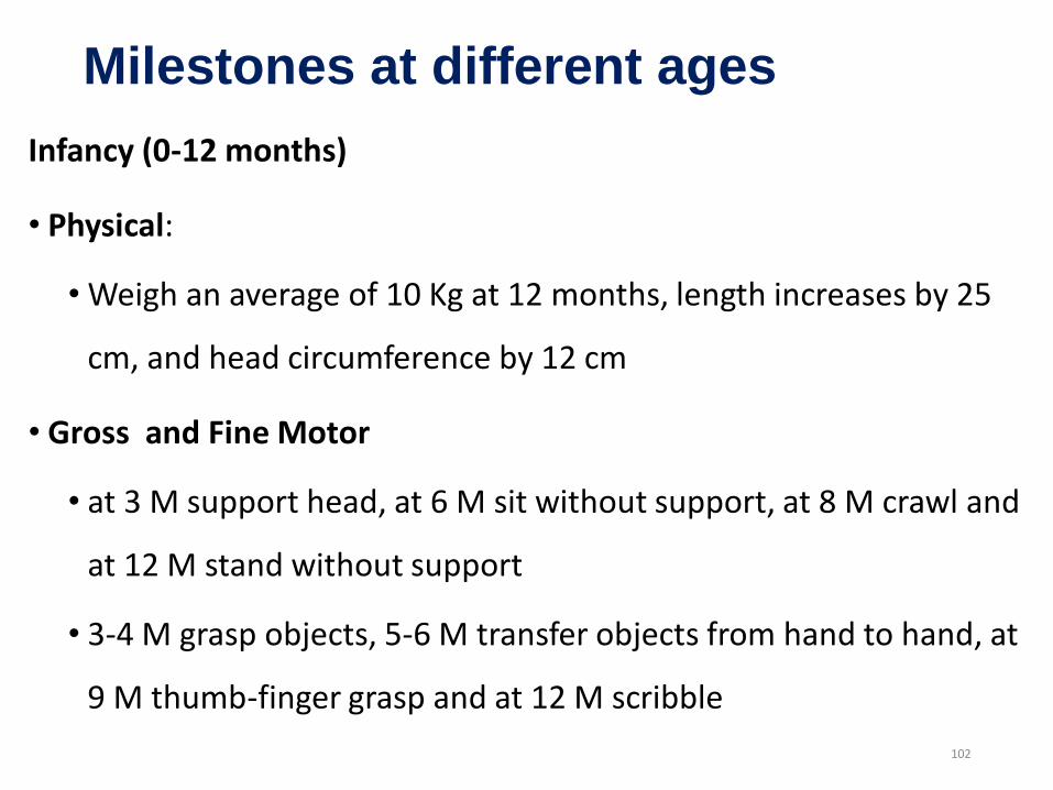
Milestones at different ages
Infancy (0-12 months)
• Physical:
• Weigh an average of 10 Kg at 12 months, length increases by 25
cm, and head circumference by 12 cm
• Gross and Fine Motor
• at 3 M support head, at 6 M sit without support, at 8 M crawl and
at 12 M stand without support
• 3-4 M grasp objects, 5-6 M transfer objects from hand to hand, at
9 M thumb-finger grasp and at 12 M scribble
102
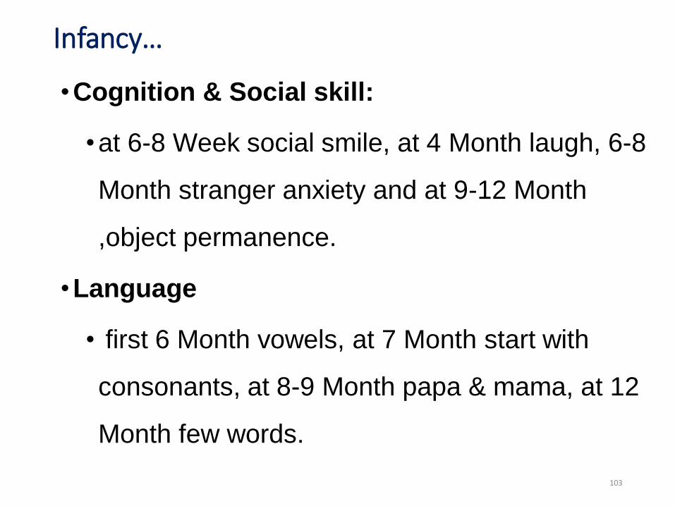
Infancy…
•Cognition & Social skill:
•at 6-8 Week social smile, at 4 Month laugh, 6-8
Month stranger anxiety and at 9-12 Month
,object permanence.
•Language
• first 6 Month vowels, at 7 Month start with
consonants, at 8-9 Month papa & mama, at 12
Month few words.
103
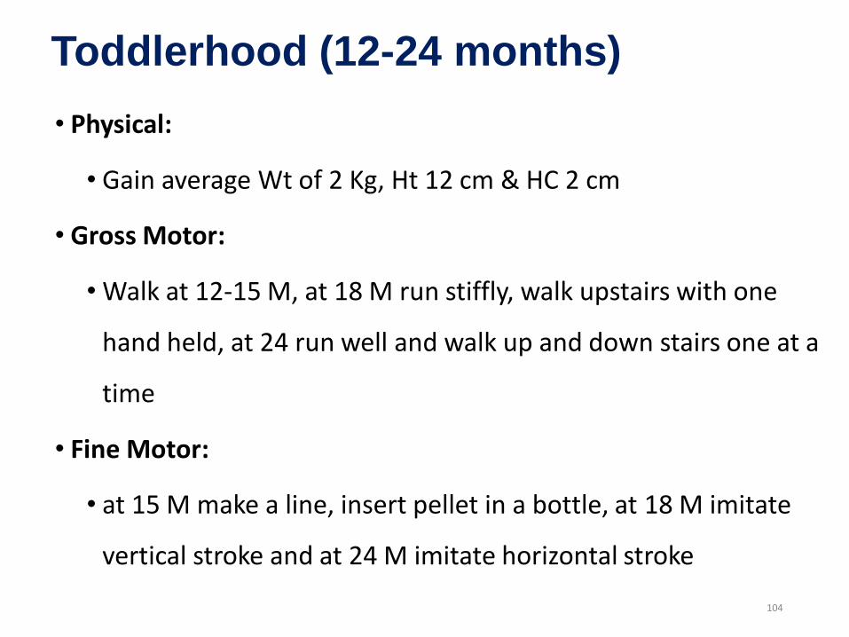
Toddlerhood (12-24 months)
• Physical:
• Gain average Wt of 2 Kg, Ht 12 cm & HC 2 cm
• Gross Motor:
• Walk at 12-15 M, at 18 M run stiffly, walk upstairs with one
hand held, at 24 run well and walk up and down stairs one at a
time
• Fine Motor:
• at 15 M make a line, insert pellet in a bottle, at 18 M imitate
vertical stroke and at 24 M imitate horizontal stroke
104
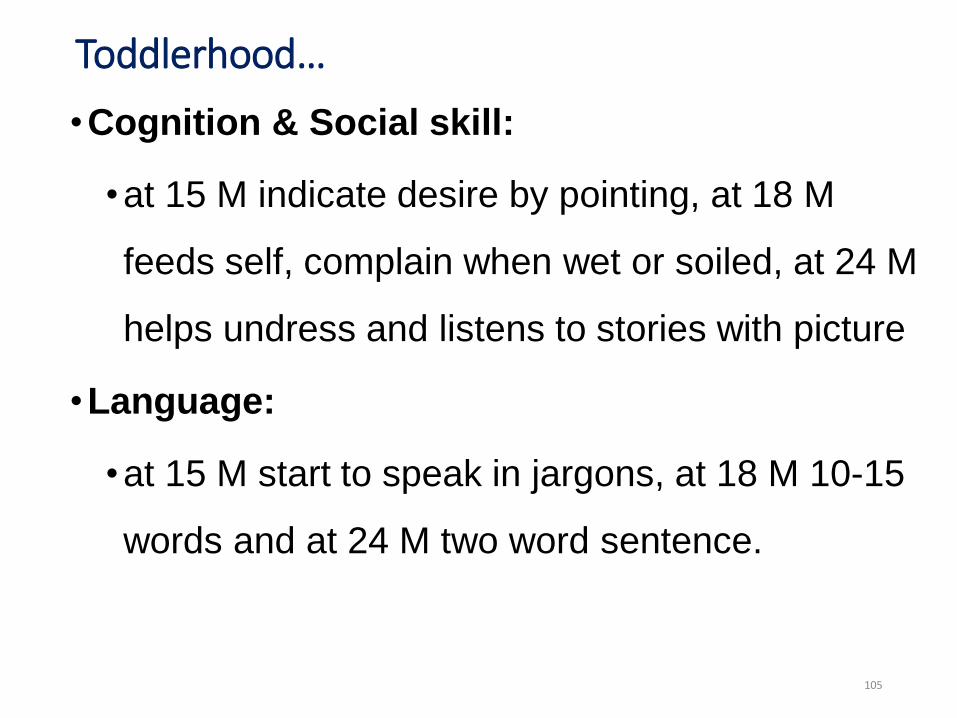
Toddlerhood…
•Cognition & Social skill:
•at 15 M indicate desire by pointing, at 18 M
feeds self, complain when wet or soiled, at 24 M
helps undress and listens to stories with picture
•Language:
•at 15 M start to speak in jargons, at 18 M 10-15
words and at 24 M two word sentence.
105
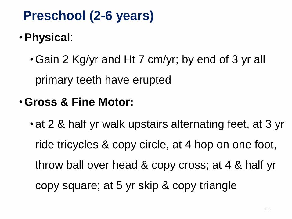
Preschool (2-6 years)
•Physical:
•Gain 2 Kg/yr and Ht 7 cm/yr; by end of 3 yr all
primary teeth have erupted
•Gross & Fine Motor:
•at 2 & half yr walk upstairs alternating feet, at 3 yr
ride tricycles & copy circle, at 4 hop on one foot,
throw ball over head & copy cross; at 4 & half yr
copy square; at 5 yr skip & copy triangle
106

Preschool…
• Cognition & Social skill:
• at 2 & half yr Know full name and pretend plays; at 3 yr
know age and sex, count 3 objects, wash hands & put shoe;
at 4 yr tell a story; at 5 yr name 4 colors, count 10 objects
correctly and dress & undress; think in prelogical
operations.
• Language
• language develop most rapidly between 2-5 yr, vocabulary
increases from 50-100 to about 2000 words; number of
words in a sentence equals age in years107
School age (6-12 years)
• Physical:
• Average weight and height gain per year is 3-3.5 kg and
6 cm respectively, HC increase by 2-3
• Motor:
• Coordination and stamina increase progressively, and
are able to perform complex movements such as
dancing
• Cognition & Language:
• Start thinking in concrete logical operations108
Value of Play and Selection of Play Material
•Play is defined as behaviour that is freely
chosen, personally directed (process of trial and
error) and intrinsically motivated.
•A successful, well-run play program needs to:
•Increase the child’s ability to cope with a
hospital environment.
109
Value of Play and Selection of Play Material
•Facilitate appropriate channels of communication
•Reduce developmental regression
•Promote confidence, self esteem and independence
•Relieving fear and anxiety
•Help professionals to assess development
•Provides opportunities to engage in meaningful
activities that enhance physical, language, social, and
cognitive development110
Play & Skill Development:
• Play can be an effective way for Children to develop skills:
• Language - name games, sing songs, rhymes/same
sound.
1. Thinking - build towers, puzzles, follow directions
• Small-muscle - string beads, cut with scissors
• Large-muscle –running, play ball, race, roller-skate etc.
• Creative – clay, paper, make-up stories, dress-up
puppets.
• Social – decisions, playing with others e.g. parts of a
play, which team going to play in etc. 111
Growth monitoring
•The most powerful tool in growth assessment
is the growth chart (Growth monitoring chart);
used in combination with accurate
measurements of height, weight, and head
circumference.
•Measurement
•Plotting
•Interpretation112

Growth monitoring….
•Growth Charts/Curves: are Graph that records
changes in the child’s growth with time compared to
normative growth rates.
• Growth parameters should be standardized and
compared with age related norms.
113

Why do we use growth curves?
•Easy and systematic way to follow changes in
growth over time.
•Height, weight and head circumference should be at
regular intervals.
•Monthly till 6 months of age and continues
accordingly.
114
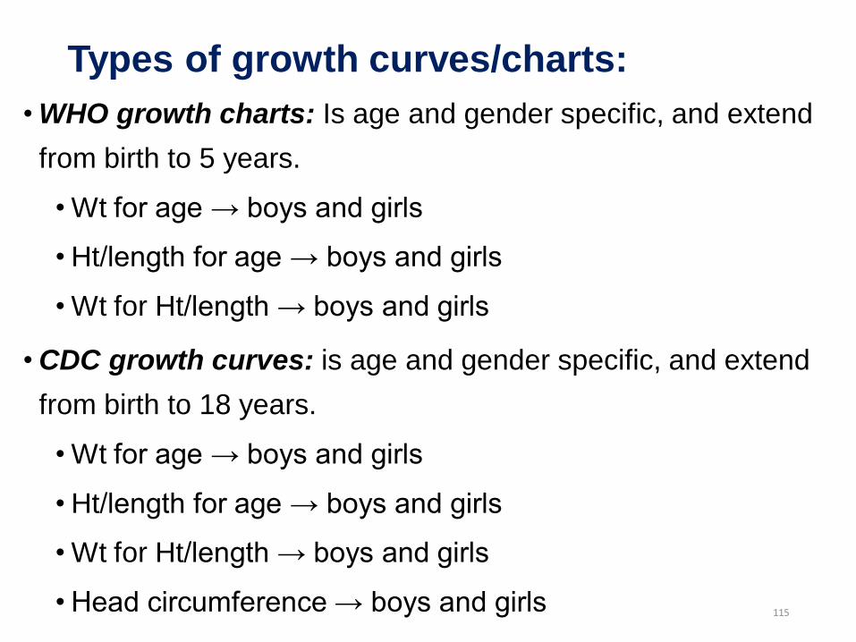
Types of growth curves/charts:
• WHO growth charts: Is age and gender specific, and extend
from birth to 5 years.
• Wt for age → boys and girls
• Ht/length for age → boys and girls
• Wt for Ht/length → boys and girls
• CDC growth curves: is age and gender specific, and extend
from birth to 18 years.
• Wt for age → boys and girls
• Ht/length for age → boys and girls
• Wt for Ht/length → boys and girls
• Head circumference → boys and girls 115
Parameters of Growth Assessment
Weight for age “growth faltering”
Height for age "stunting"
Weight for Height "wasting"
Head circumference
116

Parameters of Growth Assessment…
• N.B: Use CDC growth curves to monitor HC for < 3 yrs and Growth
for > 6 yrs of age.
• Procedures for accurate measurement:
• Accurate measurement is a key component of assessing growth.
Weight, in pounds or kilograms, must be determined using an
accurate scale.
• Head circumference is determined using a flexible tape measure run
from the supra-orbital ridge to the occiput in the path that leads to
the largest possible measurement.
117
Parameters of Growth Assessment…
•The data obtained from samples for standard
preparation are presented in 5 standard gender-
specific charts:
1. weight for age;
2. height (length and stature) for age;
3. head circumference for age;
4. weight for height (length and stature)-infants;
5. BMI for children over 2 years of age.118
Parameters of Growth Assessment…
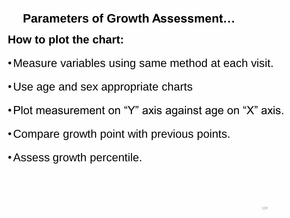
How to plot the chart:
•Measure variables using same method at each visit.
•Use age and sex appropriate charts
•Plot measurement on “Y” axis against age on “X” axis.
•Compare growth point with previous points.
•Assess growth percentile.
119
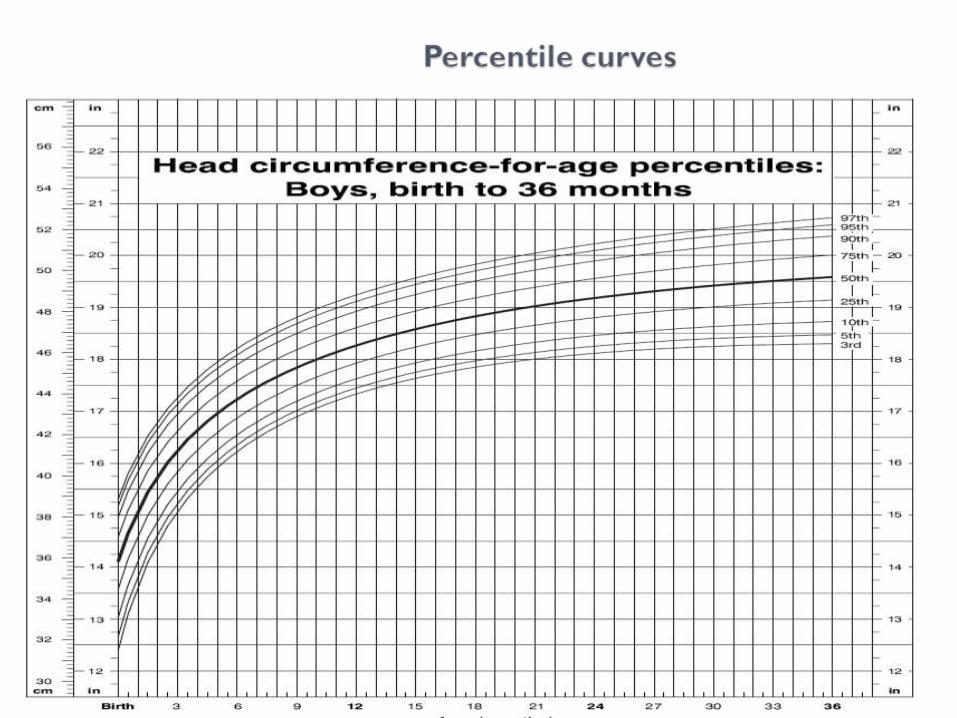
What are percentiles?
• The percentile curve indicates the percentage of children
at a given age on the x-axis whose measured value falls
below the corresponding value on the y-axis.
• The percentiles speak to the general population and
address the question “what percentage of the population
is of the same weight and height?‟.
• The median, or 50th percentile, is also termed the
standard value.120
What are percentiles…
•E.g. If a child is on the 5%, the growth chart tells you
that the child is small relative to other children of the
same age; → 95% of children of the same age are
heavier than him.
•On the weight chart for boys 0–36 mo of age, the 9
mo age line intersects the 25th percentile curve at 8.6
kg, indicating that 25% of the 9 mo old boys in the
National Center for Health Statistics sample weigh
less than 8.6 kg. 121

Continu…
• The weight-for-height charts are constructed in an analogous
fashion, with length or stature in place of age on the x-axis; the
median or standard weight for a girl measuring 110 cm is 18.2 kg.
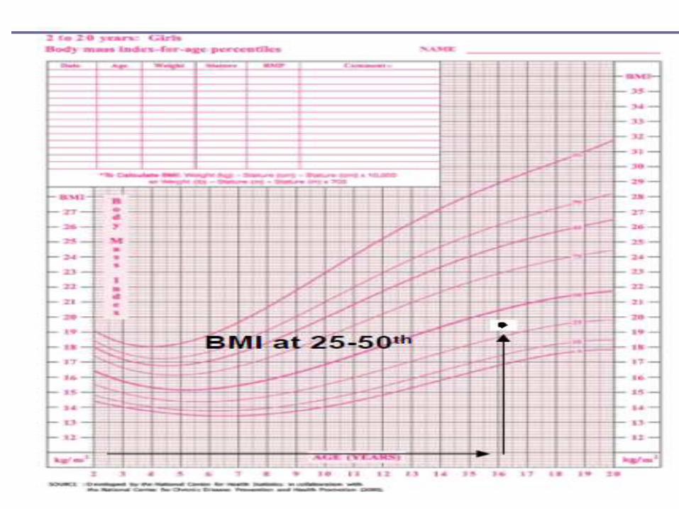
• Body mass index (BMI) is added to the standard growth charts for
children over 2 year of age. BMI can be calculated as wt. in
kg/(height in meters)2 with expression of decimals.
• BMI percentile varies with age over childhood: a 6 yr old girl with a
BMI of 21 is overweight, while a 16 year old girl with the same BMI
is just above the 50th percentile.
122
123
continue…
•Specialized charts have been developed for
children with various conditions, including very low
birth weight and prematurity; Down, Turner,
Klinefelter syndromes, and achondroplasia.
124
What to measure?1. Weight – for – age:
1. Weight for age: weight of the child (in kg) is
compared with that of a healthy child of the same
age from a reference population.
Wt for age = (Actual wt./standard wt. of the same
age)*100
125
Analysis…
•When growth parameters fall below the 5th percentile,
it is necessary to express values in percentages
•E.g. Harvard Standard
•Nutritional insufficiency must be differentiated from:
•Congenital, constitutional, familial and endocrine
causes of decreased linear growth
126
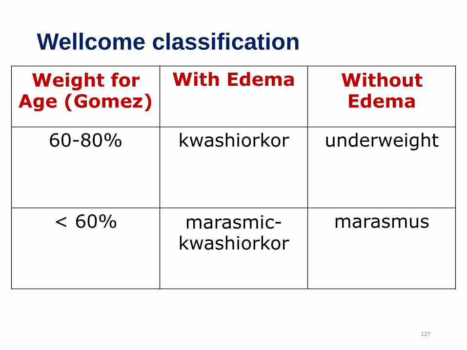
Wellcome classification
127
Weight for Age (Gomez)
With Edema Without Edema
60-80% kwashiorkor underweight
< 60% marasmic-kwashiorkor
marasmus
2. Length for age
Compares height/length with the expected height of
a healthy reference child of the same age.
Used to assess stunting which may be an index
of long-term nutritional deprivation.
128
3. Weight for length/height
Degree of wasting assessed by comparing
the child’s weight with the weight that would
be expected for a healthy child of the same
height.
A child who is < 70% of the expected weight
for height is classified as severely wasted.
129

130
4. Head circumference
Increases in size during the 1st year of life, but little
increment between 1 and 5 years of age.
Average Head circumference (cm) at birth = 35 (33 –
37)
At 6mo = 44
At 1 yr = 47
At 2 yr = 49
131
132
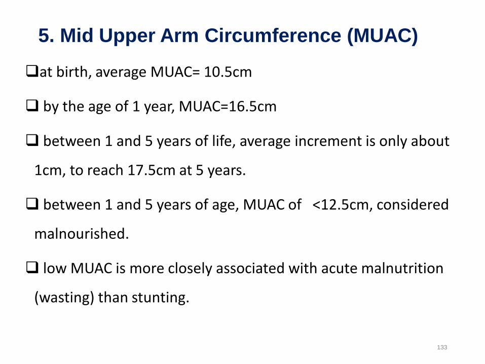
5. Mid Upper Arm Circumference (MUAC)
at birth, average MUAC= 10.5cm
by the age of 1 year, MUAC=16.5cm
between 1 and 5 years of life, average increment is only about
1cm, to reach 17.5cm at 5 years.
between 1 and 5 years of age, MUAC of <12.5cm, considered
malnourished.
low MUAC is more closely associated with acute malnutrition
(wasting) than stunting.
133
MUAC…
Useful as method of screening large number of
children, during nutritional emergencies.
less useful in long – term growth monitoring.
134
Meeting the needs of the normal children through the stages of development and parental guidance
•Nutrition: Good nutrition is the base for normal
growth and development. The first six months of life
are extremely important as the brain may suffer for
the rest of life, if the child is not getting enough
food.
135
Cont’d
•Emotional Support: It is very important to realize that
a child is a growing and developing human being and
he ought to be treated very carefully with love and
respect by everyone so he can develop in harmony.
•Love: a child who does not feel loved will not develop
properly, and will not learn as quickly as normal
children. He/she may be sad, lonely and no longer
interested in what goes on around him.
136
Cont’d
•Security: a child can only feel safe if his parents
show that they love him and take good care of him.
•He must know that his parents will look after him,
feed him when he is hungry, play with him and keep
him happy and comfortable.
•The love and security he/she gets from family helps
him to feel friendly to people outside his family when
he grows up.
137

Cont’d
•Acceptance as individual: the young child needs to
know that his mother and family love him for what he
is.
• They should not compare him with other children and
tell him that he is slow to do this or that, he is not as
good as some other child.
• Recognition of achievement: the young child needs
to know that his parents are happy and pleased when
he has learned to do something new.138
Cont’d
•Wise and consistent use of authority: children
need to know what they can and what they cannot
do. Parents must teach children how they are
expected to behave.
• Independence: as the child grows he needs to be
allowed to decide more and more things alone.
139

Cont’d
•Playing: Encourage playing even if it may be noisy
sometimes. It helps physical, mental and social
development and is also good for health.
•Language training: adults should talk with small
children and encourage them to talk about what they
are thinking
•Do not laugh when children are talking; try to
understand and be happy when they involve you in
their world. 140
Nutrition in Children
141
Learning objectives
•At the end of this chapter, students are
expected to
•Describe nutritional need and feeding of infant
and children
•Describe common forms of malnutrition
142
Feeding of infant and children
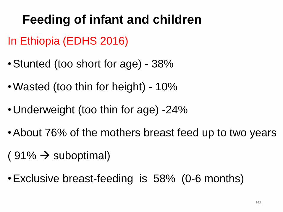
In Ethiopia (EDHS 2016)
•Stunted (too short for age) - 38%
•Wasted (too thin for height) - 10%
•Underweight (too thin for age) -24%
•About 76% of the mothers breast feed up to two years
( 91% suboptimal)
•Exclusive breast-feeding is 58% (0-6 months)
143
Feeding of infant and children
Breastfeeding
• Breast milk provides immunological, nutritional, and
psycho social advantages
• It is an ideal food for normal growth and development of
infants
• Compared to cows milk, breast milk contains more iron,
sugar, vitamin A and C, and niacin
• It has less protein and calcium than cows milk, but the
amounts present are better utilized by the baby144
Feeding of infant and children…
• Breast milk is more digestible because its fat globules are
smaller and it is pure
• It provides the baby with greater immunity to certain child
hood diseases
•Breast fed babies are less prone to intestinal upset
• Infants should be exclusively breast fed for the first
six months to achieve optimal growth and
development145
Why is breast feeding so important
• Practical factors in favor of breast milk are:
• It saves time and money
•Delivered to the baby in proper quantity and quality
•Helps the uterus to return to its normal size and
reduce post-partum bleeding.
•Gives emotional satisfaction to the mother
•Enhance bonding and attachment
146
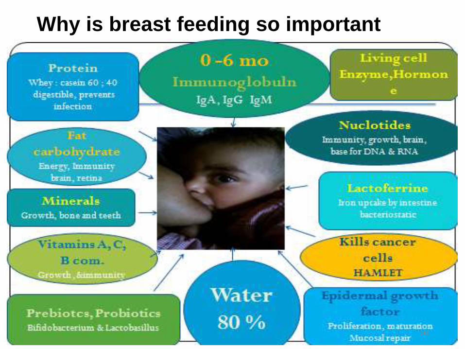
Why is breast feeding so important
•Has contraceptive benefits
•Contains the necessary nutrients needed by an
infant
•Nutrients are easily digestible and absorbable
•Provides all the water an infant needs, even in a
hot, dry climate
•Protects an infant against infection
147
Why is breast feeding so important
148

• Interferes with bonding and attachement
• More diarrhoea and persistent diarrhoea
• More frequent respiratory infections
• Malnutrition; Vitamin A deficiency
• More allergy and milk intolerance
Disadvantages of artificial feeding
Disadvantages of artificial feeding
•Increased risk of some chronic diseases
•Obesity
•Lower scores on intelligence tests
•Mother may become pregnant sooner
•Increased risk of anemia, ovarian cancer, and
breast cancer in mothers
150
Emergency situations
Malnourished children( needs support from formula milks)
risks of malnutrition
Low-birth-weight babies (Gavage feeding)
Infants of HIV-infected mothers (AFASS)
Orphans
Infants requiring tube feeding
Infants/ children with chronic illnesses etc are conditions
where formula milk is as important as mother’s breast milk.
Breast milk not adequate after 6 months
Exceptionally difficult circumstances
Contra indications of BF:
•Active maternal tuberculosis
•Maternal HIV
•Poor physical or mental cooperation of the mother
152
Instructions for the mother during BF:
• Wash hands before proceeding
• Wash nipples with warm water
• Positioning- semi-upright position
• Comfortable seated in chair with back and arm
supported
• Side lying with pillow beneath head, arm above head
• Mother should experiment the most comfortable position
• Entire body of infant turned to the mother’s breast
153
Instructions for the mother during BF:
•Both breasts are used at each feeding
•Avoid break suction by placing finger in corner of
baby’s mouth, but support breast from baby’s nose
• If a very sick infant cannot take the nipple in to his
mouth and keep it there to suck, he/she has no
attachment at all
• If not good attachment, either breast engorgement or
infant unsatisfaction has occurred
154

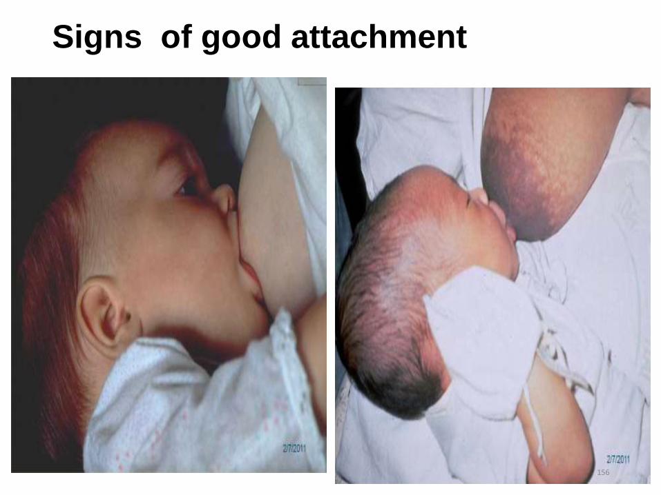
Signs of good attachment:
• Wide opened mouth of the baby
• Chin of baby touched the
mother’s breast
• More areola seen above baby’s
mouth than below
• Lower lip of baby turned out
ward
155
Signs of good attachment
156
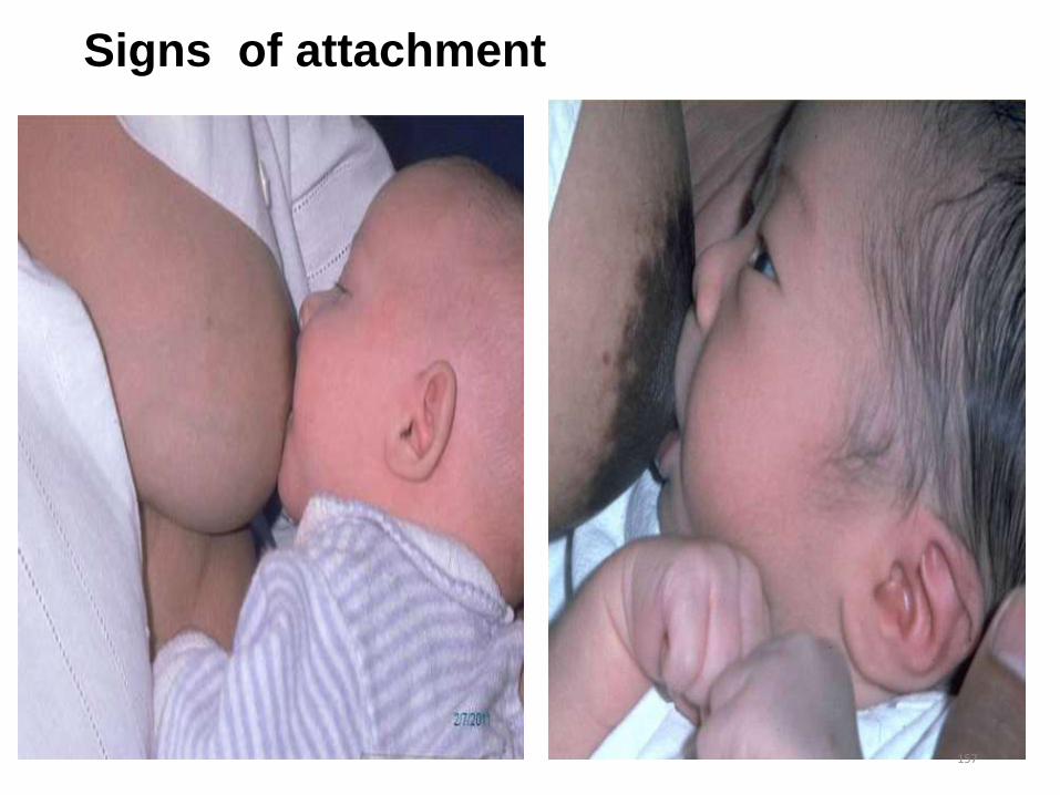
Signs of attachment
157
Signs of Good Positioning
•Infant’s head and body straight
•Head and body facing breast
•Infant’s body close to mother’s
•Supporting infant’s whole body
158
Signs for adequate breast feeding
At least 3-5 strong suckling before pausing
for breath or rest
Dimpling of cheeks may be seen while
suckling
Hearing of swallowing gurgle
Milk may be seen around the mouth leaking
out when it is excess
159
2. Complementary Feeding
•Breastfeeding is not enough for a child over 6 months
of age
•A variety of nutritious foods are needed to prevent
malnutrition
•Appropriate CF promotes growth and prevents stunting
among children between 6 and 24 months of age
• Infants are particularly vulnerable to malnutrition and
infection during the transition period when
complementary feeding begins 160

Complementary Feeding
•Energy needs from complementary foods for
infants with “average” breast milk intake in
developing countries are approximately:
•200 kcal per day at 6-8 months
•300 kcal per day at 9-11 months
•550 kcal per day at 12-23 months
161
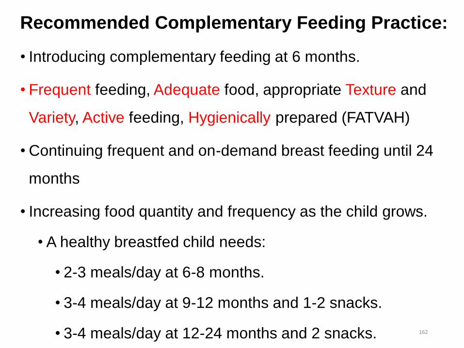
Recommended Complementary Feeding Practice:
• Introducing complementary feeding at 6 months.
• Frequent feeding, Adequate food, appropriate Texture and
Variety, Active feeding, Hygienically prepared (FATVAH)
• Continuing frequent and on-demand breast feeding until 24
months
• Increasing food quantity and frequency as the child grows.
• A healthy breastfed child needs:
• 2-3 meals/day at 6-8 months.
• 3-4 meals/day at 9-12 months and 1-2 snacks.
• 3-4 meals/day at 12-24 months and 2 snacks. 162

Recommended Complementary Feeding Practice
•Gradually increasing food consistency and
variety as the child gets older
•Practicing active Feeding
•Frequent and active feeding during and after
illness
•Practicing good hygiene and proper handling of
foods
163
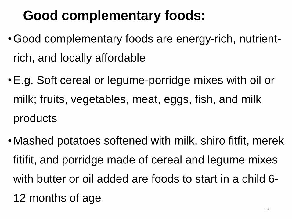
Good complementary foods:
•Good complementary foods are energy-rich, nutrient-
rich, and locally affordable
•E.g. Soft cereal or legume-porridge mixes with oil or
milk; fruits, vegetables, meat, eggs, fish, and milk
products
•Mashed potatoes softened with milk, shiro fitfit, merek
fitifit, and porridge made of cereal and legume mixes
with butter or oil added are foods to start in a child 6-
12 months of age 164
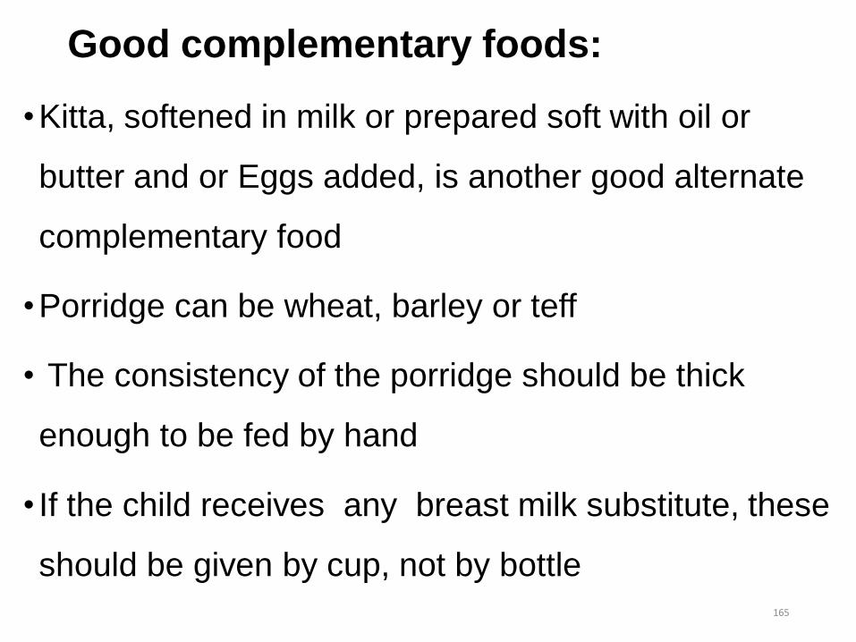
Good complementary foods:
•Kitta, softened in milk or prepared soft with oil or
butter and or Eggs added, is another good alternate
complementary food
•Porridge can be wheat, barley or teff
• The consistency of the porridge should be thick
enough to be fed by hand
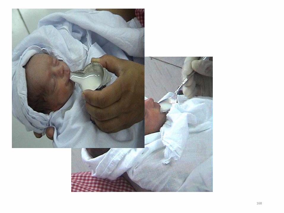
• If the child receives any breast milk substitute, these
should be given by cup, not by bottle
165
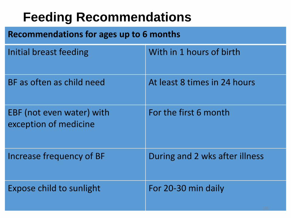
Feeding Recommendations
Recommendations for ages up to 6 months
Initial breast feeding With in 1 hours of birth
BF as often as child need At least 8 times in 24 hours
EBF (not even water) with exception of medicine
For the first 6 month
Increase frequency of BF During and 2 wks after illness
Expose child to sunlight For 20-30 min daily
166
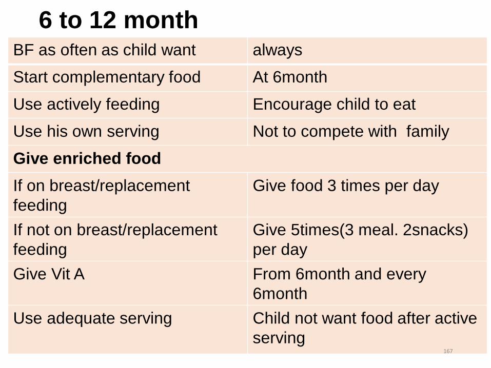
6-12 months
BF as often as child want always
Start complementary food At 6month
Use actively feeding Encourage child to eat
Use his own serving Not to compete with family
Give enriched food
If on breast/replacement
feeding
Give food 3 times per day
If not on breast/replacement
feeding
Give 5times(3 meal. 2snacks)
per day
Give Vit A From 6month and every
6month
Use adequate serving Child not want food after active
serving
6 to 12 month
167
168
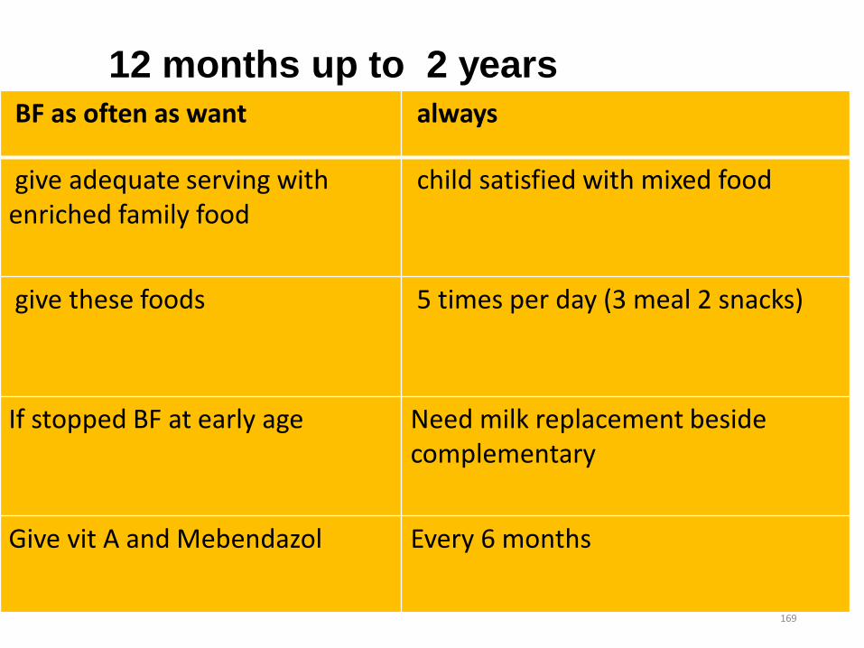
BF as often as want always
give adequate serving with enriched family food
child satisfied with mixed food
give these foods 5 times per day (3 meal 2 snacks)
If stopped BF at early age Need milk replacement beside complementary
Give vit A and Mebendazol Every 6 months
12 months up to 2 years
169
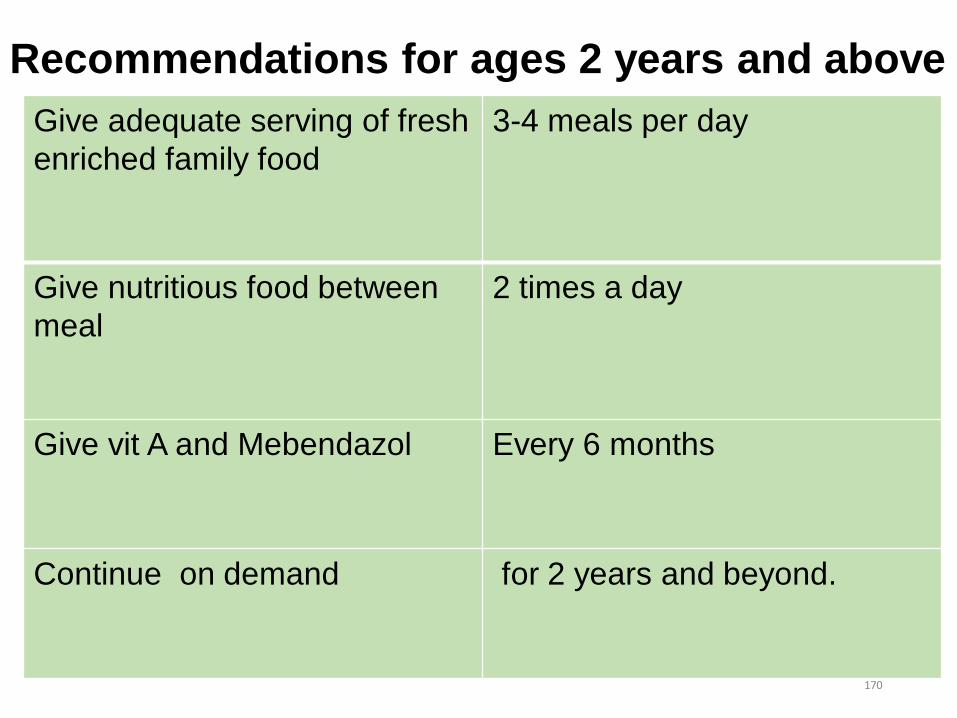
Recommendations for ages 2 years and above
Give adequate serving of fresh
enriched family food
3-4 meals per day
Give nutritious food between
meal
2 times a day
Give vit A and Mebendazol Every 6 months
Continue on demand for 2 years and beyond.
170
Malnutrition
171
Outline
• Overview
• Kwashiorkor
• Marasmus
• Stunting
• Vitamin A deficiency
• Vitamin D deficiency
• Management of Severe Acute Malnutrition (SAM)
• Childhood obesity
172
Session objectives
• At the end of this session the students will be able to:
√ Discuss kwashiorkor√ Discuss marasmus√ Differentiate the clinical presentations of
kwashiorkor from marasmus √ Explain stunting√ Describe common micronutrient deficiencies √ Discuss the management of severe acute
malnutrition√ Explain childhood obesity
173
Overview of malnutrition
Malnutrition includes:
• Macronutrient deficiency
• Micronutrient deficiency
• Over nutrition-obesity
Can be classified as:
• Acute-wasting
• Chronic-stunting
174
Kwashiorkor
• A nutritional disorder due to deficiency of protein and calories, particularly proteins, characterized by mental apathy, wasting, growth retardation, and oedema.
•
175
Etiology
• Lack of knowledge about diet
• Poverty
• Natural calamities like drought, earthquakes, etc
• Repeated infections like diarrhoea, measles, etc
• Taboos
• Religious customs (people of certain religions avoid non-vegetarian diet which has high-quality proteins)
176
Kwashiorkor ...
• Incidence is more in:
Low birth weight
Broken families
In children with whose parents are unemployed
Large families
177
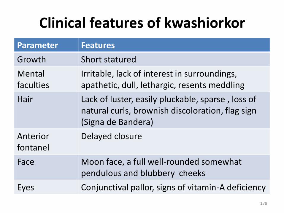
Clinical features of kwashiorkor
Parameter Features
Growth Short statured
Mental faculties
Irritable, lack of interest in surroundings, apathetic, dull, lethargic, resents meddling
Hair Lack of luster, easily pluckable, sparse , loss of natural curls, brownish discoloration, flag sign (Signa de Bandera)
Anterior fontanel
Delayed closure
Face Moon face, a full well-rounded somewhat pendulous and blubbery cheeks
Eyes Conjunctival pallor, signs of vitamin-A deficiency
178
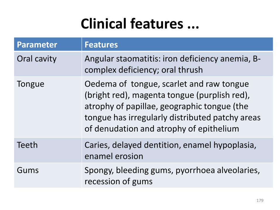
Clinical features ...Parameter Features
Oral cavity Angular staomatitis: iron deficiency anemia, B-complex deficiency; oral thrush
Tongue Oedema of tongue, scarlet and raw tongue (bright red), magenta tongue (purplish red), atrophy of papillae, geographic tongue (the tongue has irregularly distributed patchy areas of denudation and atrophy of epithelium
Teeth Caries, delayed dentition, enamel hypoplasia, enamel erosion
Gums Spongy, bleeding gums, pyorrhoea alveolaries, recession of gums
179
Clinical features ...
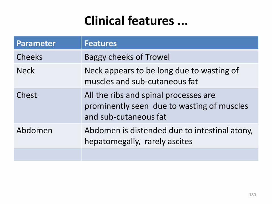
Parameter Features
Cheeks Baggy cheeks of Trowel
Neck Neck appears to be long due to wasting of muscles and sub-cutaneous fat
Chest All the ribs and spinal processes are prominently seen due to wasting of muscles and sub-cutaneous fat
Abdomen Abdomen is distended due to intestinal atony, hepatomegally, rarely ascites
180
Clinical features ...
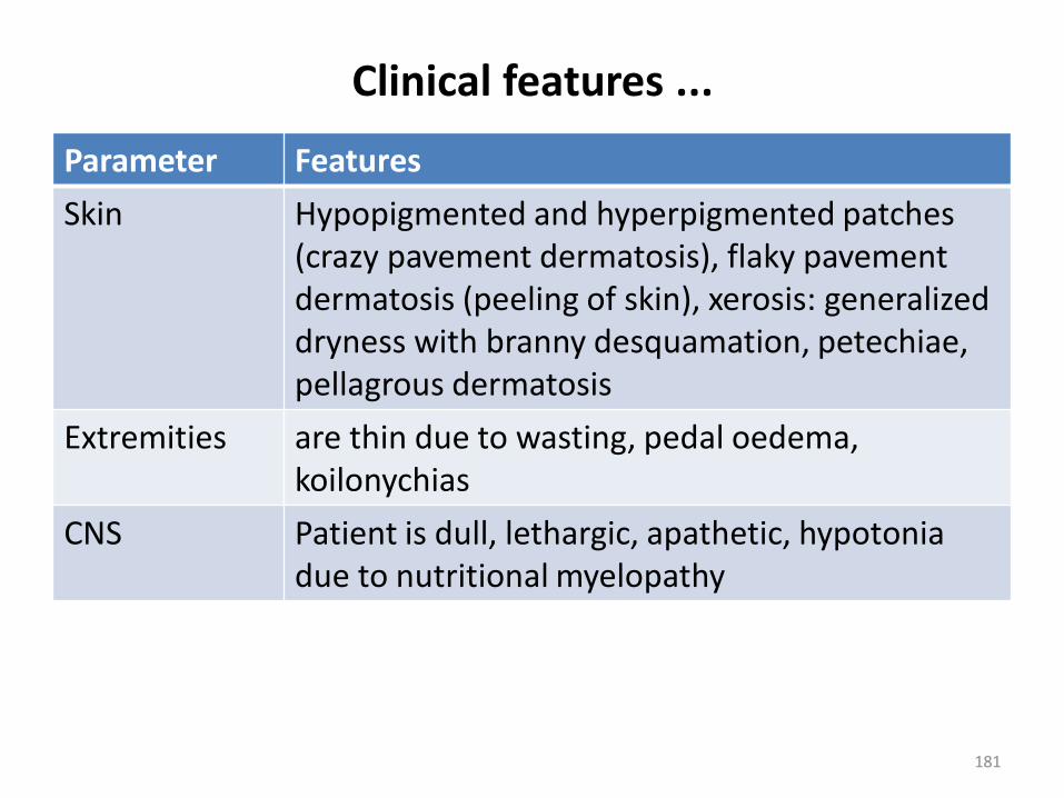
Parameter Features
Skin Hypopigmented and hyperpigmented patches (crazy pavement dermatosis), flaky pavement dermatosis (peeling of skin), xerosis: generalized dryness with branny desquamation, petechiae, pellagrous dermatosis
Extremities are thin due to wasting, pedal oedema, koilonychias
CNS Patient is dull, lethargic, apathetic, hypotonia due to nutritional myelopathy
181

Clinical features ...
Parameter Features
CVS Intensity of heart sounds is decreased due to decrease of cardiac output
Respiratory system
Features of secondary infection
GIT Hepatomegaly
Salivary glands Parotid gland is enlarged. It is firm, non-tender
182

Differential diagnosis
• Nephrotic syndrome
• Cirrhosis of liver
• Congestive heart failure
183
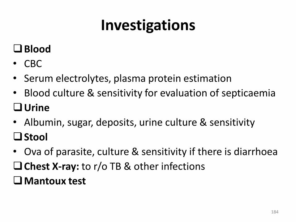
Investigations
Blood
• CBC
• Serum electrolytes, plasma protein estimation
• Blood culture & sensitivity for evaluation of septicaemia
Urine
• Albumin, sugar, deposits, urine culture & sensitivity
Stool
• Ova of parasite, culture & sensitivity if there is diarrhoea
Chest X-ray: to r/o TB & other infections
Mantoux test
184
Complications
• Hypothermia
• Hypoglycaemia
• Infections
• Electrolyte disturbances
• Dehydration
• Anemia
185

Management
• See management of SAM 2019
186
Prevention
• Health and nutrition education
• Early identification and treatment of the diseases
• Exclusive breast feeding up to six months
• Complementary feeding at the age of six months
• Family planning
• Safe and adequate water supply
• Immunization
187
Marasmus
• A nutritional disorder due to deficiency of protein and calories, particularly calories, characterized by:
Growth failure
Gross wasting
Absence of oedema
188
Etiology
1. Primary causes
• Lactation failure – the commonest cause
Lactation failure introduction of dilute & dirty formula infetions (diarrhoea)
starvation therapy due to diarrhoeamarasmus
189
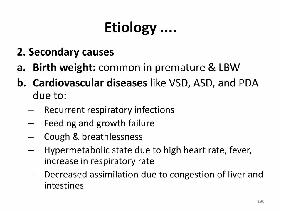
Etiology ....
2. Secondary causes
a. Birth weight: common in premature & LBW
b. Cardiovascular diseases like VSD, ASD, and PDA due to:
– Recurrent respiratory infections
– Feeding and growth failure
– Cough & breathlessness
– Hypermetabolic state due to high heart rate, fever, increase in respiratory rate
– Decreased assimilation due to congestion of liver and intestines
190

Secondary causes ...
c. Respiratory causes: TB, etcd. Gastrointestinal causes• Congenital hypertrophic pyloric stenosis-
vomiting• Congenital megacolon- diarrhoea• Cleft lip & palate – inadequate intake of feeds,
mainly breast feedse. Infections
– Repeated diarrhoea– Severe infections like congenital syphilis– Malabsorption syndrome
191

Etiology ...
3. CNS causes
• Hydrocephalus and CNS infections like tuberculosis meningitis, pyogenic meningitis can cause marasmus due to decreased intake & chronic vomiting
192

Clinical features Parameter Features
Mental faculties If there is no infection:•Patient is alert, playful, lively look•The cry is vigorous•Appetite is vigorous If there is an infection: • The child is less active, apathetic, cries in a low tone, refusal to food
Head •Hair is normal, delayed closure of anterior fontanel may be present
193
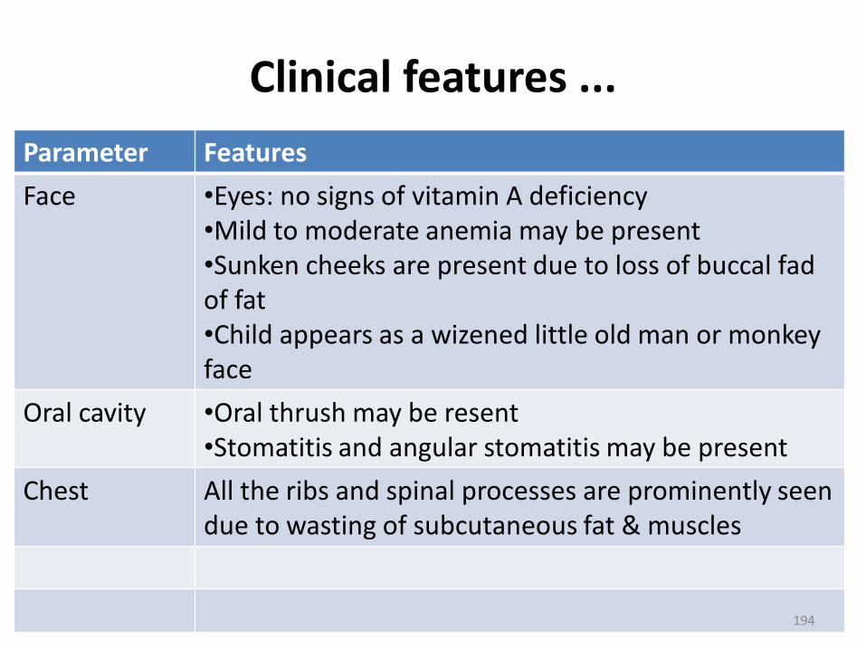
Clinical features ...
Parameter Features
Face •Eyes: no signs of vitamin A deficiency•Mild to moderate anemia may be present•Sunken cheeks are present due to loss of buccal fad of fat •Child appears as a wizened little old man or monkey face
Oral cavity •Oral thrush may be resent•Stomatitis and angular stomatitis may be present
Chest All the ribs and spinal processes are prominently seen due to wasting of subcutaneous fat & muscles
194
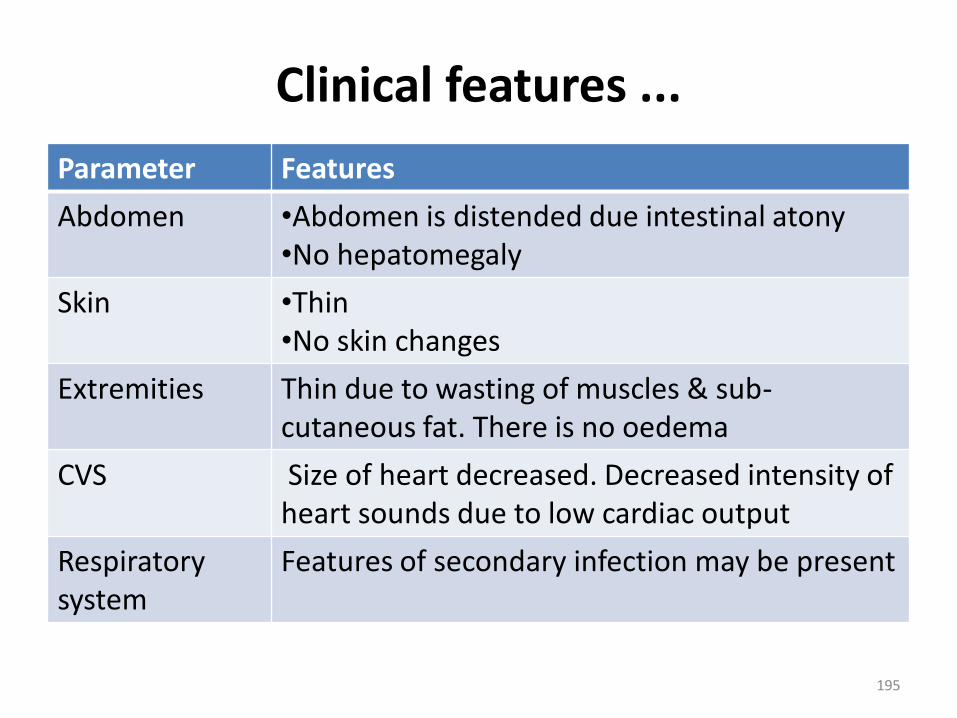
Clinical features ...
Parameter Features
Abdomen •Abdomen is distended due intestinal atony•No hepatomegaly
Skin •Thin•No skin changes
Extremities Thin due to wasting of muscles & sub-cutaneous fat. There is no oedema
CVS Size of heart decreased. Decreased intensity of heart sounds due to low cardiac output
Respiratory system
Features of secondary infection may be present
195
Investigations
• CBC
• Urine and stool culture
• Chest X-ray
• Mantoux test
196
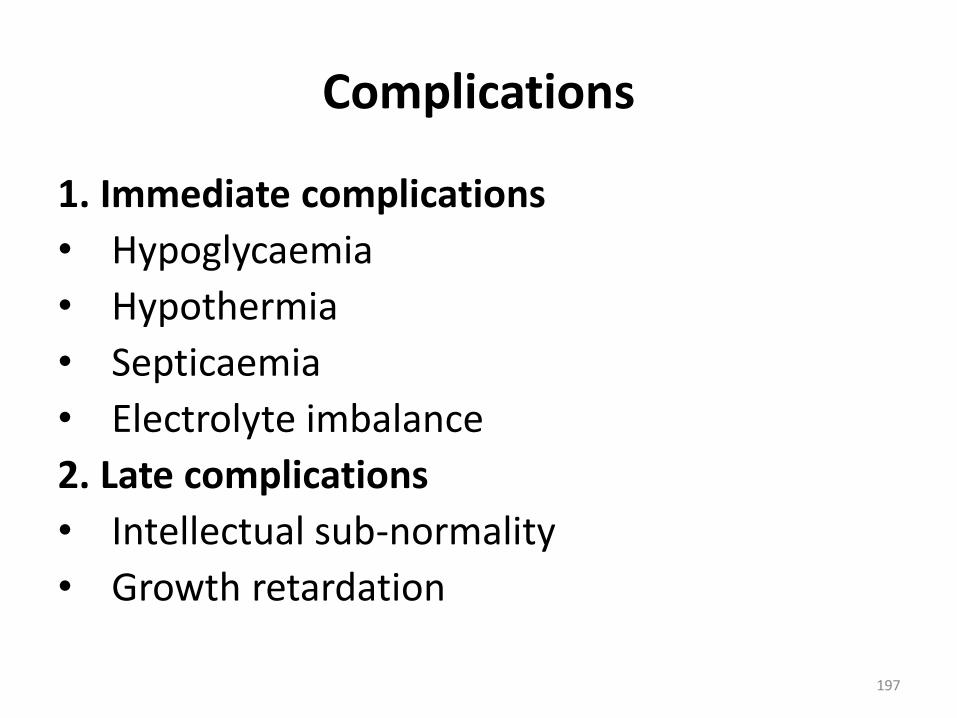
Complications
1. Immediate complications
• Hypoglycaemia
• Hypothermia
• Septicaemia
• Electrolyte imbalance
2. Late complications
• Intellectual sub-normality
• Growth retardation
197
Management
• See the management of SAM 2019
198

Vitamin A deficiency
• More common between 6 months to 3 years of age
• 50 to 80% of severe protein malnutrition patients are associated with vitamin A deficiency
199

Functions of vitamin A
• Functioning of retina
• Growth and differentiation of epithelial tissue
• Growth of bone
• Reproduction and embryonic development
• Enhances immune function, reduces the incidence of infectious diseases and may protect against the development of malignancy
200
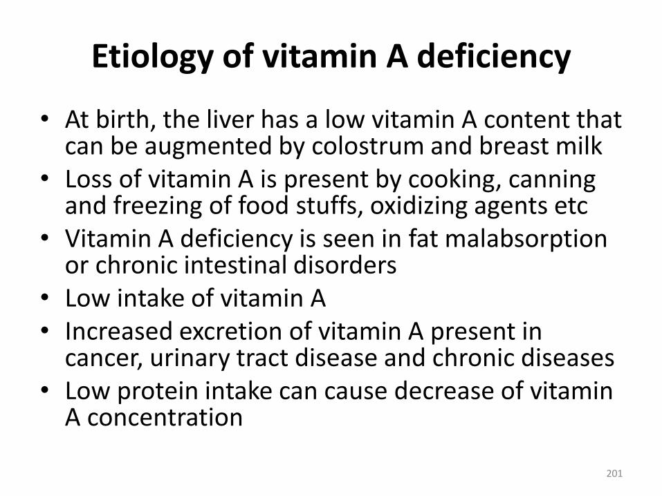
Etiology of vitamin A deficiency
• At birth, the liver has a low vitamin A content that can be augmented by colostrum and breast milk
• Loss of vitamin A is present by cooking, canning and freezing of food stuffs, oxidizing agents etc
• Vitamin A deficiency is seen in fat malabsorption or chronic intestinal disorders
• Low intake of vitamin A• Increased excretion of vitamin A present in
cancer, urinary tract disease and chronic diseases• Low protein intake can cause decrease of vitamin
A concentration
201
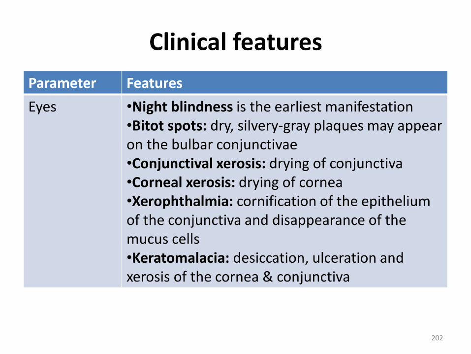
Clinical features
Parameter Features
Eyes •Night blindness is the earliest manifestation•Bitot spots: dry, silvery-gray plaques may appear on the bulbar conjunctivae •Conjunctival xerosis: drying of conjunctiva•Corneal xerosis: drying of cornea•Xerophthalmia: cornification of the epithelium of the conjunctiva and disappearance of the mucus cells•Keratomalacia: desiccation, ulceration and xerosis of the cornea & conjunctiva
202
Clinical features ...
Parameter Features
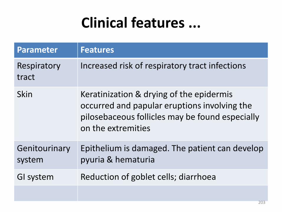
Respiratory tract
Increased risk of respiratory tract infections
Skin Keratinization & drying of the epidermisoccurred and papular eruptions involving the pilosebaceous follicles may be found especially on the extremities
Genitourinary system
Epithelium is damaged. The patient can develop pyuria & hematuria
GI system Reduction of goblet cells; diarrhoea
203
Clinical features ...
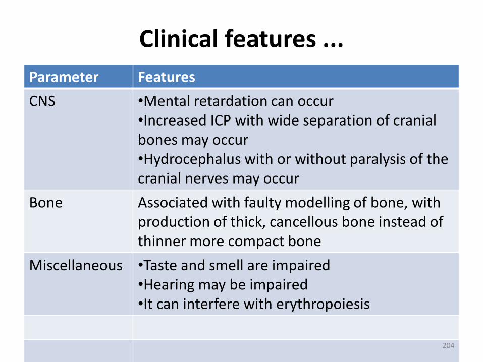
Parameter Features
CNS •Mental retardation can occur•Increased ICP with wide separation of cranial bones may occur•Hydrocephalus with or without paralysis of the cranial nerves may occur
Bone Associated with faulty modelling of bone, with production of thick, cancellous bone instead of thinner more compact bone
Miscellaneous •Taste and smell are impaired•Hearing may be impaired•It can interfere with erythropoiesis
204

Management strategies
1. Breast feeding
2. Food diversification
3. Vitamin A supplementation
4. Food fortification
205
Rickets (Nutritional)
• Bone consists of a protein matrix called osteoid and a mineral phase, principally composed of calcium and phosphate, mostly in the form of hydroxyapatite.
• Rickets, a disease of growing bone, occurs in children only before fusion of the epiphyses, and is due to unmineralized matrix at the growth plates.
206
Rickets ...
• Because growth plate cartilage and osteoid continue to expand, but mineralization is inadequate, the growth plate thickens.
• There is also an increase in the circumference of the growth plate and the metaphysis.
• This increases bone width at the location of the growth plates, causing some of the classic clinical manifestations, such as widening of the wrists and ankles
207

Etiology of rickets in general
There are many causes of rickets including
• Vitamin D disorders,
• Calcium deficiency,
• Phosphorous deficiency, and
• Distal renal tubular acidosis
208
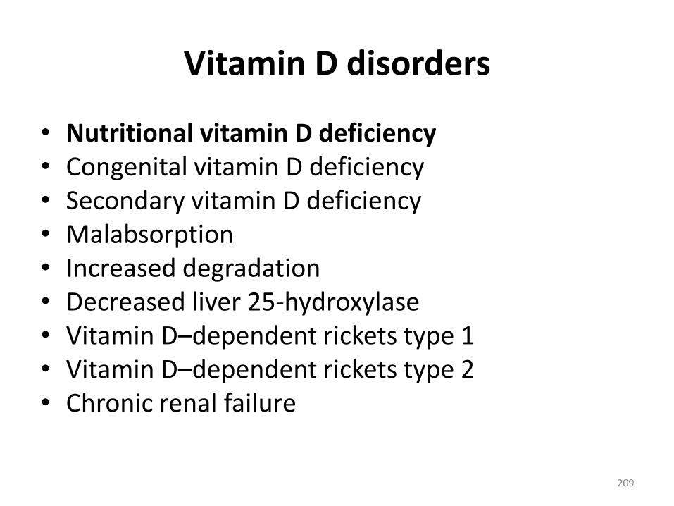
Vitamin D disorders
• Nutritional vitamin D deficiency• Congenital vitamin D deficiency• Secondary vitamin D deficiency• Malabsorption• Increased degradation• Decreased liver 25-hydroxylase• Vitamin D–dependent rickets type 1• Vitamin D–dependent rickets type 2• Chronic renal failure
209
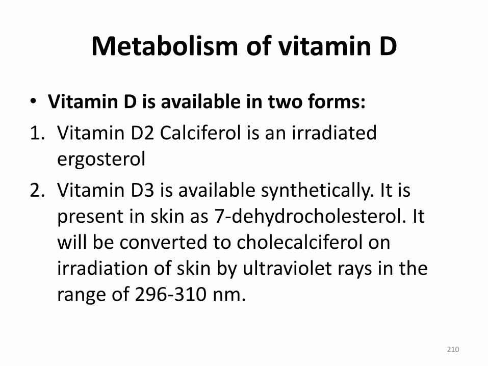
Metabolism of vitamin D
• Vitamin D is available in two forms:
1. Vitamin D2 Calciferol is an irradiated ergosterol
2. Vitamin D3 is available synthetically. It is present in skin as 7-dehydrocholesterol. It will be converted to cholecalciferol on irradiation of skin by ultraviolet rays in the range of 296-310 nm.
210
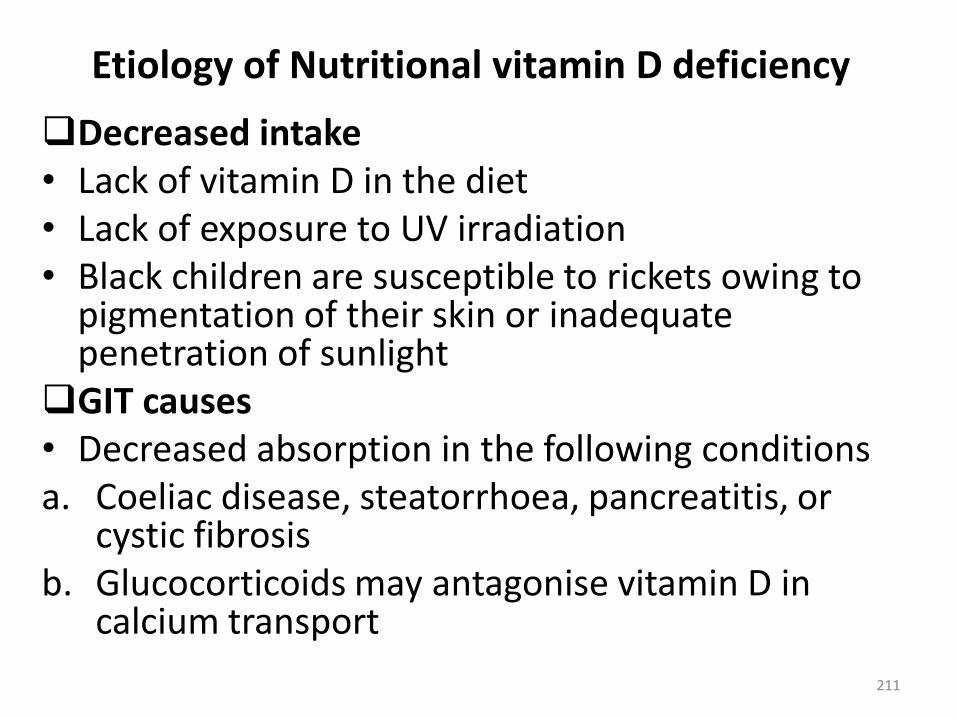
Etiology of Nutritional vitamin D deficiency
Decreased intake• Lack of vitamin D in the diet• Lack of exposure to UV irradiation• Black children are susceptible to rickets owing to
pigmentation of their skin or inadequate penetration of sunlight
GIT causes• Decreased absorption in the following conditionsa. Coeliac disease, steatorrhoea, pancreatitis, or
cystic fibrosisb. Glucocorticoids may antagonise vitamin D in
calcium transport
211
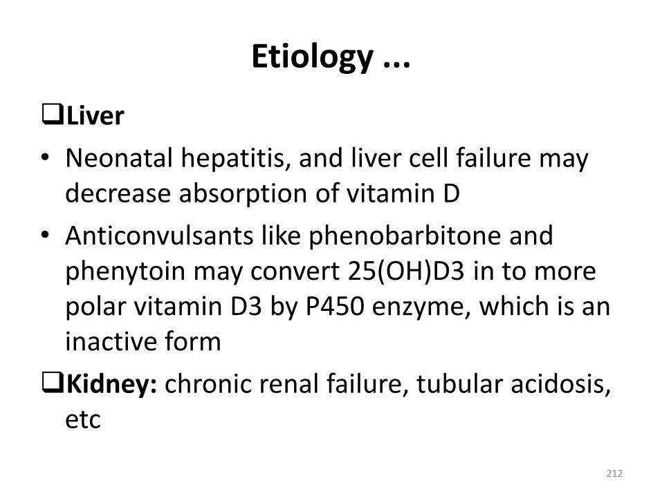
Etiology ...
Liver
• Neonatal hepatitis, and liver cell failure may decrease absorption of vitamin D
• Anticonvulsants like phenobarbitone and phenytoin may convert 25(OH)D3 in to more polar vitamin D3 by P450 enzyme, which is an inactive form
Kidney: chronic renal failure, tubular acidosis, etc
212
Incidence
• The incidence is more during the period of rapid growth, particularly between 4 months to 2 years of age
• Equal in both sexes, but it is more in male children due to rapid growth
213
Clinical Manifestations• Most manifestations of rickets are due to skeletal
changes.
• Craniotabes, a softening of the cranial bones, can be detected by applying pressure at the occiput or over the parietal bones. The sensation is similar to the feel of pressing into a Ping-Pong ball and then releasing.
• Craniotabes may also be secondary to osteogenesis imperfecta, hydrocephalus, and syphilis.
• It is a normal finding in many newborns, especially near the suture lines, but it typically disappears within a few months of birth.
214
Clinical …
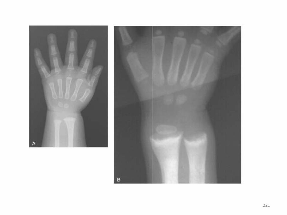
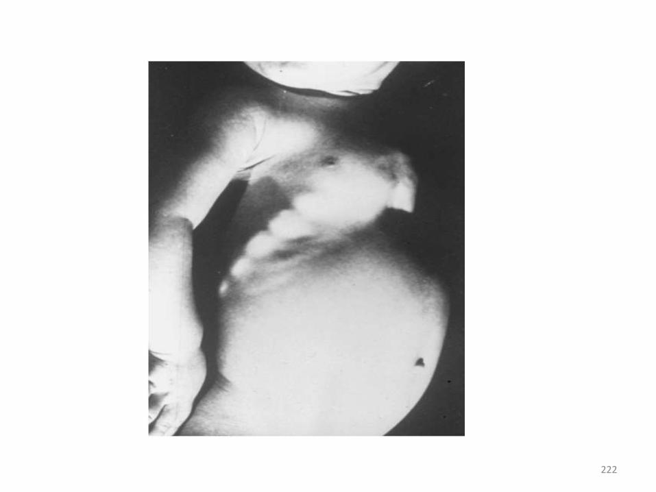
• Widening of the costochondral junctions results in a rachitic rosary; this feels like the beads of a rosary as the examiner's fingers move along the costochondral junctions from rib to rib.
• Growth plate widening is also responsible for the enlargement at the wrists and ankles.
• The horizontal depression along the lower anterior chest known as Harrison groove occurs due to pulling of the softened ribs by the diaphragm during inspiration.
215
Clinical …
• Softening of the ribs also impairs air movement and predisposes patients to atelectasis.
• The risk of pneumonia appears to be elevated in children with rickets; in Ethiopia, there may be a 13-fold higher incidence of rickets among children with pneumonia.
216
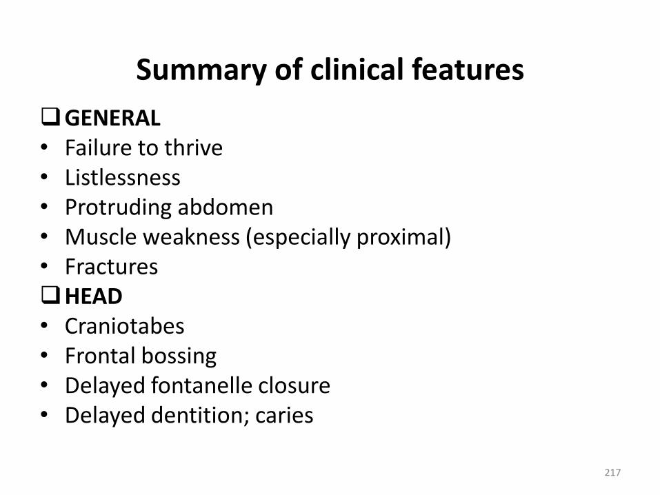
Summary of clinical features
GENERAL• Failure to thrive• Listlessness• Protruding abdomen• Muscle weakness (especially proximal)• FracturesHEAD• Craniotabes• Frontal bossing• Delayed fontanelle closure• Delayed dentition; caries
217
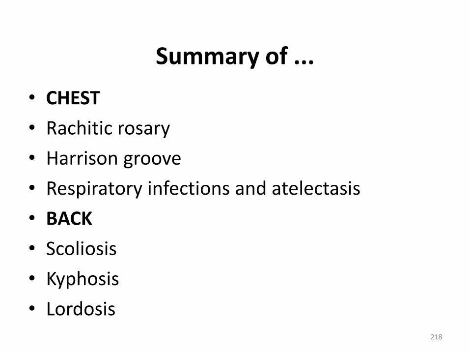
Summary of ...
• CHEST
• Rachitic rosary
• Harrison groove
• Respiratory infections and atelectasis
• BACK
• Scoliosis
• Kyphosis
• Lordosis218

Summary of ...
• EXTREMITIES
• Enlargement of wrists and ankles
• Valgus or varus deformities
• Windswept deformity (combination of valgus deformity of 1 leg with varus deformity of the other leg)
• Anterior bowing of the tibia and femur
• Leg pain
219

220
221
222

Differential diagnoses Craniotabes• Hydrocephalus • osteogenesis imperfectaCostochondrial junction enlargement • Scurvy• Chondrodystrophy• Congenital epiphysel dysplasia• Cytomegalic inclusion disease• Syphilis• Copper deficiency• Vitamin D resistant reckets
223
Treatment
• Children with nutritional vitamin D deficiency should receive vitamin D and adequate nutritional intake of calcium and phosphorus.
• Vitamin D intake of 400 IU/day, typically given as a multivitamin.
• It is important to ensure that children receive adequate dietary calcium and phosphorus; this is usually provided by milk, formula, and other dairy products.
224
Complications
• Bronchitis
• Bronchopneumonia
• Pulmonary atelectasis
• Anemia
225
Prevention
• Exposure to sunlight
• Oral administration of vitamin D
• Daily requirement of vitamin D is 10 microgram or 400IU/day
• Milk fortified with vitamin D can be given
• Vitamin D should also be given to pregnant & lactating mother
226
Management of Acute Malnutrition
Overview
Community-Based Management of Acute Malnutrition (CMAM), consists of four main components:
1. Community outreach/mobilization:
2. Inpatient Treatment/ care:
3. Outpatient Treatment Program (OTP):
4. Targeted supplementary feeding program (TSF):
227
Principle of care
Recognize signs of severe acute malnutrition
• Visible severe wasting for infants under 6 months
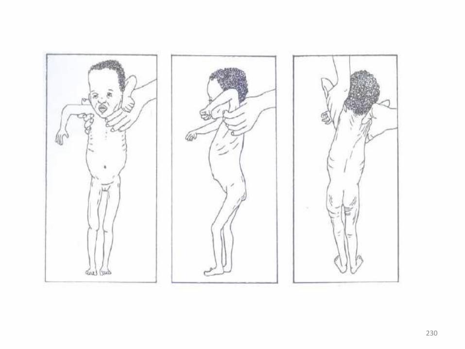
• To look for severe wasting, remove the child’s clothes. Look at the front view of the child:
• Is the outline of the child’s ribs easily seen?
• Does the skin of the upper arms look loose?
• Does the skin of the thighs look loose?
228
Recognize signs of …
• Look at the back view of the child:o Are the ribs and shoulder bones easily seen?o Is flesh missing from the buttocks?• When wasting is extreme, there are folds of skin
on the buttocks and thighs. It looks as if the child is wearing “baggy pants”.
• Because a wasted child has lost fat and muscle, this child will weigh less than other children of the same height/length and will have a low weight-for-height/Length
229
230
Recognize signs of …
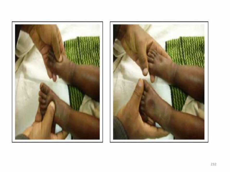
Oedema
• To check for oedema, grasp both feet so that they rest in your hand with your thumbs on top of the feet. Press your thumbs gently for three seconds or count 101,102,103.
• The child has oedema if a pit (dent) remains in both feet when you lift your thumbs
231
232
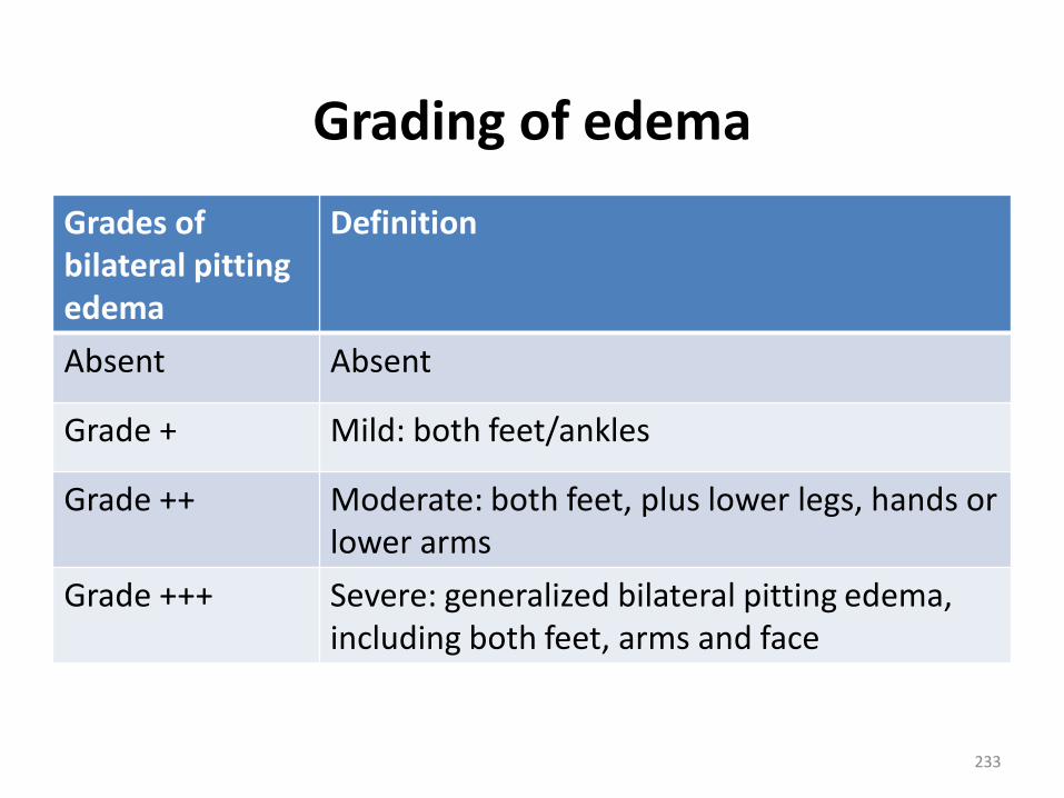
Grading of edema
Grades of bilateral pitting edema
Definition
Absent Absent
Grade + Mild: both feet/ankles
Grade ++ Moderate: both feet, plus lower legs, hands or lower arms
Grade +++ Severe: generalized bilateral pitting edema, including both feet, arms and face
233
Dermatosis
• In severe malnutrition, it is more common in children who have oedema than in wasted children.
• A child with dermatosis may have patches of skin that is abnormally light or dark in color, shedding of skin in scales or sheets, and ulceration of the skin of the perineum, groin, limbs, behind the ears, and in the armpits.
• There may be weeping lesions. There may be severe rash in the nappy area. Any break in the skin can let dangerous bacteria get into the body. When the skin is raw and weeping, this risk is very high.
234
Extent of dermatosis
• + mild: discoloration or a few rough patches of skin
• + + moderate: multiple patches on arms and/or legs
• + + + severe: flaking skin, raw skin, fissures (openings in the skin)
235

Eye signs • Bitot’s spots – superficial foamy white spots on the
conjunctiva (white part of the eye). These are associated with vitamin A deficiency.
• Pus and inflammation (redness) are signs of eye infection.
• Corneal clouding is seen as an opaque appearance of the cornea (the transparent layer that covers the pupil and iris). It is a sign of vitamin A deficiency.
• Corneal ulceration is a break in the surface of the cornea. It is a severe sign of vitamin A deficiency. If not treated, the lens of the eye may push out and cause blindness. Corneal ulceration is urgent and requires immediate treatment with vitamin A and atropine (to relax the eye).
236
Weigh and measure the child
• Carefully measure the child's length or height once on the first day. For children less than 85 cm in length, or children too weak to stand, measure the child’s length while supine (lying down).
• For children 85 cm or more, measure standing height. Note: Length is usually greater than standing height by 0.5 cm.
• If the child is 85 cm or more but cannot be measured standing, subtract 0.5 cm from the supine length.
237
Measuring length
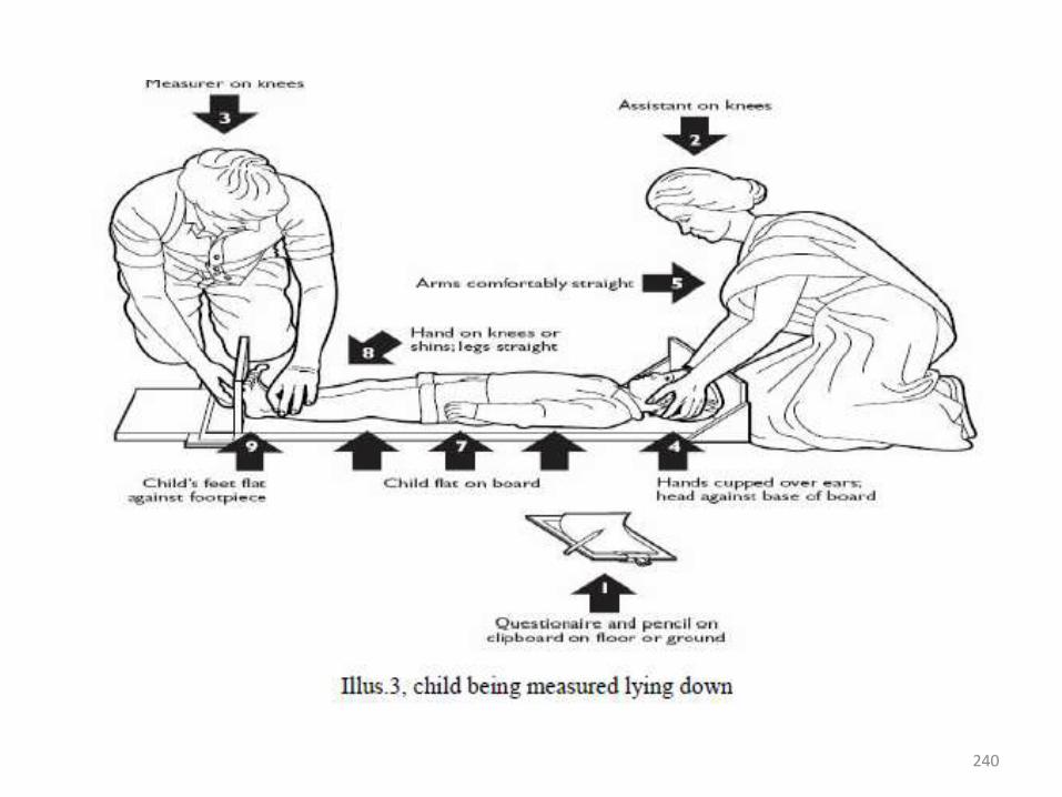
• Position the child lying on his back on the measuring board, supporting the head and placing it against the headboard.
• Position the crown of the head against the headboard, compressing the hair
• Hold the head with two hands and tilt upwards until the eyes look straight up, and the line of sight is perpendicular to the measuring board.
238
Measuring length ...
The other person should stand alongside the measuring board and:
• Support the child's trunk as the child is positioned on the board.• Place one hand on the shins or knees and press gently but firmly.• Straighten the knees as much as possible without harming the child.• With the other hand, place the foot piece firmly against the feet. • The soles of the feet should be flat on the foot piece, toes pointing
up. If the child bends the toes and prevents the foot piece touching the soles, scratch the soles slightly and slide in the foot piece when the child straightens the toes.
• Measure length to the last completed 0.1 cm and record immediately on the Multi-chart.
239
240
Measuring height
• Remove the child’s socks and shoes for accurate measurement. Also remove hair ornaments and undo braids if they interfere with measurement.
• Work with a partner. One person should kneel or crouch near the child’s feet and:
• Help the child stand with back of the head, shoulder blades, buttocks, calves and heels touching the vertical board.
• Hold the child’s knees and ankles to keep the legs straight and feet flat. Prevent children from standing on their toes.
• Young children may have difficulty standing to full height. If necessary, gently push on the tummy to help the child stand to full height.
241
Measuring height ...
• Position the head so that the child is looking straight ahead (line of sight is parallel to the base of the board).
• Place thumb and forefinger over the child’s chin to help keep the head in an upright position
• With the other hand, pull down the head board to rest firmly on top of the head and compress hair.
• Measure the height to the last completed 0.1 cm and record it immediately on the Multi chart.
242
Weigh the child
• Weigh the child as soon as possible after he arrives.
• If the child is admitted, weigh the child once daily, preferably at about the same time each day.
• The weighing time should be about one hour before or after a feed.
243
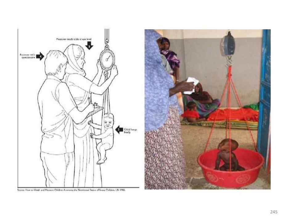
To weigh the child …
• Remove the child's clothes, but keep the child warm with a blanket or cloth while carrying to the scale.
• Put a cloth in the scale pan to prevent chilling the child.
• Adjust the scale to zero with the cloth in the pan. (If using a scale with a sling or pants or basin, adjust the scale to zero with that in place.)
• Place the naked child gently in the pan (or in the sling or pants).
• Wait for the child to settle and the weight to stabilize.• Measure weight to the nearest 100gm or as precisely
as possible. Record immediately on multi-chart.• Wrap the child immediately to re-warm.
244
245
Standardize scales
• Standardize scales daily or whenever they are moved:
• Set the scale to zero.
• Weigh one object of known weight and record the measured weight. (A container filled with stone or IV fluids etc. if the weight is accurately known.)
• Repeat the weighing of these objects and record the weights again.
• If there is a difference of 0.01 kg or more between duplicate weighing, or if a measured weight differs by 0.01 kg or more from the known standard, check the scales and adjust or replace them if necessary.
246
Measure mid-upper arm circumference.
• MUAC is measured on the upper left arm.
• To locate the correct point for measurement, the child's elbow is flexed to 90°C, with the palm facing upwards.
• A measuring tape is used to find the midpoint between the end of the shoulder (acromion) and the tip of the elbow (olecranon); this point should be marked.
• The arm is then allowed to hang freely, palm towards the thigh, and the measuring tape is placed snugly around the arm at the midpoint mark.
• The tape should not be pulled too tight or too loose.
247
Identifying a child with severe acute malnutrition
• Determine Percent-of-median based on child’s weight and length/height
• What is Percent-of-median?• It is a way of comparing a measurement, in this case a child’s
weight-for-length, to an “average” (median). • The median used in the national protocol and in this course
are NCHS reference values for weight-for-height and weight-for-length.
• It is a ratio of a child's weight to the median weight of a child of the same height in the reference, expressed as percentage.
• As a concrete example if the median (or average) weight in the reference tables for a particular height was 10 kg and a child weighed 8 kg, she/he was 80% weight-for-height.
248
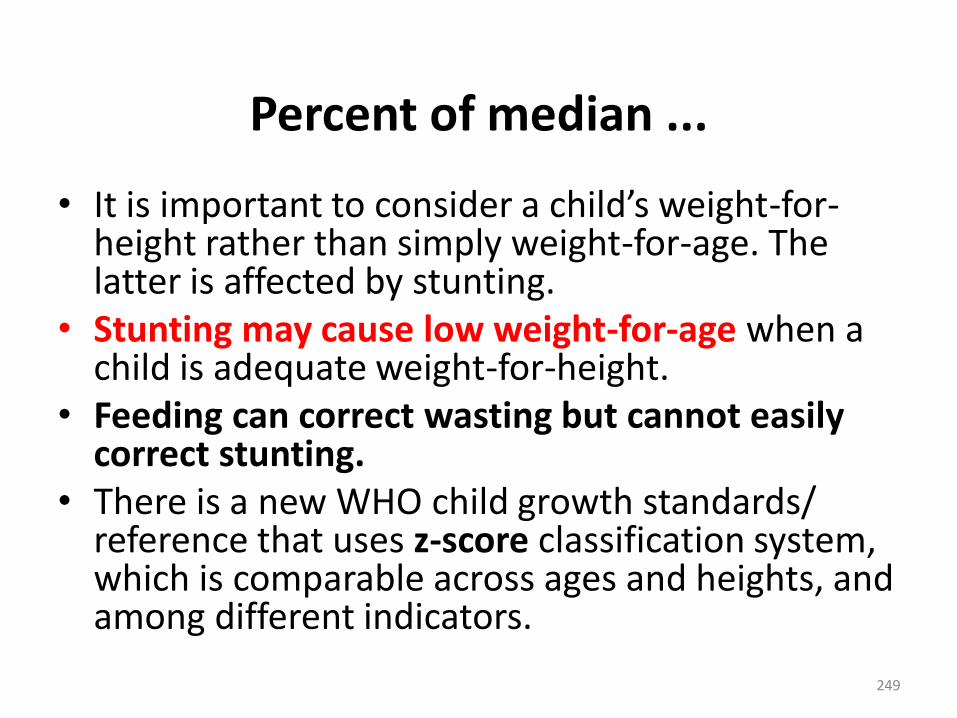
Percent of median ...
• It is important to consider a child’s weight-for-height rather than simply weight-for-age. The latter is affected by stunting.
• Stunting may cause low weight-for-age when a child is adequate weight-for-height.
• Feeding can correct wasting but cannot easily correct stunting.
• There is a new WHO child growth standards/ reference that uses z-score classification system, which is comparable across ages and heights, and among different indicators.
249

250
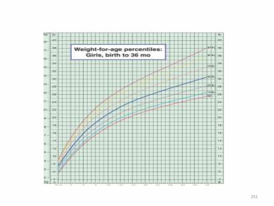
251
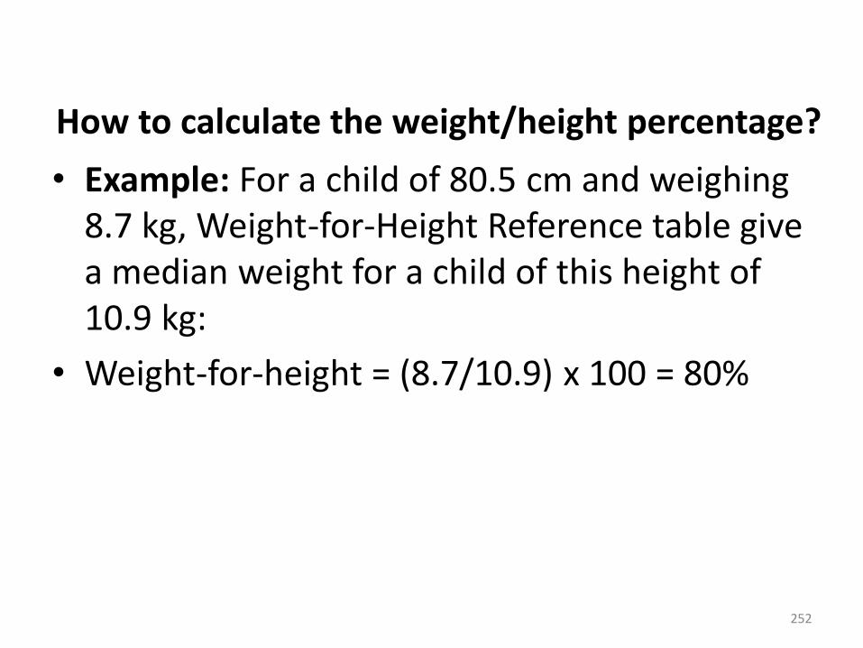
How to calculate the weight/height percentage?
• Example: For a child of 80.5 cm and weighing 8.7 kg, Weight-for-Height Reference table give a median weight for a child of this height of 10.9 kg:
• Weight-for-height = (8.7/10.9) x 100 = 80%
252
How to use the Weight-for-Height Reference table
• To use the reference table in the chart booklet or on your Weight-for-Height Reference Card:
• First, find the child's length or height in the first column of the table.
• Then look in the right columns in the same row to find the weight that correspond to the weight of the child.
• Look at the top of the column to see what the child’s percentage of median or degree of acute malnutrition .
253
... reference table
• The child’s weight may be between two percentage of median. If so, indicate that the weight is between these percentage of median by writing less than (<)
• For example, if the weight is between 70 and 75%, write down as child’s percentage of median is between 70 and 75%.
254

Recommended criteria for SAM and Admission to TFP
1. Infants less than six months or less than 3 Kg:• Weight –for- Length (WFL) less than 70% or < -3Z score
OR• Presence of pitting Oedema of both feet
OR• Visible Severe Wasting if it is difficult to determine WFL2. Children 6 months to 5 years:• Weight –for- Length (WFL) / WFH less than 70 % or < -3Z score
OR• Presence of pitting Oedema of both feet
OR• MUAC <11cm for child length greater than 65 cm
255
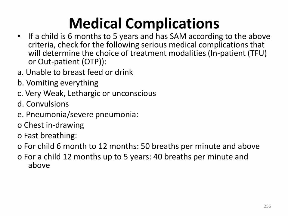
Medical Complications• If a child is 6 months to 5 years and has SAM according to the above
criteria, check for the following serious medical complications that will determine the choice of treatment modalities (In-patient (TFU) or Out-patient (OTP)):
a. Unable to breast feed or drinkb. Vomiting everythingc. Very Weak, Lethargic or unconsciousd. Convulsionse. Pneumonia/severe pneumonia:o Chest in-drawingo Fast breathing:o For child 6 month to 12 months: 50 breaths per minute and aboveo For a child 12 months up to 5 years: 40 breaths per minute and
above
256
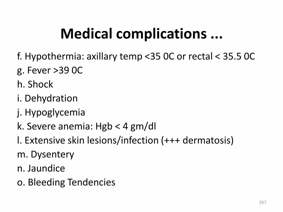
Medical complications ...
f. Hypothermia: axillary temp <35 0C or rectal < 35.5 0C
g. Fever >39 0C
h. Shock
i. Dehydration
j. Hypoglycemia
k. Severe anemia: Hgb < 4 gm/dl
l. Extensive skin lesions/infection (+++ dermatosis)
m. Dysentery
n. Jaundice
o. Bleeding Tendencies
257
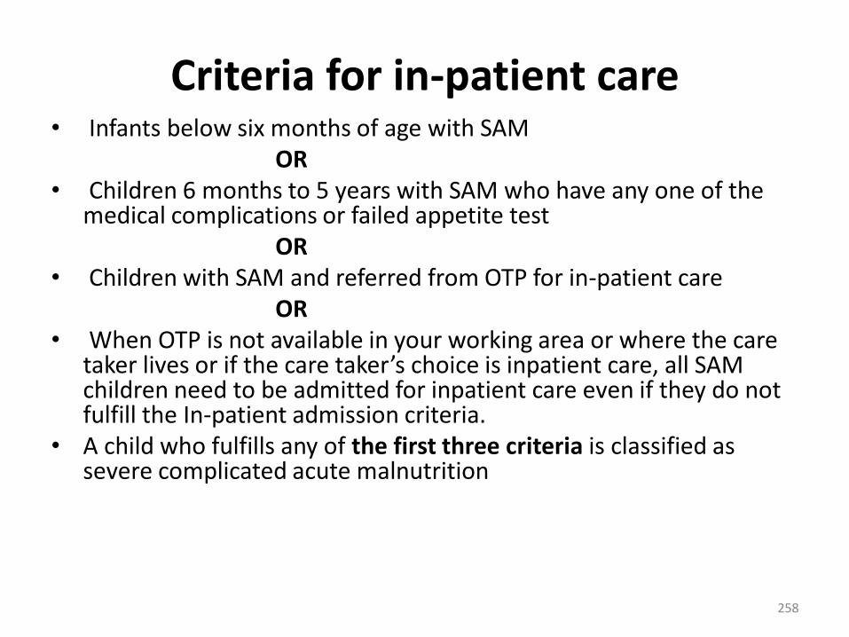
Criteria for in-patient care • Infants below six months of age with SAM
OR• Children 6 months to 5 years with SAM who have any one of the
medical complications or failed appetite testOR
• Children with SAM and referred from OTP for in-patient careOR
• When OTP is not available in your working area or where the care taker lives or if the care taker’s choice is inpatient care, all SAM children need to be admitted for inpatient care even if they do not fulfill the In-patient admission criteria.
• A child who fulfills any of the first three criteria is classified as severe complicated acute malnutrition
258
Physiology of severe malnutrition
Reductive adaptation• The systems of the body begin to “shut down”
with severe malnutrition. • The systems slow down and do less in order to
allow survival on limited calories. This slowing down is known as reductive adaptation.
• As the child is treated, the body's systems must gradually "learn" to function fully again.
• Rapid changes (such as rapid feeding or fluids) would overwhelm the systems, so feeding must be slowly and cautiously increased
259

How does reductive adaptation affect care of the child?
Presume and treat infection
• Nearly all children with severe malnutrition have bacterial infections.
• However, as a result of reductive adaptation, the usual signs of infection may not be apparent, because the body does not use its limited energy to respond in the usual ways, such as inflammation or fever.
260
Reductive adaptation ...
Do not give iron early in treatment• Due to reductive adaptation, the severely
malnourished child makes less hemoglobin than usual.
• Iron that is not used for making hemoglobin is put into storage. Thus, there is “extra” iron stored in the body, even though the child may appear anemic.
• Giving iron early in treatment will not cure anemia, as the child already has a supply of stored iron.
261
Early iron ...
• Giving iron early in treatment can also lead to “free iron” in the body. Free iron can cause problems in three ways:
1. Free iron is highly reactive and promotes the formation of free radicals, which may engage in uncontrolled chemical reactions with damaging effects.
2. Free iron promotes bacterial growth and can make some infections worse.
3. The body tries to protect itself from free iron by converting it to ferritin. This conversion requires energy and amino acids and diverts these from other critical activities.
• Later, as the child recovers and begins to build new tissue and form more red blood cells, the iron in storage will be used and supplements will be needed.
262

Provide potassium and restrict sodium• Normally the body uses a lot of energy maintaining the
appropriate balance of potassium inside the cells and sodium outside the cells.
• This balance is critical to maintaining the correct distribution of water inside the cells, around the cells and in the blood.
• In reductive adaptation, the “pump” that usually controls the balance of potassium and sodium runs slower.
• As a result, the level of sodium in the cells rises and the potassium leaks out of the cells and is lost (for example, in urine or stools).
• Fluid may then accumulate outside of the cells (as in oedema) instead of being properly distributed through the body.
263
Provide potassium …• All severely malnourished children should be given
potassium to make up for what is lost.• They should also be given magnesium, which is
essential for potassium to enter into the cells and be retained
• The commercially prepared F-75 and F-100 have enough potassium and magnesium and there is no need to supplement.
• However, if you use the F-75 and F-100 recipe that are prepared by the health facility/ locally, Combined Mineral Vitamin mixes (CMV) should be given to supplement potassium, Magnesium, and other important minerals and vitamins.
264
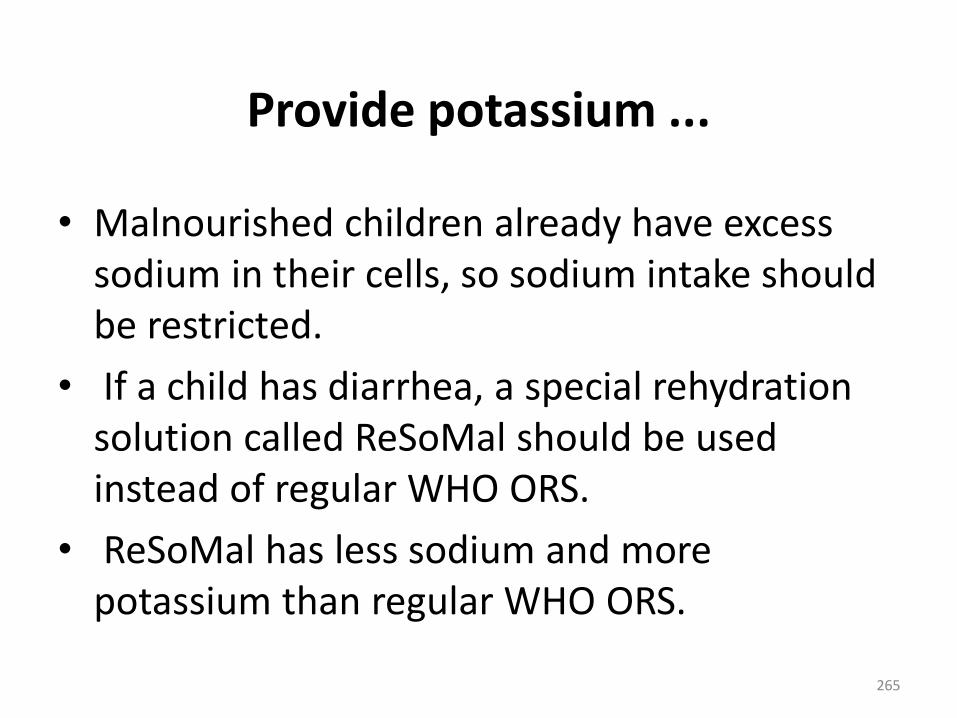
Provide potassium ...
• Malnourished children already have excess sodium in their cells, so sodium intake should be restricted.
• If a child has diarrhea, a special rehydration solution called ReSoMal should be used instead of regular WHO ORS.
• ReSoMal has less sodium and more potassium than regular WHO ORS.
265
Quiz
1. When a child is severely malnourished, why is it important to begin feeding slowly and cautiously?
2. Why should all severely malnourished children be given antibiotics?
3. Why is it dangerous to give iron early in treatment?
4. Why the regular WHO ORS is not recommended for malnourished children?
266
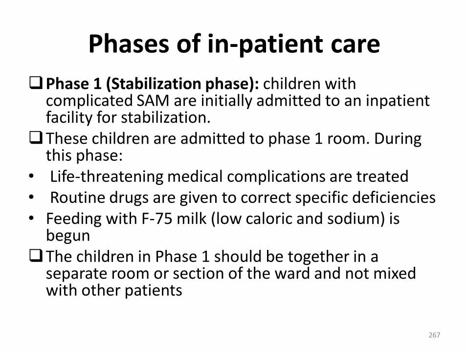
Phases of in-patient care
Phase 1 (Stabilization phase): children with complicated SAM are initially admitted to an inpatient facility for stabilization.
These children are admitted to phase 1 room. During this phase:
• Life-threatening medical complications are treated• Routine drugs are given to correct specific deficiencies• Feeding with F-75 milk (low caloric and sodium) is
begunThe children in Phase 1 should be together in a
separate room or section of the ward and not mixed with other patients
267
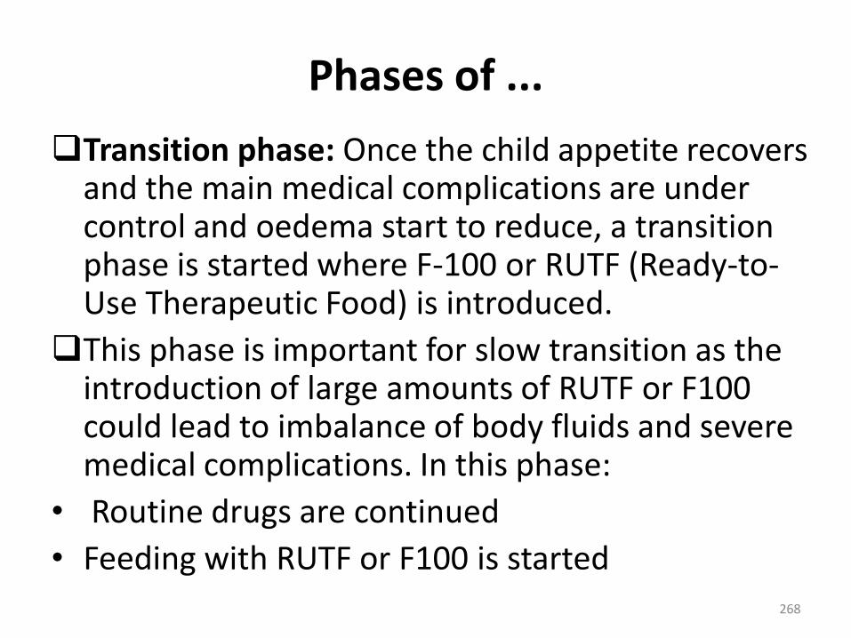
Phases of ...
Transition phase: Once the child appetite recovers and the main medical complications are under control and oedema start to reduce, a transition phase is started where F-100 or RUTF (Ready-to-Use Therapeutic Food) is introduced.
This phase is important for slow transition as the introduction of large amounts of RUTF or F100 could lead to imbalance of body fluids and severe medical complications. In this phase:
• Routine drugs are continued
• Feeding with RUTF or F100 is started268
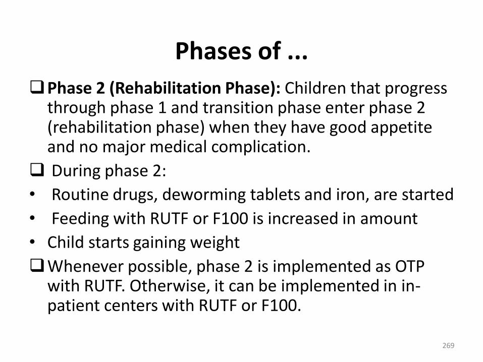
Phases of ...
Phase 2 (Rehabilitation Phase): Children that progress through phase 1 and transition phase enter phase 2 (rehabilitation phase) when they have good appetite and no major medical complication.
During phase 2:
• Routine drugs, deworming tablets and iron, are started
• Feeding with RUTF or F100 is increased in amount
• Child starts gaining weight
Whenever possible, phase 2 is implemented as OTP with RUTF. Otherwise, it can be implemented in in-patient centers with RUTF or F100.
269
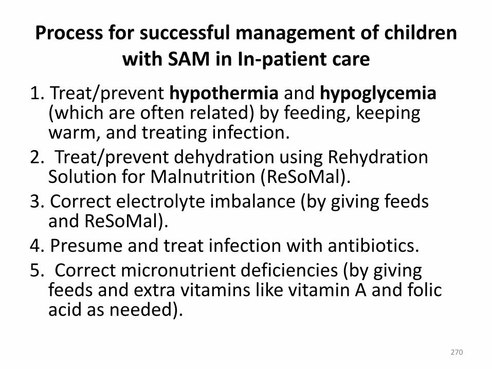
Process for successful management of children with SAM in In-patient care
1. Treat/prevent hypothermia and hypoglycemia(which are often related) by feeding, keeping warm, and treating infection.
2. Treat/prevent dehydration using Rehydration Solution for Malnutrition (ReSoMal).
3. Correct electrolyte imbalance (by giving feeds and ReSoMal).
4. Presume and treat infection with antibiotics.5. Correct micronutrient deficiencies (by giving
feeds and extra vitamins like vitamin A and folic acid as needed).
270
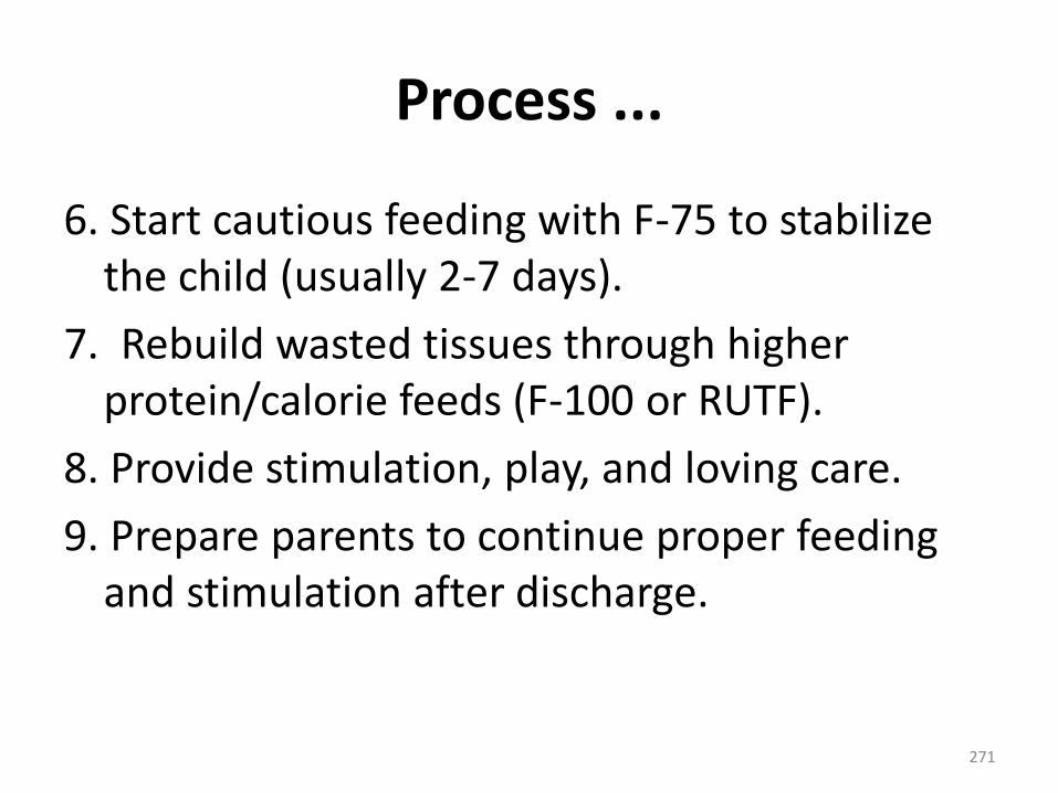
Process ...
6. Start cautious feeding with F-75 to stabilize the child (usually 2-7 days).
7. Rebuild wasted tissues through higher protein/calorie feeds (F-100 or RUTF).
8. Provide stimulation, play, and loving care.
9. Prepare parents to continue proper feeding and stimulation after discharge.
271
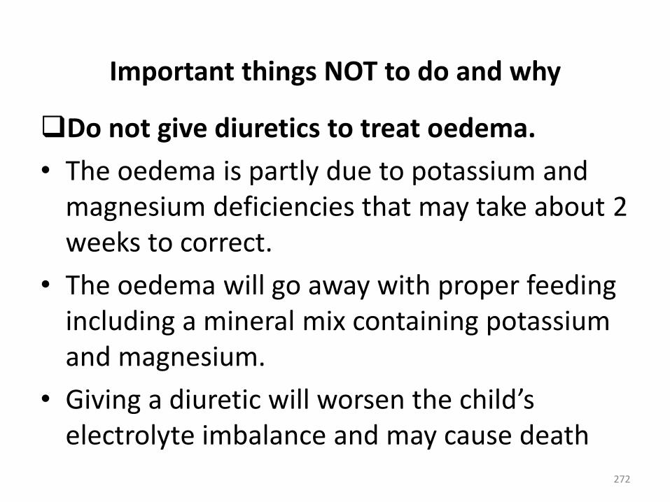
Important things NOT to do and why
Do not give diuretics to treat oedema.
• The oedema is partly due to potassium and magnesium deficiencies that may take about 2 weeks to correct.
• The oedema will go away with proper feeding including a mineral mix containing potassium and magnesium.
• Giving a diuretic will worsen the child’s electrolyte imbalance and may cause death
272
Not to do ...
Do not give iron during phase 1 and transition phase.
• Add iron only when the child is in phase 2 (usually during week 2).
• As described earlier, giving iron early in treatment can have toxic effects and interfere with the body’s ability to resist infection.
273

Not to do ...
Do not give high protein formula (over 1.5 g protein per kg body weight daily).
• Too much protein in the first days of treatment may be dangerous because the severely malnourished child is unable to deal with the extra metabolic stress involved
• Too much protein could overload the liver, heart, and kidneys and may cause death.
274
Not to do ...
Do not give IV fluids routinely
• IV fluids can easily cause fluid overload and heart failure in a severely malnourished child. Only give IV fluids to children with signs of shock.
275
MANAGEMENT OF MEDICAL COMPLICATIONS
Manage Hypoglycemia
• Hypoglycemia is a low level of glucose in the blood.
• In severely malnourished children, the level considered low is less than <54 mg/dl (< 3 mmol/litre).
• The hypoglycemic child is usually hypothermic (low temperature) as well.
• Other signs of hypoglycemia include lethargy, limpness, and loss of consciousness.
276
Treat Hypoglycemia
• If blood glucose is low or hypoglycemia is suspected, immediately give the child a 50 ml bolus of 10% glucose or 10% sucrose orally or by NG tube.
• 50 ml is a very small amount, but it can make a big difference to the child.
• Glucose is preferable because the body can use it more easily; sucrose must be broken down by the body before it can be used.
• However, give whichever is available most quickly. • If only 50% glucose solution is available, dilute one part
to four parts sterile or boiled water to make a 10% solution.
277
Treat Hypoglycemia …
• If the child can drink, give the 50 ml bolus orally. If the child is alert but not drinking, give the 50 ml by NG tube.
• If the child is lethargic, unconscious, or convulsing, give 5 ml/kg body weight of sterile 10% glucose by IV, followed by 50 ml of 10% glucose or sucrose by NG tube.
• * If the IV dose cannot be given immediately, give dose first through NG tube.
278
Manage Hypothermia Hypothermia• Hypothermia is low body temperature. • A severely malnourished child is hypothermic if the rectal
temperature is below 35.5 0C or if the auxiliary temperature is below 35 0C.
• Severely malnourished children are at greater risk of hypothermia than other children and need to be kept warm.
• The hypothermic child has not had enough calories to warm the body.
• If the child is hypothermic, he is probably also hypoglycemic. • Both hypothermia and hypoglycemia are signs that the child
has a serious systemic infection.
279
Manage Hypothermia …
Maintain temperature (prevent hypothermia)• Cover the child, including his head• Stop draughts in the room. Move the child away from
windows• Maintain room temperature of 28 and 32 0C• Keep the child covered at night• Warm your hands before touching the child• Avoid leaving the child uncovered while being
examined, weighed, etc.• Promptly change wet clothes or beddin• Dry the child thoroughly after bathing.
280
Manage Hypothermia …
Actively re-warm the hypothermic child
• In addition to keeping the child covered and keeping the room warm, use one of the following re-warming techniques if the child is hypothermic:
1. Have the mother hold the child with his skin next to her skin when possible (kangaroo technique), and cover both of them. Keep the child’s head covered. Give warm Fluid to mother.
281
Tuberculosis (TB) in Children
282

Definition
• Tuberculosis (TB) is an infectious disease caused by
Mycobacterium tuberculosis, a rod-shaped bacillus
called “acid-fast” due to its staining characteristics in
laboratory.
• Etiology:
• M.TB
• M.Bovis
• M.Africanum
• M.Microti
• M.Canetti283
Tuberculosis• Tuberculosis (TB) is a major public health problem
throughout the world
• Almost 1.3 million cases and 450,000 deaths occur in
children each year
• The burden of TB in Ethiopia is one of the highest in the
world
• Childhood TB reflects recent transmission within a
community
• Cases of TB in children usually represents between 5-15%
of all TB cases284
Tuberculosis….
• The commonest age of presentation disease is
between 1 and 4 years
• Young age is a risk factor for infection, for progression
• Children under one year of age are more liable to
develop Miliary and TB meningitis
• Most children with TB are not infectious to others
• The commonest type of TB in children is smear-
negative PTB285
Risk of transmission• Risk of transmission is dependent on :
• Index case:
• Smear positive TB
• Extensive upper lobe infiltrate & cavity
• Copious production of sputum
• Severe & forceful cough
• Not treated
• Environment:
• Poor ventilation
• Overcrowding
• Intimacy 286
Transmission
• Incubation period from infection to development of
+ve tuberculin test is 2-6wks
• Transmission is person to person (usually by air
borne mucus droplet nuclei)
287
Children at greater risk
• Children at greater risk of developing TB include:
• Children who are in close contact with a newly
diagnosed smear-positive TB case
• Children less than 5 years of age
• HIV-infected children
• Severely malnourished children
288
PATHOGENESIS• The complex of TB includes local infection at the portal of
entry & regional lymph node
• Lung is portal of entry in more than 98% of cases
• Bacilli multiply initially within alveoli & alveolar ducts
• Most are killed, some survive within non-activated
macrophages
• Macrophages carry the bacilli to regional lymph nodes by
lymphatic vessels
• If lung is portal of entry, lymph node are often involved but
paratracheal lymph node may be involved(upper lobe) 289
PATHOGENESIS
• Tissue reaction in the lung parenchyma & lymph node
intensifies over the next 2-12wks
• The parenchymal lesion of complex often heals completely by
fibrosis or calcification.
• Occasionally may continue to enlarge & result in focal
pneumonitis & pleuritis
• If caseation is intense, center of the lesion liquefies & empties into
the bronchus leaving a residual cavity
• The infection of regional LNS develop some fibrosis &
encapsulation, but healing is usually incomplete
• Viable M.TB can persist for decades within parenchymal or LN
foci290
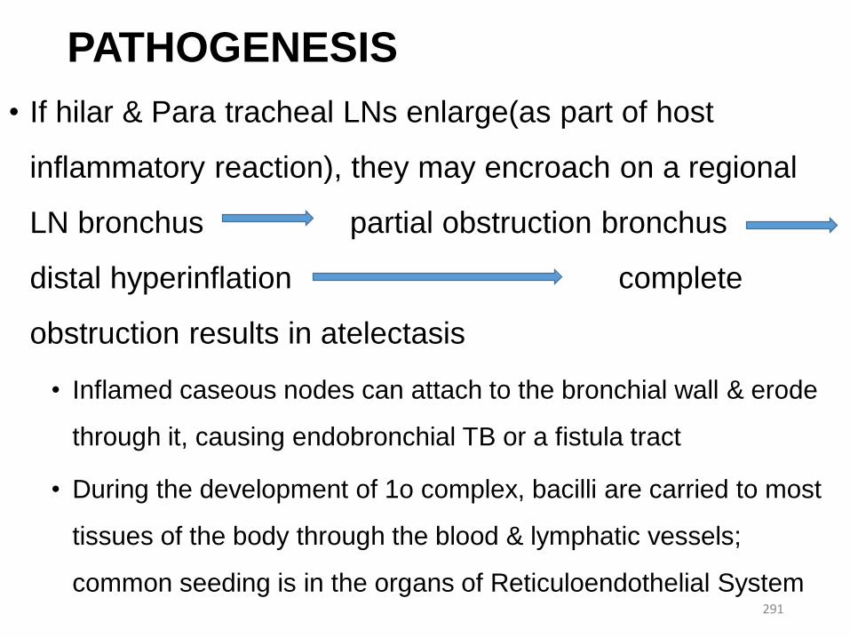
PATHOGENESIS
• If hilar & Para tracheal LNs enlarge(as part of host
inflammatory reaction), they may encroach on a regional
LN bronchus partial obstruction bronchus
distal hyperinflation complete
obstruction results in atelectasis
• Inflamed caseous nodes can attach to the bronchial wall & erode
through it, causing endobronchial TB or a fistula tract
• During the development of 1o complex, bacilli are carried to most
tissues of the body through the blood & lymphatic vessels;
common seeding is in the organs of Reticuloendothelial System291
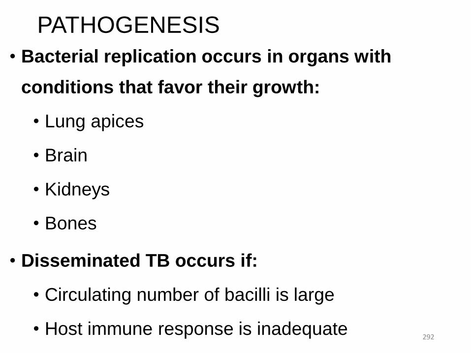
PATHOGENESIS
• Bacterial replication occurs in organs with
conditions that favor their growth:
• Lung apices
• Brain
• Kidneys
• Bones
• Disseminated TB occurs if:
• Circulating number of bacilli is large
• Host immune response is inadequate292

PATHOGENESIS
• The time b/n initial infection & clinically apparent disease
is variable:
• Disseminated & meningeal TB are early manifestations (2-6
month after infection)
• TB lymphadenitis & endobronchial TB (3-9month)
• Bones & joints take several years
• Renal TB takes decades after infection
• Pulmonary TB that occurs more than a year after infection is
usually due to endogenous regrowth of bacilli persisting in
partially encapsulated lesions
• Common site of reactivation is the apex of upper lobes
(oxygen & blood flow good) 293
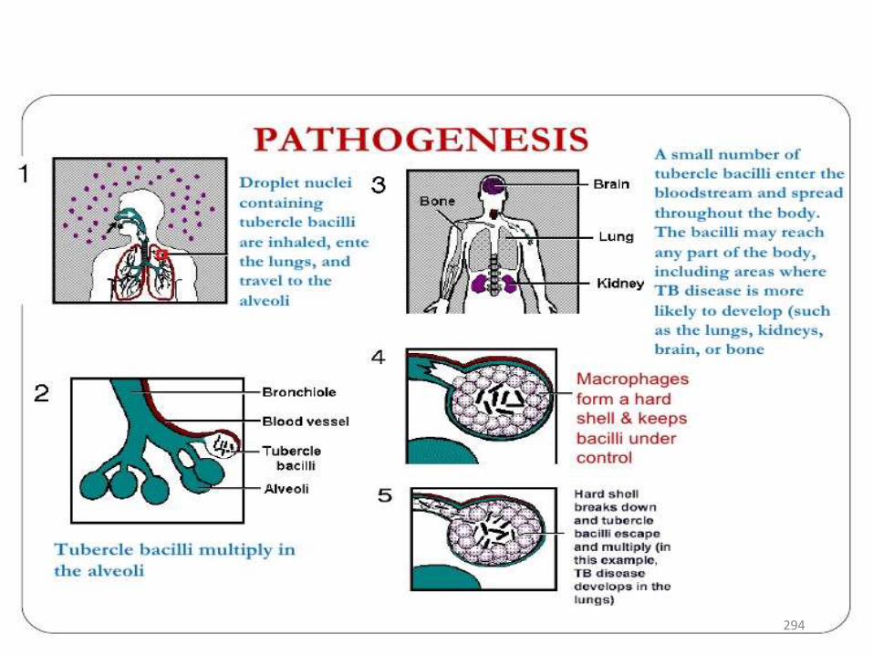
294
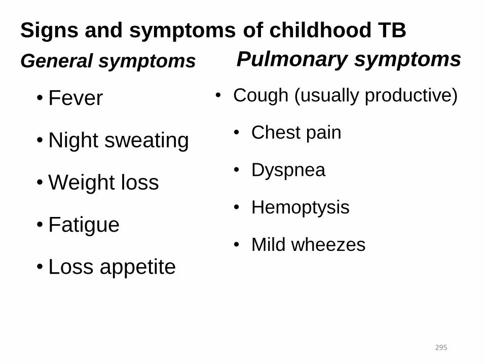
Signs and symptoms of childhood TB
General symptoms
• Fever
• Night sweating
• Weight loss
• Fatigue
• Loss appetite
Pulmonary symptoms
• Cough (usually productive)
• Chest pain
• Dyspnea
• Hemoptysis
• Mild wheezes
295

Signs and symptoms of childhood TB
• Most common clinical presentation
• Persistent respiratory symptoms
• Poor weight gain or failure to thrive
• Non-productive cough and mild dyspnea are
the most common symptoms
296
Extra-pulmonary Tuberculosis(EPTB)
• Extra-pulmonary Tuberculosis(EPTB):
• The most common forms of extrapulmonary disease in
children are TB of the superficial lymph nodes and central
nervous system (CNS)
• Children younger than 2 years of age are at risk of
disseminated disease causing miliary TB or TB meningitis
• The clinical presentation of extrapulmonary TB depends
on the site of disease
297
Miliary Tuberculosis
• Presents with constitutional features rather than
respiratory symptoms
• Early symptoms are vague and lack specificity
• Lassitude, anorexia, failure to thrive and
prolonged unexplained fever are common
• Therefore, a high index of suspicion is necessary
• TB meningitis is the commonest cause of death if
miliary TB is untreated298

CNS TB• Is the result of haematogenous spread of the bacilli to the
CNS.
• The patient may present with constitutional features and
chronic meningitis and there is gradual onset and progression
of headache and decreased consciousness
• Tuberculoma (a large solid lesion, rather like a malignant tumor)
• Present with focal neurological deficits, seizures and obstructive
hydrocephalus.
• Young children, especially those under 2 years of age, have the
highest incidence and the worst prognosis.299
Diagnosis of TB in Children• The diagnosis of TB can be made with confidence in the majority of
children sing careful clinical assessment
• There are two key factors in suspecting tuberculosis in
children:
• Identification of an infectious adult close to the child,
especially in the family
• Loss of weight or failure to thrive
• Any child with symptoms suggestive of TB, with history of exposure to an
adult or adolescent pulmonary TB patient should be investigated for TB
• Children usually develop TB within 2 years after exposure and most (90%)
within the first year
300
Diagnosis of TB in Children• Careful history (including history of TB contact and symptoms consistent
with TB)
• Clinical assessment (including serial weight)
• Diagnostic tests
• Bacteriologic confirmatory tests( ZN/FM microscopy, Xpert
MTB/RIF assay & Any child with symptoms suggestive of TB, with
history of exposure to an adult or adolescent pulmonary TB patient
should be investigated for TB culture)
• Chest X-ray
• HIV testing
• Histopathology
301
Diagnosis of TB in Children
• Key Features Suggestive of TB
• Chronic symptoms suggestive of TB
• Physical signs highly suggestive of TB
• X-ray suggestive of TB
• A positive tuberculin skin test
302
Tuberculin Skin Test(Mantoux test)
• Id, 0.1ml of PPD/purified protein derivative/, volar
surface of arms
• T-cells sensitized by prior infection are recruited to
the skin; release lymphokines that induce indurations
through local vasodilatation, edema, fibrin deposition
and recruitment of other inflammatory cells to the
area
• Amount of induration is measured 48-72hrs after
administration303
Tuberculin Skin Test(Mantoux test)
• TST is useful to support a diagnosis of TB in
children with suggestive clinical features who are
sputum smear-negative or who cannot produce
sputum
• A positive TST indicates infection:
• Positive in any child if ≥ 10 mm, irrespective of
BCG immunization
• Positive if ≥ 5 mm in HIV-infected or severely
malnourished child304
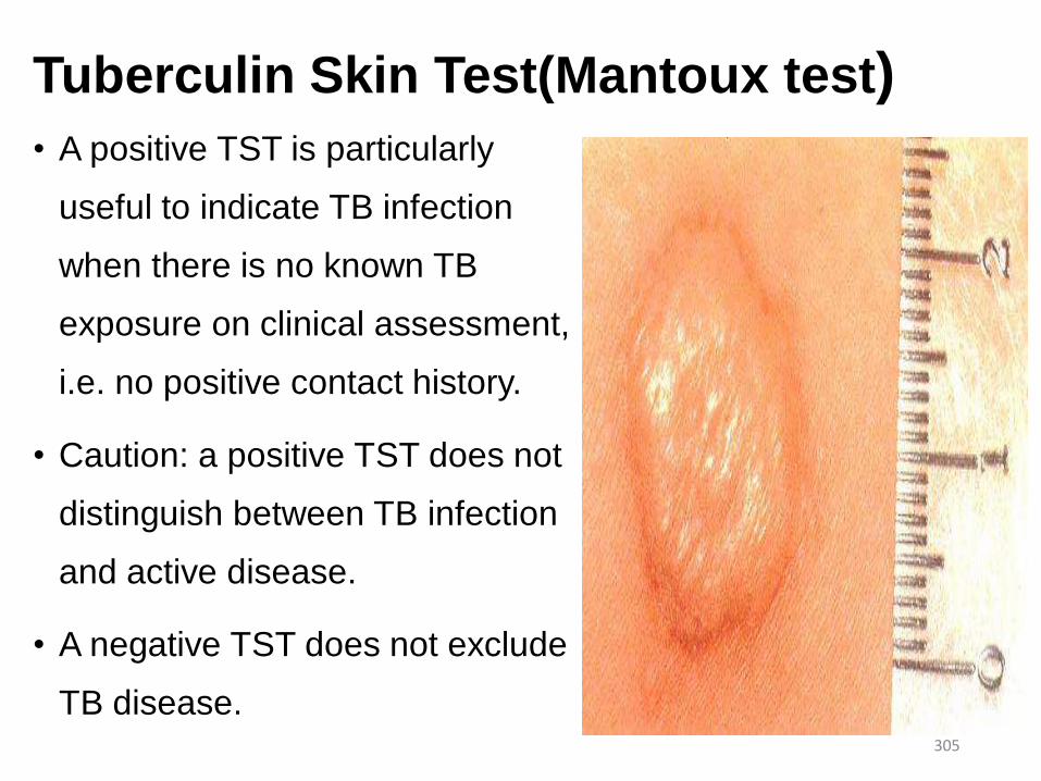
Tuberculin Skin Test(Mantoux test)• A positive TST is particularly
useful to indicate TB infection
when there is no known TB
exposure on clinical assessment,
i.e. no positive contact history.
• Caution: a positive TST does not
distinguish between TB infection
and active disease.
• A negative TST does not exclude
TB disease.305
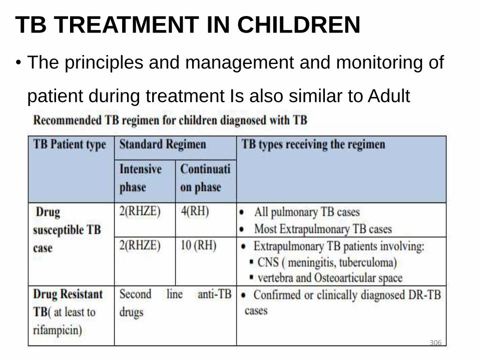
TB TREATMENT IN CHILDREN
• The principles and management and monitoring of
patient during treatment Is also similar to Adult
306
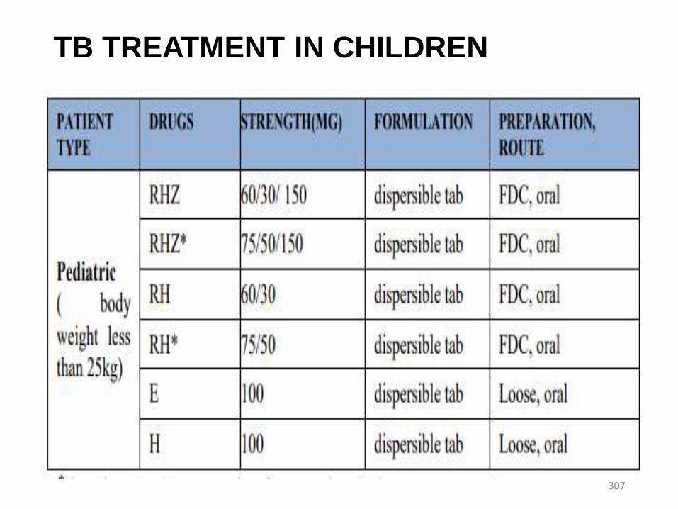
TB TREATMENT IN CHILDREN
307

TB TREATMENT IN CHILDREN
308

Second line anti TB drugs for treatment of MDR-Tb in children
• Ethionamide or prothionamide• Fluoroquinolones
• Ofloxacin• Levofloxacin• Moxifloxacin• Gatifloxacin• Ciprofloxacin
• Aminoglycosides• Kanamycin• Amikacin• Capreomycin
• Cycloserine or terizidone• paraAminosalicylic acid
309
Prevention of Childhood TB
• Case finding and treatment, which interrupts
transmission of infection between close
contacts
• Bacille Calmette-Guerin Vaccination(BCG)
• INH prophylaxis
310
Individual assignment
• HIV/AIDS
• Introduction (Definition and pathogenesis);WHO
clinical staging, ART, follow-up and advice for HIV
positive children
• Tuberculosis in HIV positive children
311
Malaria
312
Definition and Epidemiology
• An acute infection of the blood caused by protozoa of the
genus plasmodium
• Malaria is a major health problem in Africa, Asia, Central
America, Oceania, and South America
• About 40% of the world's population lives in areas where
malaria is common
• Approximately 300-500 million cases of malaria occur
every year, and 1-2 million deaths occur, most of them in
young children313
Etiology• Malaria is caused by Plasmodium species, which are
protozoal blood parasites.
• The following 5 species can infect humans:• P vivax • P falciparum • P malariae• P ovale• Plasmodium knowlesi
Mode of transmission
• Vector born -the bite of an infected female Anopheles mosquito
• Blood transfusion
• Organ transplantation
• Vertical transmission - Transplacental malaria(congenital malaria)
314

Risk factors
• Residence in, or travel through, a malarious area
• No previous exposure to malaria (hence no
immunity)
• No chemoprophylaxis or improper chemoprophylaxis
315
Life cycle and pathogenesis
• The plasmodium life cycle consists of an exoerythrocytic
(asymptomatic) stage and erythrocytic (symptomatic)
stage
• Plasmodium sporozoites are transmitted by the bite of an
infected anopheline mosquito
• The sporozoites invade hepatocytes, which divide until
schizonts are formed containing thousands of daughter
merozoites
• These rupture and release merozoites into the
bloodstream, where they invade red blood cells316
Life cycle and pathogenesis
• Within red cells, merozoites mature from ring forms to
trophozoites to multinucleated schizonts (erythrocytic
stage).
• Some merozoites differentiate into male or female
gametocytes.
• These cells are ingested by the Anopheles mosquito and
mature in the midgut, where sporozoites develop and
migrate to the salivary glands of the mosquito.
• The mosquito completes the cycle of transmission by
biting another host317
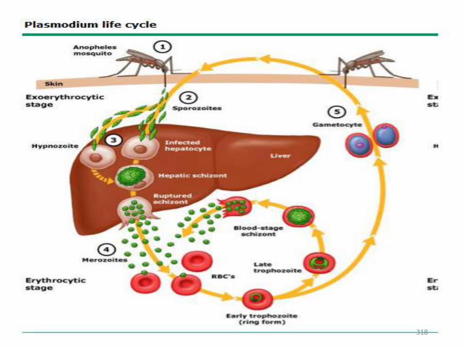
318
Life cycle and pathogenesis
• The symptoms and pathology are caused by the
asexual erythrocytic stage of the malaria parasite.
• Four pathologic process
• Fever
• Anemia
• Immunopathologic events
• Tissue anorexia resulting from cytoadherence of infected
erythrocytes
319
Life cycle and pathogenesis
• Fever-occurs when RBC rupture and release merozoites in to circulation.
• Anemia- hemolysis, sequestration of RBC in the spleen and other organs ,
and suppression of the bone marrow.
• Immunopathologic events include immune complex formation, immune
suppression, and release of inflammatory mediators such as TNF which may
be responsible for many of the pathologic features of the disease.
• Cytoadherence to vascular endothelium which occurs in p. falciparum may
lead to obstruction of blood flow and capillary damage with resultant vascular
leakage of protein and fluid , edema and tissue anorexia in the brain ,heart,
lungs, intestine and kidney
320
Clinical Manifestations
• Classic picture (shivering ,fever, and sweating) may
not be observed in Younger children.
• Children become restless, drowsy, apathetic, and
anorexic.
• Older children may report aching body, headache, and
nausea.
• Fever is usually continuous and may be very high
(40°C) from the first day.
321
Clinical Manifestations
• Flulike respiratory symptoms with mild cough and cold. These
symptoms abate in 1-2 days, with or without treatment.
• Parasitemia in neonates within 7 days of birth implies
transplacental transmission.
• This congenital malaria is usually associated with
placental parasitemia, which sometimes persists even
after adequate treatment with antimalarial drugs. Babies
have fever, are irritable, refuse feeds, and often develop
anemia, jaundice, and hepatosplenomegaly.
322
Clinical Manifestations
• Vomiting
• Seizure
• Fever (temp of 40c or more)
• Slightly tender liver
• Splenomegaly
• Anemia
• Mild jaundice
323
Clinical Manifestations
• P vivax and P ovale both give rise to hypnozoites in
the liver
• P vivax malaria may relapse for up to 3 years and P
ovale for 1-1.5 years.
• P falciparum and P malariae do not form hypnozoites,
so they do not have true relapses.
324
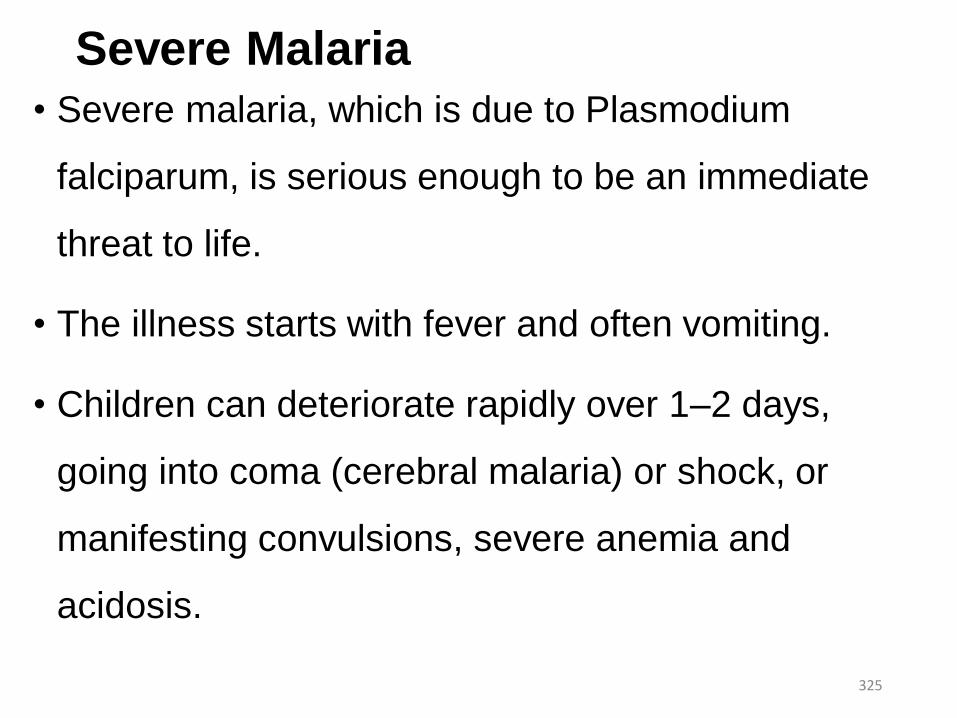
Severe Malaria
• Severe malaria, which is due to Plasmodium
falciparum, is serious enough to be an immediate
threat to life.
• The illness starts with fever and often vomiting.
• Children can deteriorate rapidly over 1–2 days,
going into coma (cerebral malaria) or shock, or
manifesting convulsions, severe anemia and
acidosis.
325
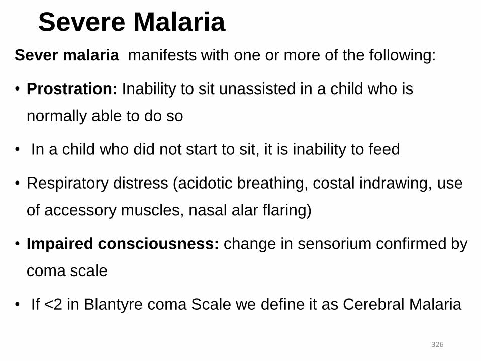
Severe Malaria Sever malaria manifests with one or more of the following:
• Prostration: Inability to sit unassisted in a child who is
normally able to do so
• In a child who did not start to sit, it is inability to feed
• Respiratory distress (acidotic breathing, costal indrawing, use
of accessory muscles, nasal alar flaring)
• Impaired consciousness: change in sensorium confirmed by
coma scale
• If <2 in Blantyre coma Scale we define it as Cerebral Malaria
326
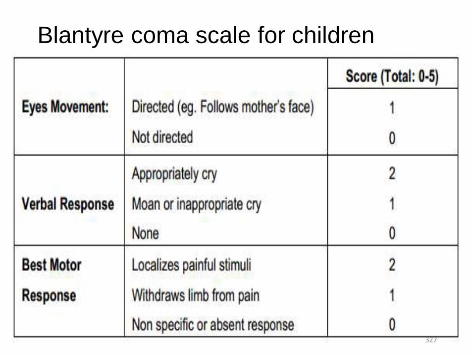
Blantyre coma scale for children
327
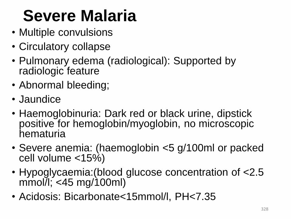
Severe Malaria • Multiple convulsions
• Circulatory collapse
• Pulmonary edema (radiological): Supported by radiologic feature
• Abnormal bleeding;
• Jaundice
• Haemoglobinuria: Dark red or black urine, dipstick positive for hemoglobin/myoglobin, no microscopic hematuria
• Severe anemia: (haemoglobin <5 g/100ml or packed cell volume <15%)
• Hypoglycaemia:(blood glucose concentration of <2.5 mmol/l; <45 mg/100ml)
• Acidosis: Bicarbonate<15mmol/l, PH<7.35 328
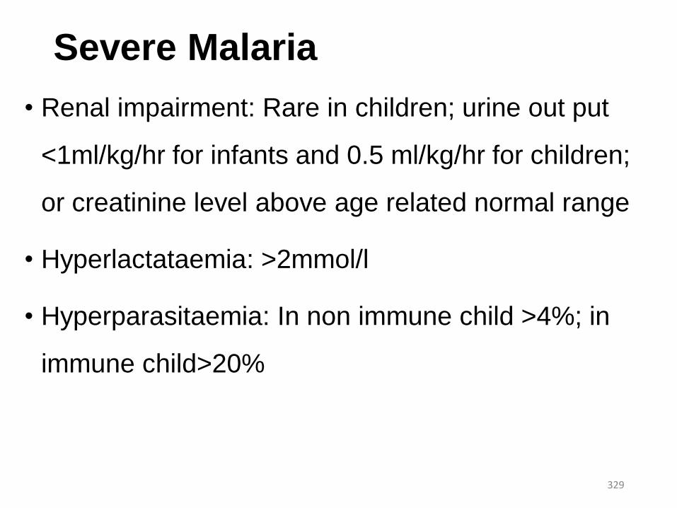
Severe Malaria
• Renal impairment: Rare in children; urine out put
<1ml/kg/hr for infants and 0.5 ml/kg/hr for children;
or creatinine level above age related normal range
• Hyperlactataemia: >2mmol/l
• Hyperparasitaemia: In non immune child >4%; in
immune child>20%
329
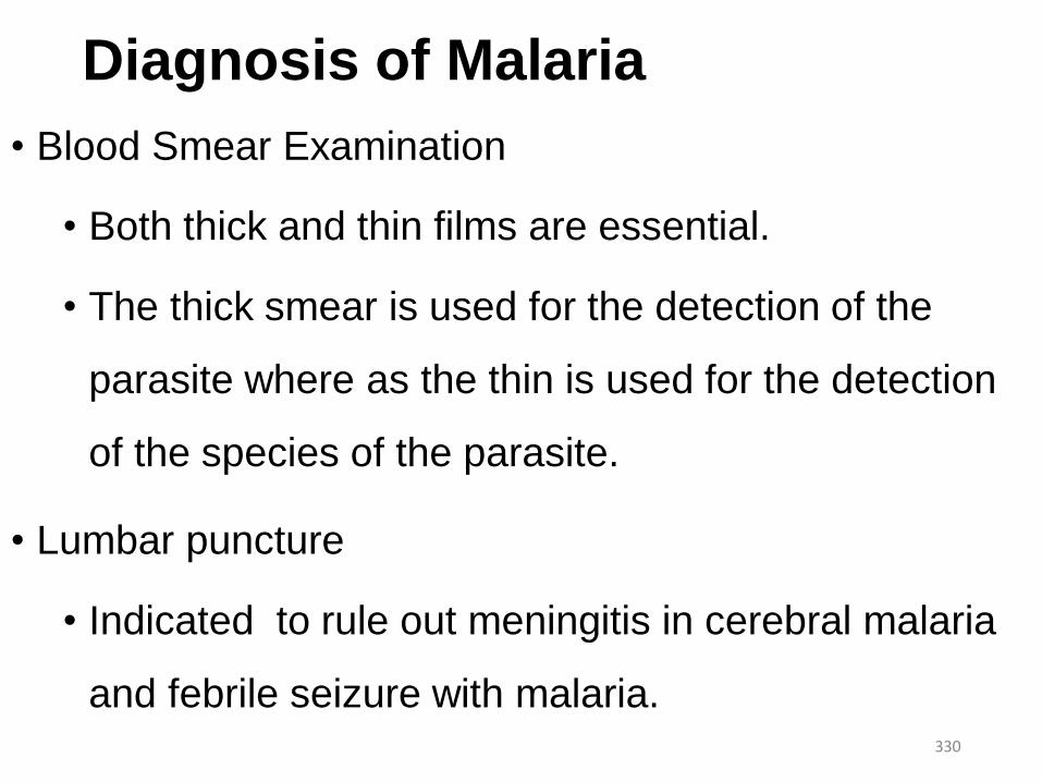
Diagnosis of Malaria
• Blood Smear Examination
• Both thick and thin films are essential.
• The thick smear is used for the detection of the
parasite where as the thin is used for the detection
of the species of the parasite.
• Lumbar puncture
• Indicated to rule out meningitis in cerebral malaria
and febrile seizure with malaria.330
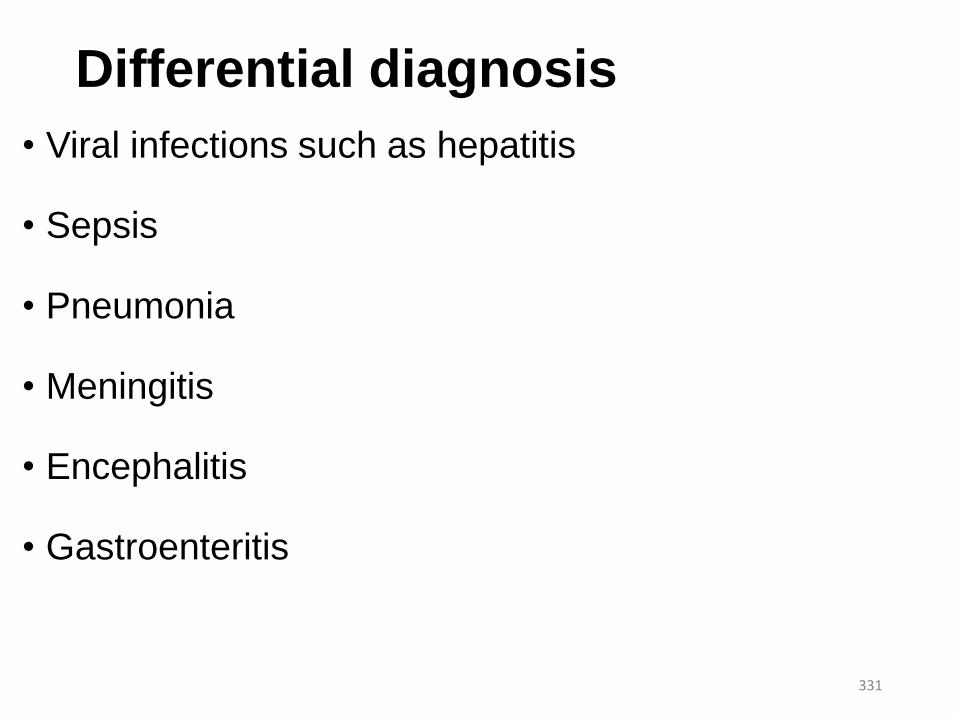
Differential diagnosis
• Viral infections such as hepatitis
• Sepsis
• Pneumonia
• Meningitis
• Encephalitis
• Gastroenteritis
331
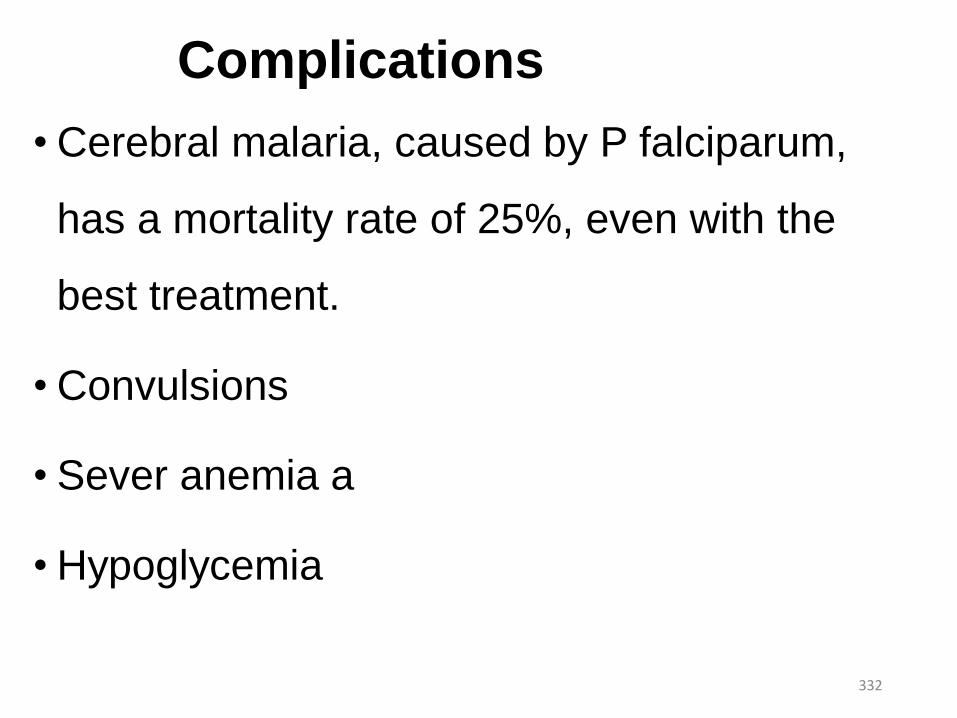
Complications
• Cerebral malaria, caused by P falciparum,
has a mortality rate of 25%, even with the
best treatment.
• Convulsions
• Sever anemia a
• Hypoglycemia
332
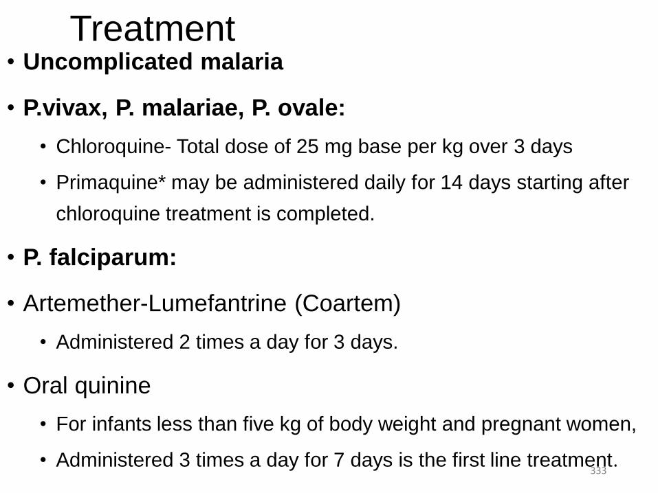
Treatment • Uncomplicated malaria
• P.vivax, P. malariae, P. ovale:
• Chloroquine- Total dose of 25 mg base per kg over 3 days
• Primaquine* may be administered daily for 14 days starting after
chloroquine treatment is completed.
• P. falciparum:
• Artemether-Lumefantrine (Coartem)
• Administered 2 times a day for 3 days.
• Oral quinine
• For infants less than five kg of body weight and pregnant women,
• Administered 3 times a day for 7 days is the first line treatment.333

Treatment of sever P. falciparum malaria
• Emergency measures : to be taken within the first hour:
• Check for hypoglycaemia and correct
• Treat convulsions with rectal diazepam
• Restore the circulating blood volume
• If the child is unconscious, minimize the risk of
aspiration pneumonia by proper positioning
• Treat severe anemia if in heart failure
• Start treatment with an effective antimalaria
334
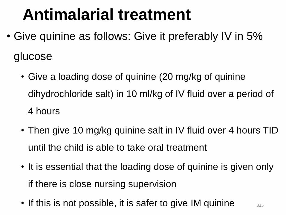
Antimalarial treatment
• Give quinine as follows: Give it preferably IV in 5%
glucose
• Give a loading dose of quinine (20 mg/kg of quinine
dihydrochloride salt) in 10 ml/kg of IV fluid over a period of
4 hours
• Then give 10 mg/kg quinine salt in IV fluid over 4 hours TID
until the child is able to take oral treatment
• It is essential that the loading dose of quinine is given only
if there is close nursing supervision
• If this is not possible, it is safer to give IM quinine 335
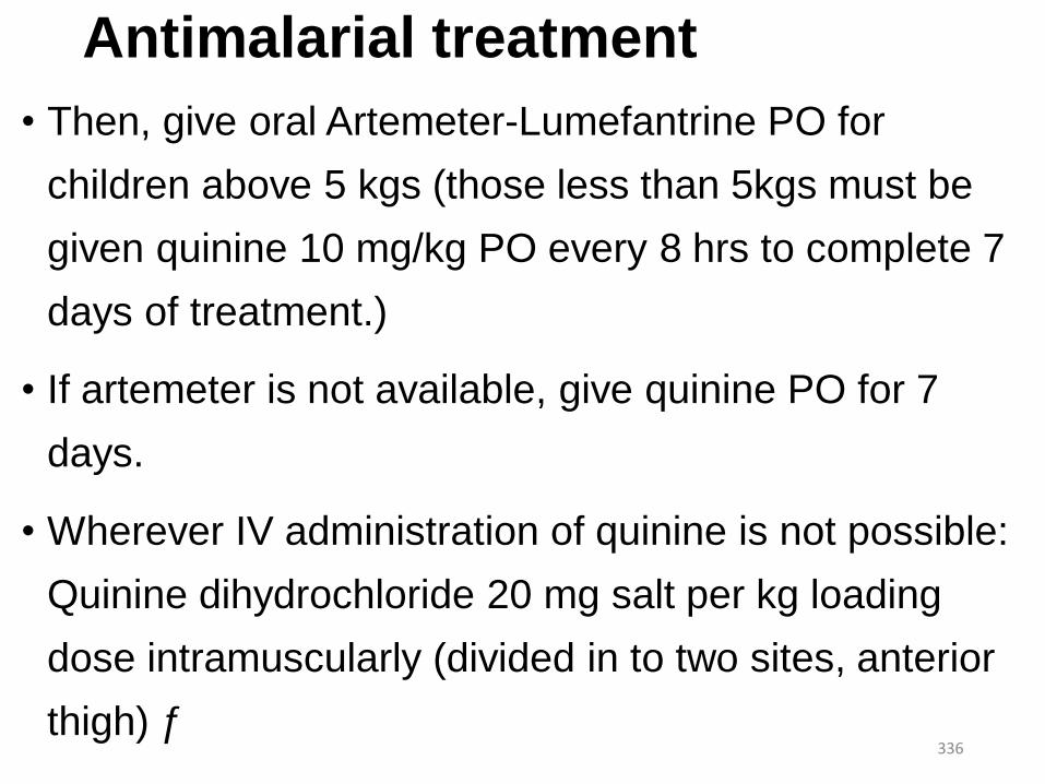
Antimalarial treatment
• Then, give oral Artemeter-Lumefantrine PO for
children above 5 kgs (those less than 5kgs must be
given quinine 10 mg/kg PO every 8 hrs to complete 7
days of treatment.)
• If artemeter is not available, give quinine PO for 7
days.
• Wherever IV administration of quinine is not possible:
Quinine dihydrochloride 20 mg salt per kg loading
dose intramuscularly (divided in to two sites, anterior
thigh) ƒ336
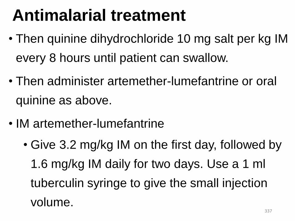
Antimalarial treatment
• Then quinine dihydrochloride 10 mg salt per kg IM
every 8 hours until patient can swallow.
• Then administer artemether-lumefantrine or oral
quinine as above.
• IM artemether-lumefantrine
• Give 3.2 mg/kg IM on the first day, followed by
1.6 mg/kg IM daily for two days. Use a 1 ml
tuberculin syringe to give the small injection
volume.337
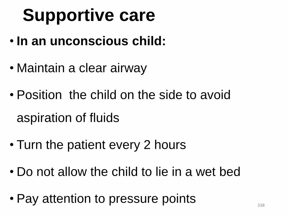
Supportive care
• In an unconscious child:
• Maintain a clear airway
• Position the child on the side to avoid
aspiration of fluids
• Turn the patient every 2 hours
• Do not allow the child to lie in a wet bed
• Pay attention to pressure points338
Supportive care
• Take the following precautions in the delivery of fluids:
• Check for dehydration and treat appropriately.
• During rehydration, examine frequently for signs of fluid
overload
• The most reliable sign is an enlarged liver.
• If, after careful rehydration, the urine output over 6 hours is
less than 0.5 ml/kg / hr, give IV furosemide.
• In children with no dehydration, ensure that they receive their
daily fluid requirements but take care not to exceed the
recommended limits.339
Prevention • Chemoprophylaxis
• Mefloquine
• Doxycycline-2mg/kg/d for >8yr of age
• Premaquine-0.3mg/kg
• Avoiding mosquito breeding sites
• Residual DDT spray or other chemicals
• Insecticide Treated Nets (ITN)
• Chemotherapy of cases347
Expanded Programme of Immunization (EPI)
EPI Target Diseases
348
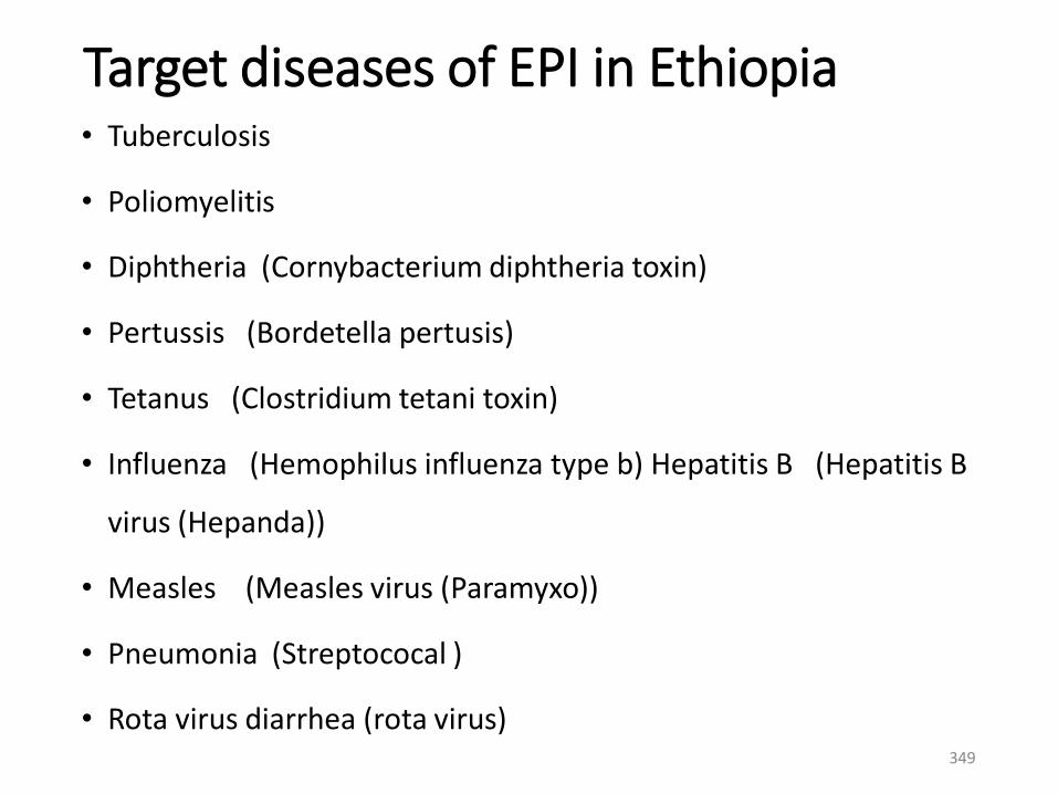
Target diseases of EPI in Ethiopia• Tuberculosis
• Poliomyelitis
• Diphtheria (Cornybacterium diphtheria toxin)
• Pertussis (Bordetella pertusis)
• Tetanus (Clostridium tetani toxin)
• Influenza (Hemophilus influenza type b) Hepatitis B (Hepatitis B
virus (Hepanda))
• Measles (Measles virus (Paramyxo))
• Pneumonia (Streptococal )
• Rota virus diarrhea (rota virus)349
Pertussis (Whooping Cough)
• Etiologic agent- A gram-negative bacterium called
Bordetella pertussis.
• Pathogenesis - The organism produces exotoxin
and affects the pharynx, larynx, trachea, bronchi,
bronchioles and sometimes the alveoli.
• Clinical features - Incubation period is 7 – 17 days.
• The symptoms of classical pertussis last about 6 weeks and
are divided into 3 stages
350
Catarrhal stage
• The onset is insidious
• Lasts 1- 2 weeks
• Cough and rhinorrhea
Paroxysmal stage
• Lasts 2-4 weeks following the infection.
• Characterized by rapid consecutive (5-15) cough before a breath
is taken and followed by deep hurried inspiration (whoop).
• Post cough vomiting is common at all ages,
351
Cont….
• Expulsion of clear tenacious mucus often followed by vomiting
• Factors stimulating cough include fright, anger, crying,
sneezing, inhalation of irritant, and overdistention of the stomach.
Convalescent stage
• It begins after 4 weeks of the illness, and is manifested by a
decrease in the frequency and severity of the paroxysms of
coughing.
• After improvement the disease may recur.
352
Diagnosis
• Clinical
• The child is well appearing and playful between
paroxysms of cough.
• Presence of children with similar illness in the family or
vicinity.
• There is no chest finding on physical examination.
• The diagnosis is usually made on the distinctive clinical
feature of the cough. To observe the classical type of
cough put tongue depressor to stimulate the coughing.
353
Cont’d• Laboratory
• WBC 15000 - 20000/mm3 (rarely to 50,000/mm3)
• 60 - 80% Lymphocytes
• Microscopy; Gram stain
• Culture to isolate the organism
• Differential Diagnosis
• Aspiration of foreign bodies
• Viral pneumonia
• Influenza
• Acute bronchitis and bronchiolitis.
354
Management
Specific measures
• Antimicrobial
• Erythromycin
• Ampicillin
• Sedatives
• Promethazine Hydrochloride
• Phenobarbitone (when there is seizure)
• Steroid
• Prednisolone
355
Cont’d
General measures
• Frequent small feeding and continue feeding if vomiting occurs
soon after meal.
• Oxygen if needed
• Severe cases especially infants are best managed in hospital.
• Prophylactic Erythromycin for all house hold members and other
contacts regardless of age, history of immunization of symptoms.
356
Complications
• Apnea
• Conjunctival hemorrhage
• Otitis media
• Pneumonia
• Atelectasis
• Encephalopathy
357
Tetanus
• An acute, spastic paralytic illness caused by tetanus toxin
(tetanospasmin), the neurotoxin produced by Clostridium
tetani
• Etiologic agent
• A gram positive anaerobic bacterium called Clostridium
tetani (Cl.tetani).
• Can survive boiling
358
Pathogenesis
• Tetanus toxin, after germination of the Cl.tetani
spores in a contaminated umbilical stump or
wound in other parts of the body, is released to the
peripheral nerves and circulation.
• This causes sustained excitatory neuronal discharge
and muscle contraction.
359
Clinical Manifestation
• Incubation period – 2 to 14 days commonly; but can
be as long as months after the injury.
• Can be divided in to two:
• Generalized tetanus –
• Trismus or lock jaw (masseter muscle spasm) is presenting
symptom in about 50%
• Headache, restlessness, and irritability are early symptoms often
followed by stiffness, dysphagia and neck muscle spasm
360
Cont’d
• Risus Sardonicus (sardonic smile) results from intractable
spasm of facial and buccal muscles
• Opisthotonus – arched posture due to involvement of
abdominal, lumbar, hip and thigh muscles (“board” like
rigidity in which the back of the head and heals touch
ground)
• Laryngeal and respiratory muscle spasm can lead to airway
obstruction and asphyxiation
• Patient remains conscious in extreme pain and
anticipation of next tetanic seizure361
Cont’d
• Tetanic spasm may be triggered by the smallest
disturbance (sight, sound or touch)
• Autonomic NS manifestations: tachycardia, arrhythmias,
labile hypertension, diaphoresis and cutaneous
vasoconstriction.
• Tetanic paralysis become more intense in the first week
after onset, and stabilizes over the ensuing one to four
weeks 362
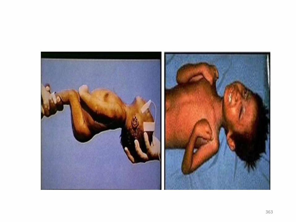
363
Cont’d
• Neonatal tetanus (Tetanus Neonatrum):
• Infantile type of generalized tetanus
• Typically manifests with in 3 – 12 days of birth
• Infection results from umbilical cord contamination during
unsanitary delivery
• Difficulty in feeding, crying, paralysis, stiffness to touch with or
without opisthotonus are characteristics.
364

Cont’d• Localized tetanus
• Painful spasm of muscles adjacent to the wound site
• May precede generalized tetanus
• Cephalic tetanus:
• A rare form of localized tetanus involving the bulbar musculature
• Follow wounds or foreign bodies in the head, nostrils or face, and
occasionally chronic otitis media.
• Characterized by retracted eye lids, deviated gaze, trismus, risus
sardonicus and spastic paralysis of the tongue.
365
Diagnosis• No diagnostic lab test
• Based on Hx and clinical finding
Differential diagnosis:
• Poisoning
• Meningitis
• Peritonitis
• Rabies
• Dental abscess
• Hypocalcemic tetany
366
Treatment
• Principles
• Eradication of C. tetani and wound environment
conducive for its multiplication.
• Neutralization of all accessible tetanus toxin-TIG
• Control of spasms-Diazepam
• Provision of meticulous supportive care
• Prevention of recurrences
367
Complications
• Respiratory arrest
• Laryngeal spasm
• Presence of autonomic nervous system disturbances.
• Prevention
• Immunization of children and women.
• Health information on harmful practices
• Training of Traditional Birth Attendants (TTBA).
368
PoliomyelitisEtiologic agent
• It is caused by polioviruses type I, II and III.
Pathogenesis
• The virus affects the anterior horn cells of the spinal cord
and several areas of the brain.
• Damage may be reversible with recovery, but it may go
on to irreversible nuclear destruction where muscle
paralysis results.
369
Clinical features • Incubation period is 6 – 14 days
• Fever, malaise, headache and muscle pain
• Nausea, vomiting, soar throat and stiffness of the neck and back with
or without paralysis.
• Paralysis usually affects the legs, more often one
• Diagnosis
• It is mainly by clinical features.
• Positive CSF – pleocytosis 20 -300 cells/mm; protein 50 -100 mg/dl by 1st
week
• Serology – a four fold or greater rise in antibody titers
• Culture – stool and naso-pharynx
370
ManagementAcute phase
• Keep the limbs position with cushions
• Apply warm packs
• Provide analgesics
• Active and passive movements are assisted by physiotherapist after the acute phase
ended.
Recovery phase
• Continue with full range of passive/active movement of the affected limb every day.
Residual phase
• Regular out patient supervision of physical, social and economic problems if
needed.
371
DiphtheriaEtiologic agent
• It is caused by Gram-positive bacterium called Corynebacterium diphtheriae.
Pathogenesis
• The bacterium produces exotoxin which causes local tissue inflammation and
necrosis.
• In cases where the pharynx is involved, there are patches of a grayish
membrane with a surrounding dull red inflammatory zone, which may cause
pharyngeal obstruction.
372
Clinical features
• The incubation period is usually 2 - 5 days.
• Sore throat which may be followed by stridor.
• Grayish white membrane seen in oropharynx.
• Upper airway obstruction by the membrane.
Diagnosis
• Clinical signs mentioned above
• Microscopy -Gram stain
373
Management
A. Specific
• Diphtheria antitoxin if the diagnosis is strongly suspected clinically.
• Antimicrobial therapy with penicillin or erythromycin
B. General
• Strict bed rest and sedation
• Intubations if needed
• Complications
• Airway obstruction
• Toxic cardiomyopathy (50 – 60% of diphtheria deaths)
• Vocal cord paralysis
374
Measles
• Etiologic agent
• It is caused by Measles Virus.
• Pathogenesis
• The essential lesion of measles is found in the skin; the
mucous membranes of the nasopharynx, bronchi, and
intestinal tract; and in the conjunctivae.
375
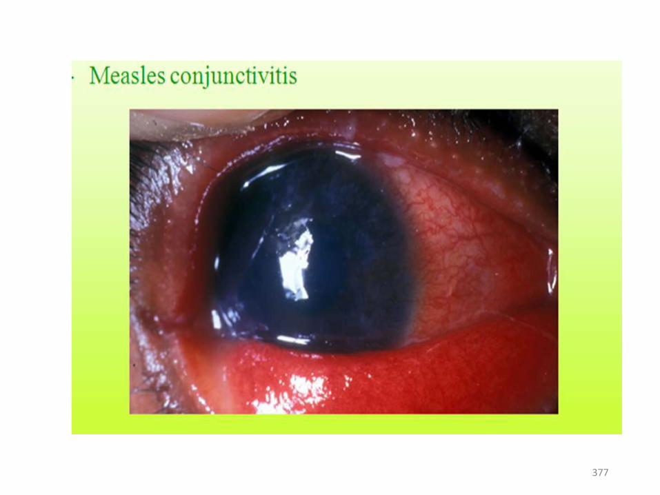
377
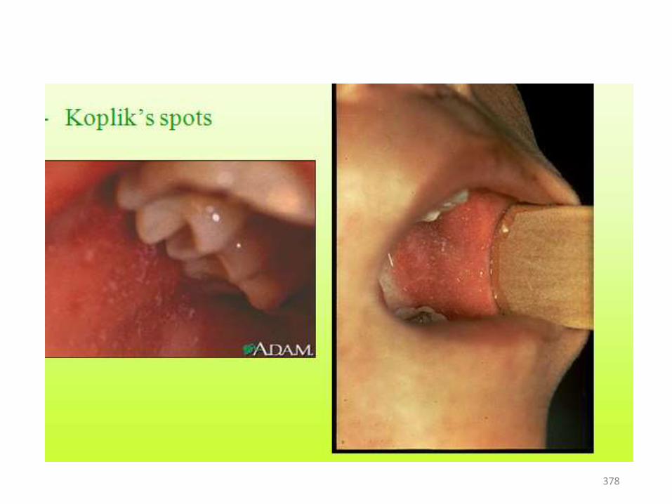
378

Diagnosis• It is made mainly by clinical features epidemiological grounds.
• Serologic - identification of Ig-M antibody. (appears 1–2 days after the onset of the rash)
• at least fourfold increase in anti-measles antibody titer
• Viral isolation from blood, urine, or respiratory secretions (by culture)
• Molecular detection by polymerase chain reaction is possible
- Case definition:
- Generalized rash lasting > 3 days and To > 38.5 oC plus
- Cough or coryza or conjunctivitis
**Management**
• Severe cases only are admitted to the hospital.
• Mothers are advised about care at home which includes – reducing fever,
maintaining hydration and nutrition.
• Serious complications are treated in hospitals.
379
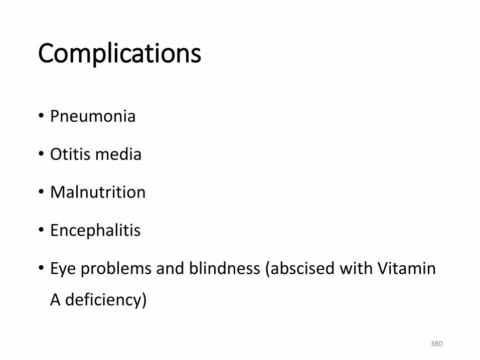
Complications
• Pneumonia
• Otitis media
• Malnutrition
• Encephalitis
• Eye problems and blindness (abscised with Vitamin
A deficiency)
380
Definitions and General concepts
• Immunization
• The process of inducing immunity artificially by either
vaccination or administration of antibodies.
• Two types:
• Active immunization – stimulation of the immune system to
produce antibodies and cellular immune response against an
infectious agent
• Passive immunization – providing temporary protection through
administration of exogenously produced antibodies.
381
Cont’d
• Vaccination
• Administration of any vaccine or toxoid for prevention of
disease.
• Types of vaccines
• Live attenuated (Polio, BCG, Measles, Rota)
• Killed (IPV, Pertussis, Hib)
• Toxoids (Tetanus, Diphtheria)
382

Expanded Programme on Immunization (EPI)/WHO
• Aims to provide free immunization for children against the
important childhood infections
• Started in 1974
• Ethiopia launched in 1980
• Main purpose:
• Prevent childhood diseases
• Provide high quality vaccines
• Surveillance of these diseases
383
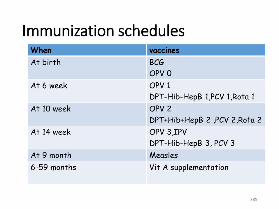
Immunization schedulesWhen vaccines
At birth BCG
OPV 0
At 6 week OPV 1
DPT-Hib-HepB 1,PCV 1,Rota 1
At 10 week OPV 2
DPT+Hib+HepB 2 ,PCV 2,Rota 2
At 14 week OPV 3,IPV
DPT-Hib-HepB 3, PCV 3
At 9 month Measles
6-59 months Vit A supplementation
385
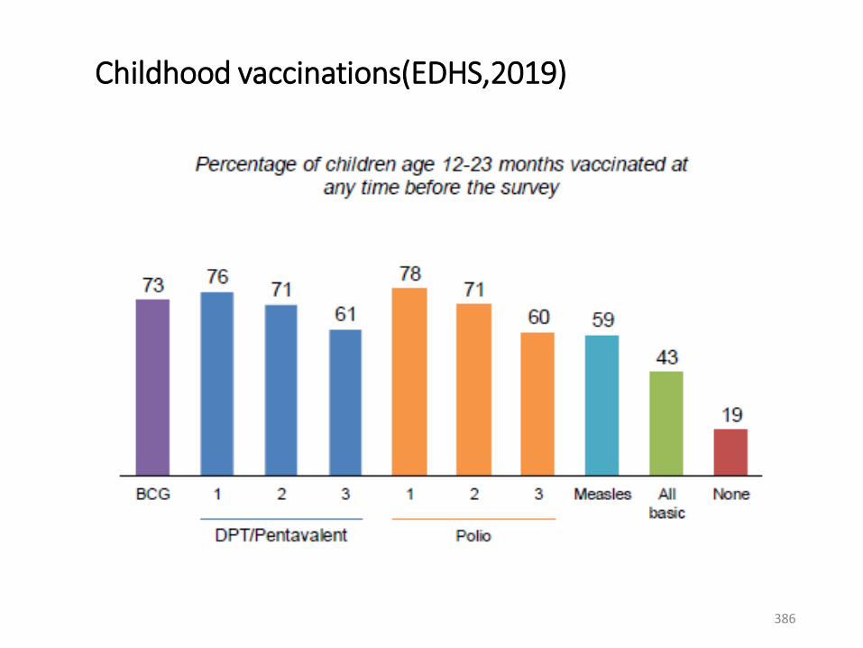
Childhood vaccinations(EDHS,2019)
386

Administering vaccine for infants
395
The strategies of EPI:
• Strategies of EPI includes:
• Routine immunization strategies
• National immunization days
• Out break response immunization
• Routine immunization strategies are classified as:
• Static immunization
• Out reach immunization
• Mopping up immunization (House to house)
396

(I) The static immunization strategy:
• Advantages: Available resources., Cold Chain
maintenance , Save time, effort and money
(II) Outreach immunization:
The outreach is carried for routine immunization
Compulsory for the targets in:
Immunization not accessible.
Low vaccination coverage
The outreach is carried during any time
Limitations:
Expensive
Cold chain failure
Difficulty to arrange 397
Cont’d(III) Mopping up Immunization:
• house-to-house immunization of OPV in high risk
districts.
• High risk districts are those:
• Where polio virus is still circulating
• Low immunization coverage.
• Transient population,
• Overcrowding poor sanitary
• Low access to health services.
398
The National Immunization Days (NIDs)
• Periodic immunization of all the eligible targets over a large
geographic areas within a short period
• Used for polio eradication and tetanus elimination.
Outbreak response immunization
• Outbreak of vaccines preventable diseases requires immunization
to be implemented.
• Before the immunization has been started, assessment of the
outbreak is mandatory.
399
Cont’d
• The assessment includes:
• The type of disease
• The magnitude of disease
• The requirement of vaccination
• The number of individuals to be vaccinated
• The number staffs required
• Other required logistics and break down of budget
400
Concepts and rates
• Immunization Coverage
• Immunization coverage is the proportion of the target
population that has been vaccinated.
• If coverage rates indicate that people are not using
immunization services, health workers and their supervisors
need to find the causes and take appropriate action.
• Comparing coverage rates for different vaccines sometimes
points toward the likely problem as well as possible
solutions.401
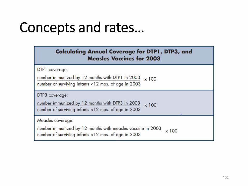
Concepts and rates…
402
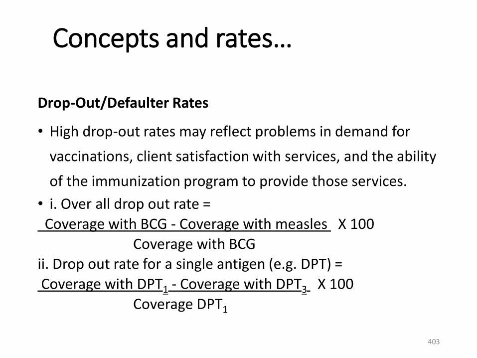
Concepts and rates…
Drop-Out/Defaulter Rates
• High drop-out rates may reflect problems in demand for
vaccinations, client satisfaction with services, and the ability
of the immunization program to provide those services.
• i. Over all drop out rate =
Coverage with BCG - Coverage with measles X 100
Coverage with BCG ii. Drop out rate for a single antigen (e.g. DPT) =
Coverage with DPT1 - Coverage with DPT3 X 100
Coverage DPT1
403
The cold chain of vaccines:• Cold Chain is a system of equipments which ensure
that correct quantity of potent vaccines reaches the people who need it.
• It is the system of storage and transportation of the vaccine at low temperature (cold condition) from the manufacture till it is consumed.
• Polio vaccine is the most sensitive vaccine to heat.
• Live attenuated vaccines are allowed to be frozen (OPV, Measles, and BCG).
• Inactivated vaccines must not be frozen ( DPT, Hib and HB).
404
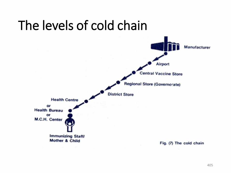
The levels of cold chain
405
Cont’d
• Refrigeration equipment:
• Refrigerator
• Cold boxes
• Vaccine carriers
• The ice packs retained in the freezer
• To stabilize the temperature of the refrigerator at the
optimum level fully frozen ice-packs are used for
lining the vaccines carriers and the cold boxes during
storing the vaccines.406
The refrigerator:
• Placed in the coolest place of the health centers away from sunlight-
• Kept locked and open only when necessary.
• Ice packs are kept in the freezer and Its temperature is recorded twice daily.
• Both the monitor and thermometer are placed in the refrigerator.
• The temperature chart is stuck on the door outside the refrigerator.
• The diluents should be kept on the lowest shelf.
407
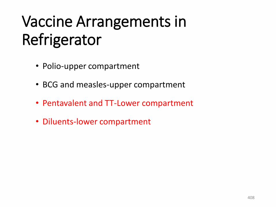
Vaccine Arrangements in Refrigerator
• Polio-upper compartment
• BCG and measles-upper compartment
• Pentavalent and TT-Lower compartment
• Diluents-lower compartment
408
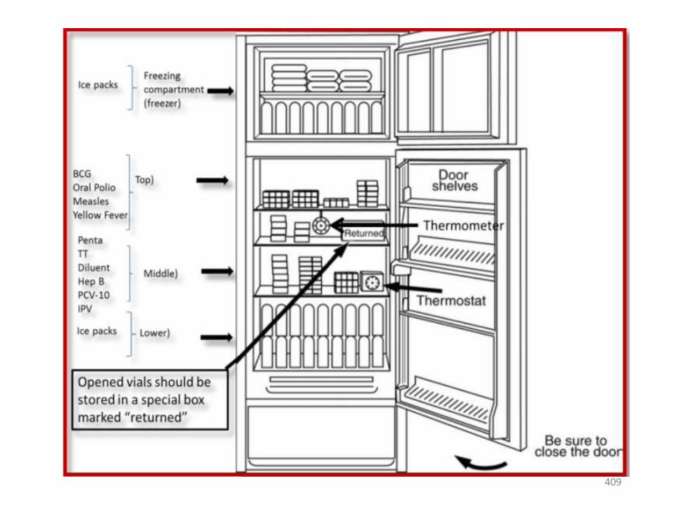
409
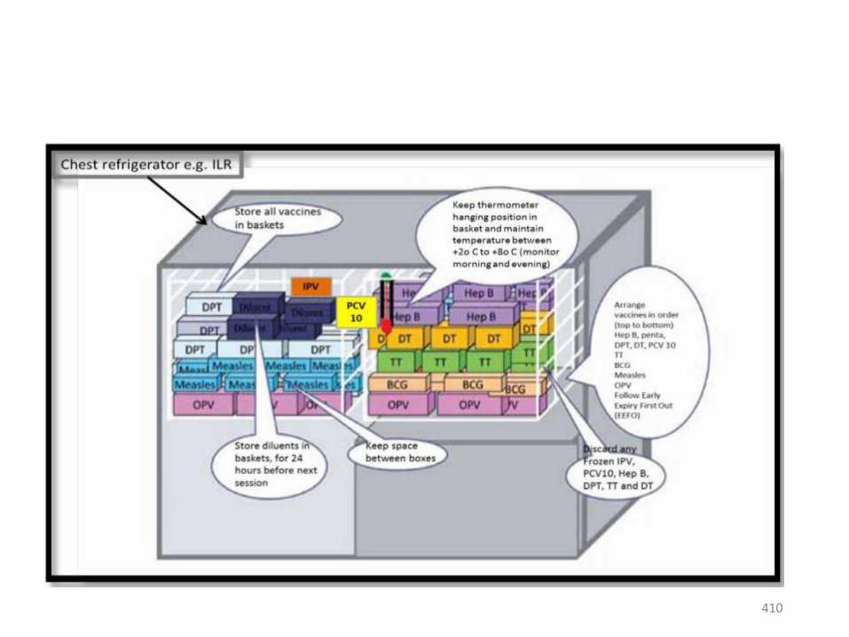
410
Tools for monitoring the cold chain:
There are four ways of monitoring the cold chain:
• Cold Chain Refrigerator Graph: regularly monitoring
the storage which should always be kept between 2
and 8 degrees celicius.
• A chart should be a fixed to the front of the refrigerator
and should be recorded at least twice in a day.
411
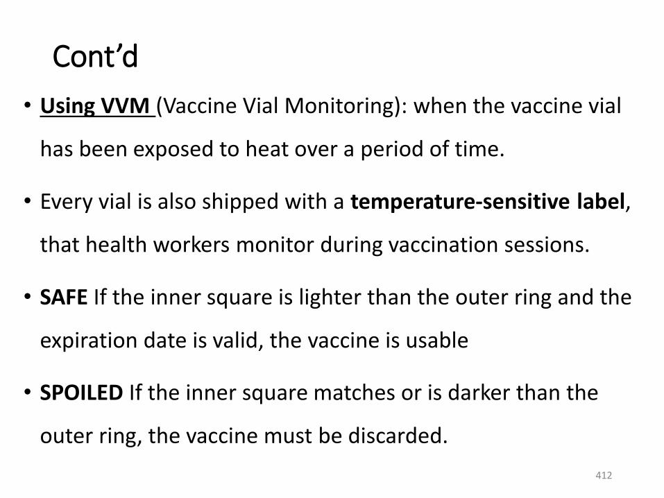
Cont’d
• Using VVM (Vaccine Vial Monitoring): when the vaccine vial
has been exposed to heat over a period of time.
• Every vial is also shipped with a temperature-sensitive label,
that health workers monitor during vaccination sessions.
• SAFE If the inner square is lighter than the outer ring and the
expiration date is valid, the vaccine is usable
• SPOILED If the inner square matches or is darker than the
outer ring, the vaccine must be discarded.
412
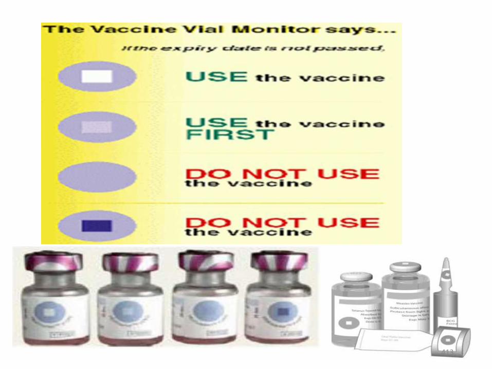
413
Cont’d• Using the cold chain monitor (freeze watch indicator):
• Read and record the temperature recorder every day to maintain
the refrigerator at 2- 8 degree centigrade.
• The shake test - DPT, hepatitis B (for Penta), TT and tetanus toxoid
vaccines can all be damaged by freezing.
• By shaking two vials, side-by-side, one that might have been
frozen and one that has never been frozen, health workers can
determine if a vaccine has spoiled.
• The vaccine should be administered shortly after withdrawal from
the vial.414
Storage of the vaccine:
• The vaccine should not kept frozen or exposed to freezing
• Store at 2°to 8°C• Shake vial vigorously before withdrawal and use. What damage the Vaccines?
1. Any defect in the cold chain.2. Out date expiry.3. Using skin antiseptic at the site of injection (e.g. BCG).4. Using the reconstituted vaccine (measles, BCG) after the
recommended period ( 6 hours)5. Exposure of the vaccine to unacceptable temperature during
the immunization session.6. Exposure of the vaccine to direct sunlight (BCG)
415
Cardiovascular Disorders
•Congestive heart failure
•Infective endocarditis
•Rheumatic fever
By Chalachew Adugna (BSc, MSc)
Department of pediatrics and child health
nursing (University of Gondar)
416
Introduction
•The Heart
•The heart is a cone-shaped, muscular organ
located between the lungs behind the sternum
•The heart muscle forms the myocardium, with
tightly interconnected cells of cardiac muscle tissue
•The pericardium is the outer membranous sac with
lubricating fluid417
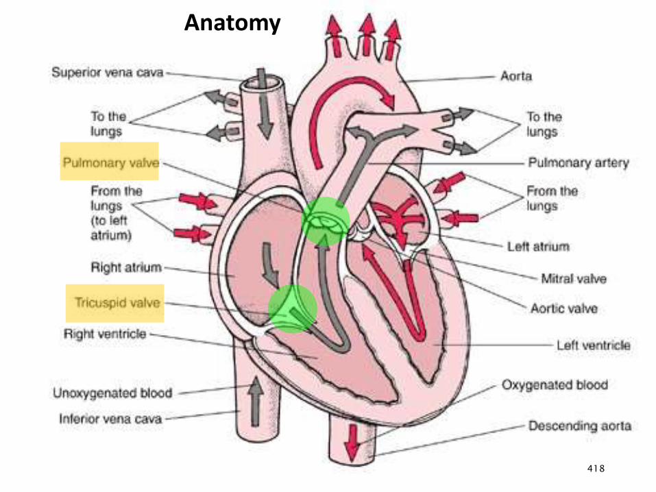
Anatomy
418
Chambers and valves of the heart
•The heart has four chambers:
• two upper, thin-walled atria
• two lower, thick-walled ventricles
•The septum is a wall dividing the right and left
sides.
•Atrioventricular valves occur between the atria and
ventricles
•The tricuspid valve on the right
•The bicuspid valve on the left 419
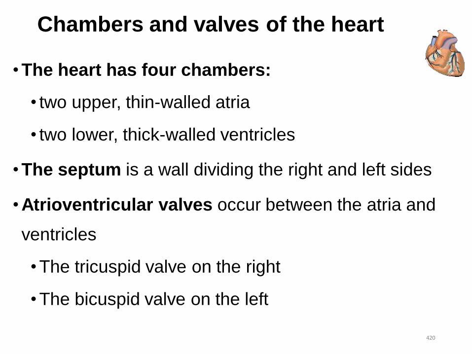
Chambers and valves of the heart
• The heart has four chambers:
• two upper, thin-walled atria
• two lower, thick-walled ventricles
• The septum is a wall dividing the right and left sides
• Atrioventricular valves occur between the atria and
ventricles
• The tricuspid valve on the right
• The bicuspid valve on the left
420
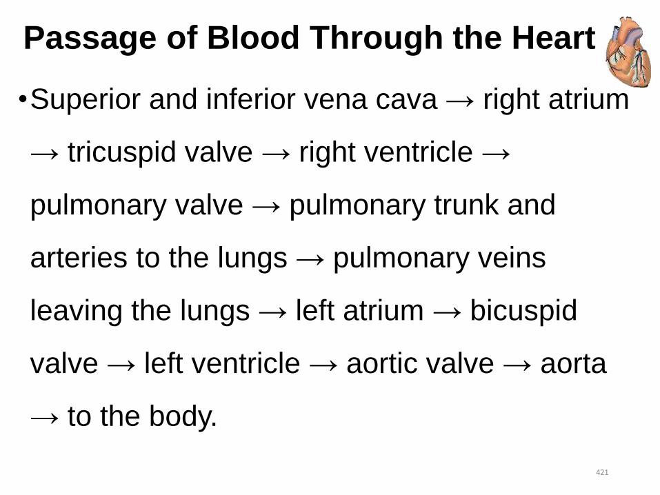
Passage of Blood Through the Heart
•Superior and inferior vena cava → right atrium
→ tricuspid valve → right ventricle →
pulmonary valve → pulmonary trunk and
arteries to the lungs → pulmonary veins
leaving the lungs → left atrium → bicuspid
valve → left ventricle → aortic valve → aorta
→ to the body.
421
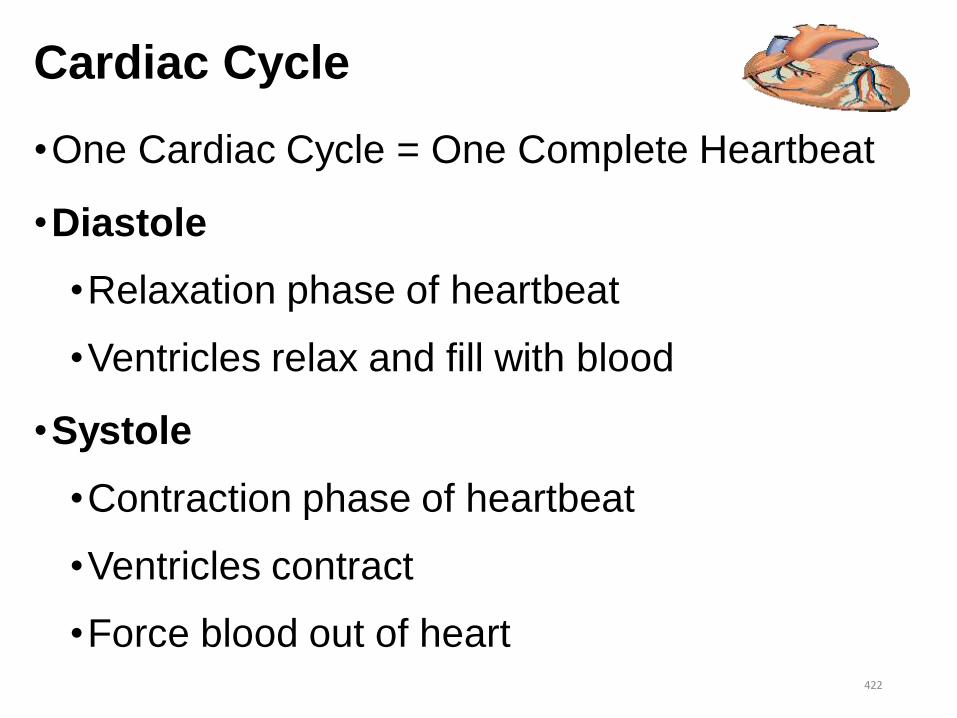
Cardiac Cycle
•One Cardiac Cycle = One Complete Heartbeat
•Diastole
•Relaxation phase of heartbeat
•Ventricles relax and fill with blood
•Systole
•Contraction phase of heartbeat
•Ventricles contract
•Force blood out of heart422
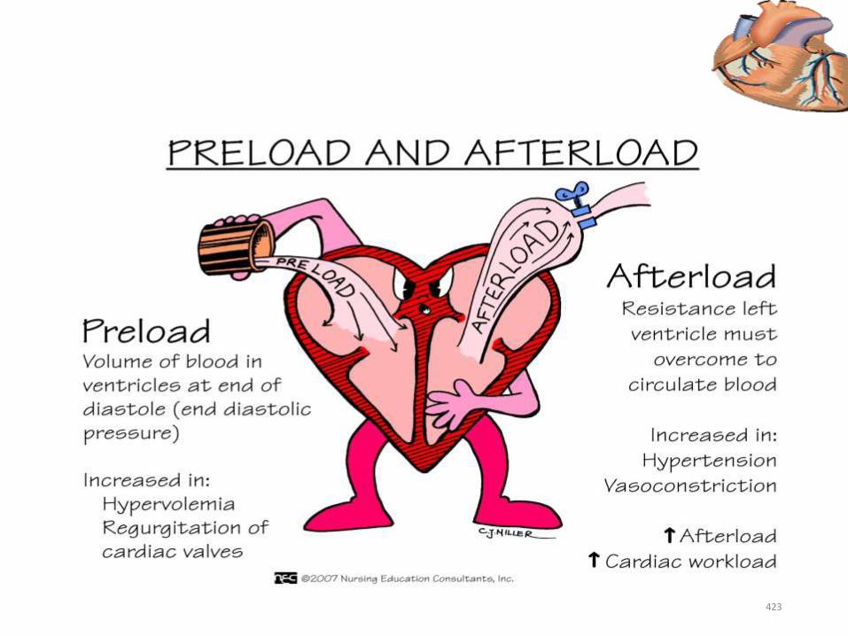
423
Congestive Heart Failure
•CHF is a condition in which the heart is unable to
pump adequate amount of blood to meet the
metabolic demand of the body
• It must not consider as a single disease entity rather
a syndrome of an over worked heart
•Heart failure is caused by ventricular pump
dysfunction, or by overload of volume (preload) or
pressure (afterload)
424
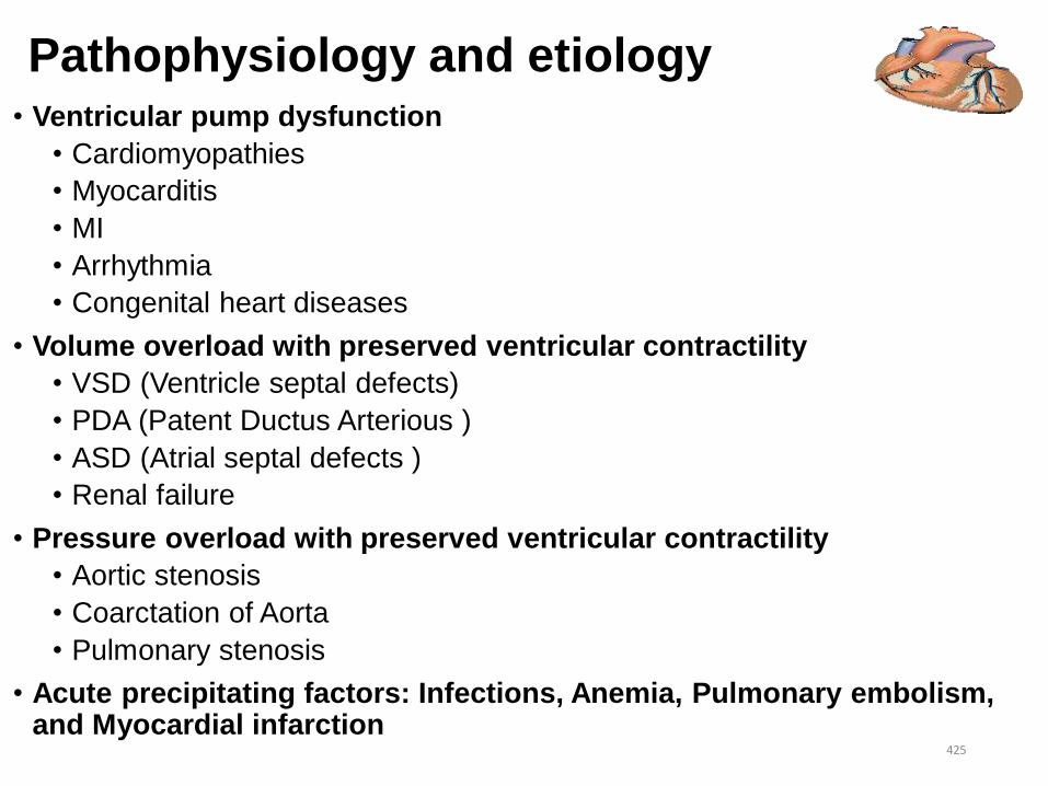
Pathophysiology and etiology • Ventricular pump dysfunction
• Cardiomyopathies
• Myocarditis
• MI
• Arrhythmia
• Congenital heart diseases
• Volume overload with preserved ventricular contractility
• VSD (Ventricle septal defects)
• PDA (Patent Ductus Arterious )
• ASD (Atrial septal defects )
• Renal failure
• Pressure overload with preserved ventricular contractility
• Aortic stenosis
• Coarctation of Aorta
• Pulmonary stenosis
• Acute precipitating factors: Infections, Anemia, Pulmonary embolism, and Myocardial infarction
425
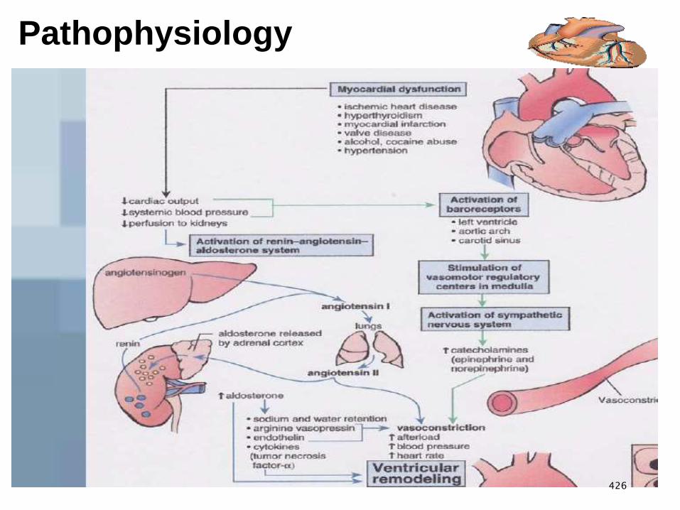
Pathophysiology
426
Clinical Features
• Fast breathing or interruption of feeding with diaphoresis
• Tachycardia (heart rate >160/minute in a child under 12 months
old; >120/minute in a child aged 12 months to 5 years)
• Basal crackles on chest exam
• Gallop rhythm on auscultation with or without murmurs
• Enlarged, tender liver
• In older children: edema of the feet, hands or face, or raised
JVP
• Severe palmar pallor may be present if severe anemia is the
cause of the heart failure427
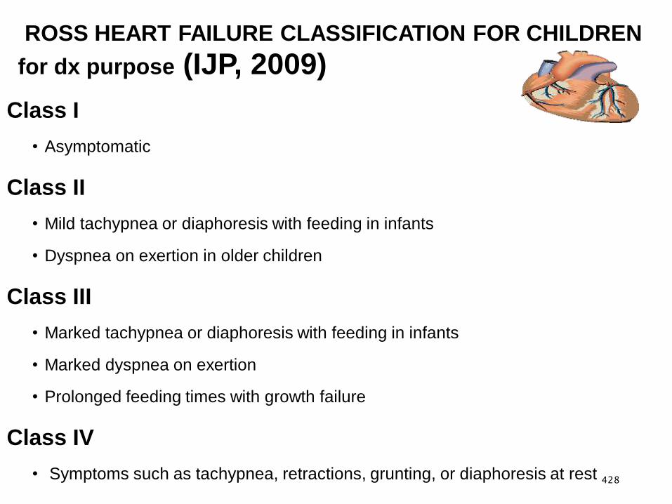
ROSS HEART FAILURE CLASSIFICATION FOR CHILDREN
for dx purpose (IJP, 2009)
Class I
• Asymptomatic
Class II
• Mild tachypnea or diaphoresis with feeding in infants
• Dyspnea on exertion in older children
Class III
• Marked tachypnea or diaphoresis with feeding in infants
• Marked dyspnea on exertion
• Prolonged feeding times with growth failure
Class IV
• Symptoms such as tachypnea, retractions, grunting, or diaphoresis at rest 428
Diagnosis
•Clinical Findings
•Chest X-ray
Cardiac enlargement
Increased pulmonary vascular bed
•Echocardiography
Chamber hypertrophy
• ECG
429

FARMINGHAM CRITERIA FOR DIAGNOSIS OF CONGESTIVE HEART FAILURE
For establishing a definite diagnosis of CHF =2major or 1 major & 2 minor
•Major Criteria• PND• Neck Vein Distention• Rales• Cardiomegaly• Acute Pulmonary Edema• S3 Gallop• Increased Venous Pressure (>16cm Of H20)
• Positive Hepatojugular Reflux
• Weight loss > 4.5kg over 5 days’ treatment
•Minor Criteria
•Extremity Edema
•Night Cough
•Dyspnea on ordinary Exertion
•Hepatomegaly
•Pleural Effusion
•Vital Capacity Reduced By 1/3 From Normal
•Tachycardia
430
Principles of management of CHF
• Treat underlying cause
• Treat the congestive state
• Prevent further deteriorations of cardiac lesions
431
Management
• Therapeutic goals for children with heart failure are:
•To relieve symptoms
•Decrease morbidity (including the risk of
hospitalization)
•Slow the progression of heart failure
• Improve patient survival
432
Management…
• Improve the cardiac efficiency/contractility force:
• Correct the underlying cause
• Administer digitalis Ex. Digoxin
• Remove accumulated fluid and sodium from the body:
• Administer diuretics Ex. Furosemide
• Restrict fluid and salt intake
• Daily measurement & Recording of intake, output and
weight
• Supplemental potassium or encourage the child to eat
potassium rich foods
433
Management…
• Improve blood & tissue oxygenation:
•Observe for signs of respiratory distress
•Position the child in sitting or semi sitting
•Administer oxygen
•Reduce the work load of the heart
•Provide adequate rest
•Provide small and frequent diet
•Provide positive pressure ventilation434
Management…
• Promote normal growth and development:
•Provide nutritious balanced diet.
• Support and educate parents:
•Adequate information about the drugs and feeding
• Encourage parental participation in child care
435
Management of CHF…
• Digoxin : 5–10 μg/kg/day, divided q12h.
• Furosemide (Lasix) - IV:1–2 mg/dose Bid.
-PO:1–4 mg/kg/day, divided qd–qid
KCL PO: one to one with the lasix(one tablet of lasix to
one tablet of KCL).
• Spironolactone : 2-3mg/kg/24hrs in 2-3 devided doses.
• ACEI: Enalapril : PO 0.08–0.5 mg/kg/dose q12–24hrs
436
Infective Endocarditis (IE)
•An acute or subacute inflammation of the endocardium
and/or heart valves that involves thrombus formation
(vegetation), which may damage the endocardial tissue
and/or valves.
•Common etiologic are:
•Streptococcus Viridans
•Staphylococcus Aureus
•Group D Streptococcus
437

Predisposing factors
Dental manipulation
Dental disease (caries, abscess)
Extra cardiac infection (lung, urinary tract, skin, bone,
abscess)
Instrumentation (urinary tract, GI tract, IV infusions)
Cardiac surgery
Injection drug use
438

Pathophysiology
Platelet deposition + fibrin cause non Bacterial thrombus /vegetation/
Patient with CHD turbulent blood flow
Nidus form Valve damage
Bacterial enter in the body
439
Clinical manifestations
•The presentation generally is indolent, with prolonged
low grade fever and a variety of somatic complaints,
including fatigue, weakness, arthralgias, myalgias,
weight loss, rigors, and diaphoresis
•The most common presenting symptoms were fever,
cardiac murmurs and dyspnea
440
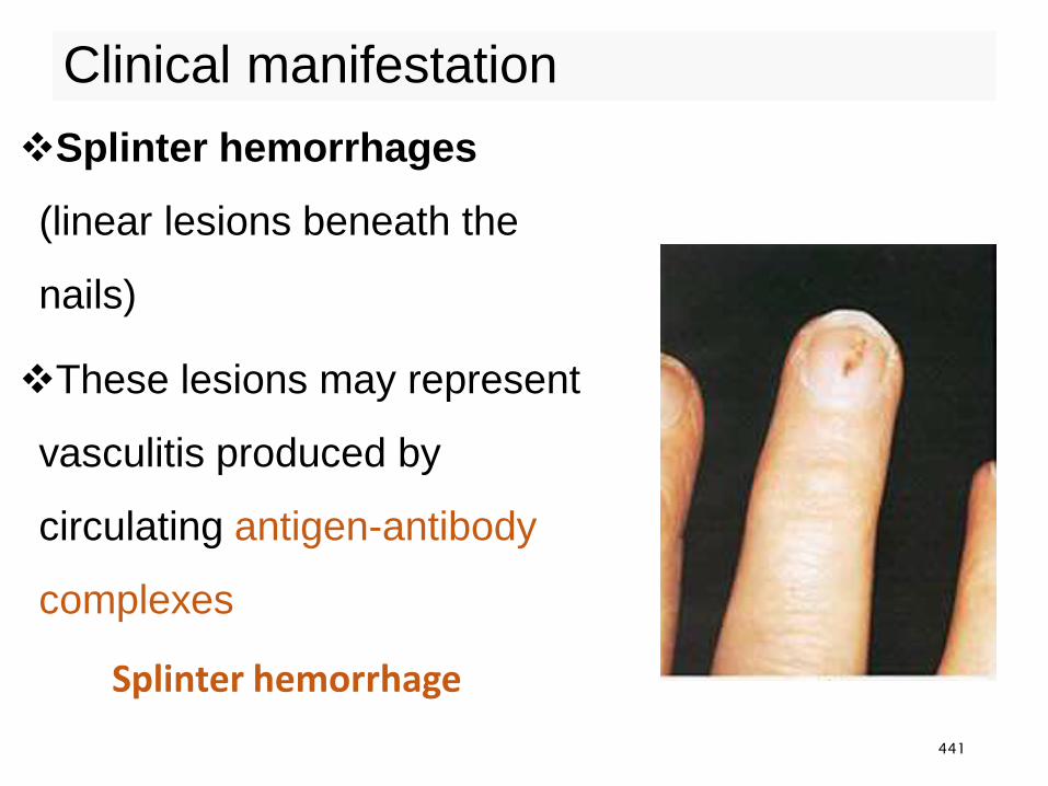
Clinical manifestation
Splinter hemorrhage
Splinter hemorrhages
(linear lesions beneath the
nails)
These lesions may represent
vasculitis produced by
circulating antigen-antibody
complexes
441
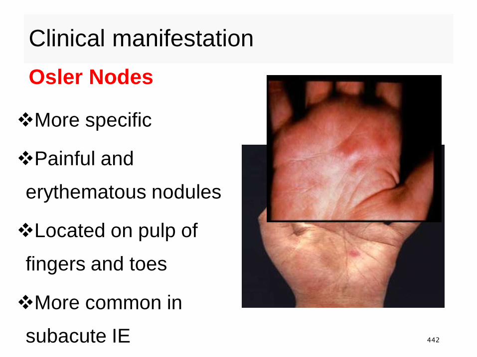
Clinical manifestation
Osler Nodes
More specific
Painful and
erythematous nodules
Located on pulp of
fingers and toes
More common in
subacute IE 442
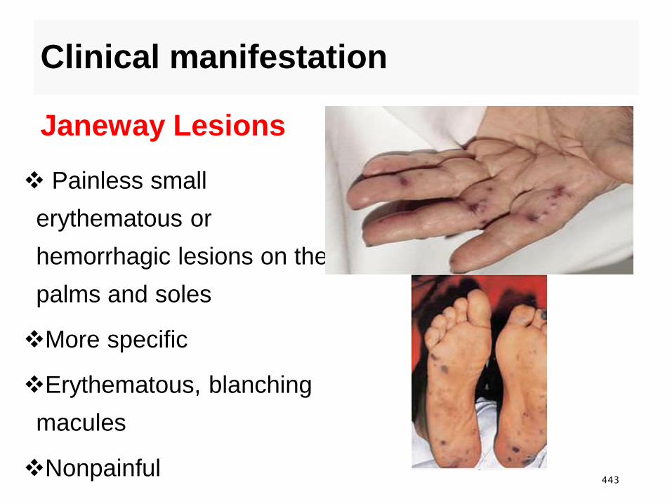
Clinical manifestation
Janeway Lesions
Painless small
erythematous or
hemorrhagic lesions on the
palms and soles
More specific
Erythematous, blanching
macules
Nonpainful443
Duke’s criteria of diagnosis IE
Major Criteria
1. Positive blood cultures :- (2 separate cultures for a
usual pathogen, 2 or more for less-typical
pathogens), and
2. Evidence of endocarditis on echocardiography
(intracardiac mass on a valve or other site,
regurgitant flow near prosthesis, abscess, partial
dehiscence of prosthetic valves, or new valve
regurgitant flow)444
Minor Criteria
Predisposing conditions
Fever ≥38oc
Embolic-vascular signs
Immune complex phenomena (glomerulonephritis,
arthritis, rheumatoid factor, Osler nodes, Roth spots)
A single, positive blood culture or
Serologic evidence of infection
Echocardiographic signs not meeting the major criteria 445
Clinical manifestation
•Common symptoms are:
•Low grade fever, chills, fatigue, arthralgia, myalgia,
malaise, night sweats, weight loss, stroke or seizure
•Physical findings:
•Tachycardia, arrhythmia
•New or changing murmur
•Splenomegaly
446
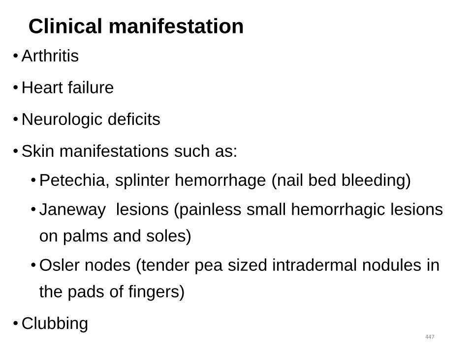
Clinical manifestation
• Arthritis
• Heart failure
• Neurologic deficits
• Skin manifestations such as:
• Petechia, splinter hemorrhage (nail bed bleeding)
• Janeway lesions (painless small hemorrhagic lesions
on palms and soles)
• Osler nodes (tender pea sized intradermal nodules in
the pads of fingers)
• Clubbing 447
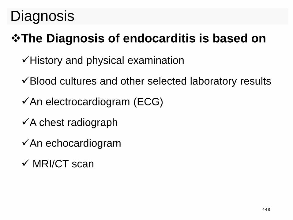
Diagnosis
The Diagnosis of endocarditis is based on
History and physical examination
Blood cultures and other selected laboratory results
An electrocardiogram (ECG)
A chest radiograph
An echocardiogram
MRI/CT scan
448
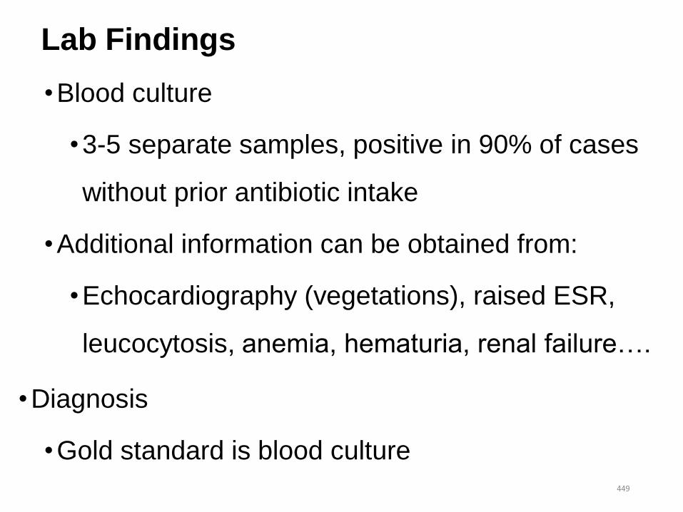
Lab Findings
•Blood culture
•3-5 separate samples, positive in 90% of cases
without prior antibiotic intake
•Additional information can be obtained from:
•Echocardiography (vegetations), raised ESR,
leucocytosis, anemia, hematuria, renal failure….
•Diagnosis
•Gold standard is blood culture
449
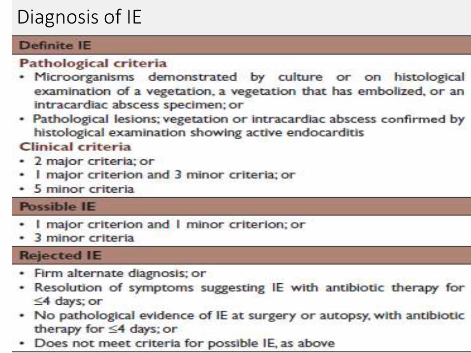
Diagnosis of IE
450

Medical management
Initial empiric antibiotic should be effective for both
Gram-positive and Gram negative organism combine
cloxacillin (100 mg /kg per 24 hours), ampicillin (300
mg/kg per 24 hr, IV) and gentamicine (3 to 4 mg / kg
per 24 hours IV)
Therapy should continue for 4 to 6 weeks (except
gentamicin for 2 weeks)
Adjustment of drugs can be done depending on blood
culture results 451
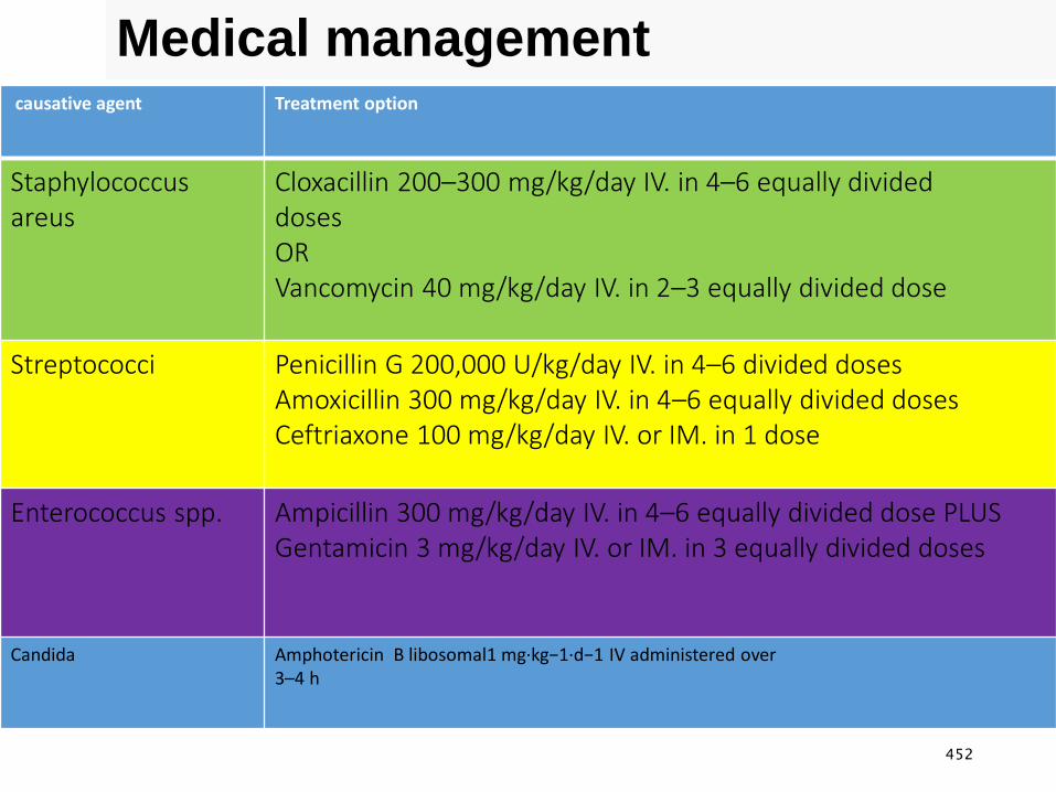
Medical managementcausative agent Treatment option
Staphylococcusareus
Cloxacillin 200–300 mg/kg/day IV. in 4–6 equally divideddoses ORVancomycin 40 mg/kg/day IV. in 2–3 equally divided dose
Streptococci Penicillin G 200,000 U/kg/day IV. in 4–6 divided dosesAmoxicillin 300 mg/kg/day IV. in 4–6 equally divided dosesCeftriaxone 100 mg/kg/day IV. or IM. in 1 dose
Enterococcus spp. Ampicillin 300 mg/kg/day IV. in 4–6 equally divided dose PLUSGentamicin 3 mg/kg/day IV. or IM. in 3 equally divided doses
Candida Amphotericin B libosomal1 mg·kg−1·d−1 IV administered over3–4 h
452
Prevention
•General dental care and oral hygiene
•Treatment of infection in high risk children
•Antibiotic prophylaxis before
•Surgical procedures of genito-urinary and gastro-
intestinal systems
453

Rheumatic Fever (RF)
• It is an auto immune inflammatory disease that occurs following
a Group A streptococcal infection
• It affects the joints, heart, skin and CNS
• RF is the leading cause of acquired heart disease
• Three percent of individuals with untreated group A
streptococcal pharyngitis confronted RF
• Epidemiologically is commonly seen in children 5-15 years of
age
• It is less likely to occur below 5 years and very rare during
infancy454
Etiology and pathogenesis
• Group A streptococcal (GAS) pharyngitis, but the bacteria
could not directly damage the organs, rather the antigen
antibody complex
• Risk Factors:
• Overcrowding
• Poor socioeconomic status
• Poor environmental hygiene
• Pathogenesis- Not completely understood
• Cytotoxicity theory
• Immunologic theory455

Clinical Features
•Major manifestations include:
•Migratory Polyarthritis- affects large joint and
migratory in nature, seen in 75% of patients, doesn't
result deformity and resolves with or without treatment
with in few days
•Carditis- inflammation of the heart muscle, has
tachycardia, murmur, pericardial friction rub, chest
pain, and gallop
•Seen in about 30% of cases 456
Clinical Features……
•Sydenham's Chorea- is characterized by jerky,
uncontrolled movement of the body, and series of
rapid movements without purpose of the face and
arms
•This occurs in less than 10% of cases
457

Clinical Features…..
•Erythematic marginatum
•Non pruritic macular lesions
which initially appear on the trunk
and inner aspects of extremities
with erythematous margins
458
Clinical Features……
•Subcutaneous Nodules- small (0.5-1cm
diameter) firm, non tender and with no skin
attachment nodules over the extensor surface
of joint
459
Minor manifestations:
•Low grade fever
•Weakness
•Fatigue
•Weight loss
•Epistaxis of unknown cause
•Abdominal pain
•Arthralgia
460
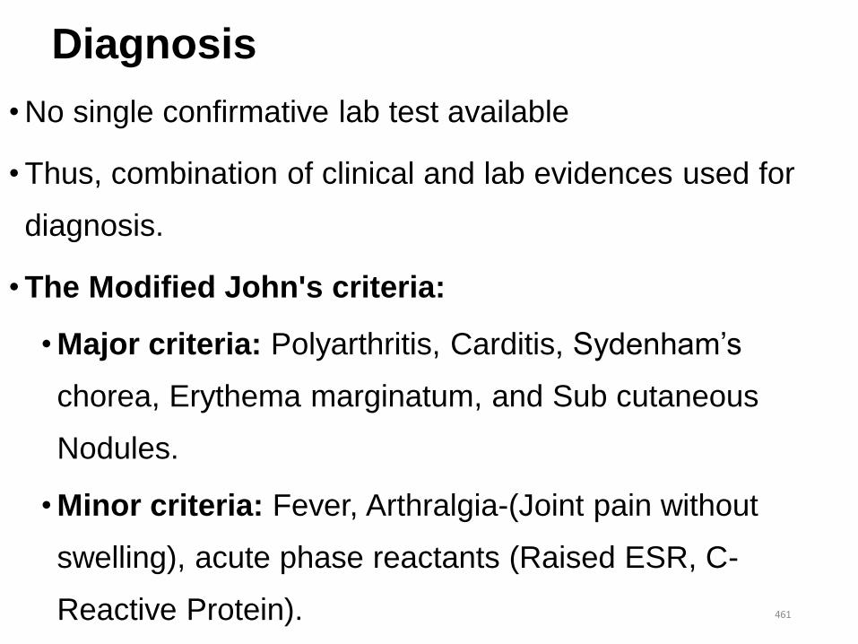
Diagnosis
• No single confirmative lab test available
• Thus, combination of clinical and lab evidences used for
diagnosis.
• The Modified John's criteria:
• Major criteria: Polyarthritis, Carditis, Sydenham’s
chorea, Erythema marginatum, and Sub cutaneous
Nodules.
• Minor criteria: Fever, Arthralgia-(Joint pain without
swelling), acute phase reactants (Raised ESR, C-
Reactive Protein). 461
DX
•Diagnosis made with:
• 2 major criteria or 1 major and 2 minor
+
• Supporting evidence for antecedent
• Streptococcal pharyngitis (mandatory)
462
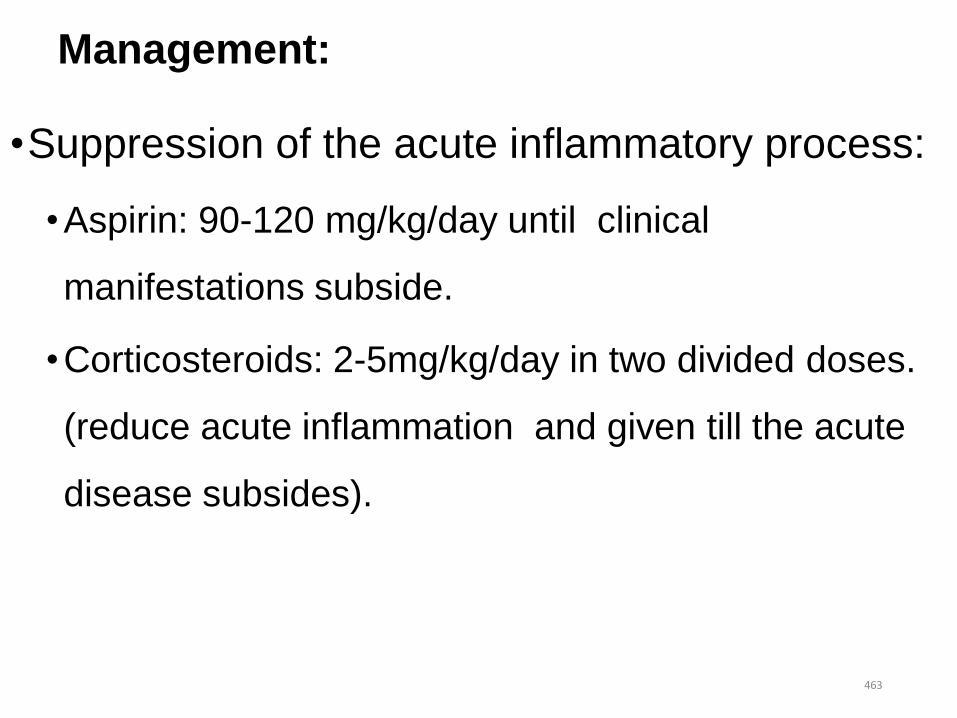
Management:
•Suppression of the acute inflammatory process:
•Aspirin: 90-120 mg/kg/day until clinical
manifestations subside.
•Corticosteroids: 2-5mg/kg/day in two divided doses.
(reduce acute inflammation and given till the acute
disease subsides).
463
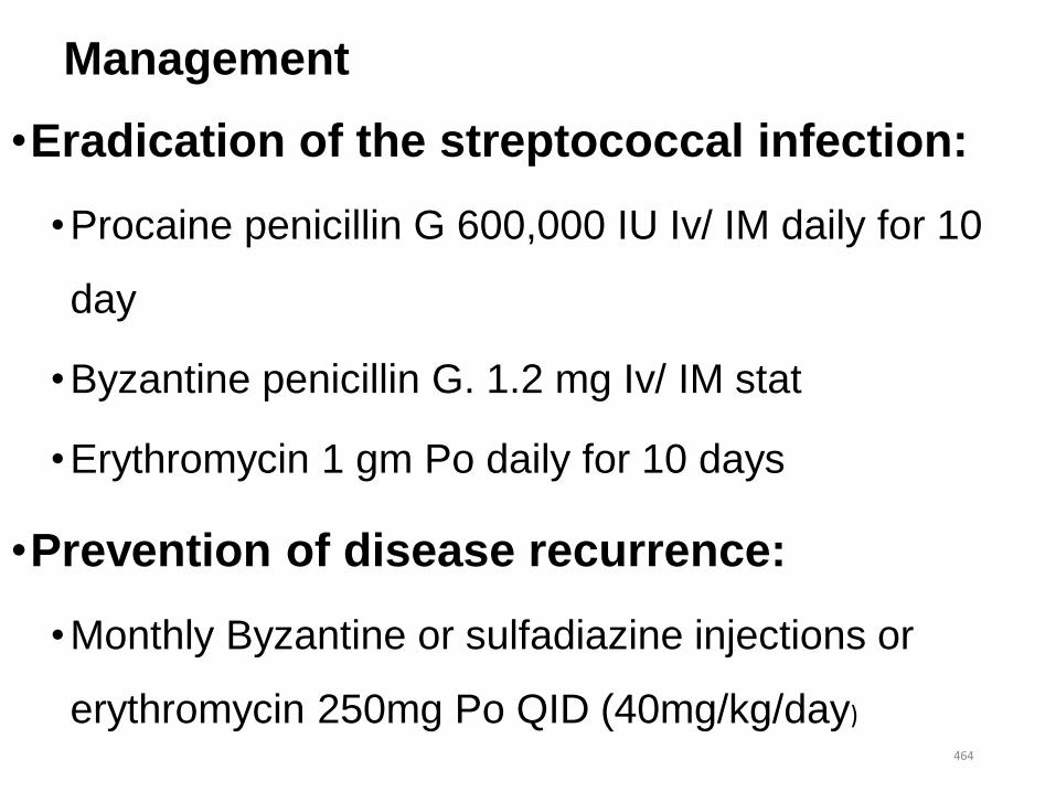
Management
•Eradication of the streptococcal infection:
•Procaine penicillin G 600,000 IU Iv/ IM daily for 10
day
•Byzantine penicillin G. 1.2 mg Iv/ IM stat
•Erythromycin 1 gm Po daily for 10 days
•Prevention of disease recurrence:
•Monthly Byzantine or sulfadiazine injections or
erythromycin 250mg Po QID (40mg/kg/day)
464
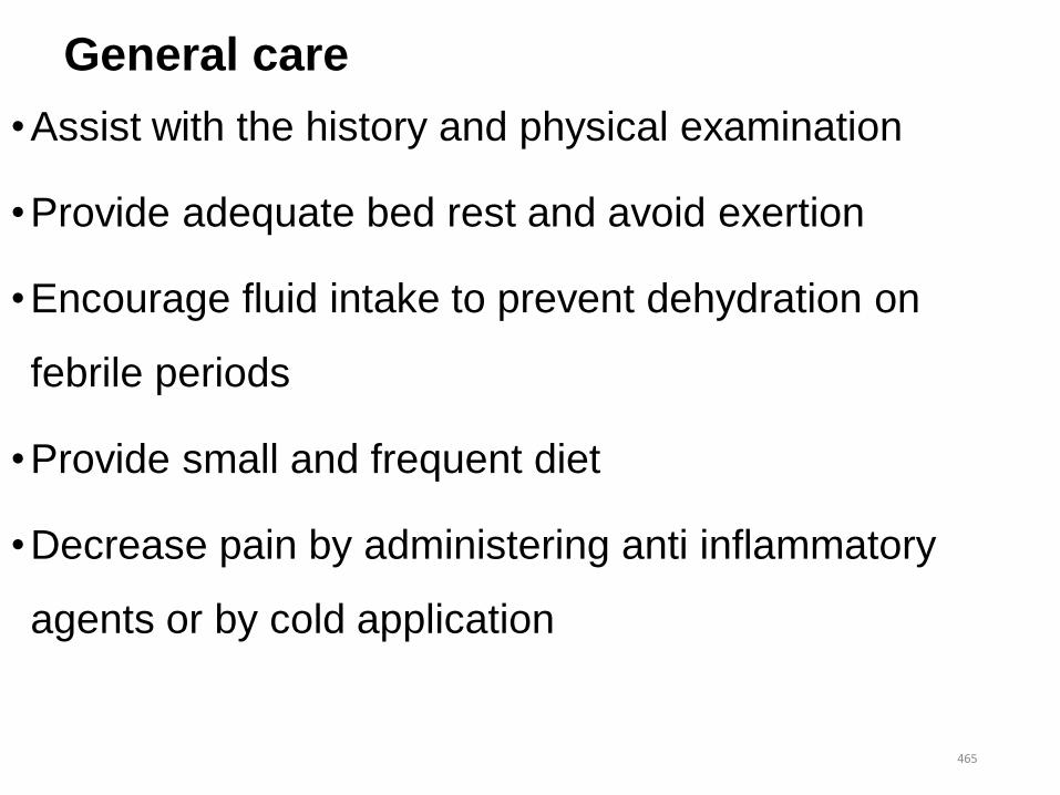
General care
•Assist with the history and physical examination
•Provide adequate bed rest and avoid exertion
•Encourage fluid intake to prevent dehydration on
febrile periods
•Provide small and frequent diet
•Decrease pain by administering anti inflammatory
agents or by cold application
465
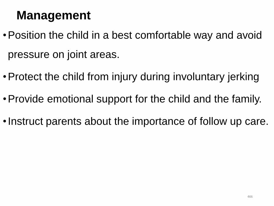
Management
•Position the child in a best comfortable way and avoid
pressure on joint areas.
•Protect the child from injury during involuntary jerking
•Provide emotional support for the child and the family.
• Instruct parents about the importance of follow up care.
466
Prevention
•Prompt antimicrobial treatment of streptococcal
pharyngitis
•Provision of prophylaxis for the prevention of
recurrent infections
467
Urinary Disorders
•Nephrotic Syndrome
•Wilms tumor
•UTI
468
Nephrotic Syndrome• It is the clinical manifestation of a large number of
glomerular disorders.
•The nephritic syndrome is characterized by :
•Proteinuria 40 mg/m 2/hr in children (>3.5mg/24hr
in adult)
•Hypoalbuminemia (<2.5g/dl)
•Hyperlipidemia
•Edema
469
Classification
•Nephritic syndrome classified as Primary and
Secondary
•Primary nephritic syndrome: associated with
primary Glomerular disease
•Secondary nephritic syndrome: when the nephritic
syndrome occurs as parts of systemic disease
•For 90% of cases the cause is unknown
470
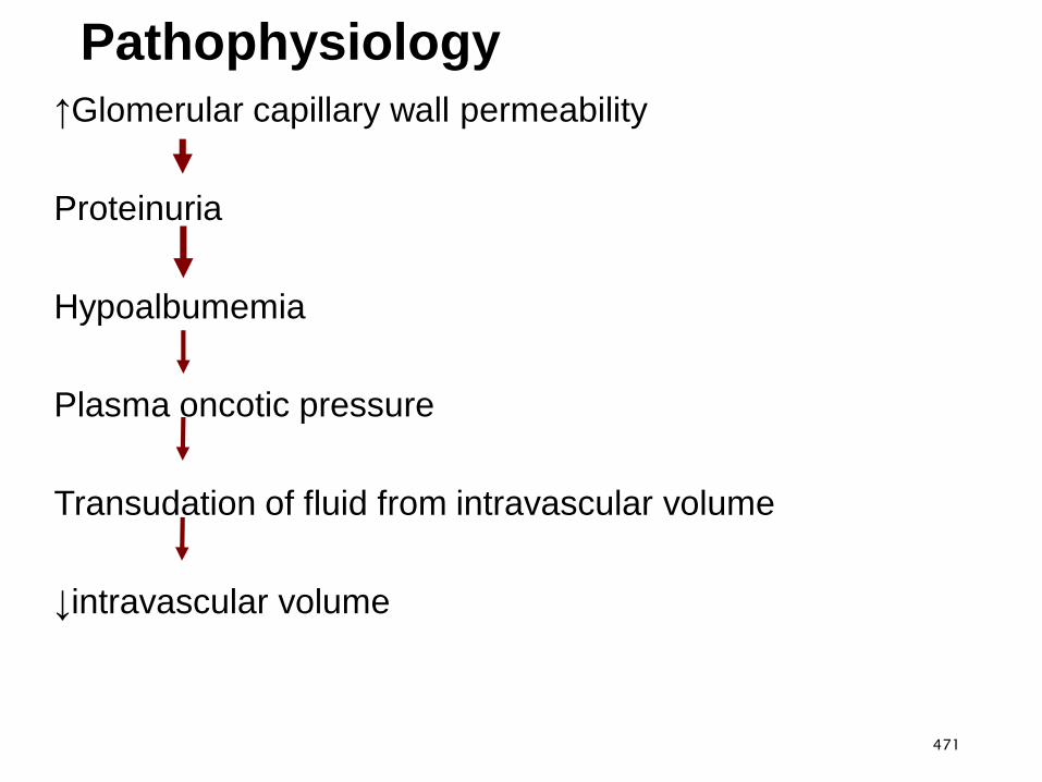
Pathophysiology
↑Glomerular capillary wall permeability
Proteinuria
Hypoalbumemia
Plasma oncotic pressure
Transudation of fluid from intravascular volume
↓intravascular volume
471
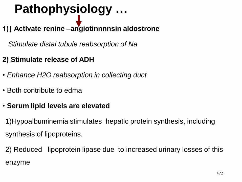
Pathophysiology …
1)↓ Activate renine –angiotinnnnsin aldostrone
Stimulate distal tubule reabsorption of Na
2) Stimulate release of ADH
• Enhance H2O reabsorption in collecting duct
• Both contribute to edma
• Serum lipid levels are elevated
1)Hypoalbuminemia stimulates hepatic protein synthesis, including
synthesis of lipoproteins.
2) Reduced lipoprotein lipase due to increased urinary losses of this
enzyme
472
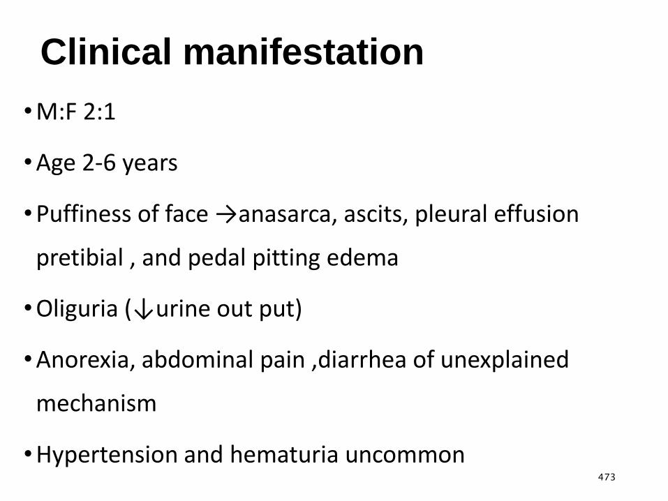
Clinical manifestation
• M:F 2:1
• Age 2-6 years
• Puffiness of face →anasarca, ascits, pleural effusion
pretibial , and pedal pitting edema
• Oliguria (↓urine out put)
• Anorexia, abdominal pain ,diarrhea of unexplained
mechanism
• Hypertension and hematuria uncommon473
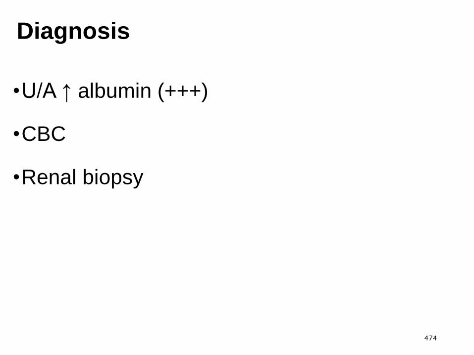
Diagnosis
•U/A ↑ albumin (+++)
•CBC
•Renal biopsy
474
Management
• The goal is reduction of protein excretion.
• Prednisone is the drug of choice
• For relapsing cases: Cyclophosphamide or Chloramphenicol
have been effective
• Encourage age appropriate diet rich in protein
• Diuretics
• Albumin transfusion
475
Management…
•Providing care during hospitalization
•Administering medications
•Maintaining proper fluid balance and assessing edema
•Providing emotional support and education for all family
members
476
Wilms tumor (Nephroblastoma)
• Wilms tumor, also known as nephroblastoma, is a complex
mixed embryonic neoplasm of the kidney
• Wilms tumor accounts for nearly 6% of all pediatric cancers and
more than 95% of all kidney tumors in children
• Survival for patients with Wilms tumor is generally excellent
around 90%
• Wilms tumor is the second most common intra-abdominal
cancer of childhood and the fifth most common pediatric
malignancy overall
477
Epidemiology
•Age 2-5 yrs (75%)
• Incidence ≈8/million children < 15 yrs
•Comprises≈ 6% of pediatric cancers
•Bilateral ≈ 7%
•Usually sporadic, but 1-2% of cases are familial
478
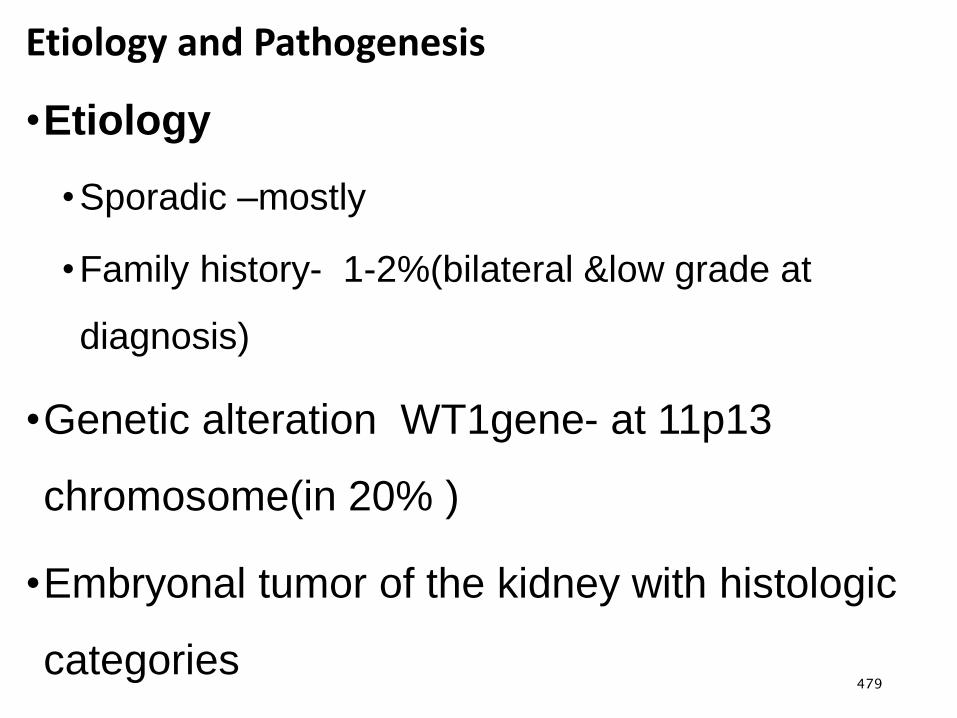
Etiology and Pathogenesis
•Etiology
•Sporadic –mostly
•Family history- 1-2%(bilateral &low grade at
diagnosis)
•Genetic alteration WT1gene- at 11p13
chromosome(in 20% )
•Embryonal tumor of the kidney with histologic
categories479
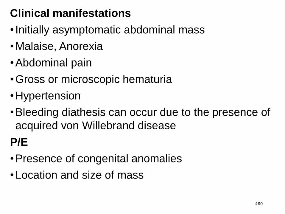
Clinical manifestations
• Initially asymptomatic abdominal mass
•Malaise, Anorexia
•Abdominal pain
•Gross or microscopic hematuria
•Hypertension
•Bleeding diathesis can occur due to the presence of
acquired von Willebrand disease
P/E
•Presence of congenital anomalies
•Location and size of mass
480
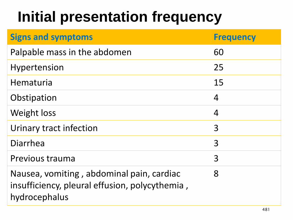
Initial presentation frequency
Signs and symptoms Frequency
Palpable mass in the abdomen 60
Hypertension 25
Hematuria 15
Obstipation 4
Weight loss 4
Urinary tract infection 3
Diarrhea 3
Previous trauma 3
Nausea, vomiting , abdominal pain, cardiac insufficiency, pleural effusion, polycythemia , hydrocephalus
8
481
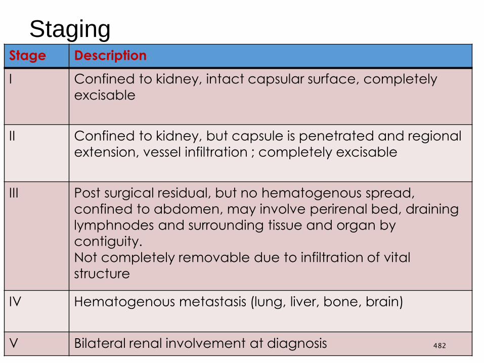
StagingStage Description
I Confined to kidney, intact capsular surface, completely
excisable
II Confined to kidney, but capsule is penetrated and regional
extension, vessel infiltration ; completely excisable
III Post surgical residual, but no hematogenous spread,
confined to abdomen, may involve perirenal bed, draining
lymphnodes and surrounding tissue and organ by
contiguity.
Not completely removable due to infiltration of vital
structure
IV Hematogenous metastasis (lung, liver, bone, brain)
V Bilateral renal involvement at diagnosis 482

Diagnosis
CBC- polycythemia, anemia, thrombocytopenia
U/A - Hematuria
Coagulation factors- PT,PTT, von Willebrand factor or
factor VII level
Ultrasound evaluation – origin, regional involvement
CT and or MRI
CXR- Pulmonary metastasis
Dx is tissue biopsy - Histologic variants
Staging – Post surgical intervention
483

Treatment
Pre-Operative chemotherapy
Nephrectomy
Chemotherapy
1. Stage I, II- Favorable histology
Vincristine + Dactinomycin
2. Stage III, IV
Vincristine
Dactinomycin
Doxorubicin
Radiation therapy 484
Prognosis
Poor prognosis
• Tumor size > 500gm
•Advanced stage ( III,IV)
•Anaplastic histology
485
Urinary tract infection (UTI)
•UTI- Infection of any component of the urinary tract
like urethritis, cystitis and pyelonephritis
•Bacteria first infect the urethra, then move to the
bladder and finally to the kidneys
•UTI tend to occur more in female than male
486
Etiology
•UTIs are caused mainly by colonic bacteria
• In girls 75-90% of all infections are caused by: E. coli,
Klebsiella spp and Proteus spp
•Others gram-positive organisms are S. saprophyticus
and enterococcus
•Adenovirus and other viral infections also can occur,
especially as a cause of cystitis
487
Risk factors
Structural anomalies
Voiding dysfunction
Uncircumcised male
Poor Hygiene
Neuropathic bladder
Female Gender
488

Pathogenesis
• The bacteria arise from the fecal flora, colonize the perineum, and
enter the bladder via the urethra
• In uncircumcised boys, the bacterial pathogens arise from the flora
beneath the prepuce
• In some cases, the bacteria causing cystitis ascend to the kidney to
cause pyelonephritis
• Rarely, renal infection occurs by hematogenous in neonates
• If bacteria ascend from the bladder to the kidney, acute pyelonephritis
can occur
489

Clinical Manifestations
Pyelonephritis is characterized by the following:
Abdominal, back, or flank pain;
Fever
Malaise
Nausea, Vomiting and, occasionally, diarrhea
Newborns can show non-specific symptoms such
as poor feeding, irritability, jaundice, and weight loss
490
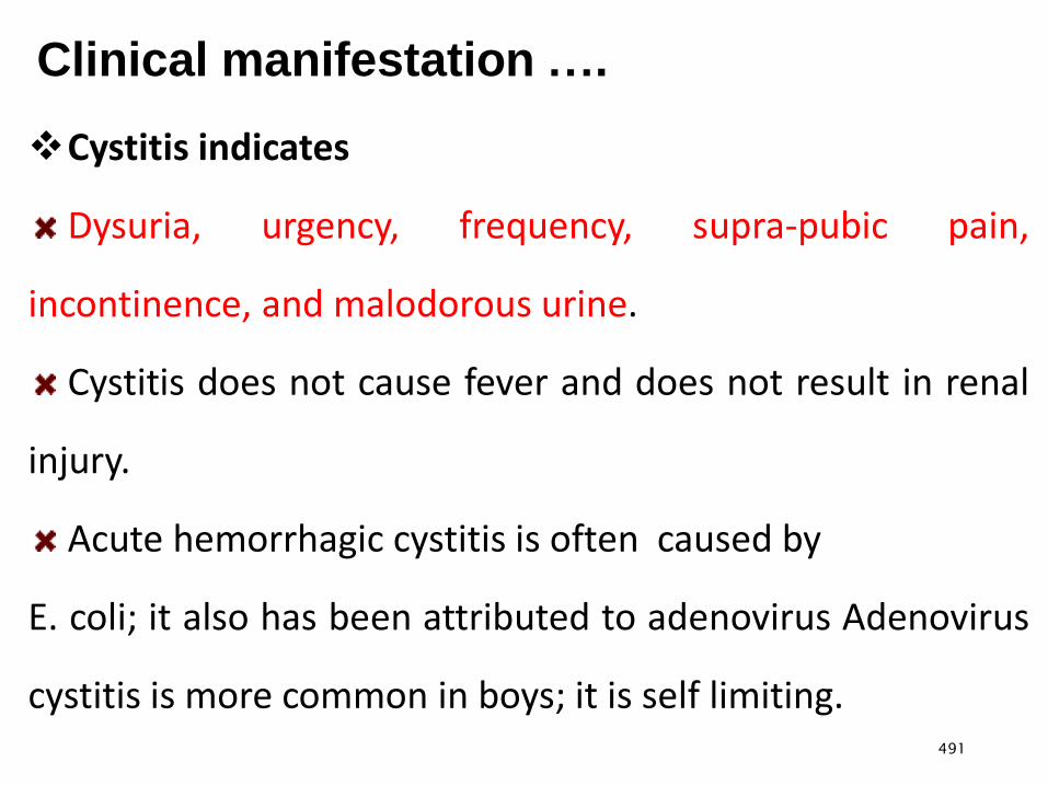
Clinical manifestation ….
Cystitis indicates
Dysuria, urgency, frequency, supra-pubic pain,
incontinence, and malodorous urine.
Cystitis does not cause fever and does not result in renal
injury.
Acute hemorrhagic cystitis is often caused by
E. coli; it also has been attributed to adenovirus Adenovirus
cystitis is more common in boys; it is self limiting.491
Clinical manifestation ….
•Urethritis refers to a condition in which there is a
positive urine culture without any manifestations of
infection; most common in girls.
•The incidence declines with increasing age.
•Some girls are mistakenly identified as having
asymptomatic bacteriuria, whereas they actually are
experiencing day or night incontinence or perineal
discomfort secondary to UTI
492
Diagnosis
Sign and Symptom
Chemical methods & microscopic (urinalysis)
Culture& sensitivity
493

Treatment
•Choose narrowest spectrum considering host factors
•Adjust therapy when sensitivities available
•Empiric therapy is directed to organism and adjusted
for age
•Ampicillin plus a second antibiotic (usually
Gentamycin or Cefotaxime) to cover for GBS,
Listeria, as well as gram negative organisms
•Vancomycin may be indicated for toxic patients
494
Prevention
If untreated can leads to end stage renal failure.
Prevention
Good hygiene practice
Early treatment
Health education
495

Neurologic Disorders
•Meningitis – inflammation of the meninges
Encephalitis – infection of the brain parenchyma
Meningoencephalitis – inflammation of brain +
meninges
Aseptic meningitis – inflammation of meninges
with sterile CSF
496
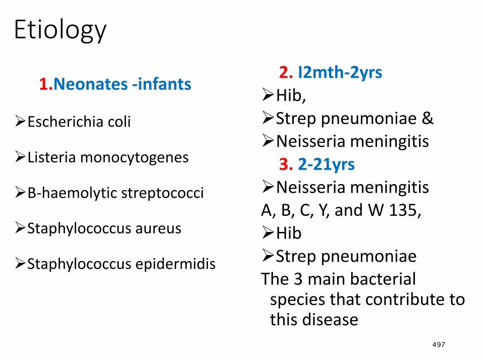
Etiology
1.Neonates -infants
Escherichia coli
Listeria monocytogenes
B-haemolytic streptococci
Staphylococcus aureus
Staphylococcus epidermidis
2. I2mth-2yrsHib, Strep pneumoniae & Neisseria meningitis
3. 2-21yrs Neisseria meningitis A, B, C, Y, and W 135,Hib Strep pneumoniaeThe 3 main bacterial
species that contribute to this disease
497

Pathophysiology• The causative organism enters the bloodstream, crosses the
blood–brain barrier, and triggers an inflammatory reaction in the
meninges
• Independent of the causative agent, inflammation of the
subarachnoid and pia mater occurs
• Increased intracranial pressure (ICP) results
• Meningeal infections generally originate in one of two ways:
either through the bloodstream from other infections (cellulitis) or
by direct extension (after a traumatic injury to the facial bones)
498
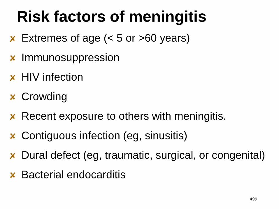
Risk factors of meningitis
Extremes of age (< 5 or >60 years)
Immunosuppression
HIV infection
Crowding
Recent exposure to others with meningitis.
Contiguous infection (eg, sinusitis)
Dural defect (eg, traumatic, surgical, or congenital)
Bacterial endocarditis
499
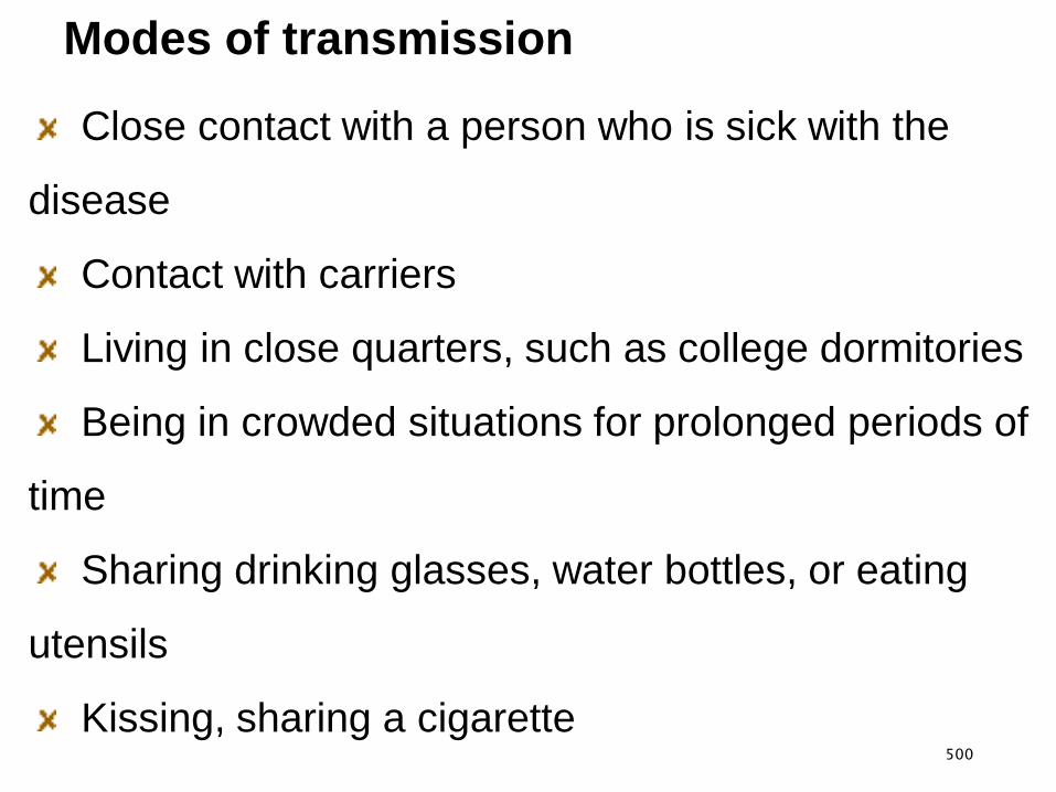
Modes of transmission
Close contact with a person who is sick with the
disease
Contact with carriers
Living in close quarters, such as college dormitories
Being in crowded situations for prolonged periods of
time
Sharing drinking glasses, water bottles, or eating
utensils
Kissing, sharing a cigarette500
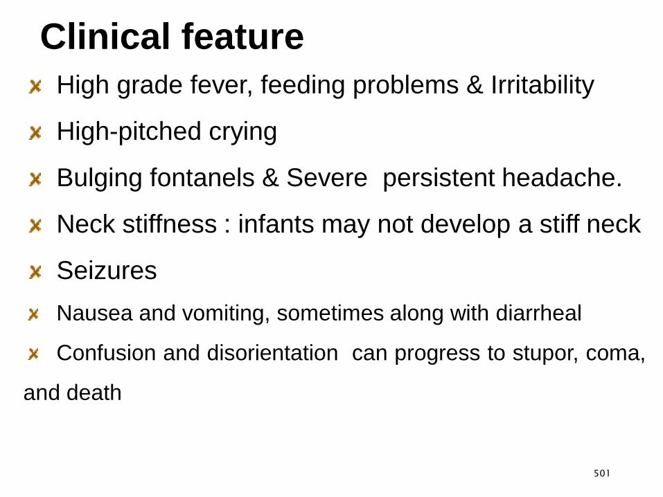
Clinical feature
High grade fever, feeding problems & Irritability
High-pitched crying
Bulging fontanels & Severe persistent headache.
Neck stiffness : infants may not develop a stiff neck
Seizures
Nausea and vomiting, sometimes along with diarrheal
Confusion and disorientation can progress to stupor, coma,
and death
501

Clinical features…
Drowsiness or sluggishness
Eye pain or sensitivity to bright light
Numbness and tingling
Nuchal rigidity or neck stiffness
502
Clinical features…
Increased intracranial pressure
Headache
Projectile vomiting
Hypertension
Bulging fontanel
Cranial sutures diastasis/separation
Coma
Cerebral hernia503
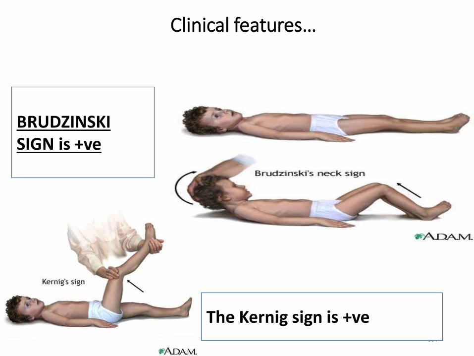
Clinical features…
504
The Kernig sign is +ve
BRUDZINSKI SIGN is +ve
Diagnosis
CM
CSF analysis Blood test
Chest X-ray
CT scan or MRI
Cultures of samples of CSF, blood, urine, mucus from
the nose and throat, and pus from skin infections.
505
Management
•Empiric treatment should be begun as soon as the
diagnosis is suspected using bactericidal agent(s) that
achieve significant levels in the CSF
Ceftriaxone 50-100 mg/kg/day IV/IM q12 hr
Vancomycin 60 mg/kg/day IV q6h.
Convulsive management
Diazepam
Phenobarbital
506
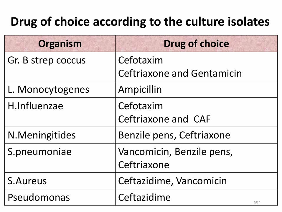
507
Organism Drug of choice
Gr. B strep coccus CefotaximCeftriaxone and Gentamicin
L. Monocytogenes Ampicillin
H.Influenzae CefotaximCeftriaxone and CAF
N.Meningitides Benzile pens, Ceftriaxone
S.pneumoniae Vancomicin, Benzile pens, Ceftriaxone
S.Aureus Ceftazidime, Vancomicin
Pseudomonas Ceftazidime
Drug of choice according to the culture isolates
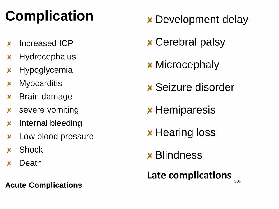
Complication
Acute Complications
Increased ICP
Hydrocephalus
Hypoglycemia
Myocarditis
Brain damage
severe vomiting
Internal bleeding
Low blood pressure
Shock
Death
Late complications
Development delay
Cerebral palsy
Microcephaly
Seizure disorder
Hemiparesis
Hearing loss
Blindness
508

Epilepsy in children
Seizure
•An episode of altered behavior or awareness
•Associated with too much excitation of a population of
nerve cells (neurons)
Epilepsy
•The tendency to have recurrent, unprovoked seizures
(brain makes seizures happen)
509
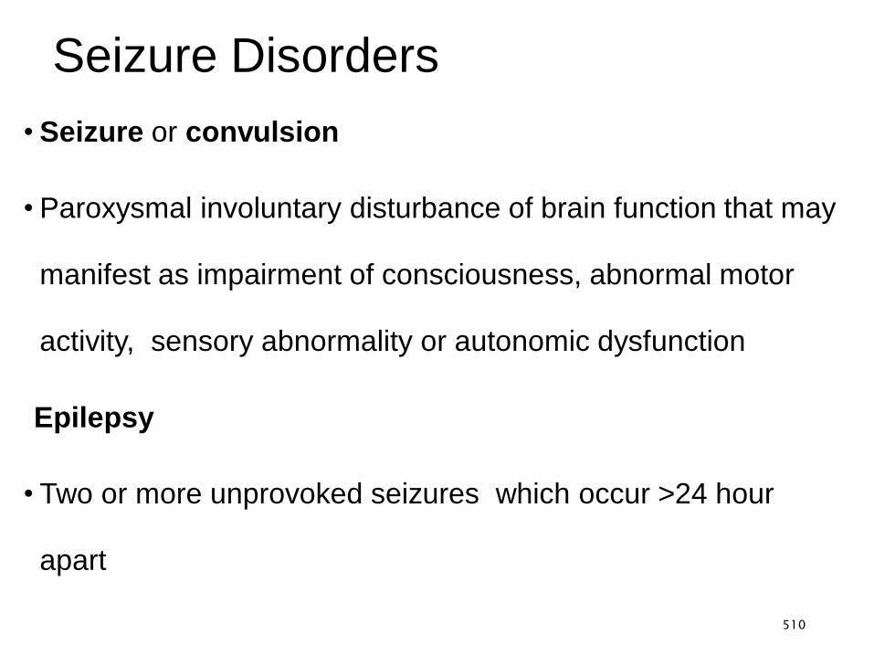
Seizure Disorders
• Seizure or convulsion
• Paroxysmal involuntary disturbance of brain function that may
manifest as impairment of consciousness, abnormal motor
activity, sensory abnormality or autonomic dysfunction
Epilepsy
• Two or more unprovoked seizures which occur >24 hour
apart
510
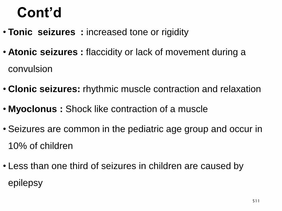
Cont’d
• Tonic seizures : increased tone or rigidity
• Atonic seizures : flaccidity or lack of movement during a
convulsion
• Clonic seizures: rhythmic muscle contraction and relaxation
• Myoclonus : Shock like contraction of a muscle
• Seizures are common in the pediatric age group and occur in
10% of children
• Less than one third of seizures in children are caused by
epilepsy
511
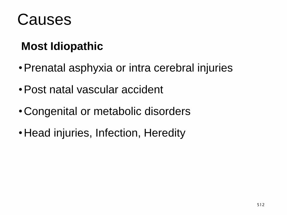
Causes
Most Idiopathic
•Prenatal asphyxia or intra cerebral injuries
•Post natal vascular accident
•Congenital or metabolic disorders
•Head injuries, Infection, Heredity
512
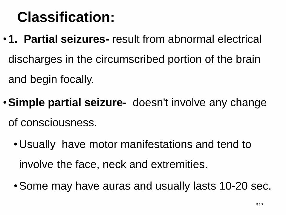
Classification:
•1. Partial seizures- result from abnormal electrical
discharges in the circumscribed portion of the brain
and begin focally.
•Simple partial seizure- doesn't involve any change
of consciousness.
•Usually have motor manifestations and tend to
involve the face, neck and extremities.
•Some may have auras and usually lasts 10-20 sec.
513
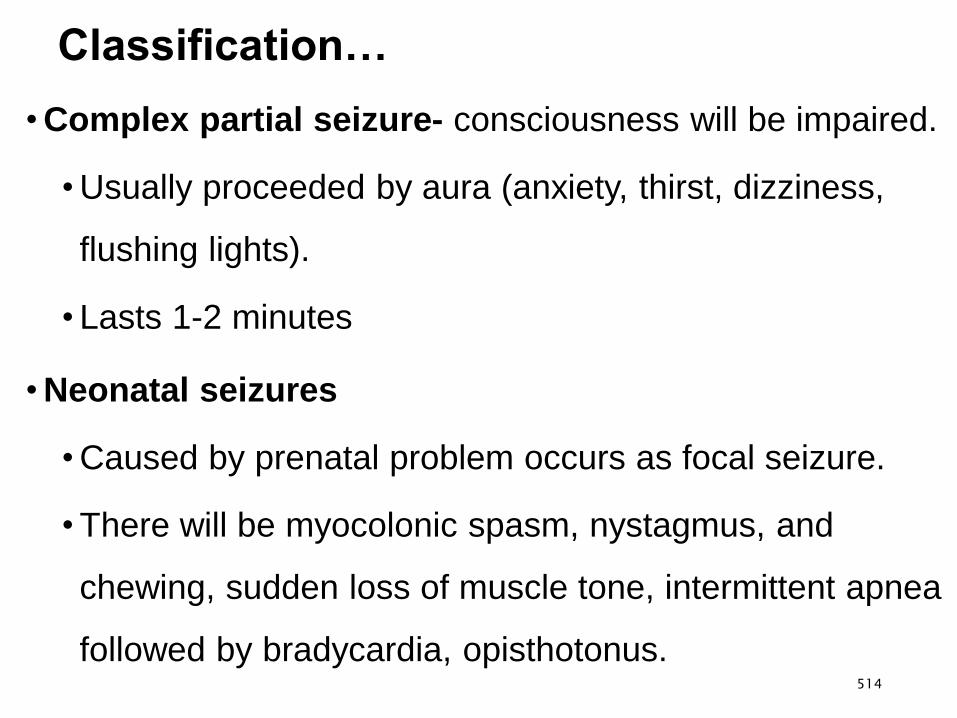
Classification…
• Complex partial seizure- consciousness will be impaired.
• Usually proceeded by aura (anxiety, thirst, dizziness,
flushing lights).
• Lasts 1-2 minutes
• Neonatal seizures
• Caused by prenatal problem occurs as focal seizure.
• There will be myocolonic spasm, nystagmus, and
chewing, sudden loss of muscle tone, intermittent apnea
followed by bradycardia, opisthotonus. 514
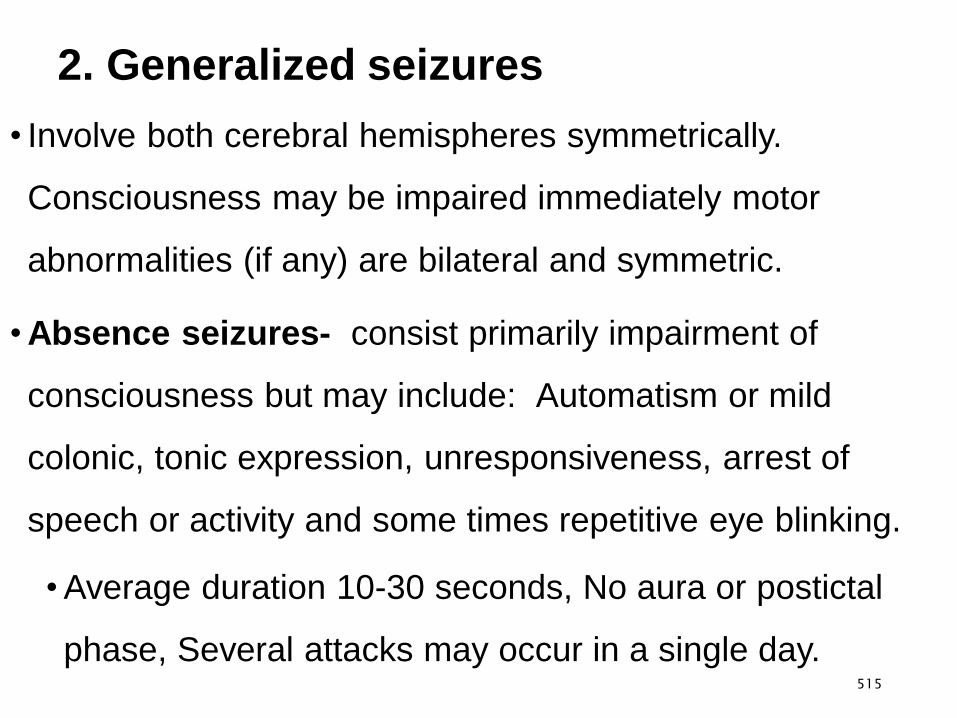
2. Generalized seizures
• Involve both cerebral hemispheres symmetrically.
Consciousness may be impaired immediately motor
abnormalities (if any) are bilateral and symmetric.
• Absence seizures- consist primarily impairment of
consciousness but may include: Automatism or mild
colonic, tonic expression, unresponsiveness, arrest of
speech or activity and some times repetitive eye blinking.
• Average duration 10-30 seconds, No aura or postictal
phase, Several attacks may occur in a single day.515
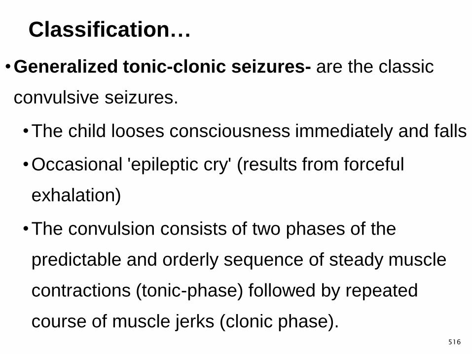
Classification…
•Generalized tonic-clonic seizures- are the classic
convulsive seizures.
•The child looses consciousness immediately and falls
•Occasional 'epileptic cry' (results from forceful
exhalation)
•The convulsion consists of two phases of the
predictable and orderly sequence of steady muscle
contractions (tonic-phase) followed by repeated
course of muscle jerks (clonic phase). 516
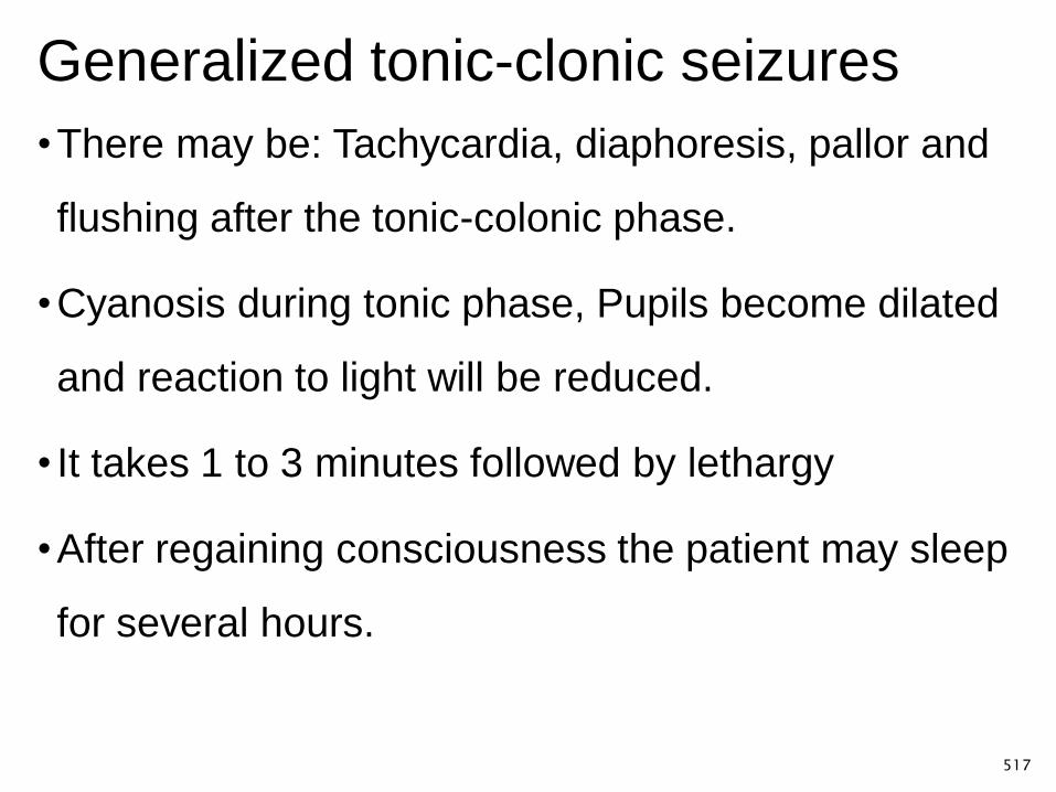
Generalized tonic-clonic seizures
•There may be: Tachycardia, diaphoresis, pallor and
flushing after the tonic-colonic phase.
•Cyanosis during tonic phase, Pupils become dilated
and reaction to light will be reduced.
• It takes 1 to 3 minutes followed by lethargy
•After regaining consciousness the patient may sleep
for several hours.
517
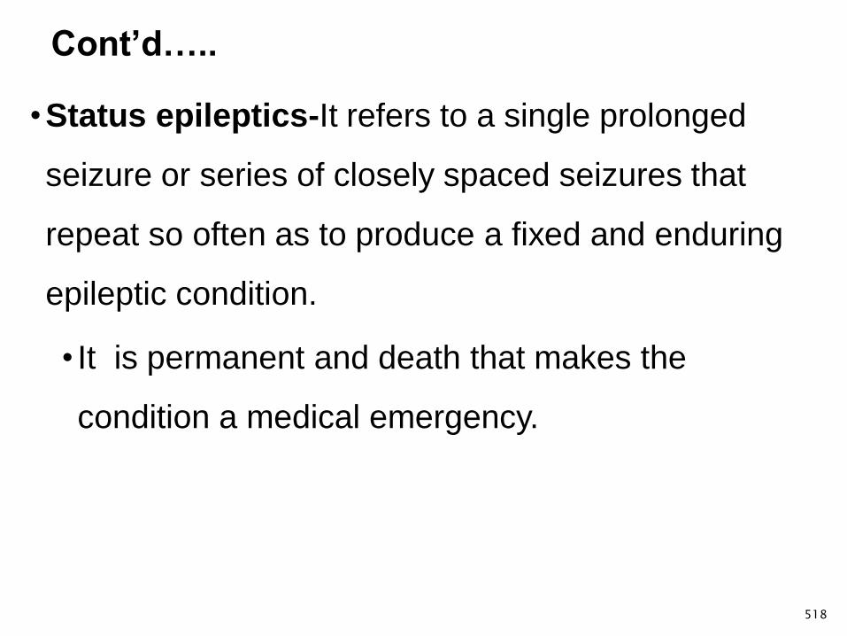
Cont’d…..
•Status epileptics-It refers to a single prolonged
seizure or series of closely spaced seizures that
repeat so often as to produce a fixed and enduring
epileptic condition.
• It is permanent and death that makes the
condition a medical emergency.
518

Treatment of Epilepsy
•Treatment is started
•Risk of seizure recurrence increases after each
seizure
•Consider treatment after 2 seizures
•Treatment for seizures
•Antiepileptic medications (AED)
•Work up to determine cause of seizure activity
519
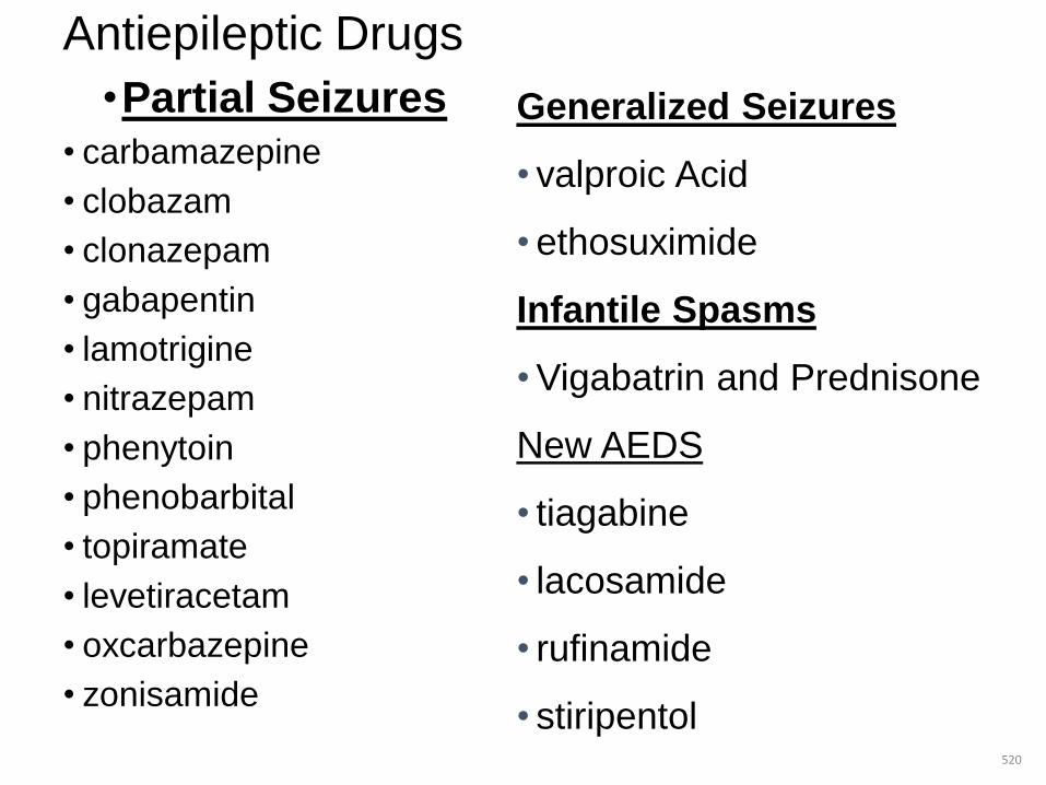
Antiepileptic Drugs
•Partial Seizures
• carbamazepine
• clobazam
• clonazepam
• gabapentin
• lamotrigine
• nitrazepam
• phenytoin
• phenobarbital
• topiramate
• levetiracetam
• oxcarbazepine
• zonisamide
520
Generalized Seizures
• valproic Acid
• ethosuximide
Infantile Spasms
• Vigabatrin and Prednisone
New AEDS
• tiagabine
• lacosamide
• rufinamide
• stiripentol
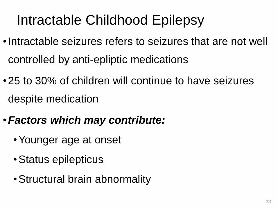
Intractable Childhood Epilepsy
• Intractable seizures refers to seizures that are not well
controlled by anti-epliptic medications
•25 to 30% of children will continue to have seizures
despite medication
•Factors which may contribute:
•Younger age at onset
•Status epilepticus
•Structural brain abnormality
521
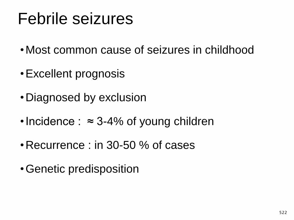
Febrile seizures
•Most common cause of seizures in childhood
•Excellent prognosis
•Diagnosed by exclusion
• Incidence : ≈ 3-4% of young children
•Recurrence : in 30-50 % of cases
•Genetic predisposition
522
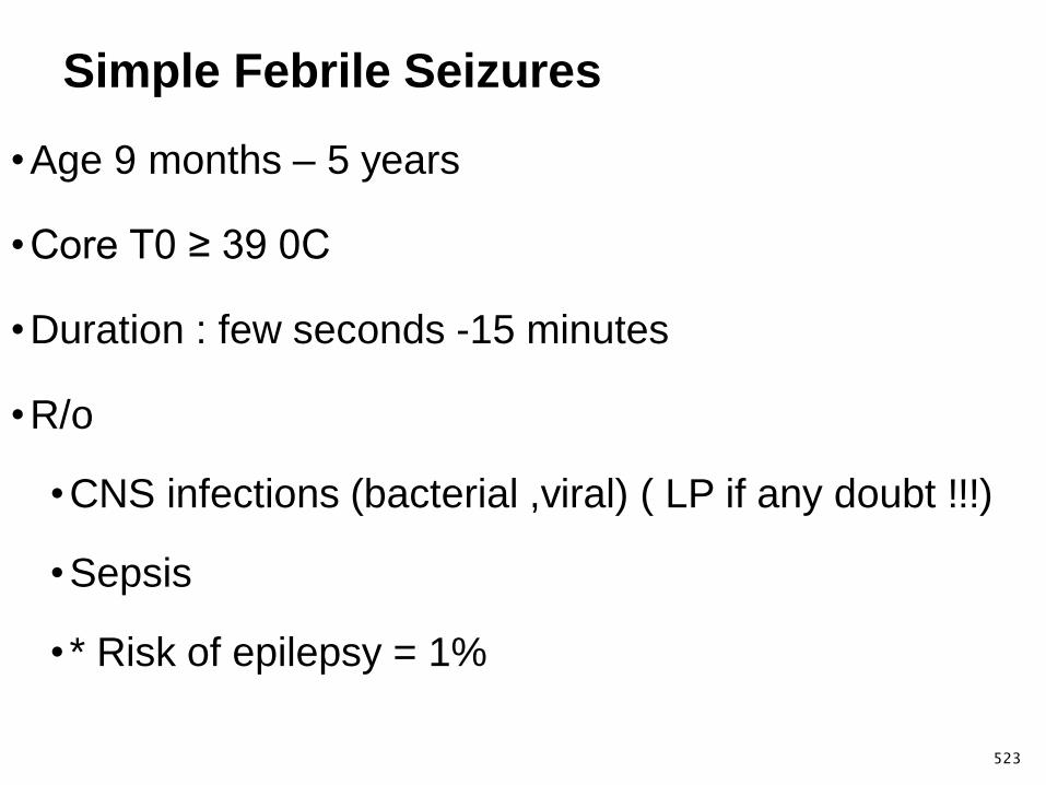
Simple Febrile Seizures
•Age 9 months – 5 years
•Core T0 ≥ 39 0C
•Duration : few seconds -15 minutes
•R/o
•CNS infections (bacterial ,viral) ( LP if any doubt !!!)
•Sepsis
• * Risk of epilepsy = 1%
523
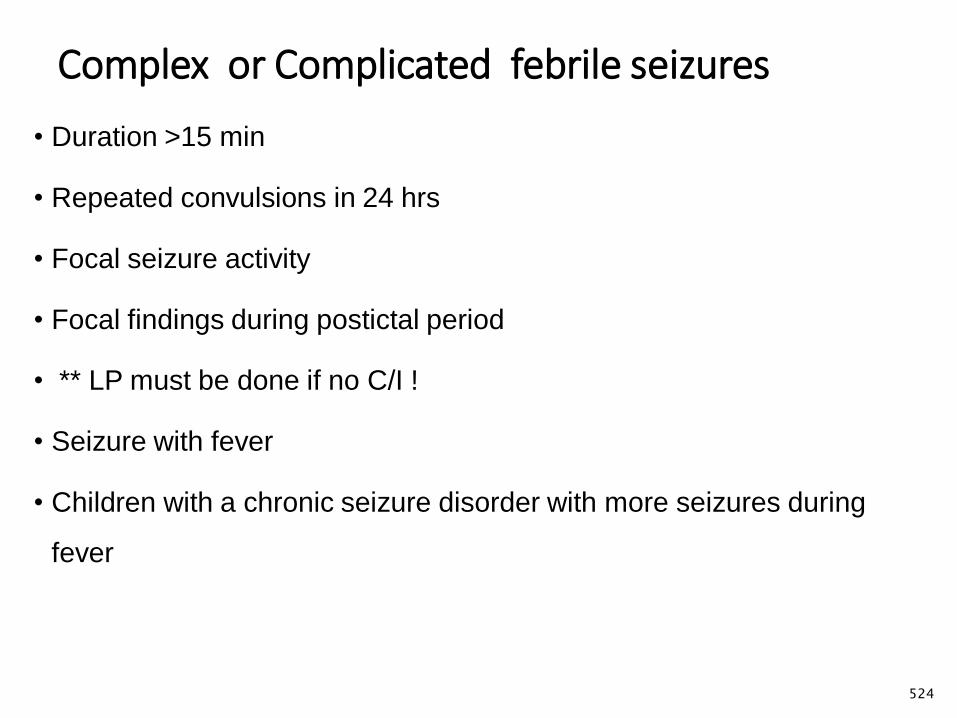
Complex or Complicated febrile seizures
• Duration >15 min
• Repeated convulsions in 24 hrs
• Focal seizure activity
• Focal findings during postictal period
• ** LP must be done if no C/I !
• Seizure with fever
• Children with a chronic seizure disorder with more seizures during
fever
524
Treatment
•Search for cause of fever
•Reassurance & education of parents
•Control fever
•Antipyretics : not shown to prevent seizure
recurrence
•Long term anticonvulsants not indicated !!
•Diazepam oral 0.3mg/kg (1mg/kg/24hr) Q 8 hourly for
duration of illness(2-3 days)
525
Seizures: Nursing Considerations
• ABC’s
• Is the patient actively seizing
• Secure an airway
• Apply oxygen or prepare to intubate if prolonged
seizure
• Assess circulation and establish IV access
• Is the patient postictal
• Support ABC’s and monitor immediately
• Reassure parents
526
Use of Oxygen During a Seizure
• For short term seizures
• No evidence oxygen by mask is effective during short
seizure
• Contraction of airway inhibits oxygen intake by mask
• Short periods of apnea (up to 45 seconds) during the tonic
phase is common.
• Blood flow reduced at the peripheries and increased to the
vital organs (200% to brain)
• Oxygen is rarely used at home for seizures
527
Nursing Management
• Emergency Care: Assist the child to a lying position; if
possible place something soft under the child head.
• Remove dangerous objects from the area and loosen tight
clothing, do not force anything into the Childs mouth .
• Allow the seizures to run its course.
• When the child relived from the seizures attack, turn him
to one side to help saliva drain from the mouth
• Treat any injuries that may have occurred during the
convulsion. 528
Cont’d
•Hospital Care Keep bed at lowest position with rails
up, Pad the side rails, pharyngeal suction, oxygen.
•Assist with diagnostic procedures, Administrator
medications appropriately and Provide emotional
support.
529
Anemia
•Anemia is defined as a reduction of the hemoglobin
concentration or red blood cell (RBC) volume below
the range of values occurring in healthy persons.
•Anemia can not be considered a specific disease
entity; it is an indication of an underlying pathologic
process or disease.
530
Classification
•Anemia can be classified based on their cause or
morphologic appearance of RBCs.
•A. Physiologic classification (based on cause)
•Sickle cell anemia: Excessive RBC destruction
•Nutritional anemia: Reduced capacity to produce
RBCs
531
B. Morphologic classification
•Microcytic e.g. Iron deficiency anemia
•Normocytic e.g. Blood loss anemia
•Macrocytic e.g. Megaloblastic anemia (Vit.B12
deficiency)
532

Pathophysiology
•When circulating hemoglobin is reduced, the
oxygen carrying capacity of the blood becomes
reduced
•During the falling of hemoglobin to below 7 to 8
g/dl , the heart tries to compensate by
increasing the rate and cardiac out put.
533
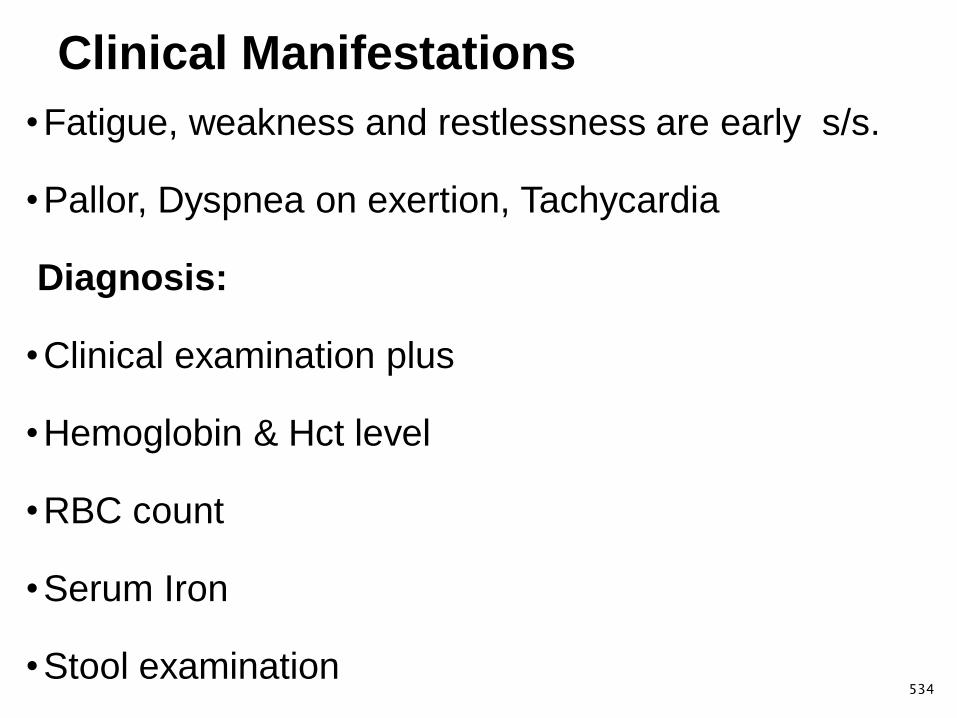
Clinical Manifestations
•Fatigue, weakness and restlessness are early s/s.
•Pallor, Dyspnea on exertion, Tachycardia
Diagnosis:
•Clinical examination plus
•Hemoglobin & Hct level
•RBC count
•Serum Iron
•Stool examination534
Management
•Depends on their cause
•Medical Management:
•Blood transfusion,
•O2 therapy,
•Specific therapy
535
General care
• Obtain a thorough history and physical examination
• Prepare the child for laboratory examination.
• Reduce oxygen demand by provision of adequate rest
• Monitor vital signs
• Administer transfusion therapy
• During blood transfusion observe for signs of reaction and as
much as possible try to prevent them
• Observe for signs of infections and complication
536
Care during transfusion of blood
•Monitor V/S before, during and after procedure.
•Do blood grouping and cross match carefully.
•Administer blood as soon as possible after it arrives
from blood bank.
• Inspect the blood for unusual color.
•Mix the blood gently but thoroughly before and during
transfusion to prevent settling of RBC.
537
Cont’d
•Remain with the child during transfusion in order to
detect reactions as early as possible.
•Complete transfusion within four hour to prevent
bacterial proliferation.
•Stop transfusion immediately if the onset of reaction
is suspected and run normal saline to keep patency
of the intravenous line.
538

Iron Deficiency Anemia
• Anemia resulting from lack of sufficient iron for synthesis of
hemoglobin is the most common hematologic disease of infancy
and childhood.
• It is more common between ages of 6 month-2 years.
• To maintain positive iron balance in childhood, about 1 mg of
iron must be absorbed each day.
• The body of a newborn infant contains about 0.5 g of iron,
whereas the adult contains 5 g.
539
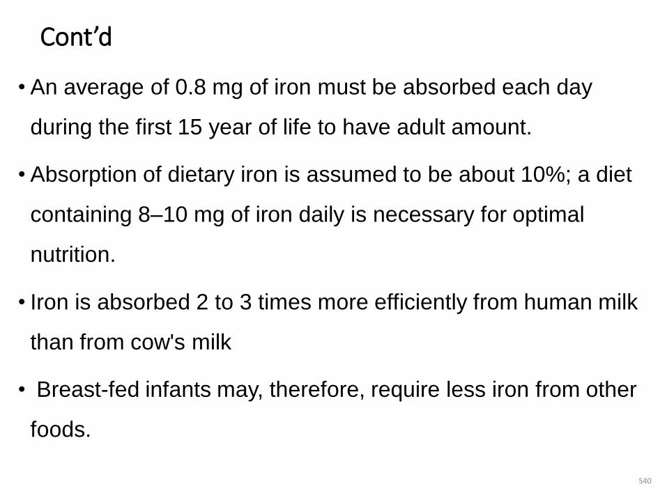
Cont’d
• An average of 0.8 mg of iron must be absorbed each day
during the first 15 year of life to have adult amount.
• Absorption of dietary iron is assumed to be about 10%; a diet
containing 8–10 mg of iron daily is necessary for optimal
nutrition.
• Iron is absorbed 2 to 3 times more efficiently from human milk
than from cow's milk
• Breast-fed infants may, therefore, require less iron from other
foods.
540
Etiology
• Insufficient iron supply at birth
• Insufficient intake of iron during periods of rapid
growth
• Impaired iron absorption
•Blood loss
541

Pathophysiology
• Iron deficiency anemia occurs over a period of time.
•Low Fe supply -> Depletion of stores -> low serum
transferring level ->low hemoglobin Synthesis
•Low Hgb Synthesis-Microcytic RBCs, Less filled with
Hgb and Reduced o2 carrying capacity of blood
542
Clinical Feature
•Pallor- palmar, nail bed and conjunctiva
•Tachycardia, systolic murmurs are often present
•Pica or Pagophagia, the desire to ingest unusual
substances such as ice or dirt, may be present.
•Affected growth, motor development, intellectual and
neuralgic functioning especially attention span,
alertness and level of learning.
543
Clinical Feature…
•When the hemoglobin level falls to <5 g/dL, irritability
and anorexia are prominent.
• In mild to moderate iron deficiency (i.e., hemoglobin
levels of 6–10 g/dL), compensatory mechanisms,
may be so effective that few symptoms of anemia are
noted, although affected children may be irritable.
544
Diagnosis
•History ( dietary history) and physical examination
•Laboratory
•Hct and Hgb level reduced (<33% and <11gm/dl
respectively).
•Low serum iron
•Stool-for the presence of parasite (hook worm)
545
Management
•Oral iron supplement 6mg/kg/day in 3 divided dose
for 3 to 4 month.
•Parenteral iron therapy 50mg/day of iron dextran IM
•Blood transfusion If the Hgb level is < 4g/dl, or, if
superimposed infection is interfering with response
to treatment.
546
Leukemia
•These are diseases that are characterized by
abnormal proliferation and maturation of the
bone marrow interfering with the production of
normal blood cells.
•They are the most common malignancies of
<15 years children with the peak age of 4
years.
547

Causes
•The exact cause of leukemia is unknown
•Several etiologic factors including
•Viruses
•Radiation
•Exposure to drugs & chemicals
•Familial predisposition
548
Classification
•Acute lymphocytic leukemia (ALL) It affects the
lymphoid cell line and believed to be curable with
chemotherapy and radiation. accounts about 80% of
child hood leukemia.
•Acute myeloid leukemia (AML)-There is
generalized progressive proliferation of immature
monocyte myelocytes from the bone marrow that
invades the blood and other tissues.
549
Clinical Features
• Onset can be abrupt or gradual and Usually non-specific
• The most classic manifestations are:
• Anemia-due to inadequate & incompetent RBC production.
• Bleeding tendency -inadequate & incompetent Platelet production.
• Infection- inadequate & incompetent white cells.
• The above triads can be manifested with:
• Pallor, Fatigue, Weakness, Dyspnea →Due to anemia,
• Purpura →due to reduced platelet, results bleedings from nose,
gum,
• Fever-due to infection
550
Diagnosis
• CBC and Bone marrow analysis
Management
• Treatment of anemia & infections
• Prevention of hemorrhage
• Alleviation of pain
• Early treatment, aggressive supportive care and continuation of
therapy
• These can be achieved by: Chemotherapy, radiation & Bone
narrow transplantation.
551
General care
• Psychosocial care
• Confirmed diagnosis of leukemia can be a very devastating and
stressful event.
• So, proper explanation and provision of emotional support is
important.
• Physical care
• Assess, manage and prevent infections, and nutritional problems.
• Prevent and manage bleeding.
• Transfuse the child if necessary.
• Provide meticulous oral, skin & hair care.
552
Diabetes Mellitus
•Diabetes mellitus (DM) is a group of metabolic
diseases characterized by chronic
hyperglycemia resulting from defects in insulin
secretion, insulin action, or both.
553
Cont’d
•The major forms of diabetes are classified:
•Type 1 DM, or T1DM : Those caused by deficiency of
insulin secretion due to pancreatic β-cell damage
•Type 2 DM, or T2DM : Those that are a consequence
of insulin resistance occurring at the level of skeletal
muscle, liver, and adipose tissue, with various degrees
of β-cell impairment
554
Cont’d
•T1DM is the most common endocrine-metabolic
disorder of childhood and adolescence, with important
consequences for physical and emotional development.
•Morbidity and mortality stem from acute metabolic
derangements and from long-term complications that
affect small and large vessels resulting in retinopathy,
nephropathy, neuropathy, ischemic heart disease, and
arterial obstruction with gangrene of the extremities.
555
Cont’d
• The acute clinical manifestations are due to
hypoinsulinemic hyperglycemic ketoacidosis.
• Autoimmune mechanisms are factors in the genesis of
T1DM; the long-term complications are related to metabolic
disturbances (hyperglycemia).
• DM is not a single entity but rather a heterogeneous group
of disorders in which there are distinct genetic patterns as
well as other etiologic and pathophysiologic mechanisms
that lead to impairment of glucose tolerance.
556
Pathophysiology
• Insulin performs a role in the storage and retrieval of cellular fuel
• Its secretion in response to feeding is modulated by the
interplay of neural, hormonal, and substrate-related
mechanisms.
• Insulin levels must be lowered to then mobilize stored energy
during the fasted state.
• Thus, in normal metabolism, there are regular swings between
the postprandial, high-insulin anabolic state and the fasted, low-
insulin catabolic state that affect liver, muscle, and adipose
tissue. 557
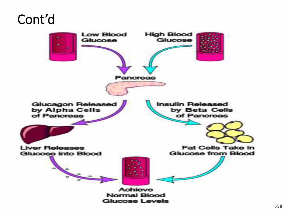
Cont’d
558
Cont’d
• T1DM is a progressive low-insulin catabolic state in which
feeding does not reverse
• With moderate insulinopenia:
• Glucose utilization by muscle and fat decreases -
postprandial hyperglycemia appears.
• Liver produces excessive glucose via glycogenolysis and
gluconeogenesis- fasting hyperglycemia begins.
• Hyperglycemia produces an osmotic diuresis (glycosuria)
• Renal threshold is exceeded (180 mg/dL; 10 mmol/L). 559
Cont’d
• Loss of calories and electrolytes, and persistent dehydration, produce
hypersecretion of stress hormones (epinephrine, cortisol, growth
hormone, and glucagon).
• Stress hormones, in turn, contribute metabolic decompensation by:
• Impairing insulin secretion (epinephrine),
• Antagonizing its action (epinephrine, cortisol, growth hormone),
• Promoting glycogenolysis, gluconeogenesis, lipolysis, and
ketogenesis (glucagon, epinephrine, growth hormone, and cortisol)
• While decreasing glucose utilization and glucose clearance
(epinephrine, growth hormone, cortisol).
560
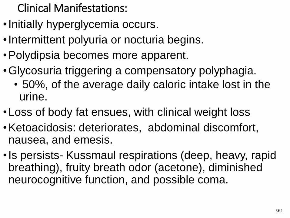
Clinical Manifestations:
• Initially hyperglycemia occurs.
• Intermittent polyuria or nocturia begins.
•Polydipsia becomes more apparent.
•Glycosuria triggering a compensatory polyphagia.
• 50%, of the average daily caloric intake lost in the urine.
•Loss of body fat ensues, with clinical weight loss
•Ketoacidosis: deteriorates, abdominal discomfort, nausea, and emesis.
• Is persists- Kussmaul respirations (deep, heavy, rapid breathing), fruity breath odor (acetone), diminished neurocognitive function, and possible coma.
561

562
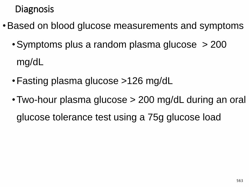
Diagnosis
•Based on blood glucose measurements and symptoms
•Symptoms plus a random plasma glucose > 200
mg/dL
•Fasting plasma glucose >126 mg/dL
•Two-hour plasma glucose > 200 mg/dL during an oral
glucose tolerance test using a 75g glucose load
563
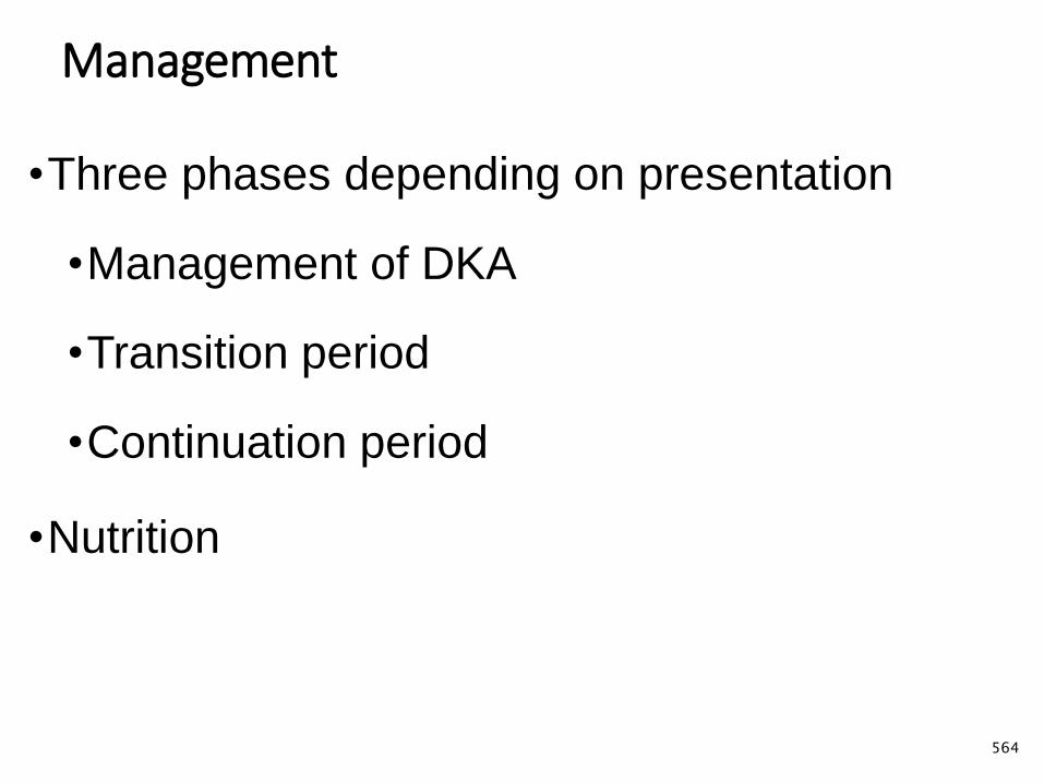
Management
•Three phases depending on presentation
•Management of DKA
•Transition period
•Continuation period
•Nutrition
564

Manage….
•Exercise
•No form of exercise should be forbbiden
•Honey moon period
•Around 75% of newly diagnosed require progressive
reduction in insulin dosage
•Psycho logic aspects
565
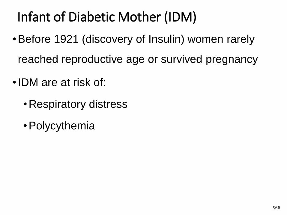
Infant of Diabetic Mother (IDM)
•Before 1921 (discovery of Insulin) women rarely
reached reproductive age or survived pregnancy
• IDM are at risk of:
•Respiratory distress
•Polycythemia
566
IDM….
•Hypoglycemia
•Electrolyte disturbance
•Growth abnormalities (LGA & SGA)
•Prevention centers on
•CLOSE MONITORING DURING
REGNANCY
567
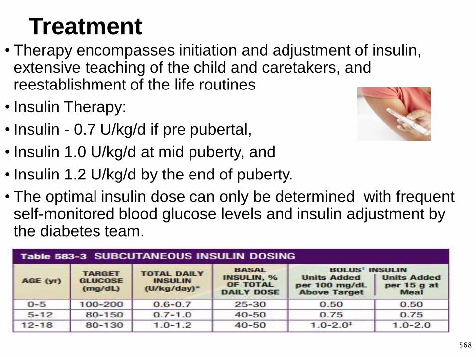
Treatment• Therapy encompasses initiation and adjustment of insulin,
extensive teaching of the child and caretakers, and reestablishment of the life routines
• Insulin Therapy:
• Insulin - 0.7 U/kg/d if pre pubertal,
• Insulin 1.0 U/kg/d at mid puberty, and
• Insulin 1.2 U/kg/d by the end of puberty.
• The optimal insulin dose can only be determined with frequent self-monitored blood glucose levels and insulin adjustment by the diabetes team.
568

Ketoacidosis management
569
Fracture:
•The anatomy, biomechanics, and physiology
of the pediatric skeletal system are different
from that of adults. This results in different
fracture patterns.
570
Pediatric fracture pattern
• Buckle (torus) — Buckle fractures follow compression failure,
often at the junction between the porous metaphysis and the
denser diaphysis.
• These injuries typically occur in the distal radius after
longitudinal trauma (eg, fall on an outstretched hand), but are
also seen in the distal tibia, fibula, and femur.
• Buckle fractures are by definition stable and can often be
managed with splinting and a single orthopedic follow-up visit
571
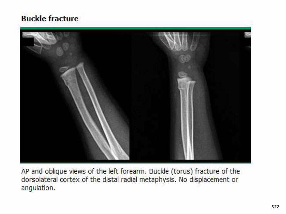
572
Pediatric fracture pattern …
• Plastic Deformation: Plastic deformation is unique to children.
• It is most commonly seen in the ulna and occasionally the fibula.
• The fracture occurs due to a force that produces microscopic
failure on the tensile side of bone and does not propagate to the
concave side.
• The bone is angulated beyond its elastic limit, but the energy is
insufficient to produce a fracture.
573
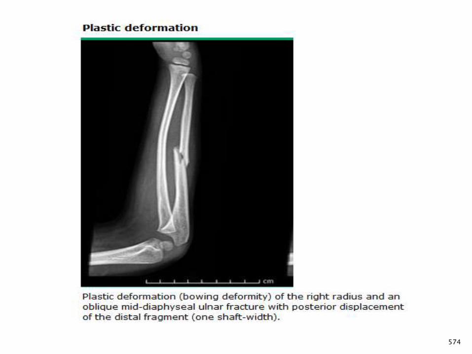
574
Pediatric fracture pattern …
• Greenstick : These fractures occur when the bone is bent, and
there is failure on the tensile (convex) side of the bone.
• The fracture line does not propagate to the concave side of the
bone
• The concave side shows evidence of microscopic failure with
plastic deformation.
• It is necessary to break the bone on the concave side as the
plastic deformation recoils it back to the deformed position.
575
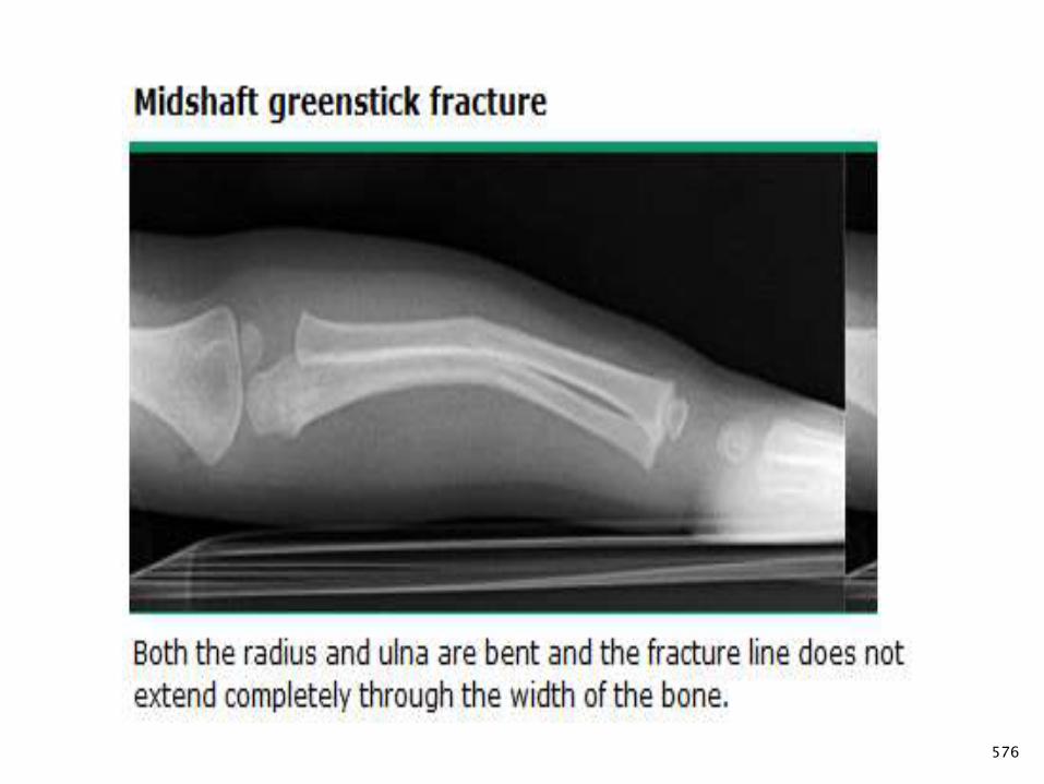
576
Specific fractures
• Clavicular- Approximately 87 percent of clavicle fractures are caused
by a fall onto the shoulder.
• Clinical presentation and examination- Because the clavicle lies close
to the skin, examination often reveals a visible bulge due to hematoma
(often with associated ecchymosis), bone angulation, or displaced
bone edges.
• Treatment - Immobilization with either a sling or figure of eight bandage
is continued until clinical union occurs (ie, the fracture site is nontender
and the patient can move the arm fully with little or no discomfort).
• Pain control and reduction of motion at the fracture site until clinical
union occurs.
577
Proximal humerus
• Humeral fractures are the second most common birth injury
after clavicle fractures.
• Fractures of the proximal humerus commonly occur due to a fall
on an outstretched hand or from a direct blow to the lateral
aspect of the shoulder.
• The child with a proximal humeral fracture typically presents
with a history of trauma, severe shoulder pain, and marked pain
on arm movement.
• Initial treatment consists of pain management, immobilization,
and radiographic evaluation.
578
Distal radius and ulna
• Forearm fractures are among the most common fractures in children,
representing 40 to 50 percent of all childhood fractures.
• The distal third of the forearm, involving the radius and/or ulna, is the
most common location.
• The child with a distal forearm fracture typically has a history of a fall
on an outstretched hand with swelling, bony pain, and/or deformity of
the distal forearm.
• Physical examination of children with distal forearm fractures should
focus on the presence of open wounds near the fracture site, careful
neurovascular assessment, and identification of any injury to the wrist
or elbow joints.
579
Cont’d
• INITIAL TREATMENT — The goals for initial care of children
with a suspected distal forearm fractures are the following:
• Identify and treat vascular compromise
• Identify open fractures
• Provide analgesia and immobilization
• Children with open fractures of the distal forearm should receive
the following initial care
• Application of a sterile dressing
• Tetanus prophylaxis, as needed
• Intravenous antibiotics directed at the most common
organisms
580
Assignment individual
1. Hemophilia
• Definition, Etiology and incidence, Pathogenesis , Clinical
manifestation, Diagnosis and Management
• 2. Failure to thrive
• Definition, Etiology and incidence, Clinical manifestation, Diagnosis, Management and Prognosis
3. Mental retardation
• Definition, Etiology and incidence, Clinical manifestation Diagnosis, Management and Prevention
581
IMNCI (2016)
Integrated Management of New born
and Childhood Illnesses
582
Introduction to
IMNCI
583
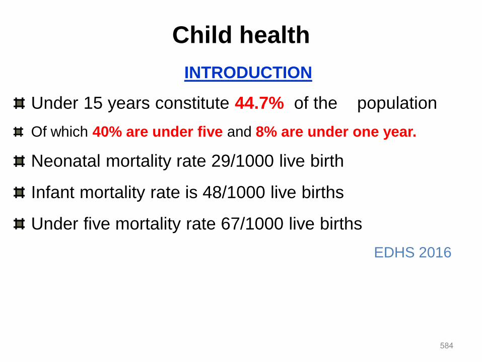
584
Child health
INTRODUCTION
Under 15 years constitute 44.7% of the population
Of which 40% are under five and 8% are under one year.
Neonatal mortality rate 29/1000 live birth
Infant mortality rate is 48/1000 live births
Under five mortality rate 67/1000 live births
EDHS 2016

585
Child health…
More than 70% of these deaths are due to the
five diseases.
Pneumonia 28.9%
Malaria 21.6%
Diarrhea 12%
Measles 5%
60% of these deaths are associated with
malnutrition
HIV/AIDS 11%
586
IMNCI
WHAT IS IMNCI?
IMNCI is a strategy to reduce morbidity &
mortality associated with the major illness.
Is the integrated strategy that combines and links
together existing child health programs.
IMNCI…
Action-oriented CLASSIFICATIONS, rather than
EXACT DIAGNOSES are used.
Using FEW CLINICAL SIGNS as possible which
health workers of diverse background can be trained
to recognize.
The IMNCI guidelines rely on detection of cases
based on SIMPLE CLINICAL SIGNS without
laboratory tests and offer EMPIRIC TREATMENT.
587
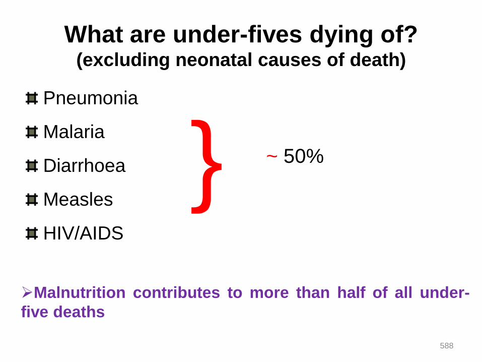
What are under-fives dying of?(excluding neonatal causes of death)
Pneumonia
Malaria
Diarrhoea
Measles
HIV/AIDS
} ~ 50%
Malnutrition contributes to more than half of all under-
five deaths
588

What are neonates dying of?
Preterm births
Severe infection
Asphyxia
Congenital anomalies
Tetanus
} ~ 75%
589
590
The Case Mgt Process
The case mgt process is presented on series of
charts which shows the sequence of steps.
The charts described the following steps.
Assess the child/ young infant: - assess a
child by checking first for general danger signs
(possible series bacterial infection in a young infant).
Assess means taking Hx & P/E.

The case mgt…
• 2. Classify the illness: means select category
or classification based on the major symptoms,
or classify a child’s illnesses using a colour-
coded triage system.
• 3. Identify Rx- After classifying all conditions,
identify specific treatments for the child.
Selecting & classification on the chart is
sufficient to identify Rx.
591
The Case Mgt …
4. Treat the child- Provide practical treatment
instructions, including teaching the caretaker how
to give oral drugs, how to feed and give fluids
during illness, and how to treat local infections at
home.
592
The Case Mgt …
5. Counsel the mother.
Assess feeding, including ASSESM’T of breastfeeding
practices, and counsel to solve any feeding problems
found. Telling her about foods & fluids to give the child & to
bring the child back to the clinic.
6. Give follow up care: - When a child is brought back
to the clinic as requested, give follow-up care and, if
necessary, reassess the child for new problems.
593
Case Mgt Process…
The case management process is
presented on two different sets of charts:
1. For infant Age birth up to 2 months and
2. For children Age 2 months up to five years.
594
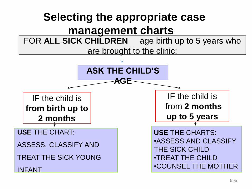
595
FOR ALL SICK CHILDREN age birth up to 5 years who
are brought to the clinic:
IF the child is
from birth up to
2 months
IF the child is
from 2 months
up to 5 years
ASK THE CHILD’S
AGE
USE THE CHART:
ASSESS, CLASSIFY AND
TREAT THE SICK YOUNG
INFANT
USE THE CHARTS:
•ASSESS AND CLASSIFY
THE SICK CHILD
•TREAT THE CHILD
•COUNSEL THE MOTHER
Selecting the appropriate case
management charts
Assess and Classify
The Sick child
Age 2 Months Up to 5 yrs
596

2 Months – 5years sick children
This age group of children are usually assessed by :
1. Checking the presence or absence of General
Danger Sign.Ask
Is the child able to drink or
breastfeed?
Does the child vomit
everything?
Has the child had
convulsions?
LOOK
See if the child is
lethargic or
unconscious.
Is the child
convulsing now? 597

2 months – 5 years sick Children...
2. Ask the four main symptoms:
Cough or difficult breathing,
Diarrhoea,
Fever, and
Ear problem.
598
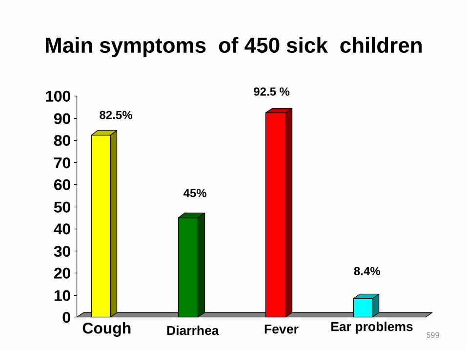
Main symptoms of 450 sick children
0
10
20
30
40
50
60
70
80
90
10082.5%
45%
92.5 %
8.4%
Cough Diarrhea Fever Ear problems599
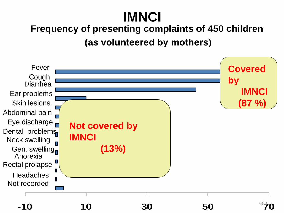
IMNCIFrequency of presenting complaints of 450 children
(as volunteered by mothers)
-10 10 30 50 70
Fever
CoughDiarrhea
Ear problems
Skin lesions
Abdominal pain
Eye discharge
Dental problemsNeck swelling
Gen. swellingAnorexia
Rectal prolapse
Headaches
Not recorded
Not covered by
IMNCI
(13%)
Covered
by
IMNCI
(87 %)
600
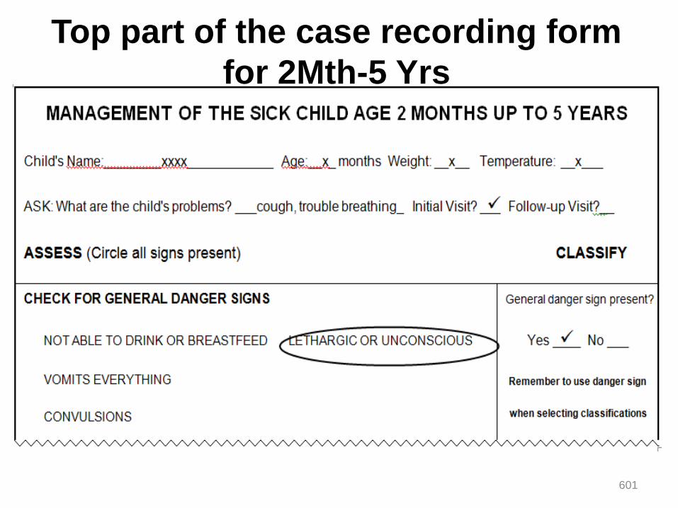
Top part of the case recording form
for 2Mth-5 Yrs
601
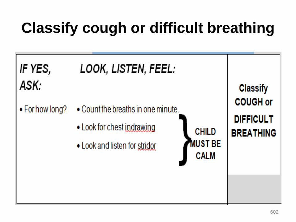
Classify cough or difficult breathing
602
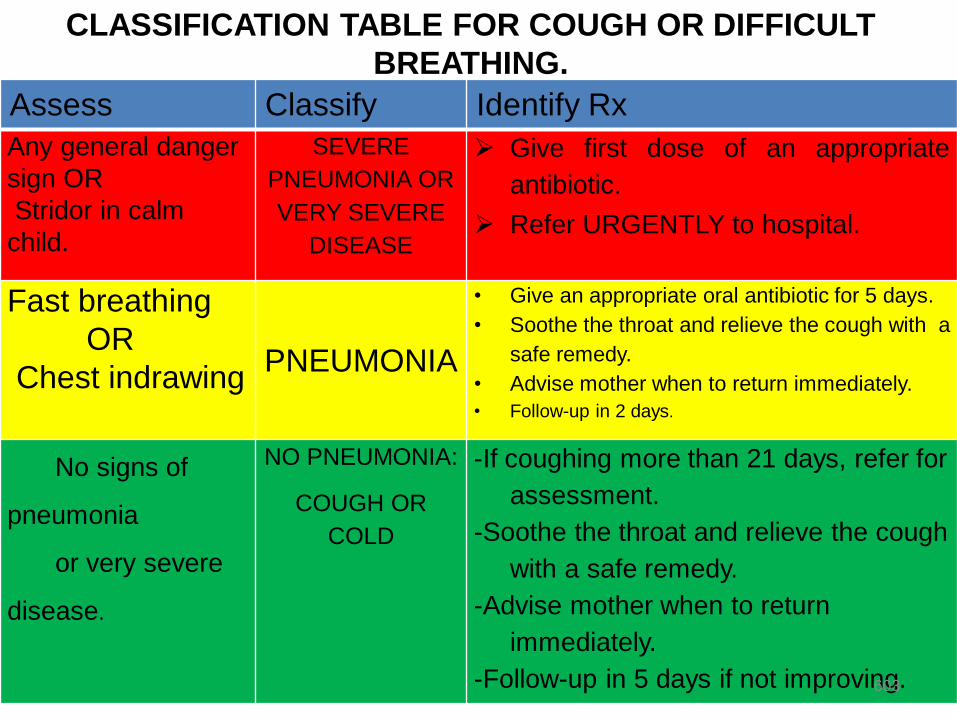
CLASSIFICATION TABLE FOR COUGH OR DIFFICULT
BREATHING.
Assess Classify Identify Rx
Any general danger
sign OR
Stridor in calm
child.
SEVERE
PNEUMONIA OR
VERY SEVERE
DISEASE
Give first dose of an appropriate
antibiotic.
Refer URGENTLY to hospital.
Fast breathing
OR
Chest indrawingPNEUMONIA
• Give an appropriate oral antibiotic for 5 days.
• Soothe the throat and relieve the cough with a
safe remedy.
• Advise mother when to return immediately.
• Follow-up in 2 days.
No signs of
pneumonia
or very severe
disease.
NO PNEUMONIA:
COUGH OR
COLD
-If coughing more than 21 days, refer for
assessment.
-Soothe the throat and relieve the cough
with a safe remedy.
-Advise mother when to return
immediately.
-Follow-up in 5 days if not improving.603
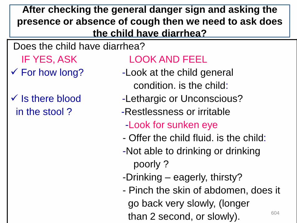
Does the child have diarrhea?
IF YES, ASK LOOK AND FEEL
For how long? -Look at the child general
condition. is the child:
Is there blood -Lethargic or Unconscious?
in the stool ? -Restlessness or irritable
-Look for sunken eye
- Offer the child fluid. is the child:
-Not able to drinking or drinking
poorly ?
-Drinking – eagerly, thirsty?
- Pinch the skin of abdomen, does it
go back very slowly, (longer
than 2 second, or slowly).
After checking the general danger sign and asking the
presence or absence of cough then we need to ask does
the child have diarrhea?
604
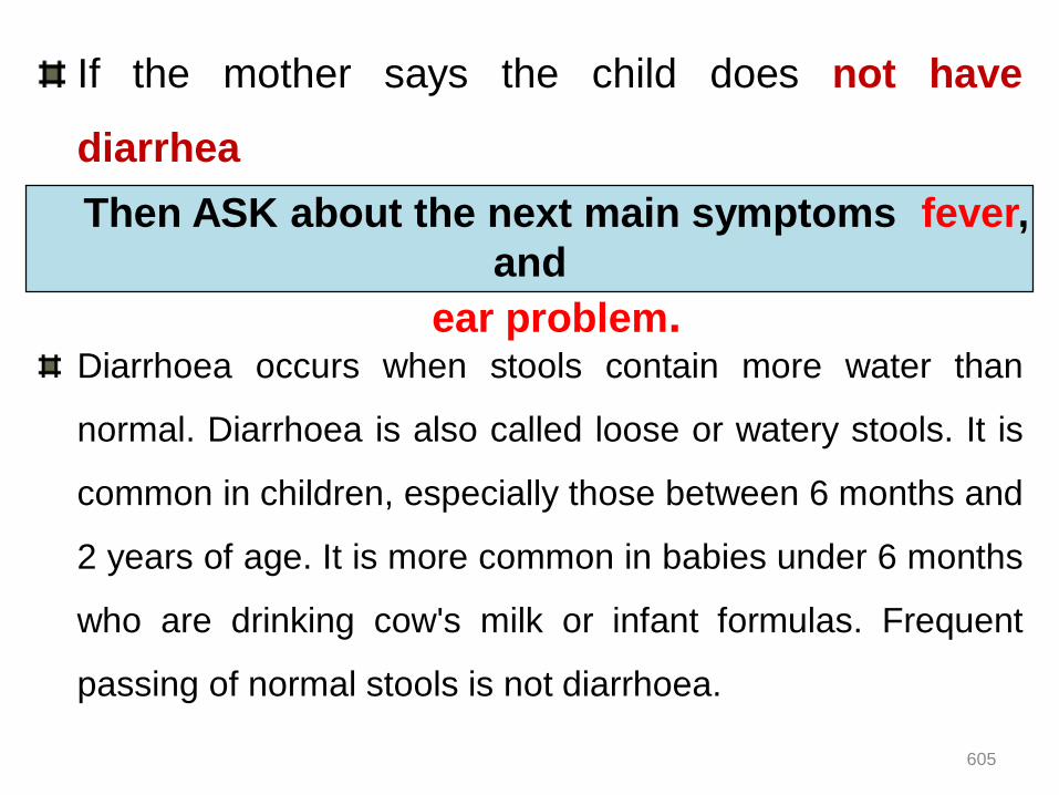
If the mother says the child does not have
diarrhea
Diarrhoea occurs when stools contain more water than
normal. Diarrhoea is also called loose or watery stools. It is
common in children, especially those between 6 months and
2 years of age. It is more common in babies under 6 months
who are drinking cow's milk or infant formulas. Frequent
passing of normal stools is not diarrhoea.
Then ASK about the next main symptoms fever,
and
ear problem.
605
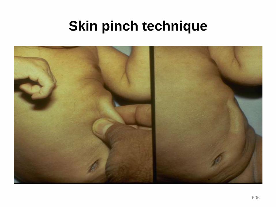
Skin pinch technique
606
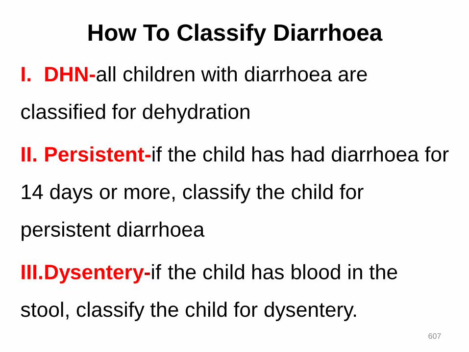
How To Classify Diarrhoea
I. DHN-all children with diarrhoea are
classified for dehydration
II. Persistent-if the child has had diarrhoea for
14 days or more, classify the child for
persistent diarrhoea
III.Dysentery-if the child has blood in the
stool, classify the child for dysentery.607
I. Classify Dehydration (DHN)
They are “3” possible classification DHN
1. Severe DHN
2.Some DHN
3. No DHN
608
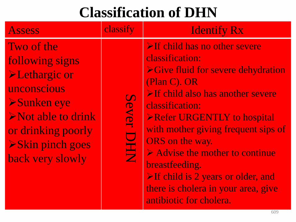
Classification of DHN
Assess classify Identify Rx
Two of the
following signs
Lethargic or
unconscious
Sunken eye
Not able to drink
or drinking poorly
Skin pinch goes
back very slowly S
ever D
HN
If child has no other severe
classification:
Give fluid for severe dehydration
(Plan C). OR
If child also has another severe
classification:
Refer URGENTLY to hospital
with mother giving frequent sips of
ORS on the way.
Advise the mother to continue
breastfeeding.
If child is 2 years or older, and
there is cholera in your area, give
antibiotic for cholera. 609

Classification of DHN…
Two of the following
signs
- Restless,
irritable
- Sunken eye
- Drinks eagerly,
thirsty
- Skin pinch goes
back slowly
So
me
DH
N
-Give fluid, Zinc supplement &
food for some DHN (plan- B)
- If the child has sever
classification:
. Refer urgently to hospital with
mother giving frequent sips of
ORS on the way
- Advise the mother to continue
B/F
- Advise the mother when to
return immediately.
- Follow up for “5” day if not
improving 610
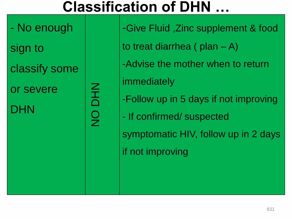
Classification of DHN …
- No enough
sign to
classify some
or severe
DHN
NO
DH
N
-Give Fluid ,Zinc supplement & food
to treat diarrhea ( plan – A)
-Advise the mother when to return
immediately
-Follow up in 5 days if not improving
- If confirmed/ suspected
symptomatic HIV, follow up in 2 days
if not improving
611
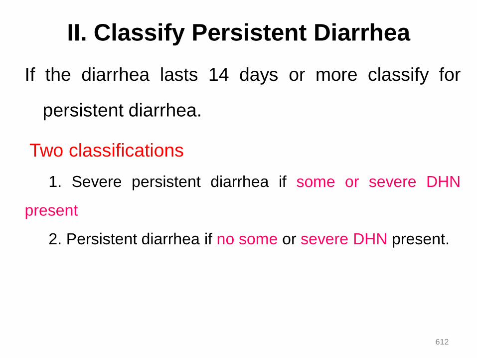
II. Classify Persistent Diarrhea
If the diarrhea lasts 14 days or more classify for
persistent diarrhea.
Two classifications
1. Severe persistent diarrhea if some or severe DHN
present
2. Persistent diarrhea if no some or severe DHN present.
612

Classify Persistent Diarrhea con’t…DHN
present
Severe -
persistent
diarrhea
Treat DHN before
referral if the child has no
severe disease
classification
Refer to hospital
Vit – A supplementation
No DHN Persistent
diarrhea
Advise the mother on
feeding a child
Follow up for in 05
day
Give vit – A 613
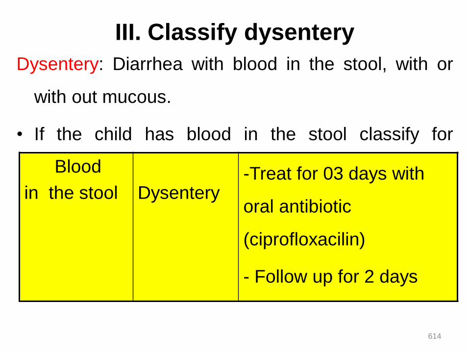
III. Classify dysentery
Dysentery: Diarrhea with blood in the stool, with or
with out mucous.
• If the child has blood in the stool classify for
dysenteryBlood
in the stool Dysentery -Treat for 03 days with
oral antibiotic
(ciprofloxacilin)
- Follow up for 2 days
614
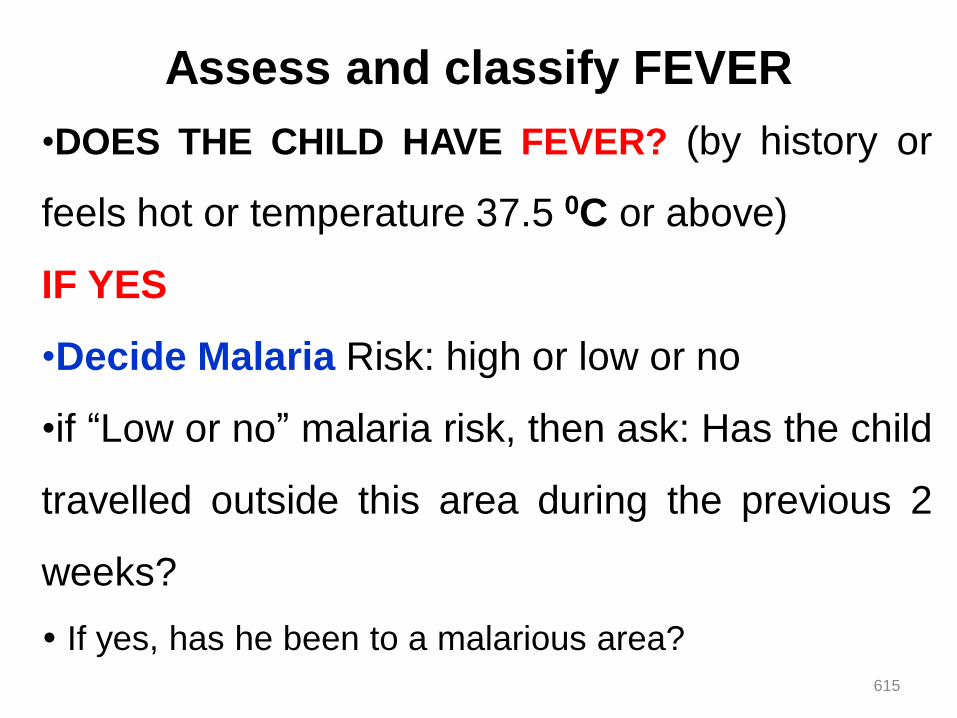
Assess and classify FEVER
•DOES THE CHILD HAVE FEVER? (by history or
feels hot or temperature 37.5 0C or above)
IF YES
•Decide Malaria Risk: high or low or no
•if “Low or no” malaria risk, then ask: Has the child
travelled outside this area during the previous 2
weeks?
If yes, has he been to a malarious area?
615
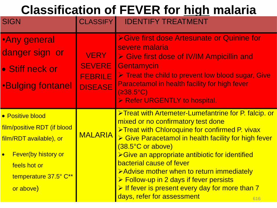
Classification of FEVER for high malaria
risk.SIGN CLASSIFY IDENTIFY TREATMENT
•Any general
danger sign or
Stiff neck or
•Bulging fontanel
VERY
SEVERE
FEBRILE
DISEASE
Give first dose Artesunate or Quinine for
severe malaria
Give first dose of IV/IM Ampicillin and
Gentamycin
Treat the child to prevent low blood sugar, Give
Paracetamol in health facility for high fever
(≥38.5°C)
Refer URGENTLY to hospital.
Positive blood
film/positive RDT (if blood
film/RDT available), or
Fever(by history or
feels hot or
temperature 37.5° C**
or above)
MALARIA
Treat with Artemeter-Lumefantrine for P. falcip. or
mixed or no confirmatory test done
Treat with Chloroquine for confirmed P. vivax
Give Paracetamol in health facility for high fever
(38.5°C or above)
Give an appropriate antibiotic for identified
bacterial cause of fever
Advise mother when to return immediately
Follow-up in 2 days if fever persists
If fever is present every day for more than 7
days, refer for assessment 616
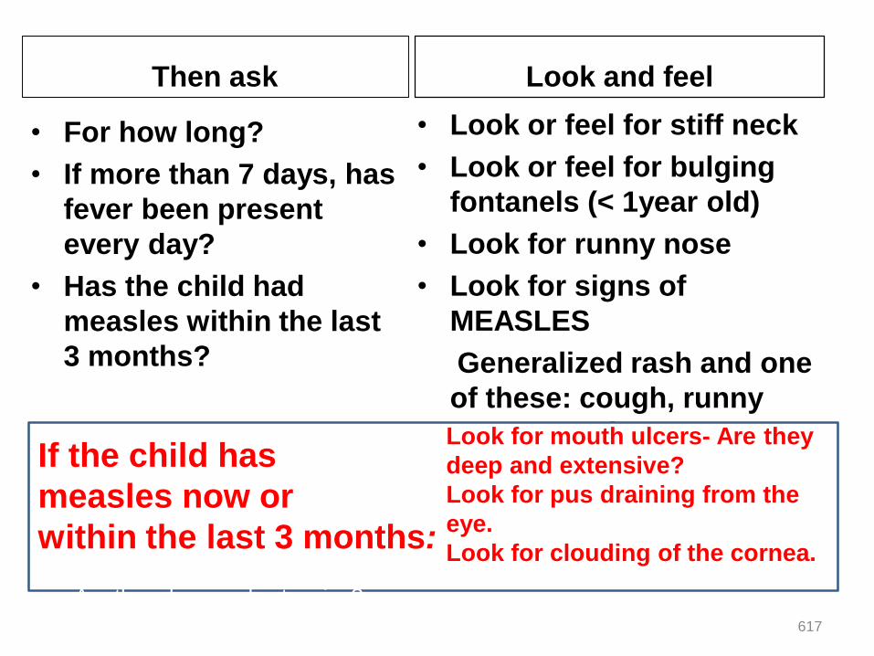
Then ask
• For how long?
• If more than 7 days, has
fever been present
every day?
• Has the child had
measles within the last
3 months?
Look and feel
• Look or feel for stiff neck
• Look or feel for bulging
fontanels (< 1year old)
• Look for runny nose
• Look for signs of
MEASLES
Generalized rash and one
of these: cough, runny
nose, or red eyes If the child has
measles now or
within the last 3 months:Look for mouth ulcers
Are they deep and extensive?
Look for pus draining from the eye.
Look for clouding of the cornea..
Look for mouth ulcers- Are they
deep and extensive?
Look for pus draining from the
eye.
Look for clouding of the cornea.
617
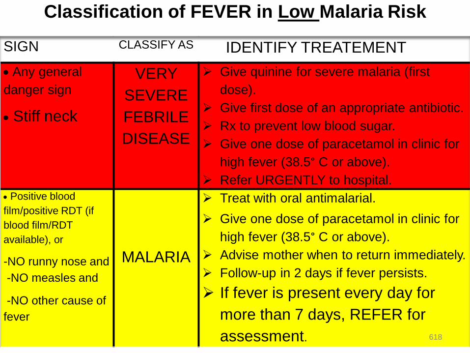
Classification of FEVER in Low Malaria Risk
SIGN CLASSIFY AS IDENTIFY TREATEMENT
Any general
danger sign
Stiff neck
VERY
SEVERE
FEBRILE
DISEASE
Give quinine for severe malaria (first
dose).
Give first dose of an appropriate antibiotic.
Rx to prevent low blood sugar.
Give one dose of paracetamol in clinic for
high fever (38.5° C or above).
Refer URGENTLY to hospital.
Positive blood
film/positive RDT (if
blood film/RDT
available), or
-NO runny nose and
-NO measles and
-NO other cause of
fever
MALARIA
Treat with oral antimalarial.
Give one dose of paracetamol in clinic for
high fever (38.5° C or above).
Advise mother when to return immediately.
Follow-up in 2 days if fever persists.
If fever is present every day for
more than 7 days, REFER for
assessment. 618
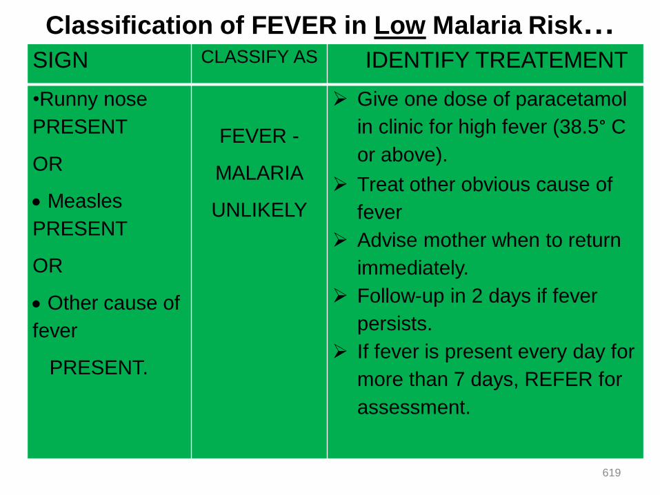
Classification of FEVER in Low Malaria Risk…SIGN CLASSIFY AS IDENTIFY TREATEMENT
•Runny nose
PRESENT
OR
Measles
PRESENT
OR
Other cause of
fever
PRESENT.
FEVER -
MALARIA
UNLIKELY
Give one dose of paracetamol
in clinic for high fever (38.5° C
or above).
Treat other obvious cause of
fever
Advise mother when to return
immediately.
Follow-up in 2 days if fever
persists.
If fever is present every day for
more than 7 days, REFER for
assessment.
619
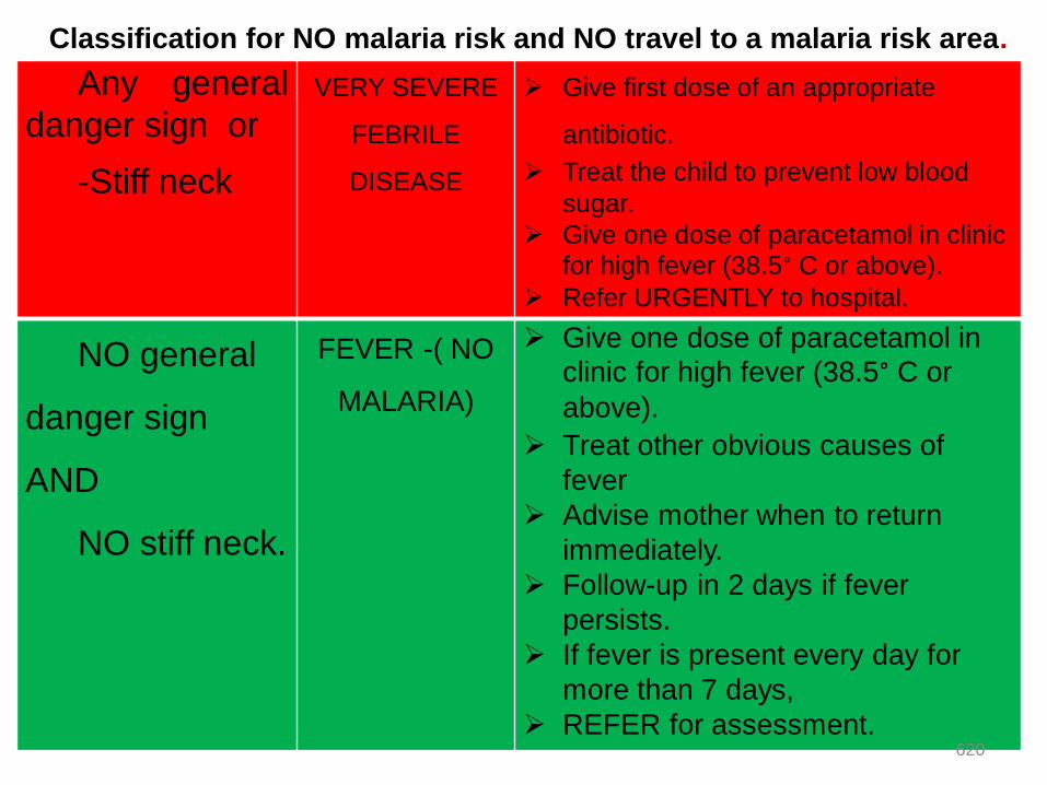
Classification for NO malaria risk and NO travel to a malaria risk area.
Any general
danger sign or
-Stiff neck
VERY SEVERE
FEBRILE
DISEASE
Give first dose of an appropriate
antibiotic.
Treat the child to prevent low blood
sugar.
Give one dose of paracetamol in clinic
for high fever (38.5° C or above).
Refer URGENTLY to hospital.
NO general
danger sign
AND
NO stiff neck.
FEVER -( NO
MALARIA)
Give one dose of paracetamol in
clinic for high fever (38.5° C or
above).
Treat other obvious causes of
fever
Advise mother when to return
immediately.
Follow-up in 2 days if fever
persists.
If fever is present every day for
more than 7 days,
REFER for assessment.620
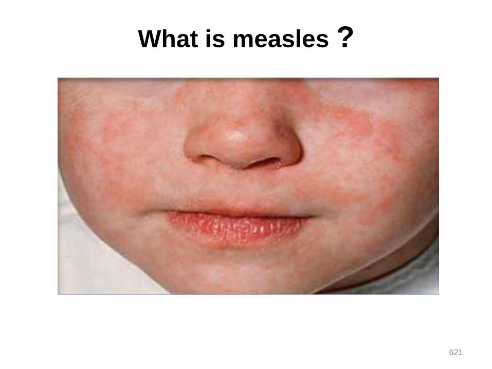
What is measles ?
621

Classification table for measles
(if measles now or within the last 3 months).
Any general danger sign
or
Clouding of cornea or
Deep or extensive
mouth ulcers.
SEVERE
COMPLIC
ATED
MEASLES
Give vitamin A therapeutic dose.
Give first dose of an appropriate
antibiotic.
If clouding of the cornea or pus
draining from the eye, apply
tetracycline eye ointment.
Refer URGENTLY to hospital.
• Pus draining from the
eye or
•Mouth ulcers
MEASLES
WITH
EYE OR
MOUTH
COMPLICA
TIONS
Give vitamin A, therapeutic dose..
If pus draining from the eye, treat
eye infection with tetracycline eye
ointment.
If mouth ulcers, treat with gentian
violet.
Follow-up in 2 days.
Measles now or within
the last 3 months
MEASLES Give vitamin A, therapeutic dose.
Advise when to return immediately
622
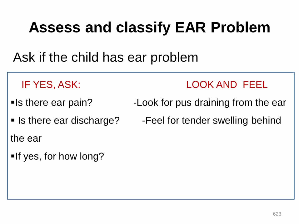
Assess and classify EAR Problem
Ask if the child has ear problem
IF YES, ASK: LOOK AND FEEL
Is there ear pain? -Look for pus draining from the ear
Is there ear discharge? -Feel for tender swelling behind
the ear
If yes, for how long?
623

Classification table for ear problem. Tender swelling
behind
the ear.MASTOIDITIS
Give first dose of an appropriate
antibiotic.
Give first dose of paracetamol for
pain.
Refer URGENTLY to hospital.
Pus is seen draining from the
ear and discharge is reported
for less than 14 days, OR
Ear pain.
ACUTE EAR
INFECTION
Give an oral antibiotic for 5 days.
Give paracetamol for pain.
Dry the ear by wicking.
Follow-up in 5 days.
Pus is seen draining from the
ear and
discharge is reported for 14
days or more.
CHRONIC EAR
INFECTION
Dry the ear by wicking.
Treat with topical quinolones
eardrops for 2 weeks
Follow-up in 5 days.
No ear pain and No
pus seen draining from
the ear.
NO EAR
INFECTIONNo additional treatment.
624
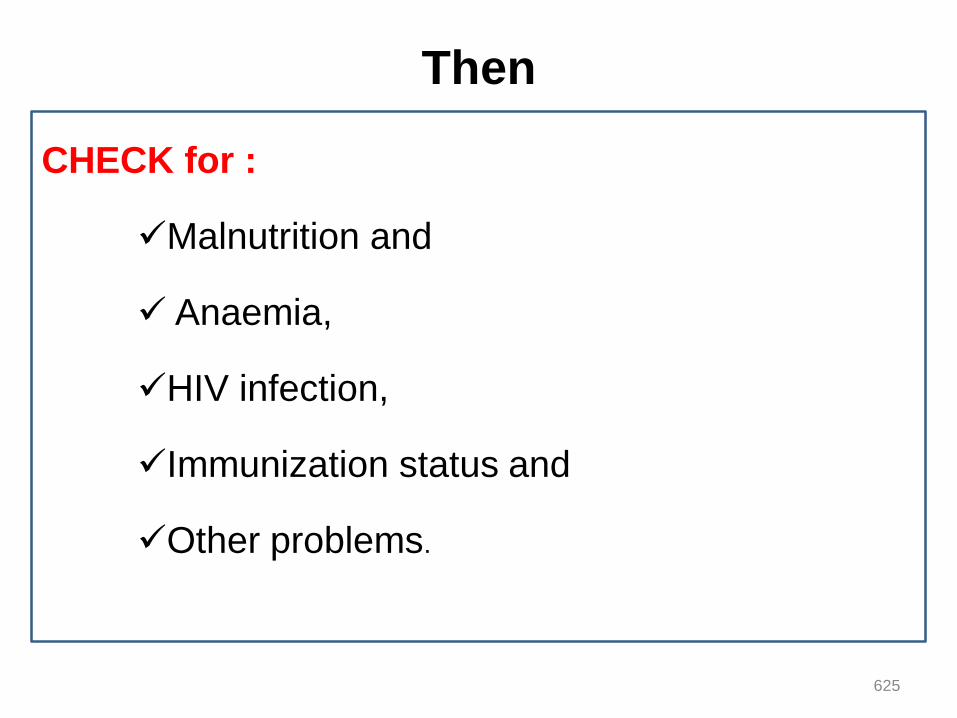
Then
625
CHECK for :
Malnutrition and
Anaemia,
HIV infection,
Immunization status and
Other problems.
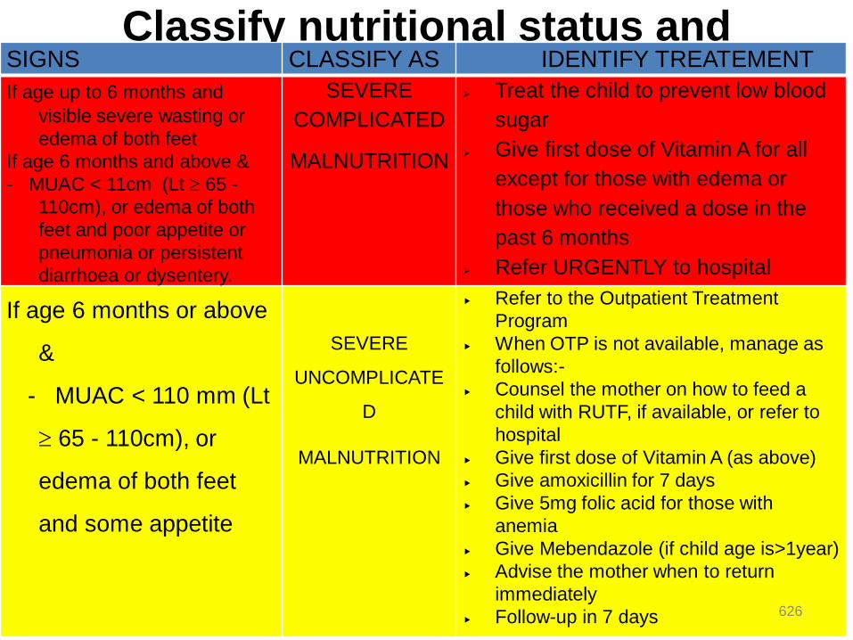
Classify nutritional status and
anemia
SIGNS CLASSIFY AS IDENTIFY TREATEMENT
If age up to 6 months and
visible severe wasting or
edema of both feet
If age 6 months and above &
- MUAC < 11cm (Lt 65 -
110cm), or edema of both
feet and poor appetite or
pneumonia or persistent
diarrhoea or dysentery.
SEVERE
COMPLICATED
MALNUTRITION
Treat the child to prevent low blood
sugar
Give first dose of Vitamin A for all
except for those with edema or
those who received a dose in the
past 6 months
Refer URGENTLY to hospital
If age 6 months or above
&
- MUAC < 110 mm (Lt
65 - 110cm), or
edema of both feet
and some appetite
SEVERE
UNCOMPLICATE
D
MALNUTRITION
Refer to the Outpatient Treatment
Program
When OTP is not available, manage as
follows:-
Counsel the mother on how to feed a
child with RUTF, if available, or refer to
hospital
Give first dose of Vitamin A (as above)
Give amoxicillin for 7 days
Give 5mg folic acid for those with
anemia
Give Mebendazole (if child age is>1year)
Advise the mother when to return
immediately
Follow-up in 7 days 626
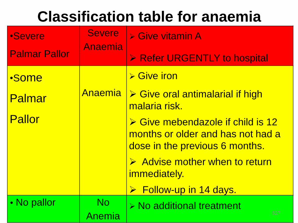
Classification table for anaemia
•Severe
Palmar Pallor
Severe
Anaemia Give vitamin A
Refer URGENTLY to hospital
•Some
Palmar
Pallor
Anaemia
Give iron
Give oral antimalarial if high
malaria risk.
Give mebendazole if child is 12
months or older and has not had a
dose in the previous 6 months.
Advise mother when to return
immediately.
Follow-up in 14 days.
No pallor No
Anemia No additional treatment
627
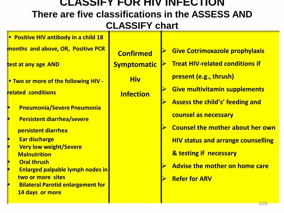
CLASSIFY FOR HIV INFECTIONThere are five classifications in the ASSESS AND
CLASSIFY chart Positive HIV antibody in a child 18
months and above, OR, Positive PCR
test at any age AND
Two or more of the following HIV -
related conditions
Pneumonia/Severe Pneumonia
Persistent diarrhea/severe
persistent diarrhea
Ear discharge Very low weight/Severe
Malnutrition Oral thrush Enlarged palpable lymph nodes in
two or more sites Bilateral Parotid enlargement for
14 days or more
Confirmed
Symptomatic
Hiv
Infection
Give Cotrimoxazole prophylaxis
Treat HIV-related conditions if
present (e.g., thrush)
Give multivitamin supplements
Assess the child's’ feeding and
counsel as necessary
Counsel the mother about her own
HIV status and arrange counselling
& testing if necessary
Advise the mother on home care
Refer for ARV
628
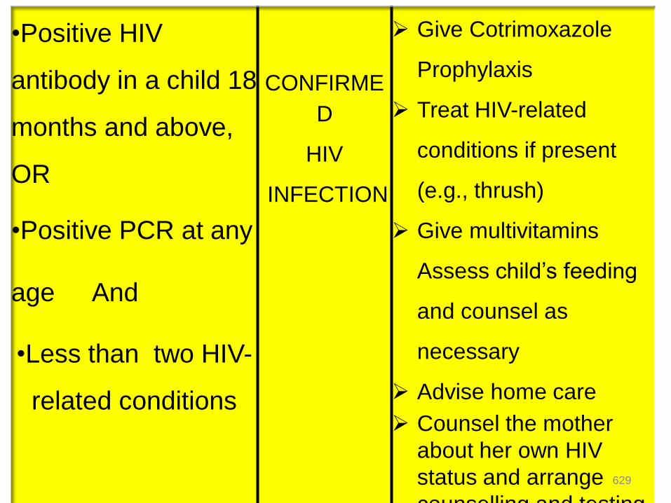
•Positive HIV
antibody in a child 18
months and above,
OR
•Positive PCR at any
age And
•Less than two HIV-
related conditions
CONFIRME
D
HIV
INFECTION
Give Cotrimoxazole
Prophylaxis
Treat HIV-related
conditions if present
(e.g., thrush)
Give multivitamins
Assess child’s feeding
and counsel as
necessary
Advise home care
Counsel the mother
about her own HIV
status and arrange
counselling and testing
629
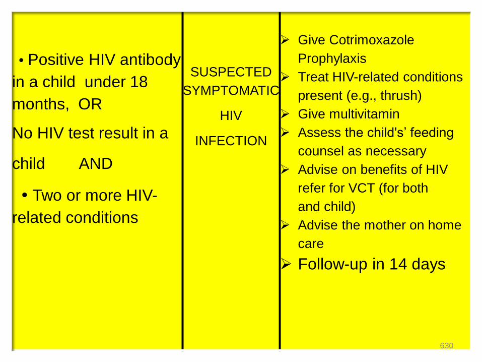
Positive HIV antibody
in a child under 18
months, OR
No HIV test result in a
child AND
Two or more HIV-
related conditions
SUSPECTED
SYMPTOMATIC
HIV
INFECTION
Give Cotrimoxazole
Prophylaxis
Treat HIV-related conditions
present (e.g., thrush)
Give multivitamin
Assess the child's’ feeding
counsel as necessary
Advise on benefits of HIV
refer for VCT (for both
and child)
Advise the mother on home
care
Follow-up in 14 days
630
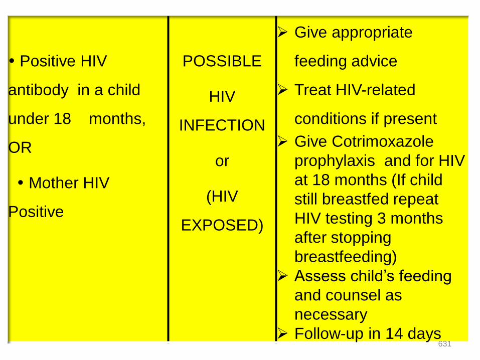
Positive HIV
antibody in a child
under 18 months,
OR
Mother HIV
Positive
POSSIBLE
HIV
INFECTION
or
(HIV
EXPOSED)
Give appropriate
feeding advice
Treat HIV-related
conditions if present
Give Cotrimoxazole
prophylaxis and for HIV
at 18 months (If child
still breastfed repeat
HIV testing 3 months
after stopping
breastfeeding)
Assess child’s feeding
and counsel as
necessary
Follow-up in 14 days631
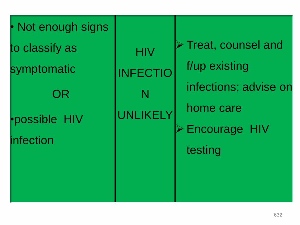
• Not enough signs
to classify as
symptomatic
OR
•possible HIV
infection
HIV
INFECTIO
N
UNLIKELY
Treat, counsel and
f/up existing
infections; advise on
home care
Encourage HIV
testing
632
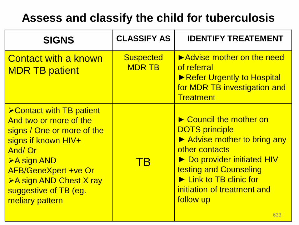
Assess and classify the child for tuberculosis
SIGNS CLASSIFY AS IDENTIFY TREATEMENT
Contact with a known
MDR TB patient
Suspected
MDR TB
►Advise mother on the need
of referral
►Refer Urgently to Hospital
for MDR TB investigation and
Treatment
Contact with TB patient
And two or more of the
signs / One or more of the
signs if known HIV+
And/ Or
A sign AND
AFB/GeneXpert +ve Or
A sign AND Chest X ray
suggestive of TB (eg.
meliary pattern
TB
► Council the mother on
DOTS principle
► Advise mother to bring any
other contacts
► Do provider initiated HIV
testing and Counseling
► Link to TB clinic for
initiation of treatment and
follow up
633
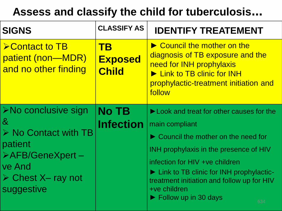
Assess and classify the child for tuberculosis...
SIGNS CLASSIFY AS IDENTIFY TREATEMENT
Contact to TB
patient (non—MDR)
and no other finding
TB
Exposed
Child
► Council the mother on the
diagnosis of TB exposure and the
need for INH prophylaxis
► Link to TB clinic for INH
prophylactic-treatment initiation and
follow
No conclusive sign
&
No Contact with TB
patient
AFB/GeneXpert –
ve And
Chest X– ray not
suggestive
No TB
Infection
►Look and treat for other causes for the
main compliant
► Council the mother on the need for
INH prophylaxis in the presence of HIV
infection for HIV +ve children
► Link to TB clinic for INH prophylactic-
treatment initiation and follow up for HIV
+ve children
► Follow up in 30 days 634
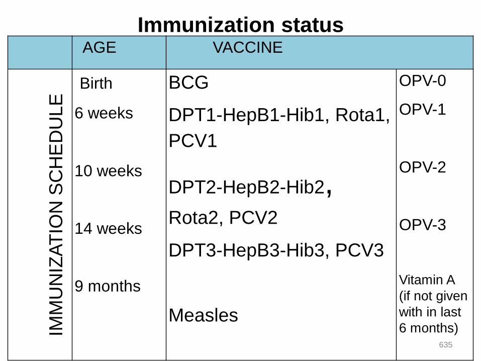
Immunization statusAGE VACCINE
IMM
UN
IZA
TIO
N S
CH
ED
ULE
Birth
6 weeks
10 weeks
14 weeks
9 months
BCG
DPT1-HepB1-Hib1, Rota1,
PCV1
DPT2-HepB2-Hib2,Rota2, PCV2
DPT3-HepB3-Hib3, PCV3
Measles
OPV-0
OPV-1
OPV-2
OPV-3
Vitamin A
(if not given
with in last
6 months) 635

Observe contraindications of
immunizationThere are only three situations at present that are
contraindications to immunization:
1. Do not give BCG to a child known to have
SYMPTOMATIC HIV INFECTION.
2. Do not give DPT2-HepB2-Hib2 or DPT3-HepB3-
Hib3 to a child who has had convulsions or shock
within 3 days of the most recent dose.
3. Do not give DPT-HepB-Hib to a child with recurrent
convulsions or another active neurological disease of
the central nervous system 636
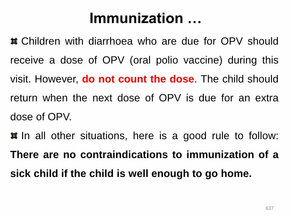
Immunization …
Children with diarrhoea who are due for OPV should
receive a dose of OPV (oral polio vaccine) during this
visit. However, do not count the dose. The child should
return when the next dose of OPV is due for an extra
dose of OPV.
In all other situations, here is a good rule to follow:
There are no contraindications to immunization of a
sick child if the child is well enough to go home.
637
Finally we have to:
Assess other problems
Counsel the mother about her own health
638
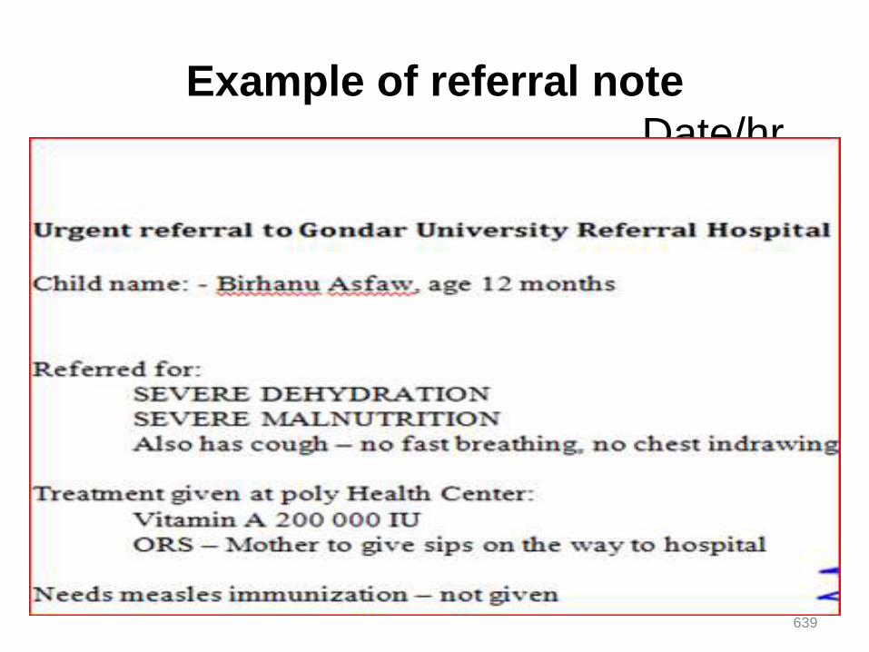
Example of referral note
Date/hrdate
639

Teach the mother to give oral drugs at
homeFollow the instructions below for every oral drug to be
given at home.
Also follow the instructions listed with each drug‘s dosage
table.
Determine the appropriate drugs and dosage for the
child‘s age or weight.
Tell the mother the reason for giving the drug to the child.
Demonstrate how to measure a dose.
Watch the mother practice measuring a dose by herself.
Ask the mother to give the first dose to her child.
Explain carefully how to give the drug, then label and
package the drug.
If more than one drug will be given, collect, count and
package each drug separately.
640
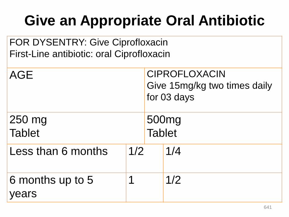
Give an Appropriate Oral Antibiotic
FOR DYSENTRY: Give Ciprofloxacin
First-Line antibiotic: oral Ciprofloxacin
AGE CIPROFLOXACIN
Give 15mg/kg two times daily
for 03 days
250 mg
Tablet
500mg
Tablet
Less than 6 months 1/2 1/4
6 months up to 5
years
1 1/2
641
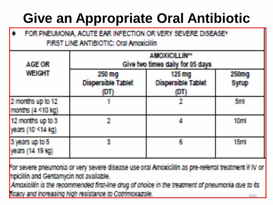
Give an Appropriate Oral Antibiotic
642
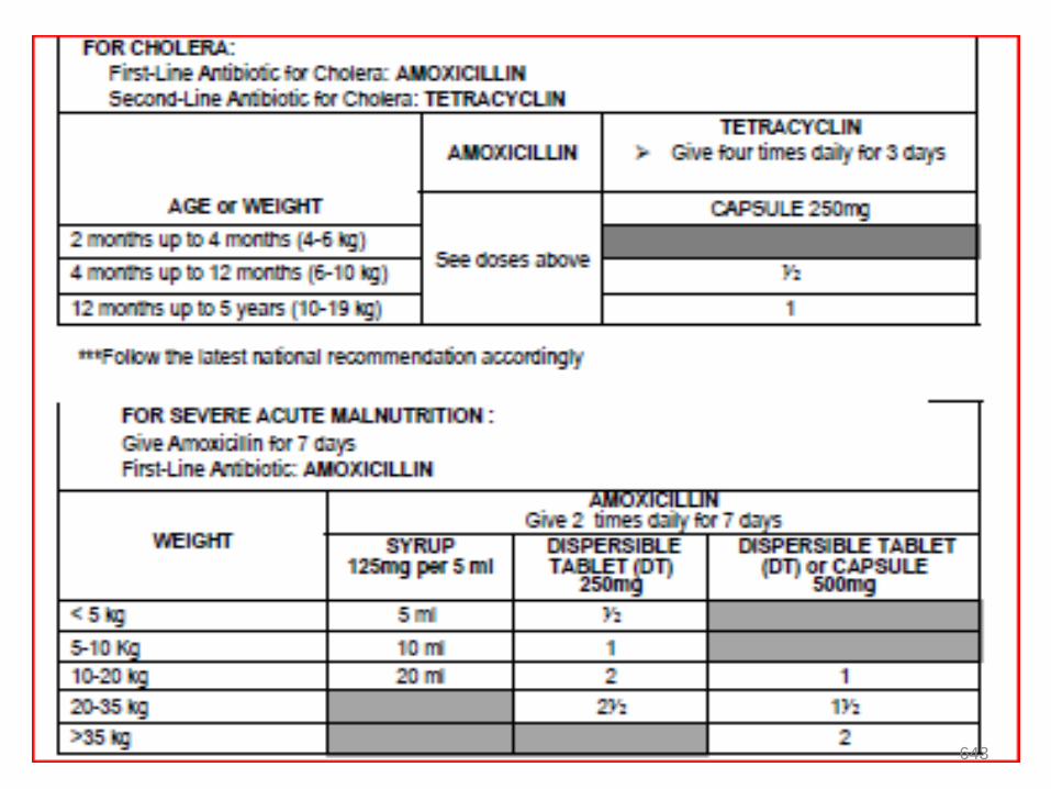
643
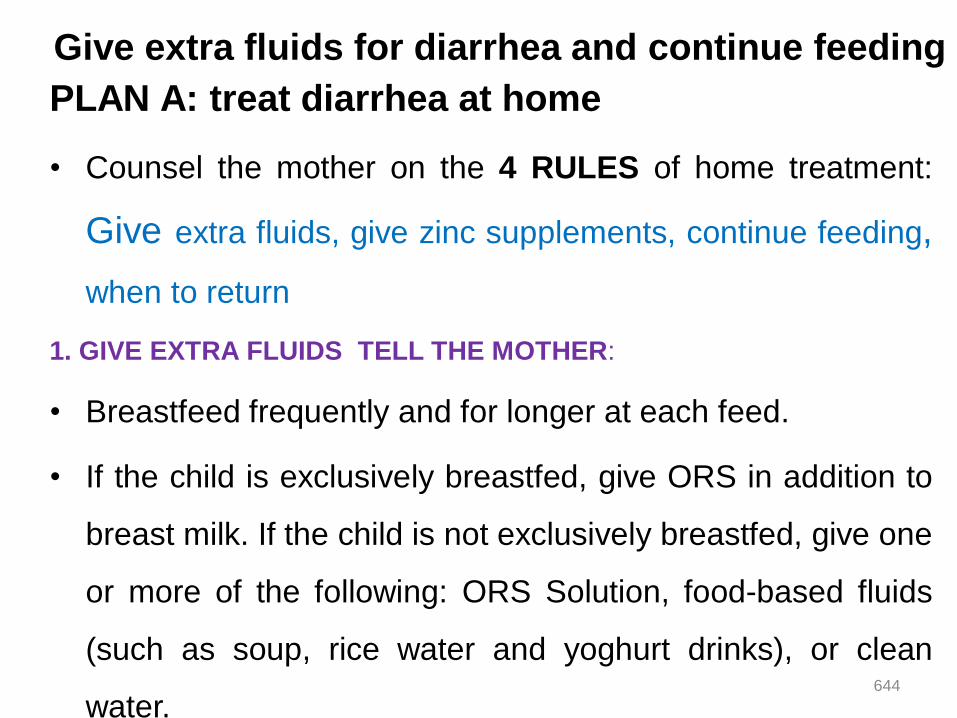
Give extra fluids for diarrhea and continue feeding
PLAN A: treat diarrhea at home
• Counsel the mother on the 4 RULES of home treatment:
Give extra fluids, give zinc supplements, continue feeding,
when to return
1. GIVE EXTRA FLUIDS TELL THE MOTHER:
• Breastfeed frequently and for longer at each feed.
• If the child is exclusively breastfed, give ORS in addition to
breast milk. If the child is not exclusively breastfed, give one
or more of the following: ORS Solution, food-based fluids
(such as soup, rice water and yoghurt drinks), or clean
water.644
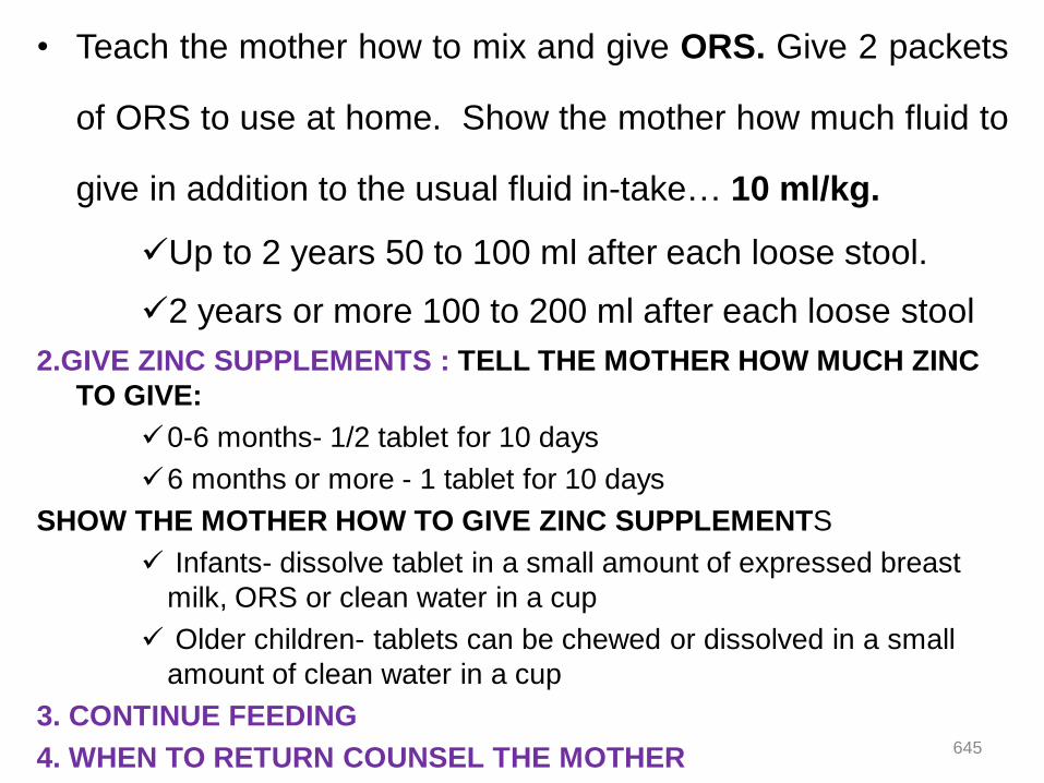
• Teach the mother how to mix and give ORS. Give 2 packets
of ORS to use at home. Show the mother how much fluid to
give in addition to the usual fluid in-take… 10 ml/kg.
Up to 2 years 50 to 100 ml after each loose stool.
2 years or more 100 to 200 ml after each loose stool
2.GIVE ZINC SUPPLEMENTS : TELL THE MOTHER HOW MUCH ZINC
TO GIVE:
0-6 months- 1/2 tablet for 10 days
6 months or more - 1 tablet for 10 days
SHOW THE MOTHER HOW TO GIVE ZINC SUPPLEMENTS
Infants- dissolve tablet in a small amount of expressed breast
milk, ORS or clean water in a cup
Older children- tablets can be chewed or dissolved in a small
amount of clean water in a cup
3. CONTINUE FEEDING
4. WHEN TO RETURN COUNSEL THE MOTHER 645
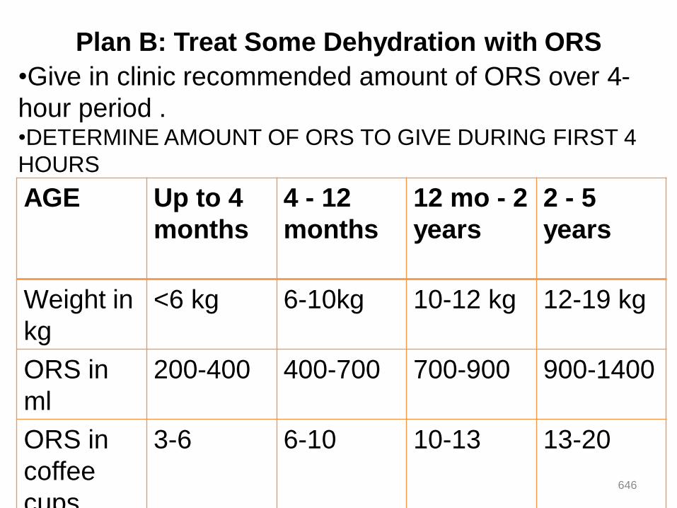
Plan B: Treat Some Dehydration with ORS
•Give in clinic recommended amount of ORS over 4-
hour period .•DETERMINE AMOUNT OF ORS TO GIVE DURING FIRST 4
HOURS
AGE Up to 4
months
4 - 12
months
12 mo - 2
years
2 - 5
years
Weight in
kg
<6 kg 6-10kg 10-12 kg 12-19 kg
ORS in
ml
200-400 400-700 700-900 900-1400
ORS in
coffee
cups
3-6 6-10 10-13 13-20
646

Plan B…
• Use the child's age only when you do not know the weight.
• The approximate amount of ORS required - 75ml/kg.
• If the child wants more ORS than shown, give more.
• For infants under 6 months who are not breastfed, also give 100-
200 ml
SHOW THE MOTHER HOW TO GIVE ORS SOLUTION:
• Give frequent small sips from a cup.
• If the child vomits, wait 10 minutes. Then continue, but more
slowly. Continue breastfeeding whenever the child wants.
647
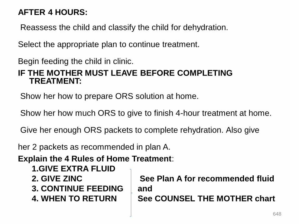
AFTER 4 HOURS:
Reassess the child and classify the child for dehydration.
Select the appropriate plan to continue treatment.
Begin feeding the child in clinic.
IF THE MOTHER MUST LEAVE BEFORE COMPLETING TREATMENT:
Show her how to prepare ORS solution at home.
Show her how much ORS to give to finish 4-hour treatment at home.
Give her enough ORS packets to complete rehydration. Also give
her 2 packets as recommended in plan A.
Explain the 4 Rules of Home Treatment:
1.GIVE EXTRA FLUID
2. GIVE ZINC See Plan A for recommended fluid
3. CONTINUE FEEDING and
4. WHEN TO RETURN See COUNSEL THE MOTHER chart
648
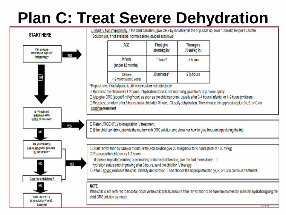
Plan C: Treat Severe Dehydration
Quickly
649

Thank you for any question, you can ask using
the following address
Chalachew Adugna (Lecturer of pediatrics
and child health nursing, University of Gondar)
Email: [email protected]
Stay at home! Be safe from COVID-19
650