Embed Size (px)
Citation preview
Vol. 31, No. 1ANTIMICROBIAL AGENTS AND CHEMOTHERAPY, Jan. 1987, p. 6-100066-4804/87/010006-05$02.00/0Copyright © 1987, American Society for Microbiology
Penetration of New Azole Compounds into the Eye and Efficacy inExperimental Candida Endophthalmitis
DORA V. SAVANI, JOHN R. PERFECT,* L. MICHAEL COBO, AND DAVID T. DURACK
Department of Medicine and Ophthalmology, Duke University Medical Center, Durham, North Carolina 27710
Received 25 April 1986/Accepted 14 October 1986
We studied the penetration of three azole compounds, ketoconazole, itraconazole, and fluconazole, into theocular tissues and fluids of rabbits in the presence and absence of ocular inflammation. Drug concentrationswere compared with those found in serum and cerebrospinal fluid. The rank order of penetration into eye tissuewas fluconazole > ketoconazole > itraconazole. Fluconazole penetrated freely into both inflamed anduninflamed eyes. The presence of inflammation improved penetration of all three compounds into ocular fluidsand tissues. Penetration of these azoles into the anterior chamber of uninflamed eyes and into the cerebrospinalfluid was similar. All three azole compounds reduced the number of yeasts found in the eye in hematogenousCandida albicans endophthalmitis in rabbits when therapy was initiated within 24 h of inoculation. However,only ketoconazole significantly reduced yeast counts in the eye when therapy was postponed for 7 days.
The incidence of ocular fungal infections has increased(10, 11, 26) due to a number of factors: increased prevalenceof immunosuppression associated with organ transplantationand malignancies; prolonged recovery from complex surgi-cal procedures; and increasing use of antibiotics, im-munosuppressive agents, intravenous catheters, and hyper-alimentation fluids. Endogenous Candida endophthalmitis(ECE) also occurs in heroin addicts (2, 9; D. W. Vastine, W.Horsley, S. B. Guth, and M. F. Goldberg, Letter, Arch.Ophthalmol. 94:1805, 1976), neonates (28, 32), and postpar-tum patients not exposed to these factors (7). The mostcommon species causing ECE is Candida albicans (8, 12).Others are Candida tropicalis (20, 26, 28), Candida stel-latoidea (12), Candida parapsilosis (20), and Candida guil-lermondi (26).Optimal therapies for oculomycoses have not been de-
fined. Amphotericin B is less than ideal: it penetrates poorlyinto ocular fluids, is toxic, and must be given intravenously.Another polyene, pimaracin, can only be given topically.Oral flucytosine penetrates well into the ocular fluids, butthis drug is active against only a limited range of pathogenicfungi, and resistance can develop during therapy. The de-velopment of azole compounds with broad-spectrum activityagainst yeasts and dimorphic fungi has expanded the thera-peutic options. Miconazole and ketoconazole have beenavailable for several years, but reported experience in eyeinfections is limited (5, 15, 21, 23). The present studyexamined the concentrations of three azole compounds,ketoconazole, fluconazole, and itraconazole, in differentcompartments of inflamed and uninflamed eyes of NewZealand white (NZW) rabbits. These three agents were thencompared for their in vivo efficacy in an established rabbitmodel (24) for hematogenous Candida infection of the eye.
MATERIALS AND METHODS
Animals. Seventy-two male NZW rabbits weighing 2 to 3kg were used. All rabbits were housed in separate cages andgiven rabbit chow (Purina Co.) and water ad libitum.Ketamine (Ketaject; Bristol Laboratories, Syracuse, N.Y.),100 to 150 mg intramuscularly, and xylazine (Rompun;
* Corresponding author.
Cutter Laboratories, Shawnee, Kans.), 15 to 25 mg intra-muscularly, were used as anesthesia for all procedures.Animals were sacrificed with intravenous pentobarbital.Organism. A clinical isolate of C. albicans (Carter strain)
was used for the endophthalmitis experiments. To preparethe inocula, C. albicans was transferred from a stock cultureonto a Sabouraud agar plate containing 100 pug of chloram-phenicol per ml and allowed to grow overnight at 37°C.Yeasts were then taken up on cotton swabs and suspended in0.015 M phosphate-buffered saline (PBS). The yeast suspen-sion was adjusted by optical density with a spectrophotom-eter (Gilford Instruments, Oberlin, Ohio) to a final concen-tration of 106 CFU/ml (24). The final concentration wasverified by a colony count of twofold dilutions of eachinoculum plated onto Sabouraud agar.
Antifungal agents. Itraconazole (Janssen Pharmaceutica,New Brunswick, N.J.) was administered orally in 50-mgcapsules containing polyethylene glycol. Fluconazole (PfizerInc., Groton, Conn.) was dissolved in sterile water to a finalconcentration of 10 mglml. The fluconazole solution waswarmed to resolubilize the drug before each treatment andinjected intravenously. Ketoconazole (Janssen Pharmaceu-tica) was administered orally as 200-mg tablets. Amphoter-icin B was prepared by dissolving the standard parenteralpreparation (E. R. Squibb and Sons, Princeton, N.J.) insterile distilled water to a concentration of 5 mg/ml andinjected intravenously.
Antifungal assay. A bioassay was used to determine azolelevels in the serum, cerebrospinal fluid (CSF), aqueoushumor, and vitreous body of each animal. The assay usedthe agar-well diffusion method of Bennett et al. (4), modifiedby the method of Jorgensen et al. for assaying azole com-pounds (25). The standard concentrations were dissolved inserum or normal saline. In preliminary experiments wefound no differences in zone sizes between azole compoundsin saline, CSF, and aqueous humor. The reproducibility ofthis assay when samples are run in triplicate is +7%. Thelower limit of sensitivity of the bioassay for ketoconazoleand itraconazole was 0.078 ,ug/ml. For fluconazole, the lowerlimit ranged between 1.56 and 3.13 p,g/ml.To assay drug in rabbit corneas, 6-mm disks of tissue
trephined from corneas were assayed by the trephine diskmethod (3, 35). Standards for this assay were prepared in
6
on May 17, 2018 by guest
http://aac.asm.org/
Dow
nloaded from
AZOLES AND CANDIDAL ENDOPHTHALMITIS 7
TABLE 1. Concentrations of azoles in rabbits with and without eye inflammation, 4 h after a single dose
Concn (mean ± SEM)
Inflamed eye Uninflamed eyeDrug and dose (mg/kg) Serum CSF Aqueous Aqueous Vitreous
(FLg/ml) (Rg/ml) Cornea humor Vitreous Comea humor body(~Lg/g) (p.g/ml) body (p.g/ml) (Rg/g) (p.g/ml) (~.Lg/ml)
Fluconazole (80, i.v.) 77.6 ± 2.7 33.2 ± 2.5 6.2 ± 1.0 50.2 ± 6.2 21.5 ± 0.1 2.1 ± 0.3 21.9 ± 3.4 16.0 ± 5.3Ketoconazole (-80, p.o.) 58.3 ± 4.5 3.1 ± 0.2 1.4 ± 0.1 33.6 ± 8.0 16.4 ± 2.7 0.7 ± 0.2 7.4 ± 5.1 2.9 ± 1.2Itraconazole (-80, p.o.) 2.13 ± 0.58 NDa 0.05 ± 0.01 0.92 ± 0.01 0.22 ± 0.04 0.03 ± 0.01 ND ND
a ND, Not detectable.
PBS, absorbed onto 6-mm filter paper disks, and plated asdescribed previously (3, 35). Candida pseudotropicalis wasused as the assay organism.
In vitro susceptibility testing. A broth dilution test adaptedfrom the turbidimetric method of Galgiani and Stevens (18)was used. Briefly, to measure MICs or IC1/2, an overnightgrowth of C. albicans Carter strain on Sabouraud slants wassuspended in PBS and then adjusted by optical density to afinal concentration of 105 CFU/ml. Serial twofold dilutions ofantifungal agents were prepared in a buffered syntheticamino acid medium for fungi (SAAMF) (18). One hundredmicroliters of the inoculum (final concentration, 0.5 x 10⁴CFU/ml) was added to 2-ml plastic tubes with drug diluted inSAAMF, mixed, and incubated for 16 to 18 h at 30°C.Growth was read by a spectrophotometer (Gilford Instru-ments). The MIC was determined as the antibiotic concen-tration which inhibited the growth of the yeast by one-halfcompared with a drug-free control (IC1/2).Drug pharmacokinetics. To produce ocular inflammation,
0.1 ml of 20% proteose was injected intravitreally into theright eye of 12 NZW rabbits. Approximately 36 h later, therabbits were randomized into four groups of three rabbitseach, and treatment was started. The groups received thefollowing treatments: group 1-fluconazole, 80 mg/kg intra-venously (i.v.); group I-itraconazole, 80 mg/kg orally(p.o.); group III-ketoconazole, 80 mg/kg p.o. Between 3and 4 h following therapy, the rabbits were anesthetized, andblood and CSF samples were aspirated. CSF was obtainedwith a 25-gauge needle on a 3-ml syringe which was intro-duced into the cisterna magna. Approximately 0.5 to 1.0 mlof CSF can be removed safely without blood contamination.Then, aqueous humor was withdrawn from both eyes. Ani-mals were sacrificed, and the eyes were enucleated, rinsed insterile water, and immediately frozen in liquid nitrogen.Tissues were dissected while still frozen and kept at -70°Cuntil assayed to determine the tissue concentrations of thedrugs (1).
Production and quantitation of Candida endophthalmitis.Sixty NZW rabbits were anesthetized, and a 1-ml suspensioncontaining 106 blastospores of C. albicans was injectedintravenously into a marginal ear vein. Approximately 24 hlater, four rabbits were sacrificed, and both eyes from eachanimal were enucleated, rinsed in sterile PBS, and placed ina sterile petri dish for dissection. The cornea, lens, and iriswere excised and discarded. The vitreous body was removedand transferred to a preweighed volumetric flask. Scleraewere opened by three radial incisions. The retina andchoroid tissues were placed in a preweighed petri dish.Samples were weighed, diluted in 1 ml of PBS, and homog-enized in a tissue homogenizer (Eberbach Corp., Ann Arbor,Mich.). One hundred microliters of the homogenate or10-fold dilutions was plated on Sabouraud agar plates con-taining 100 ,ug of chloramphenicol per ml and allowed to
grow overnight at 37°C. Colonies were counted on thefollowing day; final concentrations were expressed as CFUper gram of tissue.
Fifty-six NZW rabbits were evaluated for eye lesions byindirect ophthalmoscopy. Both eyes were examined, and themean number of foci per rabbit was recorded. Rabbits wererandomized into several treatment regimens to achieve equaldistribution of rabbits with various numbers of eye lesions ineach group.
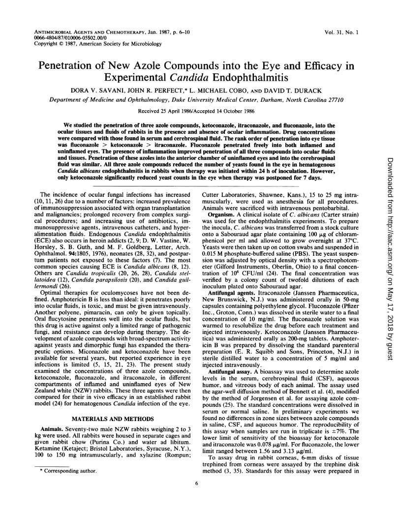
Choroid-Retina
3
2
*l~~~~~~~~od 7
a)~ ~~~Dyretinaandv ~~~~itreousboyfoavrgvausfrecrbitfern
E
ITU.
o 0 Control-I KTZ
El1 FLU0 ITZ
2
1 ~~~~~~7Days
FIG. 1. Mean quantitative counts of C. albicans in the choroid-retina and vitreous body from average values for each rabbit after notreatment, ketoconazole (KTZ, 80 mg/kg per day p.o.), itraconazole(ITZ, 80 mg/kg per day p.o.), and fluconazole (FLU, 80 mg/kg perday i.v.) are shown. Treatment was started on day 1 of infection andmaintained for 7 days.
VOL. 31, 1987
on May 17, 2018 by guest
http://aac.asm.org/
Dow
nloaded from
ANTIMICROB. AGENTS CHEMOTHER.
Treatment regimens. (i) Early therapy: 24 NZW rabbitswere started on treatment 24 h after inoculation with C.albicans and maintained on daily treatment for 7 days. Fourgroups of six rabbits each received the following: group I-itraconazole, 80 mg/kg per day p.o.; group II-ketocona-zole, 80 mg/kg per day p.o.; group III-fluconazole, 80mg/kg per day i.v.; group IV-no treatment. (ii) Late ther-apy: 32 NZW rabbits were started on antifungal agents 7days after inoculation with C. albicans and maintained ondaily treatment for 7 days. Four groups of seven rabbits eachreceived the same treatments as groups I through IV above.In this experiment an additional group of four rabbits re-ceived amphotericin B (1 mg/kg per day i.v.). Rabbits weresacrificed 24 h after the last dose in all groups, and quanti-tative cultures were performed as described above.
Statistical analysis. For comparison of yeast counts at theend of therapy, an analysis of variance of ranks, sequentiallyfollowed by pairwise comparison t tests, was used. Meanswere calculated from the average value for both eyes of eachrabbit; n = number of animals. A Student t test was used tocompare drug levels in inflamed and uninflamed eyes.
RESULTSConcentrations in serum measured 4 h after a single dose
are shown in Table 1. Both ketoconazole and itraconazolewere relatively well absorbed. A 200-mg dose of ketocona-zole and itraconazole p.o. per rabbit represents approxi-mately 80 mg/kg, similar to the intravenous dose used forfluconazole.The concentrations of the three azoles in the aqueous
humor from inflamed and uninflamed eyes and in the CSFare shown in Table 1. There were striking differences amongthe three agents. Inflammation facilitated penetration of allazoles into the aqueous humor, but fluconazole crossed bothblood-aqueous and blood-CSF barriers freely even in theabsence of inflammation. Fluconazole was found in theaqueous humor at approximately 64% of the concentration inserum, comparable to the good penetration of flucytosineinto the eye (27, 34). As we have reported previously,itraconazole could not be detected in the CSF by ourbioassay (29), nor could itraconazole be detected in theaqueous humor of uninflamed eyes. However, for inflamedeyes, the percent penetration of itraconazole into the aque-ous humor was about 45% of the simultaneous level in
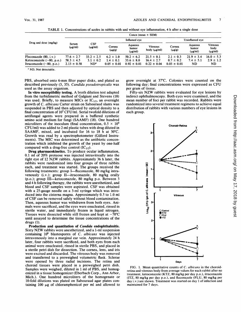
,-3La
E12
0I
'a0
--Fn
U.
tL<0-i
Choroid-Retina
I,
Vitreous
OControLZ1 FLU* KTZ*AMB*ITZ
'7 14 '7 14DAYS DAYS
FIG. 2. Quantitative cultures of the choroid-retina and vitreousbody after rabbits received no therapy, ketoconazole, itraconazole,fluconazole, or amphotericin B (AMB, 1 mg/kg i.v.) daily for 1week. Therapy was started on day 7 of infection and maintained for7 days. See the legend to Fig. 1 for other doses and abbreviations.
TABLE 2. Drug concentrations in serum of rabbits treated forCandida endophthalmitis
Time concn Mean concn inDose No. of measuredDrug (mg/kg) doses (h after SEMlast dose) ±SM
Amphotericin B 1 1 2 1.2 ± 0.041 24 0.8 ± 0.017 24 1.1± 0.03
Itraconazole 80 1 2 2.31 ± 0.41 24 4.33 ± 0.37 24 6.78 ± 0.5
Ketoconazole 80 1 2 72.09 ± 1.11 24 14.29 ± 1.87 24 11.63 ± 6.0
Fluconazole 80 1 2 65.36 ± 5.51 24 33.90 ± 2.67 24 53.85 ± 4.2
serum. Ketoconazole was present in both uninflamed andinflamed eyes by our assays, with the latter having signifi-cantly higher concentrations. Interestingly, significantlyhigher concentrations of ketoconazole were found in theaqueous humor than in CSF.The concentrations of these three azoles in the vitreous
body are shown in Table 1. These levels were approximately20 and 5% of the concentrations in serum for fluconazole andketoconazole, respectively. Intraconazole could not be de-tected in the uninflamed eye. Inflammation enhanced pene-tration of all azoles into this ocular compartment.The tissue concentrations of the azole compounds ob-
tained from corneal specimens are shown in Table 1. Mea-surable amounts of all three azoles were found in thesetissues, with higher amounts in the inflamed eye (P < 0.01)for all three azoles.The in vitro MICs of the azoles against C. albicans (Carter
strain) determined in SAAMF for amphotericin B, ketocon-azole, and fluconazole ranged between 0.4 and 0.8 ,ug/ml.The MIC of itraconazole was at least 10-fold lower thanthose of the other azoles and amphotericin B, which had anMIC of less than 0.025 ,ug/ml.The efficacy of treatment with ketoconazole, itraconazole,
and fluconazole for early Candida endophthalmitis in rabbitsis shown in Fig. 1. All three azoles significantly inhibited thegrowth of C. albicans in the choroid-retina tissue (P =0.0004) and vitreous body (P = 0.0007) when therapy wasstarted 24 h postinoculation. The F test, examining pairwiset tests, showed that all three azoles were similar in effect andbetter than no treatment. Despite low drug levels in ocularfluids and tissue, early treatment with itraconazole was atleast as effective as with ketoconazole and fluconazole.We also examined the efficacy of therapy at a more
advanced stage of infection when the number of organisms inthe retina and choroid had peaked, a feature described byprevious investigators (24). The results of late therapy,started on day 7 of infection, are shown in Fig. 2. Thenumber of organisms in the choroid-retina of animals receiv-ing amphotericin B or ketoconazole was significantly lowerthan the number in the control group after treatment (P <0.05). Some decline of yeast counts in the vitreous body inrabbits receiving these two agents occurred, but the reduc-tions were not statistically significant. Neither of the triazolecompounds, itraconazole and fluconazole, reduced the num-
8 SAVANI ET AL.
on May 17, 2018 by guest
http://aac.asm.org/
Dow
nloaded from
AZOLES AND CANDIDAL ENDOPHTHALMITIS 9
ber of yeasts found in the choroid-retina or vitreous body.Thus, these triazoles had little effect on the more establishedinfection.The various antifunigal therapies were monitored by deter-
mining the concentrations of the corresponding drugs inserum during day 1 of treatment at 2 h and 24 h and duringday 7 at 24 h after administration of the drugs (Table 2).
DISCUSSION
The rabbit provides a simple and convenient model forassessing the pharmacokihetics and efficacy of systemicantifungal antibiotics in hematogenous Candida endophthal-mitis (24). The model is reproducible, is similar in naturalevolution to human infection, and requires no interferencewith host defenses. Cultures can be obtained from differentsites of the eye.
Intraocular penetration of most drugs depends largely onthe permeability of the ocular structures, which varies ininflamed and uninflamed eyes (6). Little is known about theocular penetration of antimycotic agents. Foster andStefanyszyn (15) reported that intravenous infusion of a30-mg/kg dose of miconazole nitrate in rabbits producedpeak aqueous humor levels of 7.9 jxg/ml at 1 h; no drug wasdetected in the vitreous body. Despite another initial reportof good miconazole penetration into the eye (vitreous bodyconcentration of 0.4 jig/ml 1 h after a 700-mg miconazoleinfusion and simultaneous serum concentration of 0.7 ,.g/ml[15, 23]), parenteral miconazole was not effective in prevent-ing the development of hematogenous Candida endophthal-mitis in the rabbit model (24). Also, Blumenkranz andStevens noted progression of ECE in a 19-year-old femaleheroin addict during intravenous miconazole therapy (5).
Ketoconazole, an oral azole compound presently availablefor treatment of mycoses, is a potent inhibitor of--ergosterolsynthesis in C. albicans. It is soluble in organic solvents,absorbed well from the gastrointestinal tract, and metabo-lized by the liver; approximately 90% is protein bound.Ketoconazole has a half-life of 2 to 3 h in humans. In ourstudy a single oral dose of 80 mg of ketoconazole per kggiven to rabbits produced a 4-h mean concentration in serumof 58 + 4.5 ,ug/ml, which is 10-fold higher than that routinelyfound in humans treated with standard doses of 200 to 400mg/day. Although inflammation seems to be an importantfactor in ocular penetration of ketoconazole, we foundmeasurable amounts of ketoconazole in all ocular sitestested, even in uninflamed eyes. Recently, O'Day et al. (27)reported detectable amounts of ketoconazole in the aqueoushumor and vitreous body from a patient undergoing thera-peutic vitrectomy for fungal endophthalmitis. We measuredketoconazole in the aqueous humor of two patients andfound levels in this fluid which were approximately one-quarter of the concentrations found in simultaneous serunmsamples (unpublished data). Although the relationship be-tween penetration and successful treatment of ECE is notclear, successful therapy with ketoconazole has been de-scribed for keratomycosis in both rabbits and humans (21).In a previous study, ketoconazole failed in a rabbit ECEmodel as determined by cultures done after 7 and 14 days oftherapy. However, when ketoconazole was given for 28 daysin this model, the number of organisms was significantlylower than in nontreated controls (24). In this study we wereable to demonstrate a significant decrease in the number ofyeasts in choroid-retina tissue in ketoconazole-treated rab-bits with both early and late therapy. Although a differencein yeast counts was also seen in the vitreous fluid, the
response, to ketoconazole was not significant when therapywas initiated late. We did not examine the possibility thatprolonged treatment with this azole would have successfullytreated all sites of infection.The ocular pharmacokinetics of the two triazoles de-
scribed here are different. Itraconazole (Mr 705) is largerthan fluconazole, very hydrophobic, and more than 90%bound to protein in serum. Fluconazole is a smaller molecule(Mr 306), soluble in water, and only 10 to 20% protein boundin serum. It has a long half-life and is excreted renally.Besides all these advantageous pharmacokinetic properties,fluconazole may be given either p.o. or i.v. Whileitraconazole in vitro has been shown to be very activeagainst a variety of fungi, including Aspergillus species (19),fluconazole appears to have the better pharmacokineticprofile. However, despite the low drug levels found in theeye, itraconazole proved as effective as fluconazole in treat-ing ECE when started early in infection. Efficacy despitepoor drug availability at the site of infection recalls the effectof itraconazole in our cryptococcal meningitis rabbit model(30), in which this drug was active despite undetectable CSFconcentrations. However, no significant responses toitraconazole or fluconazole were seen when therapy wasattempted at a later stage of infection. It is disappointing thatfluconazole also did not clear the vitreous body and choroid-retina of yeasts when therapy was initiated 7 days. postinfec-tion, in view of its high concentration at the site of infection.Outcofie of treatment for ocular infection apparently isunpredictable despite excellent in vitro activity and thebioavailability of a drug at the site of infection. These resultsalso suggest that the success of some azole compounds inendogenous Candida eye infections may depend on howearly in the course of the infection they are started.The ocular pharmacokinetics of i.v. amphotericin B have
been partially defined. In animal nmodel studies amphotericinB was detected only in the aqueous humor and not in thevitreous body (24). In the human eye (29), amphotericin Bhas been found in both the vitreous body and aqueous humorafter i.v. infusion. In one study (13), 0.24 and 0.23 ,ug/mlwere found in the aqueous humor and vitreous body, respec-tively, when the amphotericin B concentration in serum was0.6 ,ug/ml. In this study, we did not attempt to study theocular penetration of amphotericin B. However, because wefelt the need for comparison with new triazole compoundsand ketoconazole, we included amphotericin B in our late-therapy regimen. i.v. amphotericin B was effective followingdaily administration of 1 mg/kg for 7 days when therapy wasinitiated 7 days postinfection. Amphotericin B concentra-tions in rabbit serum were comparable to those found inhumans. This polyene remains the standard for treatment ofoculomycosis, unsurpassed by the newer azole compoundsin this model.Although caution is needed in extending the results of
therapeutic trials from rabbits to humans, especially when asingle dose regimen is used, our study shows that oralketoconazole might be useful for some oculomycoses. Thenew triazole compounds itraconazole and fluconazole didnot fulfill the expectations of being better options thanketoconazole for ECE in the rabbit model. However, theirspectrum of antifungal activity and ocular pharmacokineticsin the rabbit make these agents worthy of further examina-tion.
LITERATURE CITED1. Abel, R., and G. L. Boyle. 1976. Dissecting ocular tissue for
intraocular drug studies. Invest. Ophthalmol. 15:216-219.
VOL. 31, 1987
on May 17, 2018 by guest
http://aac.asm.org/
Dow
nloaded from
ANTIMICROB. AGENTS CHEMOTHER.
2. Aguillar, G. L., M. S. Blumenkranz, P. R. Egbert, and J. P.McCulley. 1979. Candida endophthalmitis after intravenousdrug abuse. Arch. Ophthalmol. 97:96-100.
3. Barza, M., A. Kane, and J. Baum. 1982. The effects of infectionand probenecid on the transport of carbenicillin from the rabbitvitreous. Invest. Ophthalmol. Visual Sci. 22:720-726.
4. Bennett, J. S., E. J. Brodie, E. Brenner, and W. M. M. Kirby.1966. Simplified accurate method for antibiotic assay of clinicalspecimens. App. Microbiol. 14:170-177.
5. Blumenkranz, M. S., and D. A. Stevens. 1980. Therapy ofendogenous fungal endophthalmitis-miconazole or amphoter-icin B for coccidioidal and candidial infection. Arch.Ophthalmol. 98:1216-1219.
6. Cambie, E. 1979. General rules in ocular permeability. Bull.Soc. Belge Ophthalmol. 186:3-8.
7. Cantrill, H. S., W. P. Rodman, R. C. Ramsey, and W. H.Knobloch. 1980. Post-partum candida endophthalmitis. J. Am.Med. Assoc. 243:1163-1165.
8. Clarkson, J. G., and W. R. Green. 1979. Endogenous fungalendophthalmitis, p. 1-44. In T. D. Duane (ed.), Clinical oph-thalmology, vol. 3. Harper & Row, New York.
9. Dupont, B., and E. Drouhet. 1985. Cutaneous ocular andosteoarticular candidiasis in heroin addicts. New clinical andtherapeutic aspects in 38 patients. J. Infect. Dis. 152:577-591.
10. Edwards, J. E., Jr. 1982. Candida endophthalmitis, p. 381-397.In J. S. Remington and M. N. Swartz (ed.), Current clinicaltopics in infectious diseases, vol. 3. McGraw-Hill, New York.
11. Edwards, J. E., Jr. 1985. Candida endophthalmitis, p. 211-225.In G. P. Bodey and V. Fainstein (ed.), Candidiasis. RavenPress, New York.
12. Edwards, J. E., R. Y. Foos, and J. Z. Montgomerie. 1974. Ocularmanifestation of candida septicemia. Review of 76 cases ofhematogenous candida endophthalmitis. Medicine 53:47-75.
13. Fisher, J. F. 1983. Penetration of amphotericin B into the humaneye. J. Infect. Dis. 147:164-165.
14. Foster, C. S. 1980. Ocular toxicity of topical antifungal agents.Arch. Ophthalmol. 99:1081-1084.
15. Foster, C. S., and M. Stefanyszyn. 1979. Intraocular penetrationof miconazole in rabbits. Arch. Ophthalmol. 97:1703-1706.
16. Foster, R. K., R. L. Abott, and H. Gelender. 1980. Managementof infectious endophthalmitis. Ophthalmology 87:313-318.
17. Freeman, J. B., P. L. Davis, and L. D. Maclean. 1974. Candidaendophthalmitis associated with intravenous hyperalimentation.Arch. Surg. 108:237-240.
18. Galgiani, J. N., and D. A. Stevens. 1976. Antimicrobial suscep-tibility testing of yeast: a turbidimetric technique independent ofinoculum size. Antimicrob. Agents Chemother. 10:721-726.
19. Graybill, J. R., and J. Ahrens. 1985. Itraconazole treatment of
murine aspergillosis. Sabouraudia 23:219-223.20. Griffin, J. R., T. H. Pettit, and L. S. Fishman. 1973. Blood-borne
candida endophthalmitis: a clinical and pathologic study of 21cases. Arch. Ophthalmol. 89:450-456.
21. Ishibashi, Y., and T. Matsumoto. 1984. Oral ketoconazoletherapy for experimental candida albicans keratitis in rabbits.Sabouraudia 22:323-330.
22. Jones, D. B. 1975. Principles in the management of ocu-lomycosis. Am. J. Ophthalmol. 79:719-751.
23. Jones, D. B. 1978. Therapy of post-surgical fungal endophthal-mitis. Ophthalmology 85:357-373.
24. Jones, D. B., M. T. Green, M. S. Osato, P. H. Broberg, andL. 0. Gentry. 1981. Endogenous Candida albicans endophthal-mitis in the rabbit. Chemotherapy for systemic effort. Arch.Ophthalmol. 99:2182-2187.
25. Jorgensen, J. H., G. A. Alexander, J. R. Graybill, and D. J.Drutz. 1981. Sensitive bioassays for ketoconazole in serum andcerebrospinal fluid. Antimicrob. Agents Chemother. 20:59-62.
26. McDonnell, P. J., J. M. McDonnell, R. H. Brown, and W. R.Green. 1985. Ocular involvement in patients with fungal infec-tions. Ophthalmology 92:706-709.
27. O'Day, D. M., W. S. Head, R. D. Robinson, W. H. Stern, andJ. M. Freeman. 1985. Intraocular penetration of systemicallyadministered antifungal agents. Curr. Eye Res. 4:131-134.
28. Palmer, E. A. 1980. Endogenous candida endophthalmitis ininfants. Am. J. Ophthalmol. 89:388-395.
29. Perfect, J. R., and D. T. Durack. 1985. Penetration of imidazolesand triazoles into cerebrospinal fluid of rabbits. J. Antimicrob.Chemother. 16:81-86.
30. Perfect, J. R., D). V. Savani, and D. T. Durack. 1986. Compar-ison of itraconazole and fluconazole in the treatment of crypto-coccal meningitis and Candida pyelonephritis in rabbits. Anti-microb. Agents Chemother. 29:579-583.
31. Perraut, L. E., Jr., L. E. Perraut, B. Bleiman, and J. Lyons.1981. Successful treatment of C. albicans endophthalmitis withintravitreal amphotericin B. Arch. Ophthalmol. 99:1565-1567.
32. Speer, M. D., H. M. Hittner, and A. J. Rudolph. 1980. Candidaendophthalmitis: a manifestation of candidiasis in the neonate.South. Med. J. 73:1407-1409.
33. Stern, G. A., M. Okumoto, and G. Smolin. 1979. Combinedamphotericin B and rifampin treatment of experimental C.albicans keratitis. Arch. Ophthalmol. 79:721-722.
34. Walsh, J. A., D. A. Halft, M. H. Miller, M. R. Loran, and A. H.Friedman. 1978. Ocular penetration of 5-flucytosine. Invest.Ophthalmol. Visual Sci. 17:691-694.
35. Young, P., M. Barza, A. Kane, and J. Baum. 1979. Radioactiveand bioassay of intraocular antibiotics. Arch. Ophthalmol.97:717-720.
10 SAVANI ET AL.
on May 17, 2018 by guest
http://aac.asm.org/
Dow
nloaded from