Embed Size (px)
Citation preview

Copyright ©2001 Lippincott Wil l iams & Wilkins Bucholz, Robert W., Heckman, James D. Rockwood & Green's Fractures in Adults, 5th Edit ion
OSTEOMYELITIS
Part of "15 - COMPLICATIONS"
The term osteomyelit is usually refers to bone infections caused by bacteria; however, certain fungi such Blastomyces dermatidit is and Coccidioides immitis can occasionally be responsible (956). Over the past 20 years there has been a signif icant change in the organisms responsible for chronic osteomyelit is. The incidence of gram-negative and polymicrobial infections has increased signif icantly. The incidence of pure S. aureus infections has fal len dramatically. Osteomyelit is can arise from organisms that have reached the bone via the bloodstream (hematogenous osteomyelit is) or organisms that have reached bone via the external environment from trauma (exogenous osteomyelit is). These can further be subdivided into acute, subacute, and chronic osteomyelit is.
Classification Historically, osteomyelit is was classif ied as either acute or chronic depending on the duration of symptoms. A classif ication system based on the etiology of the osteomyelit is was developed: type I (hematogenous), type II (osteomyelit is with fracture union), type III (osteomyelit is without fracture union), and type IV (postoperative or posttraumatic osteomyelit is without fracture). Weiland et al. (968) in 1984 suggested another classif ication scheme based on the nature of the bony involvement. The categories in this system were the fol lowing: type I—open, exposed bone without soft t issue infection; type II—circumferential cortical and endosteal infection; and type III—associated with a segmental defect. May and co-workers (593) in 1989 proposed yet another classif ication scheme for osteomyelit is, focusing on the t ibia. This system was based on the nature of the bone following soft t issue and bony debridement. They proposed the fol lowing categories: type I—intact t ibia and fibula able to withstand functional loads with no reconstruction needed; type II—intact t ibia unable to withstand functional loads requiring bone graft ing; type III—<6 cm tibial defect with an intact f ibula requiring cancellous bone grant, t ibiofibular synostosis, or distraction histogenesis; type IV—<6 cm tibial defect and intact f ibula requiring distraction histogenesis, t ibiofibular synostosis, or a vascularized bone graft; and type V—≥ l6 cm tibial defect without an intact f ibula requiring a possible early amputation.
Probably the most widely used system of classif ication for adult osteomyelit is is that of Cierny and Mader (157,158). This system is based on four factors: the degree of osseous involvement, the site of involvement, the degree of impairment caused by the disease, and the general condit ion of the host (158) (Table 15-37; Fig. 15-9). Type I is medullary
P.516
P.517
Página 1 de 26Ovid: Rockwood & Green's Fractures in Adults
03/03/05http://gateway.ut.ovid.com/gw2/ovidweb.cgi

osteomyelit is (examples of which include hematogenous osteomyelit is and infections of intramedullary rods). Type II is superficial osteomyelit is confined to the bone surface. Type III is localized osteomyelit is involving the ful l thickness of the cortex. Type IV is dif fuse osteomyelit is involving the circumference of the cortex. The general condit ion of the patient is based on those factors that affect the response to infection and treatment. Class A patients have normal systemic defenses, metabolic capabil i t ies, and vascular supply to the limb. Class B patients have a local or systemic deficiency in wound healing (immunosuppressed, on corticosteroids, peripheral vascular disease). Class C patients are those in whom the treatment morbidity is worse than the presenting condit ion (156). These patients have a poor prognosis for cure.
TABLE 15-37. CIERNY AND MADER STAGING SYSTEM
Página 2 de 26Ovid: Rockwood & Green's Fractures in Adults
03/03/05http://gateway.ut.ovid.com/gw2/ovidweb.cgi

Pathogenesis of Musculoskeletal Infection Microorganisms are primitive representations and precursors of t issue cells. They often exist as microbial aggregates in a biofi lm sl ime layer colonizing surfaces within which they are resistant to antagonists. Nutrients and free energy are concentrated at the surfaces of the three-dimensional architecture of these bacterial colonies. Colonization of surfaces is not always, synonymous with infection. Bacteria such as Staphylococcus epidermidis , streptococcol species, and gram-negative groups are naturally found on skin, oral mucosa, and the intestinal tract. However, through evolution, bone and carti lage have not been intended to support bacterial colonization and thus are especially susceptible to infection.
The first step in bacterial colonization is the process of adhesion to permanent t issue or biomaterial attachment (377). Typically, the host's defense mechanisms are usually able to eliminate these bacteria. There are, however, situations in which bacteria remain viable: (a) the inoculum is larger than threshold levels, (b) host defense mechanisms are impaired, (c) the t issue on which the bacteria colonize is traumatized, (d) a foreign body is present, and (e) the surface (or t issue) is acellular or inanimate (e.g., bone, carti lage, and biomaterials).
Surface adhesion of bacteria depends on physical characteristics of the bacterium, the f luid interface, and substratum (199,338,377). Adhesion is based on time-dependent specif ic
FIGURE 15-9. The four types of chronic osteomyelitis shown diagrammatically. (From Cierny G III, Mader JT. Orthopaedics 1984;7:10, with permission.)
Página 3 de 26Ovid: Rockwood & Green's Fractures in Adults
03/03/05http://gateway.ut.ovid.com/gw2/ovidweb.cgi

protein adhesion-receptor interactions, as well as carbohydrate polymer synthesis in addit ion to charge and physical forces (377).
Bacteria randomly arrive near a bone surface or biomaterial surface by direct contamination, contiguous spreading, or hematogenous seeding. Any surface, whether a eukaryotic cell or a biomaterial, exposed to a biologic environment spontaneously acquires a glycoproteinacous f i lm (293,366,377,278,379 and 380,388). The surface is anionic and init ial ly repels bacteria (which are also anionic). Juxtaposit ion of the bacteria with a t issue surface or biomaterial is therefore accomplished by Van der Waals forces. This interaction allows bacteria to develop “irreversible” cross-links with the surface (adhesin receptor interaction) (199,256,377). Following anchorage to the surface, bacteria begin to proliferate within the polysaccharide sl ime layer and a “biofi lm-enclosed” colony of bacteria forms. Biofi lm (or sl ime) is formed by bacterial extracapsular exopolysaccharides that bind to surfaces and participate in cell-to-cell aggregation. In these biofi lms, the bacteria are resistant to host defenses and may resist antibiotic action. Thus, infection may develop, spread, and persist in this environment.
Adherence to Bone Damaged bone acts as a substratum for bacterial colonization. Bone is a relatively acellular composite structure of calcium hydroxyapatite crystals and a collagen matrix. This organic matrix is arranged as helical polypeptides composed of proline, hydroxyproline, glycine, and alanine (579). Proline-rich proteins can act as l igands for bacterial adhesion. Devital ized bone devoid of normal periosteum presents a collagen matrix to which bacteria can bind. Moreover, bone sialoprotein has also been suggested as a l igand for bacterial binding to bone (377).
Adherence to Biomaterials Many implants consist of one or more metal or polymer. Biomaterials and other foreign bodies are usually inert and susceptible to bacterial colonization because they are inanimate. Regardless of how inert a metal is, it may sti l l modulate molecular events at i ts surfaces, specif ically receptor-l igand interactions, covalent bonding, and thermodynamic interactions (293,388). The most important feature a particular metal has is its outer surface atomic layer interaction with glycoproteins and prokaryotic and eukaryotic cells. Stainless steel and cobalt-chromium and titanium alloys are resistant to corrosion through elemental composit ion, crystall ine homogeneity, and the surface oxide passivates. Surface oxides form a reactive interface with bacteria.
Hydrophobicity is often used as a parameter to evaluate the cellular adhesiveness of a biomaterial. Certain strains of bacteria owe their hydrophobicity to their synthesized “biofi lm” coating. When this coating is washed away, the bacteria become hydrophil ic. In general, hydrophobic bacteria adhere better to hydrophobic surfaces. Polymers tend to be more hydrophobic than metals (377).
Antibiotic Resistance
P.518
Página 4 de 26Ovid: Rockwood & Green's Fractures in Adults
03/03/05http://gateway.ut.ovid.com/gw2/ovidweb.cgi
Following bacterial adherence, the resistance to antibiotics increases (663,673). This resistance property seems to be dependent on the type of surface to which the organisms are attached. Organisms that adhere to hydrocarbon polymers are extremely resistant to antibiotics. These same organisms, when attachedto metals, do not resist antibiotic therapy to the same extent. It has been theorized that bacteria within biofi lms have a decreased metabolic rate and undergo phenotypic changes that may influence resistance and virulence (377). It has also been shown that bacteria adherent to surfaces are more resistant to antibiotics than free-floating bacteria.
Pathophysiology of Osteomyelitis
Acute Hematogenous Osteomyelitis I t is believed that the vascular architecture of the metaphysis, where the nutrient capil laries form sharp loops, predisposes to the establishment of infection fol lowing bacteremia. Bacteria become lodged in the small end arteries and multiply. Shortly thereafter, blood and white blood cells accumulate, result ing in further compromise of blood flow and pressure necrosis of surrounding bone. The pus moves along the haversian canals and medullary canal, which are paths of least resistance. When the pus reaches the cortical surface, it moves beneath the periosteum.
The most commonly involved organism in all age groups is S. aureus (193), which is responsible for between 50% and 70% of al l such infections in children between 1 month and 5 years of age (193). The second most common organisms are hemolytic streptococci. Both group A and group B streptococci have been implicated in hematogenous osteomyelit is, the latter particularly in the f irst 2 months of l i fe. In neonates, Haemophilus influenzae is an occasional cause of osteomyelit is, but more commonly of septic arthrit is (193).
Damage to the metaphyseal blood supply, caused by the release of bacterial toxins and the stripping of the periosteum, results in portions of the bone becoming necrotic. The inner portion of the cortex is supplied by the injured metaphyseal vessels. These areas of dead bone separated from viable bone by granulation t issue are called sequestra. The body responds to sequestra by laying down new bone, involucrum, which surrounds the area of infected bone.
Generally, there are three possible patterns of vascularity. In the infant, epiphyseal extensions of the nutrient artery may allow infections that originate in the metaphysis to seed into the epiphysis. In children, the nutrient artery terminates in end arteries and capil laries adjacent to the growth plate. An infection of the metaphysis is usually prevented from crossing the growth plate. In adults, however, the metaphysis and epiphysis are in continuity, and hematogenous osteomyelit is may be metaphyseal or epiphyseal. The high incidence of infections in the distal femur and proximal t ibia may be associated with the relative contributions these regions have to growth.
Chronic Nonhematogenous Osteomyelitis Acute osteomyelit is that is inappropriately treated can become chronic osteomyelit is.
Página 5 de 26Ovid: Rockwood & Green's Fractures in Adults
03/03/05http://gateway.ut.ovid.com/gw2/ovidweb.cgi
General factors that may predispose to this condit ion include degree of bone necrosis, nutrit ion, infecting organism, age of the patient, comorbidity, and drug abuse (193). The infecting organism varies, generally, with the cause of chronic osteomyelit is. Chronic osteomyelit is result ing from acute osteomyelit is is often caused by S. aureus; however, chronic osteomyelit is occurring after a fracture can be polymicrobial (although in most cases, it is S. aureus). Intravenous drug users are commonly found to have Pseudomonas or S. aureus infections. Gram-negative organisms are now seen in about 50% of al l cases of chronic osteomyelit is (193).
Nonhematogenous osteomyelit is is secondary to a contiguous focus of infection. The bacterial organisms enter the bone directly through interrupted tissue planes as a result of fractures or surgical procedures. Bacteria in bone by themselves are insufficient to produce osteomyelit is. A surgical or traumatic insult sets the stage for the secondary infection (646). Periosteum and muscle are injured, creating regions of cortical bone that no longer are perfused adequately. Devital ized bone presents a collagen protein matrix and acellular crystal regions to which bacteria can bind directly (379). The fundamental problem in chronic osteomyelit is is infection that devascularizes a segment of bone, leaving protected pockets of necrotic material to support bacterial growth in relative seclusion from systemic antibiotic therapy.
Posttraumatic Osteomyelitis Posttraumatic osteomyelit is is a bone infection result ing from trauma that allows pathogenic organisms to enter bone, proliferate in traumatized tissue, and cause a subsequent infection (611). The environmental condit ions that promote infection and tissue damage are present in the face of trauma. Traumatized soft t issue and bone exposes potential binding sites for bacteria such as collagen (924). Traumatized tissue also results in compromised blood supply, leading to t issue and bone necrosis, which promotes infection (924). Moreover, the f ixation devices that are required in the management of fractures serve as addit ional foci for bacterial colonization (924). In the patient with traumatic injuries, addit ional factors that contribute to the subsequent development of osteomyelit is are the presence of hypotension, inadequate debridement of the fracture site, malnutrit ion, alcoholism, and smoking (272,434,912).
Trauma may lead to interference with the host response to infection. Tissue injury or the presence of bacteria tr iggers activation of the complement cascade that leads to local vasodilation, t issue edema, migration of polymorphonuclear leukocytes (PMNs) to the site of injury, and enhanced abil i ty of phagocytes to ingest bacteria (17). Trauma has been reported to delay the inflammatory response to bacteria, to depress cell-mediated immunity, and to impair function of PMNs, including chemotaxis, superoxide production, and microbial ki l l ing (435). The commonly used system of Cierny and Mader has been shown to have greatest correlation with the general condit ion of the patient rather than the specif ics of bone involvement.
Organisms in Trauma The presence of bacteria in an open wound is not suff icient to cause infection.
P.519
Página 6 de 26Ovid: Rockwood & Green's Fractures in Adults
03/03/05http://gateway.ut.ovid.com/gw2/ovidweb.cgi
Approximately 60% to 70% of open fractures are contaminated by bacteria, but a much smaller percentage go on to develop infection (924). Moreover, the bacteria recovered from clinical infections are most l ikely to be hospital acquired pathogens such as S. aureus or gram-negative bacil l i ( including Pseudomonas aeruginosa) (390,567) (Table 15-38). However, other bacteria should be considered, depending on the environment, specif ically, C. perfringens and farm injury, Pseudomonas Aeromonas hydrophil ia fol lowing fresh water injury, and Vibrio and Erysipelothrix in salt water injury. There are some unusual organisms that can cause osteomyelit is (Table 15-39). Bacteria that cause infection are those that can colonize host t issue and cause damage. In humans, the risk of subsequent infection is highly correlated with the degree of soft t issue injury associated with an open fracture (924). Infection rates in type IIIB open fractures, those in which extensive soft t issue stripping does not al low for adequate coverage over the site, are prone to infection in up to 40% of cases (193).
Página 7 de 26Ovid: Rockwood & Green's Fractures in Adults
03/03/05http://gateway.ut.ovid.com/gw2/ovidweb.cgi
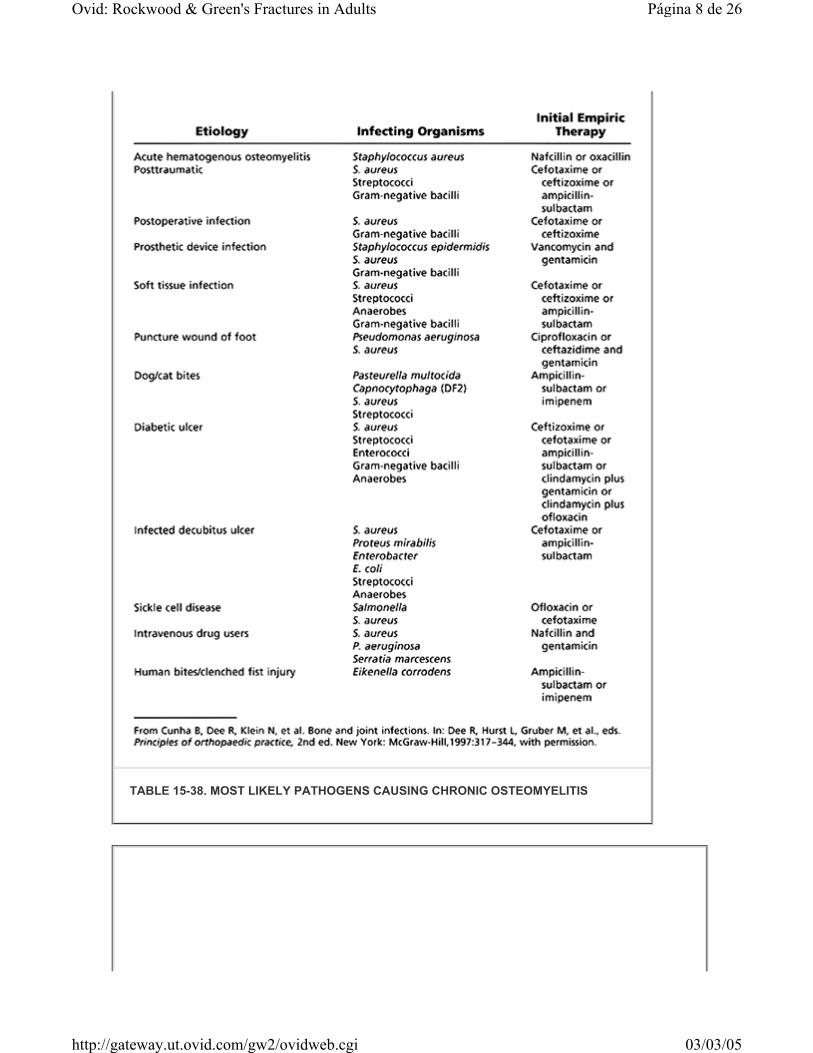
TABLE 15-38. MOST LIKELY PATHOGENS CAUSING CHRONIC OSTEOMYELITIS
Página 8 de 26Ovid: Rockwood & Green's Fractures in Adults
03/03/05http://gateway.ut.ovid.com/gw2/ovidweb.cgi

Clinical Manifestations Patients often have a history of infection of another site, such as the throat or skin, or have a history of trauma. They usually complain of substantial pain in the affected area. They may have diff iculty with weight bearing. Moreover, reduced activity, malaise, and anorexia may be exhibited. General physical f indings include fever, tachycardia, and list lessness. Local f indings include swell ing and warmth, occasional redness, tenderness to palpation, drainage, and restricted range of motion of adjacent joints.
With a history of trauma, cl inical r isk factors for infection include a history of open fracture, severe soft t issue injury, a history of substance abuse and smoking, inadequate previous treatment, and an immunocompromised state. Clinical factors affecting treatment that need to be assessed include the t ime of onset of the infection, the status of the soft t issues, the viabil i ty of the bone, the status of fracture healing, implant stabil i ty, the condit ion of the host, and the neurovascular exam.
Laboratory Findings Elevations in the white blood cell count with a left shift, erythrocyte sedimentation rate, and C-reactive proteins are often seen. The erythrocyte sedimentation rate may be normal in the f irst 48 hours but r ises to levels above 100 mm/h, and may remain elevated for weeks (193). Its gradual declination is an indicator of treatment response. Since bacteremia and septicemia are often present, blood cultures should be performed to identify infecting bacteria. Blood cultures are posit ive in about 50% to 75% of cases (193). Although not a sensit ive test for septic arthrit is, the detection of antibodies (C-reactive protein) to the teichoic acid cell wall of S. aureus has been useful in the detection of acute osteomyelit is.
TABLE 15-39. UNUSUAL ORGANISMS CAUSING OSTEOMYELITIS
Página 9 de 26Ovid: Rockwood & Green's Fractures in Adults
03/03/05http://gateway.ut.ovid.com/gw2/ovidweb.cgi

The sensit ivity approaches 82% in the acute situation and declines to 43% in chronic osteomyelit is (193).
Radiographic Imaging Within the f irst few days, soft t issues near the metaphyseal region of the bone may appear swollen. Radiographically, detectable demineralization may not be seen for at least 10 days (125). When present, i t usually signif ies trabecular bone destruction. If the infection spreads to the cortex (usually within 3 to 6 weeks), a periosteal reaction may be evident. Unfortunately, radiologic f indings in the init ial presentation of acute osteomyelit is are often normal. The most common radiographic sign of early bone infection is rarefaction, representing diffuse demineraliza-tion secondary to inflammatory hyperemia. One study reported that in cases of eventually proven osteomyelit is, 5% of radiographs were abnormal init ial ly, 33% were abnormal by 1 week, and 90% were abnormal by 4 weeks (987).
Bone Scans Technetium-99m is the principal radioisotope employed in most bone scans (37,61,102,123,186,216,260,283,292,342,407,442,623). Technetium is formed as a metastable intermediate during the decay of molybdenum-99. It is relatively inexpensive since it is readily available (201). Technetium decays with a 6-hour half- l i fe. After intravenous injection, there is rapid distribution of this agent throughout the extracellular f luid. Within several hours, more than half the dose wil l accumulate in bone, while the remainder is excreted in the urine. There is evidence to suggest that the technetium phosphates bind to both the organic and the inorganic matrix. There is preferential incorporation into metabolically active bone.
Bone images are usually acquired 2 to 4 hours fol lowing intravenous injection of the radioisotope. A tr iple phase bone scan is one that is useful for examining inflammation and related processes. Following the init ial injection, dynamic images are captured over the specif ied region. These are fol lowed by static images at later t ime points. The first phase represents the blood flow phase, the second phase immediately postinjection represents the bone pooling phase, and the third phase is a delayed image made at 3 hours when there is decreased soft t issue activity. Classically, osteomyelit is presents as a region of increased blood flow and should appear “hot” on all phases, with focal uptake in the third phase. Other processes such as a healing fracture, a loose prosthesis, and degeneration do not appear hot in the early phase despite a hot appearance in the delayed phase. Reported sensit ivit ies of bone scintigraphy for the detection of osteomyelit is vary considerably from 32% to 100%. Reported specif icit ies have ranged from 0% to 100% (806,954).
Gall ium-67 citrate binds rapidly to serum proteins, particularly transferrin (18,61,95,123,785,818). There is uptake in the blood, especially by leukocytes. Gall ium has been used in conjunction with technetium-99 to increase the specif icity of the bone scan (18,61,123,260,407). Several mechanisms have been postulated to explain the increased
P.520
P.521
Página 10 de 26Ovid: Rockwood & Green's Fractures in Adults
03/03/05http://gateway.ut.ovid.com/gw2/ovidweb.cgi

activity at sites of inflammation. Enhanced blood flow and increased capil lary permeabil i ty cause enhanced delivery. Bacteria have high iron requirements and thus avidly take up gall ium. Gall ium is strongly bound to bacterial siderophores and leukocyte lactoferins. In regions of inflammation, these proteins are available extracellularly and can bind gall ium avidly. Chemotaxis also acts to localize gal l ium-labeled white blood cells at the sites of infection. In a typical study, gall ium is injected intravenously and delayed images are acquired (at 48 to 72 hours).
The hallmark of osteomyelit is is focal increased uptake of gall ium. Unfortunately, gall ium's nonspecif ic bone uptake can be problematic since any processes causing reactive new bone formation wil l “ l ight up.” In the case of patients with fractures or a prosthesis, osteomyelit is cannot be diagnosed with gall ium alone. Most authors wil l interpret gall ium images along with bone scans. Gall ium activity is interpreted as abnormal if it is either incongruous with the bone scan activity or if there is a matching pattern with gall ium activity.
Reported sensit ivit ies and specif icit ies for the diagnosis of osteomyelit is range from 22% to 100% and 0% to 100%, respectively (806). Despite its lower than optimal diagnostic value, gall ium sti l l has advantages: (a) i t is easily administered, (b) it is the agent of choice in chronic soft t issue infections (less effective in bone infections), and (c) i t is a useful agent in following the resolution of an inflammatory process by showing a progressive decline in activity.
Indium-111–Labeled Leukocytes Due to the variable accuracy of both technetium and gall ium scans, most laboratories routinely employ indium-111–labeled leukocytes (202,203,260,295,574,598,699,802,805,817,828,940,1004). Indium white blood cell (WBC) preparations require withdrawing approximately 50 mL of autologous whole blood with a leukocyte count of at least 5,000 cells/mm3. Allowing 1 hour for optimal sedimentation of the whole blood in a heparinized syringe, the leukocyte-rich upper layer is carefully removed by catheter and placed in a centrifugation tube. After centrifugation, the leukocyte-poor plasma supernatant is removed and saved, while the leukocyte-containing pellet is resuspended in normal saline. The leukocytes are labeled with 1 mCi of indium oxine at room temperature for 30 minutes. The unbound indium and oxine are removed by centrifugation. Following intravenous injection, the labeled WBCs redistribute in the intravascular space. Immediate images show activity in the lungs, l iver, spleen, and blood pool. The half- l i fe is about 7 hours. After 24 hours, only the l iver, spleen, and bone marrow show activity. Normal-healing wounds and ful ly treated infections show no increase in uptake.
Most results show improved sensit ivity (80–100%) and specif icity (50–100%) for the diagnosis of osteomyelit is (622). Indium-labeled WBC scans are generally superior to bone scans and gall ium scans in the detection of infection. McCarthy et al. (598) have reported the diagnostic uti l i ty of indium scans in 39 patients with suspected osteomyelit is (confirmed by bone biopsy). Indium scans were 97% sensit ive and 82% specif ic for osteomyelit is. The few false-posit ive results occurred in patients with
P.522
Página 11 de 26Ovid: Rockwood & Green's Fractures in Adults
03/03/05http://gateway.ut.ovid.com/gw2/ovidweb.cgi

overlying soft t issue infections. An accompanying bone scan can aid in differentiating bone infection from soft t issue infection. In these situations, the indium scan should be performed before the bone scan to avoid false-posit ive results (from the remaining technetium uptake).
Indium-Labeled Polyclonal Immunoglobulins This nonspecif ic polyclonal immunoglobulin (IgG) prepared from human serum gamma globulin and labeled with indium via diethylenetriamine pentaacetic acid (DTPA) chelation is another agent used for the detection of osteomyelit is (700,785). Unlike the indium-labeled WBC scans, this agent is easily prepared by simply adding indium chloride to a sterile kit containing a DTPA-IgG complex. The blood half- l i fe is 24 hours and the primary uptake occurs in the liver. Compared with indium-labeled WBC scans, this technique is easier to perform and has less bone marrow uptake.
Magnetic Resonance Imaging Magnetic resonance imaging continues to play an important role in the evaluation of musculoskeletal infections (74,88,589,634,757,901,914,930). It has the spatial resolution necessary to evaluate accurately the extent of the infection in preparation for surgical treatment and localizes any abscess cavit ies. It also has the abil i ty to differentiate between infected bone and involved adjacent soft t issue structures. Images can be acquired in any orientation and there is no radiation exposure. Characteristically, active osteomyelit is displays a decreased signal on T1-weighted images and appears bright on T2-weighted images. The process represents the replacement of marrow fat with water from edema, exudate, hyperemia, and ischemia. The MRI signal characteristics that reflect osteomyelit is are intrinsically nonspecif ic: tumors and fractures can also increase the marrow water content. In patients without prior complications, MRI has been found to be sensit ive (but not specif ic) for osteomyelit is. When a fracture or prior surgery is evident, MRI is less specif ic in the diagnosis of infection. In these situations, indium-labeled WBC scans are helpful. Although l imited, studies show good results in the evaluation of acute osteomyelit is (sensit ivit ies: 92% to 100%, specif icit ies: 89% to 100%) (806).
Computed Tomography Computed tomography has assumed a lesser role in the evaluation of osteomyelit is with the widespread use of MRI (205). It remains unsurpassed, however, in the imaging of cortical bone. It is especially useful in delineating the cortical details in chronic osteomyelit is, such as sequestra and foreign bodies (902). It also is useful in evaluating the adequacy of cortical debridement in the staged treatment of chronic osteomyelit is.
Positron Emission Tomography Positron emission tomography may play an important role in the evaluation of musculoskeletal infections. The technique enables noninvasive detection and demonstration of the extent of chronic osteomyelit is with 97% accuracy (385). Positron emission tomography is especially accurate in the central skeleton within active bone
Página 12 de 26Ovid: Rockwood & Green's Fractures in Adults
03/03/05http://gateway.ut.ovid.com/gw2/ovidweb.cgi
marrow (385).
Approach to Imaging and Posttraumatic Osteomyelitis Since bone scan agents and gall ium are usually both posit ive at fracture sites, they have limited value in the detection of infection fol lowing a fracture. With no discernible uptake in reactive bone, indium-labeled WBC scans reveal superiority in the detection of infection following a fracture. In a prospective study of 20 patients with suspected osteomyelit is superimposed on a delayed union, Esterhai et al. (261) reported 100% accuracy of indium-WBC scans (261). Seabold et al. (818) have shown that using indium-WBC scans and bone scans (to differentiate between soft t issue infections) can be 97% specif ic for osteomyelit is.
In chronic or recurrent osteomyelit is, bone scans alone are of l i t t le value since they show increased uptake for up to 2 years fol lowing successful treatment and resolution of infection (365). Although gall ium scans have historically been shown to be the most optimal for fol lowing the resolution of chronic osteomyelit is, indium WBC scans appear to be superior. Merkel et al. (622), in a prospective study of 50 patients comparing indium-WBC and gall ium scans for the detection of osteomyelit is, found that indium-WBC scans were 26% more accurate than gall ium scans (83% vs. 57%).
Aspiration If the diagnosis of septic arthrit is is suspected, it must be ruled out. The most important step in the init ial diagnosis of a septic joint is aspiration. The procedure should be performed with aseptic technique. The joint should not be entered through an infected area of overlying skin. The skin should be prepared with an iodine-based solution or alcohol. An 18-gauge (or larger) needle should be introduced into the joint and the f luid aspirated. The fluid should be analyzed for cell count, differential microscopic evaluation, glucose determination, Gram stain, and culture (aerobes, anaerobes, fungi, mycobacteria).
Biopsy Identif ication of an organism and determination of antibiotic resistance patterns is crucial to a successful outcome in the management of osteomyelit is. The procedure can usually be done under f luoroscopic guidance. In general, sinus tract cultures should not be used to guide antibiotic treatment (228,332,594,677). In a prospective study, Mousa (652) found that 88.7% of sinus tract isolates were identical to operative specimens in 55 patients with chronic bone infection. These results were dependent on aspiration of material by syringe from the depths of an active f lowing sinus and immediate inoculation on culture media. Bone biopsy remains the preferred diagnostic procedure in chronic osteomyelit is. Histologic and microbiologic evaluation of percutaneous biopsy samples should be combined in cases of suspected osteomyelit is. The sensit ivity of culture in the diagnosis of osteomyelit is could be improved from 42% to 84% by the addit ion of histologic evaluation.
Molecular Diagnostics
P.523
Página 13 de 26Ovid: Rockwood & Green's Fractures in Adults
03/03/05http://gateway.ut.ovid.com/gw2/ovidweb.cgi
These procedures are being developed for diagnosis in osteomyelit is because some infections remain without an identif ied pathogen, when using standard techniques. This is particularly so in those patients who have been treated with antibiotics shortly before sample collection. These methods target specif ic macromolecules unique to the infecting pathogens, which are absent in the host cells (250,908). They have the potential to provide rapid results with high accuracy (436). The most commonly used method for the diagnosis of orthopaedic infections is polymerase chain reaction (436). Sequences within bacterial 16S ribosomal RNA have served as targets for amplif ication and detection (436). Further investigation is required before these techniques can be widely used as they lack suff icient sensit ivity and specif icity.
Treatment The management of osteomyelit is rel ies on a mult idiscipl inary approach, combining debridement, soft t issue coverage, and antimicrobial therapy to give the patient the best chance of cure (245,299,588,592).
General Approach After a cl inical diagnosis of acute osteomyelit is has been made, aspiration of the bone should be performed to identify the pathogen. All material obtained from aspiration should be sent for a Gram stain, culture and sensit ivity (aerobic/anaerobic), and fungal and mycobacterial cultures. Blood cultures should also be obtained. Even though aspiration in the early stage of disease may fail to obtain any f luid or pus, i t is very useful in obtaining the infecting organism. However, there is danger that the pus may be so thick that it may yield a negative aspiration.
In early acute hematogenous osteomyelit is, antibiotic therapy without surgical intervention may result in cure, provided the blood supply has not yet been compromised and adequate antibiotic levels in bone can be obtained. The mainstay of treatment remains rapid identif ication of the responsible pathogen and the init iation of appropriate antibiotic therapy. S. aureus is the most common cause of acute hematogenous osteomyelit is in both children over the age of 1 year and adults (377).
Historically, acute S. aureus osteomyelit is required 4 to 8 weeks of intravenous therapy (750). Subsequent studies have shown that early conversion to oral therapy can be equally effective (752,906). The decision to convert to oral therapy is based on clinical factors. Typically, fever, pain, swell ing, and local inflammation begin to resolve after 4 to 10 days of appropriate intravenous therapy. If an appropriate oral agent is available and the patient is compliant, the remainder of therapy can be completed on an outpatient basis. The duration of treatment depends on the pathogen. S. aureus or enteric gram-negative organisms require a minimum of 4 weeks of treatment. Infections caused by H. influenzae, Neisseria meningit idis , or streptococci may require only 14 days of treatment. The decision to discontinue treatment should be based on both the cl inical response of the patient and a fall ing erythrocyte sedimentation rate (197). Regardless of the method of antibiotic administration, i t appears that maintaining a peak serum bactericidal t i ter of greater than 1:8 (751) or a trough level of greater than 1:2 (973) is predictive of successful therapy.
Página 14 de 26Ovid: Rockwood & Green's Fractures in Adults
03/03/05http://gateway.ut.ovid.com/gw2/ovidweb.cgi

Indications for surgical drainage or debridement must be individualized, and include the presence of a subperiosteal abscess, a coexistent septic arthrit is, and fai lure to respond to appropriate antibiotic therapy after 36 to 48 hours (161,197).
Antibiotic Therapy Factors involved in choosing the appropriate antibiotics include infection type, infecting organism, sensit ivity results, host factors, and antibiotic characteristics (572). Antibiotic classes used in the treatment of osteomyelit is include penici l l ins, β- lactamase inhibitors, cephalosporins, other β- lactams (aztreonam and imipenam), vancomycin, cl indamycin, r i fampin, aminoglycosides, f luoroquinolones, tr imethoprim-sulfamethoxazole, metronidazole, and new agents including teicoplanin, quinupristin/dalfopristin, and oxazolidinones (572) (Table 15-40, Table 15-41, and Table 15-42).
TABLE 15-40. GRAM-POSITIVE ORGANISMS: INITIAL CHOICE OF ANTIBIOTICS FOR THERAPY (ADULT DOSES)
Página 15 de 26Ovid: Rockwood & Green's Fractures in Adults
03/03/05http://gateway.ut.ovid.com/gw2/ovidweb.cgi

Several excellent reviews have been published regarding antibiotic therapy in osteomyelit is (332,333,570,571,883). One of the most important developments in antimicrobial therapy has been the introduction of the oral quinolones. Oral ciprofloxacin has a broad spectrum of activity against both gram-posit ive and gram-negative organisms. It has a long half- l i fe, low toxicity, and excellent penetration into bone (334,372,568). In randomized trials, ciprofloxacin was as safe and effective as parenteral therapy against a wide variety of organisms, particularly P. aeruginosa , with a 2-year success rate of 77% (332). Its eff icacy
TABLE 15-41. GRAM-NEGATIVE ORGANISMS: INITIAL CHOICE OF ANTIBIOTICS FOR THERAPY (ADULT DOSES)
TABLE 15-42. ANAEROBIC ORGANISMS: INITIAL CHOICE OF ANTIBIOTICS FOR THERAPY (ADULT DOSES)
Página 16 de 26Ovid: Rockwood & Green's Fractures in Adults
03/03/05http://gateway.ut.ovid.com/gw2/ovidweb.cgi

against S. aureus is controversial, and some authors suggest the use of combination therapy for the treatment of gram-posit ive infections (332,955). The problem of emerging antibiotic resistance also is under investigation (221). This agent is not approved for use in skeletally immature patients (570).
Most coagulase-negative staphylococci are methici l l in resistant, and are recognized as virulent pathogens (332). Teicoplanin is a new glycopeptide antibiotic with activity similar to vancomycin. It has a suff iciently long half- l i fe to allow once-daily dosing. It can also be administered by intramuscular injection or rapid intravenous infusion. In several tr ials, teicoplanin was as effective as vancomycin against al l gram-posit ive organisms and had a low rate of adverse events. The prospect of once-daily intramuscular dosing and low toxicity may make this agent attractive for the outpatient treatment of methici l l in-resistant gram-posit ive infections (832).
Oral Versus Parenteral Antibiotics Four to 6 weeks of parenteral antibiotic therapy following the last debridement has become the standard length of antibiotic therapy (395,956,957). With increasing health care costs and strategies to contain these expenses, patients can be treated on an outpatient basis with a variety of indwell ing catheters such as peripheral inserted catheter l ines, heparin locks, and implantable catheters. The cost of intravenous antibiotics at home for 6 weeks can range from $3,500 to $10,000 (340). The rate of serious complications from the use of intravenous catheters ranges from 10% to 30% (181,570).
In adults, combined intravenous and oral therapy or oral therapy alone has not been well reported; however, preliminary comparative studies suggest equivalent eff icacy between oral and parenteral ciprofloxacin for susceptible organisms as well as between oral ciprofloxacin and standard parenteral antibiotic therapy (568). In a study comparing the cl inical outcome of treatment of a series of patients with osteomyelit is by single-stage, aggressive surgical debridement and appropriate soft t issue coverage fol lowed by intravenous antibiotics for 5 to 7 days and oral therapy for 6 weeks, with that of a historical series treated with the same surgical protocol and intravenous antibiotics for 6 weeks, Swiontkowski and colleagues (895) found that short-term antibiotic therapy was successful in 91% of patients, and outcome was no different between series. These results suggest that treatment fai lures are caused by inadequate debridement rather than the duration of intravenous antimicrobial treatment.
Local Antibiotics The local deposit ion of antibiotics has received increased attention (84,85,129,271,627,745,837). These techniques use a space-fi l l ing carrier agent that elutes high concentrations of antibiotic into the local t issue. Advantages include local levels of antibiotics that surpass the minimal inhibitory concentration for most pathogens, with minimal systemic levels or complications and the abil i ty to perform primary wound closure. The carrier agent can be either biologic, as in the case of bone graft, demineralized bone matrix, or calcium hydroxyapatite (186), or biologically inert, such as
P.524P.525
Página 17 de 26Ovid: Rockwood & Green's Fractures in Adults
03/03/05http://gateway.ut.ovid.com/gw2/ovidweb.cgi
polymethylmethacrylate (PMMA) or plaster of Paris (198,1000). Carrier agents such as bioabsorbable polymers (DL-lactide:glycolide polymer) (130,326,675), porous apatite-wollastonite glass ceramic, bioerodable polyanhydrides, f ibrin clot (923), and polylactide/polyglycolide implant (326) have been described. Work is also being done to incorporate antibiotics into fracture f ixation implants. In a rabbit study, Darouiche and colleagues (200) found that an antibiotic-coated intramedullary f ixation device reduced the rate of device-related osteomyelit is fol lowing infected fracture f ixation from 62% to 9%.
If staged management is not necessary, then a biologic agent may be more appropriate. It is important to remember that the elution characteristics of each antibiotic/carrier agent combination are unique and should be understood before it is used. The antibiotic should be water soluble, nontoxic to t issue, bactericidal, available in powder form, and heat stable if used in PMMA (745). Miclau and associates (627) performed a direct comparison of the elution rate of tobramycin from bone graft, demineralized bone matrix, plaster of Paris, and PMMA. Cancellous bone graft released 70% of its antibiotic load in the f irst 24 hours. Demineralized bone matrix showed a similar elution, with 45% total release in the f irst 24 hours. Neither agent was detectable at 14 days. Thus, i t is important to adjust the dose of antibiotic mixed with bone graft and demineralized bone matrix to prevent a potential ly toxic serum level secondary to rapid absorption. Plaster of Paris released 17% of i ts antibiotic load over the f irst 24 hours, with measurable elution at 21 days. PMMA eluted only 7% during the f irst 24 hours, with trace amounts detectable at 14 days.
While staged management may be more t ime-consuming, it is generally thought to be safer and more effective. In a cohort study, Chan and colleagues (144) found that a two-stage protocol using antibiotic-impregnated PMMA beads fol lowed by antibiotic-impregnated autogenous cancellous bone graft resulted in an infection arrest rate of 94.4% and had no adverse effects on bone graft incorporation.
Combinations of antibiotics and PMMA have been used extensively in the treatment of infected total joint replacements, and the elution characteristics of many antibiotics from this material have been studied thoroughly (666). Several authors have published recommendations for the appropriate mixing ratio for various antibiotic and PMMA combinations (156,745). Tobramycin/gentamicin has remained the antibiotic of choice when using antibiotic-impregnated PMMA beads, although there have been reports of the use of other antibiotics including vancomycin and ciprofloxacin. In a study evaluating in vitro elution of tobramycin and vancomycin PMMA beads and spacers from Simplex and Palacos cement, Greene and colleagues (369) found that the Palacos beads and spacers showed elution at higher levels and remained above the minimum inhibitory concentration longer than did the Simplex beads, and that tobramycin had superior elution compared to vancomycin (369). Ciprofloxacin-impregnated PMMA cement beads allowed for an elution concentration that was equivalent to the minimum inhibitory concentration for at least 7 days postimplantation (230).
The use of antibiotic-impregnated PMMA beads in the treatment of chronic osteomyelit is has been reported in several series. Blaha and colleagues (84) conducted an eight-center tr ial comparing the use of PMMA/gentamicin beads and short duration antibiotic therapy (5
P.526
Página 18 de 26Ovid: Rockwood & Green's Fractures in Adults
03/03/05http://gateway.ut.ovid.com/gw2/ovidweb.cgi
days) with conventional 4- to 6-week intravenous antibiotic therapy in a matched set. The authors noted no statistical difference in treatment success between the two groups, but the conventionally treated group had a higher rate of adverse reactions (54%) than did the PMMA/gentamicin bead group (30%), largely because of the systemic effects of the intravenous antibiotics (elevated renal and l iver function test results). The PMMA/gentamicin bead group averaged 20% fewer days in the hospital, with a signif icant cost savings. In a rabbit model, Evans and Nelson (271) found a trend toward higher cure rates when PMMA/gentamicin beads were implanted and conventional antibiotic therapy was used compared with either modality alone (271). Ostermann and colleagues (697) compared 240 open fractures treated using systemic antibiotic prophylaxis with 845 open fractures treated using local application of antibiotic beads in addit ion to prophylaxis. Local application of antibiotic beads reduced the incidence of infection from 12% to 3.7%.
While cl inical results have been good with the use of PMMA/antibiotic-impregnated beads, there are problems with their use. These include a required second surgery for their removal, local immune compromise, a short period of local bactericidal levels of antibiotics, and the presence of a substrate for bacterial colonization (572). These concerns have led to the increased use of biodegradable antibiotic-impregnated calcium hydroxyapatite implants.
The use of calcium hydroxyapatite ceramic as a biocompatible carrier for antibiotics has been investigated (837). Calcium hydroxyapatite ceramic previously has been shown to have excellent biocompatibil i ty (248) and mechanical properties. Removal is not necessary because the material is slowly resorbed and provides a calcium source necessary for new bone formation in the repair process after infection (572). This material showed elution characteristics signif icantly more prolonged than those of either PMMA or plaster of Paris. In one study, gentamicin concentrations reached a peak 8 days after implantation but sti l l showed bactericidal concentrations after 90 days, with 30% of the drug remaining. In a rat model, Solberg and colleagues (859) showed that hydroxyapatite cement as a carrier of gentamicin was an effective adjuvant in treating chronic osteomyelit is and was as effective as gentamicin-impregnated PMMA beads. A cohort study had no recurrences of infection using calcium hydroxapatite as a drug delivery system in the treatment of chronic osteomyelit is. Further research is necessary, but this class of materials may have signif icant uti l i ty in the adjuvant treatment and dead-space management of chronic osteomyelit is.
Surgical Treatment Failure to respond to treatment of acute osteomyelit is after 36 hours means that pus is probably present in the metaphysis and possibly in the subperiosteal region. The bone should be exposed at the site of maximal tenderness and swell ing. The periosteum is incised longitudinally and pus is evacuated. Specimens are sent for Gram stain, culture, and sensit ivity. All devital ized soft t issue is excised and the entire area is irr igated with several l i ters of normal saline (21). The skin may be loosely closed, but provision should be made for free drainage via drains. The uti l ity of closed suction remains unclear.
Appropriate therapy of chronic or posttraumatic osteomyelit is includes adequate drainage, thorough debridement, obliteration of dead space, stabil ization when necessary, wound
Página 19 de 26Ovid: Rockwood & Green's Fractures in Adults
03/03/05http://gateway.ut.ovid.com/gw2/ovidweb.cgi

protection, and specif ic antimicrobial therapy. The outcome of osteomyelit is depends on those factors that constitute Klemm's tr iad: (a) the vital i ty and stabil i ty of bone, (b) the virulence and antibiotic sensit ivity of the organisms, and (c) the condit ion of the soft t issue envelope. Selection of an antibiotic to which the infecting organism is sensit ive is ineffective if the antibiotic never reaches the site of infection. This problem is typical of infected tibial fractures since dead bone is not perfused.
In an obviously infected posttraumatic osteomyelit is, the priority, init ial ly, is soft t issue care and antibiotic therapy. Following appropriate culture and biopsy, a thorough debridement of al l infected soft t issues and bone is required. All nonvital t issue (including bone) should be excised and the wound left open. The extent of bone debridement is crit ical and requires experience. Remove all dry, white, dead cortex, leaving only bleeding bone. Following debridement, the infected area is thoroughly irr igated. If extensive bone loss or instabil ity persists, a procedure should be performed to achieve stabil i ty. A subsequent debridement can be done and the wound closed over antibiotic beads. Complex soft t issue reconstruction wil l depend on the stabil i ty of the bone (1014).
During the treatment process, patients require adequate nutrit ion, exercise, and encouragement to stop smoking. Antibiotic selection should be based on the organisms cultured. The duration of antibiotic therapy should be approximately 6 weeks.
Irrigating Solutions Several studies have examined the relative merits of various irr igating solutions in the debridement of infected soft t issue and bone (26,232,246,319,651,781). Irr igation with saline alone has been shown in animal studies to reduce colony counts by half in contaminated wounds (63); however, confl ict ing studies have shown no beneficial effect of saline (141,169). The effect of irr igation with various irr igating solutions in removing adherent staphylococci from bone and soft t issues has been reported (26,79). Studies have shown in vitro that while solutions such as betadine and hydrogen peroxide are effective in eliminating bacteria, they are toxic to osteoblasts. Detergents (soaps) have been shown to be the only irr igating solutions that remove addit ional bacteria above the effect of mechanical irr igation alone (26,79). Moreover, soap solutions have been found to have minimal effects on bone formation and osteoblast numbers in vitro (79). The proposed mechanism of their effect is based on the formation of micelles that overcome the strength of the interaction between the organisms and bone.
Debridement Techniques The cornerstone of the successful treatment of chronic osteomyelit is is the complete removal of al l involved bone and soft t issue. The goal is to convert a necrotic, hypoxic, infected wound to a contaminated l ive wound that can be steri l ized by appropriate antibiotic therapy. The precise debridement necessary depends on the anatomic type of osteomyelit is. An atraumatic approach with removal of al l nonviable t issue is always necessary. Sinus tracts present for more than 1 year should be excised and sent for pathologic examination to rule out an occult carcinoma (298). Soft t issue retraction should
P.527
Página 20 de 26Ovid: Rockwood & Green's Fractures in Adults
03/03/05http://gateway.ut.ovid.com/gw2/ovidweb.cgi

be minimal, and flaps should not be created.
Meticulous debridement is one of the most important init ial steps in the treatment of infected bone and soft t issue. Bone should be exposed in an extensile manner. Efforts should be made to l imit any periosteal stripping that may further devital ize the bone. Reactive new bone surrounding an area of chronic infection is l iving and usually does not require debridement. The sequestrum needs to be identif ied and removed, whereas the involucrum may be preserved. Rapid debridement may be achieved with a high-speed bur used with continuous irr igation to l imit thermal necrosis. The presence of uniform, punctate bleeding, referred to as the paprika sign, is characteristic of l iving bone.
Laser Doppler f lowmetry (LDF) may facil i tate an accurate assessment of the microvascular status of bone, thereby identifying it for removal (244,893,894). LDF is the only in vivo method of blood flow determination that provides instantaneous determinations of perfusion and is nondestructive. Duwelius and Schmidt (244) found that patients who had recurrence of infection following surgical debridement of osteomyelit is had LDF values signif icantly lower than those patients without recurrence.
Intramedullary reaming has also been suggested as a method by which to debride medullary infection. If reaming is chosen as a technique for debridement, one should overream the medullary canal by 2 mm. Lavage can be performed from the entry portal with egress through a vent or previous locking screw holes. Dull reamers and the generation of heat should be avoided to prevent further cortical necrosis.
Intramedullary reaming of the canal as a debridement technique has shown favorable results in the treatment of osteomyelit is. In one series, 25 patients with posttraumatic osteomyelit is (of whom 22 were treated with intramedullary reaming) were followed for at least 6 months (693a). At a mean postoperative evaluation of 26 months, 21 of the 22 patients were free of any recurrent infection. In a more recent study, 40 patients suffering from chronic osteomyelit is were treated with intramedullary reaming. Only four patients suffered a recurrent infection fol lowing intramedullary reaming (694).
Reaming is contraindicated if endosteal scalloping exists, or i f the medullary infection is too proximal (or distal) for a t ight reamer f i t . In these situations, a trough must be created to debride the canal directly. An oval shape of the trough is biomechanically the most desirable in comparison to other geometries and results in l i t t le diminution of the bone's torsional strength.
Stabilization Indications for stabil ization are simply those in which the stabil i ty of the bone is compromised postdebridement or there is mechanical instabil i ty of a preexisting implant. Greater than 30% loss of circumferential cortical contact or any segmental resection requires stabil ization. Options for f ixation include external f ixators, plates, and intramedullary nails.
External Fixation External f ixators can be large pin, unilateral frames or small wire circular frames. Hybrid
Página 21 de 26Ovid: Rockwood & Green's Fractures in Adults
03/03/05http://gateway.ut.ovid.com/gw2/ovidweb.cgi

frames consisting of both small wire and large pins are also available. External f ixation is the preferred method of stabil izing bone when infection exists. Following a thorough debridement of soft t issue and bone, a unilateral frame can be applied across the bony defect. This method also preserves any remaining blood supply to the debrided bone. Occasionally, it may be necessary to go above the knee or across the ankle to achieve stabil i ty. Resection of bone and shortening may be a useful technique to restore stabil i ty and improve soft t issue coverage.
When a debridement has necessitated a segmental resection of bone, the application of a circular external f ixator can stabil ize the bone ends and facil i tate distraction osteogenesis (128). While there are signif icant advantages for the surgeon in using the technique, external f ixation has its drawbacks. The length of t ime in the frame is a major problem for patients, often wait ing several months before removal. On average, about 2 months are required for every centimeter of lengthening. Moreover, healing at the docking site is not rel iable and often requires bone graft ing. Addit ionally, pin-site infections, pin loosening, and premature consolidation of bone are common in those patients treated with circular external f ixators (702).
Green (368) compared the use of circular external f ixators alone with open cancellous bone graft ing for intercalary defects in long bones. He found docking-site healing problems in the external f ixator group and donor-site morbidity in the bone graft group. Cierny and Zorn (158a) in a similar study of patients with segmental t ibial defects found that patients treated with the “I l izarov” external f ixator experienced shorter operating room times, shorter hospital stays, and similar complication rates to those treated with bone graft ing and soft t issue coverage procedures. Ueng and colleagues (928) fol lowed 15 patients with femoral diaphyseal infected nonunions managed with a two-stage protocol including antibiotic-impregnated beads, definit ive external skeletal f ixation, and staged bone graft ing, and found no recurrence of osteomyelit is (928). Similarly, Marsh and colleagues (587) found that segmental excision, distraction osteogenesis, and gradual correction of deformity was 100% effective in el iminating infection in a series of patients with osteomyelit is. The promising outcomes with external f ixation support i ts use in the treatment of bone defects in osteomyelit is.
When using external f ixation, patient selection is paramount for success. Recently, an “external f ixation risk index (EFRI)” has been proposed for identifying those patients who are at an increased risk of a major complication (failure of implant, fai lure of union) following the application of external f ixation (854). Alcohol use, low socioeconomic status, and smoking history were all independent predictors of poor outcomes with external f ixation. Moreover, the odds of having an adverse outcome increased nine times when all three were present.
Plate Fixation While plates have been used successfully in noninfected bone, they have disadvantages for use in infected bone. The extensive soft t issue dissection necessary with conventional plating techniques may further compromise the blood supply to the debrided bone. Plate f ixation should therefore be avoided as a stabil ization technique in osteomyelit is. If a plate
P.528
Página 22 de 26Ovid: Rockwood & Green's Fractures in Adults
03/03/05http://gateway.ut.ovid.com/gw2/ovidweb.cgi

currently in place is stable, i t may be retained. Bach and Hansen (42), in a randomized trial of 56 patients with open tibial shaft fractures, compared plate f ixation with external f ixation. Of the 26 patients managed with plates, 13 (50%) needed reoperation; of the 30 patients managed with external f ixation, 2 (6.7%) needed reoperation. Thus, despite the l imited sample size of 56 patients, the decrease in reoperation with external f ixation reached conventional levels of statist ical signif icance (relative risk, 0.13; 95% CI, 0.03–0.54; p <.01). The large risk difference of 43% implies that only two to three patients with open tibial fractures would have to be treated with external f ixation compared to a plate to prevent one reoperation.
Intramedullary Fixation Previous randomized trials have suggested that the overall r isk of infection following intramedullary nail ing of open tibial shaft fractures is 16.1% (95% CI, 10.6–23.5%) (78). Currently, there is insuff icient evidence from randomized trials to suggest that reamed intramedullary nail ing of open fractures results in an increased risk of infection. A recent meta-analysis of nine randomized trials comparing reamed and nonreamed intramedullary nails (n = 646 patients) found no increased risk of infection with reaming (relative risk: 0.98; 95% CI, 0.21–4.76) (76).
While intramedullary nails offer advantages over plates in the l imitation of soft t issue dissection, their load-sharing capacity, and increased abil i ty to promote bone consolidation, their role in the stabil ization of infected bone is unclear. Perry et al. (725) reported a series of actively infected t ibial nonunions treated with reamed intramedullary nail ing and local and systemic antibiotics. Thirteen of the infections become latent, whereas two remained active and eventually necessitated amputation. In a cohort study of 32 patients who had had an average of 3.2 surgical operations for osteomyelit is, Pape and colleagues (707) found that reaming of the medullary canal was successful such that 84% of patients were able to return to their previous profession and 97% were pain free.
Evidence for the treatment of an infected intramedullary nail has been derived largely from observational data. Pommer and colleagues (738) found that reaming of an infected intramedullary canal resulted in eradication of infection in all patients with init ially infected intramedullary nails compared to 62% of those with multiple operations prior to nail ing. A series of 20 patients with infection after intramedullary nail ing of the t ibia was reported by Zych and Hutson (1030). The most common pathogen isolated in their series was S. aureus , which was found in 14 patients (64%). Eleven nails were originally inserted without reaming, and nine were reamed. Treatment protocols were based on the t ime of onset of infection (acute, subacute, and chronic) and the status of bone healing. Six fractures and two nonunions in eight patients were healed at diagnosis of infection and were treated by debridement, nail removal, and antibiotics. Eight fractures and four nonunions in 12 patients were not healed. Four were treated with debridement, nail removal, and external f ixation, and four with debridement and nail retention. The overall success rate for eradicating infection was 90%.
Court-Brown et al. (183) reviewed a series of 459 patients with t ibial fractures treated by primary reamed nail ing. The incidence of infection was 1.8% in closed and Gustilo type I open fractures, 3.8% in type II , and 9.5% in type III fractures (5.5% in type IIIa, 12.5% in
Página 23 de 26Ovid: Rockwood & Green's Fractures in Adults
03/03/05http://gateway.ut.ovid.com/gw2/ovidweb.cgi

type IIIb). These authors suggested that since bone stabil ity is paramount, the nail is always retained (or exchanged for another). If there is no collection of pus and no discharge, intravenous antibiotics and bed rest are indicated unti l symptoms resolve. If a collection of pus is identif ied, i t should be incised and drained. The patient should be placed on intravenous antibiotic therapy. If the drainage persists despite these measures, the authors recommend exchange nailing with intramedullary reaming. If drainage of pus is present at the t ime of diagnosis of the infection, bone resection is often needed. Any avascular bone should be resected and the nail should be exchanged with intramedullary reaming. Soft t issue coverage of the bone should be achieved with delayed bone graft ing.
Authors' Approach to Skeletal Stabilization I t must be remembered that skeletal stabil i ty is one small part of treatment. If debridement and soft t issue coverage are inadequate, treatment wil l usually fai l . A careful evaluation of preexisting implants should be performed. If the implant is stable, i t can be retained. All unstable implants should be removed and plans made to achieve skeletal stabil i ty. If there is an unstable implant, good bone quality, a low virulence organism, a good host, and a fracture amenable to intramedullary nail ing, then intramedullary f ixation is chosen. If there is an unstable implant, poor bone quality, less optimal microbiology (gram-negative or polymicrobial infection), a poor host, and/or a fracture not amenable to intramedullary nail ing, then external f ixation is chosen. Plate f ixation is generally avoided.
Wound Dressing In choosing a wound dressing, several characteristics should be considered (262): (a) optimization of wound healing, (b) prevention of infection, (c) abil i ty to absorb exudate, (d) biocompatibi l i ty, (e) hypoallergenicity, and (f) occlusivity. Generally, there are four types of wound dressings: semiimpermeable f i lms, hydrogels, occlusive hydrocolloids, and synthetic skin substitutes. Several semiimpermeable membranes are available (OpSite, Tegaderm,
Bio-Occlusive). The permeabil i ty of these dressings to water and oxygen are 2,500 g/m2/24 hours and 7,000 mL/m2/24 hours, respectively (262). The occlusive hydrocolloids (Duoderm) have a permeabil i ty to water vapor and oxygen of 30 g/m2/24 hours and 150 mL/m2/24 hours, respectively (262). Application of a synthetic dressing is strengthened by a healthy soft t issue border around the wound. Alvarez has reported that reepithelial ization beneath a hydrocolloid dressing was greatest, fol lowed by a polyurethane fi lm, air exposure, and wet to dry dressings. However, collagen synthesis was shown to be greater in the wounds exposed to air, fol lowed by the hydrocolloid, polyurethane, and wet to dry dressings. In wounds with exposed bone, dressings should be aimed at preventing wound and bone desiccation and secondary contamination.
Soft Tissue Coverage Soft t issue reconstruction in osteomyelit is is l imited to the use of muscle f laps. The timing of f lap coverage in infected bone remains controversial. Although satisfactory results in chronic osteomyelit is have been reported with a single-stage procedure, most authors
P.529
Página 24 de 26Ovid: Rockwood & Green's Fractures in Adults
03/03/05http://gateway.ut.ovid.com/gw2/ovidweb.cgi
advocate a two-stage technique. The first step consists of thorough debridement and culture, and the init iation of empiric antibiotic coverage. Definit ive soft t issue reconstruction generally is performed in 5 to 7 days. This interval al lows t ime for the f inal culture results from the init ial debridement to be obtained and specif ic antibiotic therapy directed against al l cultured pathogens to be init iated.
Macroscopically, muscle f laps are pliable enough to completely f i l l dead space within the debridement cavity with vascularized t issue. They also serve as a vascular bed for immediate skin graft ing. Because of the markedly increased blood supply of muscle compared with skin, local oxygen tension, delivery of leukocytes, and antibiotic levels all have been shown to increase in the presence of a muscle f lap (31). Anthony and Mathes (31) fol lowed 34 consecutive patients with chronic osteomyelit is of the distal lower extremity treated with debridement, a 10- to 14-day course of culture specif ic antibiotics, and immediate muscle f lap coverage, and found a 96% success rate at a minimum 5-year follow-up. Other authors have reported success rates of 80% to 100% in the use of muscle f laps to cover osteomyelit ic wounds (32,591,712,969).
Bone Grafting Techniques Once all nonviable t issue has been removed and the defect is stable mechanically and biologically, consideration must be given to bone graft ing. Standard techniques include open cancellous bone graft ing, posterolateral bone graft ing, or soft t issue transfer before cancellous bone graft ing. Esterhai and colleagues (263) found that treatment of chronic osteomyelit is complicating nonunion and segmental defects of the t ibia was less successful with open cancellous bone graft ing.
If structural augmentation is required (greater than 30% to 50% volume loss) or nonunion is present, autogenous cancellous bone grafts usually are indicated. In a noncompromised (A-host) patient with a clean wound, these grafts can be placed directly beneath local or transferred muscle at the t ime of wound closure. Cierny (156) reported a success rate of 93% using this approach and recommended the addit ion of powdered, pathogen-specif ic antibiotic to the cancellous grafts at the t ime of insertion. Some authors recommend staged bone graft ing in systemically and/or locally compromised (B-hosts) patients, in the presence of internal f ixation, or when massive graft ing is required (greater than 50 mL) (156,608). In the interim, the osseous dead space can be maintained with antibiotic-impregnated PMMA beads. Using this technique, the patient is brought back at a later date (2 to 6 weeks) for removal of beads and definit ive graft ing when the infection is arrested and the host factors are optimized (608).
Segmental defects can be reconstructed using massive cancellous graft ing in a staged reconstruction, free bone transfer, or the bone transport techniques of I l izarov (156,368,1008). The method of I l izarov offers unique, comprehensive solutions to the problems associated with treating a large infected bone segment. Using the established techniques of stable external f ixation, atraumatic corticotomy, and appropriate delay before distraction, large skeletal defects can be spanned. This reconstructive abil i ty permits radical segmental debridement of infected regions. Instead of using necrotic cancellous bone, the dead space is slowly replaced with highly vascular regenerate bone, which has been shown to increase global blood flow to the entire extremity (464). Some authors
Página 25 de 26Ovid: Rockwood & Green's Fractures in Adults
03/03/05http://gateway.ut.ovid.com/gw2/ovidweb.cgi
suggest that muscle f laps appropriate for the soft t issue defect be used before init iating transport (288). I l izarov has shown that this may not be necessary because the skin and soft t issue wil l move with the transporting segment and close the soft t issue defect as the bone gap closes (368,464). Functional use of the l imb during treatment is encouraged.
Problems with this technique are numerous. External frames must be in place for extended periods. The patient must be compliant and motivated. Many outpatient visits and adjustments are required, and pin-tract infections are common. Alternative methods have therefore been used. Ueng and colleagues (929) fol lowed 15 patients with large infected t ibial defects managed with a two-stage protocol including antibiotic beads, local therapy, and a staged fibular osteoseptocutaneous free transfer, and found no recurrence of osteomyelit is (929).
Authors' Approach to the Treatment of Osteomyelitis The treatment of osteomyelit is requires a comprehensive and multidiscipl inary approach. The classif ication system of chronic osteomyelit is described by Cierny and Mader is extremely useful. Treatment decisions stem directly from the cl inical stage of the disease. The anatomic extent of the disease and the physiologic status of the host and the local t issues must be understood completely before surgical planning and patient counseling can begin. Previous treatment history should be documented thoroughly, including adverse reactions to antibiotics. Physical examination should include the location of sinus tracts, previous scars, and a thorough neurovascular evaluation. Laboratory testing should include a sedimentation rate and an assessment of nutrit ional status if this is questionable. Imaging adequate to define the extent of the lesion and the goals of debridement should be performed. Plastic surgery consultation should be considered if soft t issue coverage is a concern. Host factors should be optimized.
Following appropriate culture and biopsy, a thorough debridement of al l infected soft t issues and bone is required. All nonvital t issue (including bone) should be excised. The extent of bone debridement is crit ical and requires experience. Remove all dry, white, dead cortex, leaving only bleeding bone. Intramedullary reaming should be used to debride medullary infection. Following debridement, the infected area is thoroughly irr igated. If extensive bone loss or instabil i ty persists, a procedure should be performed to achieve stabil i ty. The wound should be closed over antibiotic beads to manage dead space. Empiric antibiotic therapy is begun at this t ime. Definit ive soft t issue reconstruction generally is performed in 5 to 7 days. This interval allows time for f inal culture results from the init ial debridement to be obtained and specif ic antibiotic therapy directed against all cultured pathogens to be init iated. At the t ime of definit ive wound closure, dead space management is continued with antibiotic-impregnated calcium hydroxyapatite pellets. Once any bony defect is stable mechanically and biologically (at approximately 6 weeks), consideration may be given to bone graft ing.
P.530
Página 26 de 26Ovid: Rockwood & Green's Fractures in Adults
03/03/05http://gateway.ut.ovid.com/gw2/ovidweb.cgi