Embed Size (px)
Citation preview
ISSN: 0973-4945; CODEN ECJHAO
E-Journal of Chemistry
http://www.ejchem.net 2012, 9(2), 1022-1028
Pharmacognostical and Physicochemical
Analysis of the Bark of Bauhinia tomentosa L.
S. GOPALAKRISHNAN and E. VADIVEL
Department of Pharmaceutical Chemistry
Manonmaniam Sundaranar University, Tirunelveli-627 012, India
Received 9 July 2011; Accepted 5 September 2011
Abstract: The bark of Bauhinia tomentosa L. is used for inflammation, wound,
dysentery, skin diseases and for microbial infections. In order to ensure the use of
only genuine and uniform material in preparation of herbal formulation, work on
standardization was carried out. Macroscopic, microscopic and physico-chemical
characteters determination have been carried out, which would facilitate quick
identification and selection of the drug from various adulterants.
Keywords: Pharmacognosy, Bauhinia tomentosa L, Physicochemical, microscopic, Florescence analysis.
Introduction
In the last few decades there has been an exponential growth in the field of herbal medicine.
It is getting popularized in developing and developed countries owing to its natural origin
and lesser side effects. In olden times, vaidyas used to treat patients on individual basis, and
prepared drugs according to the requirement of the patients. But the scene has been changed
now, Herbal medicines are being manufactured on a large scale in mechanical units, where
manufacturers are facing many problems such as availability of good quality raw material,
authentication of raw material, availability of standards, proper standardization methodology
of drugs and formulations, quality control parameters1,2
and etc. Correct knowledge of such
crude drugs is very important aspect in the preparation, safety and efficacy of the herbal
products. Pharmacognosy is a simple and reliable tool, by which complete information of
the crude drug can be obtained3-5
. There is a need for documentation of research work
carried out on traditional medicines6. Hence, it becomes extremely important to make an
effort towards standardization of the plant material to be used as drugs. The process of
standardization can be achieved by stepwise pharmacognostic studies.
Bauhinia is well known for the therapeutic efficacy of its different species. One of the
most important species of this genus is Bauhinia tomentosa L. Bauhinia tomentosa L. is
commonly known as “Kanjana” in Tamil and “Phalgu” in Sanskrit. The dried leaves, buds
and flowers are prescribed in dysentery7. The bruised bark is applied externally to tumors
and wounds. A decoction of the root-bark is administered for inflammation of the liver and it
is also used as a vermifuge. An infusion of the bark is also used as an astringent gargle. The
plant has been scientifically proved to have antimicrobial activity. In previous
Pharmacognostical and Physicochemical Analysis of the Bark 1023
phytochemical studies, Bauhinia tomentosa flowers have been reported to contain rutin,
quercetin, isoquercetin, and glycosides of quercetin. The literature survey reveals that there
is no systematic pharmacognostic study for this plant, The present investigation has been
planned to study the pharmacognostic characters of the bark of Bauhinia tomentosa L.
Experimental
All the chemicals and reagents used were of analytical grade purchased from Sigma Chemical
Co. (St Louis, MO, USA), Merck (Darmstadt, Germany) and Qualigens (Mumbai, India).
The medicinal plant Bauhinia tomentosa L. was collected in the month of September from
Courtallam Hills of Tirunelveli District, Tamil Nadu, India. The plant was identified by Prof. P.
Jayaraman, Plant Anatomy Research Center, West Thambaram, Chennai, Tamil Nadu, India and
the voucher specimens (MSU/PHAR/HER–138) was deposited at the Department of
Pharmaceutical Chemistry, Manonmaniam Sundaranar University, Tirunelveli, Tamilnadu, India.
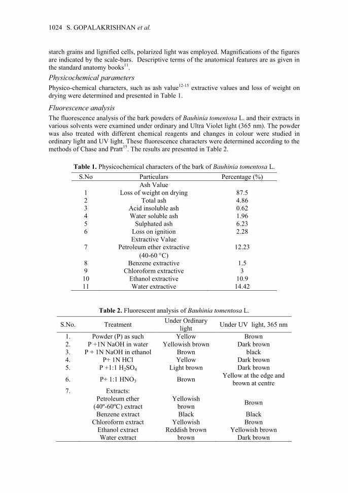
Macroscopic studies
The macroscopic evaluation was carried out for shape, size, color, and fracture of the drug .
These macroscopic characters are presented in Figure 1.
Figure 1. Bauhimia fomentosa. L.showimg its macroscopic characters.
Microscopic studies
Sectioning
The Paraffin embedded bark of Bauhinia tomentosa L. were sectioned with the help of
rotary microtome. The thickness of the section was 10 to 12 µm. Dewaxing of the section
was done by customary procedure8. The sections were stained with toluidine blue as per the
method of O’ Brien et al9. Since toluidine blue is a polychromatic stain, the staining results
were remarkably good; some cytochemical reactions were also obtained. The necessary
sections were also stained with safranin and fast–green and IKI (for starch).
For studying the stomatal morphology, venation pattern and trichome distribution,
paradermal sections (sections taken parallel to the surface of leaf) as well as clearing of leaf
with 5% sodium hydroxide or epidermal peeling was carried out by partial maceration
employing Jeffrey’s maceration fluid10
. Glycerine mounted temporary preparations were
made for macerated materials.
Photomicrographs
Microscopic descriptions of tissues were supplemented with micrographs wherever
necessary. Photographs of different magnifications were taken with Nikon Labphot 2
microscopic unit. For normal observations bright field was used, For the study of crystals,
S. GOPALAKRISHNAN et al. 1024
starch grains and lignified cells, polarized light was employed. Magnifications of the figures
are indicated by the scale-bars. Descriptive terms of the anatomical features are as given in
the standard anatomy books11
.
Physicochemical parameters
Physico-chemical characters, such as ash value12-15
extractive values and loss of weight on
drying were determined and presented in Table 1.
Fluorescence analysis
The fluorescence analysis of the bark powders of Bauhinia tomentosa L. and their extracts in
various solvents were examined under ordinary and Ultra Violet light (365 nm). The powder
was also treated with different chemical reagents and changes in colour were studied in
ordinary light and UV light. These fluorescence characters were determined according to the
methods of Chase and Pratt15
. The results are presented in Table 2.
Table 1. Physicochemical characters of the bark of Bauhinia tomentosa L.
S.No Particulars Percentage (%)
Ash Value
1 Loss of weight on drying 87.5
2 Total ash 4.86
3 Acid insoluble ash 0.62
4 Water soluble ash 1.96
5 Sulphated ash 6.23
6 Loss on ignition 2.28
Extractive Value
7 Petroleum ether extractive
(40-60 C)
12.23
8 Benzene extractive 1.5
9 Chloroform extractive 3
10 Ethanol extractive 10.9
11 Water extractive 14.42
Table 2. Fluorescent analysis of Bauhinia tomentosa L.
S.No. Treatment Under Ordinary
light Under UV light, 365 nm
1. Powder (P) as such Yellow Brown
2. P +1N NaOH in water Yellowish brown Dark brown
3. P + 1N NaOH in ethanol Brown black
4. P+ 1N HCl Yellow Dark brown
5. P +1:1 H2SO4 Light brown Dark brown
6. P+ 1:1 HNO3 Brown Yellow at the edge and
brown at centre
7. Extracts:
Petroleum ether
(40º-60ºC) extract
Yellowish
brown Brown
Benzene extract Black Black
Chloroform extract Yellowish Brown
Ethanol extract Reddish brown Yellowish brown
Water extract brown Dark brown
Pharmacognostical and Physicochemical Analysis of the Bark 1025
Results and Discussion
Macroscopic studies
Bauhinia tomentosa is an erect, branched shrub attaining a height of 1.5-3 m. The
branchlets, lower surfaces of the leaves, and pods are somewhat hairy. The leaves are 4-7 cm
long, about as wide, and split about one-third to the base, into two, with oval, rounded lobes.
The flowers are pale lemon yellow, usually in pairs on axillary peduncles. The pods are
9-11 cm long, about 1.5 cm wide, flattened, and contain 6 to 10 small seeds. These
macroscopic characters of Bauhinia tomentosa are presented in Figure 1.
Microscopic studies
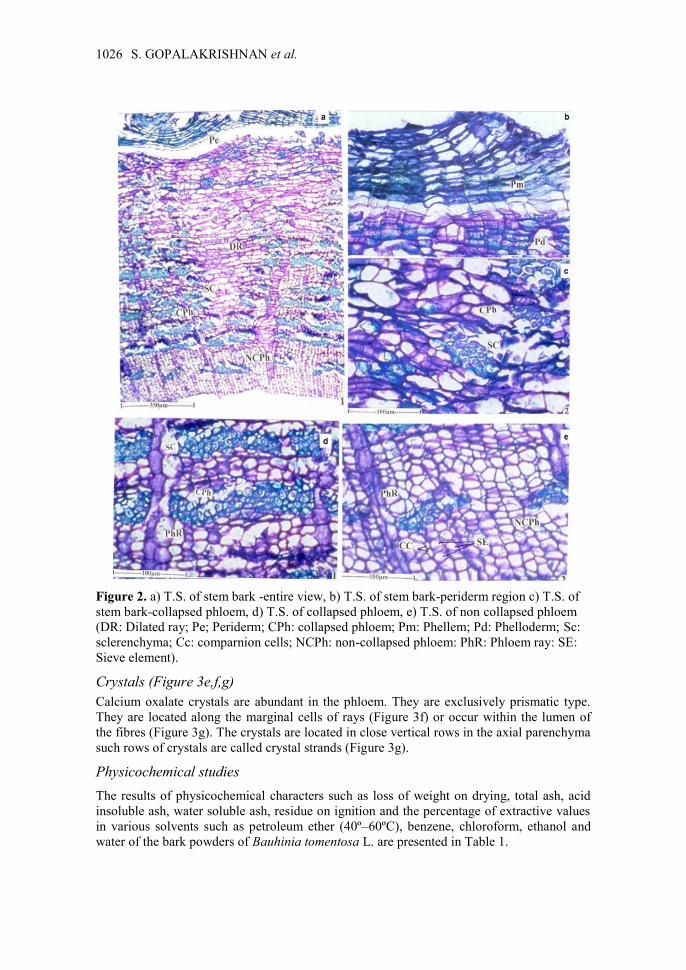
Stem bark
The bark is fairly smooth and even surface. It is grey - white. The texture is fibrous. It has
no specific odour or taste. Total thickness of the bark is 1.2 mm. The bark consists of
peridern and secondary phloem.
Periderm (Figure 2a,b)
The periderm is superficial and wavy; it is continuous. The surface exhibits shallow wide
fisseres. The periderm comprises twelve layers of phellem and seven or eight layers of
phelloderm (Figure 2b). The phellem is homogeneous; cells are tabular in shape and
suberized. The phelloderm cells are living cells with storage of ergastic substances.
Secondary phloem
It is the major part of the bark and measures about 1 mm wide. The phloem can be
differentiated into outer collapsed phloem and inner non-collapsed phloem.
Collapsed phloem
It is the wider zone measuring about 850 µm thick. The collapsed phloem is
characterized by wide, highly dilated, funnel-shaped phloem-rays which consist of
horizontal rows of tangentially stretched cells. Alternating with the dilated rays are
conical bands of sieve – elements and phloem fibres (Figure 2c). The phloem elements
are crushed into dark thin tangential lines and are located in between thick fibre
segments (Figure 2d).
Non-collapsed phloem
Non collapsed phloem consists of intact sieve elements and parenchyma cells. The phloem
rays are narrow (not dilated) and fiber segments reduced in frequency or they are absent.
The sieve elements are polyhedral in outline and are arranged in compact radial files
(Figure 2e). The sieve elements are 20 µm wide. The companion cells are small and situated
along the corners of the element (Figure 2e).
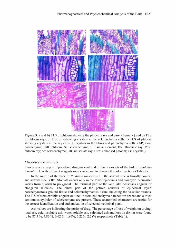
TLS view
In tangential longitudinal sections of the phloem, the phloem rays are nonstoried. They are
mostly biseriate, less frequently uniseriate or 3-seriate (Figure 3a,b,c,d). The rays are
homocellular; the cells are wide, polyhedral and compact. The biseriate rays are
220-250 µm high and 20-40 µm thick. Three-seriate are 50-60 µm thick and 250 µm high.
Phloem parenchyma cells are fusiform in shape with wedge shaped ends. They are arranged
in stories (Figure 3a,b,c,d).
S. GOPALAKRISHNAN et al. 1026
Figure 2. a) T.S. of stem bark -entire view, b) T.S. of stem bark-periderm region c) T.S. of
stem bark-collapsed phloem, d) T.S. of collapsed phloem, e) T.S. of non collapsed phloem
(DR: Dilated ray; Pe; Periderm; CPh: collapsed phloem; Pm: Phellem; Pd: Phelloderm; Sc:
sclerenchyma; Cc: comparnion cells; NCPh: non-collapsed phloem: PhR: Phloem ray: SE:
Sieve element).
Crystals (Figure 3e,f,g)
Calcium oxalate crystals are abundant in the phloem. They are exclusively prismatic type.
They are located along the marginal cells of rays (Figure 3f) or occur within the lumen of
the fibres (Figure 3g). The crystals are located in close vertical rows in the axial parenchyma
such rows of crystals are called crystal strands (Figure 3g).
Physicochemical studies
The results of physicochemical characters such as loss of weight on drying, total ash, acid
insoluble ash, water soluble ash, residue on ignition and the percentage of extractive values
in various solvents such as petroleum ether (40º–60ºC), benzene, chloroform, ethanol and
water of the bark powders of Bauhinia tomentosa L. are presented in Table 1.
Pharmacognostical and Physicochemical Analysis of the Bark 1027
Figure 3. a and b) TLS of phloem showing the phloem rays and parenchyma, c) and d) TLS
of phloem rays, e) T.S. of –showing crystals in the sclerenchyma cells, f) TLS of phloem
showing crystals in the ray cells, g) crystals in the fibres and parenchyma cells. (AP; axial
parenchyma; PhR: phloem; Sc: sclerenchyma; SE: sieve element; BR: Biseriate ray; PhR:
phloem ray; Sc: sclerenchyma; UR: uniseriate ray; CPh: collapsed phloem; Cr: crystals;).
Fluorescence analysis
Fluorescence analysis of powdered drug material and different extracts of the bark of Bauhinia
tomentosa L with different reagents were carried out to observe the color reactions (Table.2).
In the midrib of the bark of Bauhinia tomentosa L., the abaxial side is broadly conical
and adaxial side is flat. Stomata occurs only in the lower epidermis and paracytic. Vein-islet
varies from quarish to polygonal. The terminal part of the vein islet possesses angular or
elongated sclereids. The dintal part of the petiole consists of epidermal layer,
parenchymatous ground tissue and sclerenchymatous tissue enclosing the vascular strands.
The T.S of stem exhibits angular outline. In stem collenchyma batches are absent and a thick
continuous cylinder of sclerenchyma are present. These anatomical characters are useful for
the correct identification and authentication of selected medicinal plant.
Ash values are indicating the purity of drug. The percentage of loss of weight on drying,
total ash, acid insoluble ash, water soluble ash, sulphated ash and loss on drying were found
to be 87.5 %, 4.86 %, 0.62 %, 1.96%, 6.23%, 2.28% respectively (Table 1).
S. GOPALAKRISHNAN et al. 1028
The extractive values are useful to evaluate the chemical constituents present in the crude
drug and also help in estimation of specific constituents soluble in a particular solvent. The
water extractive value was found to higher (14.42%) followed by petroleum ether (40º-60ºC)
extractive value (12.23%), ethanol(10.9%), chloroform (3%), benzene (1.5%). (Table.1)
A characteristic yellowish-green fluorescence is noticed in the ethanol extract of
Bauhinia tomentosa. This characteristic fluorescence can be used as a diagnostic tool for the
correct identification of the crude drug and to test adulteration if any. In conclusion, it can be
stated that the standardization parameters used in the present investigation will provide a
way for the standardization of raw materials and prepared formulation of herbal origin as
well as answer to the latest GMP norms and FDA guidelines on standardization of herbal
drugs. This could also serve in the correct identification and preparation of a monograph on
the plant.
Acknowledgment
We are thankful to Prof. P. Jeyaraman, Director, Plant Anatomy Research Centre, West
Tambaram, Chennai - 600045. for helping in the anatomical studies.
References
1. Ali M S, Ahmed F, Pervez M K, Azhar I and Ibrahim S A, Nat Prod Res., 2005,
19(1), 53-60.
2. Agarwal A, Pharm Times., 2005, 37(6), 09- 11.
3. Nadkarni K M, Indian Materia Medica: Vol-I, 183-184.
4. Ramachandra Row L and Viswanadhan N, Proceedings of Indian Academy of
Science, 1954, 39A, 240-242.
5. Sankara Subramanian S and Nair A G R, Ind J Chemis., 1963, 1 (10), 450.
6. Dahanukar S A, Kulkarni R A and Rege N N, Ind J Pharmacol., 2000, 32, 81-118.
7. Biswas T K and Mukherjee B, Int J Lower Extremity Wounds., 2003, 2, 25–29.
8. Johansen D A, Plant Microtechnique; Mc Graw Hill Book Co: New York, 1940, 523.
9. O' Brien T P, Feder N and Mc Cull M E, Protoplasma., 1964, 59, 364-373.
10. Sass J.E, Elements of Botanical Microtechnique; Mc Graw Hill Book Co: New York,
1940, 222.
11. Esau K, Plant Anatomy; John Wiley and Sons: New York, 1965, 767.
12. Anonymous, The Ayurvedic Pharmacopoeia of India. Government of India;
Ministery of Health & Family Welfare: Published by The Controller of Publications,
Civil Lines, New Delhi, 2001; Vol.2.
13. Brain K R and Turner T D, Practical evaluation of Phytopharmaceuticals; Wright
Scientechnica: Bristol, 1975.
14. Chase C.R and Pratt R, J Am Pharm Ass (Sci Ed)., 1949, 38, 324-331.
15. Harborne J B, Phytochemical Methods; Chapman & Hall: International Edition,
Toppan Company Ltd, Japan, 1973.
Submit your manuscripts athttp://www.hindawi.com
Hindawi Publishing Corporationhttp://www.hindawi.com Volume 2014
Inorganic ChemistryInternational Journal of
Hindawi Publishing Corporation http://www.hindawi.com Volume 2014
International Journal ofPhotoenergy
Hindawi Publishing Corporationhttp://www.hindawi.com Volume 2014
Carbohydrate Chemistry
International Journal of
Hindawi Publishing Corporationhttp://www.hindawi.com Volume 2014
Journal of
Chemistry
Hindawi Publishing Corporationhttp://www.hindawi.com Volume 2014
Advances in
Physical Chemistry
Hindawi Publishing Corporationhttp://www.hindawi.com
Analytical Methods in Chemistry
Journal of
Volume 2014
Bioinorganic Chemistry and ApplicationsHindawi Publishing Corporationhttp://www.hindawi.com Volume 2014
SpectroscopyInternational Journal of
Hindawi Publishing Corporationhttp://www.hindawi.com Volume 2014
The Scientific World JournalHindawi Publishing Corporation http://www.hindawi.com Volume 2014
Medicinal ChemistryInternational Journal of
Hindawi Publishing Corporationhttp://www.hindawi.com Volume 2014
Chromatography Research International
Hindawi Publishing Corporationhttp://www.hindawi.com Volume 2014
Applied ChemistryJournal of
Hindawi Publishing Corporationhttp://www.hindawi.com Volume 2014
Hindawi Publishing Corporationhttp://www.hindawi.com Volume 2014
Theoretical ChemistryJournal of
Hindawi Publishing Corporationhttp://www.hindawi.com Volume 2014
Journal of
Spectroscopy
Analytical ChemistryInternational Journal of
Hindawi Publishing Corporationhttp://www.hindawi.com Volume 2014
Journal of
Hindawi Publishing Corporationhttp://www.hindawi.com Volume 2014
Quantum Chemistry
Hindawi Publishing Corporationhttp://www.hindawi.com Volume 2014
Organic Chemistry International
Hindawi Publishing Corporationhttp://www.hindawi.com Volume 2014
CatalystsJournal of
ElectrochemistryInternational Journal of
Hindawi Publishing Corporation http://www.hindawi.com Volume 2014