Embed Size (px)
Citation preview

Polish Journal of Microbiology2014, Vol. 63, No 3, 307–315
ORIGINAL PAPER
Introduction
Microsporum canis constitutes the main species spreading from animals to humans and causing espe-cially tinea capitis. Sometimes it can reach the nails, and in exceptional cases the pubic area in the form of tinea circinata. Several epidemiological studies undertaken in Tunisian areas revealed the re-emergence of M. canis, (Meziou et al., 2011) which did not exist in Tuni-sia before 1950 and of which the frequency has been increasing. There is not much information available on the molecular epidemiology of M. canis isolated from patients in Tunisia. Only classical identification was performed in the mycological laboratories in Tunisia.
Furthermore, identification at the strain level might be necessary for specification of the etiologic agent. Dif-ferent molecular methods were used, such as restriction fragment length polymorphism analysis of mitochon-drial (Kawasaki et al., 1996) , sequencing of the internal transcribed spacer (ITS) region of the ribosomal DNA (Graser et al., 2000; Sharma et al., 2006), sequencing of protein-encoding genes (Jung et al., 2014; Kano et al.,
2000), random amplification of polymorphic DNA (Mochizuki et al., 1997) arbitrarily primed PCR (Liu et al., 1997; Liu, et al., 2000) and PCR fingerprinting (Graser et al., 1998), have brought important progress in distinguishing between species and strains. In addi-tion, the ribosomal regions consisting of the internal transcribed spacers (ITS-1 and -2) and their interme-diary 5.8S rDNA are highly variable regions of fungal DNA and exhibit adequate variability to determine the phylogeny of closely interrelated filamentous fungi (Turin et al., 2000). These results indicate that PCR-RFLP targeting the ITS regions is a valuable tool for species identification of dermatophytes (Toshio, 2008). The present study aimed to characterize by means of phenotypic and molecular techniques a collection of 40 clinical isolates established in Tunisia from patients with M. canis infection. The assimilation of different nitrogen and carbohydrate sources was the preliminary biochemical tool used with the on the total collection.
This study aims to characterize a collection of M. canis, established from Tunisian patients, by using phenotypic and molecular tools.
Phenotypical and Molecular Characterization of Microsporum canis Strainsin North-Tunisia
CYRINE DHIEB1, BADIÂA ESSGHAIER1, DALINDA EL EUCH2
and NAJLA SADFI-ZOUAOUI1*
1 Laboratoire Microorganismes et Biomolécules Actives, Faculté des Sciences de TunisUniversité de Tunis El Manar, 2092 Tunisie
2 Service de Dermatologie et de Vénéréologie Hôpital La Rabta, 1007 Tunis, Tunisie
Submitted 3 January 2014, Revised 6 July 2014, Accepted 15 July 2014
A b s t r a c t
In this study, 40 Microsporum canis isolates were obtained from different patients from the Mycology Unit of the Hospital La Rabta (Tunis) during a 3 month period. The phenotypic identification was done by morphological characterization and biochemical tests. Molecular ana-lysis was performed by amplification of the ITS region of rDNA, the amplified region was subjected to enzymatic digestion and sequenced to evaluate phylogenetic relationships. The morphological analysis showed a considerable diversity of colonies as well as different mor-phologies of conidia and we have noted variability in the assimilation of the nitrogen and carbon sources. The PCR-RFLP results showed only one restriction pattern for each enzyme. The phylogenetic tree proves that all the strains from Tunisian patients are clonal and related with other strains from different origins. The classical methods used in the mycological laboratories are time-consuming, the PCR-RFLP analysis of the ITS is a reliable tool for the identification of M. canis strains. M. canis from infected Tunisian patients are clonal, although the isolates had different phenotypic characteristics.
K e y w o r d s: Microsporum canis, assimilation of carbon, assimilation of nitrogen, PCR-RFLP, Sequencing
* Corresponding author: Najla Sadfi Zouaoui, Laboratoire Microorganismes et Biomolécules Actives, Faculté des Sciences de Tunis, Uni-ver sité de Tunis El Manar, Campus universitaire, 2092 Tunisie; phone:+(216)71872600; fax: +(216)71885480; e-mail: [email protected]

Dhieb C. et al. 3308
Experimental
Materials and Methods
Origin of fungal strains. A total of 40 clinical iso-lates of M. canis was obtained from patients seen at the Unit of Mycology in the La Rabta Hospital of Tunis (Tunisia). All patients originated from the North of Tunisia, in the majority (95%) affected with tinea capitis and only two human cases (5%) with sycosis pubien and onychomycosis. M. canis was detected in children from 2 to 19 years old with 77.5% males and 22.5% females. The disease originated in the majority of cases from con-tact with animals (cat, dog and hamster) while few had an anthropophilic origin. Each isolate was transferred to Sabouraud dextrose agar (SAB) medium and then incu-bated at 25°C for 10 days and identified by macroscopic and microscopic characteristics of the culture strains.
Assimilation of carbon and nitrogen sources. The assimilation of carbon and nitrogen sources was tested according to Maia et al. (2001). For carbohy-drate assimilation, yeast nitrogen base (Biolife, Italia) was prepared 6.7 g/l in distilled water patched in the plugged tube and then autoclaved at 121°C for 15 min. A stock solution of 15% for carbohydrates (mannitol, maltose, mannose, sorbitol, dextrin, dulcitol, lactose, xylose, galactose) was used. Each solution was steri-lized by filtration through a filter (Millipore). A control without carbohydrate was included in each test to give a final concentration of 10 g/l. For nitrogen assimila-tion the basal medium consisted of yeast carbon base (Biolife, Italie) 11.7 g/l in distilled water, and autoclaved as described above. A stock solution of 15% for nitro-gen sources were prepared and added to medium to give a final concentration of nitrogen equivalent to 2 g/l sodium nitrate, 2.6 g/l asparagines, 2.4 g/l ammonium sulphate and 0.225 g/l urea. The strains were cultivated on HL-agar medium (sodium chloride 5 g/l, dipotas-sium hydrogen phosphate 0.3 g/l, peptone 2 g/l and agar 15 g/l) for 2 weeks at 25°C, and then a small por-tion of each mycelium was inoculated into each tube. The growth of each mycelium strain has been observed after 5 days of inoculation. Isolates were grouped using the cluster analysis UPGMA (Unweighted Pair Group Method with Arithmetic Average) (Nei et al., 1979).
Preparation of monosporic culture of Micro-sporum canis. The conidial suspension of each pure culture was prepared by flooding 10-day-old cultures of M. canis on SAB dishes with 4.5 ml of sterile distilled water and dislodging the spores with a glass rod. After serial dilution, 0.05 ml aliquots were spread-plated onto SAC plates and incubated at 25°C dur ing 48 h. Germi-nated conidia visualized with an optic micro scope were picked individually with a sterile needle and then incu-bated at 25°C during 10 days. A mycelia disk of 10 mm
diameter of each culture was inoculated in a 250 ml Erlenmeyer flask containing 100 ml of Sabouraud Broth medium. The liquid culture was then incubated at 25°C for 10 days on a rotary shaker (110 rpm) and the cul-tivated mycelium was recuperated, filtered and then frozen before being lyophilized.
DNA fungal extraction. Fungal DNA was pre-pared according to the method described by Sadfi et al. (2009). The cetyltrimethyl ammonium bromide (CTAB) method was used to extract DNA from isolates. Liquid nitrogen was added to 0.1 g of lyophilized mycelium in a mortar, and the cells were ground finely with a pestle. Genomic DNA was extracted as described by Doyle and Doyle (1987) with a few modifications. In brief, the mycelium powder was transferred to an Eppendorf tube and 700 μl lysis buffer (2% CTAB) supplemented with 0.2% (w/v) β-mercaptoethanol (Biomatic) was added. The mixture was incubated at 65°C for 40 min. An equal volume of chloroforme/isoamyl alcohol [24:1 (v/v)] was added. After homogenization of the mixture and centrifugation at 13000 rpm for 10 min at +4°C, the supernatant was recuperated into sterile Eppendorf tube. The DNA was precipitated with two volumes of cold 2-propanol supplemented with 10% (w/v) sodium acetate buffer (3 M, pH 8) at –20°C for 2 hours, washed twice 500 μl of 70% ethanol, air dried, and resuspended in 100 μl TE buffer (40 Mm Tris/HCl pH 8.0, 2 mM EDTA). The final DNA was RNase treated with incu-bation at 37°C for 30–60 min. DNA was eluted in a final volume of 200 μl, quantified on a Nanodrop ND-1000 (Nanodrop Technologies) and diluted to a concentra-tion of 50 ng/μl in PCR grade water. The DNA samples were stored at –20°C for further use.
PCR amplification of internal transcribed spac er (ITS) regions of rDNA. The ITS1-5.8S-ITS2 region of the nuclear rDNA gene cluster was amplified using prim ers ITS1 (5’-TCGGTAGGTGAACCTGCGG-3’) and ITS4 (5’-CCTCCGCTTATTGATATGC-3’) (White et al., 1990) for isolates. PCR was performed in a total reaction volume of 25 μl contain ing 5 μl of 5X buffer (Promega), 1 μl of dNTP (20 mM), 1.5 μl of MgCl2 (25 mM), 0.25 U (5 U/μl) Taq DNA polymerase, 2 μl of primer (20 pmoles/μl), and 2 μl of genomic DNA (50 ng/μl). The amplification program included an initial denaturation at 95°C for 3 min, followed by 35 cycles of denaturation at 98°C (15 s), annealing at 59°C (60 s), extension at 72°C (120 s) and a final exten-sion period of 10 min at 72°C in a Biometra thermal cycler (Germany). Following amplification, PCR prod-ucts were elec trophoresed on 1.5% agarose gels buffered with 0.5X TBE (4.5 mM Tris, 4.5 mM boric acid and 1 mM EDTA, pH 8), stained with ethidium bromide, and photographed.
RFLP. PCR amplicons of 40 isolates of M. canis were digested with restriction enzymes EcoRI, HinfI and

Characterization of Microsporum canis3 309
DdeI (Promega) in 20 μl reaction mixtures consisting of 5 μl PCR product, restriction enzyme (10 units) with the corresponding buffer (10X), and 12.8 μl H2O. The activation of each enzyme was used according to the manufacturer’s instructions, and the results were sepa-rated on 8% polyacrylamide gels, stained with ethidium bromide and visualized by UV light.
Sequencing. The PCR products were purified with enzymatic methods using ExoI and SAP and then the sequences were determined by cycle sequencing using the Taq Dye Deoxy Terminator Cycle Sequencing kit (Applied Biosystems; HTDS, Tunisia) and fragment separation in an ABI PrismTM 3130 DNA sequencer (Applied Biosystems; HTDS, Tunisia). The sequence analysis was perfomed by the Chromas Pro program.
Phylogenetic analysis. The phylogenetic analysis of isolates sequences and the M. canis sequences avail-able in the GenBank database was conducted using MEGA4.0. A neighbor-joining phylogenetic tree was produced using Hypocrea virens AN73 (HQ292945) as an out-group. The nucleotide sequences reported in this paper have been deposited in the GenBank Data-base under accession No JQ922440 until JQ922479 for E1 until E40, respectively. The reference strains used are Arthroderma otae IFM45853 (AB193632); M. canis CHUS65602 (EF581129.1), A. otea WM 05.15(EF568060) M. canis LM 2174 (EU181444), M. canis CHUS14303 (EF581130); A. otae IFM 46803 (AB193649); A. otae IFM 5286 (AB193610); M. canis strain ATCC 23828 (AY213657); M. canis strain ATCC MYA-4605 (GU291265); M. canis strain ATCC 36299 (FJ545254); A. otae isolate 574 (JN134123); M. canis strain 04-049-1860 (EU200371); M. canis strain 04-047-2395 (EU200368); A. otae isolate 268 (JN134110); A. otae strain SD100902 (HQ328946); A. otae (HQ223448); A. otae strain SD100915 (HQ395072); M. canis strain CBS566.80 (AJ252330)/M. canis strain CBS190.57 (AJ252329)/; A. otae strain A6 (HM016897); A. otae strain A11 (HM016898); M. canis (EU590655).
Results
Macroscopic and microscopic morphology. On Sabouraud’s dextrose agar, the isolates matured within 6 to 10 days producing colonies that were flat, spreading, white to cream colored, with a dense cottony, granular to coarsely fluffy to hairy surface which might show some radial grooves. Colonies usually had a bright golden yellow to brownish yellow reverse pigment (Fig. 1). In microscopic observation, the strains produced septate hyphae, macroconidia, and few or rare microconidia. Macroconidia were typically long spindle-shaped, with 5–15 cells, verrucose, thick-walled and often had a ter-minal knob. The septal walls were thin. Microconidia
were rare, unicellular and clavate to pyriform in shape. Raquet hyphae, nodular bodies, and chlamydospores might be present. Some strains were not pigmented, for example, while other, say “dysgoniques” produced dis-torted macroconidia and few, or did not produce them, and had a macroscopic aberrant (Fig. 2).
Assimilation of carbon and nitrogen sources. For the nitrogen sources all isolates assimilated sodium nitrate, asparagines and ammonium sulphate but 92.5% were able to assimilate urease. For the carbon sources, all isolates assimilated glucose, mannose and sorbitol but no assimilation was detected for dulcitol and lac-tose. For the other carbohydrates variable results for assimilation were obtained (95% for maltose, 92.5% for mannitol, 92.5% for dextrin, 90% for galactose and 17.5% for xylose) (Fig. 3).
Analysis of the numeric grouping of the biochemi-cal results. Analysis of the dendrogram, using 0.82 of the scale of the program in the Jaccard’s coefficient showed the differentiation of the isolates to assimilate different carbon and nitrogen sources the distance displayed among the isolates. This statistical analysis grouped the isolates into 2 main groups A and B. The group A included two subgroups, A1 grouping 2 iso-lates E8 that cannot assimilate dextrose, dulcitol, lac-tose and galactose and isolate E37 could not assimilate dulcitol, lactose and galactose; and the subgrouping A2 grouping two isolates E2 that could not assimilate dex-trose, dulcitol, lactose, galactose and mannitol and the isolate E35 could not assimilate dextrose, dulcitol, lac-tose, galactose and manitol. Group B which represented 70% of the isolates comprises 3 subgroups B1, B2, B3, B4 and B5. B1 grouping isolate E36 could not assimilate dulcitol, lactose and mannitol the B2 comprised one
A A’ B B’
Fig. 1. Macroscopic morphology of M. canis isolates(A, A’: strain E3; B, B’: strain E12)
(A, A’) colonies fluffy, white, reverse red-brown; (B, B’) colonies fluffy, white; reverse yellow

Dhieb C. et al. 3310
isolate E38 that could not assimilate dulcitol, lactose, xylose and maltose, the subgroup B3 grouping E15, E13 and E7 these isolates could not assimilate urease, dulcitol, lactose and xylose, B4 subgrouping included 29 isolates unable to assimilate dulcitol, lactose and glucose but were able to assimilate all the others; the subgrouping B5 comprised of E1 and E3 that could not assimilate only dulcitol and lactose (Fig. 3).
PCR. The ITS regions were successfully amplified from all the Microsporum isolates by the fungus-specific universal primers ITS1 and ITS4. Polymerase chain reaction electrophoresis showed a typical profile for all the isolates with a single band of 700 bp size.
RFLP. Amplified rDNA from isolates of M. canis was digested with EcoRI, HinfI and DdeI. Each restric-tion enzyme used showed only one restriction pattern
with two, four and five fragments for EcoRI, DdeI and HinfI, respectively (Table I). Thus, no polymorphism was detected among the isolates. The size of the vari-ous bands of digestion is presented in Table I and RFLP bands are shown in Fig. 4, 5 and 6.
Sequencing. To confirm the results of RFLP analy-sis, PCR products were sequenced and the nucleotide sequences were compared with the GenBank Database. All the strains showed a homology from 98% to 100% with M. canis (A. otae).
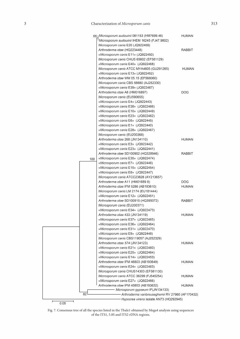
Phylogenetic analysis. The ITS phylogenetic trees show that the clinical strains isolated from different patients originating from the North of Tunisia contami-nated by animals or human sources are related. They are grouped in the same cluster and genotypically nearly identical to the strains of M. canis sequenced by the other authors. The Tunisian clinical strains of M. canis are similar to those from other clinical and animal strains (human, rabbit, dog etc.) (Fig. 7).
Discussion
M. canis is a frequent cause of tinea capitis and tinea corporis in Europe and especially in the Mediterranean area (Maraki et al., 2000). Many studies undertaken in
EcoRI present 290, 350DdeI present 90, 190, 200, 240HinfI present 140, 200, 240, 260
Table IThe PCR product patterns of RFLP analysis of rDNA from
M. canis strains using restriction enzymes EcoR1, Dde1 and Hinf1
Enzymes Restriction site Fragment size (bp)
macroconidia
microconidia
mycelia
Microconidiaon acladium
A B
C D
Fig. 2. Microscopic morphology of M. canis isolates(A) Abundant macroconidia 6–10 cells, rough-walled, wiyh thick cell walls and thick septa,microconidia clavate, sessile alongside;
(B) Some macroconidia with pyriforme microconidia; (C) Narraw macroconidia and few microconidia.

Characterization of Microsporum canis3 311
Tunisia revealed the re-emergence of M. canis (Belhadj et al., 2007). The infection can be transmitted by ani-mals, especially dogs and cats or by direct physical contact with an infected person (Gambale et al., 1993; Romano et al., 1997).
In the present study, 40 isolates of M. canis were col-lected from clinical patients originating from the north of Tunisia, aged between 2 and 19 years affected with
Fig. 3. Dendrogram of the 40 samples isolated from clinical patients. The dendrogram was constructed by UPGMA programfrom Jaccard’s similarity coefficient obtained with assimilation of carbon and nitrogen sources results.
Fig. 4. Polyacrylamide-gel electrophoresis of PCR products digested with EcoRI restriction enzyme. The ITS1-ITS4 sets of primers were used to amplify ribosomal DNA including internal
transcribed spacers (ITS). Fig. 5. Polyacrilamide-gel electrophoresis of PCR products digested with HinfI restriction enzyme. The ITS1-ITS4 sets of primers were used to amplify ribosomal DNA including internal
transcribed spacers(ITS).

Dhieb C. et al. 3312
tinea capitis, one case with sycosis pubien and another case with onychomycosis. The majority of them (82.5%) had contact with animals (cat, dog and hamster).
Moreover, from an epidemiological study carried out by Bouden-Mansour (1997), M. canis was the most frequent dermatophyte isolated from animals in Tunisia (77.2%) with a high prevalence of isolates from asymp-tomatic animals. The occurrence of zoonotic transmis-sion from animals to humans has been demonstrated by several studies (Proverbio et al., 2014; Drouot et al., 2009). For the best therapeutic procedure, identifica-tion of this dermatophyte at the species level is very important; the procedure for the identification of M. canis is usually based on phenotypical analysis by its macro- and micro-morphological features.
In mycological laboratories in Tunisia, especially in the Mycology Unit of the Hospital La Rabta (Tunisia), the identification of dermatophytes was just established by macroscopic and microscopic methods. M. canis colo nies are, in general, apiculated with low cottony texture and present pigment varying from canary yel-low pigment to ochre (Evans and Richardson, 1989; De Hoog et al., 2000). According to Brilhante et al. (2005), the morphology of M. canis colonies analyzed in dif-ferent media showed a large diversity which could be attributed to an adaptation of the strain to the varia-tion of environmental conditions. On Sabouraud agar, the isolates grew in 10 days and produce colonies that spread flat. Fluffiness was the majority. The color of the colonies varied between white-yellowish to orange-brown to brownish yellow front and upside down, this
was also described by Evans and Richardson (1989). The microscopic appearance showed the presence of hyphal cells; macroconidia in time with the distaff side with an elongated shape containing 5 to 10 stalls, pointed ends, a thick wall and verrucae surface. The microconidia were unicellular pyriform and of variable number. The abundance of fruiting bodies varied from one isolate to another and seemed to be influenced by the nature of the culture medium; for isolates E17 and E30 the absence of fructification was observed on Sabouraud agar but reappeared on the fruiting medium PDA. Marvroudeas et al. (1996) suggested that exogenous fac-tors such as the concentration of glucose or thiamine in the culture medium could affect the structure of macroconidia. Several hypotheses have been advanced to explain the phenotypic variation observed in der-matophytes. According to several studies (Makimura et al., 1999; Faggi et al., 2001; Brilhante et al., 2003; Sidrim et al., 2004), changing environmental factors such as medium composition, incubation time and temperature could be responsible for the phenotypic variation observed. Besides morphological character-istics, dermatophytes of the genus Microsporum could also be typed by means of biochemical tests such as secretion of enzymes (keratinase, collagenase, elastase, urease), assimilation of carbon and nitrogen sources and susceptibility to yeast killer toxins and antifungals (Brilhante et al., 2005). There were few variations in the assimilation of nitrogen sources, all the isolates assimi-lated nitrate and asparagine sources except for three (E7, E13 and E15) which were not able to assimilate urease. For the carbon sources, all the isolates were able to assimilate glucose, mannose and sorbitol but not dulcitol and lactose. Moreover, the assimilation of dextrose, xylose, galactose, mannitol and mannose showed variations between isolates in the collection. In contrast, Mai et al., 2001, showed that all the strains of Microsporum canis isolated from dogs and cats in Bra-zil could assimilate nitrogen sources as well as carbon sources, mannitol, maltose, mannose, sorbitol, and dex-trin and that not all the samples assimilated galactose and none of them were capable of growing with dulci-tol, lactose, or xylose as the only carbon source. This show that biochemical characteristics did not reflect the morphological ones for classification, which may explain the inadequacy of the use of different carbon sources in the typing of M. canis. The identification of M. canis was based primarily on morphological and biochemical tools (Ginnis, 1980) but the observed change requires the development of other procedures (Maia et al., 2001). The success in typing dermatophytes according to phenotyping criteria such as colony mor-phology, microscopy or biochemical reaction has been limited. Over the past years genotypic approaches have proven useful in solving the problems of identification
Fig. 6. Polyacrylamide-gel electrophoresis of PCR products digested with DdeI restriction enzyme. The ITS1-ITS4 sets of primers were used to amplify ribosomal DNA including internal
transcribed spacers (ITS).
Characterization of Microsporum canis3 313
Fig. 7. Consensus tree of all the species listed in the Tbale1 obtained by Mega4 analysis using sequencesof the ITS1, 5.8S and ITS2 rDNA regions.
Dhieb C. et al. 3314
of dermatophytes. Indeed, the genotypic characteristics are more stable and accurate in relation to phenotypic characteristics (Mochizuki et al., 2003; Liu 2000). Thus, molecular techniques have been used to better iden-tify the isolates. Several molecular studies have used PCR-RFLP to identify strains of M. canis, this technique provides a rapid and practical tool for the identification of dermatophytes (Mochizuki et al., 2003; Kanbe et al., 2003). In this study, the ITS region was subjected to enzymatic digestion by the enzymes EcoRI, DdeI and HinfI. Endonuclease analysis provides a rapid and reli-able technique for molecular differentiation of the col-lection of the isolates. The EcoRI digestion revealed the presence of two bands, while four bands were obtained by DdeI digestion for all the samples analyzed in the study. These results are in agreement with those of Brilhante et al. (2005) who have identified a collection of M. canis from veterinary and clinical origins using EcoRI, DdeI and RsaI and showed the same profiles for all the strains. Moreover, the identification of an atypical isolate of Microsporum with six other strains of M. canis by submitting the ITS region to the digestion with EcoRI and MvaI confirmed the membership of this isolate in M. canis species (Leon Mateos et al. 2006). The work of Mirzahoseini et al. (2009) showed that the HinfI enzyme cannot digest the ITS region of M. canis, but in our study we have revealed the presence of 4 bands within this enzyme. Based on the RFLP results, we con-clude that the collection of M. canis used in this study is genetically identical; such data are in agreement with Brilhante et al. (2005), and Faggi et al. (2001).
The utilization of direct sequence analysis was an important technical development in the field of amplifi-cation-based genotypic pathogen identification (Relman et al., 1993) The sequence homology within the rDNA genes of fungi (18S, 5.8S and 28S genes) and differences within the spacer regions (ITS1 and ITS2) are a genetic basis for the organization of the fungi into taxonomic groups and they provide the most relevant information, which can also be easily exchanged between labora-tories. The results of the present study show that the ITS, 5.8S and ITS2 sequences of M. canis isolates from different patients, thus supporting the hypothesis that the infections are caused by the same strain. The DNA sequences of the ITS regions of nuclear ribosome in the dermatophytes have been proven to be useful for species identification and for resolving phylogenetic relationships between close taxonomic species. Gräser et al., (2000) reported that 40 M. canis isolates display-ing different colonies morphologies did not reveal any DNA polymorphism when analyzed using molecular techniques, suggesting a strictly clonal mode of repro-duction and a strong adaptation to human skin. The present results show that the Microsporum group is het-erogeneous and that new isolates and other genome
regions need to be analyzed to elucidate the taxo-nomic relationships of a group that shows differences. According to Yu et al., (2004), the ITS region was always thought to have interspecies polymorphism and little variance at the intra-species level. Thus, our results sug-gest that strains from different origins which present the same DNA sequence in the ITS region may result from small variances in this region for intra-species isolates from patient with tinea capitis or from the environment. The accessibility of the sequencing of the amplification products, combined with the continued enrichment of databases, allows introducing the sequencing method in a rational approach to compare dermatophytes around the word so DNA sequencing is thought to be the most direct method for species discrimination. In our study of isolate differentiation, we tried to examine all the isolates of M. canis using the method of DNA sequencing. The ITS region of rDNA was chosen as the target region of the study. The results showed that the strains present some biochemical diversity to assimi-late carbon and nitrogen sources, but no genetic dif-ferences were detected between isolates from different patients. It resulted in the inference that all the isolates were a single strain which had spread among different sites and to susceptible children. M. canis strains from Tunisian patients are very clonal genetically.
AcknowledgementsThis work was supported by funds from the Ministry of Higher
Education and Scientific Research LR03ES03.
Literature
Belhadj S., H. Jeguirim, S. Anane, E. Kaouech, K. Kallel and E. Chaker. 2007. Évolution des teignes du cuir chevelu à Microsporum canis et à Trichophyton violaceum à Tunis. J. Med. Mycol. 17: 54–57. Bouden-Mansour R., S. Belhadj, L. Idir, A. Bouattour, M. Kilani and E. Chaker. 1997. Prevalence and etiology agents of animal ring-worm in the region of Tunis. Mycol. Med. 7: 145–148. Brilhante R.S., C.S. Cavalcante, F.A. Soares-Junior, R.A. Cor-deiro, J.J. Sidrim and M.F. Rocha. 2003. High rate of Microsporum canis feline and canine dermatophytoses in North east Brazil: epide-miological and diagnostic features. Mycopathologia 156(4): 303–308.Brilhante R.S., M.F. Rocha, R.A. Cordeiro, S.H. Rabenhorst, T.B. Granjeiro, A.J. Monteiro and J.J. Sirdim. 2005. Phenotypical and molecular characterization of Microsporum canis strains in north-east Brazil. J. App. Microbiol. 99(4): 776–782.De Hoog G.S., P. Mayser, G. Haase, R. Horré and A.M. Hor-revorts. 2000. A new species, Phialophora europaea, causing super-ficial infection in humans. Mycoses 43(11–12): 409–416.Doyle J.J and J.L. Doyle. 1987. A rapid DNA isolation procedure for small quantities of fresh leaf tissue. Phytochem. Bull. 19: 11–15.Drouot S., B. Mignon, M. Fratti, P. Roosje and M. Monod. 2009. Pets as the main source of two zoonotic species of the Trichophyton mentagrophytes complex in Switzerland, Arthroderma vanbreuseghemiiand, Arthroderma benhamiae. Vet. Dermatol. 20(1): 13–18.Evans E.G.V and M.D. Richardson (eds). 1989. Medical mycology – a practical approach. Oxford University Press.

Characterization of Microsporum canis3 315
Faggi E., G. Pini., E. Campisi, C. Bertellini, E. Difonzo and F. Mancianti. 2001. Application of PCR to distinguish common spe-cies of dermatophytes. J. Clin. Microbiol. 39(9): 3382–3385. Gambale W., G.E. Larsson, M.M. Moritami, B. Correa, C.R. Paula and V.M. Framil. 1993. Dermatophytes and other fungi of the hair-coat of cats without dermatophytosis in the city of Sao Paula, Brazil. Feline Prac. 21(3): 29–33. Jung H.J., S.Y. Kim, J.W. Jung, H.J. Park, Y.W. Lee, Y.B. Choe and K.J. Ahn. 2014. Identification of dermatophytes by polymerase chain reaction-restriction fragment length polymorphism analysis of metalloproteinase-1. Ann Dermatol. 26(3):338–42. Ginnis M.R. 1980. Laboratory handbook of Medical Mycology. Academic Press, Inc. New York.Graser Y., M. El Fari, W. Presber, W. Sterry and H.J. Tietz. 1998. Identification of common dermatophytes (Trichophyton, Microsporum, Epidermophyton) using polymerase chain reactions. Br. J. Dermatol. 138(4): 576–582.Graser Y., A.F. Kuijpers, M. El Fari, W. Presber and G.S. De Hoog. 2000. Molecular and conventional taxonomy of the Microsporum canis complex. Med. Mycol. 38: 143–153.Kanbe T., Y. Suzuki, A. Kamiya., T. Mochizuki, M. Fujihiro and A. Kikuchi. 2003. PCR- based identification of common dermato-phyte species using primer sets specific for the DNA topoisomerase II genes. J. Dermatol Sci. 32(2): 151–61.Kano R., K. Okabayashi, Y. Nakamura, S. Ooka, M. Kashima and M. Mizoguchi. 2000. Differences among chitin synthase 1 gene sequences in Trichophyton rubrum and T. violaceum. Med. Mycol. 38(1): 47–50.Kawasaki M., M. Aoki, H. Ishizaki, K. Nishimura and M. Miyaji. 1996. Phylogeny of Epidermophyton floccosum and other dermato-phytes. Mycopathologia 134(3): 121–128.Leon-Mateos A., C. Pares-Suarez, M.J.R. Pereiro and J. Toribio. 2006. Study of the ITS region in an atypical isolate and comparison with six species of Microsporum. Mycoses 49(6): 452–456.Liu D., S. Coloe, R. Baird and J. Pedersen. 1997. PCR identification of Trichophyton mentagrophytes var. interdigitale and T. mentagrophytes var. mentagrophytes dermatophytes with a random primer. J. Med. Microbiol 46(12): 1043–1046.Liu D., S. Coloe, R. Baird and J. Pedersen. 2000. Application of PCR to the identification of dermatophyte fungi. J. Med. Microbiol. 49(6): 493–497.Maia L.S., J.I. Dos Santos, F.C. Viani, C.E. Larsson, C.R. Paula and W. Gambale. 2001. Phenotypic characterisaton of Microsporum canis isolated from cats and dogs. Mycoses 44(11–12): 480–486.Makimura K., Y. Tamura, T. Mocgizuki, A. Hasegawa, Y. Tajri, R. Hanazawa, K. Uchida, H. Saito and H. Yamaguchi. 1999. Phy-logenetic classification and species identification of dermatophyte strains based on DNA sequences of nuclear ribosomal internal tran-scribed spacer 1 regions. J. Clin. Microbiol. 37(4): 920–924. Maraki S and Y. Tselentis. 2000. Survey on the epidemiology of Microsporum canis infections in Crete, Greece over a 5-year period. Int. J. Dermatol. 39(1): 21–24.
Mavroudeas D., A. Velegraki, J. Leonardopoulos and U. Marce-lou. 1996. Effect of glucose and thiamine concentrations on the for-mation of macroconidia in dermatophytes. Mycoses 39 (1–2): 61–66.Meziou T.J., A. Dammak, T. Zaz, M. Mseddi, S. Boudaya, L. Bouzid, F. Akrout, S. Maalej, A. Ayadi and H. Turki. 2011. Scalp ringworm tinea capitis in Tunisian infants. Med. Mal. Infect. 41 (9): 486–488.Mirzahoseini H., E. Omidiana, S. Ghahfarokhi, G. Sadeghi and R. Abyanehni. 2009. Application of PCR-RFLP to Rapid Identifica-tion of the Main Pathogenic Dermatophytes from Clinical Speci-mens. Iran. J. Publ. Health 38(1): 18–24.Mochizuki T., N. Sugie and M. Uehara. 1997. Random amplifica-tion of polymorphic DNA is useful for the differentiation of several anthropophilic dermatophytes. Mycoses 40(11–12): 405–409.Mochizuki T., H. Tanabe, M. Kawasaki, H. Ishizaki and C.J. Jackson. 2003. Rapid identification of Trichophyton tonsurans by PCR-RFLP analysis of ribosomal DNA regions. J. Dermatol. Sci. 32(1): 25–32.Nei M. and W.H. Li.1979. Mathematical model for studying genetic variation in terms of restriction endonucleases. Proc. Natl. Acad. Sci. 76(10): 5269–5273.Proverbio D., R. Perego, E. Spada, G. Bagnagatti de Giorgi, A. Della Pepa and E. Ferro. 2014. Survey of Dermatophytes in Stray Cats with and without Skin Lesions in Northern Italy. Vet. Med. Int. doi: 10.1155/2014/565470. Relman D.A. 1993. The identification of uncultured microbial pathogens. J. Infect. Dis. 168(1): 1–8.Romano C., L. Valenti and R. Barbara. 1997. Dermatophytes iso-lated from asymptomatic stray cats. Mycoses 40(11–12): 471–472.Sadfi-Zouaoui N., I. Hannachi, M. Rouaissi, M.R. Hajlaoui, M.B. Rubio, E. Monte, A. Boudabous and M.R. Hermosa. 2009. Biodiversity of Trichoderma strains in Tunisia. Can. J. Microbiol. 55(2): 154–162.Sharma R., R.C. Rajak, A.K. Pandey and Y. Graser. 2006. Inter-nal Transcribed Spacer (ITS) of rDNA of appendaged and non-appendaged strains of Microsporum gypseum revals Microsporum appendiculatum as its synonym. Antonie Van Leeuwenhoek 89(1): 197–202.Sidrim J.C., T.E.F. Meireles, L.M.P. Oliveira and M.J.N. Diogenes. 2004. Aspect Clinique des dermatophytoses. Mycol. Med. 135–161.Kanbe T. 2008. Molecular Approaches in the Diagnosis of Derma-tophytosis. Mycopathologia 166: 307–317.Turin L., F. Riva, G. Galbiati and T. Cainelli. 2000. Fast simple and highly sensitive double-rounded polymerase chain reaction assay to detect medically relevant fungi in dermatological specimens. Eur. J. Clin. Invest. 30(6): 511–518.White T.J., T. Bruns, S. Lee and J. Taylor. 1990. Amplification and direct sequencing of fungal ribosomal RNA genes for phylogenetics. A Guide to Methods and Applications, Academic Press In. Yu J., Z. Wan, W. Chen, W. Wang and R. Li. 2004. Molecular typing study of the Microsporum canis strains isolated from an outbreak of tinea capitis in a school. Mycopathologia 157(1): 37–41.