Embed Size (px)
Citation preview
Experimental Neurology 227 (2011) 79–88
Contents lists available at ScienceDirect
Experimental Neurology
j ourna l homepage: www.e lsev ie r.com/ locate /yexnr
Physiological and histopathological responses following closed rotational head injurydepend on direction of head motion
Stephanie A. Eucker a, Colin Smith b, Jill Ralston a, Stuart H. Friess c, Susan S. Margulies a,⁎a Department of Bioengineering, School of Engineering and Applied Science, University of Pennsylvania, Philadelphia, PA, USAb Department of Pathology, University of Edinburgh, Edinburgh, Scotland, UKc Department of Anesthesiology and Critical Care Medicine, The Children's Hospital of Philadelphia, Philadelphia, PA, USA
Abbreviations: CBF, cerebral blood flow; CPP, cerefluorescent microsphere; ICP, intracranial pressure; MAPSAH, subarachnoid hemorrhage; SaO2, arterial oxygen sinjury.⁎ Corresponding author. Department of Bioengineeri
Applied Science, University of Pennsylvania, 240 SkirkPhiladelphia, PA 19104, USA. Fax: +1 215 573 2071.
E-mail address: [email protected] (S.S. Marg
0014-4886/$ – see front matter © 2010 Elsevier Inc. Aldoi:10.1016/j.expneurol.2010.09.015
a b s t r a c t
a r t i c l e i n f oArticle history:Received 21 May 2010Revised 31 August 2010Accepted 21 September 2010Available online 25 September 2010
Keywords:Animal modelsBrain ischemiaBrain traumaCerebral blood flowNeuropathologySubarachnoid hemorrhage
Rotational inertial forces are thought to be the underlying mechanism for most severe brain injuries.However, little is known about the effect of head rotation direction on injury outcomes, particularly in thepediatric population. Neonatal piglets were subjected to a single non-impact head rotation in the horizontal,coronal, or sagittal direction, and physiological and histopathological responses were observed. Sagittalrotation produced the longest duration of unconsciousness, highest incidence of apnea, and largestintracranial pressure increase, while coronal rotation produced little change, and horizontal rotationproduced intermediate and variable derangements. Significant cerebral blood flow reductions were observedfollowing sagittal but not coronal or horizontal injury compared to sham. Subarachnoid hemorrhage,ischemia, and brainstem pathology were observed in the sagittal and horizontal groups but not in a singlecoronal animal. Significant axonal injury occurred following both horizontal and sagittal rotations. For bothgroups, the distribution of injury was greater in the frontal and parietotemporal lobes than in the occipitallobes, frequently occurred in the absence of ischemia, and did not correlate with regional cerebral blood flowreductions. We postulate that these direction-dependent differences in injury outcomes are due to differencesin tissue mechanical loading produced during head rotation.
bral perfusion pressure; FM,, mean arterial blood pressure;aturation; TBI, traumatic brain
ng, School of Engineering andanich Hall, 210 S. 33rd Street,
ulies).
l rights reserved.
© 2010 Elsevier Inc. All rights reserved.
Introduction
Traumatic brain injury (TBI) is a leading cause of death anddisability in infants in the U.S. (Langlois et al., 2004). These patientsmay present with irritability, seizures, loss of consciousness, andrespiratory difficulties (Gruskin and Schutzman, 1999; Johnson et al.,1995). Increased intracranial pressure (ICP) and decreased cerebralblood flow (CBF) are frequently observed (Adelson et al., 1997;Sharples et al., 1995). Radiological and pathological findings includesubdural, subarachnoid and parenchymal hemorrhages, skull frac-tures, diffuse brain swelling, axonal injury, and ischemia (Duhaimeet al., 1992; Geddes et al., 2001). However, the mechanisms of injuryare incompletely understood, and effective treatment is limited,particularly for severely injured patients.
The relative importance of primary versus secondary injuries onTBI outcomes in infants has been hotly debated. Primary injury refers
to the physical derangements, such as tissue deformation and tissuefailure, that occur during the traumatic event; it is highly dependenton the specific mechanical loading conditions to the head (Ommaya,1985). For instance, head impact frequently produces focal contu-sions, whereas non-impact rotational head motion has been shown toproduce more diffuse brain injuries (Adams et al., 1985; Gennarelli etal., 1982; Ommaya, 1985). Furthermore, falls are the most commoncause of mild to moderate TBI in infants, whereas motor vehiclecrashes and assault, which have a larger rotational component, morefrequently produce severe or fatal TBI, suggesting the importance ofrotational head motion in the most severe brain injuries (Arbogast etal., 2005; Duhaime et al., 1992). Direction of head motion may also bean important factor in TBI outcomes, and would therefore need to beconsidered when developing safety standards for automobiles andhelmets. Limited studies in adult pigs and primates have demonstrat-ed distinct physiological and histopathological responses to headrotations in different directions, likely due to brain geometryasymmetries among the different axes (Gennarelli et al., 1982,1987; Smith et al., 2000). The effect of rotation direction in thepediatric population remains to be examined.
Secondary injury encompasses the array of biological responses tothe initial traumatic event that have been observed, including ionchannel dysfunction, excitotoxicity, ischemia, and derangements inCBF and blood pressure (Bayir et al., 2003; Kochanek et al., 2000).
80 S.A. Eucker et al. / Experimental Neurology 227 (2011) 79–88
These changes can continue to develop for days following the initialtraumatic event and can be monitored clinically; thus, they arepotential targets for therapeutic intervention (Bayir et al., 2003).However, targeted therapy at a single injury mechanism has been oflimited clinical success, and it is likely that optimal treatment musttake multiple mechanisms into consideration (Kochanek et al., 2000).In particular, the coupled interaction between the primarymechanicalinsult and the initiation of secondary insults, such as CBF changes andischemia, requires further investigation.
We have previously developed a horizontal rotational brain injurymodel in neonatal piglets that recapitulates many of the clinicalfindings observed in infant TBI (Raghupathi and Margulies, 2002).However, the effects of rotation in the sagittal and coronal directionsare unknown. We hypothesize that head rotation direction affectsimmediate post-injury physiological responses, including regionalcerebral blood flow changes, unconsciousness times, and incidencesof apnea, as well as early pathological outcomes including regionalaxonal and ischemic injuries immediately following rotational closedhead injury in piglets. In addition, we identify the importance of earlycerebral blood flow reductions on the initial post-injury developmentof both axonal injury and tissue infarction.
Materials and methods
Animal preparation
All protocols were approved by the Institutional Animal Care andUse Committee of the University of Pennsylvania. Thirty-six neonatal(3–5 days old) piglets, whose level of brain development andmyelination correspond to that of human infants 1–3 months old(Armstead, 2005), were used for this study. Vital signs including heartrate, arterial oxygen saturation (SaO2), and end tidal CO2 wererecorded for the duration for each study. Supplemental oxygen andmechanical ventilator support were adjusted as needed to maintainnormoxia and normocarbia. Rectal temperature was maintainedbetween 37 and 39 °C using a heating pad and lamp.
Piglets were anesthetized with 4% isoflurane, intubated, andmechanically ventilated. Femoral artery, femoral vein, and rightcarotid artery catheters were placed for continuous mean arterialblood pressure (MAP) recording (Grass-Telefactor, Astro-Med, WestWarwick, RI), arterial blood gases, normal saline maintenanceinfusion at 4 ml/kg/h, and cerebral blood flow determination using apreviously described fluorescent microsphere technique (Euckeret al., 2010). Ligation of a single carotid artery in piglets does notalter regional CBF over a wide range of CBF values due to extensivecollateral cerebral circulation (Laptook et al., 1983). Followingcatheter placement, isoflurane was reduced to 2.25–2.75% mainte-nance levels. A pre-injury blood gas and electrolyte sample wasrecorded (Nova Biomedical, Waltham, MA).
Non-impact rotational brain injury
Brain injury was induced using a well-characterized rotationalacceleration device (Raghupathi and Margulies, 2002; Smith et al.,2000) to impart a single rapid (12–20 ms), non-impact head rotationin either the horizontal, sagittal, or coronal plane relative to thecerebrum, centered at themid-cervical spine (Fig. 1A–C, respectively).Angular velocity was measured using an angular rate sensor (ATA,Inc.) attached to the linkage sidearm and captured using a PC-baseddata acquisition system at 10 kHz (LabView, National Instruments,Austin, TX).
The first group of piglets (HOR-HIGH, n=9) received a 90°horizontal rotation, with an average peak angular velocity (PAV) of198±12 rad/s (mean±SD). The second group (COR, n=7) receiveda 90° coronal rotation, with an average peak angular velocity of 208±11 rad/s. The third group (SAG, n=6) received a 60° sagittal rotation,
with an average peak angular velocity of 166±3 rad/s. The reducedangular excursion in this groupwas due to the limited range ofmotionof the piglet neck in the sagittal direction and resulted in a lowermaximum angular velocity. To match the angular velocity loadingconditions, a second horizontal group (HOR-LOW, n=6) receiveda 90° horizontal rotation at an average peak angular velocity of 168±3 rad/s. The final group (SHAM, n=4) received all of the same pre-injury and post-injury procedures as the injured groups but did notundergo rotational injury.
Immediately prior to injury, anesthesia, supplemental oxygen, andmechanical ventilation were withdrawn, and the piglet was allowedto breathe spontaneously on room air. Following injury, the piglet wasremoved from the bite plate and supplemental oxygen was resumed,but the animal was allowed to continue breathing spontaneously.During this immediate post-injury period, the piglet was assessed forconsciousness (reflexive withdrawal to pinch stimulus) and apnea(cessation of breathing or reduced respiratory effort resulting in SaO2
b90%) every 30–60 s. If apnea occurred, mechanical ventilation wasimmediately restarted. Once the pinch reflex was positive, bothisoflurane anesthesia and mechanical ventilation were resumed.
Arterial blood gas samples were obtained at 5, 10, 15, 30, and60 min post-injury and every subsequent hour for the duration of thestudy. Supplemental oxygen was titrated as needed to maintain pre-injury pO2 levels (120–180 mmHg). Post-injury MAPwas maintainedwithin 10 mmHg of pre-injury MAP and above 40 mm Hg by titratingthe level of anesthesia and supplementing IV fluids with phenyleph-rine as needed, as this systemic vasoconstrictor has minimal effect onCBF (Strebel et al., 1998). Between 15 and 30 min post-injury, an ICPtransducer (Camino 110-4B, Integra, Plainsboro, NJ) was introducedinto the subarachnoid space over the right parietal lobe, anterior tothe coronal suture, in 29 of the 36 animals to monitor ICP post-injury.
At 6 h post-injury, the piglet was sacrificed via pentobarbitaloverdose. The brain was perfusion-fixed using 0.9% saline followed by10% neutral buffered formalin, removed, and post-fixed for N24 h in10% formalin for subsequent histological and fluorescent microsphereanalysis. We focused our assessments on a 6-h post-injury time pointbecause while βAPP staining requires 2–3 h to turn positive in braintissue after axonal injury (Hortobagyi et al., 2007), ischemic changescan take up to 6 h to become visible on H&E (Petito et al., 1987). Thisoptimized time point allowed us to capture immediate changes inaxonal injury (βAPP) and tissue ischemia (H&E), while minimizingthe influence of later secondary sequelae that also contribute to bothischemia and axonal injury.
Cerebral blood flow measurements
CBF changes were measured before and after injury using apreviously described fluorescent microsphere (FM) technique(Eucker et al., 2010) in 7 animals from HOR-HIGH, 4 from COR, 4from SAG, and 4 from HOR-LOW. Each animal received 1×10⁶ FMsbefore injury and 2×10⁶ FMs at 1 h and 6 h post-injury, using adifferent randomized FM color (yellow-green, orange, or crimson) ateach time point.
After each study, fixed brains were cut into 3-mm-thick coronalsections using a previously described piglet brain matrix (Euckeret al., 2010). Every other section was reserved for histologicalassessment. Of the remaining sections, every other section was usedfor FM analysis (Fig. 1D). This resulted in 4 total sections spacedevenly every 12 mm in the anterior-posterior direction, which couldbe subdivided into the following 9 regions: (1) right and (2) leftfrontal cerebrum including olfactory bulbs, (3) right and (4) leftparietotemporal cerebrum including basal ganglia, (5) right and (6)left occipital cerebrum including hippocampus, and (7) midbrain-pons, (8)medulla, and (9) cerebellum. Global CBFwas calculated fromthe total number of tissue microspheres in all right and left cerebralhemispheric regions (regions 1–6 above), excluding midbrain,
A
C
B
D
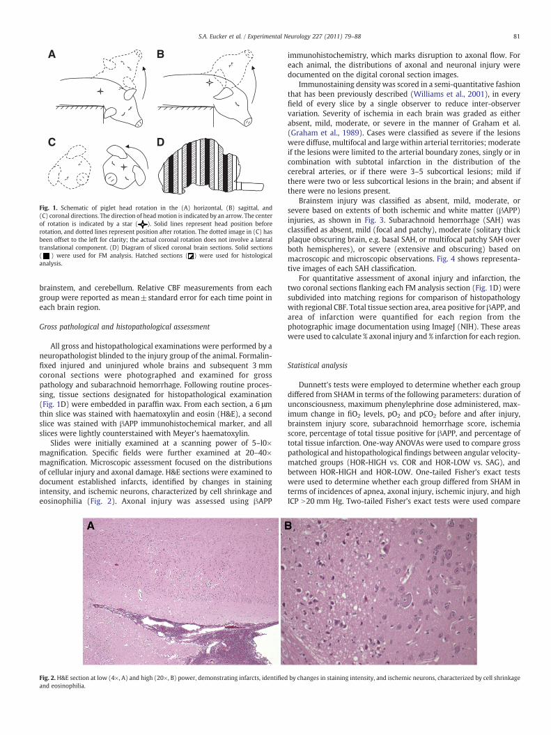
Fig. 1. Schematic of piglet head rotation in the (A) horizontal, (B) sagittal, and(C) coronal directions. The direction of headmotion is indicated by an arrow. The centerof rotation is indicated by a star ( ). Solid lines represent head position beforerotation, and dotted lines represent position after rotation. The dotted image in (C) hasbeen offset to the left for clarity; the actual coronal rotation does not involve a lateraltranslational component. (D) Diagram of sliced coronal brain sections. Solid sections( ) were used for FM analysis. Hatched sections ( ) were used for histologicalanalysis.
81S.A. Eucker et al. / Experimental Neurology 227 (2011) 79–88
brainstem, and cerebellum. Relative CBF measurements from eachgroup were reported as mean±standard error for each time point ineach brain region.
Gross pathological and histopathological assessment
All gross and histopathological examinations were performed by aneuropathologist blinded to the injury group of the animal. Formalin-fixed injured and uninjured whole brains and subsequent 3 mmcoronal sections were photographed and examined for grosspathology and subarachnoid hemorrhage. Following routine proces-sing, tissue sections designated for histopathological examination(Fig. 1D) were embedded in paraffin wax. From each section, a 6 μmthin slice was stained with haematoxylin and eosin (H&E), a secondslice was stained with βAPP immunohistochemical marker, and allslices were lightly counterstained with Meyer's haematoxylin.
Slides were initially examined at a scanning power of 5–l0×magnification. Specific fields were further examined at 20–40×magnification. Microscopic assessment focused on the distributionsof cellular injury and axonal damage. H&E sections were examined todocument established infarcts, identified by changes in stainingintensity, and ischemic neurons, characterized by cell shrinkage andeosinophilia (Fig. 2). Axonal injury was assessed using βAPP
Fig. 2. H&E section at low (4×, A) and high (20×, B) power, demonstrating infarcts, identifiedand eosinophilia.
immunohistochemistry, which marks disruption to axonal flow. Foreach animal, the distributions of axonal and neuronal injury weredocumented on the digital coronal section images.
Immunostaining density was scored in a semi-quantitative fashionthat has been previously described (Williams et al., 2001), in everyfield of every slice by a single observer to reduce inter-observervariation. Severity of ischemia in each brain was graded as eitherabsent, mild, moderate, or severe in the manner of Graham et al.(Graham et al., 1989). Cases were classified as severe if the lesionswere diffuse, multifocal and large within arterial territories; moderateif the lesions were limited to the arterial boundary zones, singly or incombination with subtotal infarction in the distribution of thecerebral arteries, or if there were 3–5 subcortical lesions; mild ifthere were two or less subcortical lesions in the brain; and absent ifthere were no lesions present.
Brainstem injury was classified as absent, mild, moderate, orsevere based on extents of both ischemic and white matter (βAPP)injuries, as shown in Fig. 3. Subarachnoid hemorrhage (SAH) wasclassified as absent, mild (focal and patchy), moderate (solitary thickplaque obscuring brain, e.g. basal SAH, or multifocal patchy SAH overboth hemispheres), or severe (extensive and obscuring) based onmacroscopic and microscopic observations. Fig. 4 shows representa-tive images of each SAH classification.
For quantitative assessment of axonal injury and infarction, thetwo coronal sections flanking each FM analysis section (Fig. 1D) weresubdivided into matching regions for comparison of histopathologywith regional CBF. Total tissue section area, area positive for βAPP, andarea of infarction were quantified for each region from thephotographic image documentation using ImageJ (NIH). These areaswere used to calculate % axonal injury and % infarction for each region.
Statistical analysis
Dunnett's tests were employed to determine whether each groupdiffered from SHAM in terms of the following parameters: duration ofunconsciousness, maximum phenylephrine dose administered, max-imum change in fiO2 levels, pO2 and pCO2 before and after injury,brainstem injury score, subarachnoid hemorrhage score, ischemiascore, percentage of total tissue positive for βAPP, and percentage oftotal tissue infarction. One-way ANOVAs were used to compare grosspathological and histopathological findings between angular velocity-matched groups (HOR-HIGH vs. COR and HOR-LOW vs. SAG), andbetween HOR-HIGH and HOR-LOW. One-tailed Fisher's exact testswere used to determine whether each group differed from SHAM interms of incidences of apnea, axonal injury, ischemic injury, and highICP N20 mm Hg. Two-tailed Fisher's exact tests were used compare
by changes in staining intensity, and ischemic neurons, characterized by cell shrinkage
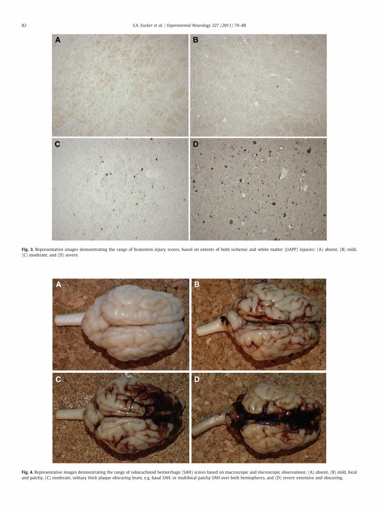
Fig. 3. Representative images demonstrating the range of brainstem injury scores, based on extents of both ischemic and white matter (βAPP) injuries: (A) absent, (B) mild,(C) moderate, and (D) severe.
Fig. 4. Representative images demonstrating the range of subarachnoid hemorrhage (SAH) scores based on macroscopic and microscopic observations: (A) absent, (B) mild, focaland patchy, (C) moderate, solitary thick plaque obscuring brain, e.g. basal SAH, or multifocal patchy SAH over both hemispheres, and (D) severe extensive and obscuring.
82 S.A. Eucker et al. / Experimental Neurology 227 (2011) 79–88
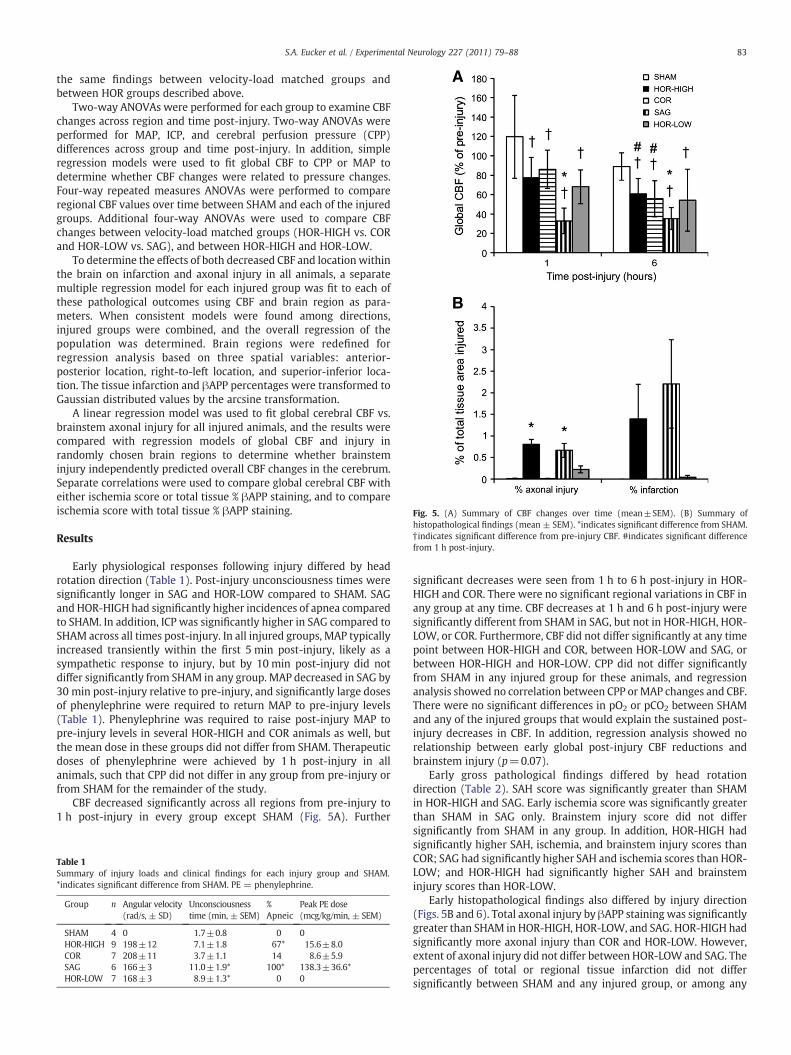
Fig. 5. (A) Summary of CBF changes over time (mean±SEM). (B) Summary ofhistopathological findings (mean ± SEM). *indicates significant difference from SHAM.† indicates significant difference from pre-injury CBF. #indicates significant differencefrom 1 h post-injury.
83S.A. Eucker et al. / Experimental Neurology 227 (2011) 79–88
the same findings between velocity-load matched groups andbetween HOR groups described above.
Two-way ANOVAs were performed for each group to examine CBFchanges across region and time post-injury. Two-way ANOVAs wereperformed for MAP, ICP, and cerebral perfusion pressure (CPP)differences across group and time post-injury. In addition, simpleregression models were used to fit global CBF to CPP or MAP todetermine whether CBF changes were related to pressure changes.Four-way repeated measures ANOVAs were performed to compareregional CBF values over time between SHAM and each of the injuredgroups. Additional four-way ANOVAs were used to compare CBFchanges between velocity-load matched groups (HOR-HIGH vs. CORand HOR-LOW vs. SAG), and between HOR-HIGH and HOR-LOW.
To determine the effects of both decreased CBF and location withinthe brain on infarction and axonal injury in all animals, a separatemultiple regression model for each injured group was fit to each ofthese pathological outcomes using CBF and brain region as para-meters. When consistent models were found among directions,injured groups were combined, and the overall regression of thepopulation was determined. Brain regions were redefined forregression analysis based on three spatial variables: anterior-posterior location, right-to-left location, and superior-inferior loca-tion. The tissue infarction and βAPP percentages were transformed toGaussian distributed values by the arcsine transformation.
A linear regression model was used to fit global cerebral CBF vs.brainstem axonal injury for all injured animals, and the results werecompared with regression models of global CBF and injury inrandomly chosen brain regions to determine whether brainsteminjury independently predicted overall CBF changes in the cerebrum.Separate correlations were used to compare global cerebral CBF witheither ischemia score or total tissue % βAPP staining, and to compareischemia score with total tissue % βAPP staining.
Results
Early physiological responses following injury differed by headrotation direction (Table 1). Post-injury unconsciousness times weresignificantly longer in SAG and HOR-LOW compared to SHAM. SAGand HOR-HIGH had significantly higher incidences of apnea comparedto SHAM. In addition, ICP was significantly higher in SAG compared toSHAM across all times post-injury. In all injured groups, MAP typicallyincreased transiently within the first 5 min post-injury, likely as asympathetic response to injury, but by 10 min post-injury did notdiffer significantly from SHAM in any group. MAP decreased in SAG by30 min post-injury relative to pre-injury, and significantly large dosesof phenylephrine were required to return MAP to pre-injury levels(Table 1). Phenylephrine was required to raise post-injury MAP topre-injury levels in several HOR-HIGH and COR animals as well, butthe mean dose in these groups did not differ from SHAM. Therapeuticdoses of phenylephrine were achieved by 1 h post-injury in allanimals, such that CPP did not differ in any group from pre-injury orfrom SHAM for the remainder of the study.
CBF decreased significantly across all regions from pre-injury to1 h post-injury in every group except SHAM (Fig. 5A). Further
Table 1Summary of injury loads and clinical findings for each injury group and SHAM.*indicates significant difference from SHAM. PE = phenylephrine.
Group n Angular velocity(rad/s, ± SD)
Unconsciousnesstime (min, ± SEM)
%Apneic
Peak PE dose(mcg/kg/min, ± SEM)
SHAM 4 0 1.7±0.8 0 0HOR-HIGH 9 198±12 7.1±1.8 67* 15.6±8.0COR 7 208±11 3.7±1.1 14 8.6±5.9SAG 6 166±3 11.0±1.9* 100* 138.3±36.6*HOR-LOW 7 168±3 8.9±1.3* 0 0
significant decreases were seen from 1 h to 6 h post-injury in HOR-HIGH and COR. There were no significant regional variations in CBF inany group at any time. CBF decreases at 1 h and 6 h post-injury weresignificantly different from SHAM in SAG, but not in HOR-HIGH, HOR-LOW, or COR. Furthermore, CBF did not differ significantly at any timepoint between HOR-HIGH and COR, between HOR-LOW and SAG, orbetween HOR-HIGH and HOR-LOW. CPP did not differ significantlyfrom SHAM in any injured group for these animals, and regressionanalysis showed no correlation between CPP or MAP changes and CBF.There were no significant differences in pO2 or pCO2 between SHAMand any of the injured groups that would explain the sustained post-injury decreases in CBF. In addition, regression analysis showed norelationship between early global post-injury CBF reductions andbrainstem injury (p=0.07).
Early gross pathological findings differed by head rotationdirection (Table 2). SAH score was significantly greater than SHAMin HOR-HIGH and SAG. Early ischemia score was significantly greaterthan SHAM in SAG only. Brainstem injury score did not differsignificantly from SHAM in any group. In addition, HOR-HIGH hadsignificantly higher SAH, ischemia, and brainstem injury scores thanCOR; SAG had significantly higher SAH and ischemia scores than HOR-LOW; and HOR-HIGH had significantly higher SAH and brainsteminjury scores than HOR-LOW.
Early histopathological findings also differed by injury direction(Figs. 5B and 6). Total axonal injury by βAPP staining was significantlygreater than SHAM in HOR-HIGH, HOR-LOW, and SAG. HOR-HIGH hadsignificantly more axonal injury than COR and HOR-LOW. However,extent of axonal injury did not differ between HOR-LOW and SAG. Thepercentages of total or regional tissue infarction did not differsignificantly between SHAM and any injured group, or among any
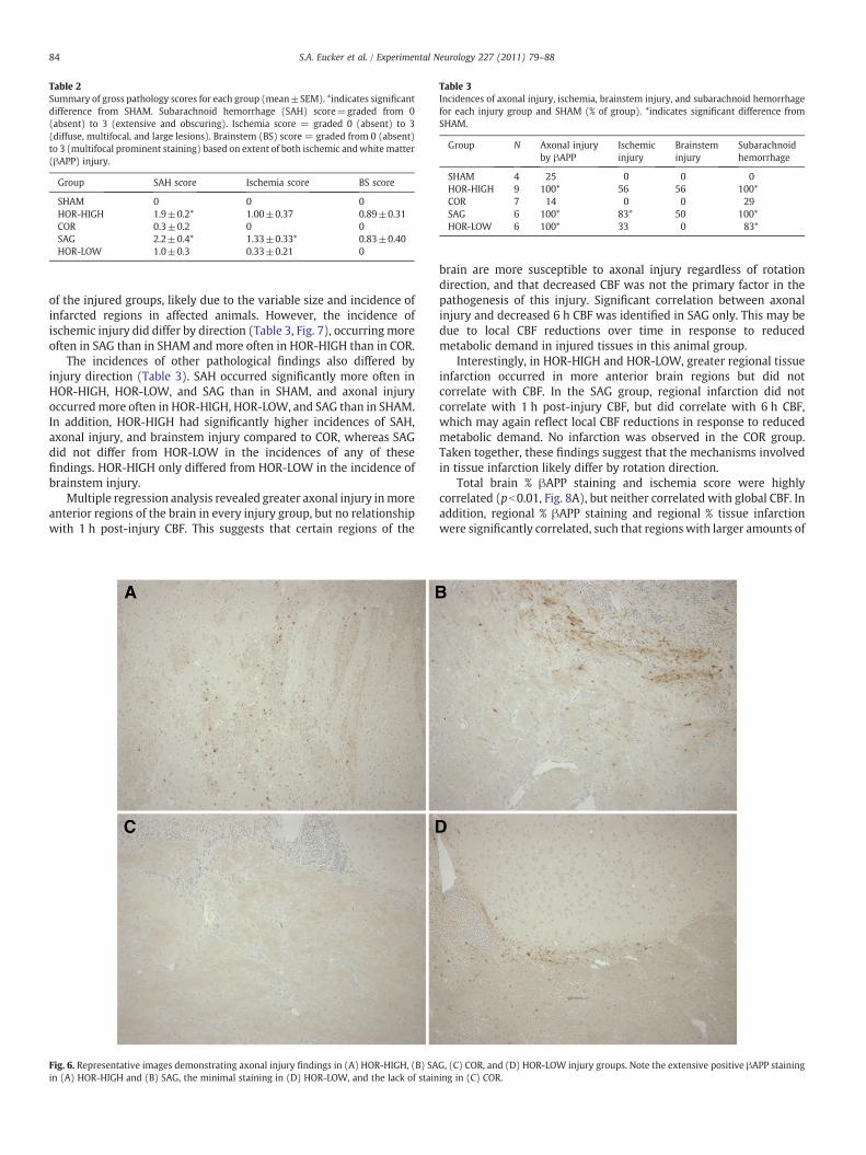
Table 2Summary of gross pathology scores for each group (mean±SEM). *indicates significantdifference from SHAM. Subarachnoid hemorrhage (SAH) score=graded from 0(absent) to 3 (extensive and obscuring). Ischemia score = graded 0 (absent) to 3(diffuse, multifocal, and large lesions). Brainstem (BS) score = graded from 0 (absent)to 3 (multifocal prominent staining) based on extent of both ischemic andwhite matter(βAPP) injury.
Group SAH score Ischemia score BS score
SHAM 0 0 0HOR-HIGH 1.9±0.2* 1.00±0.37 0.89±0.31COR 0.3±0.2 0 0SAG 2.2±0.4* 1.33±0.33* 0.83±0.40HOR-LOW 1.0±0.3 0.33±0.21 0
Table 3Incidences of axonal injury, ischemia, brainstem injury, and subarachnoid hemorrhagefor each injury group and SHAM (% of group). *indicates significant difference fromSHAM.
Group N Axonal injuryby βAPP
Ischemicinjury
Brainsteminjury
Subarachnoidhemorrhage
SHAM 4 25 0 0 0HOR-HIGH 9 100* 56 56 100*COR 7 14 0 0 29SAG 6 100* 83* 50 100*HOR-LOW 6 100* 33 0 83*
84 S.A. Eucker et al. / Experimental Neurology 227 (2011) 79–88
of the injured groups, likely due to the variable size and incidence ofinfarcted regions in affected animals. However, the incidence ofischemic injury did differ by direction (Table 3, Fig. 7), occurringmoreoften in SAG than in SHAM and more often in HOR-HIGH than in COR.
The incidences of other pathological findings also differed byinjury direction (Table 3). SAH occurred significantly more often inHOR-HIGH, HOR-LOW, and SAG than in SHAM, and axonal injuryoccurredmore often in HOR-HIGH, HOR-LOW, and SAG than in SHAM.In addition, HOR-HIGH had significantly higher incidences of SAH,axonal injury, and brainstem injury compared to COR, whereas SAGdid not differ from HOR-LOW in the incidences of any of thesefindings. HOR-HIGH only differed from HOR-LOW in the incidence ofbrainstem injury.
Multiple regression analysis revealed greater axonal injury inmoreanterior regions of the brain in every injury group, but no relationshipwith 1 h post-injury CBF. This suggests that certain regions of the
Fig. 6. Representative images demonstrating axonal injury findings in (A) HOR-HIGH, (B) SAin (A) HOR-HIGH and (B) SAG, the minimal staining in (D) HOR-LOW, and the lack of stain
brain are more susceptible to axonal injury regardless of rotationdirection, and that decreased CBF was not the primary factor in thepathogenesis of this injury. Significant correlation between axonalinjury and decreased 6 h CBF was identified in SAG only. This may bedue to local CBF reductions over time in response to reducedmetabolic demand in injured tissues in this animal group.
Interestingly, in HOR-HIGH and HOR-LOW, greater regional tissueinfarction occurred in more anterior brain regions but did notcorrelate with CBF. In the SAG group, regional infarction did notcorrelate with 1 h post-injury CBF, but did correlate with 6 h CBF,which may again reflect local CBF reductions in response to reducedmetabolic demand. No infarction was observed in the COR group.Taken together, these findings suggest that the mechanisms involvedin tissue infarction likely differ by rotation direction.
Total brain % βAPP staining and ischemia score were highlycorrelated (pb0.01, Fig. 8A), but neither correlated with global CBF. Inaddition, regional % βAPP staining and regional % tissue infarctionwere significantly correlated, such that regions with larger amounts of
G, (C) COR, and (D) HOR-LOW injury groups. Note the extensive positive βAPP staininging in (C) COR.
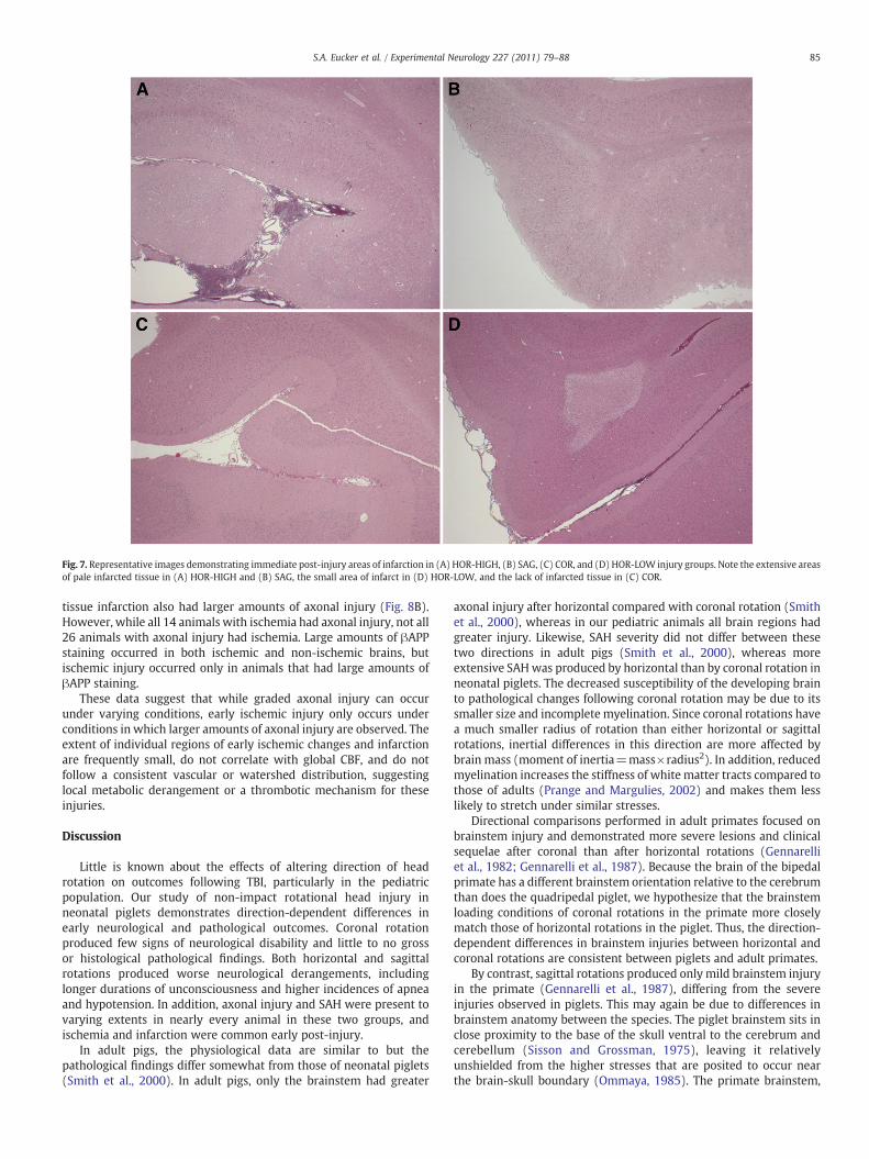
Fig. 7. Representative images demonstrating immediate post-injury areas of infarction in (A) HOR-HIGH, (B) SAG, (C) COR, and (D) HOR-LOW injury groups. Note the extensive areasof pale infarcted tissue in (A) HOR-HIGH and (B) SAG, the small area of infarct in (D) HOR-LOW, and the lack of infarcted tissue in (C) COR.
85S.A. Eucker et al. / Experimental Neurology 227 (2011) 79–88
tissue infarction also had larger amounts of axonal injury (Fig. 8B).However, while all 14 animals with ischemia had axonal injury, not all26 animals with axonal injury had ischemia. Large amounts of βAPPstaining occurred in both ischemic and non-ischemic brains, butischemic injury occurred only in animals that had large amounts ofβAPP staining.
These data suggest that while graded axonal injury can occurunder varying conditions, early ischemic injury only occurs underconditions in which larger amounts of axonal injury are observed. Theextent of individual regions of early ischemic changes and infarctionare frequently small, do not correlate with global CBF, and do notfollow a consistent vascular or watershed distribution, suggestinglocal metabolic derangement or a thrombotic mechanism for theseinjuries.
Discussion
Little is known about the effects of altering direction of headrotation on outcomes following TBI, particularly in the pediatricpopulation. Our study of non-impact rotational head injury inneonatal piglets demonstrates direction-dependent differences inearly neurological and pathological outcomes. Coronal rotationproduced few signs of neurological disability and little to no grossor histological pathological findings. Both horizontal and sagittalrotations produced worse neurological derangements, includinglonger durations of unconsciousness and higher incidences of apneaand hypotension. In addition, axonal injury and SAH were present tovarying extents in nearly every animal in these two groups, andischemia and infarction were common early post-injury.
In adult pigs, the physiological data are similar to but thepathological findings differ somewhat from those of neonatal piglets(Smith et al., 2000). In adult pigs, only the brainstem had greater
axonal injury after horizontal compared with coronal rotation (Smithet al., 2000), whereas in our pediatric animals all brain regions hadgreater injury. Likewise, SAH severity did not differ between thesetwo directions in adult pigs (Smith et al., 2000), whereas moreextensive SAHwas produced by horizontal than by coronal rotation inneonatal piglets. The decreased susceptibility of the developing brainto pathological changes following coronal rotation may be due to itssmaller size and incomplete myelination. Since coronal rotations havea much smaller radius of rotation than either horizontal or sagittalrotations, inertial differences in this direction are more affected bybrain mass (moment of inertia=mass×radius2). In addition, reducedmyelination increases the stiffness of white matter tracts compared tothose of adults (Prange and Margulies, 2002) and makes them lesslikely to stretch under similar stresses.
Directional comparisons performed in adult primates focused onbrainstem injury and demonstrated more severe lesions and clinicalsequelae after coronal than after horizontal rotations (Gennarelliet al., 1982; Gennarelli et al., 1987). Because the brain of the bipedalprimate has a different brainstem orientation relative to the cerebrumthan does the quadripedal piglet, we hypothesize that the brainstemloading conditions of coronal rotations in the primate more closelymatch those of horizontal rotations in the piglet. Thus, the direction-dependent differences in brainstem injuries between horizontal andcoronal rotations are consistent between piglets and adult primates.
By contrast, sagittal rotations produced only mild brainstem injuryin the primate (Gennarelli et al., 1987), differing from the severeinjuries observed in piglets. This may again be due to differences inbrainstem anatomy between the species. The piglet brainstem sits inclose proximity to the base of the skull ventral to the cerebrum andcerebellum (Sisson and Grossman, 1975), leaving it relativelyunshielded from the higher stresses that are posited to occur nearthe brain-skull boundary (Ommaya, 1985). The primate brainstem,
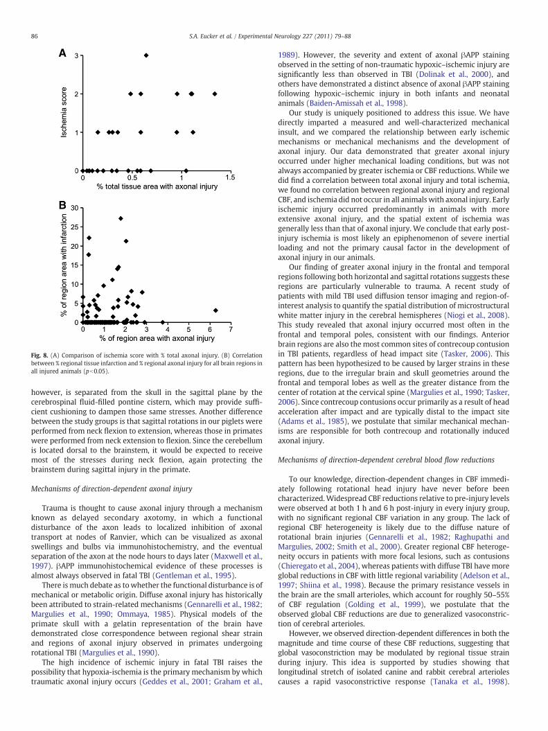
Fig. 8. (A) Comparison of ischemia score with % total axonal injury. (B) Correlationbetween % regional tissue infarction and % regional axonal injury for all brain regions inall injured animals (pb0.05).
86 S.A. Eucker et al. / Experimental Neurology 227 (2011) 79–88
however, is separated from the skull in the sagittal plane by thecerebrospinal fluid-filled pontine cistern, which may provide suffi-cient cushioning to dampen those same stresses. Another differencebetween the study groups is that sagittal rotations in our piglets wereperformed from neck flexion to extension, whereas those in primateswere performed from neck extension to flexion. Since the cerebellumis located dorsal to the brainstem, it would be expected to receivemost of the stresses during neck flexion, again protecting thebrainstem during sagittal injury in the primate.
Mechanisms of direction-dependent axonal injury
Trauma is thought to cause axonal injury through a mechanismknown as delayed secondary axotomy, in which a functionaldisturbance of the axon leads to localized inhibition of axonaltransport at nodes of Ranvier, which can be visualized as axonalswellings and bulbs via immunohistochemistry, and the eventualseparation of the axon at the node hours to days later (Maxwell et al.,1997). βAPP immunohistochemical evidence of these processes isalmost always observed in fatal TBI (Gentleman et al., 1995).
There is much debate as towhether the functional disturbance is ofmechanical or metabolic origin. Diffuse axonal injury has historicallybeen attributed to strain-related mechanisms (Gennarelli et al., 1982;Margulies et al., 1990; Ommaya, 1985). Physical models of theprimate skull with a gelatin representation of the brain havedemonstrated close correspondence between regional shear strainand regions of axonal injury observed in primates undergoingrotational TBI (Margulies et al., 1990).
The high incidence of ischemic injury in fatal TBI raises thepossibility that hypoxia-ischemia is the primary mechanism by whichtraumatic axonal injury occurs (Geddes et al., 2001; Graham et al.,
1989). However, the severity and extent of axonal βAPP stainingobserved in the setting of non-traumatic hypoxic–ischemic injury aresignificantly less than observed in TBI (Dolinak et al., 2000), andothers have demonstrated a distinct absence of axonal βAPP stainingfollowing hypoxic–ischemic injury in both infants and neonatalanimals (Baiden-Amissah et al., 1998).
Our study is uniquely positioned to address this issue. We havedirectly imparted a measured and well-characterized mechanicalinsult, and we compared the relationship between early ischemicmechanisms or mechanical mechanisms and the development ofaxonal injury. Our data demonstrated that greater axonal injuryoccurred under higher mechanical loading conditions, but was notalways accompanied by greater ischemia or CBF reductions. While wedid find a correlation between total axonal injury and total ischemia,we found no correlation between regional axonal injury and regionalCBF, and ischemia did not occur in all animals with axonal injury. Earlyischemic injury occurred predominantly in animals with moreextensive axonal injury, and the spatial extent of ischemia wasgenerally less than that of axonal injury. We conclude that early post-injury ischemia is most likely an epiphenomenon of severe inertialloading and not the primary causal factor in the development ofaxonal injury in our animals.
Our finding of greater axonal injury in the frontal and temporalregions following both horizontal and sagittal rotations suggests theseregions are particularly vulnerable to trauma. A recent study ofpatients with mild TBI used diffusion tensor imaging and region-of-interest analysis to quantify the spatial distribution of microstructuralwhite matter injury in the cerebral hemispheres (Niogi et al., 2008).This study revealed that axonal injury occurred most often in thefrontal and temporal poles, consistent with our findings. Anteriorbrain regions are also the most common sites of contrecoup contusionin TBI patients, regardless of head impact site (Tasker, 2006). Thispattern has been hypothesized to be caused by larger strains in theseregions, due to the irregular brain and skull geometries around thefrontal and temporal lobes as well as the greater distance from thecenter of rotation at the cervical spine (Margulies et al., 1990; Tasker,2006). Since contrecoup contusions occur primarily as a result of headacceleration after impact and are typically distal to the impact site(Adams et al., 1985), we postulate that similar mechanical mechan-isms are responsible for both contrecoup and rotationally inducedaxonal injury.
Mechanisms of direction-dependent cerebral blood flow reductions
To our knowledge, direction-dependent changes in CBF immedi-ately following rotational head injury have never before beencharacterized.Widespread CBF reductions relative to pre-injury levelswere observed at both 1 h and 6 h post-injury in every injury group,with no significant regional CBF variation in any group. The lack ofregional CBF heterogeneity is likely due to the diffuse nature ofrotational brain injuries (Gennarelli et al., 1982; Raghupathi andMargulies, 2002; Smith et al., 2000). Greater regional CBF heteroge-neity occurs in patients with more focal lesions, such as contusions(Chieregato et al., 2004), whereas patients with diffuse TBI have moreglobal reductions in CBF with little regional variability (Adelson et al.,1997; Shiina et al., 1998). Because the primary resistance vessels inthe brain are the small arterioles, which account for roughly 50–55%of CBF regulation (Golding et al., 1999), we postulate that theobserved global CBF reductions are due to generalized vasoconstric-tion of cerebral arterioles.
However, we observed direction-dependent differences in both themagnitude and time course of these CBF reductions, suggesting thatglobal vasoconstriction may be modulated by regional tissue strainduring injury. This idea is supported by studies showing thatlongitudinal stretch of isolated canine and rabbit cerebral arteriolescauses a rapid vasoconstrictive response (Tanaka et al., 1998).
87S.A. Eucker et al. / Experimental Neurology 227 (2011) 79–88
Furthermore, ultrastructural observations following stretch injury ofguinea pig optic nerves (Maxwell et al., 1991) or head injury in baboons(Maxwell et al., 1988) demonstrate widespread microvascular endo-thelial dysfunction in bilateral optic nerves or cerebral white matter,respectively. The mechanism of stretch-induced vasoconstriction maybe myogenic or mediated by alterations in levels of vasoactivebiochemical signals, such as increases in endothelial-derived vascon-strictors, including endothelin, thromboxane, and opoids and/ordecreases in both production of and response to vasodilatory signals(Armstead, 2005; Golding et al., 1999).
CBF reductions may also occur secondary to decreased tissuemetabolism (Sharples et al., 1995). Coupling of CBF with metabolicdemand is suggested by the greater correlation of CBF with bothaxonal injury and infarction in SAG at 6 h compared with 1 h post-injury. However, the head rotation direction-dependent effects ofrotational injury on cerebral metabolic rate are unknown.
The direction-dependent differences in CBF may be secondary tofunctional brainstem injury after sagittal rotation. SAG had a highincidence of apnea and required significantly higher doses ofphenylephrine to maintain normal MAP, indicating greater brainstemdysfunction. While brainstem regulation of CBF is not well under-stood, abnormal increases or decreases in the activity of theseregulatory pathways may lead to post-traumatic microvasculardysfunction. Dysfunction may extend into the upper cervical regionwhere the sympathetic nuclei are located, leading to an abnormalupregulation of the sympathetic vasoconstrictive response (Shibata etal., 1993).
Alternatively, the greater CBF reductions in SAG may be due tohigher strains at the base of the brain during rotation in this direction,causing greater stretch of the carotid arteries as they enter thecranium. The greater degree of brainstem dysfunction in SAGmay alsobe due to these higher strains and may be an epiphenomenon, ratherthan a causal factor, of the CBF reductions. While isolated large arteryconstriction normally hasminimal effects on CBF due tomicrovascularautoregulation, in the setting of trauma-induced endothelial dysfunc-tion large artery constriction may result in profound decreases inglobal CBF. The absence of this response in the HOR group is likelybecause only one carotid artery is stretched during rotation in thisdirection, and an intact Circle of Willis re-distributes blood from thepatent vessel to the entire brain (Sisson and Grossman, 1975).
Mechanisms of direction-dependent ischemic injury
We found a high incidence of early ischemic injury, 47% of allinjured animals, consistent with the high incidence of hypoxic–ischemic injuries seen in human pediatric and adult TBI fatalities(Geddes et al., 2001; Graham et al., 1989). However, neither ischemiascore nor regional infarction correlated with global or regional CBF,respectively. Interestingly, the incidence and severity of ischemicinjury differed by head rotation direction, with SAG producing theworst, HOR-HIGH and HOR-LOW producing intermediate amounts,and COR producing no ischemic injury or infarct. Early infarctionnever occurred in the absence of axonal injury andwas more frequentin regions with more extensive axonal injury. Together, these resultssuggest that early ischemic injuries following trauma are tissue strain-dependent. This hypothesis is further supported by a study inprimates, which found a strong inverse power-law relationshipbetween physical model-predicted maximum principal strains andregional brain tissue ATP levels, where lower ATP levels were usedindicate reduced oxygen metabolism (Thibault et al., 1991).
Apnea is a frequent sequela of TBI in children and adults and maylead to hypoxic–ischemic brain injury (Geddes et al., 2001; Johnsonet al., 1995). While we also observed a high incidence of apnea in ouranimals, mechanical ventilatory support was immediately initiatedfor blood oxygen saturations b90%. Thus, the ischemic injury observedin our animals is unlikely to be due to systemic hypoxia.
Interestingly, although there was no significant regional CBFvariation in any injury group, tissue ischemia and infarction weremuch more heterogeneously distributed. Regions of ischemia andinfarction were typically small, usually b10% of the area of a singlebrain region, but multi-focal in both location and vascular territory.One possible reason is that localized regions of increased tissuemetabolic rate and/or decreased CBF are smaller than the resolution ofour FM measurements. Another possibility is that the local ischemicthreshold is altered secondary to changes in local tissuemetabolic rate(Sharples et al., 1995). The final possibility, and the most commoncause of infarction, is endothelial injury leading to thrombusformation and localized vascular occlusion (Cotran et al., 1999).
Conclusions
Early injury outcomes including regional cerebral blood flow andregional tissue pathology differ by head rotation direction followingnon-impact inertial injury. Sagittal rotations resulted in the worstphysiological dysfunction and cerebral blood flow reductions, whileboth sagittal and horizontal rotations produced the greatest degreesof tissue pathology. Coronal rotations did not result in any significantphysiological or pathological sequelae. Regional axonal injury andinfarction did not correlate with regional cerebral blood flow. Thedirection-dependent differences in immediate post-injury outcomesare likely due to differences in mechanical loading (e.g., tissue strain)produced during head rotation.
Acknowledgments
The authors would like to thank Dr. Nicole Ibrahim, Dr. BrittanyCoats, Rahul Natesh, Sarah Casey, and Alison Agres for their valuabletechnical expertise. This research was funded by the American HeartAssociation and NIH R01 NS39679.
References
Adams, J.H., Doyle, D., Graham, D.I., Lawrence, A.E., McLellan, D.R., Gennarelli, T.A.,Pastuszko, M., Sakamoto, T., 1985. The contusion index: a reappraisal in human andexperimental non-missile head injury. Neuropathol. Appl. Neurobiol. 11, 299–308.
Adelson, P.D., Clyde, B., Kochanek, P.M., Wisniewski, S.R., Marion, D.W., Yonas, H., 1997.Cerebrovascular response in infants and young children following severe traumaticbrain injury: a preliminary report. Pediatr. Neurosurg. 26, 200–207.
Arbogast, K.B., Margulies, S.S., Christian, C.W., 2005. Initial neurologic presentation inyoung children sustaining inflicted and unintentional fatal head injuries. Pediatrics116, 180–184.
Armstead, W.M., 2005. Age and cerebral circulation. Pathophysiology 12, 5–15.Baiden-Amissah, K., Joashi, U., Blumberg, R., Mehmet, H., Edwards, A.D., Cox, P.M., 1998.
Expression of amyloid precursor protein (beta-APP) in the neonatal brain followinghypoxic ischaemic injury. Neuropathol. Appl. Neurobiol. 24, 346–352.
Bayir, H., Kochanek, P.M., Clark, R.S., 2003. Traumatic brain injury in infants andchildren: mechanisms of secondary damage and treatment in the intensive careunit. Crit. Care Clin. 19, 529–549.
Chieregato, A., Fainardi, E., Servadei, F., Tanfani, A., Pugliese, G., Pascarella, R., Targa, L.,2004. Centrifugal distribution of regional cerebral blood flow and its time course intraumatic intracerebral hematomas. J. Neurotrauma 21, 655–666.
Cotran, R.S., Kumar, V., Collins, T., Robbins, S.L., 1999. Robbins pathologic basis ofdisease. W. B. Saunders.
Dolinak, D., Smith, C., Graham, D.I., 2000. Global hypoxia per se is an unusual cause ofaxonal injury. Acta Neuropathol. (Berl) 100, 553–560.
Duhaime, A.C., Alario, A.J., Lewander, W.J., Schut, L., Sutton, L.N., Seidl, T.S., Nudelman, S.,Budenz, D., Hertle, R., Tsiaras, W., et al., 1992. Head injury in very young children:mechanisms, injury types, and ophthalmologic findings in 100 hospitalizedpatients younger than 2 years of age. Pediatrics 90, 179–185.
Eucker, S.A., Hoffman, B.D., Natesh, R., Ralston, J., Armstead, W.M., Margulies, S.S., 2010.Development of a fluorescent microsphere technique for rapid histologicaldetermination of cerebral blood flow. Brain Res. 1326, 128–134.
Geddes, J.F., Hackshaw, A.K., Vowles, G.H., Nickols, C.D., Whitwell, H.L., 2001.Neuropathology of inflicted head injury in children. I. Patterns of brain damage.Brain 124, 1290–1298.
Gennarelli, T.A., Thibault, L.E., Adams, J.H., Graham, D.I., Thompson, C.J., Marcincin, R.P.,1982. Diffuse axonal injury and traumatic coma in the primate. Ann. Neurol. 12,564–574.
Gennarelli, T.A., Thibault, L.E., Tomei, G., Wiser, R., Graham, D., Adams, J., 1987.Directional Dependence of Axonal Brain Injury Due to Centroidal and Non-Centroidal Acceleration. STAPP Car Crash Conference, New Orleans, LA, pp. 49–53.
88 S.A. Eucker et al. / Experimental Neurology 227 (2011) 79–88
Gentleman, S.M., Roberts, G.W., Gennarelli, T.A., Maxwell, W.L., Adams, J.H., Kerr, S.,Graham, D.I., 1995. Axonal injury: a universal consequence of fatal closed headinjury? Acta Neuropathol. (Berl) 89, 537–543.
Golding, E.M., Robertson,C.S., Bryan Jr., R.M., 1999. Theconsequencesof traumaticbrain injuryon cerebral blood flow and autoregulation: a review. Clin. Exp. Hypertens. 21, 299–332.
Graham, D.I., Ford, I., Adams, J.H., Doyle, D., Teasdale, G.M., Lawrence, A.E.,McLellan, D.R.,1989. Ischaemic brain damage is still common in fatal non-missile head injury. J.Neurol. Neurosurg. Psychiatry 52, 346–350.
Gruskin, K.D., Schutzman, S.A., 1999. Head trauma in children younger than 2 years: arethere predictors for complications? Arch. Pediatr. Adolesc. Med. 153, 15–20.
Hortobagyi, T., Wise, S., Hunt, N., Cary, N., Djurovic, V., Fegan-Earl, A., Shorrock, K.,Rouse, D., Al-Sarraj, S., 2007. Traumatic axonal damage in the brain can be detectedusing beta-APP immunohistochemistry within 35 min after head injury to humanadults. Neuropathol. Appl. Neurobiol. 33, 226–237.
Johnson, D.L., Boal, D., Baule, R., 1995. Role of apnea in nonaccidental head injury.Pediatr. Neurosurg. 23, 305–310.
Kochanek, P.M., Clark, R.S., Ruppel, R.A., Adelson, P.D., Bell, M.J., Whalen, M.J.,Robertson, C.L., Satchell, M.A., Seidberg, N.A., Marion, D.W., Jenkins, L.W., 2000.Biochemical, cellular, and molecular mechanisms in the evolution of secondarydamage after severe traumatic brain injury in infants and children: lessons learnedfrom the bedside. Pediatr. Crit. Care Med. 1, 4–19.
Langlois, J. A., Rutland-Brown,W., and Thomas, K. E., 2004. Traumatic brain injury in theUnited States: emergency department visits, hospitalizations, and deaths, Centersfor Disease Control and Prevention, National Center for Injury Prevention andControl. Centers for Disease Control and Prevention, National Center for InjuryPrevention and Control, Atlanta, GA.
Laptook, A.R., Stonestreet, B.S., Oh, W., 1983. The effect of carotid artery ligation onbrain blood flow in newborn piglets. Brain Res. 276, 51–54.
Margulies, S.S., Thibault, L.E., Gennarelli, T.A., 1990. Physical model simulations of braininjury in the primate. J. Biomech. 23, 823–836.
Maxwell, W.L., Irvine, A., Adams, J.H., Graham, D.I., Gennarelli, T.A., 1988. Response ofcerebral microvasculature to brain injury. J. Pathol. 155, 327–335.
Maxwell, W.L., Irvine, A., Watt, C., Graham, D.I., Adams, J.H., Gennarelli, T.A., 1991. Themicrovascular response to stretch injury in the adult guinea pig visual system. J.Neurotrauma 8, 271–279.
Maxwell, W.L., Povlishock, J.T., Graham, D.L., 1997. A mechanistic analysis ofnondisruptive axonal injury: a review. J. Neurotrauma 14, 419–440.
Niogi, S.N., Mukherjee, P., Ghajar, J., Johnson, C., Kolster, R.A., Sarkar, R., Lee, H., Meeker,M., Zimmerman, R.D., Manley, G.T., McCandliss, B.D., 2008. Extent of microstruc-
tural white matter injury in postconcussive syndrome correlates with impairedcognitive reaction time: a 3T diffusion tensor imaging study of mild traumatic braininjury. AJNR Am. J. Neuroradiol. 29, 967–973.
Ommaya, A.K., 1985. Biomechanics of Head Injury: Experimental Aspects. In: Nahum,A.M., Melvin, J. (Eds.), The Biomechanics of Trauma. Appleton-Century-Crofts,Norwalk, CT, pp. 245–269.
Petito, C.K., Feldmann, E., Pulsinelli, W.A., Plum, F., 1987. Delayed hippocampal damagein humans following cardiorespiratory arrest. Neurology 37, 1281–1286.
Prange, M.T., Margulies, S.S., 2002. Regional, directional, and age-dependent propertiesof the brain undergoing large deformation. J. Biomech. Eng. 124, 244–252.
Raghupathi, R., Margulies, S.S., 2002. Traumatic axonal injury after closed head injury inthe neonatal pig. J. Neurotrauma 19, 843–853.
Sharples, P.M., Stuart, A.G., Matthews, D.S., Aynsley-Green, A., Eyre, J.A., 1995. Cerebralblood flow and metabolism in children with severe head injury. Part 1: relation toage, Glasgow coma score, outcome, intracranial pressure, and time after injury. J.Neurol. Neurosurg. Psychiatry 58, 145–152.
Shibata, M., Einhaus, S., Schweitzer, J.B., Zuckerman, S., Leffler, C.W., 1993. Cerebralblood flow decreased by adrenergic stimulation of cerebral vessels in anesthetizednewborn pigs with traumatic brain injury. J. Neurosurg. 79, 696–704.
Shiina, G., Onuma, T., Kameyama, M., Shimosegawa, Y., Ishii, K., Shirane, R., Yoshimoto, T.,1998. Sequential assessment of cerebral blood flow in diffuse brain injury by 123I-iodoamphetamine single-photon emission CT. AJNR Am. J. Neuroradiol. 19, 297–302.
Sisson, S., Grossman, J.D., 1975. The Anatomy of the Domestic Animals. W. B. Saunders.Smith, D.H., Nonaka, M., Miller, R., Leoni, M., Chen, X.H., Alsop, D., Meaney, D.F., 2000.
Immediate coma following inertial brain injury dependent on axonal damage in thebrainstem. J. Neurosurg. 93, 315–322.
Strebel, S.P., Kindler, C., Bissonnette, B., Tschaler, G., Deanovic, D., 1998. The impact ofsystemic vasoconstrictors on the cerebral circulation of anesthetized patients.Anesthesiology 89, 67–72.
Tanaka, Y., Shigenobu, K., Nakayama, K., 1998. Inhibitory actions of variousvasorelaxants on the myogenic contraction induced by quick stretch studied incanine cerebral artery. Eur. J. Pharmacol. 356, 225–230.
Tasker, R.C., 2006. Changes in white matter late after severe traumatic brain injury inchildhood. Dev. Neurosci. 28, 302–308.
Thibault, L.E., Boock, R.J., Gennarelli, T.A., 1991. Strain dependent ischemia in braintissue as a function of inertial loading of the head. IRCOBI, Berlin, pp. 101–113.
Williams, S., Raghupathi, R., MacKinnon, M.A., McIntosh, T.K., Saatman, K.E., Graham,D.I., 2001. In situ DNA fragmentation occurs in whitematter up to 12 months afterhead injury in man. Acta Neuropathol. (Berl) 102, 581–590.