Embed Size (px)
Citation preview
J. Water Environ. Nanotechnol., 3(2): 95-105 Spring 2018
RESEARCH ARTICLE
Phytofabrication of fluorescent silver nanoparticles from Leucaena leucocephala L. leaves and their biological activitiesSuresh Ghotekar1,2*, Ajay Savale1, Shreyas Pansambal3
1Department of Chemistry, KKHA Arts, SMGL Comm. and SPHJ Science College, Chandwad 423 101, Savitribai Phule Pune University, Maharashtra, India2Department of Applied Science and Humanities, G. M. Vedak Institute of Technology, Tala, Raigad 402 111, University of Mumbai, Maharashtra, India3Department of Chemistry, S.N. Arts, D.J.M. Comm. and B.N.S. Science College, Sangamner 422 605, Savitribai Phule Pune University, Maharashtra, India
Received: 2017.12.06 Accepted: 2018.02.18 Published: 2018.04.30
ABSTRACTThe aim of this study was to expand an ecofriendly route for the fabrication of spherical shape silver nanoparticles (AgNPs) using an aqueous extract of Leucaena leucocephala L. leaves to act as stabilizing and reducing agent. Several biomolecules present in plant extract are accountable for single step reduction of metal ions into nanoparticles. The synthesized AgNPs were characterized by X-ray diffraction (XRD) profile, Fourier transform infrared (FTIR) spectroscopy, field emission scanning electron microscopy (FESEM), transmission electron microscopy (TEM), Energy-dispersive X-ray spectroscopy (EDS) and Photoluminescence. Besides these, AgNPs evinced potent antibacterial, antimalarial and antimycobacterial activity against Pseudomonas aeruginosa, Streptococcus pyogenes, Staphylococcus aureus, Escherichia coli, Salmonella typhi, Bacillus subtilis, Plasmodium falciparum and Mycobacterium tuberculosis. The results suggest that the efficiently synthesized AgNPs can be used as potential candidates for various medicinal applications in bionanotechnology based industries.
Keywords: AgNPs; Biological activity; Leucaena leucocephala L.; Nanotechnology
How to cite this articleGhotekar S, Savale A, Pansambal S. Phytofabrication of fluorescent silver nanoparticles from Leucaena leucocephala L. leaves and their biological activities. J. Water Environ. Nanotechnol., 2018; 3(2): 95-105. DOI: 10.22090/jwent.2018.02.001
ORIGINAL RESEARCH PAPER
This work is licensed under the Creative Commons Attribution 4.0 International License.To view a copy of this license, visit http://creativecommons.org/licenses/by/4.0/.
* Corresponding Author Email: [email protected]
INTRODUCTIONNoble metal nanoparticles are amending enormous
significance for the past decades due to their interesting and miraculous applications in the field of material science, biotechnology, bio-engineering, catalysis, optoelectronics, water treatment, chemistry and metal consumer products [1-2]. Amongst them, AgNPs evince novel properties depending upon their specific size, morphology and crystalline structure [3]. On account of their widespread utility, synthesis of AgNPs have been practiced via various methods such as electrochemical [4], microwave-assisted [5], radiation assisted [6], chemical reduction method [7], thermal decomposition, chemical and
photochemical reactions [8], via micro-organisms [9] and plant extract. In an environmental standpoint, bio-inspired methods proffer ample benefits over chemical and physical methods as they are economically affordable, environmental benign, easy to scale up, non-mephitic and simple processes [10]. Hence, synthesis of AgNPs using plant extract has already been popular and well documented in the literature. Various plants such as Artemisia annua [11], Ziziphora tenuior [12], Azadirechta indica [13], Eucalyptus chamaniana [14], Boerhaavia diffusa [15], Pistacia atlantica [16], Pomegranate peel [17], Mulberry leaves [18], Nelumbo nucifera [19], Cissus quadrangularis [20],
96
S. Ghotekar et al. / Green synthesis of Nanoparticles
J. Water Environ. Nanotechnol., 3(2): 95-105 Spring 2018
Eucalyptus hybrid [21], Catharanthus roseus [22], Sesivium portulacastrum[23], Mangosteen [22], Acaltpha indica [25], Caralluma fimbriata [26], Ocimum sanctum [27] and unripe fruit of Annona reticulata [28] are utilized for approachable biogenic synthesis of AgNPs. Moreover, the biological activity of AgNPs has long been recognized against a number of bacterial strains or microorganisms [29] and notably, AgNPs played an overriding role as a catalyst due to their interesting properties for several transformations [30]. The use of hazardous chemical methods have become the major concern for the scientific community and to get rid of it, to develop green protocols for industrial scale up and material synthesis is the sole option.
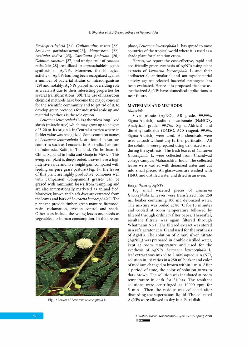
Leucaena leucocephala L. is a thornless long-lived shrub (miracle tree) which may grow up to heights of 5-20 m. Its origin is in Central America where its fodder value was recognized. Some common names of Leucaena leucocephala L. are found in various countries such as Leucaena in Australia, Lamtoro in Indonesia, Katin in Thailand, Yin ho huan in China, Subabul in India and Guaje in Mexico. This evergreen plant is deep-rooted. Leaves have a high nutritive value and live weight gain compared with feeding on pure grass pasture (Fig. 1). The leaves of this plant are highly productive; combines well with campanion (companion) grasses can be grazed with minimum losses from trampling and are also internationally marketed as animal feed. Moreover, brown and black dyes are extracted from the leaves and bark of Leucaena leucocephala L. The plant can provide timber, green manure, firewood, resin, reclamation, erosion control and shade. Other uses include the young leaves and seeds as vegetables for human consumption. In the present
phase, Leucaena leucocephala L. has spread to most countries of the tropical world where it is used as a shade plant for plantation crops.
Herein, we report the cost-effective, rapid and eco-friendly green synthesis of AgNPs using plant extracts of Leucaena leucocephala L. and their antibacterial, antimalarial and antimycobacterial activity against selected bacterial pathogens has been evaluated. Hence it is proposed that the as-synthesized AgNPs have biomedical applications in near future.
MATERIALS AND METHODSMaterials
Silver nitrate (AgNO3, AR grade, 99.99%, Sigma-Aldrich), sodium bicarbonate (NaHCO3, Analytical grade, 99.7%, Sigma-Aldrich) and dimethyl sulfoxide (DMSO, ACS reagent, 99.9%, Sigma-Aldrich) were used. All chemicals were used as such without any further purification. All the solutions were prepared using deionized water during the synthesis. The fresh leaves of Leucaena leucocephala L. were collected from Chandwad college campus, Maharashtra, India. The collected leaves were washed with deionized water and cut into small pieces. All glassware’s are washed with HNO3 and distilled water and dried in an oven.
Biosynthesis of AgNPs10g small wizened pieces of Leucaena
leucocephala L. leaves were transferred into 250 mL beaker containing 100 mL deionized water. The mixture was boiled at 80 oC for 15 minutes and cooled at room temperature followed by filtered through ordinary filter paper. Thereafter, resultant filtrate was again filtered through Whatmann No.1. The filtered extract was stored in a refrigerator at 4 oC and used for the synthesis of AgNPs. The solution of 2 mM silver nitrate (AgNO3) was prepared in double distilled water, kept at room temperature and used for the synthesis of AgNPs. Leucaena leucocephala L. leaf extract was mixed to 2 mM aqueous AgNO3 solution in 1:8 ratios in a 250 ml beaker and color of medium changed to brown within 1 min. After a period of time, the color of solution turns to dark brown. The solution was incubated at room temperature in dark for 24 hrs. The resultant solutions were centrifuged at 10000 rpm for 5 min. Then the residue was collected after discarding the supernatant liquid. The collected AgNPs were allowed to dry in a Petri dish.
Figures captions
Fig. 1. Leaves of Leucaena leucocephala L.
Fig. 1: Leaves of Leucaena leucocephala L.
S. Ghotekar et al. / Green synthesis of Nanoparticles
J. Water Environ. Nanotechnol., 3(2): 95-105 Spring 2018 97
Characterization of the Synthesized AgNPsThe completely dried powder of synthesized
AgNPs was used for the Fourier transform Infra-red (FT-IR) (JASCO 4100) analysis. Transmission electron microscopic (TEM) images were taken on Philips CM 200 operated at accelerating voltages of 20 and 200 kV. X-ray diffraction (XRD) pattern of AgNPs was obtained using Bruker D8-Advanced Diffractometer (λ=1.54 Å) from which average crystal size of AgNPs was calculated. The surface morphology and elemental study of synthesized AgNPs were carried out by field emission scanning electron microscopy (FE-SEM) and Energy Dispersive Spectroscopy (EDS) (JEOL JSM-6701). Photoluminescence studies were evaluated by using fluorescence spectrophotometer (Jobin Yvon Flurolog-3-11, Spectrofluorimeter).
Phytochemical Screening-Qualitative AnalysisFresh aqueous extract of Leucaena leucocephala
L. leaves was used for phytochemical screening-Qualitative analysis. Phytochemical screening was carried out by a standard method [31].
Antibacterial Activity of Synthesized AgNPsThe antibacterial activity of synthesized AgNPs
was determined by using disc diffusion method. This method was employed against selected human pathogens i.e. Pseudomonas aeruginosa, Streptococcus pyogenes, Staphylococcus aureus, Escherichia coli, Salmonella typhi and Bacillus subtilis obtained from Institute of Microbial Technology, Chandigarh, India. The nutrient agar medium (g/l) plates were prepared, well sterilized and solidified. After solidification, bacterial cultures spread evenly over the plate, and then the different concentration of AgNPs solution was poured into each plate. Thereafter, these plates were incubated in an incubator at 37 0C for 24 hrs and measured zone of inhibition of selected bacterial strains against standard reference drugs.
In Vitro Antimalarial Screening of Synthesized AgNPs
In vitro antimalarial assay was carried out in 96 well microtitre plates according to the microassay protocol of antimalarial activity [32]. The cultures of Plasmodium falciparum strain was maintained in medium RPMI-1640 supplemented with 25 mM HEPES, 0.23% NaHCO3, 1% D-glucose and 10% heat-inactivated human serum. The asynchronous parasites of Plasmodium falciparum
were synchronized after 5% D-sorbitol treatment to get only the ring stage parasitized cells. For carrying out the assay, an initial ring stage parasitemia of 3% hematocrit in a total volume of 200 µl of medium RPMI-1640 was determined by Jaswant Singh Bhattacharya (JSB) staining [33] to evaluate the percent parasitemia and uniformly maintained with 50% RBCs (O+ve). The culture plates were incubated at 37 ºC in a candle jar. After 36 hrs incubation, thin blood smears from every cell was prepared and stained with JSB stain. Thereafter, slides were microscopically observed to note that maturation of the ring stage parasites into schizonts and trophozoites in the presence of different concentrations of the synthesized AgNPs. Therein, synthesized AgNPs concentration which inhibits the complete maturation into schizonts was recorded as the minimum inhibitory concentration (MIC). Chloroquine and Quinine were used as the reference drugs for experiments.
In Vitro Antimycobacterial Screening of Synthesized AgNPs
The antimycobacterial screening for green synthesized AgNPs was obtained for Mycobacterium tuberculosis H37RV, by using L. J. (Lowenstein and Jensen) MIC method [34]. Stock solutions of primary 1000, 500, 250 and secondary 200, 100, 62.5, 50, 25, 12.5, 6.25, 3.25 μg/ml of AgNPs in DMSO were added in the liquid L. J. Medium and then media was sterilized. A culture of Mycobacterium tuberculosis H37RV growing on L. J. medium was harvested in 0.85% saline in bijou bottles. These tubes were then incubated at 37°C for 24 hrs followed by streaking of Mycobacterium tuberculosis H37RV. The growth of bacilli was seen after 12 days, 22 days and finally 28 days of incubation respectively. Thereafter, AgNPs containing tubes were compared with control tubes where medium alone was incubated with Mycobaterium tuberculosis H37RV. The concentration at which no development of colonies occurred or < 20 colonies was taken as MIC concentration of test compound. The standard strain Mycobacterium tuberculosis H37RV was tested against reference drug isoniazid [35].
RESULTS AND DISCUSSIONXRD analysis
The crystal structure and crystallite size of synthesized AgNPs were confirmed by using the
98
S. Ghotekar et al. / Green synthesis of Nanoparticles
J. Water Environ. Nanotechnol., 3(2): 95-105 Spring 2018
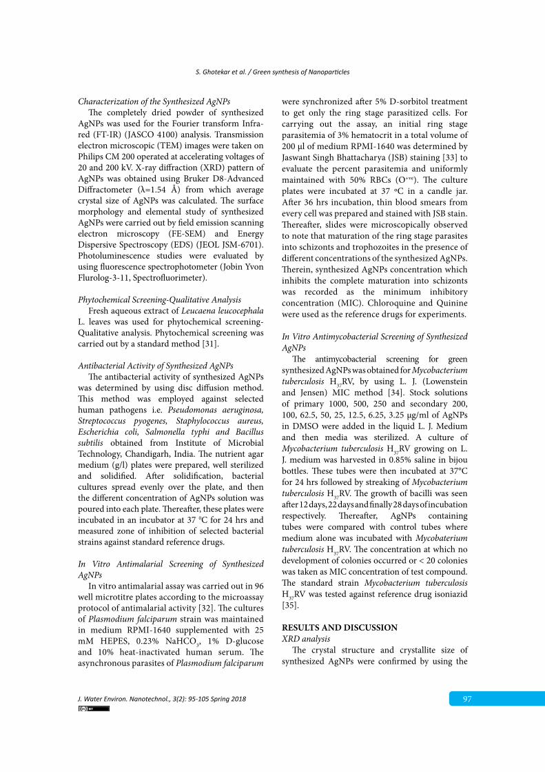
XRD pattern (Fig. 2) in the 2θ range of 20º to 80º. The prominent and intense peaks at 2θ=38.00º, 44.27º, 64.47º and 77.30º corresponding to the (111), (200), (220) and (311) Bragg’s reflections of the face-centered cubic (FCC) crystal structure (JCPDS card no. 04-0783) of AgNPs, respectively. The average crystallite size (D) of AgNPs was calculated by using Scherrer’s equation.
D = Kλ /β COSθ (1)
Where, D is the crystal size of synthesized AgNPs (nm), θ is Bragg angle (degrees), λ is the wavelength of the X-ray source used (1.54060 Å), β is the angular width at the half maximum of the diffraction peak (radians) and K is the constant of Scherrer’s equation which is generally, for the spherically grown nanoparticles 0.94. Therefore, in our present investigation, we have used K= 0.94 to calculate the D value for synthesized material. The average crystal size of the synthesized AgNPs is estimated to be around 32-50 nm. The overall
optostructural study indicated that synthesized material has a pure face-centered cubic crystal structure with nanocrystalline nature and was good agreement with TEM and FE-SEM results.
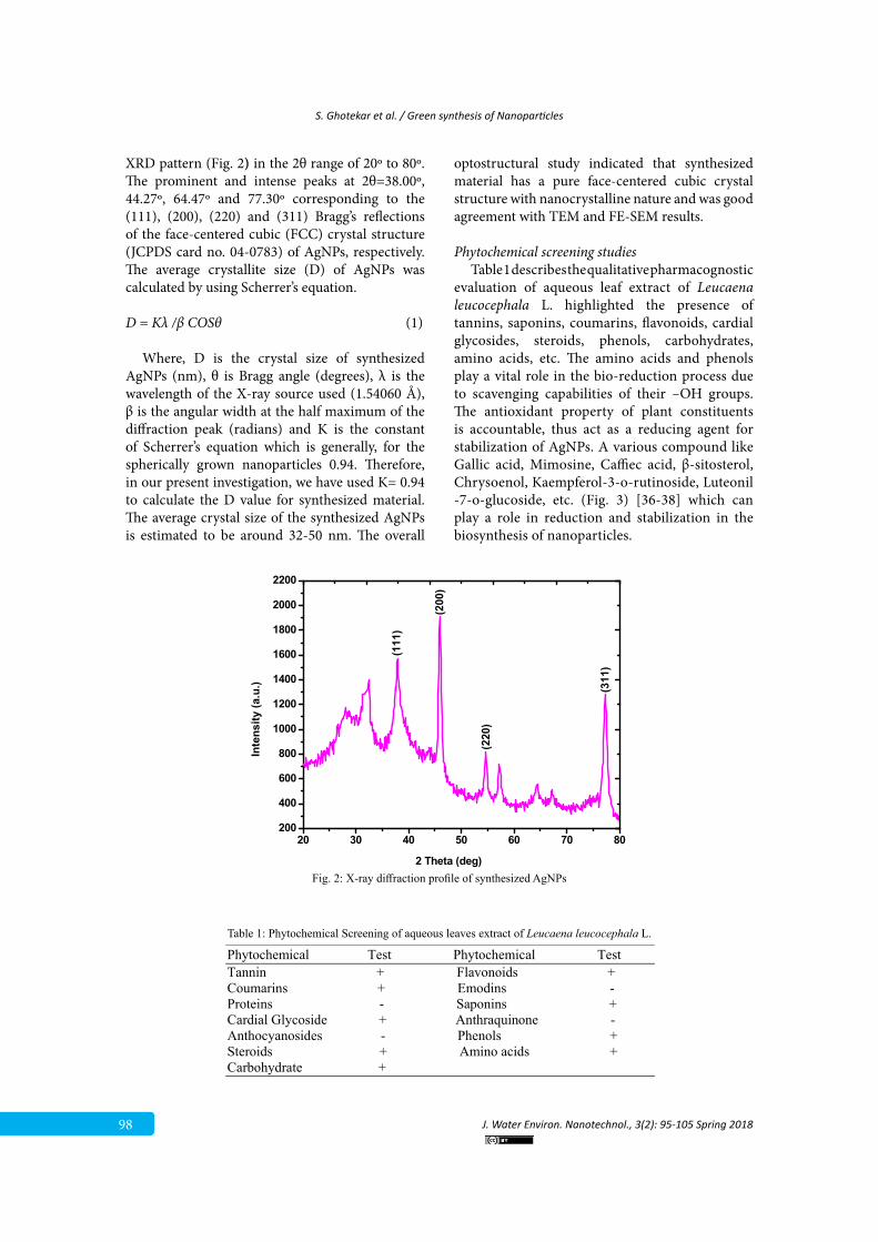
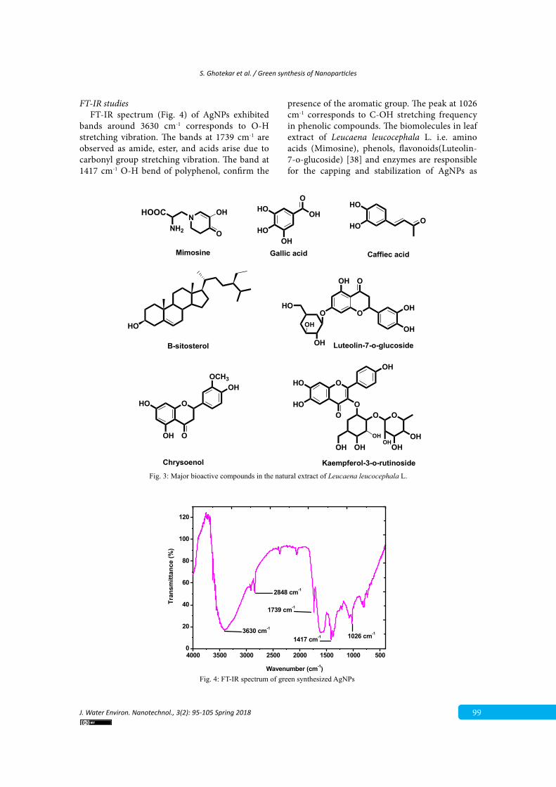
Phytochemical screening studiesTable 1 describes the qualitative pharmacognostic
evaluation of aqueous leaf extract of Leucaena leucocephala L. highlighted the presence of tannins, saponins, coumarins, flavonoids, cardial glycosides, steroids, phenols, carbohydrates, amino acids, etc. The amino acids and phenols play a vital role in the bio-reduction process due to scavenging capabilities of their –OH groups. The antioxidant property of plant constituents is accountable, thus act as a reducing agent for stabilization of AgNPs. A various compound like Gallic acid, Mimosine, Caffiec acid, β-sitosterol, Chrysoenol, Kaempferol-3-o-rutinoside, Luteonil -7-o-glucoside, etc. (Fig. 3) [36-38] which can play a role in reduction and stabilization in the biosynthesis of nanoparticles.
20 30 40 50 60 70 80200
400
600
800
1000
1200
1400
1600
1800
2000
2200
(311
)
(220
)
(200
)
(111
)
Inte
nsity
(a.u
.)
2 Theta (deg) Fig. 2. X-ray diffraction profile of synthesized AgNPs
Fig. 2: X-ray diffraction profile of synthesized AgNPsTables
Table 1. Phytochemical Screening of aqueous leaves extract of Leucaena leucocephala L.
Phytochemical Test Phytochemical Test Tannin + Flavonoids + Coumarins + Emodins - Proteins - Saponins + Cardial Glycoside + Anthraquinone - Anthocyanosides - Phenols + Steroids + Amino acids + Carbohydrate +
Table 1: Phytochemical Screening of aqueous leaves extract of Leucaena leucocephala L.
S. Ghotekar et al. / Green synthesis of Nanoparticles
J. Water Environ. Nanotechnol., 3(2): 95-105 Spring 2018 99
FT-IR studiesFT-IR spectrum (Fig. 4) of AgNPs exhibited
bands around 3630 cm-1 corresponds to O-H stretching vibration. The bands at 1739 cm-1 are observed as amide, ester, and acids arise due to carbonyl group stretching vibration. The band at 1417 cm-1 O-H bend of polyphenol, confirm the
presence of the aromatic group. The peak at 1026 cm-1 corresponds to C-OH stretching frequency in phenolic compounds. The biomolecules in leaf extract of Leucaena leucocephala L. i.e. amino acids (Mimosine), phenols, flavonoids(Luteolin-7-o-glucoside) [38] and enzymes are responsible for the capping and stabilization of AgNPs as
NNH2
HOOC
O
OH
Mimosine
HO
B-sitosterol
O
HO
HO
Caffiec acid
O
OCH3OH
O
HO
OH
Chrysoenol
OOHO
OH
OH
OH O
OH
OH
OHHO
HOO
OH
Gallic acid
O
OH
O
HO
HO OO O
OH OHOH
OHOH
OH
Kaempferol-3-o-rutinoside
Luteolin-7-o-glucoside
Fig. 3. Major bioactive compounds in the natural extract of Leucaena leucocephala L.
Fig. 3: Major bioactive compounds in the natural extract of Leucaena leucocephala L.
4000 3500 3000 2500 2000 1500 1000 5000
20
40
60
80
100
120
3630 cm-1
2848 cm-1
1739 cm-1
1417 cm-1
Wavenumber (cm-1)
Tran
smitt
ance
(%)
1026 cm-1
Fig. 4. FT-IR spectrum of green synthesized AgNPs
Fig. 4: FT-IR spectrum of green synthesized AgNPs
100
S. Ghotekar et al. / Green synthesis of Nanoparticles
J. Water Environ. Nanotechnol., 3(2): 95-105 Spring 2018
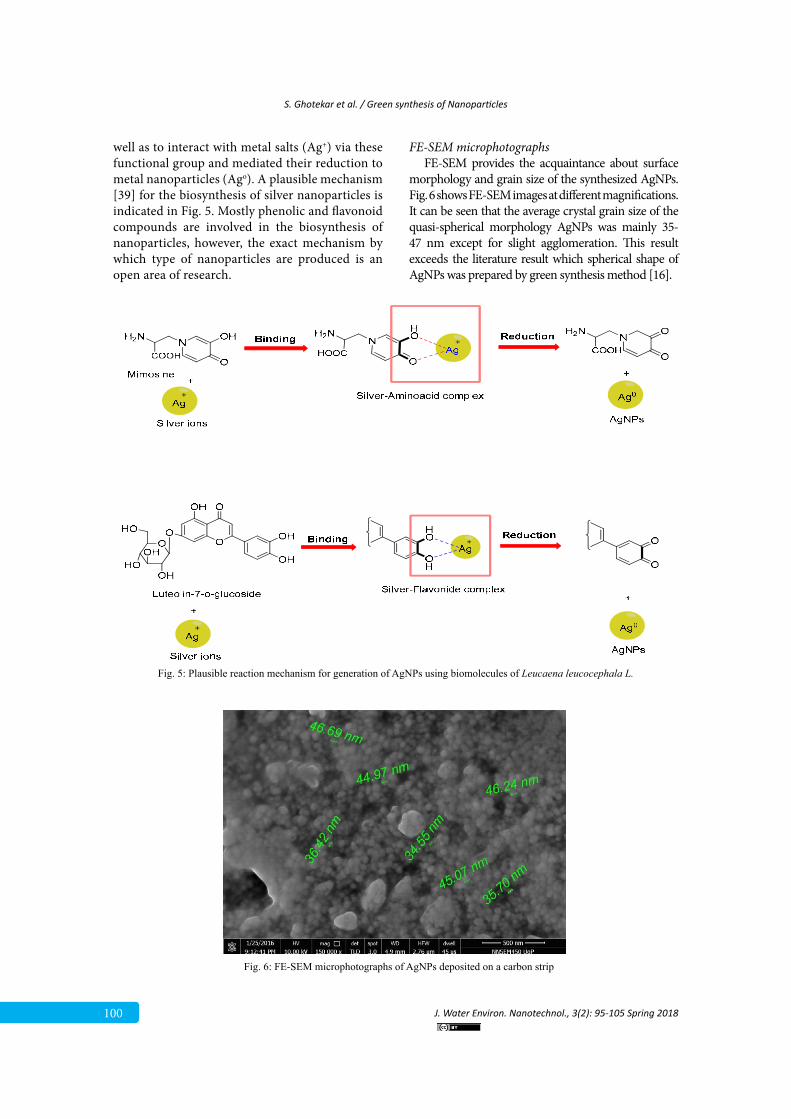
well as to interact with metal salts (Ag+) via these functional group and mediated their reduction to metal nanoparticles (Ago). A plausible mechanism [39] for the biosynthesis of silver nanoparticles is indicated in Fig. 5. Mostly phenolic and flavonoid compounds are involved in the biosynthesis of nanoparticles, however, the exact mechanism by which type of nanoparticles are produced is an open area of research.
FE-SEM microphotographsFE-SEM provides the acquaintance about surface
morphology and grain size of the synthesized AgNPs. Fig. 6 shows FE-SEM images at different magnifications. It can be seen that the average crystal grain size of the quasi-spherical morphology AgNPs was mainly 35-47 nm except for slight agglomeration. This result exceeds the literature result which spherical shape of AgNPs was prepared by green synthesis method [16].
Fig. 5. Plausible reaction mechanism for generation of AgNPs using biomolecules of Leucaena leucocephala L.
Fig. 5: Plausible reaction mechanism for generation of AgNPs using biomolecules of Leucaena leucocephala L.
Fig. 6. FE-SEM microphotographs of AgNPs deposited on a carbon strip
Fig. 6: FE-SEM microphotographs of AgNPs deposited on a carbon strip
S. Ghotekar et al. / Green synthesis of Nanoparticles
J. Water Environ. Nanotechnol., 3(2): 95-105 Spring 2018 101
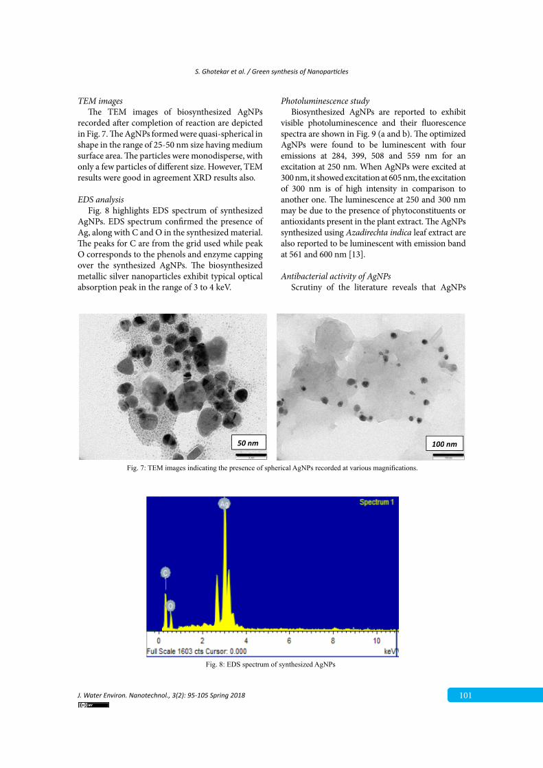
TEM imagesThe TEM images of biosynthesized AgNPs
recorded after completion of reaction are depicted in Fig. 7. The AgNPs formed were quasi-spherical in shape in the range of 25-50 nm size having medium surface area. The particles were monodisperse, with only a few particles of different size. However, TEM results were good in agreement XRD results also.
EDS analysis Fig. 8 highlights EDS spectrum of synthesized
AgNPs. EDS spectrum confirmed the presence of Ag, along with C and O in the synthesized material. The peaks for C are from the grid used while peak O corresponds to the phenols and enzyme capping over the synthesized AgNPs. The biosynthesized metallic silver nanoparticles exhibit typical optical absorption peak in the range of 3 to 4 keV.
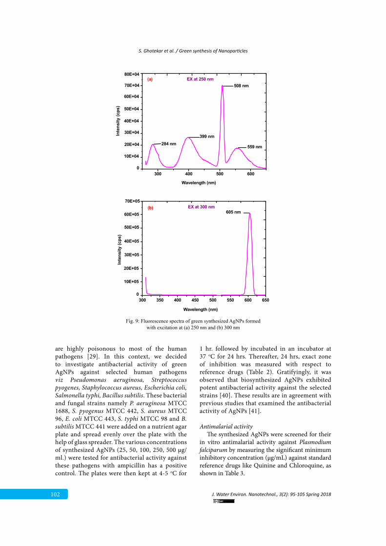
Photoluminescence study Biosynthesized AgNPs are reported to exhibit
visible photoluminescence and their fluorescence spectra are shown in Fig. 9 (a and b). The optimized AgNPs were found to be luminescent with four emissions at 284, 399, 508 and 559 nm for an excitation at 250 nm. When AgNPs were excited at 300 nm, it showed excitation at 605 nm, the excitation of 300 nm is of high intensity in comparison to another one. The luminescence at 250 and 300 nm may be due to the presence of phytoconstituents or antioxidants present in the plant extract. The AgNPs synthesized using Azadirechta indica leaf extract are also reported to be luminescent with emission band at 561 and 600 nm [13].
Antibacterial activity of AgNPsScrutiny of the literature reveals that AgNPs
Fig. 7. TEM images indicating the presence of spherical AgNPs recorded at various
magnifications.
50 nm 100 nm
Fig. 7: TEM images indicating the presence of spherical AgNPs recorded at various magnifications.
Fig. 8. EDS spectrum of synthesized AgNPs
Fig. 8: EDS spectrum of synthesized AgNPs
102
S. Ghotekar et al. / Green synthesis of Nanoparticles
J. Water Environ. Nanotechnol., 3(2): 95-105 Spring 2018
300 400 500 600
559 nm284 nm399 nm
508 nm
0
10E+04
20E+04
30E+04
40E+04
50E+04
60E+04
70E+04
80E+04
Inte
nsity
(cps
)
Wavelength (nm)
(a) EX at 250 nm
300 350 400 450 500 550 600 650
605 nm
Wavelength (nm)
0
10E+05
20E+05
30E+05
40E+05
50E+05
60E+05
70E+05
Inte
nsity
(cps
)
(b) EX at 300 nm
Fig. 9. Fluorescence spectra of green synthesized AgNPs formed with excitation at (a) 250 nm
and (b) 300 nm
Fig. 9: Fluorescence spectra of green synthesized AgNPs formed with excitation at (a) 250 nm and (b) 300 nm
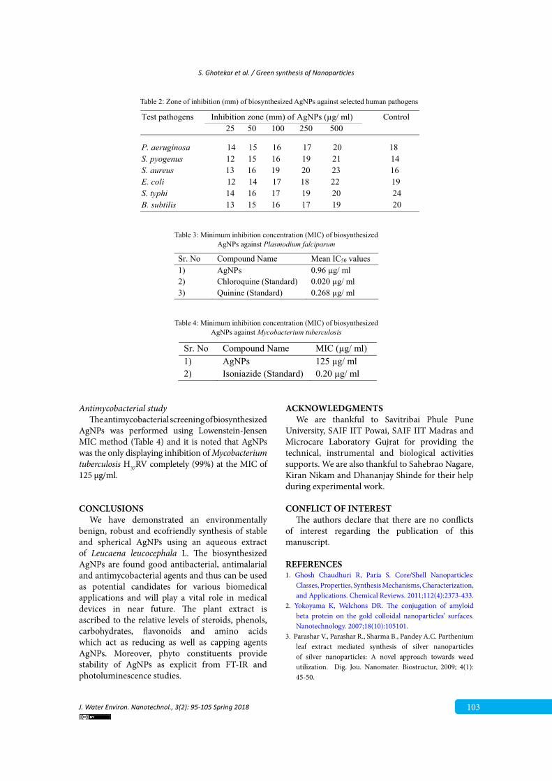
are highly poisonous to most of the human pathogens [29]. In this context, we decided to investigate antibacterial activity of green AgNPs against selected human pathogens viz Pseudomonas aeruginosa, Streptococcus pyogenes, Staphylococcus aureus, Escherichia coli, Salmonella typhi, Bacillus subtilis. These bacterial and fungal strains namely P. aeruginosa MTCC 1688, S. pyogenus MTCC 442, S. aureus MTCC 96, E. coli MTCC 443, S. typhi MTCC 98 and B. subtilis MTCC 441 were added on a nutrient agar plate and spread evenly over the plate with the help of glass spreader. The various concentrations of synthesized AgNPs (25, 50, 100, 250, 500 µg/ ml.) were tested for antibacterial activity against these pathogens with ampicillin has a positive control. The plates were then kept at 4-5 oC for
1 hr. followed by incubated in an incubator at 37 oC for 24 hrs. Thereafter, 24 hrs, exact zone of inhibition was measured with respect to reference drugs (Table 2). Gratifyingly, it was observed that biosynthesized AgNPs exhibited potent antibacterial activity against the selected strains [40]. These results are in agreement with previous studies that examined the antibacterial activity of AgNPs [41].
Antimalarial activity The synthesized AgNPs were screened for their
in vitro antimalarial activity against Plasmodium falciparum by measuring the significant minimum inhibitory concentration (µg/mL) against standard reference drugs like Quinine and Chloroquine, as shown in Table 3.
S. Ghotekar et al. / Green synthesis of Nanoparticles
J. Water Environ. Nanotechnol., 3(2): 95-105 Spring 2018 103
Antimycobacterial studyThe antimycobacterial screening of biosynthesized
AgNPs was performed using Lowenstein-Jensen MIC method (Table 4) and it is noted that AgNPs was the only displaying inhibition of Mycobacterium tuberculosis H37RV completely (99%) at the MIC of 125 μg/ml.
CONCLUSIONSWe have demonstrated an environmentally
benign, robust and ecofriendly synthesis of stable and spherical AgNPs using an aqueous extract of Leucaena leucocephala L. The biosynthesized AgNPs are found good antibacterial, antimalarial and antimycobacterial agents and thus can be used as potential candidates for various biomedical applications and will play a vital role in medical devices in near future. The plant extract is ascribed to the relative levels of steroids, phenols, carbohydrates, flavonoids and amino acids which act as reducing as well as capping agents AgNPs. Moreover, phyto constituents provide stability of AgNPs as explicit from FT-IR and photoluminescence studies.
ACKNOWLEDGMENTSWe are thankful to Savitribai Phule Pune
University, SAIF IIT Powai, SAIF IIT Madras and Microcare Laboratory Gujrat for providing the technical, instrumental and biological activities supports. We are also thankful to Sahebrao Nagare, Kiran Nikam and Dhananjay Shinde for their help during experimental work.
CONFLICT OF INTERESTThe authors declare that there are no conflicts
of interest regarding the publication of this manuscript.
REFERENCES1. Ghosh Chaudhuri R, Paria S. Core/Shell Nanoparticles:
Classes, Properties, Synthesis Mechanisms, Characterization, and Applications. Chemical Reviews. 2011;112(4):2373-433.
2. Yokoyama K, Welchons DR. The conjugation of amyloid beta protein on the gold colloidal nanoparticles’ surfaces. Nanotechnology. 2007;18(10):105101.
3. Parashar V., Parashar R., Sharma B., Pandey A.C. Parthenium leaf extract mediated synthesis of silver nanoparticles of silver nanoparticles: A novel approach towards weed utilization. Dig. Jou. Nanomater. Biostructur, 2009; 4(1): 45-50.
Table 3. Minimum inhibition concentration (MIC) of biosynthesized AgNPs against Plasmodium
falciparum
Sr. No Compound Name Mean IC50 values 1) AgNPs 0.96 µg/ ml 2) Chloroquine (Standard) 0.020 µg/ ml 3) Quinine (Standard) 0.268 µg/ ml
Table 2. Zone of inhibition (mm) of biosynthesized AgNPs against selected human pathogens
Test pathogens Inhibition zone (mm) of AgNPs (µg/ ml) Control 25 50 100 250 500
P. aeruginosa 14 15 16 17 20 18 S. pyogenus 12 15 16 19 21 14 S. aureus 13 16 19 20 23 16 E. coli 12 14 17 18 22 19 S. typhi 14 16 17 19 20 24 B. subtilis 13 15 16 17 19 20
Table 2: Zone of inhibition (mm) of biosynthesized AgNPs against selected human pathogens
Table 3: Minimum inhibition concentration (MIC) of biosynthesized AgNPs against Plasmodium falciparum
Table 4. Minimum inhibition concentration (MIC) of biosynthesized AgNPs against
Mycobacterium tuberculosis
Sr. No Compound Name MIC (µg/ ml) 1) AgNPs 125 µg/ ml 2) Isoniazide (Standard) 0.20 µg/ ml
Table 4: Minimum inhibition concentration (MIC) of biosynthesized AgNPs against Mycobacterium tuberculosis
104
S. Ghotekar et al. / Green synthesis of Nanoparticles
J. Water Environ. Nanotechnol., 3(2): 95-105 Spring 2018
4. Hirsch T, Zharnikov M, Shaporenko A, Stahl J, Weiss D, Wolfbeis OS, et al. Size-Controlled Electrochemical Synthesis of Metal Nanoparticles on Monomolecular Templates. Angewandte Chemie International Edition. 2005;44(41):6775-8.
5. Nadagouda MN, Speth TF, Varma RS. Microwave-Assisted Green Synthesis of Silver Nanostructures. Accounts of Chemical Research. 2011;44(7):469-78.
6. Cheng Y, Yin L, Lin S, Wiesner M, Bernhardt E, Liu J. Toxicity Reduction of Polymer-Stabilized Silver Nanoparticles by Sunlight. The Journal of Physical Chemistry C. 2011;115(11):4425-32.
7. Ghotekar S.K., Vaidya P.S., Pande S.N., Pawar S.P. Synthesis of silver nanoparticles by using 3-methyl pyrazol 5-one (chemical reduction method) and its characterizations. Int. J. Multidis. Res and Deve, 2015; 2(5): 419-422.
8. Jen-La Plante I, Zeid TW, Yang P, Mokari T. Synthesis of metal sulfide nanomaterials via thermal decomposition of single-source precursors. Journal of Materials Chemistry. 2010;20(32):6612.
9. Sunkar S., Nachiyar C.V. Microbial synthesis and characterization of silver nanoparticles using the endophytic bacterium bacillus cereus: a novel source in the benign synthesis. Global J. Med. Res, 2012; 12(2): 43-50.
10. Mittal AK, Chisti Y, Banerjee UC. Synthesis of metallic nanoparticles using plant extracts. Biotechnology Advances. 2013;31(2):346-56.
11. Khatoon N, Ahmad R, Sardar M. Robust and fluorescent silver nanoparticles using Artemisia annua: Biosynthesis, characterization and antibacterial activity. Biochemical Engineering Journal. 2015;102:91-7.
12. Sadeghi B, Gholamhoseinpoor F. A study on the stability and green synthesis of silver nanoparticles using Ziziphora tenuior (Zt) extract at room temperature. Spectrochimica Acta Part A: Molecular and Biomolecular Spectroscopy. 2015;134:310-5.
13. Ahmed S, Saifullah, Ahmad M, Swami BL, Ikram S. Green synthesis of silver nanoparticles using Azadirachta indica aqueous leaf extract. Journal of Radiation Research and Applied Sciences. 2016;9(1):1-7.
14. Sulaiman GM, Mohammed WH, Marzoog TR, Al-Amiery AAA, Kadhum AAH, Mohamad AB. Green synthesis, antimicrobial and cytotoxic effects of silver nanoparticles using Eucalyptus chapmaniana leaves extract. Asian Pacific Journal of Tropical Biomedicine. 2013;3(1):58-63.
15. Vijay Kumar PPN, Pammi SVN, Kollu P, Satyanarayana KVV, Shameem U. Green synthesis and characterization of silver nanoparticles using Boerhaavia diffusa plant extract and their anti bacterial activity. Industrial Crops and Products. 2014;52:562-6.
16. Sadeghi B, Rostami A, Momeni SS. Facile green synthesis of silver nanoparticles using seed aqueous extract of Pistacia atlantica and its antibacterial activity. Spectrochimica Acta Part A: Molecular and Biomolecular Spectroscopy. 2015;134:326-32.
17. Goudarzi M, Mir N, Mousavi-Kamazani M, Bagheri S, Salavati-Niasari M. Biosynthesis and characterization
of silver nanoparticles prepared from two novel natural precursors by facile thermal decomposition methods. Scientific Reports. 2016;6(1).
18. Singh J., Singh N., Rathi A., Kukkar D., Rawat M. Facile Approach to Synthesize and Characterization of Silver Nanoparticles by Using Mulberry Leaves Extract in Aqueous Medium and its Application in Antimicrobial Activity. J Nanostruct, 2017; 7(2):134-140.
19. Vanaja M, Gnanajobitha G, Paulkumar K, Rajeshkumar S, Malarkodi C, Annadurai G. Phytosynthesis of silver nanoparticles by Cissus quadrangularis: influence of physicochemical factors. Journal of Nanostructure in Chemistry. 2013;3(1):17.
20. Huang J, Li Q, Sun D, Lu Y, Su Y, Yang X, et al. Biosynthesis of silver and gold nanoparticles by novel sundried Cinnamomum camphora leaf. Nanotechnology. 2007;18(10):105104.
21. Dubay M., Bhadauria S., Kushwah B.S. Green synthesis of nanosilver particles from extract of Eucalyptus hybrida (safeda) leaf. Dig. J. Nanomat. Biostruc, 2009; 4(3): 537-543.
22. Ponarulselvam S, Panneerselvam C, Murugan K, Aarthi N, Kalimuthu K, Thangamani S. Synthesis of silver nanoparticles using leaves of Catharanthus roseus Linn. G. Don and their antiplasmodial activities. Asian Pacific Journal of Tropical Biomedicine. 2012;2(7):574-80.
23. Nabikhan A, Kandasamy K, Raj A, Alikunhi NM. Synthesis of antimicrobial silver nanoparticles by callus and leaf extracts from saltmarsh plant, Sesuvium portulacastrum L. Colloids and Surfaces B: Biointerfaces. 2010;79(2):488-93.
24. Veerasamy R, Xin TZ, Gunasagaran S, Xiang TFW, Yang EFC, Jeyakumar N, et al. Biosynthesis of silver nanoparticles using mangosteen leaf extract and evaluation of their antimicrobial activities. Journal of Saudi Chemical Society. 2011;15(2):113-20.
25. Krishnaraj C, Jagan EG, Rajasekar S, Selvakumar P, Kalaichelvan PT, Mohan N. Synthesis of silver nanoparticles using Acalypha indica leaf extracts and its antibacterial activity against water borne pathogens. Colloids and Surfaces B: Biointerfaces. 2010;76(1):50-6.
26. Pande S.N., Bharati K.T., Wakchure S.K., Ghotekar S.K., Gujrathi D.B., Phatangare N.D. Green synthesis of silver nanoparticles by Caralluma Fimbriata & its characterization. Ind. J. App. Res, 2015; 5(2): 749-750.
27. Singhal G, Bhavesh R, Kasariya K, Sharma AR, Singh RP. Biosynthesis of silver nanoparticles using Ocimum sanctum (Tulsi) leaf extract and screening its antimicrobial activity. Journal of Nanoparticle Research. 2011;13(7):2981-8.
28. Ghotekar S.K., Pande S.N., Pansambal S.S., Sanap D.S., Mahale K.M., Sonawane B. Biosynthesis of Silver Nanoparticles Using Unripe Fruit Extract of Annona reticulata L. and its Characterization. World J. Pharm. and Pharm. Sci, 2015; 4(11): 1304-1312.
29. Ahmed S, Ahmad M, Swami BL, Ikram S. A review on plants extract mediated synthesis of silver nanoparticles for antimicrobial applications: A green expertise. Journal of Advanced Research. 2016;7(1):17-28.
30. A. Bhosale M, M. Bhanage B. Silver Nanoparticles: Synthesis,
S. Ghotekar et al. / Green synthesis of Nanoparticles
J. Water Environ. Nanotechnol., 3(2): 95-105 Spring 2018 105
Characterization and their Application as a Sustainable Catalyst for Organic Transformations. Current Organic Chemistry. 2015;19(8):708-27.
31. Farnsworth NR. Biological and Phytochemical Screening of Plants. Journal of Pharmaceutical Sciences. 1966;55(3):225-76.
32. Rieckmann KH, Campbell GH, Sax LJ, Ema JE. DRUG SENSITIVITY OF PLASMODIUM FALCIPARUM. The Lancet. 1978;311(8054):22-3.
33. Singh J.S.B. J. S. B stain- A Review. Ind. J. Malar, 1956; 10: 117-129
34. Anargyros P., Astill D.S., Lim I.S. Comparison of improved BACTEC and Lowenstein-Jensen media for culture of mycobacteria from clinical specimens. J. Clin. Microbio, 1990; 28(6): 1288-1291
35. Pansambal S. Phytosynthesis and Biological Activities of Fluorescent CuO Nanoparticles Using Acanthospermum hispidum L. Extract. Journal of Nanostructures. 2017;7(3).
36. Abdelhady N, Abdallah G. HPLC/MS/MS Study of Phenolic Compounds of Leucaena leucocephala Legumes Monitored with Their in vitro Antihyperglycemic Activity. European Journal of Medicinal Plants. 2016;17(4):1-9.
37. Chen C-Y, Wang Y-D. Steroids from the Whole Plants of Leucaena Leucocephala. American Journal of Analytical Chemistry. 2010;01(01):31-3.
38. Hassan RA, Tawfik WA, Abou-Setta LM. The flavonoid constituents of Leucaena Leucocephala growning in Egypt, and their biological activity. African Journal of Traditional, Complementary and Alternative Medicines. 2013;11(1).
39. Kumar B, Kumari S, Cumbal L, Debut A, Camacho J, H. Gallegos E, et al. Pomosynthesis And Biological Activity Of Silver Nanoparticles Using Passiflora Tripartita Fruit Extracts. Advanced Materials Letters. 2015;6(2):127-32.
40. Aher Y.B., Jain G.H., Patil G.E., Savale A.R., Ghotekar S.K., Pore D.M., Pansambal S.S., Deshmukh K.K. Biosynthesis of copper oxide nanoparticles using leaves extract of Leucaena leucocephala L. and their promising upshot against the selected human pathogens. Int. J. Mole. and Clin. Micro, 2017; 7(1): 776-786.
41. Jyoti K, Baunthiyal M, Singh A. Characterization of silver nanoparticles synthesized using Urtica dioica Linn. leaves and their synergistic effects with antibiotics. Journal of Radiation Research and Applied Sciences. 2016;9(3):217-27.