Embed Size (px)
Citation preview
University of MontanaScholarWorks at University of MontanaGraduate Student Theses, Dissertations, &Professional Papers Graduate School
2018
PIG TRAUMA MODELS: A CIVILIANPERSPECTIVE ON AR-15 POST-CRANIALSKELETAL TRAUMALauren M. KenneyUniversity of Montana, Missoula
Let us know how access to this document benefits you.Follow this and additional works at: https://scholarworks.umt.edu/etd
Part of the Biological and Physical Anthropology Commons, and the Other AnthropologyCommons
This Thesis is brought to you for free and open access by the Graduate School at ScholarWorks at University of Montana. It has been accepted forinclusion in Graduate Student Theses, Dissertations, & Professional Papers by an authorized administrator of ScholarWorks at University of Montana.For more information, please contact [email protected].
Recommended CitationKenney, Lauren M., "PIG TRAUMA MODELS: A CIVILIAN PERSPECTIVE ON AR-15 POST-CRANIAL SKELETAL TRAUMA"(2018). Graduate Student Theses, Dissertations, & Professional Papers. 11204.https://scholarworks.umt.edu/etd/11204
i
PIG TRAUMA MODELS: A CIVILIAN PERSPECTIVE ON AR-15
POST-CRANIAL SKELETAL TRAUMA
By
LAUREN MEREDITH KENNEY
Bachelor of Arts, Western Kentucky University, Bowling Green, Kentucky, 2013
Thesis
presented in partial fulfillment of the requirements
for the degree of
Master of Arts
in Anthropology, Forensic Option
University of Montana
Missoula, MT
May 2018
Approved by:
Scott Whittenburg, Dean of The Graduate School
Graduate School
Dr. Randall Skelton, Chair
Department of Anthropology
Dr. Kirsten Green, Co-Chair
Department of Anthropology
Dr. Dusten Hollist, Co-Chair
Department of Sociology
ii
COPYRIGHT
by
Lauren Meredith Kenney
2018
All Rights Reserved
iii
Table of Contents
Chapter 1: Introduction . . . . . . . . . . . . . . . . . . . . . . . . . . . . . . . . . . . . . . . . . . . . . . . . . . . . . . 1
Chapter 2: Literature Review . . . . . . . . . . . . . . . . . . . . . . . . . . . . . . . . . . . . . . . . . . . . . . . . . 3
Historical Review of Firearms . . . . . . . . . . . . . . . . . . . . . . . . . . . . . . . . . . . . . . . . . . . . 3
Basics of Firearms and Ammunition . . . . . . . . . . . . . . . . . . . . . . . . . . . . . . . . . . . . . . . . 4
Size . . . . . . . . . . . . . . . . . . . . . . . . . . . . . . . . . . . . . . . . . . . . . . . . . . . . . . . . . . . . . . . . . 4
Bullet Construction . . . . . . . . . . . . . . . . . . . . . . . . . . . . . . . . . . . . . . . . . . . . . . . . . . . . 5
Projectile Velocity . . . . . . . . . . . . . . . . . . . . . . . . . . . . . . . . . . . . . . . . . . . . . . . . . . . . . 6
Basics of Bullet Travel . . . . . . . . . . . . . . . . . . . . . . . . . . . . . . . . . . . . . . . . . . . . . . . . . . 7
Impact and Effects Bullets . . . . . . . . . . . . . . . . . . . . . . . . . . . . . . . . . . . . . . . . . . . . . . . 8
Wound Beveling . . . . . . . . . . . . . . . . . . . . . . . . . . . . . . . . . . . . . . . . . . . . . . . . . 11
Wound Shape . . . . . . . . . . . . . . . . . . . . . . . . . . . . . . . . . . . . . . . . . . . . . . . . . . . 12
Wound Size . . . . . . . . . . . . . . . . . . . . . . . . . . . . . . . . . . . . . . . . . . . . . . . . . . . . 13
Fracture Type . . . . . . . . . . . . . . . . . . . . . . . . . . . . . . . . . . . . . . . . . . . . . . . . . . 14
Fracture Lines . . . . . . . . . . . . . . . . . . . . . . . . . . . . . . . . . . . . . . . . . . . . . . . . . . 14
Swine, Civilian, and Military Trauma Models. . . . . . . . . . . . . . . . . . . . . . . . . . . . . . . . 16
Chapter 3: Materials and Methods . . . . . . . . . . . . . . . . . . . . . . . . . . . . . . . . . . . . . . . . . . . . . 19
Animal Procurement and Preparation . . . . . . . . . . . . . . . . . . . . . . . . . . . . . . . . . . . . . . 19
Study Area . . . . . . . . . . . . . . . . . . . . . . . . . . . . . . . . . . . . . . . . . . . . . . . . . . . . . . . . . . . 21
Instrumentation Used . . . . . . . . . . . . . . . . . . . . . . . . . . . . . . . . . . . . . . . . . . . . . . . . . . . 21
Experimental Protocol . . . . . . . . . . . . . . . . . . . . . . . . . . . . . . . . . . . . . . . . . . . . . . . . . . 22
The Experiment . . . . . . . . . . . . . . . . . . . . . . . . . . . . . . . . . . . . . . . . . . . . . . . . . . . . . . . . 23
Butchering and Breakdown of Samples . . . . . . . . . . . . . . . . . . . . . . . . . . . . . . . . . . . . . 26
Cleaning the Samples . . . . . . . . . . . . . . . . . . . . . . . . . . . . . . . . . . . . . . . . . . . . . . . . . . . 26
Shipping the Samples to Montana . . . . . . . . . . . . . . . . . . . . . . . . . . . . . . . . . . . . . . . . . 27
Data Collection and Analysis Protocol . . . . . . . . . . . . . . . . . . . . . . . . . . . . . . . . . . . . . 28
Chapter 4: Results . . . . . . . . . . . . . . . . . . . . . . . . . . . . . . . . . . . . . . . . . . . . . . . . . . . . . . . . . . 30
Skeletal Inventory . . . . . . . . . . . . . . . . . . . . . . . . . . . . . . . . . . . . . . . . . . . . . . . . . . . . . . 30
Pig A Results . . . . . . . . . . . . . . . . . . . . . . . . . . . . . . . . . . . . . . . . . . . . . . . . . . . . . . . . . 30
Right Humerus . . . . . . . . . . . . . . . . . . . . . . . . . . . . . . . . . . . . . . . . . . . . . . . . . . . . 31
Left Humerus . . . . . . . . . . . . . . . . . . . . . . . . . . . . . . . . . . . . . . . . . . . . . . . . . . . . 31
Right Scapula . . . . . . . . . . . . . . . . . . . . . . . . . . . . . . . . . . . . . . . . . . . . . . . . . . . . 33
Left Scapula . . . . . . . . . . . . . . . . . . . . . . . . . . . . . . . . . . . . . . . . . . . . . . . . . . . . . 34
Thoracic Vertebrae . . . . . . . . . . . . . . . . . . . . . . . . . . . . . . . . . . . . . . . . . . . . . . . 34
Rib Fragments . . . . . . . . . . . . . . . . . . . . . . . . . . . . . . . . . . . . . . . . . . . . . . . . . . . 36
Sternum Portion . . . . . . . . . . . . . . . . . . . . . . . . . . . . . . . . . . . . . . . . . . . . . . . . . . 37
Left Os Coxa . . . . . . . . . . . . . . . . . . . . . . . . . . . . . . . . . . . . . . . . . . . . . . . . . . . . 37
Right Femur . . . . . . . . . . . . . . . . . . . . . . . . . . . . . . . . . . . . . . . . . . . . . . . . . . . . . 38
Left Femur . . . . . . . . . . . . . . . . . . . . . . . . . . . . . . . . . . . . . . . . . . . . . . . . . . . . . . 39
Right Tibia . . . . . . . . . . . . . . . . . . . . . . . . . . . . . . . . . . . . . . . . . . . . . . . . . . . . . . 40
Left Tibia . . . . . . . . . . . . . . . . . . . . . . . . . . . . . . . . . . . . . . . . . . . . . . . . . . . . . . . 41
Right Fibula . . . . . . . . . . . . . . . . . . . . . . . . . . . . . . . . . . . . . . . . . . . . . . . . . . . . . 42
Left Fibula . . . . . . . . . . . . . . . . . . . . . . . . . . . . . . . . . . . . . . . . . . . . . . . . . . . . . . 43

iv
Metacarpal . . . . . . . . . . . . . . . . . . . . . . . . . . . . . . . . . . . . . . . . . . . . . . . . . . . . . . . . 43
Bullet Fragments . . . . . . . . . . . . . . . . . . . . . . . . . . . . . . . . . . . . . . . . . . . . . . . . . . . 44
Pig B Results . . . . . . . . . . . . . . . . . . . . . . . . . . . . . . . . . . . . . . . . . . . . . . . . . . . . . . . . . . 44
Left Humerus . . . . . . . . . . . . . . . . . . . . . . . . . . . . . . . . . . . . . . . . . . . . . . . . . . . . . 45
Left Scapula . . . . . . . . . . . . . . . . . . . . . . . . . . . . . . . . . . . . . . . . . . . . . . . . . . . . . . 46
Rib Fragment . . . . . . . . . . . . . . . . . . . . . . . . . . . . . . . . . . . . . . . . . . . . . . . . . . . . . . 46
Right Femur . . . . . . . . . . . . . . . . . . . . . . . . . . . . . . . . . . . . . . . . . . . . . . . . . . . . . . 47
Right Os Coxa . . . . . . . . . . . . . . . . . . . . . . . . . . . . . . . . . . . . . . . . . . . . . . . . . . . . . 47
Left Os Coxa . . . . . . . . . . . . . . . . . . . . . . . . . . . . . . . . . . . . . . . . . . . . . . . . . . . . . . 48
Chapter 5: Discussion . . . . . . . . . . . . . . . . . . . . . . . . . . . . . . . . . . . . . . . . . . . . . . . . . . . . . . . . 49
Chapter 6: Conclusions . . . . . . . . . . . . . . . . . . . . . . . . . . . . . . . . . . . . . . . . . . . . . . . . . . . . . . . 52
References Cited . . . . . . . . . . . . . . . . . . . . . . . . . . . . . . . . . . . . . . . . . . . . . . . . . . . . . . . . . . . . . 54
v
List of Tables
Table 1. Biological measurements of Pig A and Pig B including weight, length,
abdominal and thoracic circumference . . . . . . . . . . . . . . . . . . . . . . . . . . . . . . . . . . . . . . . . . . . . 20
List of Figures
Figure 1: The two pigs procured for the experiment . . . . . . . . . . . . . . . . . . . . . . . . . . . . . . . . . . . 20
Figure 2: The Moore Family Farm sign . . . . . . . . . . . . . . . . . . . . . . . . . . . . . . . . . . . . . . . . . . . . 21
Figure 3: Pig A hanging upright from tractor and hayfork with green livestock marker
on areas selected for impact with AR-15 . . . . . . . . . . . . . . . . . . . . . . . . . . . . . . . . . . . . . . . . . . . . 24
Figure 4: Pig B hanging upright from tractor and hayfork with green livestock marker
on areas selected for impact with AR-15 . . . . . . . . . . . . . . . . . . . . . . . . . . . . . . . . . . . . . . . . . . . . 25
Figure 5: Author, Lauren Kenney, cleaning samples in the makeshift lab created in
a garage . . . . . . . . . . . . . . . . . . . . . . . . . . . . . . . . . . . . . . . . . . . . . . . . . . . . . . . . . . . . . . . . . . . . . 27
Figure 6: The make-up of the skeletal elements for consideration for each pig model;
a. Pig A elements b. Pig B elements . . . . . . . . . . . . . . . . . . . . . . . . . . . . . . . . . . . . . . . . . . . . . . . 30
Figure 7: The right humerus of Pig A;
a. Anterior view b. Lateral view c. Posterior view d. Medial view . . . . . . . . . . . . . . . . . . . . . . . . 31
Figure 8: The left humerus of Pig A;
a. Anterior view b. Medial view c. Posterior view (with exit wound/third fragment)
d. Posterior view (without exit wound/third fragment) e. Lateral view . . . . . . . . . . . . . . . . . . . . 32
Figure 9: The right scapula of Pig A;
a. Posterior view b. Anterior view . . . . . . . . . . . . . . . . . . . . . . . . . . . . . . . . . . . . . . . . . . . . . . . . . 33
Figure 10: The left scapula of Pig A;
a. Posterior view b. Anterior view . . . . . . . . . . . . . . . . . . . . . . . . . . . . . . . . . . . . . . . . . . . . . . . . . 34
Figure 11: Thoracic vertebra (T8) of Pig A;
a. Superior view b. Inferior view c. Anterior view . . . . . . . . . . . . . . . . . . . . . . . . . . . . . . . . . . . . 35
Figure 12: Thoracic vertebra (T9) of Pig A;
a. Superior view b. Inferior view c. Posterior view . . . . . . . . . . . . . . . . . . . . . . . . . . . . . . . . . . . . 35
Figure 13: Mostly complete ribs from Pig A which show damage to the sternal ends;
a. Superior view b. Inferior view . . . . . . . . . . . . . . . . . . . . . . . . . . . . . . . . . . . . . . . . . . . . . . . . . . 36
vi
Figure 14: Sternal end rib fragments from Pig A;
a. Superior view b. Inferior view . . . . . . . . . . . . . . . . . . . . . . . . . . . . . . . . . . . . . . . . . . . . . . . . . . 36
Figure 15: Vertebral end rib fragments from Pig A;
a. Superior view b. Inferior view . . . . . . . . . . . . . . . . . . . . . . . . . . . . . . . . . . . . . . . . . . . . . . . . . . 36
Figure 16: The sternum portion from Pig A;
a. Posterior view b. Anterior view . . . . . . . . . . . . . . . . . . . . . . . . . . . . . . . . . . . . . . . . . . . . . . . . 37
Figure 17: The left os coxa of Pig A;
a. Lateral view b. Medial view . . . . . . . . . . . . . . . . . . . . . . . . . . . . . . . . . . . . . . . . . . . . . . . . . . . 38
Figure 18: The right femur of Pig A;
a. Anterior view b. Lateral view c. Posterior view d. Medial view . . . . . . . . . . . . . . . . . . . . . . . . 39
Figure 19: The right femur of Pig A;
Butterfly fracture image from Dino-Lite . . . . . . . . . . . . . . . . . . . . . . . . . . . . . . . . . . . . . . . . . . . . 39
Figure 20: The left femur of Pig A;
a. Anterior view b. Lateral view c. Medial view d. Posterior view . . . . . . . . . . . . . . . . . . . . . . . . 40
Figure 21: The left femur of Pig A;
Exit wound image from Dino-lite . . . . . . . . . . . .. . . . . . . . . . . . . . . . . . . . . . . . . . . . . . . . . . . . . 40
Figure 22: The right tibia of Pig A;
a. Anterior view b. Lateral view c. Posterior view d. Medial view . . . . . . . . . . . . . . . . . . . . . . . . 41
Figure 23: The left tibia of Pig A;
a. Anterior view b. Lateral view c. Posterior view d. Medial view . . . . . . . . . . . . . . . . . . . . . . . . 41
Figure 24: The proximal end of the right fibula from Pig A;
a. Anterior view b. Posterior view . . . . . . . . . . . . . . . . . . . . . . . . . . . . . . . . . . . . . . . . . . . . . . . . . 42
Figure 25: The distal end of the right fibula from Pig A;
a. Anterior view b. Posterior view . . . . . . . . . . . . . . . . . . . . . . . . . . . . . . . . . . . . . . . . . . . . . . . . . 42
Figure 26: The left fibula from Pig A;
a. Anterior view b. Posterior view . . . . . . . . . . . . . . . . . . . . . . . . . . . . . . . . . . . . . . . . . . . . . . . . 43
Figure 27: The metacarpal of Pig A;
a. Anterior view b. Posterior view d. Medial view . . . . . . . . . . . . . . . . . . . . . . . . . . . . . . . . . . . . 43
Figure 28: The five bullet fragments collected from Pig A . . . . . . . . . . . . . . . . . . . . . . . . . . . . . . 44
vii
Figure 29: One of the largest bullet fragments recovered from Pig A, magnified
using the Dino-Lite to show the outer copper coating and rifling
(image from Dino-Lite) . . . . . . . . . . . . . . . . . . . . . . . . . . . . . . . . . . . . . . . . . . . . . . . . . . . . . . . . . 44
Figure 30: The left humerus of Pig B;
a. Anterior view b. Lateral view c. Posterior view d. Medial view . . . . . . . . . . . . . . . . . . . . . . . 45
Figure 31: The left scapula of Pig B;
a. Posterior view b. Inferior posterior view c. Anterior view . . . . . . . . . . . . . . . . . . . . . . . . . . . . 46
Figure 32: The rib fragment from Pig B;
a. Anterior view b. Posterior view . . . . . . . . . . . . . . . . . . . . . . . . . . . . . . . . . . . . . . . . . . . . . . . . .46
Figure 33: The right femur of Pig B;
a. Anterior view b. Lateral view c. Posterior view d. Medial view . . . . . . . . . . . . . . . . . . . . . . . . 47
Figure 34: The right os coxa of Pig B;
a. Lateral view b. Medial view . . . . . . . . . . . . . . . . . . . . . . . . . . . . . . . . . . . . . . . . . . . . . . . . . . . 48
Figure 35: The left os coxa of Pig B;
a. Lateral view b. Medial view . . . . . . . . . . . . . . . . . . . . . . . . . . . . . . . . . . . . . . . . . . . . . . . . . . . 48
viii
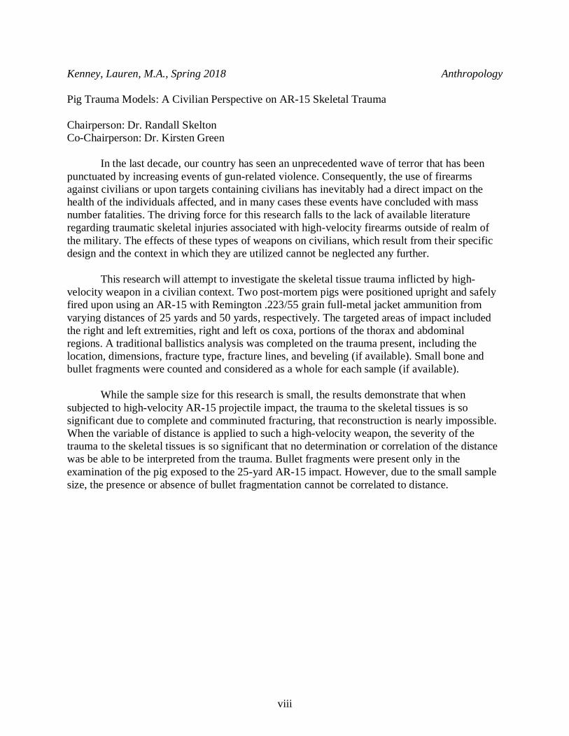
Kenney, Lauren, M.A., Spring 2018 Anthropology
Pig Trauma Models: A Civilian Perspective on AR-15 Skeletal Trauma
Chairperson: Dr. Randall Skelton
Co-Chairperson: Dr. Kirsten Green
In the last decade, our country has seen an unprecedented wave of terror that has been
punctuated by increasing events of gun-related violence. Consequently, the use of firearms
against civilians or upon targets containing civilians has inevitably had a direct impact on the
health of the individuals affected, and in many cases these events have concluded with mass
number fatalities. The driving force for this research falls to the lack of available literature
regarding traumatic skeletal injuries associated with high-velocity firearms outside of realm of
the military. The effects of these types of weapons on civilians, which result from their specific
design and the context in which they are utilized cannot be neglected any further.
This research will attempt to investigate the skeletal tissue trauma inflicted by high-
velocity weapon in a civilian context. Two post-mortem pigs were positioned upright and safely
fired upon using an AR-15 with Remington .223/55 grain full-metal jacket ammunition from
varying distances of 25 yards and 50 yards, respectively. The targeted areas of impact included
the right and left extremities, right and left os coxa, portions of the thorax and abdominal
regions. A traditional ballistics analysis was completed on the trauma present, including the
location, dimensions, fracture type, fracture lines, and beveling (if available). Small bone and
bullet fragments were counted and considered as a whole for each sample (if available).
While the sample size for this research is small, the results demonstrate that when
subjected to high-velocity AR-15 projectile impact, the trauma to the skeletal tissues is so
significant due to complete and comminuted fracturing, that reconstruction is nearly impossible.
When the variable of distance is applied to such a high-velocity weapon, the severity of the
trauma to the skeletal tissues is so significant that no determination or correlation of the distance
was be able to be interpreted from the trauma. Bullet fragments were present only in the
examination of the pig exposed to the 25-yard AR-15 impact. However, due to the small sample
size, the presence or absence of bullet fragmentation cannot be correlated to distance.
ix
Acknowledgments
This research has been somewhat of an uphill battle from the beginning; one that has
been not only filled with mental but emotional struggles. As I sat in my apartment piecing
together the hundreds of fragments of bone produced from this research, seventeen people were
killed at Marjory Stoneman Douglas High School in Parkland, Florida. Therefore, while it will
never be enough, this research is dedicated to the victims and survivors of gun-violence.
For those that have helped to bridge the many gaps that opened along the way, you have
not gone unnoticed. Dr. Randall Skelton, Dr. Kirsten Green, and Dr. Dusten Hollist; my gratitude
for your involvement is my committee can never be expressed. Thank you.
To my parents, who began this journey with me in a U-Haul nearly two years and 2,000
miles ago. I will never be able to repay you for the time, money, and effort that you have
dedicated to all of my endeavors. To Mom, your endless positive affirmations that I can, in fact,
“do this” can never be repaid. And to Dad, for always saying “yes”, even if in the end we both
know it should have been a “no”. I love you both, and I will always be your best girl.
And finally, to Joe Michael and Andy Joe Moore, two amazing educators who have not
only dedicated their lives to children but saw my vision and the need for this research. I sincerely
could not have done any of this without you.
1
Chapter 1: Introduction
In the last decade, our country has seen an unprecedented wave of terror that has been
punctuated by increasing events of gun-related violence. While civilian mass shootings in Las
Vegas, Kentucky, and Florida only position as the most recent, conservative estimates show that
such events have risen four-fold from 1999 to 2006 (Kashuk et al., 2009; Wolf et al., 2009).
Consequently, the use of high-velocity firearms against civilians or upon targets containing
civilians has inevitably had a direct impact on the health of the individuals affected, and in many
cases these events have concluded with mass number fatalities (Coupland and Meddings, 1999;
Coupland and Samnegaard, 1999; Kashuk et al., 2009; Wolf et al., 2009).
The driving force for this research is due to the lack of available literature regarding the
traumatic skeletal injuries associated with high-velocity firearms outside of realm of the military
(Champion et al., 2003; Lichte et al., 2010). The trauma encountered from such weapons in the
civilian setting differ greatly from what is seen in military combatants in terms of the
epidemiology, the mechanism of wounding, and pathophysiologic trajectory (Champion et al.,
2003; Steadman and Haglund, 2005; Shin et al., 2015). While there has been significant research
conducted on combat injuries, it must be recognized that the trauma inflicted from high-velocity
firearms has many unique considerations that must be addressed with regard to a civilian context.
The effects of these types of weapons on civilians, resulting from their specific design, and the
context in which they are being utilized cannot be neglected any further (Coupland and
Meddings, 1999; Coupland and Samnegaard, 1999; Cukier, 2002).
With that said, there is much to be gained from research that focuses on high-velocity
firearm trauma. The recognition of the differences within the wounding patterns could be
applicable to the study of trauma in an anthropological or human rights context, which could in

2
turn be beneficial to future scenarios involving civilians in mass atrocity events around the world
(Coupland and Samnegaard, 1999; Mabry et al., 2000; Champion et al., 2003; Steadman and
Haglund, 2005; Buchanan, 2011; Steflj and Darden, 2013).

3
Chapter 2: Literature Review
Historical Review of Firearms
The history of firearms constitutes a rather lengthy and complex subject. However, it is
one that parallels the trauma associated with such weapons and therefore must be reviewed. To
begin, gunpowder is thought to have originated in China during the 9th century, but it was not
until the 13th century in Europe that experimentation on weaponry begins to be seen in the
historical literature (Frost and Denton, 2015). Shortly after the 13th century, evidence for the use
of firearms became predominant during warfare and conflict (Frost and Denton, 2015).
Over the last several centuries, firearms and their associated ballistics have been greatly
refined, making them more accurate with increased firing capacity (Ezell, 2002; Frost and
Denton, 2015). Lock-works and loading mechanisms have progressed from muzzle-loading
matchlocks, flintlocks, and cap-and-ball arms to more modern, self-contained cartridges with
rapid firing power (Frost and Denton, 2015). Additionally, improved night vision equipment, as
well as high-powered scopes allow for rapid target acquisition and precision during distance
shooting (Ezell, 2002).
Historically, primary weapons have been the most frequently observed during conflict
and can be divided into two types: explosive munitions and small arms (U.S. Army, 2013; Smith
and Bellamy, 2016). Explosive munitions include artillery weapons such as grenades, mortars,
bombs, rockets, mines, and IEDs (Improvised Explosive Devices) (U.S. Army, 2013). Small
arms include weapons such as pistols, rifles, and machine guns (Byers, 2011; U.S. Army, 2013).
Many of the trends for small arms usage since World War II, include weapons such as rifles and
machine guns that have an increased firing capacity, lighter bullets, and increased muzzle
4
velocity (Ezell, 2002). Small arms are also the preferred weapons utilized in most murders and
suicides because of their high rates of lethality (Byers, 2011).
Surprisingly, nearly 80% of the small arms weapons in the world (approximately 650
million) reside in the hands of civilians; the United States alone accounts for about 270 million,
or about 90 firearms for every 100 people (Tejan and Lindsey, 1998; Cukier, 2002). For this
particular research only small arms, specifically rifles (the AR-15), will be taken into
consideration due to their accessibility within the modern civilian population, as well as their
prevalence in many recent civilian mass shootings (Tejan and Lindsey, 1998; Buchanan, 2011).
Basics of Firearms and Ammunition
Because of the wide variety presently available on the market, firearms and their
associated ammunition also constitute a lengthy and complex subject (Barach et al., 1986a;
Barach et al., 1986b; Byers, 2011). Firearms and ammunition come in a variety of sizes and
powers, with each possessing its own specific wounding pattern and characteristics (Byers,
2011). However, the common characteristic of all small arms weaponry is a tube of variable
length, called the barrel. The barrel is complete with an attached chamber that receives the unit
of ammunition containing a bullet (Stefanopoulos et al., 2016). Bullets, or projectiles, are
customarily described as being either high or low velocity, and this description roughly
corresponds to the two main categories of small arms: handguns and rifles.
Size
Size can refer to either the diameter of a projectile, and/or the diameter of the barrel. This
diameter can be as small as 0.05 of an inch to 0.950 of an inch, and is measured in terms of
caliber, gauge, or number (Barach et al., 1986a; Barach et al., 1986b; Byers, 2011).

5
Bullet Construction
Construction, with regard to a bullet, refers to a number of factors including the profile,
the internal composition, and covering (jacketing) (Barach et al., 1986a; Barach et al., 1986b;
Byers, 2011). There are three basic bullet profiles, and these include sharp, blunt, and hollow-
point (Barach et al., 1986a; Barach et al., 1986b; Di Maio, 1993; Berryman et al., 1995; Byers,
2011).
A sharp profile is commonly utilized in rifle ammunition, while blunt and hollow-point
ammunition is frequently utilized in handguns (Barach et al., 1986a; Barach et al., 1986b; Di
Maio, 1993; Berryman et al., 1995; Byers, 2011). Characteristically, blunt profile ammunition
has either a flat or rounded tip, while hollow-points are identified by an indention on their tips.
When compared to sharp profile ammunition, both blunt and hollow-point bullets are more likely
to deform on impact and would be expected to cause greater trauma and larger wounds in bone
(especially exit wounds) (Barach et al., 1986a; Barach et al., 1986b; Di Maio, 1993; Berryman et
al., 1995; Byers, 2011).
There are two basic types of internal composition within bullets. The most common
composition is lead, which is preferred because of its weight and its ability to deform more than
iron or steel (Barach et al., 1986a; Barach et al., 1986b; Di Maio, 1993; Berryman et al., 1995;
Byers, 2011). Some bullets however, are intentionally constructed to fragment upon impact and
this is usually seen within round or blunt profiles (Barach et al., 1986a; Barach et al., 1986b; Di
Maio, 1993; Berryman et al., 1995; Byers, 2011). Fragmenting bullets are usually composed of
small rounded pellets that scatter after the rupture of the casing upon impact with the target
(Byers, 2011).
6
The presence or absence of a jacket is the final factor of bullet construction. A jacket
refers to a thin copper (or other metal) coating that encapulates the outside of the bullet (Barach
et al., 1986a; Barach et al., 1986b; Di Maio, 1993; Berryman et al., 1995; Byers, 2011). Some
coatings cover the entire projectile, called full-metal jackets, while other coatings cover only a
portion of the projectile (Barach et al., 1986a; Barach et al., 1986b; Di Maio, 1993; Berryman et
al., 1995; Byers, 2011). Fully jacketing a projectile helps to reduce deformation and
fragmentation during passage through the body. Hence, projectiles that are non-jacketed are
more likely to deform during passage through bodily tissues (Barach et al., 1986a; Barach et al.,
1986b; Di Maio, 1993; Berryman et al., 1995; Byers, 2011).
Projectile Velocity
While there is debate, some research suggests that the velocity of a projectile has the
greatest contributing effect on its wounding potential (Barach et al., 1986a; Barach et al., 1986b;
Di Maio, 1993; Berryman et al., 1995; Byers, 2011). Velocity refers to the speed at which a
projectile exits the barrel of the weapon and impacts a target (Morse et al., 1983; Barach et al.,
1986a; Barach et al., 1986b; Byers, 2011). Low-velocity projectiles consist of those that are used
in handguns, while high-velocity projectiles are most commonly used within shotguns and rifles
(Byers, 2011; Shin et al., 2015).
Traditionally, ballistics have been defined in terms of their specific projectile velocities
by way of distance and time (fps or m/s) (Coupland and Samnegaard, 1999). While low velocity
is generally considered to be less than 2,000 fps or 350 m/s, high velocity (while less defined), is
considered to start at approximately 600-700 m/s and can exceed up to 700-960 m/s (Byers,
2011; Smith and Bellamy, 2016). Both low-velocity and high-velocity projectiles lead to tissue
7
damage that is based on the overall amount of kinetic energy (KE= ½ MV2) imparted onto the
tissue or body portion impacted (Shin et al., 2015). The kinetic energy transferred is also
determined by several factors, including the projectile’s size, construction, the velocity, the
kinetic energy of the projectile at bodily contact, the distance traveled prior to striking the body
and after penetration, the entrance profile and path taken by the projectile, and the biological
characteristics of the bodily tissue affected (Shin et al., 2015).
Basics of Bullet Travel
In small arms weaponry, like the rifle, the bullet is accelerated down the barrel under
extremely high pressure that is created by the build-up of the expanding gasses from a
combustion propellant (Stefanopoulos et al., 2016). When a bullet is expelled from the barrel of
most modern firearms, it travels in the direction of the target while spinning along its long axis
(Byers, 2011). Spiral grooves cut into the internal surface of the guns’ barrel, called rifling,
impart a spin so that the projectile will go straighter for a longer distance (Byers, 2011).
At the beginning of its flight, a bullet’s long axis is parallel to it flight path (trajectory). If
the angle of the discharging weapon remains perpendicular (90o angle) to the intended target, a
circular wound will be presented. However, after the bullet leaves the barrel and travels some
distance, it is likely to start to tumble (Byers, 2011). If the discharging weapon is not at a 90o
angle, a noncircular outline will result (Byers, 2011). The effect is that the bullet’s long axis is no
longer parallel to its trajectory, and when it eventually reaches the target, the projectile may
create an uncharacteristic noncircular wound (Byers, 2011).

8
Impact and Effects of Bullets
The severity and lethality of any firearm wound is directly associated to the dynamics of
the projectile, the wound track taken, and the proximity of the wound track to vital organs, blood
vessels, and bone (Smith and Bellamy, 2016). The type of firearm utilized, as well as the
subsequent effects, are dependent on many factors including the interaction on the anatomical
location, the number of shots, the technical specification of the ammunition used, the effects
from the properties of the projectile, the velocity, and the angle of trajectory (Champion et al.,
2003; Steadman and Haglund, 2005; Buchanan, 2011). The preponderance and severity of the
injuries can also be dependent on variables found within the environment including buildings or
fences, as well as any personal protective gear in the form of advanced helmets and body armor
that may be worn by the individual (Blair et al., 2012). All of these factors can subsequently
influence the trajectory of the bullet through the body and the pattern of damage to tissues,
organs, and bones (Smith and Bellamy, 2016).
As a projectile penetrates the body, rupturing of the tissues encountered by the leading
edge of the advancing bullet occurs, along with the crushing injury of any bones in the direct
path (Trott, 1988; Klein et al., 2007; Stefanopoulos et al., 2014). As the tissue detaches, it creates
a vacuum behind the projectile for a few thousandths of a second, much like a torpedo creates
when traveling underwater (Buchanan, 2011; Stefanopoulos et al., 2014). The projectile causes
both a temporary and permanent cavitation event marked by subsequent tissue expansion and
damage from shearing, percussion, and vibratory forces (Shin et al., 2015).
When a bullet strikes a target with enough energy, it will create a perforating wound
(Byers, 2011). The location of entry is called a penetrating entry wound, and where it leaves,
provided that it has enough energy to do so, is called the exit wound (Byers, 2011).
9
The residual wound tract which remains after the complete passage of a projectile, is commonly
referred to as the permanent wound tract or cavity, indicated by a central defect in the body along
with any surrounding area of irreversible tissue damage (Stefanopoulos et al., 2014) A
permanent cavity is also characterized by a localized defect area of cell necrosis that is
proportional to the size of the projectile as it passes through the body (U.S. Army, 2013). The
process of permanent cavitation occurs at exceedingly high velocities, usually greater than 600
m/s, and it is an extremely dynamic phenomenon; the greater the speed of the bullet, the larger
the permanent cavity (Buchanan, 2011; Stefanopoulos et al., 2014).
Alternatively, a temporary cavity is characterized by the lateral displacement of tissue
which occurs after the passage of a projectile through tissue. Elastic tissues, such as the muscle,
blood vessels, and skin may be pushed away from the projectile but retained through rebounding
(Steflj and Darden, 2013; U.S. Army, 2013). Inelastic tissues, such as bone may actually fracture
or be ruptured several centimeters from the bullet track (Klein, 2007; Buchanan, 2011; U.S.
Army, 2013).
If the projectile enters the target intact, its original characteristics will be directly
imparted into the target (Byers, 2011). However, if the projectile comes into contact with an
intermediate object, it will usually deform and fragment, especially if it is of non-jacketed
construction (Burke and Rowe, 1992; Byers, 2011). The same can be said for projectiles that
travel through significant amounts of bodily tissues. Splinters from bullet fragments can become
embedded in soft tissues or can cause the chipping or fragmentation of bone (Burke and Rowe,
1992; Byers, 2011). The fracturing of bone can happen so severely that portions can become
fragmented into minute pieces. When a wound (entry/exit) is formed through bone, fracture lines
can radiate outward from, and in some cases, encircle the area of impact (Berryman and Symes,

10
1998; Byers, 2011). Projectiles that impact the bone with enough force generally result in the
creation of complete fractures, with both displacement of the bone and fracture lines (Byers,
2011).
A bullet retained within the tissue is one which has utilized and delivered all of its energy
creating only an entrance wound. Alternatively, a bullet that has perforated the skeletal tissue
completely, also called a through-and-through, is indicated not only by an entrance wound, but
also an exit wound (Stefanopoulos et al., 2014). Low-velocity projectiles tend to cause only
localized tissue damage that is directly related to the actual size of the projectile itself (Shin et al,
2015). Drill-hole defects, which are common in low-velocity penetration are more common
along the metaphyseal region of long bones due to the greater proportion of cancellous bone
(Stefanopoulos et al., 2014; Smith and Bellamy, 2016).
High-velocity projectiles on the other hand, have multiple mechanisms of tissue and
bone destruction that occur secondarily to the forces and energy imparted on the tissues (Shin et
al., 2015). High-velocity projectiles typically produce comminuted fractures due to the explosive
effects of cavitation that is associated with the properties of the marrow cavity (Blair et al.,
2012). Thus, high-velocity weapon injuries often result in tissue defects, complete with
significant fracturing of the underlying bone, varying degrees of thermal trauma, and
contamination due to imbedded particulates and foreign debris (Shin et al., 2015). Specifically,
injuries to the extremities often result in severe comminuted fractures, or permanent bone and
joint deformities (Klein et al., 2007; Buchanan, 2011).
Overall, bone injuries from ballistics are a more complex process than that which occurs
during the penetration of soft tissues. Bone tissues cause a marked slowing of the penetrating
projectile, which can be expected due to its greater density, related mechanical properties, and

11
hardness (Griffiths and Clasper, 2006; Hodgetts et al., 2006; Manring et al., 2009; Stefanopoulos
et al., 2014). These characteristics of bone may actually cause the bullet to become deformed or
even fragment upon entering the body (Griffiths and Clasper, 2006; Stefanopoulos et al., 2014).
The M-16 rifle, or its civilian version the AR-15 used in this research, is one of the most
common weapons utilized during worldwide conflicts (Klein, 2007). With the particular
projectiles used in such rifles, either full-metal jacketed or ball, there is only a 25 cm path of
tissue disruption, explaining why relatively minimal tissue damage may be seen in some wounds
(Trott, 1988). Alternatively, damage to bone can be significant with such weaponry, and will be
of importance for this research.
Wound Beveling
When a projectile comes into contact with bone, if perforation occurs, the bone will
deform. This deformation creates a funnel shaped hole larger where the projectile exits, than
where the projectile entered the bone (Berryman and Symes, 1998; Byers, 2011). The funneling
effect is called beveling, and can be categorized into three types: inward, outward, and reverse.
Inward beveling can be seen at the direct site of a projectiles’ entry into the bone. The
area of beveling on the outer surface of the bone is usually smaller than the area of beveling
inside (Berryman and Symes, 1998; Byers 2011). Outward beveling occurs at the area the
projectile exits the bone. In contrast to entry wounds, the inner hole beveling will be smaller than
the outer hole beveling (Byers, 2011). Reverse beveling can also occur, which can be simply
described as beveling that occurs in the opposite direction of either the entrance or exit of a
wound and is usually considerably smaller than the inward or outward beveling that it opposes
(Berryman and Symes, 1998; Byers 2011).
12
Wound Shape
The wound shape that a bullet or projectile creates is dependent on several factors. These
can include the way that the bullet is constructed, its angle of trajectory, the angle of its axis, and
the type of wound (entry or exit) that is creates (Byers, 2011). Regardless, projectile wounds can
be categorized into one of four distinct shapes. These include round, oval, keyhole, or irregular
wounds (Byers, 2011).
Round and oval wounds are either circular or elliptical in outline, respectively. Round
wounds are most likely to occur when the angle of trajectory and the angle of the bullet’s axis is
perpendicular (90o) to the surface of the bone (Byers, 2011). Round wounds are commonly seen
in entry, rather than exit wounds, and in the smaller of area of a wound’s beveling (Byers, 2011).
Because of their construction, most projectiles will create round entry wounds. However, due to
their non-deforming nature, jacketed projectiles can often create rounded exit wounds (Byers,
2011). Oval wounds are more likely to occur when the projectile’s angle of trajectory is not
perpendicular to the bone’s surface when impact occurs, or if the projectile begins to tumble
before impact (Byers, 2011). Oval wounds more likely to be visible in entry rather than exit
wounds, as well as in the smaller area of a wound’s beveling (Byers, 2011). While any bullet
construction can produce an oval wound, jacketed projectiles often create oval exit wounds
(Byers, 2011).
Keyhole wounds can be created by any bullet construction and are usually characterized
by a circular defect at one end of the wound and triangular defect at the other (Byers, 2011).
There is usually a round entrance defect with inward beveling that is connected to a splayed
triangular exit wound with outward beveling (Dixon, 1982; Byers, 2011). Most often caused by
13
projectiles that graze the bone, keyhole wounds can also often be seen in exit wounds qualifying
them as both an entry and exit wound (Dixon, 1982; Byers, 2011).
Irregular wounds are the final shape that can be made by a projectile and can manifest in
both entry and exit wounds (Huelke and Darling, 1963; Stewart, 1979; Byers, 2011). Irregular
wounds are named for their lack of conformity in outline and show no discernable shape or
pattern. They can take on a variety of shapes from jagged circular to irregular rectangle (Byers,
2011). Irregular wounds are often the result of shattering and extensive fragmentation, that gives
the appearance that the bone has exploded (Byers, 2011). Because blunt and hollow-point bullets
are more likely to deform upon impact to skeletal tissues, Byers (2011) believes that it is
reasonable to conclude that irregular wounds are more common with such projectiles.
Wound Size
There are many factors that can influence the size of the wound that is created from a
projectile. However, the most important of these are the type of wound created (entry or exit),
and the bullet’s specific characteristics including its caliber, construction, and velocity (Byers,
2011). It is important to keep in mind that exit wounds are often larger than entry wounds, and
larger caliber ammunition usually creates larger wounds (Byers, 2011).
Ann Ross’ (1996) research in cranial gunshot entrance wounds demonstrated a
considerable overlap between all calibers of projectiles and their associated wounds. In her
analysis, smaller caliber projectiles caused wounds that were much larger. Larger-than-caliber
entrance wounds, in her opinion, appeared to be related to the thickness of the bone, meaning
that thicker bone tends to cause a deformation of the bullet on impact and therefore may cause
larger entry wounds (Ross, 1996; Byers, 2011). Other factors that Ross believed could affect
14
entry wound size included the age of the individual or interference in trajectory (Byers, 2011). In
young individuals, bones may be more flexible allowing them to bend slightly with impact,
creating a smaller entry wound after retraction of the bone (Ross, 1996). A bullet which passes
through a structure disrupting trajectory may ricochet causing fragmentation of the bullet,
leaving it smaller in size than the original caliber, in turn, creating a smaller wound (Ross, 1996).
Fracture Types
Abnormal forces of tension, compression, torsion, bending, and shearing can create gross
fracturing of skeletal tissues when they are applied (White and Folkins, 2005). Often used to
describe the features of bone breakage, the possible fracture types include complete, incomplete
or “greenstick”, comminuted, and compound fractures (White and Folkins, 2005).
A complete fracture is one in which the broken ends of the bone become separated, while
an incomplete or greenstick fracture is marked by a combination of breakage and bending of the
bone (White and Folkins, 2005). Comminuted fractures are those that are characterized by bone
shattering and splintering, whereas in compound fractures, the splintering of bone actually
perforates outward through the skin (White and Folkins, 2005).
Fracture Lines
The impact of projectiles on bone may also cause fracture lines to form. It is generally
understood that more powerful, and higher-velocity weaponry causes more extensive fracturing
(Byers, 2011). There are three main categories of fracture lines, and these include radiating,
concentric, butterfly, and irregular fractures. Radiating fracture lines originate from the site of
projectile impact, especially at entrance wounds and move outward in all directions (Byers,

15
2011). Radiating fracture lines tend to follow the areas of weakness and least resistance on the
bone. If a radiating fracture line encounters another fracture, a foramen, or a suture line it will
usually dissipate, or continue to follow the path of least resistance (Rhine and Curran, 1990).
Concentric fracturing lines must also be considered and appear as a series of concentric
circles at various intervals away from an entry or exit wound, whose center point is the location
of bullet impact (Smith et al., 1987; Byers, 2011). The production of concentric fracture lines is
dependent on the power of the weapon, and more powerful, and higher-velocity weapons are
more likely to cause these types of fracture lines (Byers, 2011). Because concentric fracture lines
occur later in the fracturing sequence, their power can dissipate to a stop when they encounter
other radiating fracture lines (Byers, 2011). Hart (2005) notes that concentric fracture lines from
projectiles show external beveling due to fracturing that occurs from the inner to the outer table,
angling away from the point of impact.
The final two fracture lines tend to impact only the long bones of the skeleton, and these
include butterfly and irregular fracture lines. Butterfly fracture lines occur at and around the site
of the bullet impact on the diaphysis (Huelke and Darling, 1964; Byers, 2011). They appear as
lozenge-shaped lines extending along the long axis of the bone (Huelke and Darling, 1964;
Byers, 2011). When the bullet strikes near the center of the bone, the fracture lines will present
bilaterally, extending up and down from the site of the bullet’s impact (Huelke and Darling,
1964; Byers, 2011). If the bullet impacts away from the center of the bone, the lines may result
as only unilaterally (Huelke and Darling, 1964; Byers, 2011). Irregular fracture lines usually
occur when the bullet exits. In long bones especially, exiting of the bullet through the bone
usually causes extensive outward shattering from which no pattern can be discerned. Langley’s
16
(2007) research on gunshot wounds notes the commonality of such irregular fracturing within the
ribs.
Pig, Civilian, and Military Trauma Models
While pig models as proxies for human decomposition studies have been tested under
considerable scrutiny, pigs have emerged as one of the preferred large animal trauma models
because of their anatomic and physiologic comparability with humans (Swindle, 2010). Pigs and
humans have similar skin, subcutaneous tissues, abdominal organs, as well as similar skeletal
anatomy. In addition, they can be procured to simulate an adult human size at 5-6 months of age,
depending upon the breed selected. For example, a 50kg pig has a similar mass of a young adult
male (Chen et al., 2006; Swindle, 2010). Because of this, there has been an increased interest in
the development of animal models for trauma, and pigs have become one of the primary species
of interest (Swindle, 2010).
A study conducted by Chen et al. (2006) examined the penetrating trauma from high-
energy projectiles with increased velocities of over 1000 m/s). The study attempts to characterize
the mechanical and biomechanical alterations caused by hypervelocity ballistic impacts
generated by a spherical ball to the hind limb of a pig (Chen et al., 2006). Using projectiles with
known velocities of 1000 m/s, 2000 m/s, 3000 m/s, and 4000 m/s, the authors demonstrate that
the severity of the trauma was positively correlated with the velocity of the projectile. During
experimentation, all of the projectiles penetrated the hind limbs. However, the authors
demonstrate that projectiles with velocities at 4000 m/s penetrate the body, but do not exit (Chen
et al., 2006). The data also suggests that with increasing projectile velocity, the entrance wound
becomes larger than the projectile (Chen et al., 2006). Unfortunately, Chen et al. (2006) only
17
provides data on the soft tissue damage, while information regarding the trauma to the skeletal
elements of the hind limb when tested under these conditions is not presented.
Klein, Shatz, and Bejaro (2007) present a case of a civilian inflicted with two gunshot
wounds from an unknown distance. Although the weapon was never recovered, in this case it is
believed to have been an AR-15 (Klein et al., 2007). Upon physical examination, it was
discovered that the individual had been shot in the arm (indicated by an entry and exit wound
with obvious deformity), as well through the right of the sternum at the 4th rib, and through the
left 10th rib (Klein et al., 2007). Radiographs demonstrated that the bullets had fragmented along
the wound path, and a comminuted fracture of the right humerus also contained fragments of the
projectile (Klein et al., 2007).
As stated previously, the driving force for this research falls to the lack of available
literature regarding traumatic skeletal injuries associated with high-velocity firearms outside of
realm of the military (Lichte et al., 2010). With that in mind, all other case studies presented
represent military samples. While they will be included, it must be recognized that trauma has
many unique considerations in both civilian and military arenas with regard to high-velocity
firearms and may render them not applicable for this research.
A study conducted in 2009 by Bauman et al. exposed pig models to various levels of
explosive blasts, from varying distances within a confined structure (tube) (Bauman et al., 2009;
Swindle, 2010). The pigs were fitted with body armor in order to simulate exposure in the open
field during military conflict (Bauman et al., 2009; Swindle, 2010). Similarly, Boutillier et al.
(2017) also exposed pigs to explosive blasts, with specific focus directed towards the thoracic
response (Boutillier et al., 2017). In both cases, the traumatic injuries presented were primary in
nature, meaning that they did little to no damage to the skeletal tissues, but instead inflicted

18
surface tissues or delivered low-velocity blunt force trauma that was not lethal (Chen et al.,
2006; Bauman et al., 2009; Swindle, 2010; Boutillier et al., 2017).
In 2016, Blair et al. conducted research through the utilization of the Joint Theater
Trauma Registry on spinal column injuries among American military personal in Iraq and
Afghanistan, between October 2001 to December 2009 (Blair et al., 2016). Of the 10,979
samples, gunshots and their associated trauma accounted for 15% of the casualties (Blair et al.,
2016). Nearly all injuries to the spinal column were fractures, including fractures of the
transverse processes, compression fractures, and burst fractures (Blair et al., 2016). At high-
velocities the authors where able to demonstrate secondary skeletal damage within the vertebrae
due to projectile fragmentation (Blair et al., 2016).
This research will attempt to investigate the skeletal trauma inflicted by a high-velocity
firearm, specifically the AR-15 in a civilian context, and asks if the distance between the victim
and perpetrator can be interpreted from the trauma present. It is hypothesized, if biological pig
models are exposed to high-velocity AR-15 impact, then the trauma will differ from that
recorded for traditional ballistics trauma within the literature. If varying distances are then
applied to such a high-velocity weapon, it is also hypothesized that the trauma to the skeletal
tissues will be so significant that no determination of the distance will be able to be interpreted
from the trauma presented.
19
Chapter 3: Materials and Methods
In order to examine the skeletal tissue trauma inflicted by an AR-15 in a civilian context, two
post-mortem pigs were used to simulate human biological models. The animals used during this
study were not sacrificed for the purpose of research, but instead were initially intended for
human consumption. The experimentation portion of this research was completed in one day,
including the procurement of the samples, as well as the breakdown and butchering. Post-
experiment cleaning, processing, inventory, and analysis followed. The experiment in its entirety
was filmed and photographed for future reference.
Animal Procurement and Preparation
Two complete, fully fleshed, young-male, Yorkshire pigs were procured from a local
breeder and sausage manufacturer (Moore’s Sausage LLC) in Barren County, Kentucky on
January 15th, 2018 (Figure 1). Food-grade pigs were procured in order to cut down on
experimental costs, and by purchasing directly from the breeder all internal organs were able to
be left in-situ to better simulate civilian biology. The price per pig was determined at the
discretion of the supplier, which equated to approximately $1.02 per pound. The two pigs were
humanely euthanized by the supplier within 45 minutes of being used for the experiment.
Unfortunately, due to the method of euthanasia, the integrity of the skull was compromised and
will not be under consideration for this research.
The pigs were designated a letter, either A or B, which corresponded to a pre-determined
variable of distance (25 yards or 50 yards). Pig A was designated as the target for 25 yards, while
Pig B was designated as the target for 50 yards. For each pig, measurements were collected
20
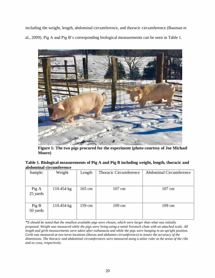
including the weight, length, abdominal circumference, and thoracic circumference (Bauman et
al., 2009). Pig A and Pig B’s corresponding biological measurements can be seen in Table 1.
Figure 1: The two pigs procured for the experiment (photo courtesy of Joe Michael
Moore)
Table 1. Biological measurements of Pig A and Pig B including weight, length, thoracic and
abdominal circumference
Sample: Weight Length Thoracic Circumference Abdominal Circumference
Pig A
25 yards
110.454 kg 165 cm 107 cm 107 cm
Pig B
50 yards
110.454 kg 159 cm 109 cm 109 cm
*It should be noted that the smallest available pigs were chosen, which were larger than what was initially
proposed. Weight was measured while the pigs were living using a metal livestock chute with an attached scale. All
length and girth measurements were taken after euthanasia and while the pigs were hanging in an upright position.
Girth was measured at two torso locations (thorax and abdomen circumference) to insure the accuracy of the
dimensions. The thoracic and abdominal circumferences were measured using a tailor ruler at the areas of the ribs
and os coxa, respectively.

21
Study Area
The study area was located on private property belonging to the Moore family in Barren
County, Kentucky, approximately 20 miles east of Glasgow, Kentucky (Figure 2). The site
consisted of a level area of unused farmland free of grazing animals or properties, and within
close proximity to the butchering and breakdown location. Ground covering at the study area
consisted of grass and limestone gravel.
Figure 2: The Moore Family Farm sign (photo courtesy of Joe Michael Moore)
Instrumentation Used
Initially, a moveable, wooden structure was built with the intention of being erected on
the private property belonging to the Moore family who graciously provided the area for the
experiment to be conducted. The gallows-type structure would have allowed for the pigs to be
hung with rope and fired upon safely, in an upright, standing position similar to that of a human.
However, due to the weight of the pigs and the materials used in the structure, it was decided that
22
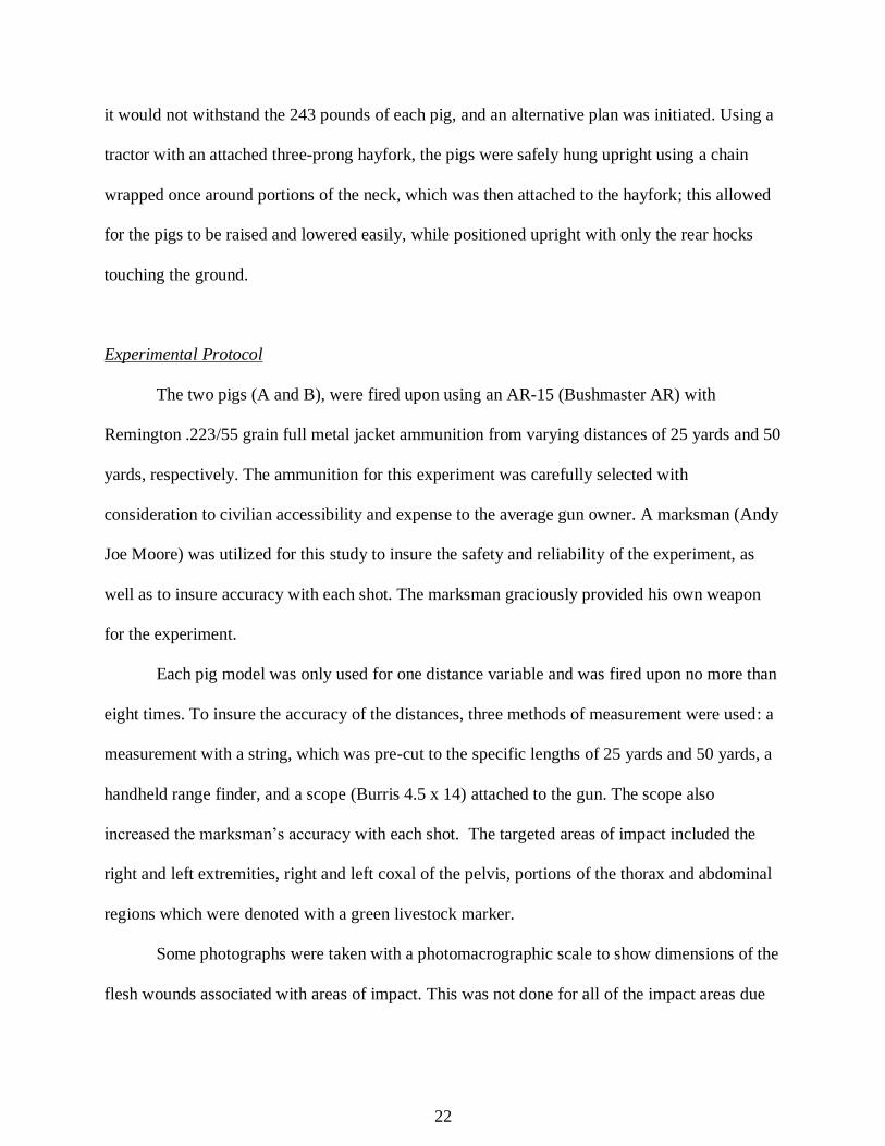
it would not withstand the 243 pounds of each pig, and an alternative plan was initiated. Using a
tractor with an attached three-prong hayfork, the pigs were safely hung upright using a chain
wrapped once around portions of the neck, which was then attached to the hayfork; this allowed
for the pigs to be raised and lowered easily, while positioned upright with only the rear hocks
touching the ground.
Experimental Protocol
The two pigs (A and B), were fired upon using an AR-15 (Bushmaster AR) with
Remington .223/55 grain full metal jacket ammunition from varying distances of 25 yards and 50
yards, respectively. The ammunition for this experiment was carefully selected with
consideration to civilian accessibility and expense to the average gun owner. A marksman (Andy
Joe Moore) was utilized for this study to insure the safety and reliability of the experiment, as
well as to insure accuracy with each shot. The marksman graciously provided his own weapon
for the experiment.
Each pig model was only used for one distance variable and was fired upon no more than
eight times. To insure the accuracy of the distances, three methods of measurement were used: a
measurement with a string, which was pre-cut to the specific lengths of 25 yards and 50 yards, a
handheld range finder, and a scope (Burris 4.5 x 14) attached to the gun. The scope also
increased the marksman’s accuracy with each shot. The targeted areas of impact included the
right and left extremities, right and left coxal of the pelvis, portions of the thorax and abdominal
regions which were denoted with a green livestock marker.
Some photographs were taken with a photomacrographic scale to show dimensions of the
flesh wounds associated with areas of impact. This was not done for all of the impact areas due
23
to the fact that this research is primarily focused on the skeletal tissue trauma. However, it may
be important in some instances to note the small size of the entrance wounds and large size of the
exit wound, with respect to the skeletal trauma present.
The Experiment
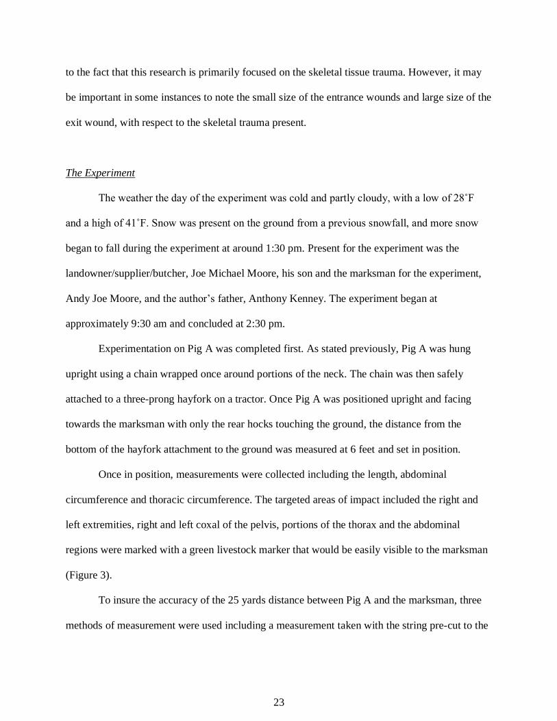
The weather the day of the experiment was cold and partly cloudy, with a low of 28˚F
and a high of 41˚F. Snow was present on the ground from a previous snowfall, and more snow
began to fall during the experiment at around 1:30 pm. Present for the experiment was the
landowner/supplier/butcher, Joe Michael Moore, his son and the marksman for the experiment,
Andy Joe Moore, and the author’s father, Anthony Kenney. The experiment began at
approximately 9:30 am and concluded at 2:30 pm.
Experimentation on Pig A was completed first. As stated previously, Pig A was hung
upright using a chain wrapped once around portions of the neck. The chain was then safely
attached to a three-prong hayfork on a tractor. Once Pig A was positioned upright and facing
towards the marksman with only the rear hocks touching the ground, the distance from the
bottom of the hayfork attachment to the ground was measured at 6 feet and set in position.
Once in position, measurements were collected including the length, abdominal
circumference and thoracic circumference. The targeted areas of impact included the right and
left extremities, right and left coxal of the pelvis, portions of the thorax and the abdominal
regions were marked with a green livestock marker that would be easily visible to the marksman
(Figure 3).
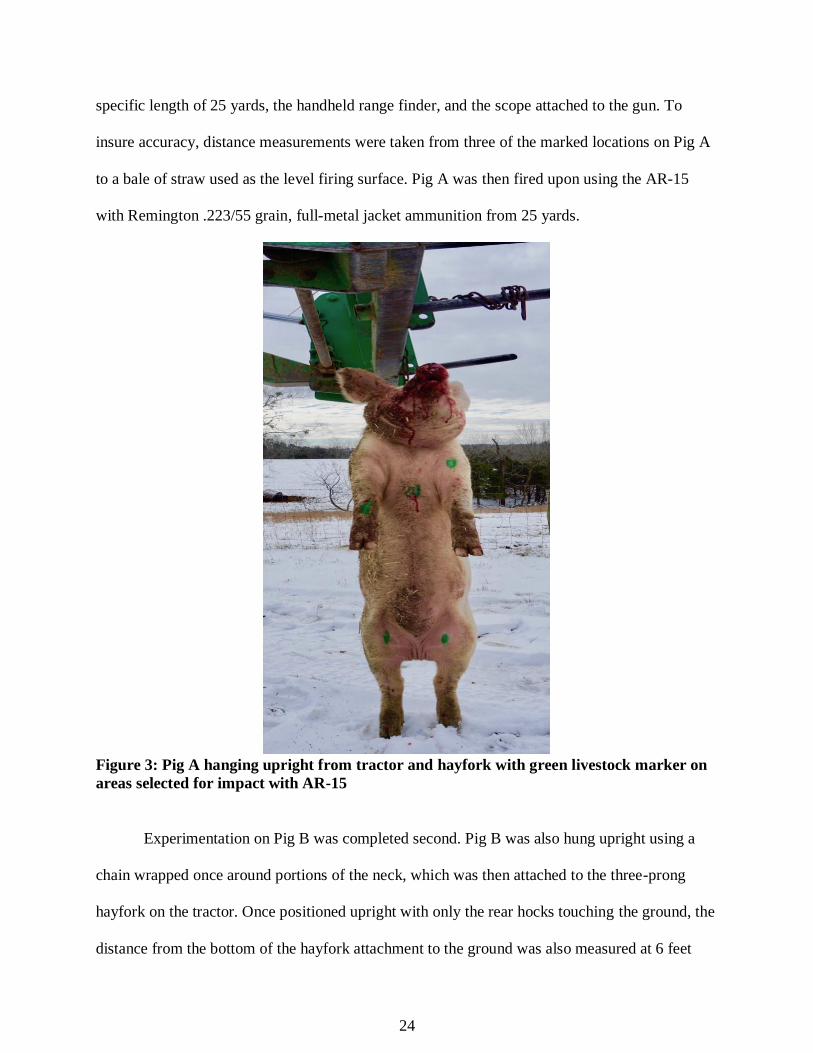
To insure the accuracy of the 25 yards distance between Pig A and the marksman, three
methods of measurement were used including a measurement taken with the string pre-cut to the
24
specific length of 25 yards, the handheld range finder, and the scope attached to the gun. To
insure accuracy, distance measurements were taken from three of the marked locations on Pig A
to a bale of straw used as the level firing surface. Pig A was then fired upon using the AR-15
with Remington .223/55 grain, full-metal jacket ammunition from 25 yards.
Figure 3: Pig A hanging upright from tractor and hayfork with green livestock marker on
areas selected for impact with AR-15
Experimentation on Pig B was completed second. Pig B was also hung upright using a
chain wrapped once around portions of the neck, which was then attached to the three-prong
hayfork on the tractor. Once positioned upright with only the rear hocks touching the ground, the
distance from the bottom of the hayfork attachment to the ground was also measured at 6 feet
25
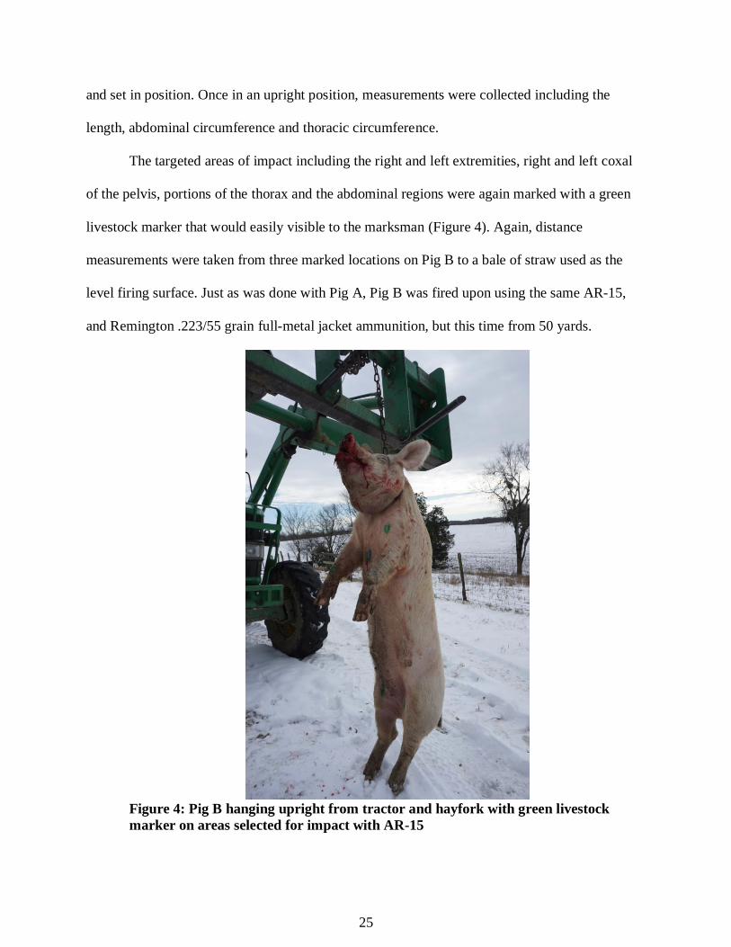
and set in position. Once in an upright position, measurements were collected including the
length, abdominal circumference and thoracic circumference.
The targeted areas of impact including the right and left extremities, right and left coxal
of the pelvis, portions of the thorax and the abdominal regions were again marked with a green
livestock marker that would easily visible to the marksman (Figure 4). Again, distance
measurements were taken from three marked locations on Pig B to a bale of straw used as the
level firing surface. Just as was done with Pig A, Pig B was fired upon using the same AR-15,
and Remington .223/55 grain full-metal jacket ammunition, but this time from 50 yards.
Figure 4: Pig B hanging upright from tractor and hayfork with green livestock
marker on areas selected for impact with AR-15
26
Butchering and Breakdown of Samples
Individual breakdown and processing of each pig occurred immediately, and separately
after each was fired upon with the help of a trained butcher. Processing included the careful
removal of long bones via disarticulation at and around the joint surfaces, as well as the removal
of as much tissue from around the targeted areas of impact in order to speed up the cleaning
process. Careful consideration was taken by the butcher to ensure that minimal cut marks were
imparted onto the bone. Some areas including those around the vertebral column and ribs were
removed in large sections through the use of a hand saw.
For areas needing saw use, consideration was taken by the butcher to overcompensate the
area taken surrounding the trauma insuring that no damage was done to the bone. Each pig was
disarticulated separately to ensure that no mixing of the samples occurred. For storage and
transport to the cleaning and processing location, the butchered samples were placed into large
garbage bags that were labeled with the associated sample letter and portion. The labeled
garbage bags were then placed into five-gallon buckets designated with the corresponding
sample letter.
Cleaning the Samples
Each pig was also cleaned and processed separately to ensure that no mixing of the
samples occurred. The samples were cleaned only with a Dawn detergent and warm water
solution. Two propane burners fitted with large cooking pots and straining basket for easy
retrieval of larger bones were used, as well as two additional large crockpots (Figure 5). All
heating devices were kept at low temperatures to help minimize the amount of cracking and
damage imparted on the bones from heating. Only wooden utensils, as well as wooden skewers

27
were used to assist in the removal of excess flesh from the bones. All processing liquids were
poured through a 1/8” screen that was fitted to a 5-gallon bucket, and all meat was screened for
bone and bullet fragments as well. Approximately 8 gallons of meat was shredded by hand and
screened for fragments of bone and bullets. The entire cleaning, processing, and drying time for
both samples, totaled 75 hours. The samples were labeled (A or B) with a black Sharpie and
bagged within paper sandwich bags according to the associated firing distance.
Figure 5: Author, Lauren Kenney, cleaning samples in the makeshift lab created
in a garage
Shipping the Samples to Montana
Three plastic storage containers were used and designated either A or B with a label. The
samples from Pig A were contained within two of the boxes, while Pig B’s samples could be
contained into one. The skeletal samples were still contained within paper bags marked with the
corresponding, letter, distance, and element name if available at the time. All bags were placed
28
into their associated container in levels, alternating bubble wrap and brown packing paper. The
plastic containers were securely closed using locking lids and heavy-duty duct tape.dd
All three plastic containers were shipped in a single cardboard box surrounded by bubble
wrap, shipping paper, and packing peanuts to Missoula, Montana. Before taping closed the box, I
included a note which explained the oddity of the contents and their importance to my degree if
for some reason it needed to be opened during transport. The package was closed with red
“fragile” tape and was insured for $500.00; approximately the total cost of the pigs. The box and
samples arrived in perfect condition on January 26th, 2018.
Data Collection and Analysis Protocol
Bullet wounds can be analyzed in order to determine information about the type of
weapon utilized, as well as the victim’s relationship to that weapon (Byers, 2011). A traditional
ballistics analysis will be completed on the elements impacted, including the collection of data
on the trauma present (Byers, 2011). This analysis will include detailing the placement and
location, wound type, size, shape, fracture types, fracture lines, and beveling (if possible)
(Huelke and Darling, 1964; Stewart, 1979; Dixon, 1982; Smith et al., 1987; Ross, 1996;
Berryman and Symes, 1998; Hart, 2005; White and Folkins, 2005; Byers, 2011).
In order to see the characteristics commonly associated with ballistics trauma,
reconstruction needed to be completed. The re-attachment of any fragmentation that could be
repaired was completed with Elmer’s glue. It should be noted that because young pigs were used
for this experiment, many of the epiphyses were not yet fused. These epiphyses were connected
with Elmer’s glue in order to provide a better representation of the full element as it would be in-
situ, as well as a better representation of any trauma that may be present. The analysis of each

29
pig was completed separately and began with an overall inventory of the skeletal elements
present. All remaining small fragments for each pig were counted and weighed separately using
a digital scale and will be considered for each individual pig as a whole. Bullet fragments will be
counted, if present. Photographs were taken throughout the process using a Sony alpha 1500
digital camera.
30
Chapter 4: Results
Skeletal Inventory
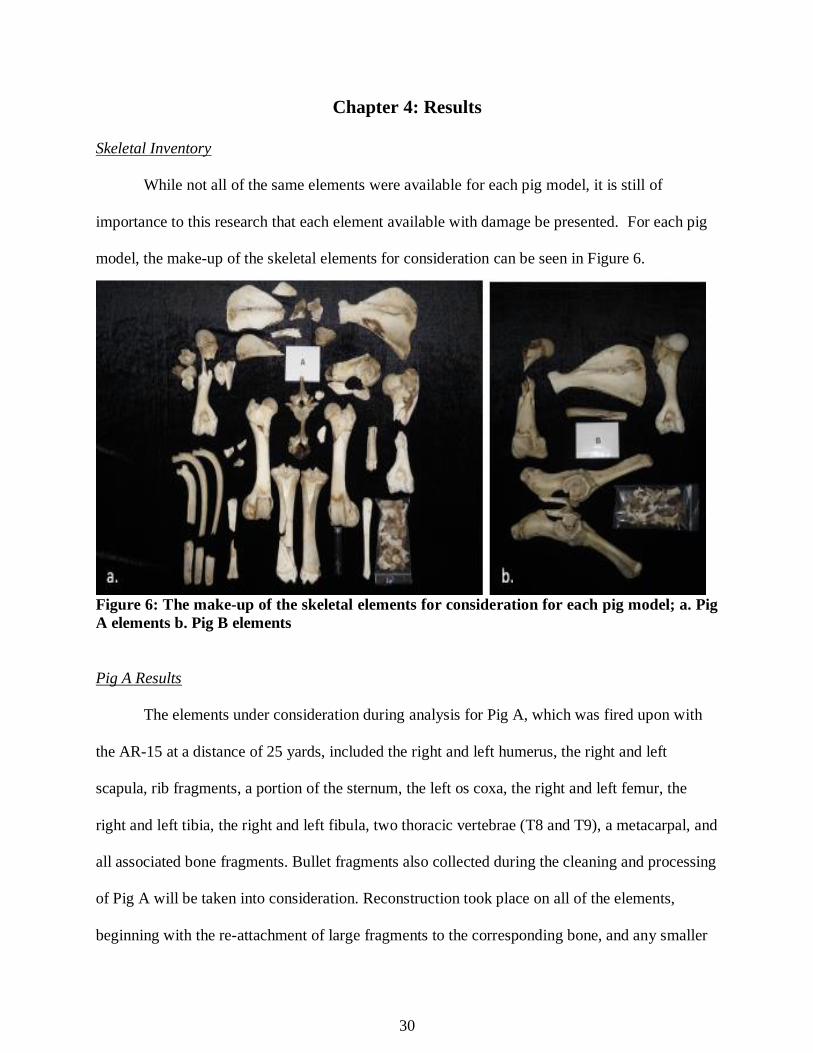
While not all of the same elements were available for each pig model, it is still of
importance to this research that each element available with damage be presented. For each pig
model, the make-up of the skeletal elements for consideration can be seen in Figure 6.
Figure 6: The make-up of the skeletal elements for consideration for each pig model; a. Pig
A elements b. Pig B elements
Pig A Results
The elements under consideration during analysis for Pig A, which was fired upon with
the AR-15 at a distance of 25 yards, included the right and left humerus, the right and left
scapula, rib fragments, a portion of the sternum, the left os coxa, the right and left femur, the
right and left tibia, the right and left fibula, two thoracic vertebrae (T8 and T9), a metacarpal, and
all associated bone fragments. Bullet fragments also collected during the cleaning and processing
of Pig A will be taken into consideration. Reconstruction took place on all of the elements,
beginning with the re-attachment of large fragments to the corresponding bone, and any smaller

31
fragments (n=153) unable to be reconstructed or positively associated to a specific skeletal
element were weighed together, totaling 52 grams.
Right Humerus
The right humerus of Pig A was extremely fragmented and needed a significant amount
of reconstruction before analysis could take place due to complete and comminuted fracturing
(Figure 7). There is not a typical entry or exit wound that can be defined by one of the common
shapes, and no beveling is present. Therefore, wound size cannot be determined. However, there
is a probable area of impact due to the number of radiating fracture lines present throughout the
diaphysis that travel up and through the anatomical neck. The epiphyses of what would be the
greater and lesser tubercules of the head have been damaged beyond repair, but two fragments
clearly correspond to the element.
Figure 7: The right humerus of Pig A (arrows indicate radiating fracture lines); a. Anterior
view b. Lateral view c. Posterior view d. Medial view
Left Humerus
The left humerus of Pig A was also extremely fragmented after impact and needed a
significant amount of reconstruction before analysis could take place. After reconstruction, the
32
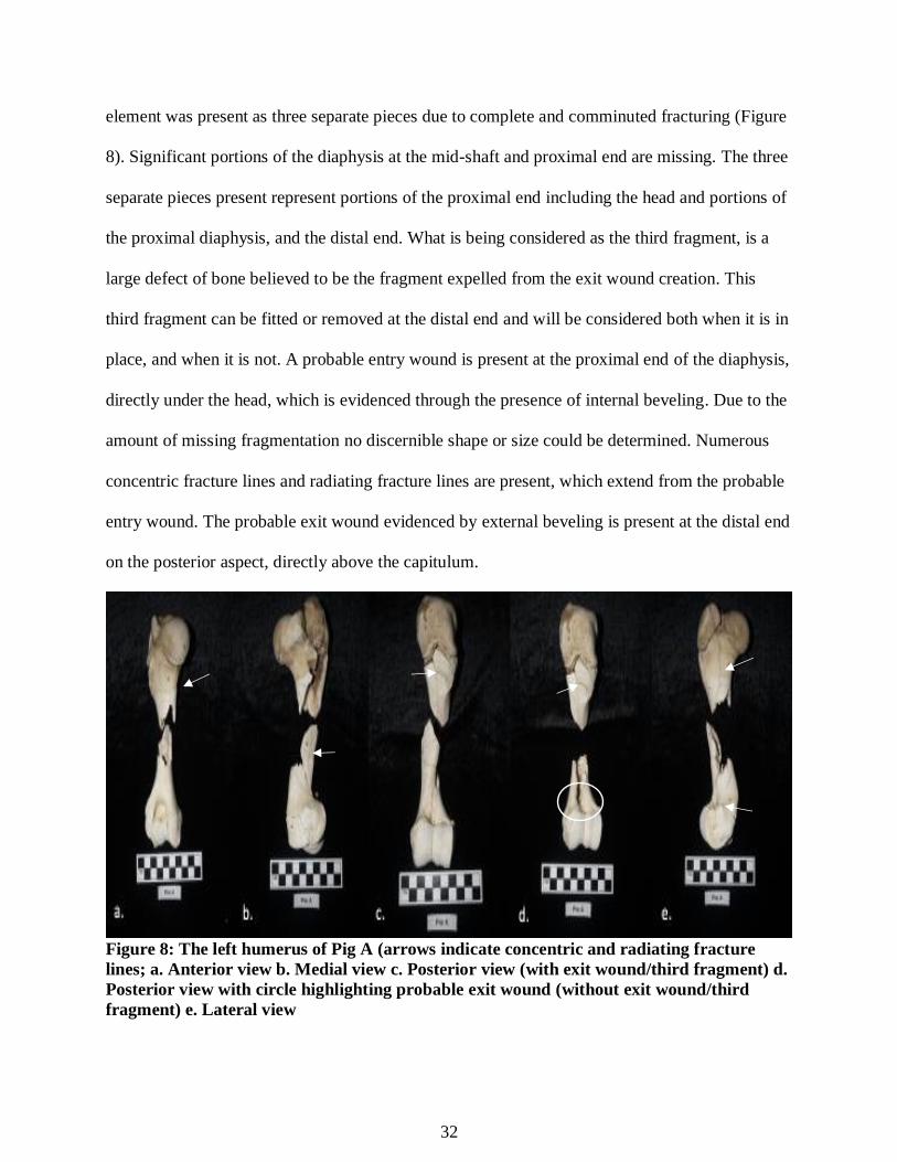
element was present as three separate pieces due to complete and comminuted fracturing (Figure
8). Significant portions of the diaphysis at the mid-shaft and proximal end are missing. The three
separate pieces present represent portions of the proximal end including the head and portions of
the proximal diaphysis, and the distal end. What is being considered as the third fragment, is a
large defect of bone believed to be the fragment expelled from the exit wound creation. This
third fragment can be fitted or removed at the distal end and will be considered both when it is in
place, and when it is not. A probable entry wound is present at the proximal end of the diaphysis,
directly under the head, which is evidenced through the presence of internal beveling. Due to the
amount of missing fragmentation no discernible shape or size could be determined. Numerous
concentric fracture lines and radiating fracture lines are present, which extend from the probable
entry wound. The probable exit wound evidenced by external beveling is present at the distal end
on the posterior aspect, directly above the capitulum.
Figure 8: The left humerus of Pig A (arrows indicate concentric and radiating fracture
lines; a. Anterior view b. Medial view c. Posterior view (with exit wound/third fragment) d.
Posterior view with circle highlighting probable exit wound (without exit wound/third
fragment) e. Lateral view
33
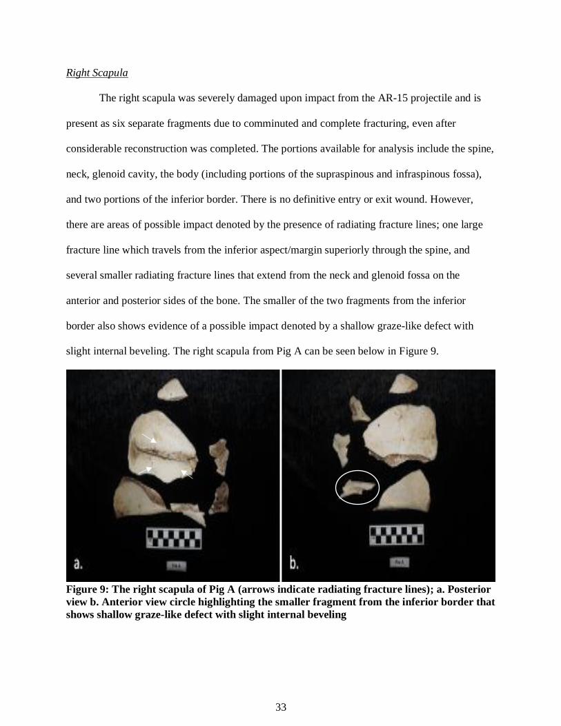
Right Scapula
The right scapula was severely damaged upon impact from the AR-15 projectile and is
present as six separate fragments due to comminuted and complete fracturing, even after
considerable reconstruction was completed. The portions available for analysis include the spine,
neck, glenoid cavity, the body (including portions of the supraspinous and infraspinous fossa),
and two portions of the inferior border. There is no definitive entry or exit wound. However,
there are areas of possible impact denoted by the presence of radiating fracture lines; one large
fracture line which travels from the inferior aspect/margin superiorly through the spine, and
several smaller radiating fracture lines that extend from the neck and glenoid fossa on the
anterior and posterior sides of the bone. The smaller of the two fragments from the inferior
border also shows evidence of a possible impact denoted by a shallow graze-like defect with
slight internal beveling. The right scapula from Pig A can be seen below in Figure 9.
Figure 9: The right scapula of Pig A (arrows indicate radiating fracture lines); a. Posterior
view b. Anterior view circle highlighting the smaller fragment from the inferior border that
shows shallow graze-like defect with slight internal beveling
34
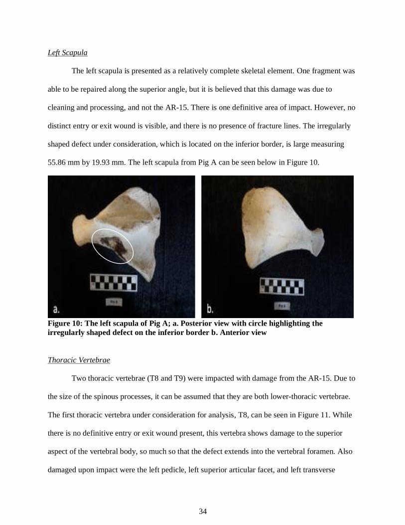
Left Scapula
The left scapula is presented as a relatively complete skeletal element. One fragment was
able to be repaired along the superior angle, but it is believed that this damage was due to
cleaning and processing, and not the AR-15. There is one definitive area of impact. However, no
distinct entry or exit wound is visible, and there is no presence of fracture lines. The irregularly
shaped defect under consideration, which is located on the inferior border, is large measuring
55.86 mm by 19.93 mm. The left scapula from Pig A can be seen below in Figure 10.
Figure 10: The left scapula of Pig A; a. Posterior view with circle highlighting the
irregularly shaped defect on the inferior border b. Anterior view
Thoracic Vertebrae
Two thoracic vertebrae (T8 and T9) were impacted with damage from the AR-15. Due to
the size of the spinous processes, it can be assumed that they are both lower-thoracic vertebrae.
The first thoracic vertebra under consideration for analysis, T8, can be seen in Figure 11. While
there is no definitive entry or exit wound present, this vertebra shows damage to the superior
aspect of the vertebral body, so much so that the defect extends into the vertebral foramen. Also
damaged upon impact were the left pedicle, left superior articular facet, and left transverse
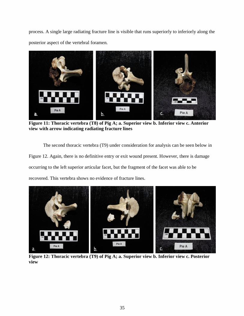
35
process. A single large radiating fracture line is visible that runs superiorly to inferiorly along the
posterior aspect of the vertebral foramen.
Figure 11: Thoracic vertebra (T8) of Pig A; a. Superior view b. Inferior view c. Anterior
view with arrow indicating radiating fracture lines
The second thoracic vertebra (T9) under consideration for analysis can be seen below in
Figure 12. Again, there is no definitive entry or exit wound present. However, there is damage
occurring to the left superior articular facet, but the fragment of the facet was able to be
recovered. This vertebra shows no evidence of fracture lines.
Figure 12: Thoracic vertebra (T9) of Pig A; a. Superior view b. Inferior view c. Posterior
view
36
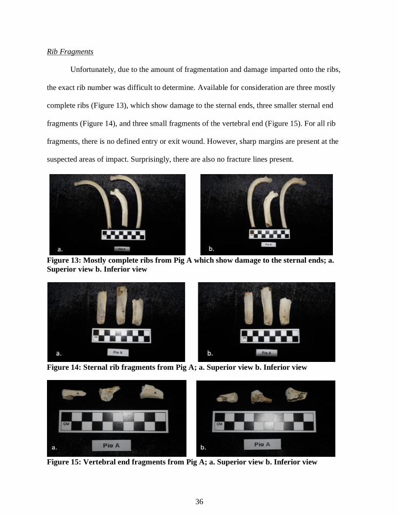
Rib Fragments
Unfortunately, due to the amount of fragmentation and damage imparted onto the ribs,
the exact rib number was difficult to determine. Available for consideration are three mostly
complete ribs (Figure 13), which show damage to the sternal ends, three smaller sternal end
fragments (Figure 14), and three small fragments of the vertebral end (Figure 15). For all rib
fragments, there is no defined entry or exit wound. However, sharp margins are present at the
suspected areas of impact. Surprisingly, there are also no fracture lines present.
Figure 13: Mostly complete ribs from Pig A which show damage to the sternal ends; a.
Superior view b. Inferior view
Figure 14: Sternal rib fragments from Pig A; a. Superior view b. Inferior view
Figure 15: Vertebral end fragments from Pig A; a. Superior view b. Inferior view

37
Sternum Portion
The sternum of Pig A was also damaged from the impact of the AR-15 fire. One portion
is available for consideration, which shows a wedge-shaped defect that damaged that articular
surface on the lower margin. This defect demonstrates no characteristics of an entry or exit
wound, and there are no fracture lines present. The sternum portion from Pig A can be seen in
Figure 16.
Figure 16: The sternum portion of Pig A (arrows indicate wedge-shaped defect); a.
Posterior view b. Anterior view
Left Os Coxa
The left os coxa of Pig A (Figure 17) is presented as three pieces after reconstruction.
However, the damage to the pubic symphysis, indicated by accidental saw striations, will not be
considered because it more than likely was created during butchering and processing rather than
from the AR-15 impact. There is damage to the ilium, but no definitive entry or exit wound is
visible. The margins of the damage are sharp, and there is no beveling visible. There is a small,
shallow, circular depression with striations that travel into the affected area of the ilium, which

38
could be evidence of a possible bone graze from a bullet. Surprisingly, no fracture lines are
present.
Figure 17: The left os coxa of Pig A; a. Lateral view with circle highlighting circular
depression with striations; evidence of a possible bone graze from a bullet b. Medial view
Right Femur
After cleaning and processing, the right femur of Pig A (Figure 18) was presented as four
separate fragments due to complete and comminuted fracturing. After significant reconstruction,
the skeletal element is mostly complete. There is a probable entry wound present on the lateral
side of the proximal diaphysis, but no internal beveling is present to corroborate the impact.
Radiating fracture lines extend outward from this suspected area of projectile entrance, and these
extend to create a larger butterfly fracture with the characteristic v-shaped defect (Figure 19). An
exit wound is likely on the posterior aspect of the proximal diaphysis, but this area was
accidentally reconstructed during the reconnection of fragments.
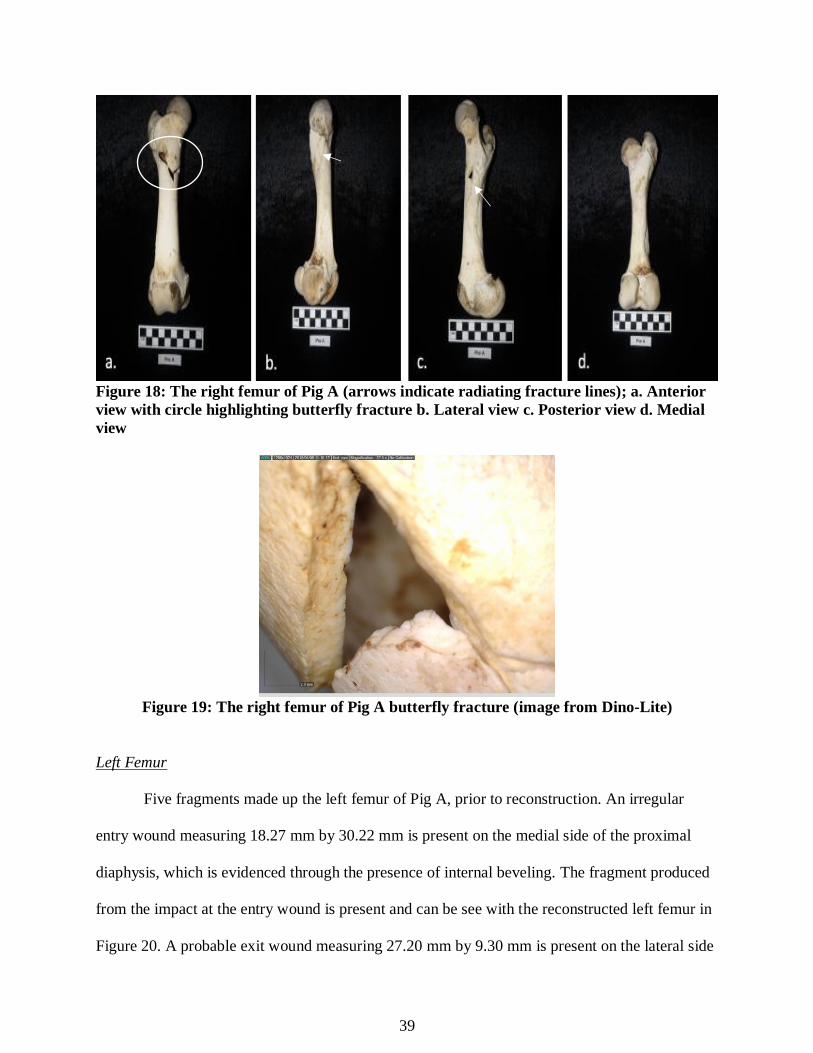
39
Figure 18: The right femur of Pig A (arrows indicate radiating fracture lines); a. Anterior
view with circle highlighting butterfly fracture b. Lateral view c. Posterior view d. Medial
view
Figure 19: The right femur of Pig A butterfly fracture (image from Dino-Lite)
Left Femur
Five fragments made up the left femur of Pig A, prior to reconstruction. An irregular
entry wound measuring 18.27 mm by 30.22 mm is present on the medial side of the proximal
diaphysis, which is evidenced through the presence of internal beveling. The fragment produced
from the impact at the entry wound is present and can be see with the reconstructed left femur in
Figure 20. A probable exit wound measuring 27.20 mm by 9.30 mm is present on the lateral side
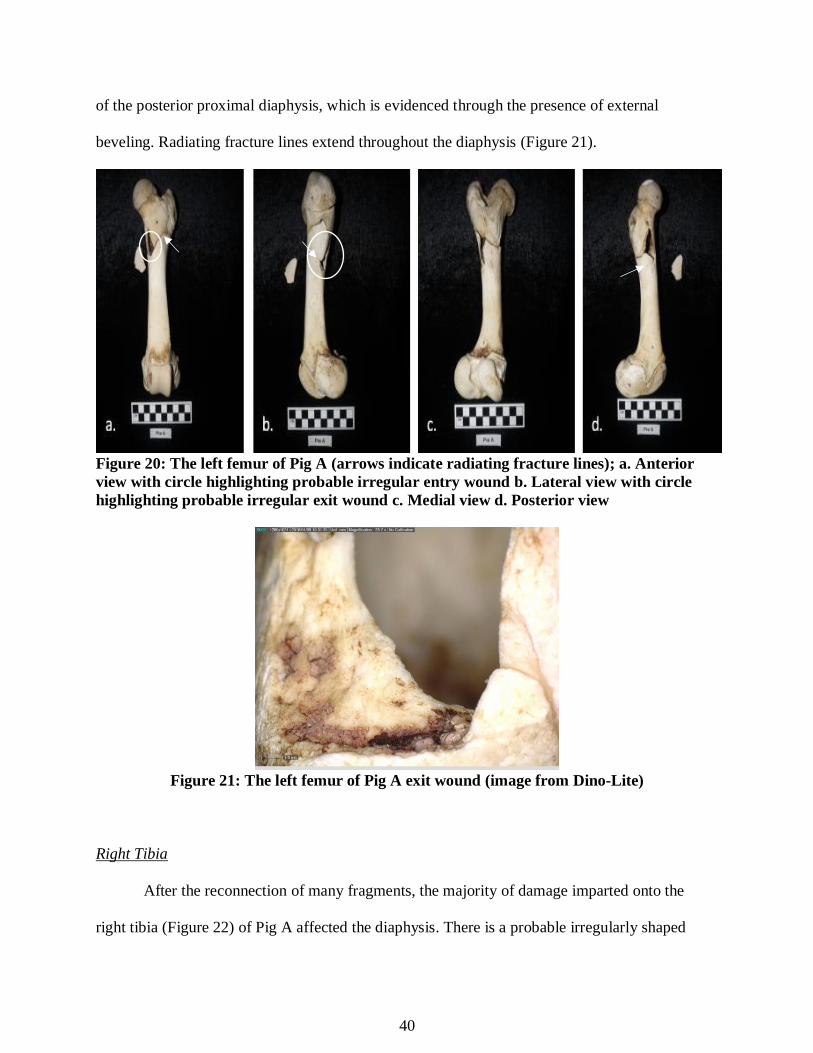
40
of the posterior proximal diaphysis, which is evidenced through the presence of external
beveling. Radiating fracture lines extend throughout the diaphysis (Figure 21).
Figure 20: The left femur of Pig A (arrows indicate radiating fracture lines); a. Anterior
view with circle highlighting probable irregular entry wound b. Lateral view with circle
highlighting probable irregular exit wound c. Medial view d. Posterior view
Figure 21: The left femur of Pig A exit wound (image from Dino-Lite)
Right Tibia
After the reconnection of many fragments, the majority of damage imparted onto the
right tibia (Figure 22) of Pig A affected the diaphysis. There is a probable irregularly shaped

41
entry wound present at the distal end near the center of the diaphysis. Internal beveling is present
on the superior margin of the entry wound. A probable exit wound is present on the posterior
evidenced through the presence of external beveling. The entry and exit wounds cannot be
measured due to the lack of full margins from fragmentation. Radiating fracture lines are visible
throughout the length of the diaphysis spanning from the proximal to distal ends.
Figure 22: The right tibia of Pig A (arrows indicate radiating fracture lines); a. Anterior
view with circle highlighting probable entry wound b. Lateral view c. Posterior view with
circle highlighting probable exit wound d. Medial view
Left Tibia
The left tibia from Pig A is present as a mostly complete skeletal element. There is
definitive impact damage to the distal end on the medial side of the bone which produced a
single, large radiating fracture line that travels up the diaphysis medially to laterally.
Unfortunately, there is no definitive entry or exit wound with the associated beveling that can be
measured. The left tibia from Pig A can be seen below in Figure 23.
Figure 23: The left tibia of Pig A; a. Anterior view b. Lateral view with arrow indicating
radiating fracture line c. Posterior view d. Medial view with circle highlighting probable
impact area

42
Right Fibula
The right fibula of Pig A is present as two separate pieces: the proximal end (Figure 24),
and the distal end (Figure 25). Unfortunately, no definitive entry or exit wound is present or
visible. However, there is significant damage to both fragments. The margins of the affected
areas are sharply broken by complete and comminuted fracturing. At the distal end, a sharp v-
shaped defect is located on the posterior with slight external beveling.
Figure 24: The proximal end of the right fibula of Pig A; a. Anterior view b. Posterior view
Figure 25: The distal end of the right fibula of Pig A; a. Anterior view with circle
highlighting the v-shaped defect b. Posterior view

43
Left Fibula
The left fibula of Pig A is present only as a portion of the diaphysis and proximal end
(Figure 26). Unfortunately, no definitive entry or exit wound is present or visible. However,
there is significant damage to the distal end. The margins of the affected area are sharply
fragmented indicating comminuted fracturing.
Figure 26: The left fibula from Pig A; a. Anterior view b. Posterior view
Metacarpal
One metacarpal is available for consideration (Figure 27). The number is unknown due to
the damage to the proximal end, as well as to the superior facets which help with identification.
There is no defined entry or exit wound present but radiating fractures that extend from the
defected areas may indicate the point of impact. While no beveling is present, the entry wound is
likely on the anterior side on the proximal end.
Figure 27: The metacarpal of Pig A (arrows indicate radiating fracture lines); a. Anterior
view b. Posterior view d. Medial view
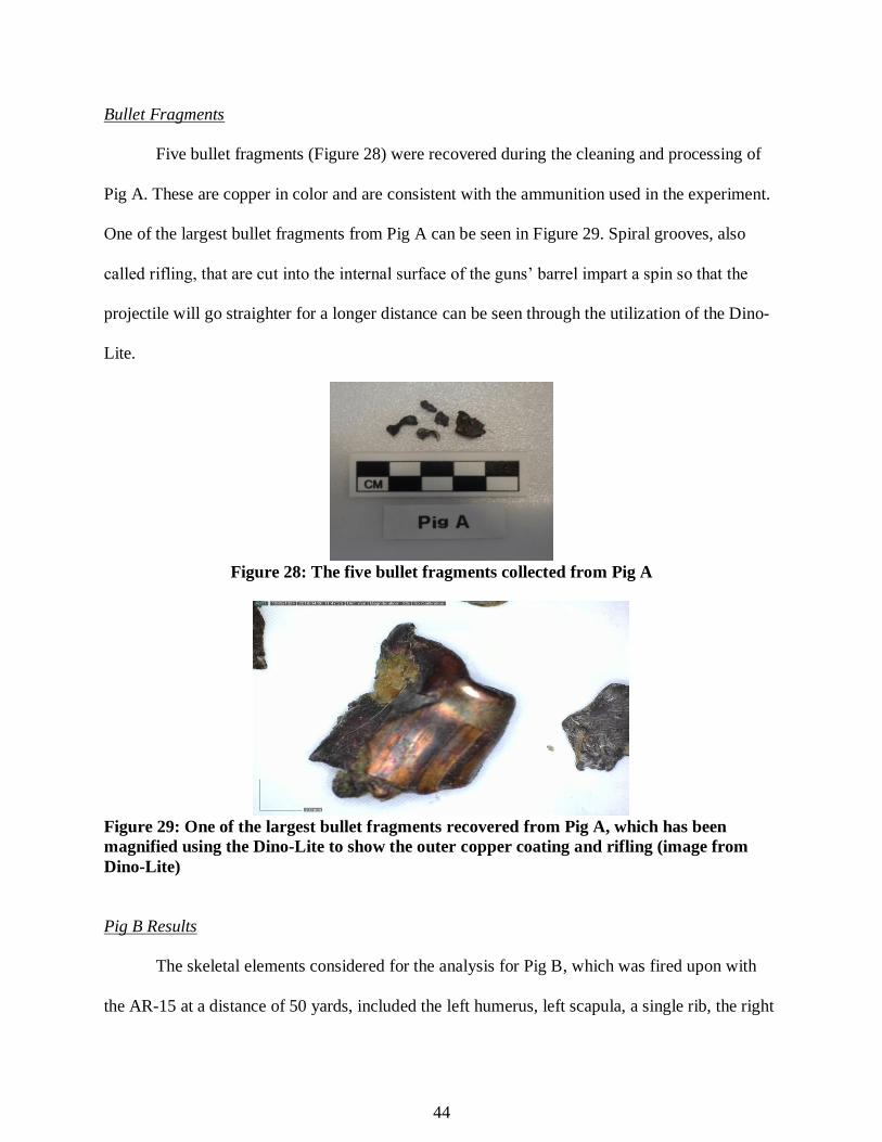
44
Bullet Fragments
Five bullet fragments (Figure 28) were recovered during the cleaning and processing of
Pig A. These are copper in color and are consistent with the ammunition used in the experiment.
One of the largest bullet fragments from Pig A can be seen in Figure 29. Spiral grooves, also
called rifling, that are cut into the internal surface of the guns’ barrel impart a spin so that the
projectile will go straighter for a longer distance can be seen through the utilization of the Dino-
Lite.
Figure 28: The five bullet fragments collected from Pig A
Figure 29: One of the largest bullet fragments recovered from Pig A, which has been
magnified using the Dino-Lite to show the outer copper coating and rifling (image from
Dino-Lite)
Pig B Results
The skeletal elements considered for the analysis for Pig B, which was fired upon with
the AR-15 at a distance of 50 yards, included the left humerus, left scapula, a single rib, the right
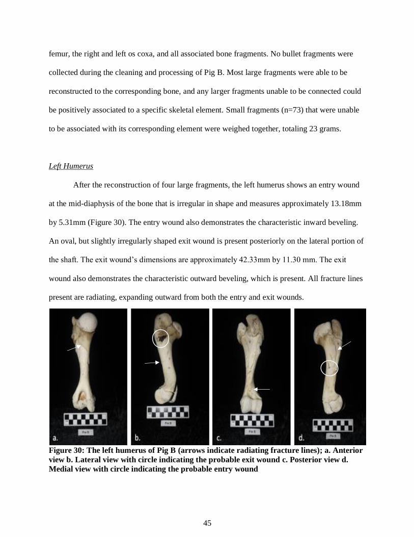
45
femur, the right and left os coxa, and all associated bone fragments. No bullet fragments were
collected during the cleaning and processing of Pig B. Most large fragments were able to be
reconstructed to the corresponding bone, and any larger fragments unable to be connected could
be positively associated to a specific skeletal element. Small fragments (n=73) that were unable
to be associated with its corresponding element were weighed together, totaling 23 grams.
Left Humerus
After the reconstruction of four large fragments, the left humerus shows an entry wound
at the mid-diaphysis of the bone that is irregular in shape and measures approximately 13.18mm
by 5.31mm (Figure 30). The entry wound also demonstrates the characteristic inward beveling.
An oval, but slightly irregularly shaped exit wound is present posteriorly on the lateral portion of
the shaft. The exit wound’s dimensions are approximately 42.33mm by 11.30 mm. The exit
wound also demonstrates the characteristic outward beveling, which is present. All fracture lines
present are radiating, expanding outward from both the entry and exit wounds.
Figure 30: The left humerus of Pig B (arrows indicate radiating fracture lines); a. Anterior
view b. Lateral view with circle indicating the probable exit wound c. Posterior view d.
Medial view with circle indicating the probable entry wound
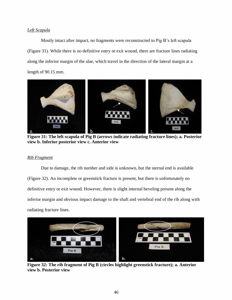
46
Left Scapula
Mostly intact after impact, no fragments were reconstructed to Pig B’s left scapula
(Figure 31). While there is no definitive entry or exit wound, there are fracture lines radiating
along the inferior margin of the alae, which travel in the direction of the lateral margin at a
length of 90.15 mm.
Figure 31: The left scapula of Pig B (arrows indicate radiating fracture lines); a. Posterior
view b. Inferior posterior view c. Anterior view
Rib Fragment
Due to damage, the rib number and side is unknown, but the sternal end is available
(Figure 32). An incomplete or greenstick fracture is present, but there is unfortunately no
definitive entry or exit wound. However, there is slight internal beveling present along the
inferior margin and obvious impact damage to the shaft and vertebral end of the rib along with
radiating fracture lines.
Figure 32: The rib fragment of Pig B (circles highlight greenstick fracture); a. Anterior
view b. Posterior view

47
Right Femur
After the re-attachment of as many fragments as possible, the right femur of Pig B
remained in two separate pieces (Figure 33). The head consisted of seven reconstructed
fragments and the distal end was made up of eight fragments due to comminuted fracturing.
There is unfortunately no definitive entry wound. However, there is extensive and obvious
impact damage to the diaphysis, especially around the proximal end near the neck and head. An
irregularly shaped exit wound with external beveling is present on the medial side of the mid-
diaphysis measuring 16.67 mm by 12.27 mm. Radiating fracture lines are also present extending
from the wound. Triangular shaped fracture lines that are characteristic of a butterfly fracture are
present on the posterior aspect.
Figure 33: The right femur of Pig B (arrows indicate radiating fracture lines); a. Anterior
view b. Lateral view c. Posterior view d. Medial view with circle highlighting irregular exit
wound
Right Os Coxa
The right os coxa (Figure 34) was presented as four fragments that were able to be
reconstructed before analysis. While there is no definitive entry or exit wound present, there is

48
extensive damage from impact resulting in the loss of significant bone structure to the connecting
area of the acetabulum and the ilium. This damaged portion shows a v-shaped margin, which is
indicative of a possible butterfly fracture. Radiating fracture lines extend from the suspected area
of impact.
Figure 34: The right os coxa of Pig B; a. Lateral view b. Medial view with arrow indicating
radiating fracture lines
Left Os Coxa
The left os coxa (Figure 35) was presented as three fragments that were able to be
reconstructed before analysis. Again, there is no definitive entry or exit wound present.
However, there is damage from projectile impact resulting in the severe fracturing to the
connecting area of the acetabulum and the ilium. Radiating fracture lines extend from the
suspected area of impact.
Figure 35: The left os coxa of Pig B; a. Lateral view b. Medial view with arrows indicating
radiating fracture lines, and circle highlighting probable area of impact
49
Chapter 5: Discussion
The data collected during this study, while limited due to sample size, clearly
demonstrates the severity of trauma that could be imparted onto the human skeleton by an AR-
15. Each pig model fired upon received extensive skeletal damage highlighted by comminuted
fracturing and complete fragmentation. All elements that received damage needed significant
amounts of reconstruction, which would inevitably be a daunting and costly task for any
recovery or identification effort.
Research from Blair et al. (2012) suggests that high-velocity projectiles typically
produce comminuted fractures due to the explosive effects of cavitation that is associated with
the properties of the marrow cavity. While this holds true for much of the trauma imparted to the
extremities of the samples presented here, when impacted with the AR-15 the bones could only
react in one way, and that was to explode into numerous fragments (complete and comminuted
fracturing) regardless of element impacted or marrow cavity type.
Based on the analysis, high-velocity weaponry like the AR-15, did not strictly follow
traditional wound patterning or show many of the common characteristics (entry wounds and
exit wounds with margins for measuring, beveling) normally needed for a traditional ballistic
analysis. In fact, many of the common characteristics that have been published in other research
are not applicable and the wound characteristics that are presented here are very inconsistent
with what have been previously recorded. The fact that no entry or exit wounds are visible for a
majority of the skeletal elements is likely due to the nature of velocity, and amount of
fragmentation that occurred to the bone.
It was anticipated, based on the research conducted by Chen et al. (2016), as well as
Klein et al. (2007), that any projectile associated with high-velocity weaponry, especially an AR-

50
15, had the potential to fracture the bone and the severity of the damage would more than likely
prohibit a correlation with distance. It is because of such obscure patterning, that there may never
be a clear indicator or marker of distance for ballistics, especially when such high-velocity
weapons are used.
One interesting finding centers around the amount of fragmentation for Pig A, which was
significantly greater to that of Pig B. It should be considered that the number and type of
elements available for each pig model was not consistent and could play a role in these
unproportioned findings. However, it cannot be ignored that the shorter firing distance for Pig A
provided increased accuracy for the marksman, which in turn created more impacts on the bone,
and therefore, more elements for analysis.
The five bullet fragments recovered from Pig A during processing are also an
unexpected outcome. There are several assumptions that can be made regarding this
phenomenon, but unfortunately no positive correlations can be made to distance. The literature
suggests that sharp profiled, jacketed ammunition are less likely to deform or fragment on impact
(Barach et al., 1986a; Barach et al., 1986b; Di Maio, 1993; Berryman et al., 1995; Byers, 2011).
However, this data suggests otherwise. In fact, the ammunition used during this experiment was
full metal jacketed, with a sharp profile, yet they still fragmented.
As with any aspect of science or forensic work there is always the question of a topic’s
ability to withstand scrutiny and be accurately replicated. It is important to note that the sample
size for this research is extremely small, and initially this study was visualized with the use of at
least five whole pig models. It is without a doubt a factor that impeded any statistical
significance to follow in analysis. Due to financial constraints and whole pig procurement
difficulties there was unfortunately no way to alleviate sample size issues in this study.
51
There are also many other variables that could have been chosen for this experiment, and
it is suggested that they be considered in future testing. Ideally, multiple distances would need to
be tested, with multiple samples available for each distance. Further research could open up the
possibility for the consideration of a number of other variables that were unable to be examined
here. Experiments utilizing the collection and recovery of fired bullets could be beneficial for a
focus on the ballistic characteristics of different types of ammunition and their associated trauma
patterns. For example, examining various types of ammunition could be suggested, like using
5.56 AR-15 ammunition versus the .223 that was used for this research. Also, examining the
traumatic differences found within the various grains of ammunition, or jacketed versus non-
jacketed projectiles. Other questions to consider would be if the various brands of ammunition
have an effect on the types of trauma or amount present, or if other factors such as those found
within the environment (weather and temperature) or clothing effect the patterns and prevalence
of trauma, or amount of bullet fragmentation.
Ideally, all skeletal elements would be included in testing as well, and while that would
have been ideal for this experiment, it is clearly evident that this would have taken an extreme
number of shots and a significant amount of ammunition. Fearing that multiple shots would, for
a lack of a better term “Swiss –cheese” the sample beyond recognition or data collection, it was
decided that minimal impacts would be best. The effects of high-velocity weaponry to the skull
must also be considered for future research, therefore cranial trauma must also be examined.
52
Chapter 6: Conclusion
The purpose of this study was to examine the skeletal tissue trauma inflicted by a high-
velocity firearm, specifically the AR-15 in a civilian context. It was hypothesized that when
biological pig models are exposed to high-velocity AR-15 fire, it was expected that the trauma
would differ from that recorded for traditional ballistics trauma within the literature. If varying
distances are then applied to such a high-velocity weapon, it was also hypothesized that the
trauma to the skeletal tissues would be so significant that no determination of the distance would
be able to be interpreted from the trauma present.
With regard to the first statement tested, while not confidently rejecting the hypothesis,
the data suggest far too many corroborating instances of high fragmentation that is consistent
with the comminuted and complete fracturing anticipated with high-velocity weaponry through
the literature. Therefore, the hypothesis here must be rejected. The only inconsistencies that
arise fall to the lack of available literature that details the effects of such weapons on skeletal
elements beyond the extremities.
The latter statement, again, while not confidently, must be accepted. While it cannot be
ignored that the significantly greater amount of fragmentation for Pig A, compared to that of that
of Pig B could be an indicator of distance, the number and type of elements presented for
analysis skews the proportional findings. The five bullet fragments recovered from Pig A during
processing are also an unexpected outcome that could be attributed to distance, but unfortunately
no positive correlations can be made on such a limited sample.
While this study does not show significant statistical evidence that follows traditional
ballistics analysis, the results should not be indiscriminately accepted or rejected without further
research being conducted that utilizes more variables, and a considerably larger sample size. It is
53
also without question that further research of this type is a necessity that must continue to be
undertaken.
It has been shown that there is inevitably much that can be gained from the understanding
of the patterns and type of high-velocity firearm trauma, especially when they are used against
civilians, and the applicability of such research is vast. The recognition of the patterns that are
seen in civilians could be applicable to the study of trauma in an anthropological context, which
could in turn, be beneficial to future scenarios involving civilians in mass atrocity events around
the world.
On a humanitarian and anthropological level, studies such as the one presented here could
assist during the analysis of civilian mass graves in war torn countries. Such research could also
be applicable to a number of other fields, including those in medicine and emergency response;
understanding how the human body reacts to such high-velocity weaponry could help assist in
triage management and could be utilized by physicians in medical trauma units when deciding
how to treat patients with similar wound patterns. With that said, regardless of its sample size
issues, it is with hope that this study was still preformed as a precedent that not only sparks
future research, but conversations on a broader political spectrum.

54
References Cited
Barach E, Tomlanovich M, and Nowak R. 1986a. Ballistics: A Pathophysiologic Examination
of Wounding Mechanisms of Firearms: Part I. Journal of Trauma. 36(3):225-235.
Barach E, Tomlanovich M, and Nowak R. 1986b. Ballistics: A Pathophysiologic Examination
of Wounding Mechanisms of Firearms: Part II. Journal of Trauma. 26(4):374-383.
Bauman R, Ling G, Tong L, Januszkiewicz A, Agoston D, Delanrolle N, Kim Y, Ritzel D, Bell
R, Ecklund R, Armonda R, Bandak F, and Parks S. 2009. An Introductory
Characterization of a Combat-Casualty-Care Relevant Swine Model of Closed Head
Injury Resulting from Exposure to Explosive Blast. Journal of Neurotrauma. 6(26):841-
860.
Berryman H and Symes S. 1998. Recognizing Gunshot and Blunt Cranial Trauma Through
Fracture Interpretation. In: Reichs, K, ed. Forensic Odontology, Advances in the
Identification of Human Remains. 2nd ed. Springfield, IL: Charles C. Thomas.
Berryman H, Smith O, and Symes S. 1995. Diameter of Cranial Gunshot Wounds as a Function
of Bullet Caliber. Journal of Forensic Sciences. 40:751-754.
Blair J, Patzkowski J, Schoenfeld A, Rivera J, Grenier E, Lehman R, and Hsu J. 2012. The
Skeletal Research Consortium. Spinal Column Injuries Among Americans in the Global
War on Terror. The Journal of Bone and Joint Surgery. 94(135): 1-9.
Boutillier J, De Mezzo S, Deck C, Magan P, and Willinger R. 2017. Chest Response Assessment
of Post-Mortem Swine Under Blast Loadings. Journal of Biomechanics. In press.
Buchanan C. 2011. The Health and Human Rights of Survivors of Gun Violence: Charting a
Research and Policy Agenda. Health and Human Rights.13:2: 50-63.
Burke T and Rowe W. 1992. Bullet Ricochet: A Comprehensive Review. Journal of Forensic
Sciences. 37:1254-1260.
Byers S. 2011. Introduction to Forensic Anthropology. Upper Saddle River, NJ: Prentice Hall
Pearson Education Inc..
Champion R, Bellamy R, Roberts P, and Leppaniemi A. 2003. A Profile of Combat Injury. The
Journal of Trauma. 54: S13-S19.
Coupland R and Meddings D. 1999. Mortality Associated with Use of Weapons in Armed
Conflicts, Wartime Atrocities, and Civilian Mass Shootings: Literature Review. British
Medical Journal. 319:7207:407-410.
Coupland R and Samnegaard H. 1999. Effect of Type and Transfer of Conventional Weapons on
Civilian Injuries: Retrospective. British Medical Journal. 319:7207: 410-412.
55
Cukier W. 2002. Small Arms and Light Weapons: A Public Health Approach. The Brown
Journal of World Affairs. 9:1: 261-280.
Chen J, Zhang B, Chen W, Kang J, Chen K, Wang A, and Wang J. 2006. Local and Distant
Trauma After Hypervelocity Ballistic Impact to the Pig Hind Limb. Springer Open.
doi:10.1186/s40064-016-3160-y. Accessed 2017, Oct 05.
Di Maio V. 1993. Gunshot Wounds. Practical Aspects of Firearms, Ballistics, and Forensic
Techniques. Boca Raton, FL: CRC Press.
Dixon D. 1982. Keyhole lesions in Gunshot Wounds to the Skull and Direction of Fire. Journal
of Forensic Sciences. 27:555-566.
Ezell V. 2002. Small Arms: Dominating Conflict in the Early Twenty-First Century. The Brown
Journal of World Affairs. 9:1: 305-310.
Frost R and Denton J. 2015. Forensic Pathology of Firearm Wounds. MedScape.
Griffiths D and Clasper J. 2006. Military limb injuries/ballistic fractures. Current Orthopaedics.
20:5:346-353.
Hart G. 2005. Fracture Pattern Interpretation in the Skull: Differentiating Blunt Force from
Ballistic Trauma Using Concentric Fractures. Journal of Forensic Sciences. 50(60):1276-
1281.
Hodgetts T, Mahoney P, Russell Q, and Byers M. 2006. ABC to <C> ABC: Redefining the
Military Trauma Paradigm. Emergency Medical Journal. 23: 745-747.
Huelke D and Darling J. 1964. Bone Fractures Produced by Bullets. Journal of Forensic
Sciences. 9(4):461-469.
Kashuk J, Halperin P, Caspi G, Colwell C, and Moore E. 2009. Bomb Explosions in Acts of
Terrorism: Evil Creativity Challenges Our Trauma System. Journal of the American
College of Surgeons. 209:1: 134-140.
Klein Y, Shatz D, and Bejarano P. 2007. Blast-Induced Colon Perforation Secondary to Civilian
Gunshot Wound. European Journal of Trauma and Emergency Surgery. 3: 298-
300.
Langley N. 2007. An Anthropological Assessment of Gunshot Wounds to the Chest. Journal of
Forensic Sciences. 52(3):532-537.
Lichte P, Oberbeck R, Binnebösel M, Wildenauer R, Pape H, and Kobbe P. 2010. A Civilian
56
Perspective on Ballistic Trauma and Gunshot Injuries. Scandinavian Journal of Trauma,
Resuscitation and Emergency Medicine. 18: 1-8.
Mabry R, Holcomb J, Baker A, Cloonan C, Uhorchak J, Perkins D, Canfield A, and Hagmann J.
2009. United States Army Rangers in Somalia: An Analysis of Combat Casualties on an
Urban Battlefield. The Journal of Trauma: Injury, Infection, and Critical Care. 49:3:
515-529.
Manring M, Hawk A, Calhoun H, and Andersen C. 2009. Treatment of War Wounds: A
Historical Review. Clinical Orthopedic Related Research. 467:8: 2168-2191.
Morse D, Crusoe D, and Smith H. 1983. Handbook of Forensic Archaeology and Anthropology.
Tallahassee, FL: Rose Printing.
Peterson B. 1991. External Beveling of Cranial Entrance Wounds. Journal of Forensic Sciences.
36:1592-1595.
Reichs K. 1986. Forensic Odontology, Advances in the Identification of Human Remains. 2nd ed.
Springfield, IL: Charles C. Thomas.
Rhine A and Curran B. 1990. Multiple Gunshot Wounds of the Head: An Anthropological
View. Journal of Forensic Science.35:1236-1245.
Ross A. 1996. Caliber Estimation from Cranial Entrance Defect Measurements. Journal of
Forensic Science. 41:629-633.
Shin E, Sabino J, Nanos G, and Valerio I. 2015. Ballistic Trauma: Lessons Learned from Iraq
and Afghanistan. Seminars in Plastic Surgery. 29:1: 10-19.
Smith A and Bellamy R. 2016. Understanding Weapons Effects: A Fundamental Precept in the
Professional Preparation of Military Physicians. Journal of Military and Veterans’
Health. 24:3.
Smith O, Berryman H, and Lahern C. 1987. Cranial Fracture Patterns and Estimate of Direction
from Low Velocity Gunshot Wounds. Journal of Forensic Science.32:1416-1421.
Steadman D and Haglund W. 2005. The Scope of Anthropological Contributions to
Human Rights Investigations. Journal of Forensic Science. 50:1: 1-8.
Stefanopoulos P, Hadjigerorgiou G, Filippakis K, and Gyftokostas D. 2014. Gunshot Wounds: A
Review of Ballistics Related to Penetrating Trauma. Journal of Acute Disease. 178-185.
Steflj I and Darden J. 2013. Making Civilian Casualties Count: Approaches to Documenting the
Human Cost of War. Human Rights Review. 14: 347-366.
Stewart T. 1979. Essentials of Forensic Anthropology. Springfield, IL: Charles C. Thomas.
57
Swindle M. 2010. Homeostasis Models: Trauma Models in Swine. Sinclair Bio-Resources. 1-3.
Symes S, Berryman H and Smith O. 1998. Saw Marks in Bone: Introduction and Examination of
Residual Kerf Contour. In: Reichs, K, ed. Forensic Odontology, Advances in the
Identification of Human Remains. 2nd ed. Springfield, IL: Charles C. Thomas.
Tejan J and Lindsey R. 1998. Management of Civilian Gunshot Injuries of the Femur: A Review
of the Literature. Injury.29:1: SA18-SA22.
Trott A. 1988. Mechanisms of surface soft tissue Trauma. Annals of Emergency Medicine. 17:12:
1279-1283.
U.S. Army. 2013. Weapons Effects and War Wounds: Clinical Practice Guidelines. Emergency
War Surgery. 2013; 1-16
White T and Folkins P. 2005. The Human Bone Manual. Burlington, MA: Elsevier Academic
Press.
Wolf S, Bebarta V, Bonnett C, Pons P, and Cantrill S. 2009. Blast Injuries. The Lancet. 374:
405-415.