Embed Size (px)
Citation preview

Instructions for use
Title Pigment compositions are linked to the habitat types in dinoflagellates
Author(s) Yamada, Norico; Tanaka, Ayumi; Horiguchi, Takeo
Citation Journal of plant research, 128(6), 923-932https://doi.org/10.1007/s10265-015-0745-4
Issue Date 2015-11
Doc URL http://hdl.handle.net/2115/63700
Rights The final publication is available at Springer via http://dx.doi.org/[10.1007/s10265-015-0745-4]
Type article (author version)
File Information Pigment compositions are linked to the habitat types in dinoflagellates.pdf
Hokkaido University Collection of Scholarly and Academic Papers : HUSCAP
1
The name of the authors: Norico Yamada1, Ayumi Tanaka2, Takeo Horiguchi3
Title: Pigment compositions are linked to the habitat types in dinoflagellates
Running title: Habitat-related Pigments in Dinoflagellates
The affiliations and addresses of the authors:
1:Department of Natural History Sciences, Graduate School of Science, Hokkaido University,
Sapporo, 060-0810, Japan
2:Institute of Low Temperature Science, Hokkaido University, Sapporo, 060-0819, Japan,
JST/CREST
3:Department of Biological Sciences, Faculty of Science, Hokkaido University, Sapporo,
060-0810, Japan

2
Abstract
Compared to planktonic species, benthic dinoflagellates living in sandy beach or seafloor
are a little known about ecology, physiology, even their existences. In a previous study, we
discovered 132,173-cyclopheophorbide a enol (cPPB-aE) from sand-dwelling benthic
dinoflagellates. This enol had never been detected in phytoplankton despite it is a chlorophyll
a catabolite. We speculated from this discovery that habitat selection might be linked to
pigment compositions in dinoflagellates. To test the hypothesis of habitat selection linking to
pigment compositions, we conducted extensive analysis of pigments with high performance
liquid chromatography (HPLC) for 40 species using 45 strains of dinoflagellates including
three habitat types; sand-dwelling benthic forms, tidal pool inhabitants and planktonic species.
Those 40 dinoflagellates are also able to distinguished into two types based on their
chloroplast origins; red alga-derived secondary chloroplasts and diatom-derived tertiary ones.
By plotting the pigments profiles onto three habitats, we noticed that twelve pigments
including cPPB-aE were found to occur only in benthic sand-dwelling species of red
alga-derived type. The similar tendency was also observed in dinoflagellates with
diatom-derived chloroplasts, i.e. additional sixteen pigments including chl c3 were found only
in sand-dwelling forms. This is the first report of the occurrence of chl c3 in dinoflagellate
3
with diatom-derived chloroplasts. These results clarify the far greater diversity of pigments
are produced by the dinoflagellates living in sand regardless of chloroplast types relative to
those of planktonic and tidal pool forms. Dinoflagellates seem to produce a part of their
pigments in response to their habitats.
Key words; benthic, diatom, dinoflagellate, HPLC, planktonic, pigment
Abbreviation; cPPB-aE: 132,173-cyclopheophorbide a enol, PCDi: pigments common to all
dinotoms, PCPe: pigments common to all peridinin-type dinoflagellates, UC: unknown
carotenoid
Introduction
Dinoflagellates are unicellular eukaryotes mainly known as marine primary producers.
About 77% of species are marine planktonic which drift near the surface of the sea. On the
other hand, although in the minority, the benthic marine dinoflagellates account for about 8%
of living species (Taylor et al. 2008, Hoppenrath et al. 2014). They have been recorded
mainly from sandy beaches (e.g. Herdman 1922), intertidal flats (e.g. Hoppenrath et al. 2007),
tidal pools in rocky shores (e.g. Horiguchi and Chihara 1988), sandy sea floors (e.g. Yamada

4
et al. 2013), and the surface of either seaweeds (e.g. Parsons and Preskitt 2007) or of
sediments (e.g. Faust et al. 2008). Some benthic representatives swim in the interstitial spaces
between sand grains, while others attach themselves to the substrata, existing as non-motile
vegetative cells or temporary cysts (e.g. Horiguchi and Chihara 1987, Murray and Patterson
2002, Saburova et al. 2009).
Tidal pools, considered as part of benthic habitats, (Hoppenrath et al. 2014) were one of
our focal habitats because dinoflagellates here show an intermediate life cycle between
benthic and planktonic organisms (Horiguchi and Chihara 1988). They are planktonic cells
when the tide is low, but when the pool is flooded at high tide, they become benthic by
forming temporary cysts attaching to the rock surfaces. Because of this intermediary
behavioral pattern, we regarded tidal pool dinoflagellates should be distinguished from
sand-dwelling dinoflagellates or planktonic forms.
Dinoflagellates can be further distinguished by chloroplast types. More than 95% of
photosynthetic dinoflagellates possess a red algal derived secondary chloroplast (Zhang et al.
1999, Taylor et al. 2008), which contains chlorophyll a/c2 with peridinin, a xanthophyll
unique to dinoflagellates. This typical chloroplast type in dinoflagellate called
‘peridinin-type’. In contrast, some groups of dinoflagellates have replaced their

5
peridinin-type chloroplasts with one from other microalgae, belonging to Haptophyta (Tengs
et al. 2000, Zapata et al. 2012), Chlorophyta (Watanabe et al. 1990, Matsumoto et al. 2012) or
Bacillariophyta (diatoms) (Riley and Wilson 1967, Chesnick et al. 1997, Takano et al. 2008,
Zhang et al. 2011, Zhang et al. 2014).
In a previous study, we discovered 132,173-cyclopheophorbide a enol (cPPB-aE) for the
first time in photosynthetic organisms (Yamada et al. 2014). Through our pigment analytical
survey, we discovered that the enol was only detected in sand-dwelling benthic
dinoflagellates (Yamada et al. 2014). From this result, we came to an idea that habitat
selection might be linked to pigment compositions. Although recently Zapata et al. (2012)
reported pigment compositions of 64 dinoflagellate species, including wide taxonomic groups,
little attention was made to relate pigment profiles and habitat types. Therefore, to date, no
comprehensive studies relating pigment composition to habitat types have not been available.
Here we report on the pigment profiles of 40 species (using 45 strains) of dinoflagellates
by HPLC. We distinguished them into three habitat types; 29 species (31 strains) of
sand-dwelling benthic forms, three tidal pool inhabitants (five strains), and eight planktonic
species (nine strains). In this study, a hierarchical cluster analysis was employed to classify
HPLC pigments data into habitat groups and to assess the effects of environmental factors on

6
pigment compositions.
These 40 dinoflagellates can be divided into one of two chloroplast types: of red
alga-derived (peridinin-type) secondary chloroplast and diatom-derived tertiary one. We
treated 34 species (35 strains) of peridinin-type species and six species (ten strains) of
dinoflagellates having diatom-derived chloroplasts; the latter dinoflagellates are collectively
called ‘dinotoms’ (Imanian et al. 2010). Dinotoms are known to possess chlorophylls c1 and
c2 with fucoxanthin as the major xanthophyll (Mandelli 1968, Jeffery et al. 1975, Withers et
al. 1977, Tamura et al. 2005). The endosymbiont diatoms originated from four species
belonging to different four genera: Chaetoceros (Horiguchi and Takano 2006), Cyclotella
(Zhang et al. 2014), Discostella (Takano et al. 2008, Zhang et al. 2011), and Nitzschia
(Chesnick et al. 1997, Tamura et al. 2005). Six dinotoms analyzed in this study can be
distinguished to two diatom species derived types, i.e. possessing a Nitzschia-type
endosymbiont (Tamura et al. 2005, Pienaar et al. 2007) (represented by five species, nine
strains), and Discostella-type (Zhang et al. 2011) (represented by one species, two strains). If
our hypothesis that pigment profiles are related to habitat were valid, it would be expected
that, regardless of chloroplast types, similar tendencies could be seen between habitat types.
In addition to dinoflagellates, we included pigment data from the three free-living diatoms;

7
Cylindrotheca closterium (Ehrenberg) Reimann & JC Lewin, Nitzschia sp. and Tabularia sp.
A comparison of pigment composition between endosymbiont diatoms and free-living one
would provide insight into whether pigment modifications occurred after endosymbiotic
event (s) or not.
Materials and methods
Cultures and species identifications
All culture strains used here were isolated from sandy beaches, sandy sea floors, tidal
pools or the surface of seawater or freshwater (STable 1). Each sand sample was placed in a
plastic cup and enriched with Daigo’s IMK medium (marine, Nihon Pharmaceutical Co., Ltd.,
Tokyo) or with URO medium (freshwater, Kimura and Ishida 1985), and cultured at 15, 20 or
25ºC, which were close to water temperature at sampling site of each species, with an
illumination of 60 µmol·photons·m-2·s-1 under a 16:8 h light:dark cycle without suspension.
Dinoflagellate cells that appeared in the cup were isolated using capillary pipettes with
several rinses in sterilized medium under an inverted microscope and subsequently clonal
cultures were established. The culture strains were maintained in petri dishes in IMK, URO
or f/2 medium (marine, Guillard and Ryther 1962) using the same conditions indicated above.

8
In these petri dishes, planktonic species can swim freely without agitation, while benthic
species can attach to the inside wall of petri dishes or swim near surface of substrata and
spend benthic life cycle. All culture strains were identified morphologically using the light
microscope (Figs. S1 and S2) and verified by analysis of molecular data (Figs. S3 and S4,
Table S2).
Pigment extraction and HPLC analysis
After being cultured for 1 to 4 months, each culture was centrifuged at 10000g for 5 min
and the cells were pelleted. For the species firmly attaching to the inside wall of petri dish,
the cells were collected by gentle sweeping by clean paint brush. Then the cells were
centrifuged as above. The pelleted cells were suspended in 100% acetone and homogenized
by stainless beads (5 mm in diameter, TCS0-0100, Bio medical science, Tokyo) for 1 min
using a ShakeMaster grinding apparatus (BioMedical Science, Tokyo). The homogenates
were centrifuged for 15 min at 22000g. The pigments in the supernatant were separated on a
Symmetry C8 column (150 X 4.6 mm, Waters, Milford) according to a method reported
previously (Zapata et al. 2000). The elution profiles (Fig. 1) were monitored by measuring
the absorbance at 450 nm (SPD-M10A, Shimadzu, Kyoto), and the pigments were identified

9
by their retention times and spectral patterns.
Hierarchical clustering analysis
The distributions of detected pigments were analyzed by software R with the Ward method,
which clusters orderly two objects with smaller differences. For binary similarity measure,
the Sokal and Michener index (Sokal and Michener 1958) was used because we regarded
both states of pigment existence, i.e. presence or absence of pigments, are important. This
index considers equally both attributes, presence or absence, between two objects (Choi et al.
2010).
Results
The chloroplasts of dinoflagellates analyzed originated from phylogenetically different
algae. Therefore, the results for peridinin-type dinoflagellates with red algal-derived
chloroplasts and for dinotoms with diatom-derived chloroplasts are considered separately.
[Peridinin-type dinoflagellates]
Pigment compositions
Forty-two pigments were detected in 35 strains (representing 34 species) of peridinin-type

10
dinoflagellates (Fig. S5). Five of them were chlorophylls and related molecules, i.e.
chlorophyll c2 (chl c2, peak 4), chlorophyll c1 (chl c1, peak 5), 132,173-cyclopheophorbide a
enol (cPPB-aE, peak 36), chlorophyll a (chl a, peak 49) and pheophythin a (peak 53) (in
order of retention time). Chl c2, chl a and pheophythin a were detected in all samples.
cPPB-aE is thought to be degradation product of pheophythin a (Ma and Dolphin 1999,
Louda et al. 2000).
Thirty-seven carotenoids were detected in total of which only twelve were identifiable,
peridininol (peak 1), peridinin (peak 6), a peridinin-like carotenoid (peak 7), violaxanthin
(peak 14), diadinochrome (peak 18), diadinoxanthin (peak 19), dinoxanthin (peak 22),
diatoxanthin (peak 24), a diadinochrome-like carotenoid (peak 25), zeaxanthin (peak 27), a
lutein-like carotenoid (peak 28) and β-carotene (peak 54) (in order of retention time). The
other twenty-five pigments were unknown carotenoids (UCs). All the dinoflagellates
analyzed commonly had the following ten carotenoids; peridininol, peak 2 UC, peridinin, a
peridinin-like carotenoid, peak 11 UC, diadinoxanthin, dinoxanthin, zeaxanthin, lutein-like
carotenoid (except in Stylodinium littorale Horiguchi & Chihara, Analysis Number 23),
β-carotene.
The pigments (chlorophylls and carotenoids) detected in all peridinin-type dinoflagellates

11
are here designated as PCPe (Pigments common to all peridinin-type dinoflagellates) (Fig.
S5).
Habitat-type distribution of pigment profiles
We grouped the peridinin-type dinoflagellates into three habitat categories; benthic,
sand-dwellers (25 species, 26 strains), tidal pool inhabitants (two species, two strains) and
planktonic forms from sea surface (seven species, seven strains). The cluster analysis based
on HPLC pigments categorized them into 4 groups (Fig. 2). These clusters represent the
pigments only detected from sand-dwellers (cluster A), from tidal pool inhabitants and
sand-dwellers (cluster B), from planktonic species and sand-dwellers (cluster C), and from all
habitat type dinoflagellates (cluster D), respectively.
Cluster A is consisted of pigments detected only from sand-dwelling dinoflagellates. The
presence of twelve pigments, i.e. cPPB-aE, peak 16, 17, 33, 37, 39, 40, 42, 43, 44, 51 and 52
UCs, was restricted to this habitat. However, these pigments are not universally shared by all
sand-dwellers. In particular, each of peak 33, 37, 39, 40, 42, 43 and 44 UCs were only
detected in one respective dinoflagellate (Fig. S5). The remaining restricted pigments were
detected in two (peak 16 and 17 UCs), three (peak 52 UC), six (cPPB-aE) or seven species

12
(peak 51 UC) of sand-dwelling dinoflagellates.
Cluster B contained two pigments, i.e. peak 12 UC and chl c1. The former was specific to
tidal pool member, while the latter was found in the species from both tidal pool and sand.
There were no pigments limited in distribution to planktonic species. Pigments from
planktonic species were restricted to clusters C and D, and these pigments were also found in
other habitat type species. On the contrary, sand-dwelling species possessed most diverse
pigments, i.e. found in all clusters 1 - 4, and all pigments detected in planktonic and tidal pool
dinoflagellates were found in at least one sand-dwelling species except for peak 12 UC which
is tidal pool specific.
Phylogenetic distribution of pigment profiles
Based on morphology and molecular phylogeny inferred from SSU rDNA data, we
identified the dinoflagellate species (Figs. S1 and S3). The species analyzed in this study
consisted of phylogenetically wide range of groups. Figure S6 shows the similarity of
pigment composition among species by clustering analysis. In some cases, closely related
species, i.e. congeneric species, show similar type of pigment profiles (boxed groups in Fig.
S6), while in other instances little similarities have been detected even between the species
13
within the same genus. Correlations between the phylogenetic positions and pigment
compositions were not strongly supported.
[Dinotoms]
Pigment compositions
Thirty-eight pigments were detected from ten strains, representing six species of dinotoms
(Fig. S7). Five chlorophylls and its derivatives were detected, in order of retention time,
chlorophyll c3 (chl c3, peak 3), chlorophyll c2 (chl c2, peak 4), chlorophyll c1 (chl c1, peak 5),
chlorophyll a (chl a, peak 49) and pheophythin a (peak 53). cPPB-aE was not detected.
Additionally, thirty-three carotenoids were detected in dinotoms, eight of which were
identified as a fucoxanthin-like carotenoid (peak 9), fucoxanthin (peak 10), diadinoxanthin
(peak 19), diatoxanthin (peak 24), zeaxanthin (peak 27), a lutein-like carotenoid (peak 28),
β-ψ carotene (peak 50) and β-carotene (peak 54). The remaining twenty-five pigments were
UCs. Ten pigments were detected from all dinotoms (PCDi: Pigments common to all
dinotoms) include chl c2, chl c1, fucoxanthin, diadinoxanthin, peak 21 UC, zeaxanthin, chl a,
β-ψ carotene, pheophythin a, β-carotene (in order of retention time, Fig. S7).

14
Habitat type / Phylogenetic distribution of pigment profiles
The molecular phylogenetic analyses supported a monophyletic dinotom grouping (Figs.
S3 and S4). Our strains were divided into four main clades within the monophyletic group,
the Durinskia cf. baltica (Levander) Carty & Cox clade (No.36 and 37), the Durinskia sp.
clade (No.38, 39 and 40), the Galeidinium spp. clade (No.41, 42 and 43) and the
Peridiniopsis cf. kevei Grigorszky clade (No.44 and 45) (Fig. S4). These analyzed dinotoms
were also categorized to three habitats as in the case for the peridinin-type dinoflagellates;
four benthic sand-dwelling species (D. cf. baltica and three species of Galeidinium spp.
clade), one tidal pool inhabitant (Durinskia sp.) and one planktonic species (P. cf. kevei).
Figure 3 shows that all detected pigments plotted on habitat types were grouped to mainly
four types by cluster analysis; pigments detected only from sand-dwellers (cluster A), from
tidal pool inhabitants (cluster B), mainly from planktonic species (cluster C) and common
pigments for all habitat type dinotoms (cluster D).
Cluster A was consisted of specific pigments for sand-dwelling four species (five strains).
These pigments could be separated to three pigment groups. The first group contained nine
pigments, peak 23, 26, 33, 34, 35, 39, 41, 43 and 44 UCs, and these were shared by
Galeidinium rugatum Tamura & Horiguchi (No.41) and Unidentified coccoid dinotom 1
15
(No.42). The second group consisted of three pigments, peak 16, 45 and 46 UCs, which were
shared by three species in Galeidinium spp. clade (No.41, 42 and 43). The third group
included four pigments, chl c3, peak 29, 47 and 48 UCs, that were shared by all
sand-dwelling dinotoms. The number of shared pigments seems to reflect phylogenetic
closeness between them based on phylogenetic analysis of SSU rDNA data (Fig. S4); the
highest number of shared specific pigments, were detected in the closest species, G. rugatum
(No.41) and Unidentified coccoid dinotom 1 (No.42).
We analyzed only one species as representative of tidal pool (three strains) and planktonic
habitats (two strains), respectively. In cluster B, peak 40 UC was shown to be specific for
tidal pool species, Durinskia sp. (No.38, 39 and 40). Pigments in cluster C were mainly from
freshwater planktonic species, P. cf. kevei (No.44 and 45), and could be separated to two
categories; peak 13 and 52 UCs which were only detected in planktonic species, and peak 12
and 15 UCs which were shared by planktonic and sand-dwelling species. PCDi and the
pigments detected from all habitat type dinotoms were included in cluster D.
Some pigments appeared in the positions that were difficult to interpret. A fucoxanthin-like
carotenoid, a lutein-like carotenoid, peak 30, 32 and 38 UCs were detected, but showed no
clear correlations with habitat types.

16
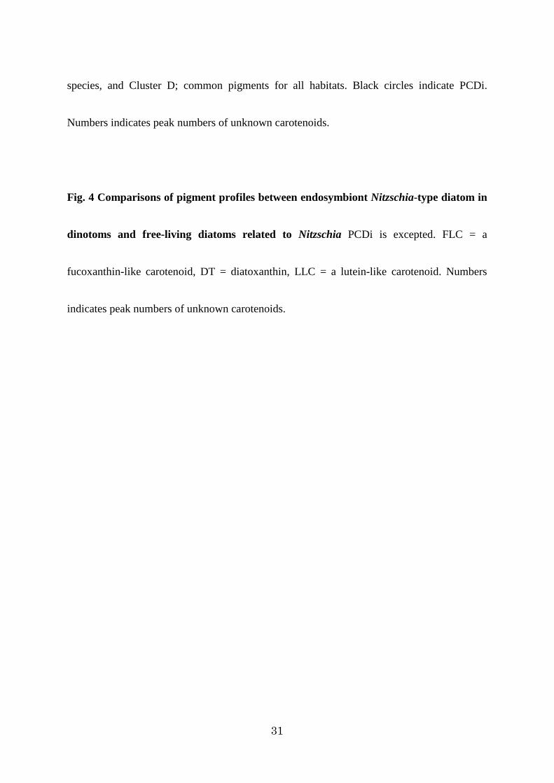
Comparison between pigment profiles of Nitzschia-type dinotoms and free-living diatoms
It is known that the species of the genus Durinskia and members of the Galeidinium spp.
clade possess a Nitzschia-type diatom endosymbiont (Tamura et al. 2005, Pienaar et al. 2007
and unpublished data). To compare the pigment compositions of dinotoms to those of
free-living diatoms, we added three diatoms, Cylindrotheca closterium (No.46), Nitzschia sp.
(No.47) and Tabularia sp. (No.48) as representatives for HPLC analyses (Fig. 4 and Fig. S7).
Free-living diatoms shared all PCDi with the dinotoms with the exception of β-ψ carotene
(Fig. S7). Other detected pigments from these diatoms were also detected in Nitzschia-type
dinotoms. Moreover Nitzschia-type dinotoms possessed many pigments that have not been
detected in free-living diatoms (Fig. 4). Additionally, the pigment compositions were
different among the Nitzschia-type dinotoms. Sand-dwelling three species in Galeidinium spp.
clade produced the highest number of specific pigments for dinotoms.
DISCUSSION
Validity of detected pigments
Recently Zapata et al. (2012) reported pigment compositions of 64 dinoflagellate species

17
(122 strains). This is the only comprehensive research for pigments using a HPLC that dealt
with both peridinin-type dinoflagellates and dinotoms, which allows us to compare to our
results. Most of common pigments of both dinoflagellate types in this study (PCPe and PCDi)
are corresponding with detected pigments in Zapata et al. (2012). However, the number of
detected pigments in this study greatly exceeds that of Zapata et al. (2012). Most of such
additional pigments reported here, including cPPB-aE, chl c3 and many UCs, have never
been detected from peridinin-type dinoflagellates and dinotoms.
In the interpretation of pigment compositions, it should also be considered the culture age
because the strains used in this study had been cultured for periods ranging from 1-4
month(s). Because each species shows different cell division rate, it was necessary to use
different culture ages for optimal harvesting. Cultures of all strains were checked for cell
density and cell morphology using the inverted microscope for suitability for the HPLC
analysis. The physiological conditions of cultures should also be considered whether the
differences of culture age affect cellular condition or not.. Therefore, we measured the
photosynthetic activities of three species cultured for different periods, i.e. one and three/four
month(s). Fv/Fm ratio of three benthic species showed no significant differences in
photosynthetic activities were found between different culture periods by t-test (Table S3).
18
We suggest, therefore, that the all strains used for pigment analyses were in healthy state and
detected pigments including many unknown carotenoids were not formed by stress condition.
Why these minor pigments have never been mentioned in the previous pigment studies in
dinoflagellates? One possible reason might be that the previous studies simply ignored most
unknown pigments. Minor peaks have been neglected in most of the previous reports because
these reports aimed to identify the type of chloroplast of a target dinoflagellate. However,
these minor pigments are equally-characteristic for dinoflagellates because they cannot be
detected in other microalgae, such as cyanobacteria, the chlorophyte Chlamydomonas
reinhardtii (data not shown), and diatoms (Fig. 4).
Are pigment profiles related to phylogenetic positions?
In this study, we analyzed 34 peridinin-type species and six dinotom species. In some
closely related species of peridinin-type dinoflagellates, it was shown that their pigment
profiles are similar to each other (Fig. S6). In the dinotoms, it was also indicated their
pigment profiles were somewhat reflected in the phylogenetic positions; within
sand-dwelling four species, the more closely related species based on molecular phylogeny
shared higher number of pigments (Fig.4 and Fig. S4). Thus it seems that pigment

19
compositions relate with phylogenetic positions, but we noticed that these closely related
dinoflagellates are, at the same time, all sand-dwelling dinoflagellates. Such observations
lead to the hypothesis that the habitat has a role to play in determining to produce the specific
pigments; we think that some closely related species are living in similar environment, and
thus, spending similar life cycle, and these conditions affect on their pigment profiles.
Pigment profiles are related to habitat
We noticed cPPB-aE and eleven unknown carotenoids were restricted to benthic
sand-dwelling species in peridinin-type dinoflagellates, although seven of them were from
one each representative species. The distributions of peak 12 UC and chl c1 were also
restricted. The former was found in only a tidal pool dinoflagellate, while the latter has been
detected in eight sand-dwelling and one tidal pool species. On the other hand, no specific
pigments were identifiable for planktonic dinoflagellates.
In dinotoms, chl c3 and fifteen unknown carotenoids were only detected in sand-dwelling
forms. The tidal pool dinotom had also the unique peak 40 UC. Contrary to peridinin-type
dinoflagellates in which no specific pigments were found, the freshwater planktonic dinotom
uniquely has 13 and 52 UCs.

20
These results highlighted that the far greater diversity of pigments produced by both
chloroplast type dinoflagellates living in sand relative to those of planktonic and tidal pool
forms. We suggest that dinoflagellates respond to their habitat in the production of their
pigments.
The results of pigment analysis relative to three free-living diatoms reinforce the idea that
diverse pigment of benthos is a response brought about by the host dinoflagellates. The
number of pigments in all Nitzschia-type dinotoms is in general, greater than that in
free-living diatoms. Further, the sand-dwelling species of the Galeidinium spp. clade even
show a greater diversity of pigment profile even compared to those of other Nitzschia-type
dinotoms.
Interestingly, all of specific pigments for sand-dwelling dinoflagellates both chloroplast
types are minor pigments, and most are unknown carotenoids. These unknown carotenoids
possess almost the same absorbance spectra (data not shown), so it is difficult to contemplate
that they have more than one function. Most of these UCs are suggested as intermediates or
degradation products of one or several novel carotenoid(s) (Takaichi personal
communication). However, the identity of the main carotenoid(s) remains obscure, and
therefore, the further studies are needed to determine the structure and functions of these

21
individual UCs. Only cPPB-aE and chl c3 were specifically identified and the presence of chl
c3 is the first report from dinotoms.
All specific pigments are rare one, and such pigments are detected only from a part of
sand-dwelling species, indicated to us that these pigments are produced not constantly but
occasionally depending on their physiological conditions. Similar situation can be found in
the case of chl c1 production. Although the biosynthetic pathway of chlorophyll c is still
unknown, one pathway that chl c1 and c3 are synthesized from chl c2 independently (Beale
1999, Green 2011) has been proposed. Chl c1 is thought to be synthesized by the reduction of
the 8-vinyl group of chl c2 (Green 2011). Recently it was suggested that DVR (3,8-divinyl
chlorophyllide reductase), an enzyme that functions as a chlorophyll a-synthesizing enzyme,
has low specificity for substrate and can also work to produce chl c1 from chl c2 (Ito and
Tanaka 2014). We suggest that the production of chl c1 takes place whenever chl c2 exists,
although that the amount of chl c1 produced is often too small to detect. Actually, although it
is generally known that peridinin-type dinoflagellates have chl a and c2, chl c1 has been
detected in quite a few of dinoflagellates from both planktonic and benthic species in
previous studies (e.g. Carreto et al. 2001, Fraga et al. 2011, Wakahama et al. 2012, Zapata et
al. 2012) and these examples indicate the presence of above mechanism (Ito and Tanaka

22
2014). Like chl c1, most of these minor pigments detected in this study must be produced by
the mechanism not under control as intermediates or degradation products.
Why, then, do the only sand-dwelling benthic dinoflagellates produce these specific extra
pigments? At this moment, we have no answer for this question. To answer this question,
further photosynthetic studies, such as the determination of the structure and roles of the
unknown carotenoids, the synthetic pathway, enzymes of these pigments and ecological
responses within the microhabitat, are needed.
Acknowledgements
We gratefully acknowledge Ms. Junko Kishimoto for technical assistance with the HPLC
analyses. We also appreciate to Ms. Saeka Takabayashi for cluster analyses of detected
pigments. Our thanks go to Dr. Stuart D. Sym for reading the manuscript and collecting No.
39 and 40 of Durinskia sp. We also thank to Dr. Ryuta Terada and Captain M. Uchiyama and
the crew of T/S Nansei-maru, Faculty of Fisheries, Kagoshima University, for their kind help
in collecting the underwater samples. This work was partly supported by the Grant-in-Aid by
the Japan Society for the Promotion of Science (JSPS) (No.24370034). Eleven of the strains
used in this study were collected during the field trips for the project entitled ‘Biodiversity
23
and evolution of algae in the Indo-Pacific: a Japan/South Africa comparison’ (Strategic
International Research Cooperative Program) supported by Japan Science and Technology
Agency. This research was also supported by JSPS Research Fellowships for young scientists
(No.322).
References
Beale SI (1999) Enzymes of chlorophyll biosynthesis. Photosynth Res 60:43-73
Carreto JI, Carignan MO, Montoya NG (2001) Comparative studies on mycosporine-like
amino acids, paralytic shellfish toxins and pigment profiles of the toxic dinoflagellates
Alexandrium tamarence, A. catenella and A. minutum. Mar Ecol Prog Ser 49:49-60
Chesnick JM, Kooistra WHCF, Wellbrock U, Medlin LK. (1997) Ribosomal RNA analysis
indicates a benthic pennate diatom ancestry for the endosymbionts of the
dinoflagellates Peridinium foliaceum and Peridinium balticum (Pyrrhophyta). J
Eukaryot Microbiol 44:314-20
Choi SS, Cha SH, Tappert CC (2010) A survey of binary similarity and distance measures. J
Syst Cybern Inf 8:43-48
Faust MA, Vandersea MW, Kibler SR, Tester PA (2008) Prorocentrum levis, a new benthic

24
species (dinophyceae) from a mangrove island, twin cays, belize. J Phycol 44:232-240
Fraga S, Rodríguez F, Caillaud A, Diogène J, Raho N, Zapata M (2011) Gambierdiscus
excentricus sp. nov. (Dinophyceae), a benthic toxic dinoflagellate from the Canary
Islands (NE Atlanic Ocean). Harmful Algae 11:10-22
Green BR (2011) After the primary endpsymbiosis: an update on the chromalveolate
hypothesis and the origins of algae with Chl c. Photosynth Res 107:103-115
Guillard RR, Ryther JH (1962) Studies of marine planktonic diatoms. I. Cyclotella nana
Hustedt, and Detonula confervacea (Cleve) Gran. Can J Microbiol 8:229-239
Herdman EC (1922) Notes on dinoflagellates and other organisms causing discolouration of
the sand at Port Erin II. Trans Lpool Bio Soc 36:15-30
Hoppenrath M, Horiguchi T, Miyoshi Y, Selina M, Taylor MFJR, Leander BS (2007)
Taxonomy, phylogeny, biogeography, and ecology of Sabulodinium undulatum
(Dinophyceae), including an emended description of the species. Phycol Res
55:159-175
Hoppenrath M, Murray SA, Chomérat N, Horiguchi T (2014) Marine benthic dinoflagellates
– unveiling their worldwide biodiversity. Schweizerbart, Stuttgart
Horiguchi T, Chihara M (1987) Spiniferodinium galeiforme, a new genus and species of
25
benthic dinoflagellates (Phytodiniales, Pyrrhophyta ) from Japan. Phycologia
26:478-487
Horiguchi T, Chihara M (1988) Life-cycle, behavior and morphology of a new tide pool
dinoflagellate, Gymnodinium-pyrenoidosum sp-nov (Gymnodiniales, Pyrrhophyta). Bot
Mag Tokyo 101:255-265
Horiguchi T, Takano Y (2006) Serial replacement of a diatom endosymbiont in the marine
dinoflagellate Peridinium quinquecorne (Peridiniales, Dinophyceae). Phycol Res
54:193-200
Imanian B, Pombert J, Keeling P (2010) The complete plastid genomes of the two ‘dinotoms’
Durinskia baltica and Kryptoperidinium foliaceum. Plos One 5:e10711
Ito H, Tanaka A (2014) Evolution of a new chlorophyll metabolic pathway driven by the
dynamic changes in enzyme promiscuous activity. Plant Cell Physiol 55:593-603
Jeffrey SW, Sielicki M, Haxo FT (1975) Chroloplast pigment patterns in dinoflagellates. J
Phycol 11:374-384
Kimura B, Ishida Y (1985) Photophagotrophy in Uroglena americana, Chrysophyceae. Jpn J
Limnol 46:315-318

26
Louda JW, Loitz JW, Rudnick DT, Baker EW (2000) Early diagenetic alteration of
chlorophyll-a and bacteriochlorophyll-a in a contemporaneous marl ecosystem; Florida
Bay. Org Geochem 31:1561-1580
Ma L, Dolphin D (1999) The metabolites of dietary chlorophylls. Phytochemistry 50:195-202
Mandelli EF (1968) Carotenoid pigments of the dinoflagellate Glenodinium foliaceum Stein.
J Phyccol 4:347-348
Matsumoto T, Kawachi M, Miyashita H, Inagaki Y (2012) Prasinoxanthin is absent in the
green-colored dinoflagellate Lepidodinium chlorophorum strain NIES-1868: pigment
composition and 18S rDNA phylogeny. J Plant Res 125:705-711
Murray S, Patterson, DJ (2002) The benthic dinoflagellate genus Amphidinium in
south-eastern Australian waters, including three new speices. Eur J Phycol 37:279-298
Parsons ML, Preskitt LB (2007) A survey of epiphytic dinoflagellates from the coastal waters
of the island of Hawai‘i. Harmful Algae 6:658-669
Pienaar RN, Sakai H, Horiguchi T (2007) Description of a new dinoflagellate with a diatom
endosymbiont, Durinskia capensis sp. nov. (Peridiniales, Dinophyceae) from South
Africa. J Plant Res 120:247-258
Riley JP, Wilson TRS (1967) The pigments of some marine phytoplankton species. J Mar

27
Biol Assoc UK 47:351-361
Saburova N, Al-Yamani F, Polikarpov I (2009) Biodiversity of free-living flagellates in
Kuwait’s intertidal sediments. Bio Risk 3:97-110.
Sokal RR, Michener CD (1958) A statistical method for evaluating systematic relationships.
Kans Univ Sci Bull 38:1409-1438
Takano Y, Hansen G, Fujita D, Horiguchi T (2008) Serial replacement of diatom
endosymbionts in two freshwater dinoflagellates, Peridiniopsis spp. (Peridiniales,
Dinophyceae). Phycologia 47:41-53
Tamura M, Shimada S, Horiguchi T (2005) Galeidinium rugatum gen. et sp. nov.
(Dinophyceae), a new coccoid dinoflagellate with a diatom endosymbiont. J Phycol
41:658-671
Taylor FJR, Hoppenrath M, Saldarriaga JF (2008) Dinoflagellate diversity and distribution.
Biodivers Conserv 17:407-18.
Tengs T, Dahlberg OJ, Shalchian-Tabrizi K, Klaveness D, Rudi K, Delwiche CF, Jakobsen
KS (2000) Phylogenetic analyses indicate that the 19’
hexanoyloxy-fucoxanthin-containing dinoflagellates have tertiary plastids of haptophyte
origin. Mol Biol Evol 17:718-29.

28
Wakahama T, Okuyama H, Maoka T, Takaichi S (2012) Unique carotenoid lactoside, P457, in
Symbiodinium sp. of dinoflagellate. Acta Biochim Pol 59:155-157
Watanabe MM, Suda S, Inouye I, Sawaguchi T, Chihara M (1990) Lepidodinium viride gen.
et sp. nov. (Gymnodiniales, Dinophyta), a green dinoflagellate with a chlorophyll a- and
b-containing endosymbiont. J Phycol 26:741-51.
Withers NW, Cox ER, Tomas R, Haxo FT (1977) Pigments of the dinoflagellate Peridinium
balticum and its photosynthetic endosymbiont. J Phycol 13:354-358
Yamada N, Terada R, Tanaka A, Horiguchi T (2013) Bispinodinium angelaceum gen. et sp.
nov. (Dinophyceae), a new sand-dwelling dinoflagellate from the seafloor off
Mageshima Island, Japan. J Phycol 49:555-569
Yamada N, Tanaka A, Horiguchi T (2014) cPPB-aE is discovered from photosynthetic
benthic dinoflagellates. J Phycol 50:101-107
Zapata M, Rodríguez F, Grarrido, JL (2000) Separation of chlorophylls and carotenoids from
marine phytoplankton: a new HPLC method using a reversed phase C8 column and
pyridine-containing mobile phases. Mar Ecol Prog Ser 195:29-45
Zapata M, Fraga S, Rodríguez F, Garrido JL (2012) Pigment-based chloroplast types in
dinoflagellates. Mar Ecol Prog Ser 465:33-52

29
Zhang Q, Liu G, Hu Z (2011) Morphological differences and molecular phylogeny of
freshwater blooming species, Peridiniopsis spp. (Dinophyceae) from China. Eur J
Protistol 47:149-160
Zhang Q, Liu G, Hu Z (2014) Description of a new freshwater bloom-forming dinoflagellate
with a diatom endosymbiont, Peridiniopsis minima sp. nov. (Peridiniales, Dinophyceae)
from China. Algological Studies 146:119-133
Zhang ZD, Green BR, Cavalier-Smith,T (1999) Single gene circles in dinoflagellate
chloroplast genomes. Nature 400;155-159
FIGURES
Fig. 1 HPLC chromatograms of the detected pigments of analyzed dinoflagellates (a)
Peridinin-type dinoflagellate, (b) Dinotom. Pigments were extracted from Pyrocystis sp., No.
20; planktonic (a1), Alexandrium hiranoi, No.1; tidal pool living (a2), Bispinodinium
angelaceum, No.11; sand-dwelling (a3), Peridiniopsis cf. kevei, No.45; planktonic (b1),
Durinskia sp., No.39; tidal pool living (b2), Unidentified coccoid dinoflagellate 1, No.42;
sand-dwelling (b3). The absorbance at 450 nm monitored. Black circles indicate typical
pigments for dinoflagellates. Double black circles indicate characteristic pigments for each

30
chloroplast type. Peak numbers are same to Figs. S5 and S7. Peak 4 = chl c2, peak 6 =
peridinin, peak 10 = fucoxanthin, peak 19 = diadinoxanthin, peak 22 = dinoxanthin, peak 24
= diatoxanthin, peak 27 = zeaxanthin, peak 49 = chl a, peak 54 = β-carotene.
Fig. 2 Result of clustering analysis of pigment distribution according to habitat-type in
peridinin-type dinoflagellates: sand-dwelling benthic, tidal pool inhabiting, planktonic
The binary similarity and dissimilarity measure is from Sokal and Michener index. Cluster A;
pigments only detected from sand-dwellers, Cluster B; from tidal pool inhabiters and
sand-dwellers, Cluster C; from planktonic species and sand-dwellers, Cluster D; from all
habitat type dinoflagellates. Black circles indicate PCPe. Numbers indicates peak numbers of
unknown carotenoids.
Fig. 3 Result of clustering analysis of pigment distribution according to habitat-type in
dinotoms: sand-dwelling benthic, tidal pool inhabiting, planktonic The binary similarity
and dissimilarity measure is from Sokal and Michener index. Cluster A; only from
sand-dwellers, Cluster B; from tidal pool inhabiters, Cluster C; mainly from planktonic
31
species, and Cluster D; common pigments for all habitats. Black circles indicate PCDi.
Numbers indicates peak numbers of unknown carotenoids.
Fig. 4 Comparisons of pigment profiles between endosymbiont Nitzschia-type diatom in
dinotoms and free-living diatoms related to Nitzschia PCDi is excepted. FLC = a
fucoxanthin-like carotenoid, DT = diatoxanthin, LLC = a lutein-like carotenoid. Numbers
indicates peak numbers of unknown carotenoids.

Title: Pigment compositions are linked to the habitat types in dinoflagellates
Journal name: Journal of plant research
The name of the authors: Norico Yamada1, Ayumi Tanaka2, Takeo Horiguchi3
E-mail of the corresponding author: [email protected]

Supplementary materials and methods DNA extraction and PCR amplification
DNA extractions were performed by using the benzyl chloride method (Zhu et al. 1993) or by
using the QuickExtract FFPE RNA Extraction Kit (Epicentre, Wisconsin) from the same culture
strains that used for HPLC analyses. For the latter method, several dinoflagellate cells (1 to 10 cells)
were isolated using capillary pipettes under an inverted microscope and transferred into 10μl of
QuickExtract FFPE solution. The sample was then heated at 56 ºC for 1 hour and then 98 ºC for 2
min. The solution was used as template DNA. The PCR amplification process consisted of 1 initial
cycle of denaturation at 94 ºC for 5 min, followed by 40 cycles of denaturation at 94 ºC for 30 s,
annealing at 55 ºC for 30 s, and extension at 72ºC for 1 min. The final extension cycle was at 72 ºC
for 7 min. The primer combinations are as follows; SR1b (F) - SR3 (R); SR2spin (F) - SR7 (R); SR4
(F) - SR9p(R); SR6 (F) - SR11(R); SR8 (F) - SR12b (R) (Nakayama et al. 1996, Yamaguchi and
Horiguchi 2005, Yamada et al. 2014). For Peridiniopsis cf. kevei, the following newly-designed
primers were used in stead of SR7; SR7PP (R): TAACGACCTCCAATCTCTAG (the pair primer is
SR2spin), and in stead of SR6-SR11 pair; SR6PP (F): GCATTCGTATTTAACTGTCA-SR11PP (R):
CATCACGATGCGTTTTAAC. The PCR products were purified and sequenced using an ABI
PRISM Big Dye Terminator (Applied Biosystems, Foster City). The sequence reactions were run on
a DNA autosequencer ABI PRISM 3730 DNA Analyzer (Applied Biosystems, Foster City). Both
forward and reverse strands were sequenced.
Sequence analysis
The accession numbers of sequences included in the alignments are shown in Supplemental Table
2. The SSU rDNA sequences were aligned manually, based on the published secondary structure of
the SSU rRNA molecule, using the alveolate taxa available at the rRNA server
(http://www.psb.ugent.be/rRNA) (database no longer available). As outgroups, the apicomplexan
organisms, Sarcocystis muris (Blancbard) Alexieff and Toxoplasma gondii Nicolle et Manceaux,
were used for peridinin-type dinoflagellates, while the dinoflagellates Ceratium fusus Ehrenberg and
Gonyaulax spinifera (Claparède & Lachmann) Diesing were designated for dinotoms. The aligned
sequences were analyzed by Bayesian method using the MrBayes 3.1.2. (Huelsenbeck and Ronquist
2001). The GTR+I+G model was selected by MrModeltest 2.2 (Nylander et al. 2004) as a suitable
evolutionary model. Markov chain Monte Carlo iterations were carried out until 50,000,000
generations were attained, when the average standard deviations of split frequencies fall bellows 0.01
indicating convergence of the iterations.
Photosynthetic activity of culture strains.

Chlorophyll fluorescence was determined by a PAM fluorometer (PAM 101/102/103, Heinz
Waltz, Effeltrich, Germany). Strains of cultured for one, three or four month(s) under continuous
light (60 μmol·photons ·m−2·s−1), respectively were dark-adapted for 20 min.

Supplementary tables
Table S1 The list of dinoflagellate strains, sampling stations and culture conditions (next page)
1Habitat-type: P = Planktonic, S = Sand-dwelling and T = Tidal pool. Unit of culture temperature =
degrees C, All samples were cultured under the 60 μmol· photons / m2 · s-1, light: dark = 16:8hours.
Some of the dinoflagellates were treated as undescribed, novel species (No.8, 12, 17, 19, 27 and the
dinoflagellates included in unidentified group) or currently-unidentifiable to the species rank (No.1,
2, 20, 21, 24 and 25) because for precise identification, SEM observation is needed.


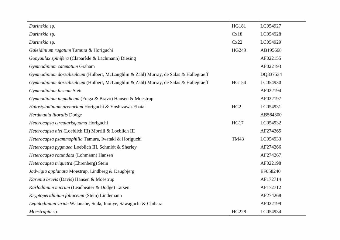
Table S2 Genbank accession numbers of samples used in this study
Species Name Strain Accession Number
Adenoides eludens (Herdman) Balech EF492484
Akashiwo sanguinea (Hirasaka) Hansen & Moestrup U41085
Alexandrium hiranoi Kita & Fukuyo AY641564
Alexandrium hiranoi Kita & Fukuyo HG3 LC056070
Alexandrium insuetum Balech AB088298
Alexandrium minutum Halim U27499
Alexandrium ostenfeldii (Paulsen) Balech & Tangen AJ535384
Alexandrium pseudogonyaulax (Biecheler) Horiguchi ex Kita & Fukuyo AB088302
Alexandrium tamarense (Lebour) Balech AF022191
Alexandrium tamutum Montresor, Beran & John AJ535378
Alexandrium taylori Balech AJ535385
Alexandrium sp. 1 HG222 LC056069
Alexandrium sp. 2 NY008 LC056068
Amphidiniella sedentaria Horiguchi AB212091
Amphidiniella sedentaria Horiguchi HG156 LC057317
Amphidiniopsis dragescoi (Balech) Hoppenrath, Selina, Yamaguchi & Leander AY238479
Amphidinium carterae Hulburt AF009217
Amphidinium cupulatisquama Tamura & Horiguchi HG149 LC056067
Amphidinium gibbosum (Maranda & Shimizu) Jørgensen & Murray L13719
Amphidinium gibbosum (Maranda & Shimizu) Jørgensen & Murray NY004 AB863027
Amphidinium massartii Biecheler AF274255

Amphidinium operculatum Claparède & Lachmann AB704006
Amphidinium cf. rhynchocephalum Anissimowa AY443012
Amphidinium steinii Lemmermann HG214 LC054920
Amphidinium steinii Lemmermann HG220 LC054921
Amphidinium sp. AB626895
Amphidinium sp. HG115 AB477347
Amphidinium sp. HG213 LC054922
Archaeperidinium minutum (Kofoid) Jørgensen AB564308
Azadinium spinosum Elbrächter & Tillmann JN680857
Biecheleria baltica Moestrup, Lindberg, & Daugbjerg EF058252
Biecheleria natalensis (Horiguchi & Pienaar) Moestrup Cx7 LC054923
Bispinodinium angelaceum Yamada & Horiguchi HG236 AB762397
Borghiella tenuissima (Lauterborn) Moestrup, Hansen & Daugbjerg AY443025
Ceratium fusus Ehrenberg AF022153
Ceratocorys sp. NY002 LC054924
Cochlodinium polykrikoides Margalef AY421781
Dinophysis acuta Ehrenberg AJ506973
Dinophysis norvegica Claparède & Lachmann AJ506974
Diplopsalis lenticula Bergh AB716909
Durinskia agilis (Kofoid & Swezy) Saburova, Chomérat & Hoppenrath JF514516
Durinskia baltica (Levander) Carty & Cox AF231803
Durinskia cf. baltica (Levander) Carty & Cox HG171 LC054925
Durinskia cf. baltica (Levander) Carty & Cox HG265 LC054926
Durinskia capensis Pienaar, Sakai & Horiguchi AB271107
Durinskia sp. HG181 LC054927
Durinskia sp. Cx18 LC054928
Durinskia sp. Cx22 LC054929
Galeidinium rugatum Tamura & Horiguchi HG249 AB195668
Gonyaulax spinifera (Claparède & Lachmann) Diesing AF022155
Gymnodinium catenatum Graham AF022193
Gymnodinium dorsalisulcum (Hulbert, McLaughlin & Zahl) Murray, de Salas & Hallegraeff DQ837534
Gymnodinium dorsalisulcum (Hulbert, McLaughlin & Zahl) Murray, de Salas & Hallegraeff HG154 LC054930
Gymnodinium fuscum Stein AF022194
Gymnodinium impudicum (Fraga & Bravo) Hansen & Moestrup AF022197
Halostylodinium arenarium Horiguchi & Yoshizawa-Ebata HG2 LC054931
Herdmania litoralis Dodge AB564300
Heterocapsa circularisquama Horiguchi HG17 LC054932
Heterocapsa niei (Loeblich III) Morrill & Loeblich III AF274265
Heterocapsa psammophilla Tamura, Iwataki & Horiguchi TM43 LC054933
Heterocapsa pygmaea Loeblich III, Schmidt & Sherley AF274266
Heterocapsa rotundata (Lohmann) Hansen AF274267
Heterocapsa triquetra (Ehrenberg) Stein AF022198
Jadwigia applanata Moestrup, Lindberg & Daugbjerg EF058240
Karenia brevis (Davis) Hansen & Moestrup AF172714
Karlodinium micrum (Leadbeater & Dodge) Larsen AF172712
Kryptoperidinium foliaceum (Stein) Lindemann AF274268
Lepidodinium viride Watanabe, Suda, Inouye, Sawaguchi & Chihara AF022199
Moestrupia sp. HG228 LC054934

Nematodinium sp. FJ947039
Paragymnodinium shiwhaense Kang, Jeong, Moestrup & Shin AM408889
Pfiesteria piscicida Steidinger & Burkholder AY112746
Pentapharsodinium tyrrhenicum (Balech) Montressor, Zingone & Marino AF022201
Peridiniopsis borgei Lemmermann EF058241
Peridiniopsis cf. kevei Grigorszky AB353770
Peridiniopsis cf. kevei Grigorszky DA08 LC054935
Peridiniopsis cf. kevei Grigorszky HG327 LC054936
Peridiniopsis niei Liu & Hu HM596542
Peridiniopsis penardii (Lemmermann) Bourrelly AB353771
Peridiniopsis polonicum (Woloszynska) Bourrelly AY443017
Peridinium aciculiferum Lemmermann AY970653
Peridinium cinctum (Müller) Ehrenberg DQ166209
Peridinium quinquecorne Abé AB246744
Peridinium willei Huitfeldt-Kaas AF274272
Plagiodinium belizeanum Faust & Balech HG225 LC054937
Plagiodinium sp. HG177 LC054938
Polarella glacialis Montresor, Procaccini & Stoecker AF099183
Prorocentrum lima (Ehrenberg) Stein Y16235
Prorocentrum maculosum Faust Y16236
Prorocentrum micans Ehrenberg M14649
Prorocentrum minimum (Pavillard) Schiller AY421791
Pseudopfiesteria shumwayae (Glasgow & Burkholder) Litaker, Steidinger, Mason, Shields & Tester AF080098
Pyrocystis lunula (Schütt) Schütt AF274274

Pyrocystis noctiluca Murray ex Haeckel AF022156
Pyrocystis sp. NY007 LC054939
Sabulodinium undulatum Saunders & Dodge DQ975474
Scrippsiella hangoei (Schiller) Larsen AY970662
Scrippsiella precaria Montresor & Zingone DQ847435
Scrippsiella sweeneyae Loeblich III AF274276
Scrippsiella trochoidea (Stein) Balech ex Loeblich III AF274277
Scrippsiella sp. NY012 LC054940
Spiniferodinium galeiforme Horiguchi & Chihara GU295203
Spiniferodinium galeiforme Horiguchi & Chihara TM57 LC054941
Stylodinium littorale Horiguchi & Chihara NY017 LC054942
Symbiodinium californium Banaszak, Iglesias-Prieto & Trench AF225965
Symbiodinium corculorum Trench L13717
Symbiodinium goreaui Trench & Blank EF036539
Symbiodinium microadriaticum Freudenthal M88521
Symbiodinium sp. 1 HG193 AB863030
Symbiodinium sp. 2 NY010 AB863031
Testudodinium corrugatum (Larsen & Patterson) Horiguchi, Tamura & Yamaguchi HG163 AB704004
Testudodinium corrugatum (Larsen & Patterson) Horiguchi, Tamura & Yamaguchi TM-85 AB704003
Testudodinium maedaense Katsumata & Horiguchi MAE-18 AB704005
Testudodinium testudo (Herdman) Horiguchi, Tamura, Katsumata & Yamaguchi KOM-30 AB704002
Testudodinium sp. HG230 LC054943
Thecadinium petasatum Kofoid & Skogsberg GU295204
Thoracosphaera heimii (Lohmann) Kamptner AF274278

Thoracosphaera heimii (Lohmann) Kamptner HG252 LC054944
Togula britannica (Herdman) Jørgensen, Murray & Daugbjerg AY443010
Togula jolla Jørgensen, Murray & Daugbjerg AF274252
Unidentified athecate dinoflagellate 1 NY005 AB863028
Unidentified athecate dinoflagellate 2 HG167 AB863029
Unidentified athecate dinoflagellate 3 NY003 LC054945
Unidentified coccoid dinotom 1 HG180 LC054946
Unidentified coccoid dinotom 2 HG204 LC054947
Unidentified thecate dinoflagellate 1 NY011 LC054948
Unidentified thecate dinoflagellate 2 NY013 LC054949
Unidentified thecate dinoflagellate 3 HG151 LC054950
Unidentified thecate dinoflagellate 4 NY014 LC054951
Outgroup
Sarcocystis muris M64244
Toxoplasma gondii L24381
Diatom
Cylindrotheca closterium NY018 LC054954
Nitzschia sp. NY060 LC054952
Tabularia sp. NY059 LC054953

Table S3 The Fv/Fm ratio using PAM method of three dinoflagellates which were cultured in one, three or four months The Fv/Fm ratio
by PAM fluorometer were measured in three times for each strain and the averages of these values were used for t-test with two-tailed test
(p<0.05). In Durinskia sp. (No.38), three months cultured strain was used because cells of this species become the deformation after maintained
in four months. One month = cultured from 1 April 2015 to 1 May 2015. Three months = cultured from 7 January 2015 to 6 April 2015. Four
months = cultured from 3 December 2014 to 6 April 2015.
Supplementary figures
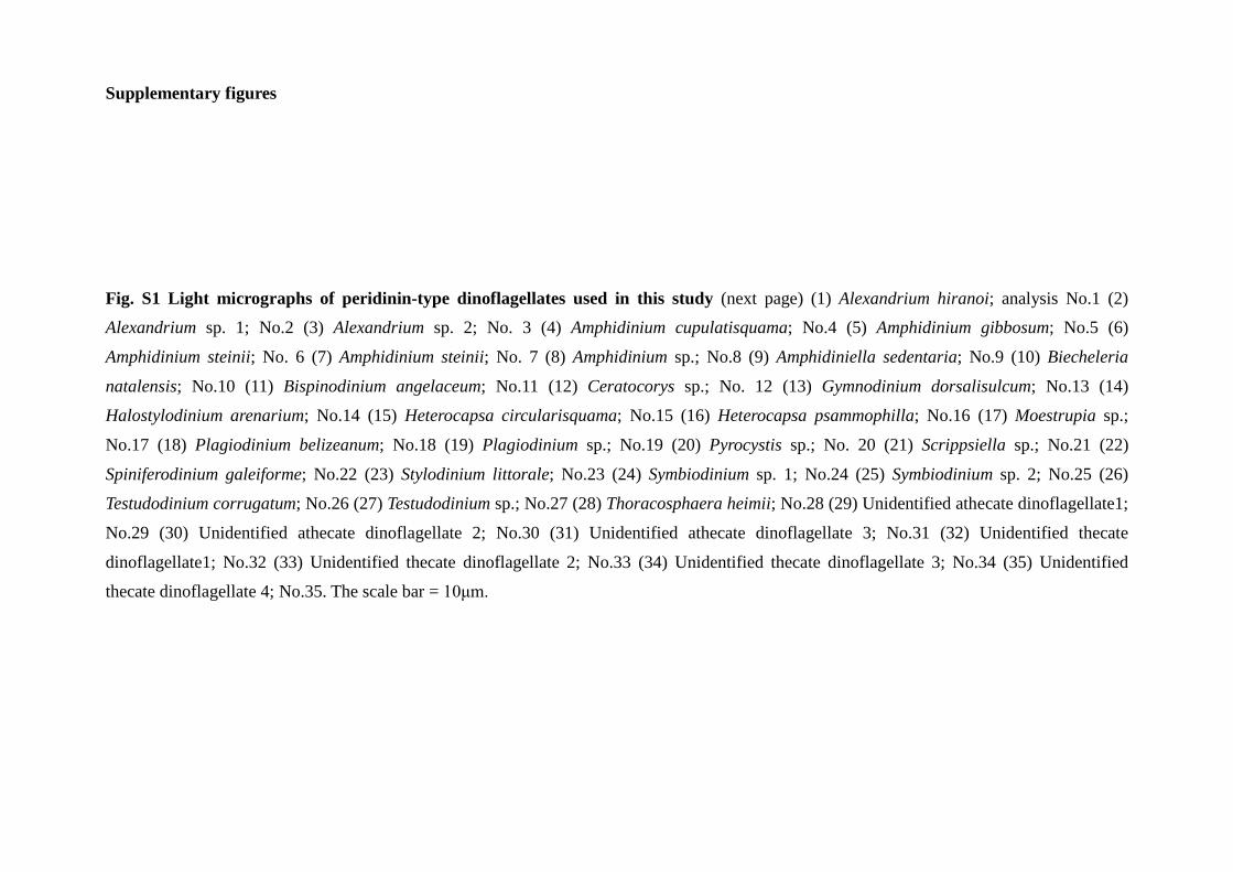
Fig. S1 Light micrographs of peridinin-type dinoflagellates used in this study (next page) (1) Alexandrium hiranoi; analysis No.1 (2)
Alexandrium sp. 1; No.2 (3) Alexandrium sp. 2; No. 3 (4) Amphidinium cupulatisquama; No.4 (5) Amphidinium gibbosum; No.5 (6)
Amphidinium steinii; No. 6 (7) Amphidinium steinii; No. 7 (8) Amphidinium sp.; No.8 (9) Amphidiniella sedentaria; No.9 (10) Biecheleria
natalensis; No.10 (11) Bispinodinium angelaceum; No.11 (12) Ceratocorys sp.; No. 12 (13) Gymnodinium dorsalisulcum; No.13 (14)
Halostylodinium arenarium; No.14 (15) Heterocapsa circularisquama; No.15 (16) Heterocapsa psammophilla; No.16 (17) Moestrupia sp.;
No.17 (18) Plagiodinium belizeanum; No.18 (19) Plagiodinium sp.; No.19 (20) Pyrocystis sp.; No. 20 (21) Scrippsiella sp.; No.21 (22)
Spiniferodinium galeiforme; No.22 (23) Stylodinium littorale; No.23 (24) Symbiodinium sp. 1; No.24 (25) Symbiodinium sp. 2; No.25 (26)
Testudodinium corrugatum; No.26 (27) Testudodinium sp.; No.27 (28) Thoracosphaera heimii; No.28 (29) Unidentified athecate dinoflagellate1;
No.29 (30) Unidentified athecate dinoflagellate 2; No.30 (31) Unidentified athecate dinoflagellate 3; No.31 (32) Unidentified thecate
dinoflagellate1; No.32 (33) Unidentified thecate dinoflagellate 2; No.33 (34) Unidentified thecate dinoflagellate 3; No.34 (35) Unidentified
thecate dinoflagellate 4; No.35. The scale bar = 10μm.


Fig. S2 Light micrographs of dinotoms and free-living diatoms used in this study (next page) (A) Durinskia cf. baltica (A1) analysis number
36, collected from Tokashiki Islands, Okinawa, Japan (A2) No.37, collected from Odo Beach, Okinawa, Japan (B) Durinskia sp. (B1) No.38,
collected from Marina Beach, South Africa (B2) No.39, collected from Marina Beach, South Africa (B3) No.40, collected from Marina Beach,
South Africa (C) Galeidinium rugatum; No.41. Non-motile cell. (D) Unidentified coccoid dinotom 1; No.42. Non-motile cell. (E) Unidentified
coccoid dinotom 2; No.43. Non-motile cell. (F) Peridiniopsis cf. kevei (F1) No.44, sampling from Shikotu-Lake, Hokkaido, Japan (F2) No.45,
sampling from Biwa-Lake, Shiga, Japan. (G) Cylindrotheca closterium, free-living diatom close to endosymbiont diatom Nitzschia of (A) to (E);
No.46. (H) Nitzschia sp. free-living diatom; No.47. (I) Tabularia sp. free-living diatom; No.48. The scale bar = 5μm. The other the scale bar =
10μm.


Fig. S3 The Bayesian molecular phylogeny inferred from SSU rDNA of Dinophyta including
peridinin-type dinoflagellates and dinotoms Numbers on the major nodes represent posterior
probability. Only PP>0.5 is shown. Analyzed strains and numbers are shown by bold type. Gray
boxes with asterisk indicate dinotoms.
Fig.
S4
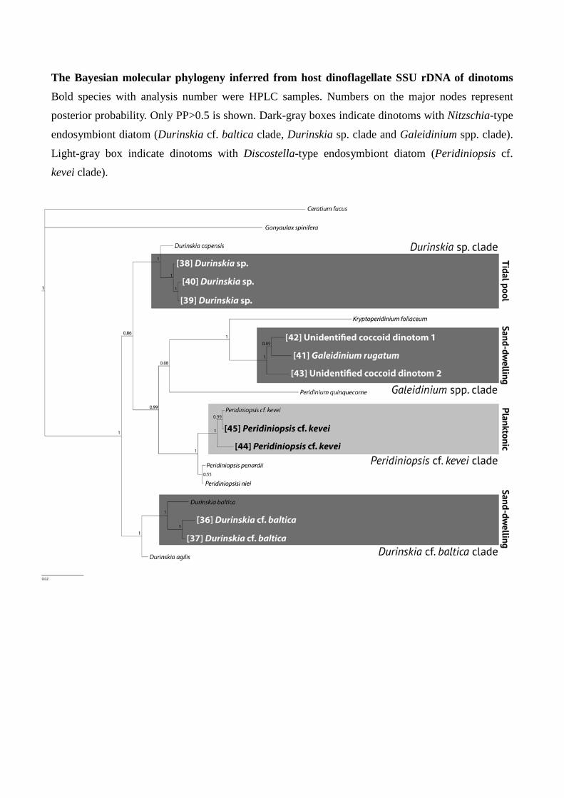
The Bayesian molecular phylogeny inferred from host dinoflagellate SSU rDNA of dinotoms
Bold species with analysis number were HPLC samples. Numbers on the major nodes represent
posterior probability. Only PP>0.5 is shown. Dark-gray boxes indicate dinotoms with Nitzschia-type
endosymbiont diatom (Durinskia cf. baltica clade, Durinskia sp. clade and Galeidinium spp. clade).
Light-gray box indicate dinotoms with Discostella-type endosymbiont diatom (Peridiniopsis cf.
kevei clade).
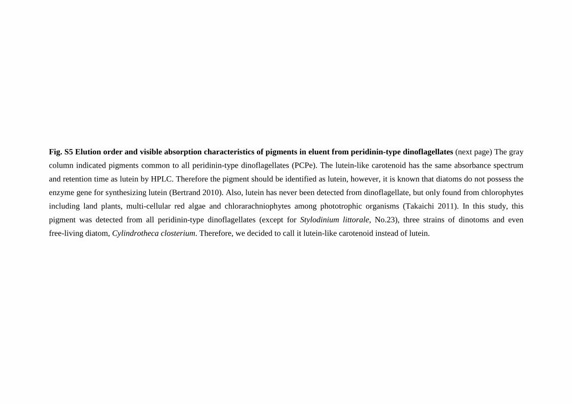
Fig. S5 Elution order and visible absorption characteristics of pigments in eluent from peridinin-type dinoflagellates (next page) The gray
column indicated pigments common to all peridinin-type dinoflagellates (PCPe). The lutein-like carotenoid has the same absorbance spectrum
and retention time as lutein by HPLC. Therefore the pigment should be identified as lutein, however, it is known that diatoms do not possess the
enzyme gene for synthesizing lutein (Bertrand 2010). Also, lutein has never been detected from dinoflagellate, but only found from chlorophytes
including land plants, multi-cellular red algae and chlorarachniophytes among phototrophic organisms (Takaichi 2011). In this study, this
pigment was detected from all peridinin-type dinoflagellates (except for Stylodinium littorale, No.23), three strains of dinotoms and even
free-living diatom, Cylindrotheca closterium. Therefore, we decided to call it lutein-like carotenoid instead of lutein.
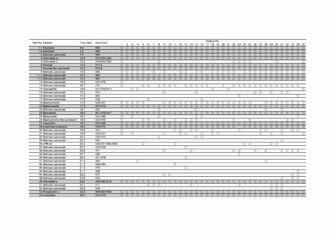

Fig. S6 Result of clustering analysis of pigment distribution according to species in peridinin-type dinoflagellates (next page) The binary
similarity and dissimilarity measure is from Sokal and Michener index. Gray columns indicated the species possessing similar pigment
compositions each other between close related species. Numbers indicates peak numbers of unknown carotenoids.


Fig. S7 Elution order and visible absorption characteristics of pigments in eluent from dinotoms and free-living diatoms The gray column
indicated pigments common to all dinotoms (PCDi). Diatom 1 is Cylindrotheca closterium, No.46. Diatom 2 is Nitzschia sp., No.47. Diatom 3 is
Tabularia sp., No.48.

References in supplementary files Bertrand M (2010) Carotenoid biosynthesis in diatoms. Phytosynth Res 106:89-102
Huelsenbeck JP, Ronquist F (2001) MrBayes: Bayesian inference of phylogenetic trees.
Bioinformatics 17:754-755
Nakayama T, Watanabe S, Mitsui K, Uchida H, Inouye I (1996) The phylogenetic
relationship between the Chlamydomonadales and Chlorococcales inferred from
18S rDNA sequence data. Phycol Res 44:47-55.
Nylander JAA, Ronquist F, Huelsenbeck JP, Nieves-Aldrey JL (2004) Bayesian
phylogenetic analysis of combined data. Syst Biol 53:47-67
Takaichi S (2011) Carotenoids in Algae: Distribution, Biosyntheses and Functions. Mar
Drungs 9:1101-1118
Yamaguchi A, Horiguchi T (2005) Molecular phylogenetic study of the heterotrophic
dinoflagellate genus Protoperidinium (Dinophyceae) inferred from small subunit
rRNA gene sequences. Phycol Res 53:30-42
Zhu H, Qu F, Zhu LH (1993) Isolation of genomic DNAs from plants, fungi and
bacteria using benzyl-chloride. Nucleic Acids Res 21:5279-80