Embed Size (px)
Citation preview
pISSN: 2234-8646 eISSN: 2234-8840http://dx.doi.org/10.5223/pghn.2014.17.4.257Pediatr Gastroenterol Hepatol Nutr 2014 December 17(4):257-262 PGHNCase Report
PEDIATRIC GASTROENTEROLOGY, HEPATOLOGY & NUTRITION
The Complex Surgical Management of the First Case of Severe Combined Immunodeficiency and Multiple Intestinal Atresias Surviving after the Fourth Year of Life
Riccardo Guanà, Salvatore Garofano, Elisabetta Teruzzi, Simona Vinardi, Giulia Carbonaro, Alessia Cerrina, Isabella Morra*, Davide Montin†, Alessandro Mussa† and Jürgen Schleef
Divisions of Pediatric General Surgery and *Pathology, †Department of Pediatric and Public Health Sciences, University of Torino, Regina Margherita Children’s Hospital, Torino, Italy
Severe combined immunodeficiency (SCID) is a life-threatening syndrome of recurrent infections and gastro-intestinal alterations due to severe compromise of T cells and B cells. Clinically, most patients present symptoms before the age of 3 months and without intervention SCID usually results in severe infections and death by the age of 2 years. Its association with intestinal anomalies as multiple intestinal atresias (MIA) is rare and worsens the prog-nosis, resulting lethal. We describe the case of a four year-old boy with SCID-MIA. He presented at birth with meco-nium peritonitis, multiple ileal atresias and underwent several intestinal resections. A targeted Sanger sequencing revealed a homozygous 4-bp deletion (c.313ΔTATC; p.Y105fs) in tetratricopeptide repeat domain 7A (TTC7A).He experienced surgical procedures including resection and stricturoplasty. Despite parenteral nutrition-associated liver disease, the patient is surviving at the time of writing the report. Precocious immune system assessment, scrutiny of TTC7A mutations and prompt surgical procedures are crucial in the management.
Key Words: Severe combined immunodeficiency, Multiple intestinal atresias, Intestinal resection
Received:July 25, 2014, Revised:September 18, 2014, Accepted:October 3, 2014
Corresponding author: Riccardo Guanà, Department of Pediatric General Surgery, Regina Margherita Children’s Hospital, Piazza Polonia 94, 10126 Torino, Italy. Tel: +39-11-3131792, Fax: +39-11-3135486, E-mail: [email protected]
Copyright ⓒ 2014 by The Korean Society of Pediatric Gastroenterology, Hepatology and NutritionThis is an openaccess article distributed under the terms of the Creative Commons Attribution NonCommercial License (http://creativecommons.org/licenses/by-nc/3.0/) which permits unrestricted noncommercial use, distribution, and reproduction in any medium, provided the original work is properly cited.
INTRODUCTION
Severe combined immunodeficiency with multi-ple intestinal atresias (SCID-MIA) is an extremely rare hereditary disease characterized by intestinal obstructions and life-threatening immune system defects involving both humoral and cellular immune
systems. The genetic bases of SCID-MIA have been recently unravelled by Samuels et al. [1]: exome se-quencing experiments demonstrated that the dis-ease is caused by inactivating autosomal recessive mutation in the tetratricopeptide repeat domain 7A (TTC7A) gene, coding for a protein expressed in both thymic epithelial cells and thymocytes. TTC7A in-
258 Vol. 17, No. 4, December 2014
Pediatr Gastroenterol Hepatol Nutr
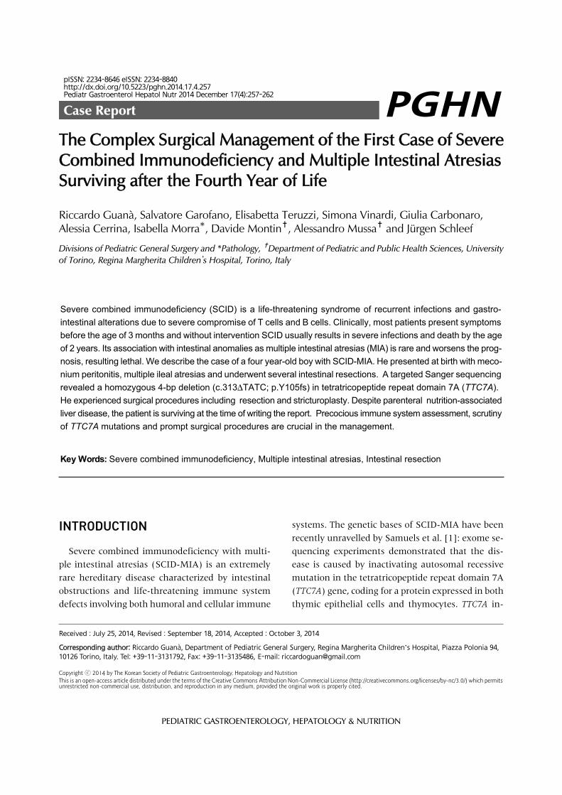
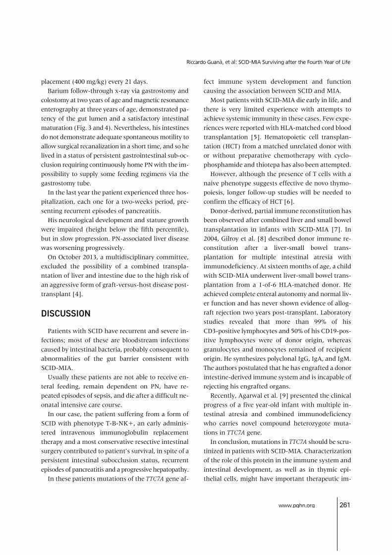
Fig. 1. Intra-operative findingsat the time of the first lapa-rotomy and intestinal reco-nstruction.
activation lead to severe lymphoid depletion in the thymus and peripheral lymphoid tissues from pa-tients with SCID-MIA. Loss-of- function mutation in TTC7A was also associated with very early onset in-flammatory bowel diseases [2]. Functional studies demonstrated that the mutations cause defects in enterocytes and T cells that lead to severe apoptotic enterocolitis, with or without immunodeficiency [2]. Study by Chen et al. [3] confirmed these ob-servations in eight unrelated cases. Our report describes the long and difficult ther-apeutic management of one patient among these eight that to our knowledge nowadays is the lon-gest-surviving.
CASE REPORT
The patient was admitted to our hospital at birth, on July 2010, with diagnosis of abdominal distension and respiratory failure. The baby was born to parents of Bosnian origin, from elective caesarean section at the 34th week of gestation, with 2.710 g neonatal weight, 45 cm length, 32.4 cm head circumference, and 5/8 Apgar score at 1 and 5 minutes, respectively.
His general condition at birth was poor and the baby underwent nasotracheal intubation. At a prenatal ul-trasonography, intestinal perforation consistent with duodenal or jejunal atresia associated with poly-hydramnios was suspected. At birth an echocardiog-raphy demonstrated a patent arterial duct with bidir-ectional shunting and patent foramen ovale with a negligible left to right shunt. Abdominal X-ray evi-denced a clear-cut gastric air bubble, but absence of intestinal gas in all remaining abdominal fields in-dicating duodenal or jejunal atresia. On an emergent laparotomy, meconium peritonitis due to intestinal perforation was accompanied by 16 intestinal lesions of intestinal atresias and severe stenosis with in-testinal malrotation. These traits were distanced by minimum of 3 cm and maximum of 15 cm. The sole trait without stenosis was the first jejunal loop. In ad-dition, cecal atresia was present (Fig. 1). Serials of re-section and anastomosis on three loci of ileal atresia (each one of 6, 8, and 11 cm) followed by five ileal strictureplasties were performed. Also, colic atresia and ileocecal atresia were treated with an “en bloc” re-section, including cecal appendix, using a gastro-in-testinal anastomosis stapler and creating an ileo-colic

www.pghn.org 259
Riccardo Guanà, et al: SCID-MIA Surviving after the Fourth Year of Life
Table 1. Immunological Features at 10 Months of Age
Parameter Patient Normal range
Lymphocytes (cells/μL)CD3+ (cells/μL)CD4+ (cells/μL)CD8+ (cells/μL)CD19+ (cells/μL)CD16/56+ (cells/μL)RTE (CD4+45RA+31+ cells/μL)Proliferation to PHA (SI)IgG (mg/dL)IgA (mg/dL)IgM (mg/dL)
62016913517
231235
157
<6<25
4,000-6,1002,400-3,3001,600-2,200
820-1,6001,000-1,600
270-1,100800-4,000
>67223-1,099
0-730-100
RTE: recent thymic emigrants lymphocytes, PHA: phytohemag-glutinin, SI: stimulation index, Ig: immunoglobulin.
termino-lateral anastomosis. A colostomy at the de-scending colon was followed due to additional atresia at descending and sigmoid colons. The remaining ileum was estimated to be approximately 70 cm long. Histopathological examination showed the classic as-pect of intestinal lumen fibrosis. In the post-operative period, parenteral nutrition (PN) was established through a central venous catheter. On August 2010 bowel obstruction due to multiple adhesions neces-sitated treatment with intestinal anastomosis. At the second laparotomy, tenacious adhesions were present between ileal loops and the caudate lobe of the liver that caused an obstruction of the pyloric region. Adhesiolysis of all adhesions was performed. On September 2010, a further episode of bowel ob-struction occurred. At the laparotomy, tenacious ad-hesions that compacted all the intestinal loops to form a rigid coil were present and adhesiolysis on each lesion was performed. A CoSeal surgical sealant (Baxter Healthcare Corporation, Deerfield, IL, USA) was applied on the intestinal loop at the end of the op-eration for prevent spontaneous intestinal adhesion. On September 2010, we positioned a permanent cen-tral venous catheter in the left subclavian vein for long term PN. On March 2011, peritoneal adhesiolysis was necessary for a newly developed bowel occlusion. The lysis of adhesions allowed the highlighting of a discrete maturation of the intestine in terms of size and length as well as the presence of numerous ileal stenotic areas. Performing of seven enteroplasties in Heineke-Mikulizt fashion was done in corre-spondence at the ileal stenotic traits. We also posi-tioned naso-jejunal two-way polyurethane enteral tube. On March 2011, dehiscence of the surgical wound occurred. Wound revision and closure with in-terposition of mesh was necessary. Due to infection on the abdominal wall prosthetic patch, on April 2011, it was necessary to remove the device from the abdomen and to close the abdominal wound. During the operation, under fluoroscopic control, Iomeron (Bracco Imaging SpA, Milano, Italy) was adminis-tered through the stoma, allowing the highlighting of a complete channeling of the intestine up to the duo-denum and the absence spillage of the contrast agent.
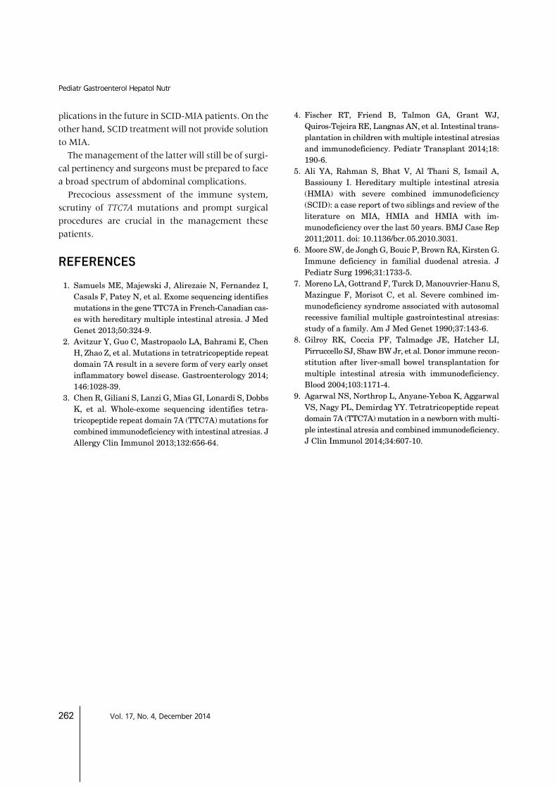
A radiographic intestinal transit was completed, ad-ministering contrast material through the nasogastric tube allowing an assessment of the patency of stom-ach, duodenum and remaining trait of the bowel. The clinical course was complicated by multiple epi-sodes of sepsis caused by Pseudomonas aeruginosa, one with the development of an abdominal abscess, and Candida albicans. Laboratory investigations at the age of ten months showed severe T- and B-cell lymphope-nia, absent proliferation to phytohemagglutinin and anti-CD3, and profound hypogammaglobulinemia (the nadir value of serum levels of immunoglobulin (Ig) G was 57, IgA and IgM values were undetectable), confirming the diagnosis of SCID (Table 1). Anti-fun-gal, anti-viral and anti-bacterial prophylaxes were started, together with regular intravenous immu-noglobulin replacement therapy. Whole-exome sequencing was performed on the patient and healthy family members. The analysis on the patient identified a novel deleterious variant in the gene TTC7A. Sanger sequencing to validate the deleterious variant was performed and revealed a ho-mozygous 4-bp deletion (c.313ΔTATC) in exon 2 of TTC7A (Fig. 2). Therefore, the variant is predicted to lead to a frameshift mutation at codon 105 (p.Y105fs) and subsequent nonsense-mediated decay of the re-sulting ribonucleic acid transcript, which means the strong possibility of SCID-MIA. The deletion was also present in the parents but

260 Vol. 17, No. 4, December 2014
Pediatr Gastroenterol Hepatol Nutr
Fig. 2. Sanger sequencing revealed a homozygous 4-bp deletion(c.313ΔTATC) in exon 2 of TTC7A.
Fig. 4. Magnetic resonance enterography at 3 years of age, evidencing absence of intestinal stenosis or dilation (A) and progressive develop-ment and growth of the gut(B).
Fig. 3. Barium follow-throughx-ray via gastrostomy and colostomy at 2 years of age, demonstrating patency of the gut lumen (A) and a satis-factory intestinal maturation (B).
with a heterozygote mutation and no anomaly was found in the brothers. A multidisciplinary team meeting involving im-munologist, infectious disease specialist, anaesthesi-ologist, pediatric surgeon and social worker was organized. The team explained to the parents the poor prognosis of their baby, and it was decided to proceed with only supportive therapies. A percutaneous endoscopic gastrostomy was per-formed on September 2011 in order to decompress the stomach and small bowel and to avoid the discomfort of the nasogastric tube. In the following months the clinical improvement of the patient allowed a pro-tected hospital discharge. The patient was discharged on May 2012 and followed-up monthly. His therapy consisted of acyclovir, fluconazole, antibiotic prophy-laxis administered daily, and immunoglobulin re-
www.pghn.org 261
Riccardo Guanà, et al: SCID-MIA Surviving after the Fourth Year of Life
placement (400 mg/kg) every 21 days. Barium follow-through x-ray via gastrostomy and colostomy at two years of age and magnetic resonance enterography at three years of age, demonstrated pa-tency of the gut lumen and a satisfactory intestinal maturation (Fig. 3 and 4). Nevertheless, his intestines do not demonstrate adequate spontaneous motility to allow surgical recanalization in a short time, and so he lived in a status of persistent gastrointestinal sub-oc-clusion requiring continuously home PN with the im-possibility to supply some feeding regimens via the gastrostomy tube. In the last year the patient experienced three hos-pitalization, each one for a two-weeks period, pre-senting recurrent episodes of pancreatitis. His neurological development and stature growth were impaired (height below the fifth percentile), but in slow progression. PN-associated liver disease was worsening progressively. On October 2013, a multidisciplinary committee, excluded the possibility of a combined transpla-ntation of liver and intestine due to the high risk of an aggressive form of graft-versus-host disease post- transplant [4].
DISCUSSION
Patients with SCID have recurrent and severe in-fections; most of these are bloodstream infections caused by intestinal bacteria, probably consequent to abnormalities of the gut barrier consistent with SCID-MIA. Usually these patients are not able to receive en-teral feeding, remain dependent on PN, have re-peated episodes of sepsis, and die after a difficult ne-onatal intensive care course. In our case, the patient suffering from a form of SCID with phenotype T-B-NK+, an early adminis-tered intravenous immunoglobulin replacement therapy and a most conservative resective intestinal surgery contributed to patient’s survival, in spite of a persistent intestinal subocclusion status, recurrent episodes of pancreatitis and a progressive hepatopathy. In these patients mutations of the TTC7A gene af-
fect immune system development and function causing the association between SCID and MIA. Most patients with SCID-MIA die early in life, and there is very limited experience with attempts to achieve systemic immunity in these cases. Few expe-riences were reported with HLA-matched cord blood transplantation [5]. Hematopoietic cell transplan-tation (HCT) from a matched unrelated donor with or without preparative chemotherapy with cyclo-phosphamide and thiotepa has also been attempted. However, although the presence of T cells with a naive phenotype suggests effective de novo thymo-poiesis, longer follow-up studies will be needed to confirm the efficacy of HCT [6]. Donor-derived, partial immune reconstitution has been observed after combined liver and small bowel transplantation in infants with SCID-MIA [7]. In 2004, Gilroy et al. [8] described donor immune re-constitution after a liver-small bowel trans-plantation for multiple intestinal atresia with immunodeficiency. At sixteen months of age, a child with SCID-MIA underwent liver-small bowel trans-plantation from a 1-of-6 HLA-matched donor. He achieved complete enteral autonomy and normal liv-er function and has never shown evidence of allog-raft rejection two years post-transplant. Laboratory studies revealed that more than 99% of his CD3-positive lymphocytes and 50% of his CD19-pos-itive lymphocytes were of donor origin, whereas granulocytes and monocytes remained of recipient origin. He synthesizes polyclonal IgG, IgA, and IgM. The authors postulated that he has engrafted a donor intestine-derived immune system and is incapable of rejecting his engrafted organs. Recently, Agarwal et al. [9] presented the clinical progress of a five year-old infant with multiple in-testinal atresia and combined immunodeficiency who carries novel compound heterozygote muta-tions in TTC7A gene. In conclusion, mutations in TTC7A should be scru-tinized in patients with SCID-MIA. Characterization of the role of this protein in the immune system and intestinal development, as well as in thymic epi-thelial cells, might have important therapeutic im-
262 Vol. 17, No. 4, December 2014
Pediatr Gastroenterol Hepatol Nutr
plications in the future in SCID-MIA patients. On the other hand, SCID treatment will not provide solution to MIA. The management of the latter will still be of surgi-cal pertinency and surgeons must be prepared to face a broad spectrum of abdominal complications. Precocious assessment of the immune system, scrutiny of TTC7A mutations and prompt surgical procedures are crucial in the management these patients.
REFERENCES
1. Samuels ME, Majewski J, Alirezaie N, Fernandez I, Casals F, Patey N, et al. Exome sequencing identifies mutations in the gene TTC7A in French-Canadian cas-es with hereditary multiple intestinal atresia. J Med Genet 2013;50:324-9.
2. Avitzur Y, Guo C, Mastropaolo LA, Bahrami E, Chen H, Zhao Z, et al. Mutations in tetratricopeptide repeat domain 7A result in a severe form of very early onset inflammatory bowel disease. Gastroenterology 2014; 146:1028-39.
3. Chen R, Giliani S, Lanzi G, Mias GI, Lonardi S, Dobbs K, et al. Whole-exome sequencing identifies tetra-tricopeptide repeat domain 7A (TTC7A) mutations for combined immunodeficiency with intestinal atresias. J Allergy Clin Immunol 2013;132:656-64.
4. Fischer RT, Friend B, Talmon GA, Grant WJ, Quiros-Tejeira RE, Langnas AN, et al. Intestinal trans-plantation in children with multiple intestinal atresias and immunodeficiency. Pediatr Transplant 2014;18: 190-6.
5. Ali YA, Rahman S, Bhat V, Al Thani S, Ismail A, Bassiouny I. Hereditary multiple intestinal atresia (HMIA) with severe combined immunodeficiency (SCID): a case report of two siblings and review of the literature on MIA, HMIA and HMIA with im-munodeficiency over the last 50 years. BMJ Case Rep 2011;2011. doi: 10.1136/bcr.05.2010.3031.
6. Moore SW, de Jongh G, Bouic P, Brown RA, Kirsten G. Immune deficiency in familial duodenal atresia. J Pediatr Surg 1996;31:1733-5.
7. Moreno LA, Gottrand F, Turck D, Manouvrier-Hanu S, Mazingue F, Morisot C, et al. Severe combined im-munodeficiency syndrome associated with autosomal recessive familial multiple gastrointestinal atresias: study of a family. Am J Med Genet 1990;37:143-6.
8. Gilroy RK, Coccia PF, Talmadge JE, Hatcher LI, Pirruccello SJ, Shaw BW Jr, et al. Donor immune recon-stitution after liver-small bowel transplantation for multiple intestinal atresia with immunodeficiency. Blood 2004;103:1171-4.
9. Agarwal NS, Northrop L, Anyane-Yeboa K, Aggarwal VS, Nagy PL, Demirdag YY. Tetratricopeptide repeat domain 7A (TTC7A) mutation in a newborn with multi-ple intestinal atresia and combined immunodeficiency. J Clin Immunol 2014;34:607-10.


![2234 McConnell Grace[1] OK](https://img.pdfslide.net/doc/110x75/577cdd8e1a28ab9e78ad4741/2234-mcconnell-grace1-ok.jpg)