Embed Size (px)
Citation preview
Kathleen W. Pojunas' David L. Daniels Alan L. Williams
M. Kristin Thorsen Victor M. Haughton
This article appears in the March/April 1986 issue of AJNR and the June 1986 issue of AJR.
Received January 30, 1985; accepted after revision August 5, 1985.
1 All authors: Department of Radiology, Medical College of Wisconsin, Froedtert Memorial Lutheran Hospital, 9200 W. Wisconsin Ave., Milwaukee, WI 53226. Address reprint requests to K. W. Pojunas.
AJNR 7:271-274, March/April 1986 0195-6108/86/0702-0271 © American Society of Neuroradiology
Pituitary and Adrenal CT of Cushing Syndrome
271
Determination of the site of excessive hormone production in Cushing syndrome is possible with biochemical tests in 80% of cases. High-resolution CT of both the pituitary and adrenal glands was used to evaluate eight patients with surgically verified ACTHsecreting pituitary microadenomas and one patient with ectopic Cushing syndrome. Three ACTH-secreting microadenomas were demonstrated by CT. Adrenal CT was normal in six of the eight patients with pituitary tumors. The patient with ectopic ACTH production had mild unilateral adrenal gland enlargement and a normal pituitary CT scan. Normal adrenal or pituitary CT scans do not exclude Cushing syndrome.
The diagnosis of Cushing syndrome and the differentiation of pituitary, adrenal, and ectopic (i .e., outside the pituitary or adrenal) sites of excess ACTH or cortisol production are usually possible with endocrinologic tests [1, 2] . However, in 20% of cases, pituitary and ectopic sites of excess ACTH production cannot be distinguished from one another by biochemical testing [2, 3].
The earliest experience with CT of the sella suggested that ACTH-secreting microadenomas had a different appearance than prolactin-secreting tumors [4, 5]. Thin axial sections with sagittal and coronal reformation were much less reliable in localizing ACTH-secreting than prolactin-secreting pituitary tumors [6 , 7]. The results of imaging ACTH-secreting microadenomas with optimal techniques and direct coronal imaging have not been described.
We evaluated the efficacy of high-resolution CT imaging of the pituitary gland in patients with surgically verified corticotropin-secreting neoplasms to determine whether ACTH-secreting microadenomas could be reliably detected. The adrenal glands of each patient were also studied with CT and the results correlated with pituitary images, laboratory data, and clinical findings to help define the role of CT in differentiating ectopic and pituitary sites of excess ACTH production.
Materials and Methods
Cases selected for review had laboratory confirmation of Cushing syndrome, pathologic verification of a corticotropin-secreting neoplasm , and high-resolution CT studies of the pituitary and adrenal glands. Laboratory confirmation included at least elevated plasma cortisol, urinary 17-ketogenic steroids (17 KGS) , 17-hydroxycorticosteroids (17-0HCS) , and failure of low-dose (1 mgj24 hr) dexamethasone to suppress urinary 17 -OHCS and plasma cortisol.
CT scans were obtained on a General Electric 8800 or 9800 scanner. For pituitary imaging , 42 g of either 30% or 60% iodinated contrast material was injected at about 30 mljmin into an antecubital vein via a 19 gauge needle whi le the patient was supine on the scanner table. When about half of the contrast material (21 g I) had been infused, the patient was pOSitioned with the head hyperextended in a head-holder for direct coronal imaging of the pituitary gland. A lateral localizer image was obtained from which slice locations and gantry angulation were selected by means of a cursor and computer program. Contiguous images with 1.5 mm beam collimation were obtained from the tuberculum to the dorsum sellae. Technical factors for the
272 POJUNAS ET AL. AJNR:7, March/April1986
8
8800 scanner were 576 views, 120 kV, 600 mA, 9.6 sec scan time, and 3.3 msec pulse width code; for the 9800 scanner, 120 kV, 200 mA, and 4 sec scan time were used. Single-level dynamic scans of the pituitary gland were obtained in three patients (8].
For CT imaging of the adrenal glands, 5-mm-thick contiguous axial scans were obtained. Intravenous and oral contrast material were not routinely used. Technical factors for the 8800 scanner were 576 views , 5.6 sec scan time, 120 kV, 320 mA, and 3 msec pulse width code; for the 9800 scanner, 120 kV, 170 mA, and 3 sec scan time were selected.
Pituitary gland height and appearance and infundibulum position were obtained from radiographic reports dictated by a neuroradiologist (D . L. D., A. L. W., orV. M. H.) at the time of the initial examination. All pituitary scans were reviewed independently by a fourth neuroradiologist (K. W. P.) . Adrenal CT scans were reviewed for this report by an abdominal CT radiologist (M. K. T.).
Results
Nine patients, all women 21-43 years old, constituted the study group. Eight subsequently had transsphenoidal resection of an ACTH-secreting pituitary microadenoma. The ninth had Cushing syndrome presumably secondary to liver metastases producing corticotropin-releasing factor (CRF).
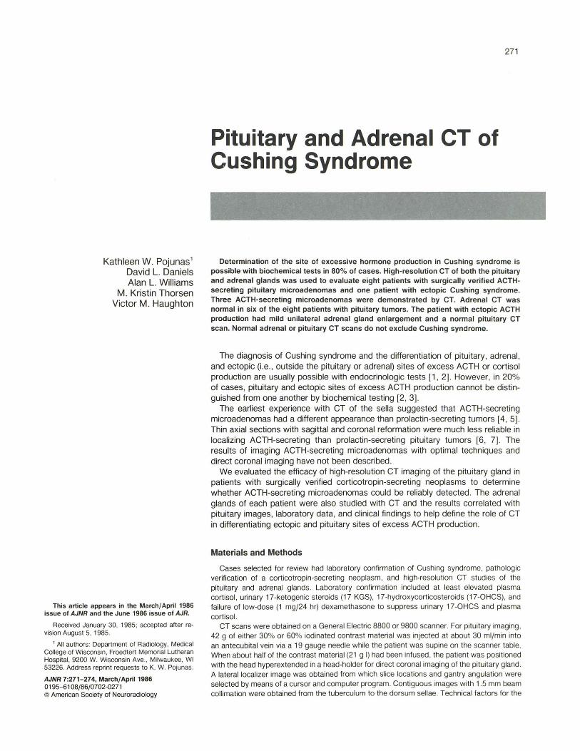
Fig. 1.-A, Well defined region of decreased contrast enhancement within pituitary gland (arrow) to left of midline. Surgical exploration revealed "mucoid" basophilic adenoma at site of CT abnormality. B, Single-level dynamic scan of this patient revealed normal midline "pituitary tuft" (arrow) .
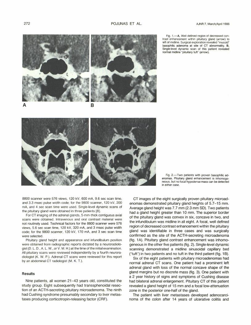
Fig. 2.- Two patients with proven basophilic adenomas. Pituitary gland enhancement is inhomogeneous, but no focal hypodense mass can be detected in either case.
CT images of the eight surgically proven pituitary microadenomas demonstrated pituitary gland heights of 5.7-15 mm. Average gland height was 7.7 mm (2 .3 mm SD). Two patients had a gland height greater than 10 mm. The superior border of the pituitary gland was convex in six, concave in two, and the infundibulum was midline in all eight. A focal , well defined region of decreased contrast enhancement within the pituitary gland was identifiable in three cases and was surgically confirmed as the site of the ACTH-secreting microadenoma (fig. 1 A) . Pituitary gland contrast enhancement was inhomogeneous in the other five patients (fig . 2). Single-level dynamic scanning demonstrated a midline sinusoidal capillary bed ("tuft") in two patients and no tuft in the third patient (fig. 1 B).
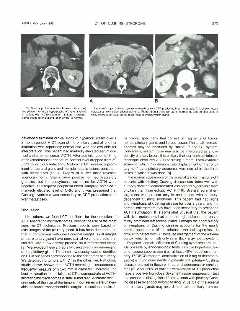
Six of the eight patients with pituitary microadenomas had normal adrenal CT scans. One patient had a prominent left adrenal gland with loss of the normal concave shape of the gland margins but no discrete mass (fig. 3). One patient with a 2 year history of signs and symptoms of Cushing disease had bilateral adrenal enlargement. Pituitary CT of this patient revealed a gland height of 15 mm and a focal low-attenuation zone in the posterior one-half of the gland.
The patient with liver metastases developed adenocarcinoma of the colon after 14 years of ulcerative colitis and
AJNR:7, March/April 1986 CT OF CUSHING SYNDROME 273
A B
Fig. 3.-Loop of unopacified bowel (solid arrow) lies adjacent to mildly hyperplastic left adrenal gland in patient with ACTH-secreting pituitary microadenoma. Right adrenal gland (open arrow) is normal.
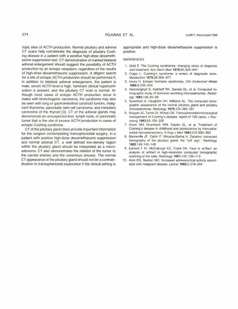
Fig. 4.-Ectopic Cushing syndrome resulting from CRF-producing liver metastasis. A, Multiple hepatic metastases from colon adenocarcinoma. Right adrenal gland (arrow) is normal. B, Left adrenal gland is mildly enlarged (arrow) , but no focal mass is evident within gland.
developed fulminant clinical signs of hypercortisolism over a 2 month period. A CT scan of the pituitary gland at another institution was reportedly normal and was not available for interpretation. This patient had markedly elevated serum cortisol and a normal serum ACTH. After administration of 8 mg of dexamethasone, her serum cortisol level dropped from 59 f.Lg/dl to 22 (63% reduction). Abdominal CT revealed a prominent left adrenal gland and multiple hepatic lesions consistent with metastases (fig. 4). Biopsy of a liver mass revealed adenocarcinoma. Stains were positive for neurosecretory granules, but immunocytochemical stains for ACTH were negative. Subsequent peripheral blood sampling revealed a markedly elevated level of CRF, and it was presumed that Cushing syndrome was secondary to CRF production from liver metastases.
Discussion
Like others, we found CT unreliable for the detection of ACTH-secreting microadenomas, despite the use of the most sensitive CT techniques. Previous reports were based on axial images of the pituitary gland. It has been demonstrated that in comparison with direct coronal images, axial images of the pituitary gland have more partial-volume artifacts that can simulate a low-density process on a reformatted image [9]. We avoided these artifacts by using direct coronal imaging of the pituitary gland . The three low-density lesions identified on CT in our series corresponded to the adenomas at surgery. We detected no tumors with CT in the other five. Pathologic studies have shown that ACTH-secreting microadenomas frequently measure only 2-3 mm in diameter. Therefore, the best explanation for the failure of CT to demonstrate all ACTHsecreting microadenomas is small tumor size. Accurate measurements of the size of the tumors in our series were unavailable because transsphenoidal surgical resection results in
pathologic specimens that consist of fragments of tumor, normal pituitary gland, and fibrous tissue. The small microaden om as may be obscured by "noise" in the CT system. Conversely, system noise may also be interpreted as a lowdensity pituitary lesion. It is unlikely that our contrast infusion technique obscured ACTH-secreting tumors. Even dynamic scanning, which may demonstrate displacement of the "pituitary tuft" by a pituitary adenoma, was normal in the three cases in which it was done [8].
The normal appearance of the adrenal glands in six of eight patients with pituitary Cushing disease correlates well with autopsy data that demonstrated less adrenal hyperplasia from pituitary than from ectopic ACTH [10]. Bilateral adrenal enlargement was present only in one patient with pituitarydependent Cushing syndrome. This patient had had signs and symptoms of Cushing disease for over 2 years , and the adrenal enlargement may have been secondary to prolonged ACTH stimulation. It is somewhat unusual that the patient with liver metastases had a normal right adrenal and only a mildly prominent left adrenal gland. Perhaps the short history of symptoms of Cushing disease accounts for the nearly normal appearance of the adrenals. Adrenal hyperplasia is difficult to detect with CT because enlargement of the adrenal cortex, which is normally only 2 mm thick, may not be evident.
Diagnosis and classification of Cushing syndrome are usually possible by endocrinologic tests. Positive high-dose dexamethasone suppression (i .e., at least 40% reduction of urinary 17 -OHCS after oral administration of 8 mg of dexamethasone) is found consistently in patients with pituitary Cushing disease, but not in those with adrenal adenomas or carcinomas [2]. About 20% of patients with ectopic ACTH production have a positive high-dose dexamethasone suppression test and cannot be distinguished from patients with pituitary Cushing disease by endocrinologic testing [2, 3] . CT of the adrenal and pituitary glands may help differentiate pituitary from ec-
274 POJUNAS ET AL. AJNR :7, March/April 1986
topic sites of ACTH production. Normal pituitary and adrenal CT scans help corroborate the diagnosis of pituitary Cushing disease in a patient with a positive high-dose dexamethasone suppression test. CT demonstration of marked bilateral adrenal enlargement should suggest the possibility of ACTH production by an ectopic neoplasm, regardless of the results of high-dose dexamethasone suppression. A diligent search for a site of ectopic ACTH production should be performed if, in addition to bilateral adrenal enlargement, the patient is male, serum ACTH level is high , fulminant clinical hypercortisolism is present, and the pituitary CT scan is normal. Although most cases of ectopic ACTH production occur in males with bronchogenic carcinoma, the syndrome may also be seen with lung or gastrointestinal carcinoid tumors, malignant thymoma, pancreatic islet-cell carcinoma, and medullary carcinoma of the thyroid [3]. CT of the adrenal glands may demonstrate an unsuspected liver, lymph node, or pancreatic tumor that is the site of excess ACTH production in cases of ectopic Cushing syndrome.
CT of the pituitary gland does provide important information for the surgeon contemplating transsphenoidal surgery. In a patient with positive high-dose dexamethasone suppression and normal adrenal CT, a well defined low-denSity region within the pituitary gland should be interpreted as a microadenoma. CT also demonstrates the relation of the tumor to the carotid arteries and the cavernous sinuses. The normal CT appearance of the pituitary gland should not be a contraindication to transsphenoidal exploration if the clinical setting is
appropriate and high-dose dexamethasone suppression is positive.
REFERENCES
1. Gold E. The Cushing syndromes: changing views of diagnosis and treatment. Ann Intern Med 1979;90 :829-844
2. Crapo L. Cushing 's syndrome: a review of diagnostic tests. Metabolism 1979;28:955-977
3. Imura H. Ectopic hormone syndromes. Clin Endocrinol Metab 1980;9: 235-249
4. Hemminghytt S, Kalkhoff RK, Daniels DL, et al. Computed tomographic study of hormone secreting microadenomas. Radiology 1983;146:65-69
5. Syvertson A, Haughton VH, Williams AL. The computed tomographic appearance of the normal pituitary gland and pituitary microadenomas. Radiology 1979;133: 385-391
6. Boggan JE, Tyrrell JV, Wilson CB. Transsphenoidal microsurgical management of Cushing's disease: report of 100 cases. J Neurosurg 1983;59 : 195-200
7. Styne OM, Grumbach MM, Kaplan SL, et al. Treatment of Cushing's disease in chi ldhood and adolescence by transsphenoidal microadenectomy. N EnglJ Med 1984;310:889-893
8. Bonneville JF, Cattin F, Moussa-Bacha H. Dynamic computed tomography of the pituitary gland: the "tuft sign. " Radiology 1983;149 : 145-148
9. Earnest F IV, McCullough EC, Frank DA. Face or artifact: an analysis of artifact in high-resolution computed tomographic scanning of the sella. Radiology 1981 ;140: 1 09-113
10. Alloh EN , Skelton MO. Increased adrenocortical activity associated with malignant disease. Lancet 1960;2:278-284