Embed Size (px)
Citation preview
Place and role of the pathology in the medicine.
Structure of pathology and methods of investigation
Dr. Attila Zalatnai

TREATMENTS
THE DOCTOR HIM (HER)SELF
Technical arsenal
Operative techniques
Pharmacology
DISEASES
Molecular diagnostics
Genetic diagnostics
Radiologic diagnostics
Laboratory diagnostics
Clinical picture
Abnormal regulations
Morphological alterations
THE HUMAN
BEING
Psychology
FunctionPhysiology, biochemistry, molecular
mechanisms
StructureMacroscopic anatomy, embryology,
histology, subcellular morphology,
molecular level
STRUCTURE OF THE MEDICINE
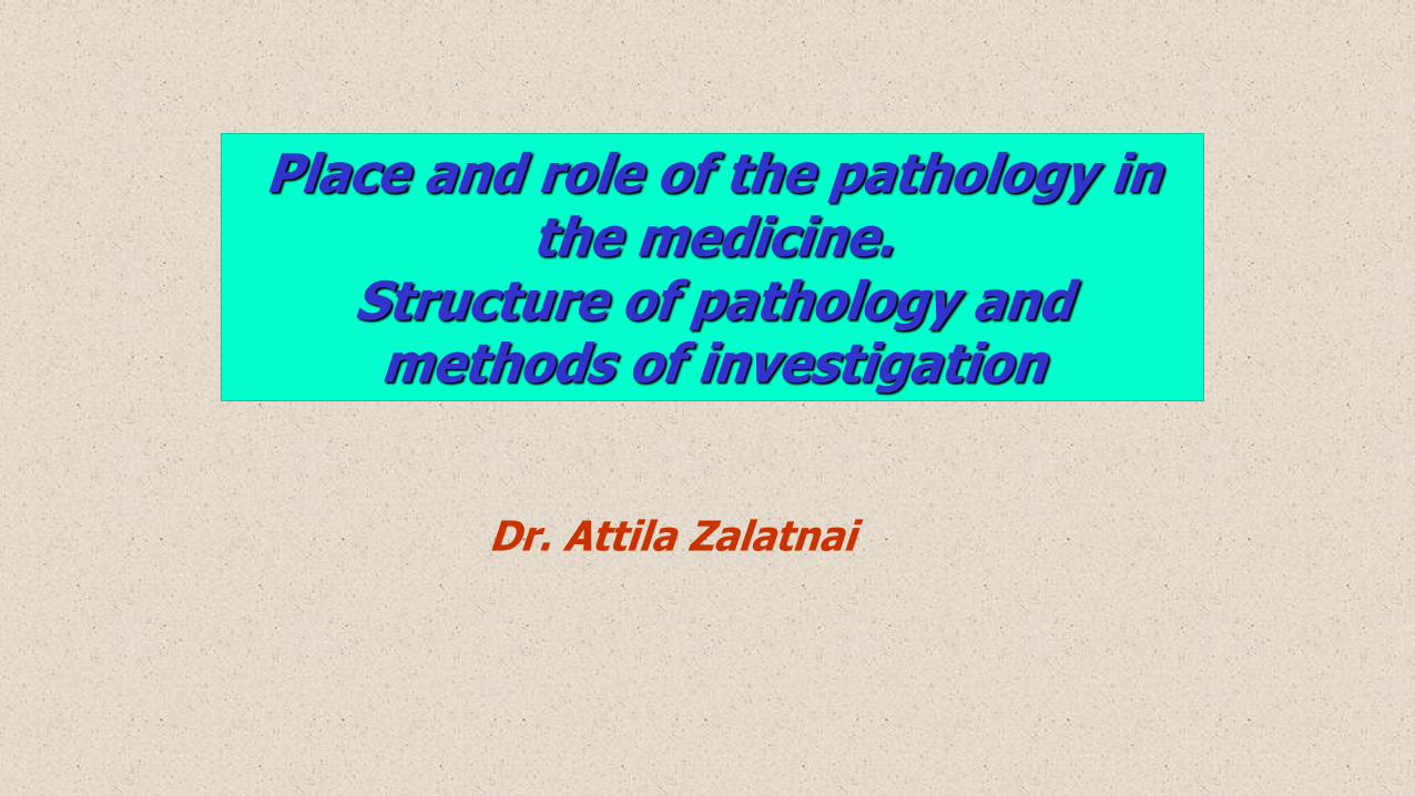
Clinical symptoms
DISEASE
Laboratory alterations
Pathological alterations
- macroscopic level
- light microscopic level
- ultrastructural level
- molecular level
Imaging methods
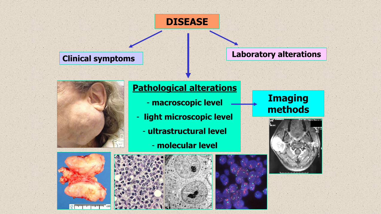
Without pathology there is no modern diagnostics!
Law: „all surgically removed tissue must be sent to pathology!”
routine: 8 % formalin
other studies: 0,9 % NaCl
never should be dry!
Except: tooth
normal placenta
nail
eye lens
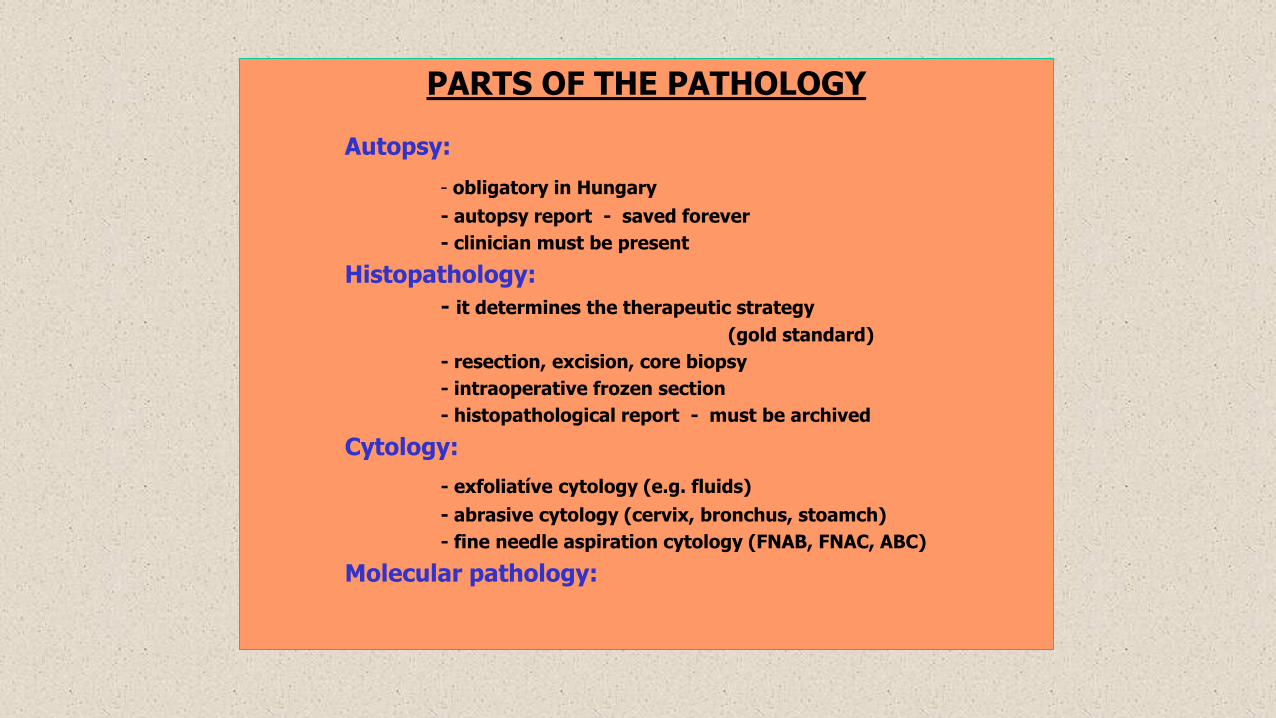
PARTS OF THE PATHOLOGY
Autopsy:
- obligatory in Hungary
- autopsy report - saved forever
- clinician must be present
Histopathology:
- it determines the therapeutic strategy
(gold standard)
- resection, excision, core biopsy
- intraoperative frozen section
- histopathological report - must be archived
Cytology:
- exfoliatíve cytology (e.g. fluids)
- abrasive cytology (cervix, bronchus, stoamch)
- fine needle aspiration cytology (FNAB, FNAC, ABC)
Molecular pathology:


Autopsy
- controls the clinical diagnoses (quality control)
- provides epidemiological data
- characterization of newly discovereddiseases
- better understanding of the naturalhistory of malignant diseases
- recognition of the morphologicalalterations (medical teaching)
- exclusion of forensic cases
- defence against the malpractice charges
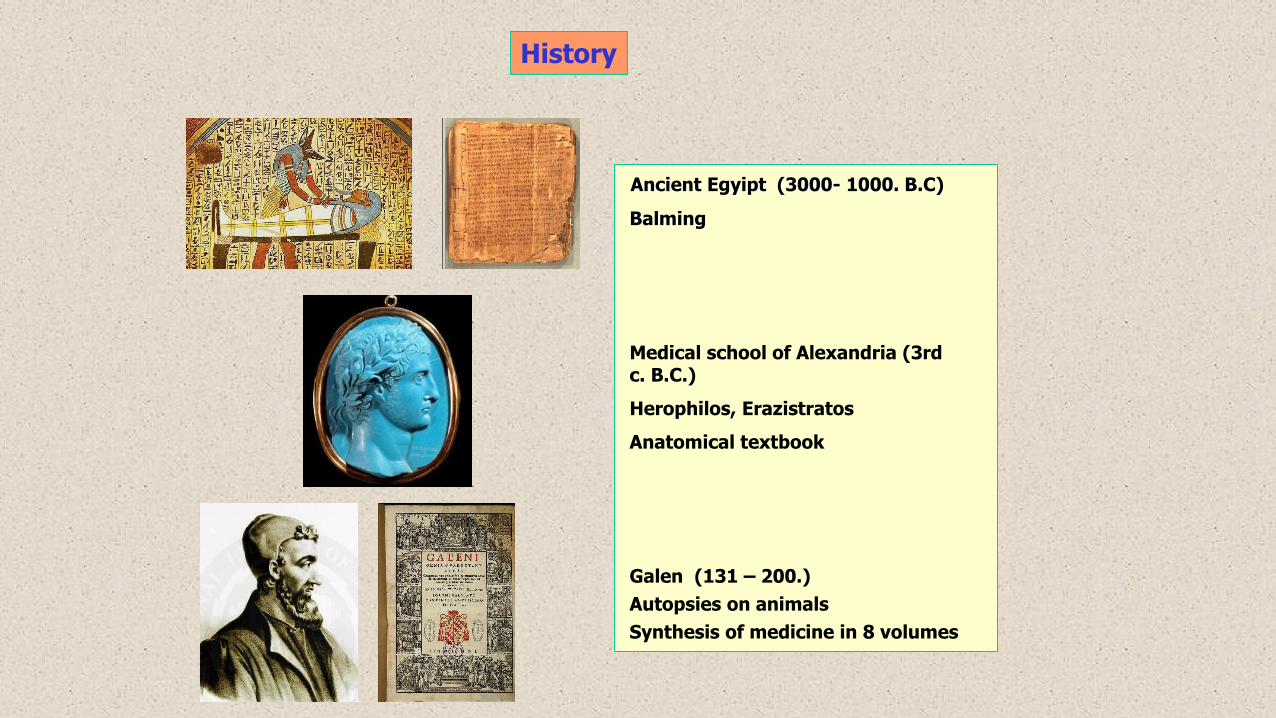
History
Ancient Egyipt (3000- 1000. B.C)
Balming
Medical school of Alexandria (3rd c. B.C.)
Herophilos, Erazistratos
Anatomical textbook
Galen (131 – 200.)
Autopsies on animals
Synthesis of medicine in 8 volumes
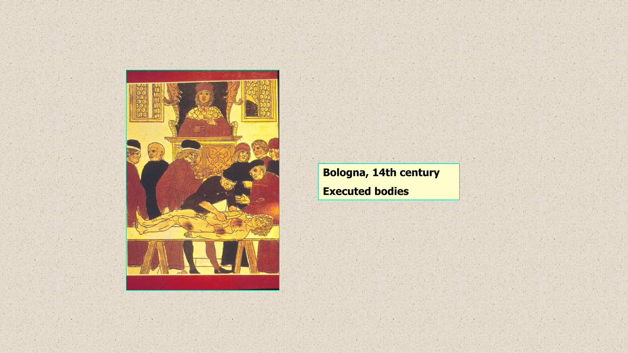
Bologna, 14th century
Executed bodies
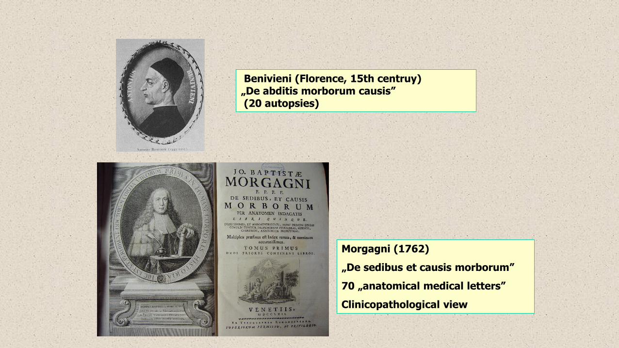
Benivieni (Florence, 15th centruy) „De abditis morborum causis”(20 autopsies)
Morgagni (1762)
„De sedibus et causis morborum”
70 „anatomical medical letters”
Clinicopathological view
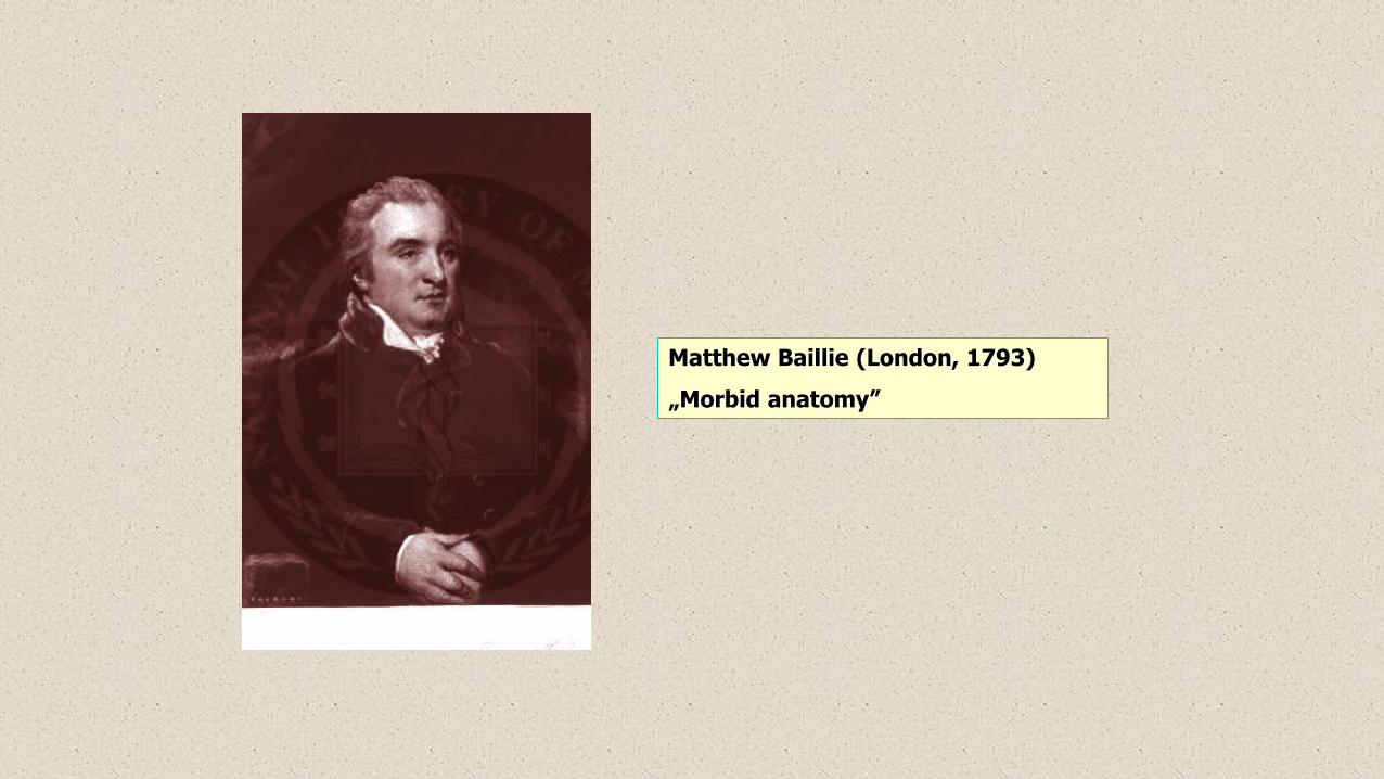
Matthew Baillie (London, 1793)
„Morbid anatomy”
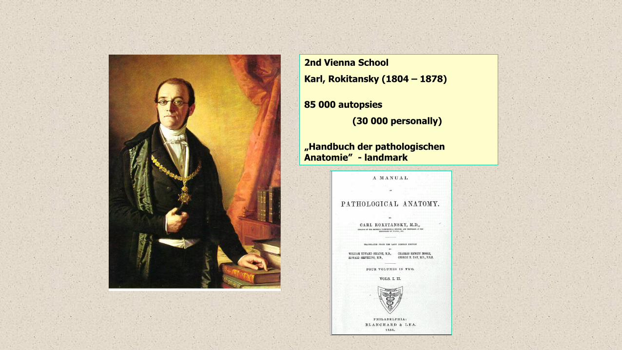
2nd Vienna School
Karl, Rokitansky (1804 – 1878)
85 000 autopsies
(30 000 personally)
„Handbuch der pathologischen Anatomie” - landmark
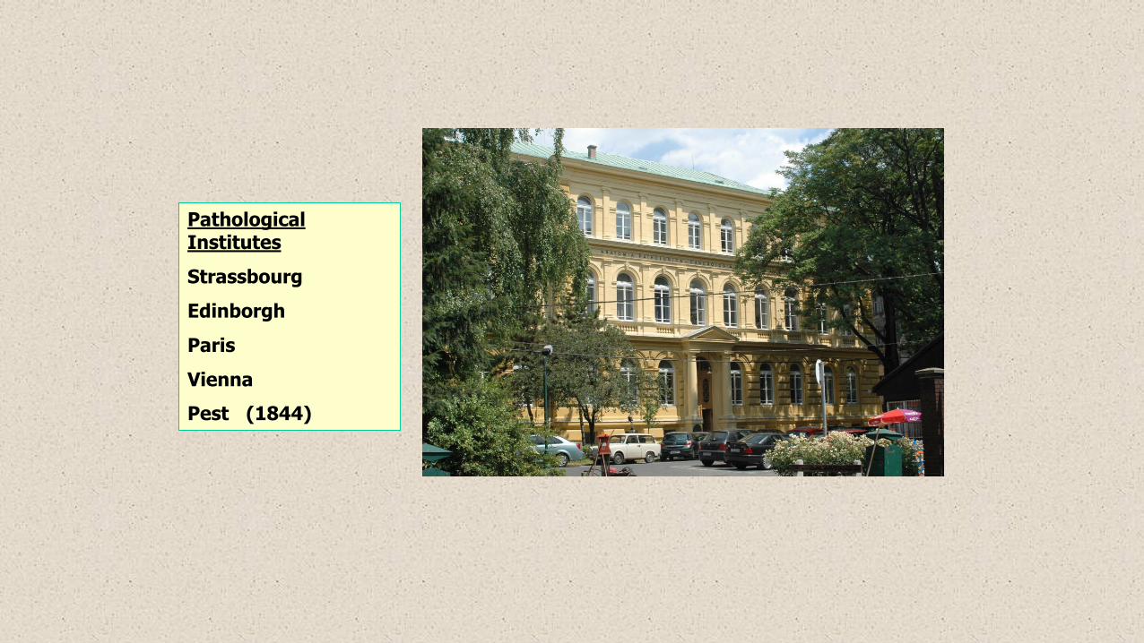
Pathological Institutes
Strassbourg
Edinborgh
Paris
Vienna
Pest (1844)

Histopathology
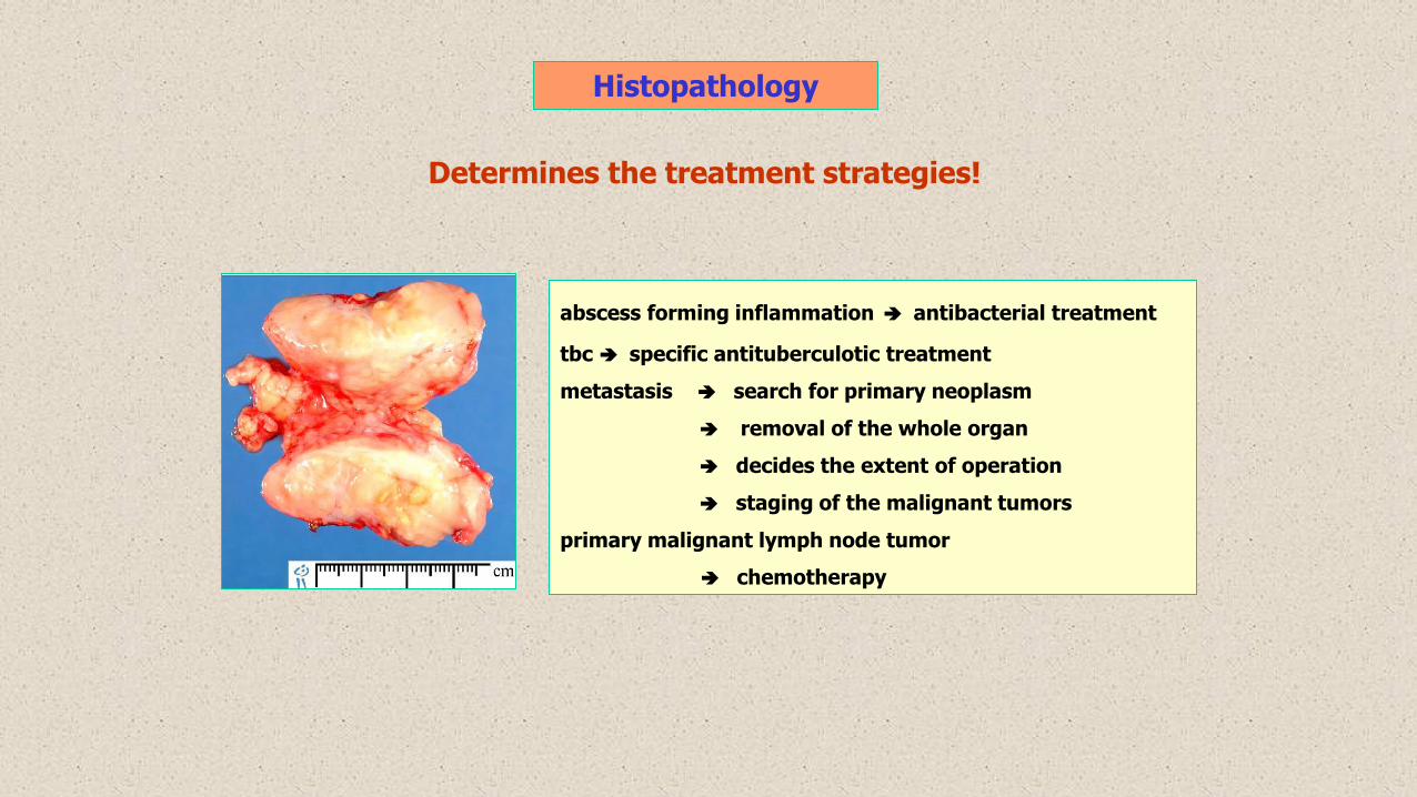
Determines the treatment strategies!
abscess forming inflammation antibacterial treatment
tbc specific antituberculotic treatment
metastasis search for primary neoplasm
removal of the whole organ
decides the extent of operation
staging of the malignant tumors
primary malignant lymph node tumor
chemotherapy

- characterization of the effusions
- screening for preneoplastic conditions
- recognition of the nature of focal lesions
(breast, thyroid, liver, salivary gland, brain…)
Cytology
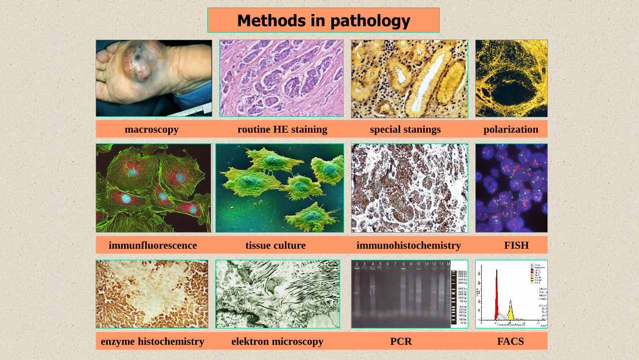
Methods in pathology
macroscopy routine HE staining special stanings polarization
immunfluorescence tissue culture immunohistochemistry FISH
enzyme histochemistry elektron microscopy PCR FACS

Adaptation tissue reactions
(atrophy, hypertrophy, hyperplasia,
metaplasia)
Dr. Attila Zalatnai
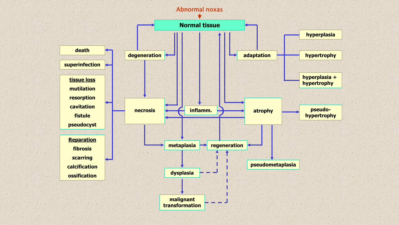
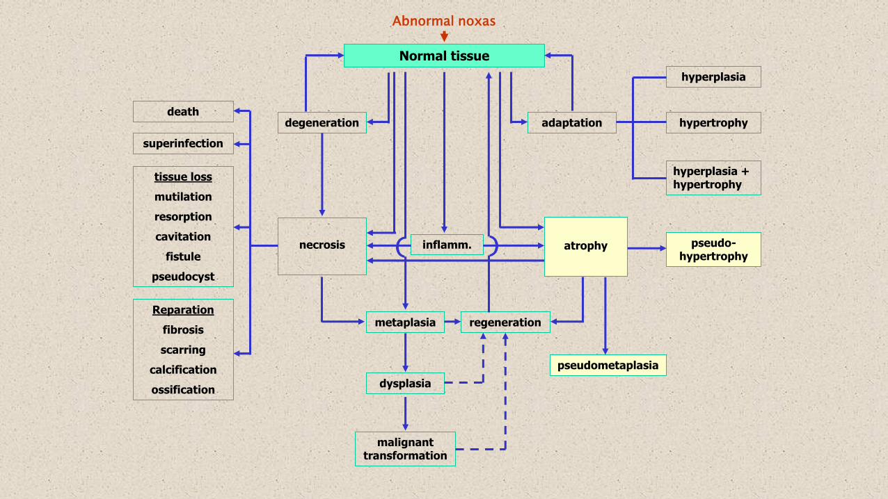
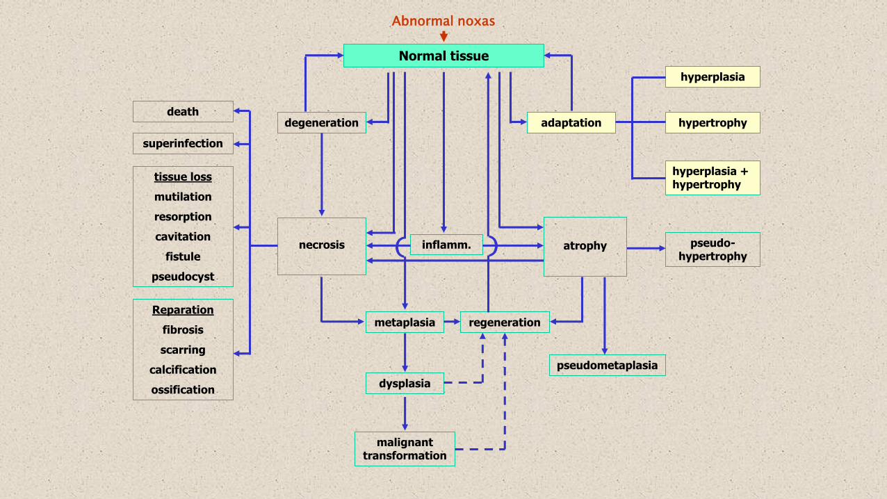
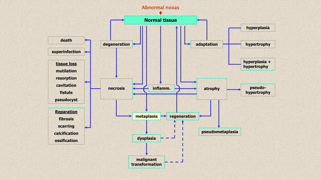
Normal tissue
Abnormal noxas
degeneration adaptation
hyperplasia
hypertrophy
hyperplasia + hypertrophy
necrosis
death
superinfection
tissue loss
mutilation
resorption
cavitation
fistule
pseudocyst
Reparation
fibrosis
scarring
calcification
ossification
metaplasia
inflamm. atrophy
pseudometaplasia
pseudo-hypertrophy
regeneration
dysplasia
malignant transformation
((

Degenerations (dystrophies)
Results of metabolic cellular damage, morphological apprearances of the intracellulardisturbances
Many different causes – nonspecific!
- tissue hypoxia, shift in pH
- intracellular ionic imbalance
- toxic damage ártalmak
- distant effect of severe generalized inflammations
- burn, freezing
- intoxications
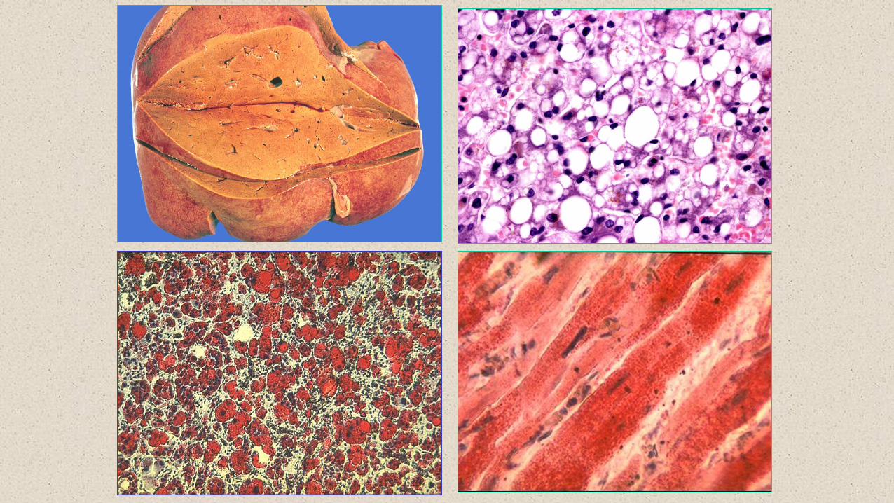
Parenchymatous degeneration (cloudy swelling)
Hydropic degeneration
Vacuolic degeneration
Fatty degeneration
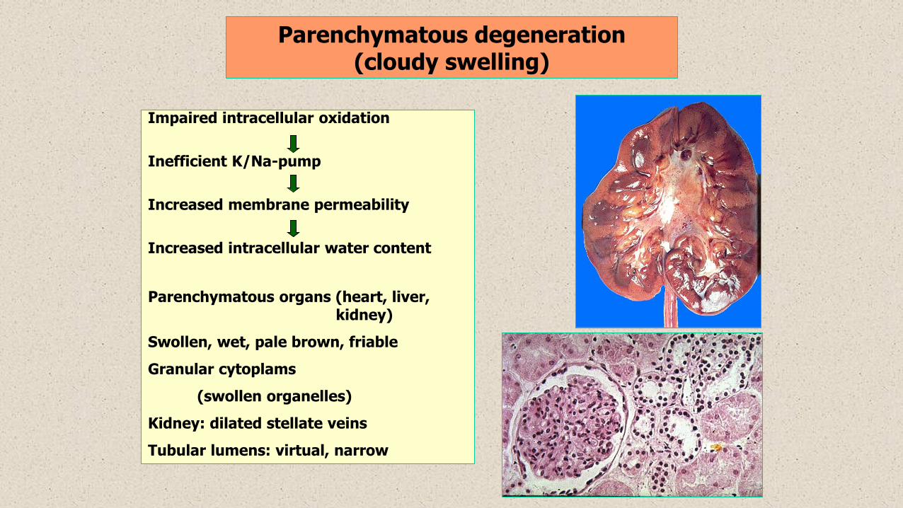
Parenchymatous degeneration (cloudy swelling)
Impaired intracellular oxidation
Inefficient K/Na-pump
Increased membrane permeability
Increased intracellular water content
Parenchymatous organs (heart, liver, kidney)
Swollen, wet, pale brown, friable
Granular cytoplams
(swollen organelles)
Kidney: dilated stellate veins
Tubular lumens: virtual, narrow
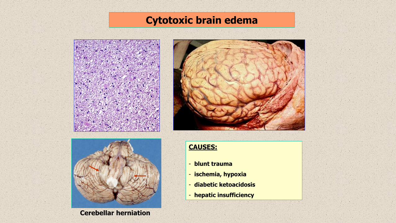
Cytotoxic brain edema
Cerebellar herniation
CAUSES:
- blunt trauma
- ischemia, hypoxia
- diabetic ketoacidosis
- hepatic insufficiency
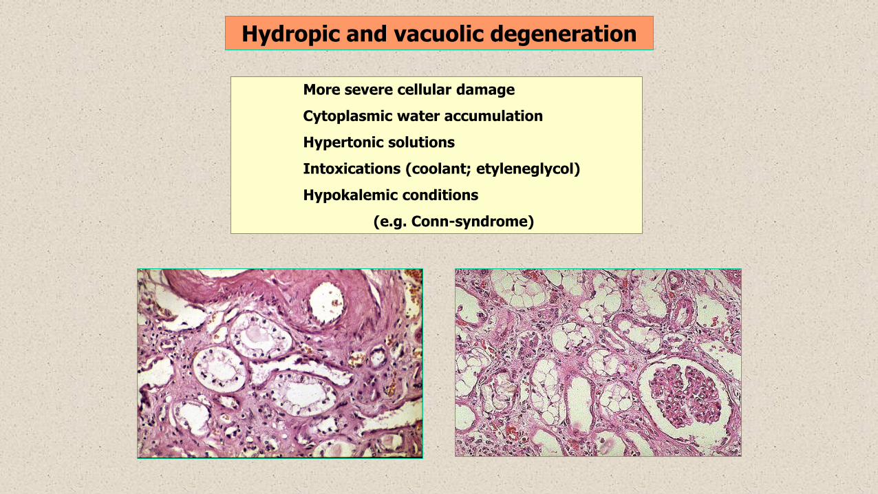
Hydropic and vacuolic degeneration
More severe cellular damage
Cytoplasmic water accumulation
Hypertonic solutions
Intoxications (coolant; etyleneglycol)
Hypokalemic conditions
(e.g. Conn-syndrome)
Fatty degeneration
Accumulation of neutral lipids (triglicerides) in the cytoplasm
Depending on the size of lipid droplets: microvesicular – macrovesicular
Increased trigliceride-synthesis Impraired trigliceride-excretion
- hyperlipidemias - impaired protein synthesis
- increased fatty acid synthesis - alcoholism
- decreased fatty acid oxidation (disturbed membrane transport)
(hypoxia, acidosis) - shortage of lipotropic materials
Characteristic localizations: LIVER, heart (diffuse, or „tiger heart”), kidney
Normal tissue
Abnormal noxas
degeneration adaptation
hyperplasia
hypertrophy
hyperplasia + hypertrophy
necrosis
death
superinfection
tissue loss
mutilation
resorption
cavitation
fistule
pseudocyst
Reparation
fibrosis
scarring
calcification
ossification
metaplasia
inflamm. atrophy
pseudometaplasia
pseudo-hypertrophy
regeneration
dysplasia
malignant transformation
((
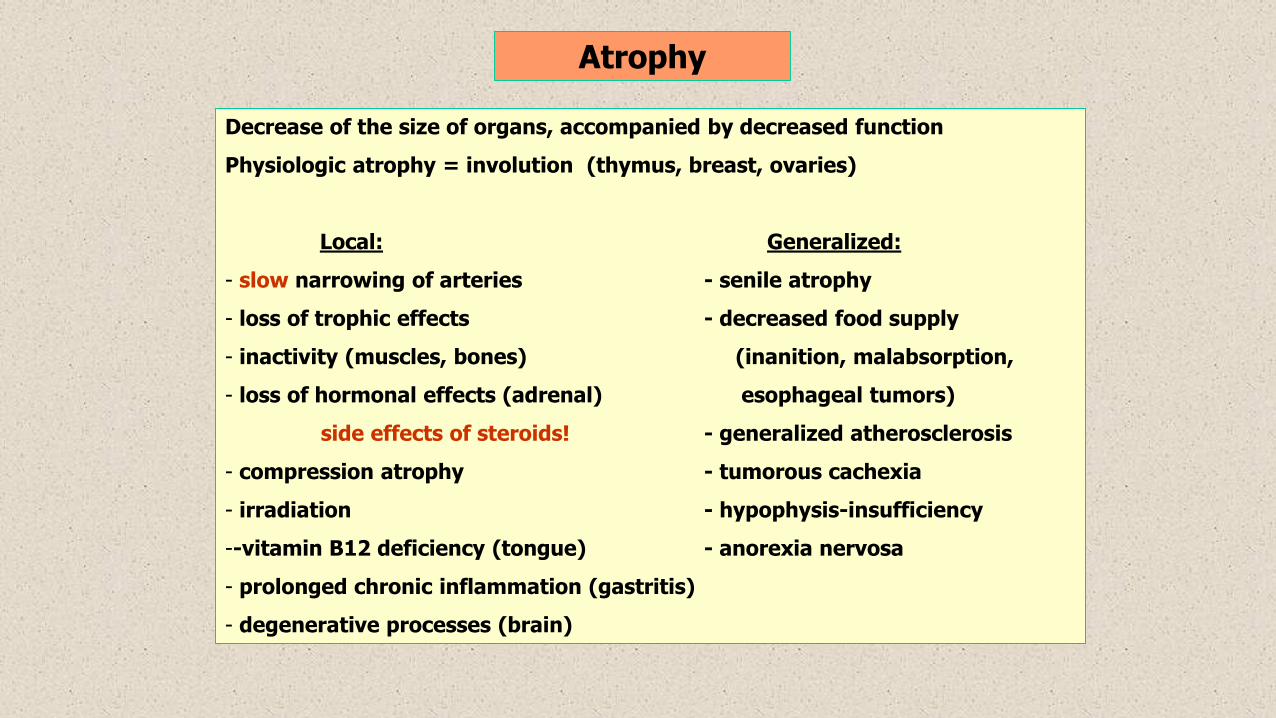
Atrophy
Decrease of the size of organs, accompanied by decreased function
Physiologic atrophy = involution (thymus, breast, ovaries)
Local: Generalized:
- slow narrowing of arteries - senile atrophy
- loss of trophic effects - decreased food supply
- inactivity (muscles, bones) (inanition, malabsorption,
- loss of hormonal effects (adrenal) esophageal tumors)
side effects of steroids! - generalized atherosclerosis
- compression atrophy - tumorous cachexia
- irradiation - hypophysis-insufficiency
--vitamin B12 deficiency (tongue) - anorexia nervosa
- prolonged chronic inflammation (gastritis)
- degenerative processes (brain)
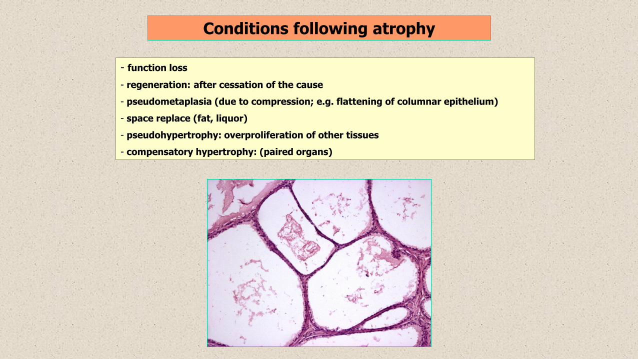
Conditions following atrophy
- function loss
- regeneration: after cessation of the cause
- pseudometaplasia (due to compression; e.g. flattening of columnar epithelium)
- space replace (fat, liquor)
- pseudohypertrophy: overproliferation of other tissues
- compensatory hypertrophy: (paired organs)
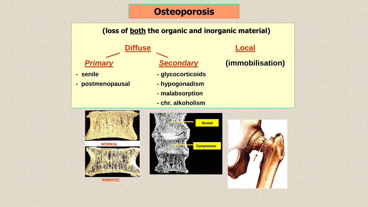
(loss of both the organic and inorganic material)
Diffuse Local
Primary Secondary (immobilisation)
- senile - glycocorticoids
- postmenopausal - hypogonadism
- malabsorption
- chr. alkoholism
Normal
CompressionNORMAL
POROTIC
Osteoporosis
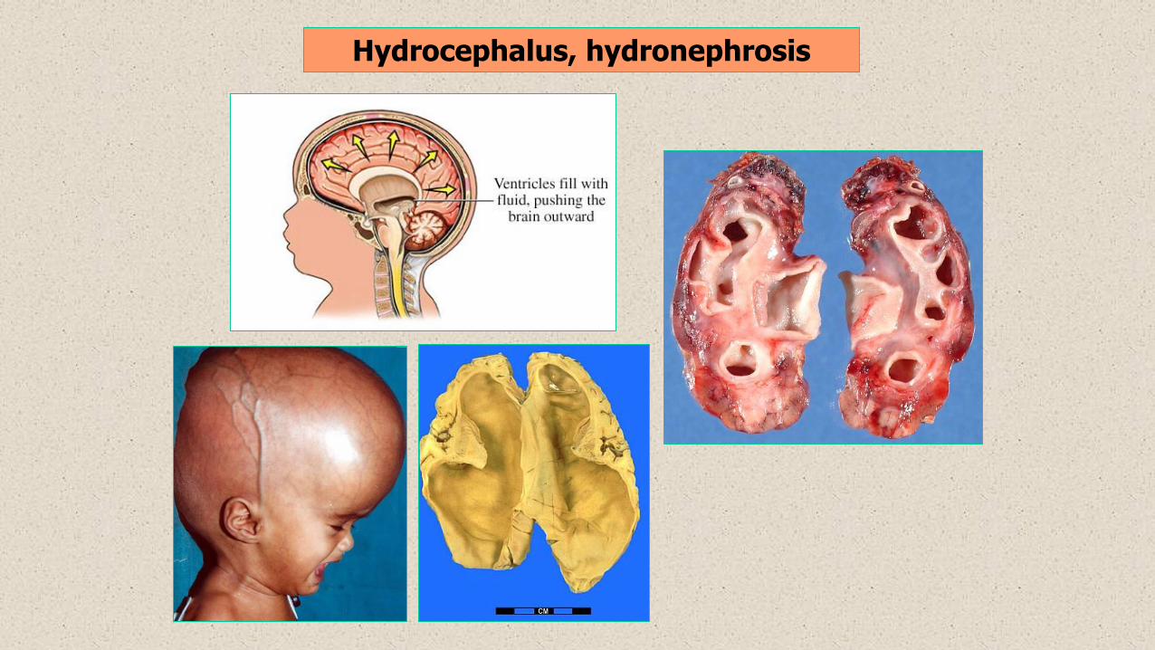
Hydrocephalus, hydronephrosis
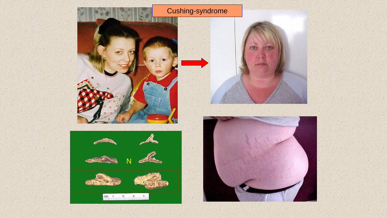
Cushing-syndrome
N
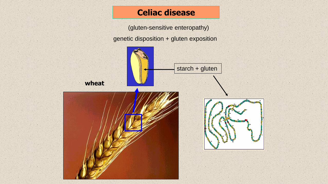
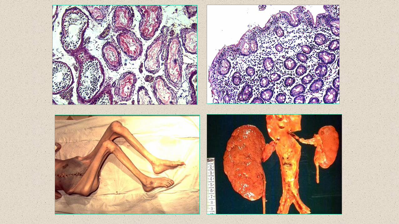
Celiac disease
(gluten-sensitive enteropathy)
genetic disposition + gluten exposition
starch + gluten
wheat

villi
Lieberkühn-
crypts
4
1
NORMAL
CELIAC DISEASE
Celiac disease
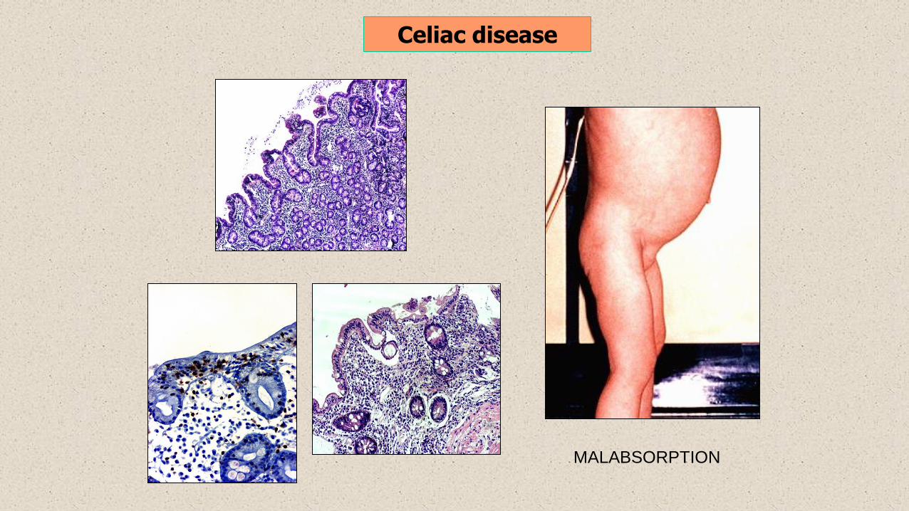
Celiac disease
MALABSORPTION
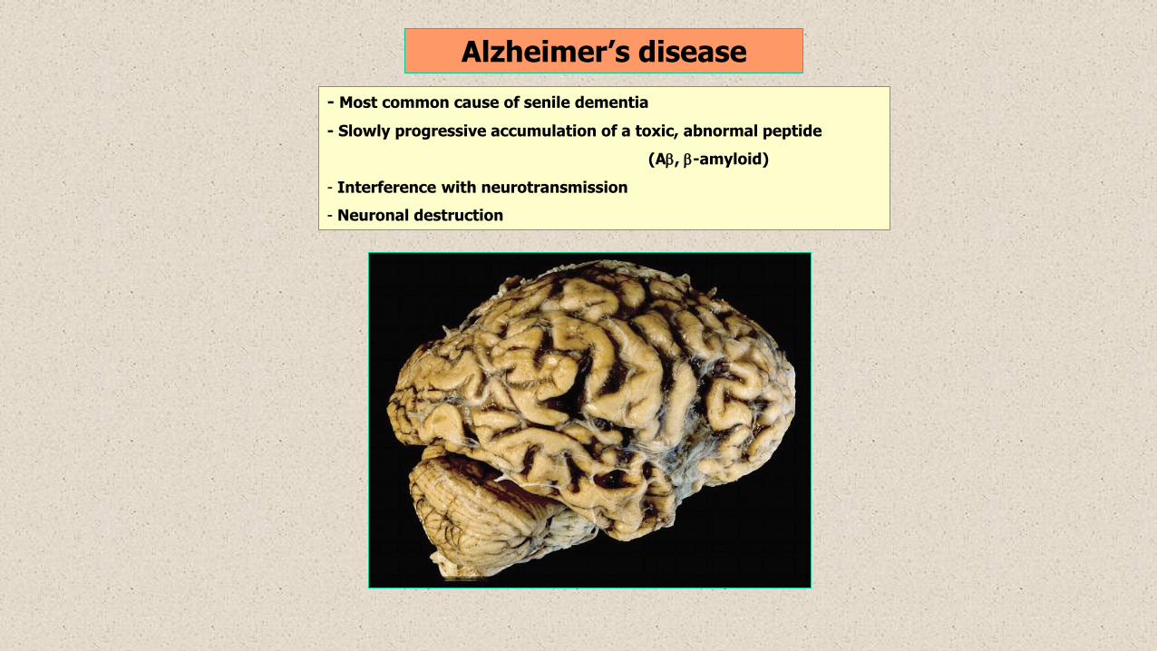
Alzheimer’s disease
- Most common cause of senile dementia
- Slowly progressive accumulation of a toxic, abnormal peptide
(A, -amyloid)
- Interference with neurotransmission
- Neuronal destruction
Normal tissue
Abnormal noxas
degeneration adaptation
hyperplasia
hypertrophy
hyperplasia + hypertrophy
necrosis
death
superinfection
tissue loss
mutilation
resorption
cavitation
fistule
pseudocyst
Reparation
fibrosis
scarring
calcification
ossification
metaplasia
inflamm. atrophy
pseudometaplasia
pseudo-hypertrophy
regeneration
dysplasia
malignant transformation
((

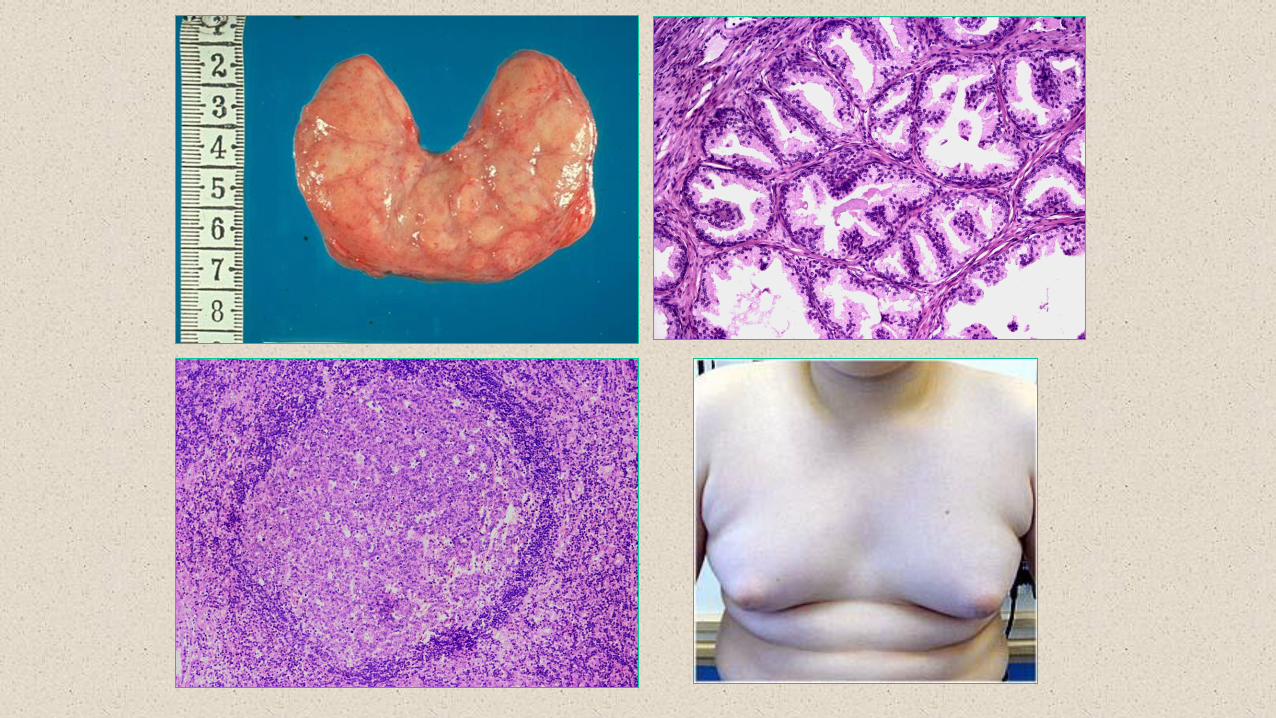
Hyperplasia
Increase of size of organs due to numerical excess of the cells
Prolonged demand
Organs /tissues that are capable of division
Prerequisite: good blood supply
Causative factors:
- hormonal effects (adrenal cortex, prostata, male breast, acromegaly)
- prolonged antigenic stimulus (follicular hyperplasia)
- drugs (cyclosporin A – gingival hyperplasia)
- metabolic causes (obesity)
- compensatory
- unknown
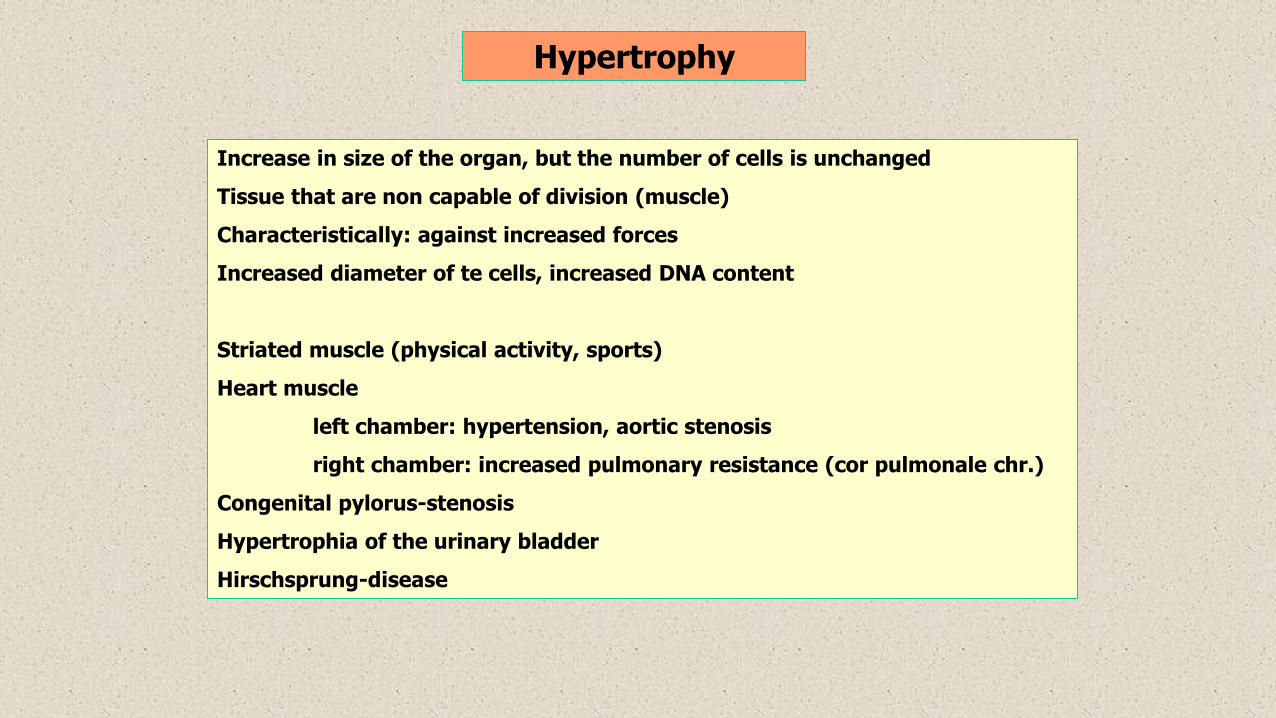
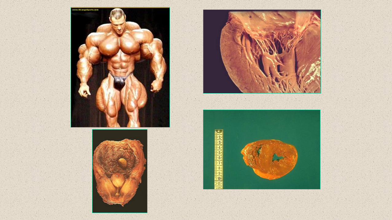
Hypertrophy
Increase in size of the organ, but the number of cells is unchanged
Tissue that are non capable of division (muscle)
Characteristically: against increased forces
Increased diameter of te cells, increased DNA content
Striated muscle (physical activity, sports)
Heart muscle
left chamber: hypertension, aortic stenosis
right chamber: increased pulmonary resistance (cor pulmonale chr.)
Congenital pylorus-stenosis
Hypertrophia of the urinary bladder
Hirschsprung-disease
Normal tissue
Abnormal noxas
degeneration adaptation
hyperplasia
hypertrophy
hyperplasia + hypertrophy
necrosis
death
superinfection
tissue loss
mutilation
resorption
cavitation
fistule
pseudocyst
Reparation
fibrosis
scarring
calcification
ossification
metaplasia
inflamm. atrophy
pseudometaplasia
pseudo-hypertrophy
regeneration
dysplasia
malignant transformation
((
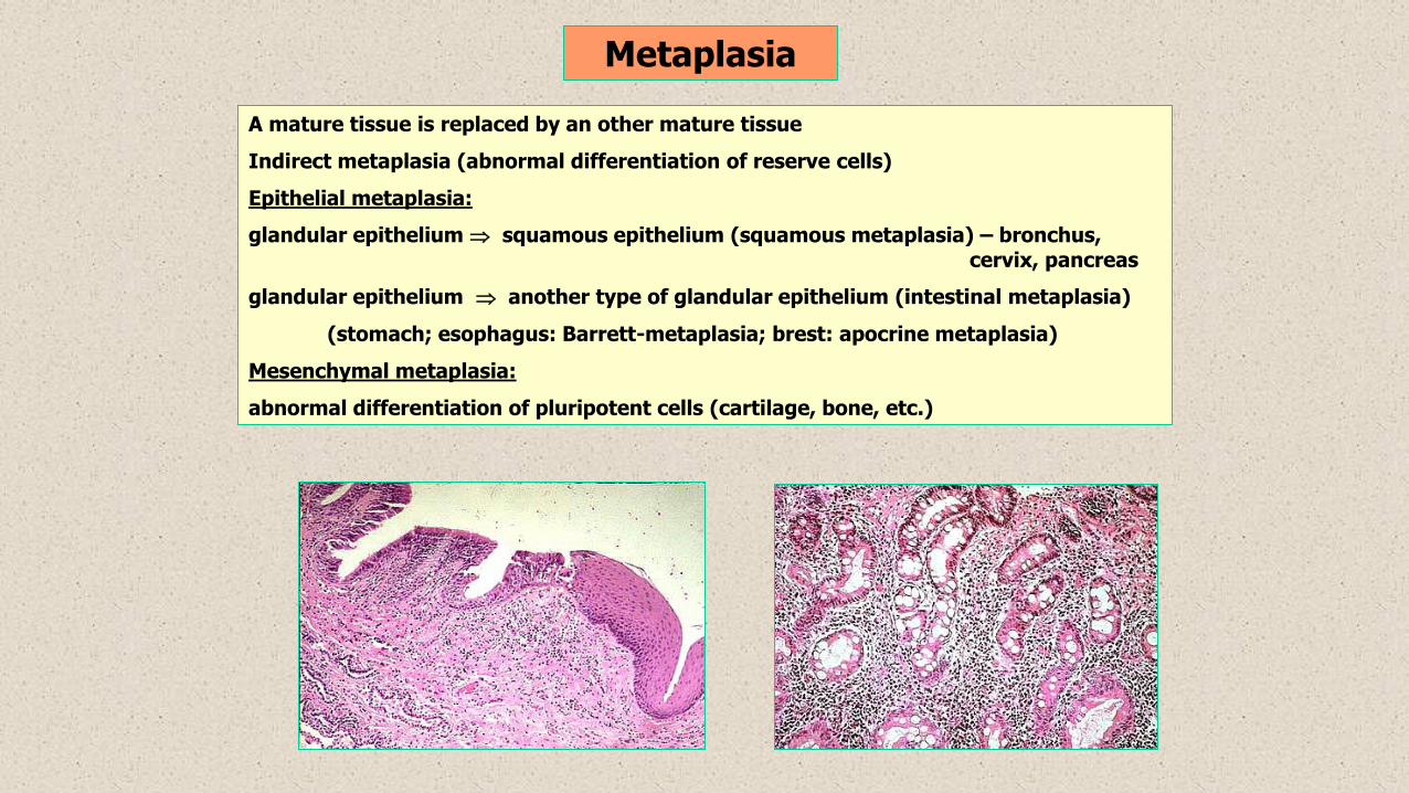
Metaplasia
A mature tissue is replaced by an other mature tissue
Indirect metaplasia (abnormal differentiation of reserve cells)
Epithelial metaplasia:
glandular epithelium squamous epithelium (squamous metaplasia) – bronchus,cervix, pancreas
glandular epithelium another type of glandular epithelium (intestinal metaplasia)
(stomach; esophagus: Barrett-metaplasia; brest: apocrine metaplasia)
Mesenchymal metaplasia:
abnormal differentiation of pluripotent cells (cartilage, bone, etc.)