Embed Size (px)
Citation preview
Histochem Cell Biol (2008) 130:21–54
DOI 10.1007/s00418-008-0420-0REVIEW
The desmosome and pemphigus
Jens Waschke
Accepted: 12 March 2008 / Published online: 3 April 2008© Springer-Verlag 2008
Abstract Desmosomes are patch-like intercellular adher-ing junctions (“maculae adherentes”), which, in concertwith the related adherens junctions, provide the mechanicalstrength to intercellular adhesion. Therefore, it is not sur-prising that desmosomes are abundant in tissues subjectedto signiWcant mechanical stress such as stratiWed epitheliaand myocardium. Desmosomal adhesion is based on theCa2+-dependent, homo- and heterophilic transinteraction ofcadherin-type adhesion molecules. Desmosomal cadherinsare anchored to the intermediate Wlament cytoskeleton byadaptor proteins of the armadillo and plakin families. Des-mosomes are dynamic structures subjected to regulationand are therefore targets of signalling pathways, which con-trol their molecular composition and adhesive properties.Moreover, evidence is emerging that desmosomal compo-nents themselves take part in outside-in signalling underphysiologic and pathologic conditions. Disturbed desmo-somal adhesion contributes to the pathogenesis of a numberof diseases such as pemphigus, which is caused by autoan-tibodies against desmosomal cadherins. Beside pemphigus,desmosome-associated diseases are caused by other mecha-nisms such as genetic defects or bacterial toxins. Becausemost of these diseases aVect the skin, desmosomes areinteresting not only for cell biologists who are inspired bytheir complex structure and molecular composition, butalso for clinical physicians who are confronted with
patients suVering from severe blistering skin diseases suchas pemphigus. To develop disease-speciWc therapeuticapproaches, more insights into the molecular compositionand regulation of desmosomes are required.
Keywords Desmosomes · Desmogleins · Pemphigus · Autoantibodies · Steric hindrance · Desmoglein compensation
Introduction
Desmosomes are intercellular adhering junctions serving toattach neighbouring cells to each other. They are mostnumerous in tissues subjected to signiWcant mechanicalstress such as the stratiWed squamous epithelia of the skin(Bizzozero 1864) and of mucous membranes (Farquhar andPalade 1963) as well as the myocardium (Fawcett andSelby 1958). Moreover, desmosomes are found in simpleepithelia and in non-epithelial cells such as the meningealcells of the arachnoidea (Gusek 1962) and the folliculardendritic cells of lymph follicles (Swartzendruber 1965).
Desmosomes were discovered as cell contacts in themiddle of the nineteenth century (Calkins and Setzer 2007).By the means of light microscopy, desmosomes were Wrstdescribed in the epidermis by the Italian pathologist Bizzo-zero in (1864). In his histology text book, the anatomistJosef SchaVer from Vienna introduced the term “desmo-some”, by combining the greek words “desmos” (bond) and“soma” (body) although, to that time, he, like most othersin the Weld believed that desmosomes were cytoplasm-Wlledintercellular bridges (SchaVer 1920). It took almost anothercentury until Keith Porter, using electron microscopy, wasable to conWrm the basic observation of Bizzozero that des-mosomes rather are contacts between adjacent cells and to
J. WaschkeInstitute of Anatomy and Cell Biology, University of Würzburg, Koellikerstr. 6, 97070 Würzburg, Germany
J. Waschke (&)Institute of Anatomy and Cell Biology, Julius-Maximilians-University, Koellikerstr. 6, 97070 Würzburg, Germanye-mail: [email protected]
123
22 Histochem Cell Biol (2008) 130:21–54
allow the Wrst description on desmosome ultrastructure(Porter 1956). With these new technical advances at hand,several studies were performed in the following years onthe distribution and organization of desmosomes in varioustissues. In addition, starting in the 1970s, biochemicalapproaches and molecular cloning techniques were appliedto identify the desmosomal components and to characterizetheir interactions (Drochmans et al. 1978; Moll et al. 1986;Moll and Franke 1982; Schwarz et al. 1990; Skerrow andMatoltsy 1974).
SigniWcant insights into the regulation of desmosomaladhesion also came from the Weld of dermatology since itwas demonstrated that autoantibodies in patients suVeringfrom the autoimmune blistering skin diseases pemphigusvulgaris (PV), and pemphigus foliaceus (PF), are directedto Ca2+-sensitive cell surface proteins within desmosomes(Eyre and Stanley 1987, 1988; Jones et al. 1986b; Karpatiet al. 1993), which were identiWed as the desmosomal cad-herins desmoglein 1 (Dsg 1) and Dsg 3 (Amagai et al.1991; Koulu et al. 1984). The term “pemphigus” comesfrom the greek word “pemphix” (blister) and is being usedin dermatology since 1791 (Schmidt et al. 2000), longbefore it was found that pemphigus is associated with auto-antibodies against keratinocyte surface antigens (Beutnerand Jordon 1964) and that these antibodies are suYcient tocause acantholysis, i.e. loss of cell–cell adhesion, in humanskin in vivo and in vitro (Anhalt et al. 1982; Schiltz andMichel 1976). The Wnal break-through was the Wnding thatautoantibodies against the extracellular domains of Dsg 3and Dsg 1 in PV and in PF are pathogenic (Amagai et al.1995, 1994a, 1992). Therefore, autoantibodies from pem-phigus patients have been used to characterize the mecha-nisms involved in the regulation of desmosomal adhesion.Except from pemphigus, other diseases in which desmo-somal adhesion is altered by mutations or bacterial toxinshelped to elucidate the functional role of the diVerent des-mosomal components.
During the last past several years, a number of compre-hensive reviews have been published on both desmosomestructure and function (Dusek et al. 2007b; Garrod et al.2002; Getsios et al. 2004b; Green and Simpson 2007;Holthofer et al. 2007; Kitajima 2002; Kottke et al. 2006;Muller et al. 2008a; Yin and Green 2004) and/or on themechanisms involved in pemphigus pathogenesis (Amagai2003; Hashimoto 2003; Lanza et al. 2006; Payne et al.2004; Sharma et al. 2007; Sitaru and Zillikens 2005;Stanley and Amagai 2006), which indicates that the per-spective of the existing model of the desmosome and itsrole in pemphigus pathogenesis are constantly reshaped.Moreover, because even textbook knowledge such as on themolecular composition of myocardial intercalated discsneeds revision (Borrmann et al. 2006; Franke et al. 2006), itbecomes obvious that after almost 150 years of desmosome
research, our knowledge is still far from complete. Thisarticle focuses on the mechanisms regulating desmosomaladhesion, which are compromised in diseases such aspemphigus.
The ultrastructure and composition of desmosomes
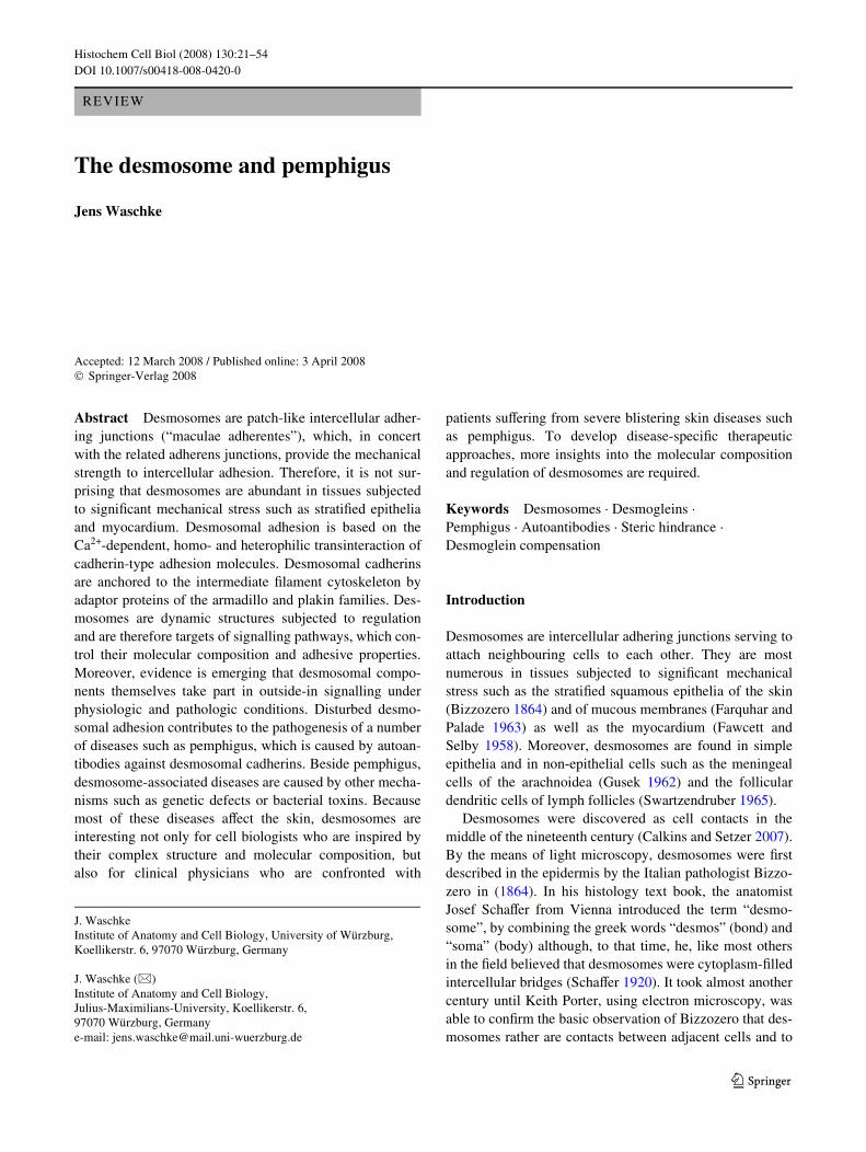
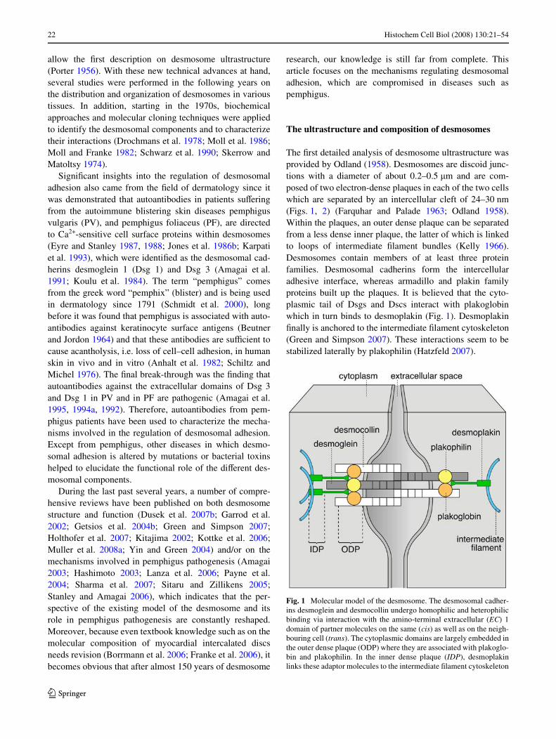
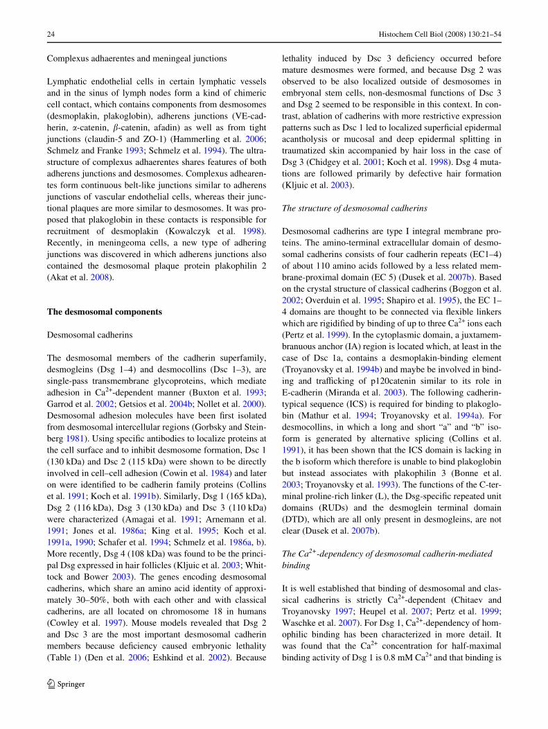
The Wrst detailed analysis of desmosome ultrastructure wasprovided by Odland (1958). Desmosomes are discoid junc-tions with a diameter of about 0.2–0.5 �m and are com-posed of two electron-dense plaques in each of the two cellswhich are separated by an intercellular cleft of 24–30 nm(Figs. 1, 2) (Farquhar and Palade 1963; Odland 1958).Within the plaques, an outer dense plaque can be separatedfrom a less dense inner plaque, the latter of which is linkedto loops of intermediate Wlament bundles (Kelly 1966).Desmosomes contain members of at least three proteinfamilies. Desmosomal cadherins form the intercellularadhesive interface, whereas armadillo and plakin familyproteins built up the plaques. It is believed that the cyto-plasmic tail of Dsgs and Dscs interact with plakoglobinwhich in turn binds to desmoplakin (Fig. 1). DesmoplakinWnally is anchored to the intermediate Wlament cytoskeleton(Green and Simpson 2007). These interactions seem to bestabilized laterally by plakophilin (Hatzfeld 2007).
Fig. 1 Molecular model of the desmosome. The desmosomal cadher-ins desmoglein and desmocollin undergo homophilic and heterophilicbinding via interaction with the amino-terminal extracellular (EC) 1domain of partner molecules on the same (cis) as well as on the neigh-bouring cell (trans). The cytoplasmic domains are largely embedded inthe outer dense plaque (ODP) where they are associated with plakoglo-bin and plakophilin. In the inner dense plaque (IDP), desmoplakinlinks these adaptor molecules to the intermediate Wlament cytoskeleton
123
Histochem Cell Biol (2008) 130:21–54 23
Desmosomes and desmosome-like junctions
Adhering junctions are divided into two main forms: (1)desmosomes, which serve as anchoring structures for inter-mediate Wlaments to desmosomal cadherins, and (2) adher-ens junctions, which contain cell-type speciWc adhesionmolecules from the cadherin super-family that are linked tothe actin cytoskeleton. Both, desmosomes and adherensjunctions can be found as constituents of more elaboratedcell contact complexes. Moreover, chimeric cell contactsexist which share features of both adherens junctions anddesmosomes.
Junctional complexes
Polarized epithelial cells display junctional complexeslocated at the uppermost section of the baso-lateral mem-brane. In apico-basal direction, the complex is composed ofthe zonula occludens (tight junction), the zonula adherensand a desmosome (macula adherens) (Farquhar and Palade1963). Accompanied by a line of separated desmosomes,the zonula occludens and the zonula adherens span theentire cell by forming continuous junction belts. Thesejunctional complexes are regarded as hallmarks of pola-rized epithelial cells but diVer in terms of size and ultra-structure in cell type-speciWc manner.
Area composita
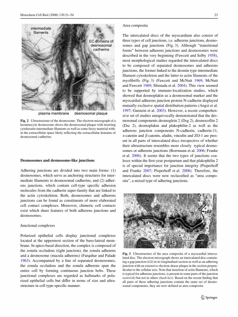
The intercalated discs of the myocardium also consist ofthree types of cell junctions, i.e. adherens junctions, desmo-somes and gap junctions (Fig. 3). Although “transitionalforms” between adherens junctions and desmosomes weredescribed in the very beginning (Fawcett and Selby 1958),most morphological studies regarded the intercalated discsto be composed of separated desmosomes and adherensjunctions, the former linked to the desmin type intermediateWlament cytoskeleton and the latter to actin Wlaments of themyoWbrills (Fig. 3) (Fawcett and McNutt 1969; McNuttand Fawcett 1969; Shimada et al. 2004). This view seemedto be supported by immuno-localization studies, whichshowed that desmoplakin as a desmosomal marker and themyocardial adherens junction protein N-cadherin displayedmutually exclusive spatial distribution patterns (Angst et al.1997; Gutstein et al. 2003). However, a recent comprehen-sive set of studies unequivocally demonstrated that the des-mosomal components desmoglein 2 (Dsg 2), desmocollin 2(Dsc 2), desmoplakin and plakophilin-2 as well as theadherens junction components N-cadherin, cadherin-11,�-catenin and �-catenin, afadin, vinculin and ZO-1 are pres-ent in all parts of intercalated discs irrespective of whethertheir ultrastructure resembles more closely typical desmo-somes or adherens junctions (Borrmann et al. 2006; Frankeet al. 2006). It seems that the two types of junctions coa-lesce within the Wrst-year postpartum and that plakophilin 2is of special importance for junction integrity (PieperhoVand Franke 2007; PieperhoV et al. 2008). Therefore, theintercalated discs were now reclassiWed as “area compo-sita”, a mixed type of adhering junctions.
Fig. 2 Ultrastructure of the desmosome. The electron micrograph of akeratinocyte desmosome shows the desmosomal plaque with insertingcytokeratin intermediate Wlaments as well as some fuzzy material with-in the extracellular space likely reXecting the extracellular domains ofdesmosomal cadherins
Fig. 3 Ultrastructure of the area composita of a myocardial interca-lated disc. The electron micrograph shows an intercalated disc contain-ing a gap junction (GJ) in its longitudinal section as well as an adheringjunction with an extensive electron-dense plaque in the section perpen-dicular to the cellular axis. Note that insertion of actin Wlaments, whichis typical for adherens junctions, is present in some parts of the junction(asterisk) but not in others (hash key). Based on the recent Wnding thatall parts of these adhering junctions contain the same set of desmo-somal components, they are now deWned as area composita
123
24 Histochem Cell Biol (2008) 130:21–54
Complexus adhaerentes and meningeal junctions
Lymphatic endothelial cells in certain lymphatic vesselsand in the sinus of lymph nodes form a kind of chimericcell contact, which contains components from desmosomes(desmoplakin, plakoglobin), adherens junctions (VE-cad-herin, �-catenin, �-catenin, afadin) as well as from tightjunctions (claudin-5 and ZO-1) (Hammerling et al. 2006;Schmelz and Franke 1993; Schmelz et al. 1994). The ultra-structure of complexus adhaerentes shares features of bothadherens junctions and desmosomes. Complexus adhearen-tes form continuous belt-like junctions similar to adherensjunctions of vascular endothelial cells, whereas their junc-tional plaques are more similar to desmosomes. It was pro-posed that plakoglobin in these contacts is responsible forrecruitment of desmoplakin (Kowalczyk et al. 1998).Recently, in meningeoma cells, a new type of adheringjunctions was discovered in which adherens junctions alsocontained the desmosomal plaque protein plakophilin 2(Akat et al. 2008).
The desmosomal components
Desmosomal cadherins
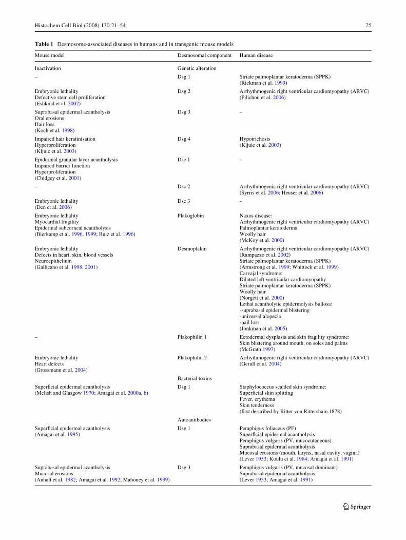
The desmosomal members of the cadherin superfamily,desmogleins (Dsg 1–4) and desmocollins (Dsc 1–3), aresingle-pass transmembrane glycoproteins, which mediateadhesion in Ca2+-dependent manner (Buxton et al. 1993;Garrod et al. 2002; Getsios et al. 2004b; Nollet et al. 2000).Desmosomal adhesion molecules have been Wrst isolatedfrom desmosomal intercellular regions (Gorbsky and Stein-berg 1981). Using speciWc antibodies to localize proteins atthe cell surface and to inhibit desmosome formation, Dsc 1(130 kDa) and Dsc 2 (115 kDa) were shown to be directlyinvolved in cell–cell adhesion (Cowin et al. 1984) and lateron were identiWed to be cadherin family proteins (Collinset al. 1991; Koch et al. 1991b). Similarly, Dsg 1 (165 kDa),Dsg 2 (116 kDa), Dsg 3 (130 kDa) and Dsc 3 (110 kDa)were characterized (Amagai et al. 1991; Arnemann et al.1991; Jones et al. 1986a; King et al. 1995; Koch et al.1991a, 1990; Schafer et al. 1994; Schmelz et al. 1986a, b).More recently, Dsg 4 (108 kDa) was found to be the princi-pal Dsg expressed in hair follicles (Kljuic et al. 2003; Whit-tock and Bower 2003). The genes encoding desmosomalcadherins, which share an amino acid identity of approxi-mately 30–50%, both with each other and with classicalcadherins, are all located on chromosome 18 in humans(Cowley et al. 1997). Mouse models revealed that Dsg 2and Dsc 3 are the most important desmosomal cadherinmembers because deWciency caused embryonic lethality(Table 1) (Den et al. 2006; Eshkind et al. 2002). Because
lethality induced by Dsc 3 deWciency occurred beforemature desmosmes were formed, and because Dsg 2 wasobserved to be also localized outside of desmosomes inembryonal stem cells, non-desmosmal functions of Dsc 3and Dsg 2 seemed to be responsible in this context. In con-trast, ablation of cadherins with more restrictive expressionpatterns such as Dsc 1 led to localized superWcial epidermalacantholysis or mucosal and deep epidermal splitting intraumatized skin accompanied by hair loss in the case ofDsg 3 (Chidgey et al. 2001; Koch et al. 1998). Dsg 4 muta-tions are followed primarily by defective hair formation(Kljuic et al. 2003).
The structure of desmosomal cadherins
Desmosomal cadherins are type I integral membrane pro-teins. The amino-terminal extracellular domain of desmo-somal cadherins consists of four cadherin repeats (EC1–4)of about 110 amino acids followed by a less related mem-brane-proximal domain (EC 5) (Dusek et al. 2007b). Basedon the crystal structure of classical cadherins (Boggon et al.2002; Overduin et al. 1995; Shapiro et al. 1995), the EC 1–4 domains are thought to be connected via Xexible linkerswhich are rigidiWed by binding of up to three Ca2+ ions each(Pertz et al. 1999). In the cytoplasmic domain, a juxtamem-branuous anchor (IA) region is located which, at least in thecase of Dsc 1a, contains a desmoplakin-binding element(Troyanovsky et al. 1994b) and maybe be involved in bind-ing and traYcking of p120catenin similar to its role inE-cadherin (Miranda et al. 2003). The following cadherin-typical sequence (ICS) is required for binding to plakoglo-bin (Mathur et al. 1994; Troyanovsky et al. 1994a). Fordesmocollins, in which a long and short “a” and “b” iso-form is generated by alternative splicing (Collins et al.1991), it has been shown that the ICS domain is lacking inthe b isoform which therefore is unable to bind plakoglobinbut instead associates with plakophilin 3 (Bonne et al.2003; Troyanovsky et al. 1993). The functions of the C-ter-minal proline-rich linker (L), the Dsg-speciWc repeated unitdomains (RUDs) and the desmoglein terminal domain(DTD), which are all only present in desmogleins, are notclear (Dusek et al. 2007b).
The Ca2+-dependency of desmosomal cadherin-mediated binding
It is well established that binding of desmosomal and clas-sical cadherins is strictly Ca2+-dependent (Chitaev andTroyanovsky 1997; Heupel et al. 2007; Pertz et al. 1999;Waschke et al. 2007). For Dsg 1, Ca2+-dependency of hom-ophilic binding has been characterized in more detail. Itwas found that the Ca2+ concentration for half-maximalbinding activity of Dsg 1 is 0.8 mM Ca2+ and that binding is
123
Histochem Cell Biol (2008) 130:21–54 25
Table 1 Desmosome-associated diseases in humans and in transgenic mouse models
Mouse model Desmosomal component Human disease
Inactivation Genetic alteration
– Dsg 1 Striate palmoplantar keratoderma (SPPK) (Rickman et al. 1999)
Embryonic lethalityDefective stem cell proliferation(Eshkind et al. 2002)
Dsg 2 Arrhythmogenic right ventricular cardiomyopathy (ARVC) (Pilichou et al. 2006)
Suprabasal epidermal acantholysisOral erosionsHair loss(Koch et al. 1998)
Dsg 3 –
Impaired hair keratinisationHyperproliferation (Kljuic et al. 2003)
Dsg 4 Hypotrichosis (Kljuic et al. 2003)
Epidermal granular layer acantholysisImpaired barrier functionHyperproliferation(Chidgey et al. 2001)
Dsc 1 –
– Dsc 2 Arrhythmogenic right ventricular cardiomyopathy (ARVC)(Syrris et al. 2006; Heuser et al. 2006)
Embryonic lethality(Den et al. 2006)
Dsc 3 –
Embryonic lethalityMyocardial fragilityEpidermal subcorneal acantholysis(Bierkamp et al. 1996, 1999; Ruiz et al. 1996)
Plakoglobin Naxos disease:Arrhythmogenic right ventricular cardiomyopathy (ARVC)Palmoplantar keratodermaWoolly hair(McKoy et al. 2000)
Embryonic lethalityDefects in heart, skin, blood vesselsNeuroepithelium(Gallicano et al. 1998, 2001)
Desmoplakin Arrhythmogenic right ventricular cardiomyopathy (ARVC)(Rampazzo et al. 2002)Striate palmoplantar keratoderma (SPPK)(Armstrong et al. 1999; Whittock et al. 1999)Carvajal syndrome:Dilated left ventricular cardiomyopathyStriate palmoplantar keratoderma (SPPK)Woolly hair(Norgett et al. 2000)Lethal acantholytic epidermolysis bullosa:-suprabasal epidermal blistering-universal alopecia-nail loss(Jonkman et al. 2005)
– Plakophilin 1 Ectodermal dysplasia and skin fragility syndrome:Skin blistering around mouth, on soles and palms(McGrath 1997)
Embryonic lethalityHeart defects(Grossmann et al. 2004)
Plakophilin 2 Arrhythmogenic right ventricular cardiomyopathy (ARVC)(Gerull et al. 2004)
Bacterial toxins
SuperWcial epidermal acantholysis(Melish and Glasgow 1970; Amagai et al. 2000a, b)
Dsg 1 Staphylococcus scalded skin syndrome:SuperWcial skin splittingFever, erythemaSkin tenderness(Wrst described by Ritter von Rittershain 1878)
Autoantibodies
SuperWcial epidermal acantholysis(Amagai et al. 1995)
Dsg 1 Pemphigus foliaceus (PF)SuperWcial epidermal acantholysisPemphigus vulgaris (PV, mucocutaneous)Suprabasal epidermal acantholysisMucosal erosions (mouth, larynx, nasal cavity, vagina)(Lever 1953; Koulu et al. 1984; Amagai et al. 1991)
Suprabasal epidermal acantholysisMucosal erosions(Anhalt et al. 1982; Amagai et al. 1992; Mahoney et al. 1999)
Dsg 3 Pemphigus vulgaris (PV, mucosal dominant)Suprabasal epidermal acantholysis(Lever 1953; Amagai et al. 1991)
123

26 Histochem Cell Biol (2008) 130:21–54
highly cooperative with the Hill coeYcient being ¸5 (Was-chke et al. 2007). This indicates that Dsg 1 binding isstrong only at extracellular Ca2+concentrations higher than0.8 mM. Although the exact extracellular Ca2+concentra-tion within the epidermis is unknown, it has been shownthat a gradient exists with low Ca2+concentrations in thebasal layers and high concentrations in the superWcial epi-dermis (Elias et al. 2002; Menon and Elias 1991). There-fore, if homophilic binding of Dsg 1 occurs in vivo, it has tobe considered that it may contribute to eVective intercellu-lar adhesion only in the superWcial epidermis.
Transinteraction mechanisms of desmosmal cadherins
At present, it is still a matter of debate how desmosmal cad-herins interact with each other in vivo. However, severallines of evidence indicate that the N-terminal EC 1 domainis important. Similar to classical cadherins, desmosomalcadherins contain a cell adhesion recognition (CAR) sitecontaining a central alanine residue (Blaschuk et al. 1990).However, instead of the conserved tripeptide HAVsequence of classical cadherins, the sequence is YAT forDsc 1 and RAL for Dsg 1, respectively (Tselepis et al.1998). Peptides derived from these sequences were able toblock homophilic adhesion mediated by Dsg 1 and Dsc 1and to inhibit desmosomal adhesion in epithelial cells whenthe peptides for Dsg 1 and Dsc 1 were applied together,indicating that the CAR site in the EC 1 domain is criticalfor maintenance of desmosmal adhesion (Runswick et al.2001; Tselepis et al. 1998). This hypothesis is supported bystudies in which mechanisms underlying the loss of kerati-nocyte cohesion in pemphigus were investigated. AK23, amonoclonal Dsg 3 antibody from a PV mouse modeldirected against the predicted binding motif of the EC 1,has been shown to be pathogenic in vivo whereas anti-bodies targeting other parts of the Dsg 3 extracellular domainwere not (Tsunoda et al. 2003). Together with the recentWnding that AK 23 similar to Dsg 3 antibodies from PVpatients, which are known to be also primarily directedagainst the N-terminal part of EC 1 (Sekiguchi et al. 2001),is able to directly interfere with Dsg 3 binding (Heupelet al. 2007), these data demonstrate that interaction of EC 1is crucial for Dsg 3 binding. In addition to these functionalstudies, morphologic studies sought to address the mode ofdesmosomal cadherin interaction within desmosomes byusing electron tomography imaging of epidermal tissue(Al-Amoudi et al. 2007; He et al. 2003). Based on predic-tions from the C-cadherin crystal structure, He and col-leagues reported that in mouse epidermis severaldesmosomal cadherins form knots in which cadherins dis-play stochastic arrangement. In these knots, desmosomalcadherins seemed to interact via their EC 1 domains withboth molecules on the same cell in cis as well as with
molecules from opposing cells in trans (He et al. 2003). Al-Amoudi and co-workers reWned the technique by employ-ing cryo-electron microscopy in human epidermis. TheyconWrmed cis- and trans-interactions of the EC 1 domains,possibly via insertion of the tryptophane 2 into the hydro-phobic pocket of the CAR site (Al-Amoudi et al. 2007).However, they found that cis-interactions of the EC 4–5regions may also occur and that desmosomal cadherinsrather show periodically zipper-like arrangements similarto classical cadherins (Boggon et al. 2002).
Homophilic and heterophilic binding of desmosomal cadherins
In contrast to classical cadherins from adherens junctionswhich primarily bind in homophilic manner, data indicatethat desmosomal cadherins undergo both homophilic andheterophilic transinteraction. Using EC 1–2 fragments ofDsg 2 and Dsc 2, it was shown that homophilic bindingoccurs in vitro (Syed et al. 2002). Similarly, homophilicbinding of Dsg 3 was found (Amagai et al. 1994b). Whenrecombinant proteins consisting of the complete extracellu-lar domain were used for atomic force microscopy (AFM)measurements, it was demonstrated that the unbindingforce of single homophilically transinteracting moleculeswas about 37–68 pN (depending on the retrace velocity300–6,000 nm/s) for Dsg 1 with a lifetime of 0.17 s andabout 50 pN for Dsg 3 (Heupel et al. 2007; Waschke et al.2005, 2007), which is in the same range as observed forother types of cadherins characterized by the same methodsuch as VE-cadherin, N-cadherin or LI-cadherin (Baum-gartner et al. 2003, 2000; Wendeler et al. 2007). These dataindicate that the molecular binding properties of homo-philic adhesion of desmosomal cadherins may be compara-ble to other cadherins.
Heterophilic binding of Dsg 2 to Dsc 1 or Dsc 2 wasalso found on the molecular level (Chitaev and Troyanov-sky 1997; Syed et al. 2002) but no interaction of Dsg 1with Dsg 3 (Heupel et al. 2007). Aggregation assays oftransfected cells indicated that in cells, heterophilic bind-ing of Dsgs and Dscs might be of even greater importancethan homophilic binding to induce strong intercellularadhesion (Kowalczyk et al. 1996; Marcozzi et al. 1998;Runswick et al. 2001) and that adhesion is strictly depen-dent on the ratio of the respective Dsgs and Dscs (Getsioset al. 2004a). This view is supported by the recent Wndingthat a conditional Dsc 3-deWciency in mice induced asevere pemphigus-like phenotype with epidermal blister-ing (Chen et al. 2007). Because antibodies in typical pem-phigus are usually not directed against Dsc 3 but againstDsg 1 and Dsg 3, it has to be considered that heterophilicbinding of these three molecules is important for epider-mal cohesion in vivo.
123
Histochem Cell Biol (2008) 130:21–54 27
Armadillo family proteins
From the Armadillo family, plakoglobin and plakophilins1–3 are important components of desmosomes.
Plakoglobin
Plakoglobin (82 kDa), also termed �-catenin, is the onlyessential desmosomal component which is also found intypical adherens junctions (Cowin et al. 1986; Franke et al.1989, 1983). The gene encoding for plakoglobin wasmapped to chromosome 17 (Aberle et al. 1995). Plakoglo-bin binds to the cytoplasmic cadherin-typical sequence ofDsgs and Dscs via its Wrst three armadillo repeat domains(Chitaev et al. 1998). Because the same binding site isrequired for interaction of plakoglobin to �-catenin, the lat-ter is excluded from desmosomes. Similarly, although thearmadillo repeat domain of �-catenin can also bind to Dsg2, its Xanking domains inhibit this interaction, which mayexplain the absence of �-catenin from desmosomes (Troya-novsky et al. 1996; Wahl et al. 1996). Plakoglobin has beendemonstrated to interact with other desmosomal plaquecomponents such as desmoplakin, plakophilins and alsowith cytokeratin Wlaments (Bonne et al. 2003; Chen et al.2002; Kowalczyk et al. 1997; Smith and Fuchs 1998). Theimportance of this linker function can be concluded fromstudies in which inactivation of plakoglobin led to embryoniclethality due to mechanical fragility of the myocardiumand, when mice are viable, to subcorneal skin blisteringindicating that plakoglobin is essential for desmosomalstability (Table 1) (Bierkamp et al. 1996; Ruiz et al.1996).
Besides its function as a desmosomal adaptor protein,plakoglobin seems to be involved in nuclear signalling. Ithas been shown that plakoglobin, comparable to �-cateninin the canonical wnt signalling pathway, confers transcrip-tional activity together with TCF-4/LEF transcription fac-tors (Maeda et al. 2004). This mechanism seems to interferewith �-catenin-mediated transcription (Hu et al. 2003).Because plakoglobin like �-catenin is a target of glycogensynthase kinase-3 �, which drives proteosomal degradationof both proteins (Kodama et al. 1999; Muller et al. 2008a;Williamson et al. 2006), a complex pattern of direct andindirect transcriptional regulation seems likely. A targetgene of Lef-1/plakoglobin signalling is c-Myc, the expres-sion of which was inhibited in keratinocytes but enhancedin transformed rat kidney epithelial cells (Kolligs et al.2000; Kolly et al. 2007; Williamson et al. 2006). This indi-cates that the role of plakoglobin transcriptional regulationis strictly cell type-dependent. In keratinoytes, c-Mycrepression by plakoglobin is required to stop proliferationand to allow terminal diVerentiation (Williamson et al.2006). Another potential target gene is the anti-apoptotic
molecule Bcl–XL, which was found to be upregulated inplakoglobin-deWcient cells leading to reduced apoptosisand thus might also be repressed by plakoglobin (Duseket al. 2007a). Taken together, plakoglobin serves as func-tional linker between intercellular adhesion and regulationof the cell cycle. This might also be important for cancerprogression because many tumors are characterized by lossof plakoglobin expression.
Plakophilins
Plakophilin 1 (80 kDa) was Wrst identiWed as an “acces-sory” plaque protein because, in contrast to plakoglobin anddesmoplakin, it was found in cells from certain stratiWedand complex epithelia only (Franke et al. 1983; Hatzfeldet al. 1994; Heid et al. 1994). Afterwards, it became clearthat plakophilin 2 (100 kDa) is ubiquitously present in alldesmosomes and also plakophilin 3 (87 kDa) is present inmost simple and stratiWed epithelia (Mertens et al. 1999;Schmidt et al. 1999). The genes encoding plakophilin 1, 2and 3 are located on chromosomes 1, 12, and 11, respec-tively (Bonne et al. 1998). Plakophilin 1 and 2 exist in twosplice variants with a shorter “a” and a longer “b” form(Mertens et al. 1996; Schmidt et al. 1997). In addition totheir localization in desmosomes, plakophilins 1 and 2 arealso found in the karyoplasm in a variety of cells and plako-philin 1 b is exclusively located in the nucleus. Plakophilin2 deWciency leads to embryonic death due to heart defectsindicating that the presence of at least one member of theplakophilin family is required (Table 1) (Grossmann et al.2004). Under these conditions, cytokeratin Wlaments wereretracted from cell borders and desmoplakin was localizedin the cytoplasm rather than at desmosomes in cardiomyo-cytes, demonstrating the relevance of plakophilin 2 to des-mosplakin recruitment. Because cardiomyocytes in contrastto epithelial cells express only plakophilin 2 but not plako-philin 1 and 3, defects were present in the heart only,whereas epithelia were not aVected.
Plakophilins can directly interact with all other desmo-somal components including cytokeratins via the aminoter-minal head domain (Bonne et al. 2003; Hatzfeld 2007;Hatzfeld et al. 2000). Plakophilin 1 recruits desmosomalcomponents to the cell membrane, increases size and num-ber of desmosomes and therefore seems to be a scaVoldingprotein, which induces desmosome assembly (Hatzfeldet al. 2000; Kowalczyk et al. 1999; Wahl 2005). On theother hand, because plakophilin 1 is located in the denseinner desmosomal plaque whereas cytokeratin Wlamentsonly loop into the outer plaque, it is believed that plakophi-lin 1 enhances desmoplakin lateral interactions but does notdirectly associate with cytokeratin Wlaments in vivo(Hatzfeld 2007; North et al. 1999). It has been demonstratedthat desmosome formation is mediated by the aminoterminal
123
28 Histochem Cell Biol (2008) 130:21–54
domain, whereas recruitment of plakophilin 1 itself to theplasma membrane is dependent on the carboxyterminalregion (Sobolik-Delmaire et al. 2006).
In addition to its function in the regulation of desmo-some assembly, plakophilins may also regulate signallingmechanisms, both at cell borders as well as in the nucleus.Plakophilin 1 associates with actin Wlaments at cell bordersand has been reported to interact with a GTP exchange fac-tor (GEF) for Rho and thereby could regulate activity ofRho GTPases similar to the closely related p120-catenin,which is known to inhibit Rho A and to activate Rac 1 andCdc42 (Anastasiadis and Reynolds 2001; Hatzfeld 2007).In addition to their localization within the desmosomalplaque, plakophilin 1 and 2 are also present inside nucleusand plakophilin 2 has been found to be part of the polymer-ase III complex which is responsible for generation oftRNA and rRNA (Mertens et al. 2001). By these two mecha-nisms, it is possible that plakophilins may regulate celladhesion and cell growth (Hatzfeld 2007).
Plakin family proteins
Plakin family proteins are linkers between the cytoskeletonand cell–cell or cell–matrix contacts (JeVerson et al. 2007).Desmoplakin, which exists in two spice variants of a pro-tein encoded by a single gene on chromosome 6 (desmopla-kin I: 322 kDa; desmoplakin II: 259 kDa), is an essentialcomponent of the desmosomal plaque and therefore isregarded as the prototype of this family (Armstrong et al.1999; Hatsell and Cowin 2001; Mueller and Franke 1983;Sonnenberg and Liem 2007). Other members such as plec-tin, envoplakin and periplakin were also found in desmo-somes, but their signiWcance for the structure and functionof desmosomes is less clear. Especially, plectin is primarilyimportant in hemidesmosmes, which anchor epithelia to theextracellular matrix.
Desmoplakin consists of an aminoterminal plakindomain, which can interact with all other desmosomalplaque proteins such as plakoglobin and plakophilins butalso with Dsc 1a (Kowalczyk et al. 1997; Smith and Fuchs1998; Troyanovsky et al. 1994b). The central coiled-coilrod domain, which is important for dimerization, is fol-lowed by the carboxyterminal tail consisting of three globu-lar subdomains with several plakin-repeats, which serve aslinkers for diVerent intermediate Wlament types (Choi et al.2002; Green et al. 1990; Stappenbeck and Green 1992). Itis well established that desmoplakin is the main linker pro-tein between the desmosomal cadherin–plakoglobin com-plex and the intermediate Wlament cytoskeleton. This hasbeen shown in vitro and was ultimately demonstrated indesmoplakin-deWcient mice, which had a reduced numberof desmosomes and died at embryonic stage just afterimplantation (Table 1) (Bornslaeger et al. 1996; Gallicano
et al. 1998). Similar to the Wndings in epidermal-speciWcdesmoplakin-deWcient mice, which suVered from skin blis-tering, desmosomes were not anchored to intermediate Wla-ments (Vasioukhin et al. 2000). Moreover, whendesmoplakin was rescued in extraembryonic tissues so thatembryos further developed, defects were present not only inthe myocardium and epidermis but also in the vasculatureand in the neuroepithelium, underlining the importance ofdesmoplakin for tissue diVerentiation (Gallicano et al.2001).
Diversity of desmosomes in diVerent tissues and speciWc epithelial layers
Although it was discovered about 25 years ago that thestructure of desmosomes is not identical in all types of cellsand tissues (Giudice et al. 1984; Jones et al. 1986b), theknowledge on the diversity of desmosomes is still uncom-plete and matter of discussion (Garrod et al. 2002; Getsioset al. 2004b; Green and Simpson 2007; Hatzfeld 2007; Hol-thofer et al. 2007; Kottke et al. 2006; Yin and Green 2004).The diversity of desmosomes has implications for tissuediVerentiation and also is of high-medical relevancebecause diseases caused by genetic alteration of or by anautoimmune reponse against a speciWc desmosomal com-ponent may aVect only certain but not all desmosome-con-taining tissues.
Some desmosomal components such as Dsg 2, Dsc 2 andthe plaque proteins desmoplakin, plakoglobin and plako-philin 2 are ubiquitously expressed in all cells and tissues inwhich desmosomes are found. Plakophilin 3 is present inmost simple epithelia except hepatocytes as well as in strat-iWed epithelia, whereas plakophilin 1 is restricted to strati-Wed and complex epithelia. In epithelia, the desmosomalcadherins show typical expression patterns. Simple epithe-lia and urothelium usually express Dsg 2 and Dsc 2 only.Apparently, exceptions are the additional presence of Dsg 1in the mucosa of uterus, stomach, intestine and in epitheliaof liver and pancreas as well as the expression of Dsc 1 inintestine and liver or Dsc 3 in stomach, prostate, salivarygland and urothelium. Dsg 4 has a unique tissue distribu-tion in skin and several simple epithelia such as those pres-ent in pancreas, salivary glands, testis, prostate and hepaticepithelium.
The Dsg 1/Dsc 1 and Dsg 3/Dsc 3 pairs are largely con-Wned to stratiWed epithelia where the expression patterns ofthe Dsg and Dsc isoforms usually conform. Interestingly, inthe stratiWed corneal epithelium, only Dsg 1 and Dsc1 arepresent indicating that these desmosomal cadherins in theabsence of Dsg 1 and Dsg 3 are suYcient to maintain cohe-sion in stratiWed epithelia also. In the epidermis, the plaqueproteins plakoglobin, desmoplakin and plakophilin 3 areexpressed in all layers (Fig. 4). In contrast, plakophilins 1
123
Histochem Cell Biol (2008) 130:21–54 29
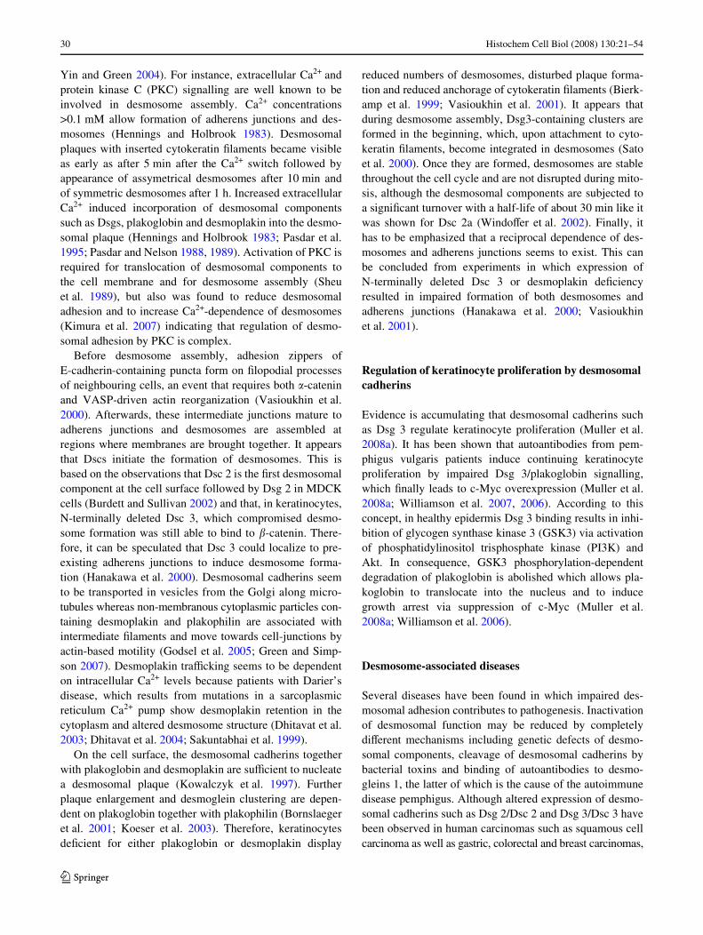
and 2 display inverse distribution with plakophilin 1 beingmore abundant in the superWcial epidermis. These inverseexpression patterns are also typical for the Dsg 1/Dsc 1 andDsg 3/Dsc 3 pairs. Dsg 1/Dsc 1 are the predominant desmo-somal cadherins in the superWcial epidermis, whereas Dsg3/Dsc 3 are primarily expressed in the lower epidermis. Incontrast to Dsg 1, which can be detected in some desmo-somes in keratinocytes of the basal layer also, Dsc 1 andDsg 4 are absent in the basal layer (Dusek et al. 2007b;Mahoney et al. 2006; Spindler et al. 2007). Because Dsg 3is expressed throughout the spinous layers (Fig. 5), theexpression patterns of Dsg 1 and Dsg 3 largely overlap inhuman adult epidermis (Mahoney et al. 2006; Spindleret al. 2007). Dsc 2 is enriched in the deep epidermis withlower levels in the superWcial epidermis, whereas Dsg 2 isrestricted to basal and suprabasal cells but is present in veryfaint amounts only (Mahoney et al. 2006) indicating thatthis pair of proteins may be primarily important for cellcohesion in simple epithelia and myocardium rather than incomplex epithelia. However, it is unclear at present whichdesmosomal cadherin isoforms are capable to heretophili-cally bind to each other and thus interpretation of these dis-tribution patterns with respect to their relevance formechanical adhesion is preliminary. In multilayered squa-mous epithelium of mucous membranes, for instance of theoral cavity, Dsg 1 and Dsg 3 are strongly expressedthroughout all layers, whereas Dsg 4 shows strong expres-sion in superWcial layers but is missing in the basal layer(Mahoney et al. 2006).
It is important to note that the speciWc distribution pat-terns of desmosomal components in stratiWed epithelia areimportant for epithelial diVerentiation and function (Greenand Simpson 2007). It was shown that forced overexpres-sion of Dsg 3 in the suprabasal epidermis led to abnormaldiVerentiation and hyperproliferation as well as perinatallethality due to transepidermal water loss (Elias et al. 2001;Merritt et al. 2002). Similarly, forced suprabasal Dsg 2 and
Dsc 3 overexpression resulted in hyperproliferation andformation of papillomas, possibly via altered �-catenin/wntsignalling (Brennan et al. 2007; Hardman et al. 2005).
Desmosome assembly and disassembly
The mechanisms participating in desmosome assembly anddisassembly have been reviewed in detail elsewhere (Gets-ios et al. 2004b; Green and Simpson 2007; Kitajima 2002;
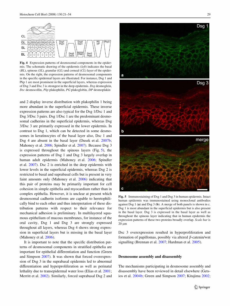
Fig. 4 Expression patterns of desmosomal components in the epider-mis. The schematic drawing of the epidermis (left) indicates the basal(BL), spinous (SL), granular (GL) and corneal (CL) layer of the epider-mis. On the right, the expression patterns of desmosomal componentsin the speciWc epidermal layers are illustrated. For instance, Dsg 1 andPkp 1 are most prominent in the superWcial layers, whereas expressionof Dsg 3 and Dsc 3 is strongest in the deep epidermis. Dsg desmoglein,Dsc desmocollin, Pkp plakophilin, PG plakoglobin, DP desmoplakin
Fig. 5 Immunostaining of Dsg 1 and Dsg 3 in human epidermis. Intacthuman epidermis was immunostained using monoclonal antibodiesagainst Dsg 1 (a) and Dsg 3 (b). A merge of both panels is shown in c.Dsg 1 is most abundant in the superWcial epidermis but is also presentin the basal layer. Dsg 3 is expressed in the basal layer as well asthroughout the spinous layer indicating that in human epidermis theexpression patterns of these two proteins broadly overlap. Scale bar is20 �m
123
30 Histochem Cell Biol (2008) 130:21–54
Yin and Green 2004). For instance, extracellular Ca2+ andprotein kinase C (PKC) signalling are well known to beinvolved in desmosome assembly. Ca2+ concentrations>0.1 mM allow formation of adherens junctions and des-mosomes (Hennings and Holbrook 1983). Desmosomalplaques with inserted cytokeratin Wlaments became visibleas early as after 5 min after the Ca2+ switch followed byappearance of assymetrical desmosomes after 10 min andof symmetric desmosomes after 1 h. Increased extracellularCa2+ induced incorporation of desmosomal componentssuch as Dsgs, plakoglobin and desmoplakin into the desmo-somal plaque (Hennings and Holbrook 1983; Pasdar et al.1995; Pasdar and Nelson 1988, 1989). Activation of PKC isrequired for translocation of desmosomal components tothe cell membrane and for desmosome assembly (Sheuet al. 1989), but also was found to reduce desmosomaladhesion and to increase Ca2+-dependence of desmosomes(Kimura et al. 2007) indicating that regulation of desmo-somal adhesion by PKC is complex.
Before desmosome assembly, adhesion zippers ofE-cadherin-containing puncta form on Wlopodial processesof neighbouring cells, an event that requires both �-cateninand VASP-driven actin reorganization (Vasioukhin et al.2000). Afterwards, these intermediate junctions mature toadherens junctions and desmosomes are assembled atregions where membranes are brought together. It appearsthat Dscs initiate the formation of desmosomes. This isbased on the observations that Dsc 2 is the Wrst desmosomalcomponent at the cell surface followed by Dsg 2 in MDCKcells (Burdett and Sullivan 2002) and that, in keratinocytes,N-terminally deleted Dsc 3, which compromised desmo-some formation was still able to bind to �-catenin. There-fore, it can be speculated that Dsc 3 could localize to pre-existing adherens junctions to induce desmosome forma-tion (Hanakawa et al. 2000). Desmosomal cadherins seemto be transported in vesicles from the Golgi along micro-tubules whereas non-membranous cytoplasmic particles con-taining desmoplakin and plakophilin are associated withintermediate Wlaments and move towards cell-junctions byactin-based motility (Godsel et al. 2005; Green and Simp-son 2007). Desmoplakin traYcking seems to be dependenton intracellular Ca2+ levels because patients with Darier’sdisease, which results from mutations in a sarcoplasmicreticulum Ca2+ pump show desmoplakin retention in thecytoplasm and altered desmosome structure (Dhitavat et al.2003; Dhitavat et al. 2004; Sakuntabhai et al. 1999).
On the cell surface, the desmosomal cadherins togetherwith plakoglobin and desmoplakin are suYcient to nucleatea desmosomal plaque (Kowalczyk et al. 1997). Furtherplaque enlargement and desmoglein clustering are depen-dent on plakoglobin together with plakophilin (Bornslaegeret al. 2001; Koeser et al. 2003). Therefore, keratinocytesdeWcient for either plakoglobin or desmoplakin display
reduced numbers of desmosomes, disturbed plaque forma-tion and reduced anchorage of cytokeratin Wlaments (Bierk-amp et al. 1999; Vasioukhin et al. 2001). It appears thatduring desmosome assembly, Dsg3-containing clusters areformed in the beginning, which, upon attachment to cyto-keratin Wlaments, become integrated in desmosomes (Satoet al. 2000). Once they are formed, desmosomes are stablethroughout the cell cycle and are not disrupted during mito-sis, although the desmosomal components are subjected toa signiWcant turnover with a half-life of about 30 min like itwas shown for Dsc 2a (WindoVer et al. 2002). Finally, ithas to be emphasized that a reciprocal dependence of des-mosomes and adherens junctions seems to exist. This canbe concluded from experiments in which expression ofN-terminally deleted Dsc 3 or desmoplakin deWciencyresulted in impaired formation of both desmosomes andadherens junctions (Hanakawa et al. 2000; Vasioukhinet al. 2001).
Regulation of keratinocyte proliferation by desmosomal cadherins
Evidence is accumulating that desmosomal cadherins suchas Dsg 3 regulate keratinocyte proliferation (Muller et al.2008a). It has been shown that autoantibodies from pem-phigus vulgaris patients induce continuing keratinocyteproliferation by impaired Dsg 3/plakoglobin signalling,which Wnally leads to c-Myc overexpression (Muller et al.2008a; Williamson et al. 2007, 2006). According to thisconcept, in healthy epidermis Dsg 3 binding results in inhi-bition of glycogen synthase kinase 3 (GSK3) via activationof phosphatidylinositol trisphosphate kinase (PI3K) andAkt. In consequence, GSK3 phosphorylation-dependentdegradation of plakoglobin is abolished which allows pla-koglobin to translocate into the nucleus and to inducegrowth arrest via suppression of c-Myc (Muller et al.2008a; Williamson et al. 2006).
Desmosome-associated diseases
Several diseases have been found in which impaired des-mosomal adhesion contributes to pathogenesis. Inactivationof desmosomal function may be reduced by completelydiVerent mechanisms including genetic defects of desmo-somal components, cleavage of desmosomal cadherins bybacterial toxins and binding of autoantibodies to desmo-gleins 1, the latter of which is the cause of the autoimmunedisease pemphigus. Although altered expression of desmo-somal cadherins such as Dsg 2/Dsc 2 and Dsg 3/Dsc 3 havebeen observed in human carcinomas such as squamous cellcarcinoma as well as gastric, colorectal and breast carcinomas,
123

Histochem Cell Biol (2008) 130:21–54 31
mutations are usually absent (Bazzi and Christiano 2007).Therefore, the role of desmosomal cadherins in cancer isunclear at present.
Genetic diseases
Mutations in desmosomal plaque components in humansaVect the myocardium as well as the epidermis with itsappendages (Table 1). Mutations in genes for the essentialdesmosomal plaque components desmoplakin and plako-globin result in heart, skin and hair defects (Bazzi andChristiano 2007; Green and Simpson 2007; McGrath2005). In contrast, genetic alterations of Dsg 2, Dsc 2 andplakophilin 2 selectively lead to heart defects because theseare the only isoforms of their protein families in the myo-cardium. On the other hand, mutations in Dsg 1 and plako-philin 1, which are primarily expressed in the epidermis,cause skin defects whereas loss of Dsg 4 in hair folliclesresults in hair loss.
Genetic heart defects
Interestingly, all defects of desmosomal components caus-ing heart defects such as desmoplakin, plakoglobin, plako-philin 2, Dsg 2 and Dsc 2 lead to the phenotype ofarrhythmogenic right ventricular cardiomyopathy (ARVC)which is clinically characterized by right bundle block andarrhythmia and histologically by Wbrofatty replacement ofcardiomyocytes, possibly due to impaired cell adhesioncaused by loss and alterations of desmosomes (Asimakiet al. 2007; Gerull et al. 2004; McKoy et al. 2000; Pilichouet al. 2006; Rampazzo et al. 2002; Syrris et al. 2006;Heuser et al. 2006). This is in line with embryonic lethalitydue to myocardial rupture in mice models deWcient in theseproteins. Therefore, the thinnest parts of the right ventricleare the most severely aVected, but left ventricle involve-ment also occurs (van Tintelen et al. 2007). However, Wbro-fatty transdiVerentiation of cardiomyocytes cannot besimply explained by impaired desmosomal adhesion, butrather seems to be caused by altered wnt/ �-catenin signal-ling in response to nuclear translocation of plakoglobin(Garcia-Gras et al. 2006).
Genetic defects of skin and its appendages
HaploinsuYciency of the gene encoding Dsg 1 results inthe autosomal dominant skin disease striate palmoplantarkeratoderma (SPPK), which is characterized by linearlyarranged thickening of the stratum corneum on the palms,soles, knees, ankles and Wnger knuckles (Milingou et al.2006; Rickman et al. 1999). However, blisters are absentindicating that disturbed diVerentiation is the primarymechanism underyling this entity rather than a loss of
keratinocyte adhesive function. Similarly, mutated Dsg 4leads to autosomal recessive inherited hypotrichosis due todefective hair follicle diVerentiation, a phenotype related tothe lanceolate hair mouse (Kljuic et al. 2003). In contrast,ablation of plakophilin 1 results in the recessive skin-fragi-lity ectodermal dysplasia syndrome, which in 1997 was theWrst genetic desmosome-associated disease to be described(McGrath 2005). Here, both loss and alterations of desmo-somes and lacking insertion of cytokeratin Wlaments due toinability to recruit desmoplakin cause skin blisteringaround the mouth as well as on palms and soles accompa-nied by dystrophic hair and nails (McGrath et al. 1997;McMillan and Shimizu 2001).
Mutations in plaque proteins with involvement of various tissues
Mutations in plakoglobin are the cause of Naxos disease inwhich ARVC and palmoplantar keratoderma are associatedwith woolly hair (McKoy et al. 2000). Interestingly, in con-trast to plakoglobin-deWcient mice (Bierkamp et al. 1996),acantholysis is absent indicating that some mechanicalfunctions of plakoglobin are maintained in these patients.
The most variable phenotypes are the consequence ofdesmoplakin alterations. An autosomal recessive disorderwith dilated cardiomyopathy, keratoderma and woolly haircalled Carvajal syndrome is comparable to Naxos disease(Norgett et al. 2000). HaploinsuYciency leads to SPPK,whereas non-sense mutations are accompanied by skin fra-gility leading to blisters in the face as well as on extremitiesand trunk and also with wolly hair (Armstrong et al. 1999;Whittock et al. 1999, 2002). However, the most severe dis-order is lethal acantholytic epidermolyis bullosa, which iscaused by C-terminally truncated desmoplakin and wasfatal in a 10-day-old hair and nailless newborn due toextensive blistering leading to transcutaneous Xuid loss(Jonkman et al. 2005). On the ultra-structural level, desmo-somes were reduced in desmoplakin-related SPPK similarto SPPK caused by mutations in Dsg 1 (Wan et al. 2004),but not in lethal acantholytic epidermolysis bullosa. How-ever, desmosome shedding, alterations of desmosomalplaques and impaired cytokeratin insertion were typicallyassociated with desmoplakin mutations (Jonkman et al.2005; Norgett et al. 2000; Wan et al. 2004). At present, it isunclear why diVerent mutations aVect diVerent tissues.
Infectious diseases
Staphylococcal scalded skin syndrome (SSSS), which wasWrst described by Ritter von Rittershain in 1878, is the sys-temic variant of epidemic pemphigus neonatorum or spo-radic bullous impetigo and is characterized by superWcialepidermal splitting accompanied by fever, erythema and
123
32 Histochem Cell Biol (2008) 130:21–54
skin tenderness (Farrell 1999; Lyell 1983; Stanley andAmagai 2006). Most cases are caused by staphylococcalexfoliative toxin (ET), a serine protease, which has beenshown to selectively cleave Dsg 1 between EC 3 and 4 inconformation-dependent manner, but not Dsg 3 or E-cad-herin (Table 1) (Amagai et al. 2000a, 2002; Hanakawaet al. 2002; Melish and Glasgow 1970). Assuming that ETdoes not cleave other superWcially expressed desmosomalcadherins such as Dsg 4 or Dsc 1, SSSS is a good examplethat extensive epidermal blistering can be induced by prote-olytic cleavage of a single adhesion molecule. According toits bacterial pathogenesis, SSSS can be eVectively treatedwith antibiotics (Stanley and Amagai 2006).
Pemphigus
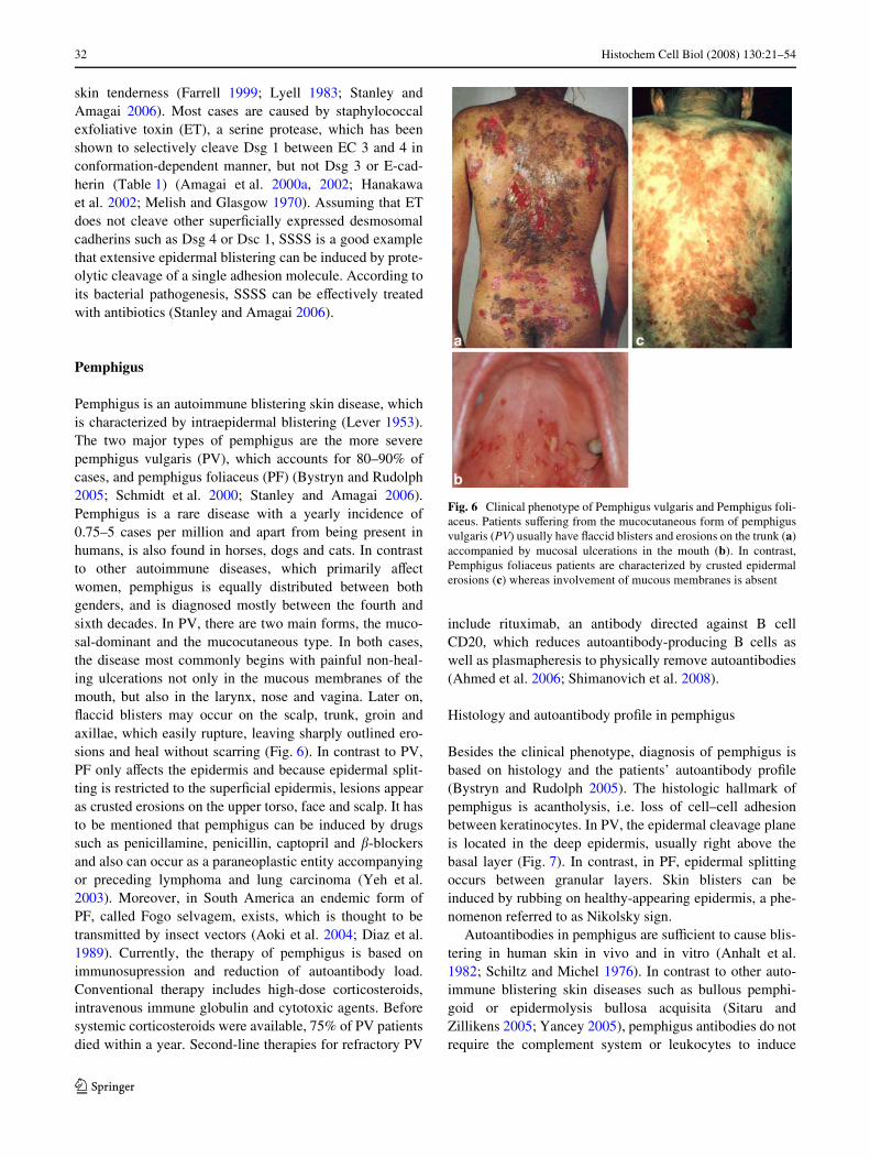
Pemphigus is an autoimmune blistering skin disease, whichis characterized by intraepidermal blistering (Lever 1953).The two major types of pemphigus are the more severepemphigus vulgaris (PV), which accounts for 80–90% ofcases, and pemphigus foliaceus (PF) (Bystryn and Rudolph2005; Schmidt et al. 2000; Stanley and Amagai 2006).Pemphigus is a rare disease with a yearly incidence of0.75–5 cases per million and apart from being present inhumans, is also found in horses, dogs and cats. In contrastto other autoimmune diseases, which primarily aVectwomen, pemphigus is equally distributed between bothgenders, and is diagnosed mostly between the fourth andsixth decades. In PV, there are two main forms, the muco-sal-dominant and the mucocutaneous type. In both cases,the disease most commonly begins with painful non-heal-ing ulcerations not only in the mucous membranes of themouth, but also in the larynx, nose and vagina. Later on,Xaccid blisters may occur on the scalp, trunk, groin andaxillae, which easily rupture, leaving sharply outlined ero-sions and heal without scarring (Fig. 6). In contrast to PV,PF only aVects the epidermis and because epidermal split-ting is restricted to the superWcial epidermis, lesions appearas crusted erosions on the upper torso, face and scalp. It hasto be mentioned that pemphigus can be induced by drugssuch as penicillamine, penicillin, captopril and �-blockersand also can occur as a paraneoplastic entity accompanyingor preceding lymphoma and lung carcinoma (Yeh et al.2003). Moreover, in South America an endemic form ofPF, called Fogo selvagem, exists, which is thought to betransmitted by insect vectors (Aoki et al. 2004; Diaz et al.1989). Currently, the therapy of pemphigus is based onimmunosupression and reduction of autoantibody load.Conventional therapy includes high-dose corticosteroids,intravenous immune globulin and cytotoxic agents. Beforesystemic corticosteroids were available, 75% of PV patientsdied within a year. Second-line therapies for refractory PV
include rituximab, an antibody directed against B cellCD20, which reduces autoantibody-producing B cells aswell as plasmapheresis to physically remove autoantibodies(Ahmed et al. 2006; Shimanovich et al. 2008).
Histology and autoantibody proWle in pemphigus
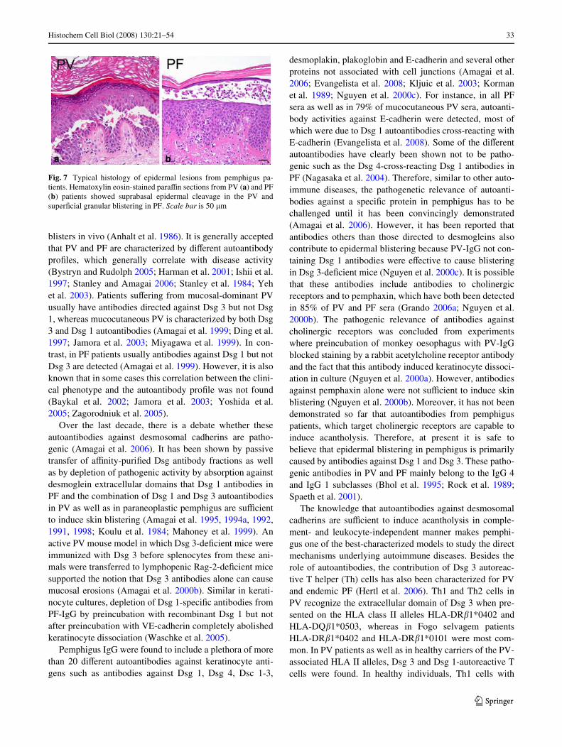
Besides the clinical phenotype, diagnosis of pemphigus isbased on histology and the patients’ autoantibody proWle(Bystryn and Rudolph 2005). The histologic hallmark ofpemphigus is acantholysis, i.e. loss of cell–cell adhesionbetween keratinocytes. In PV, the epidermal cleavage planeis located in the deep epidermis, usually right above thebasal layer (Fig. 7). In contrast, in PF, epidermal splittingoccurs between granular layers. Skin blisters can beinduced by rubbing on healthy-appearing epidermis, a phe-nomenon referred to as Nikolsky sign.
Autoantibodies in pemphigus are suYcient to cause blis-tering in human skin in vivo and in vitro (Anhalt et al.1982; Schiltz and Michel 1976). In contrast to other auto-immune blistering skin diseases such as bullous pemphi-goid or epidermolysis bullosa acquisita (Sitaru andZillikens 2005; Yancey 2005), pemphigus antibodies do notrequire the complement system or leukocytes to induce
Fig. 6 Clinical phenotype of Pemphigus vulgaris and Pemphigus foli-aceus. Patients suVering from the mucocutaneous form of pemphigusvulgaris (PV) usually have Xaccid blisters and erosions on the trunk (a)accompanied by mucosal ulcerations in the mouth (b). In contrast,Pemphigus foliaceus patients are characterized by crusted epidermalerosions (c) whereas involvement of mucous membranes is absent
123
Histochem Cell Biol (2008) 130:21–54 33
blisters in vivo (Anhalt et al. 1986). It is generally acceptedthat PV and PF are characterized by diVerent autoantibodyproWles, which generally correlate with disease activity(Bystryn and Rudolph 2005; Harman et al. 2001; Ishii et al.1997; Stanley and Amagai 2006; Stanley et al. 1984; Yehet al. 2003). Patients suVering from mucosal-dominant PVusually have antibodies directed against Dsg 3 but not Dsg1, whereas mucocutaneous PV is characterized by both Dsg3 and Dsg 1 autoantibodies (Amagai et al. 1999; Ding et al.1997; Jamora et al. 2003; Miyagawa et al. 1999). In con-trast, in PF patients usually antibodies against Dsg 1 but notDsg 3 are detected (Amagai et al. 1999). However, it is alsoknown that in some cases this correlation between the clini-cal phenotype and the autoantibody proWle was not found(Baykal et al. 2002; Jamora et al. 2003; Yoshida et al.2005; Zagorodniuk et al. 2005).
Over the last decade, there is a debate whether theseautoantibodies against desmosomal cadherins are patho-genic (Amagai et al. 2006). It has been shown by passivetransfer of aYnity-puriWed Dsg antibody fractions as wellas by depletion of pathogenic activity by absorption againstdesmoglein extracellular domains that Dsg 1 antibodies inPF and the combination of Dsg 1 and Dsg 3 autoantibodiesin PV as well as in paraneoplastic pemphigus are suYcientto induce skin blistering (Amagai et al. 1995, 1994a, 1992,1991, 1998; Koulu et al. 1984; Mahoney et al. 1999). Anactive PV mouse model in which Dsg 3-deWcient mice wereimmunized with Dsg 3 before splenocytes from these ani-mals were transferred to lymphopenic Rag-2-deWcient micesupported the notion that Dsg 3 antibodies alone can causemucosal erosions (Amagai et al. 2000b). Similar in kerati-nocyte cultures, depletion of Dsg 1-speciWc antibodies fromPF-IgG by preincubation with recombinant Dsg 1 but notafter preincubation with VE-cadherin completely abolishedkeratinocyte dissociation (Waschke et al. 2005).
Pemphigus IgG were found to include a plethora of morethan 20 diVerent autoantibodies against keratinocyte anti-gens such as antibodies against Dsg 1, Dsg 4, Dsc 1-3,
desmoplakin, plakoglobin and E-cadherin and several otherproteins not associated with cell junctions (Amagai et al.2006; Evangelista et al. 2008; Kljuic et al. 2003; Kormanet al. 1989; Nguyen et al. 2000c). For instance, in all PFsera as well as in 79% of mucocutaneous PV sera, autoanti-body activities against E-cadherin were detected, most ofwhich were due to Dsg 1 autoantibodies cross-reacting withE-cadherin (Evangelista et al. 2008). Some of the diVerentautoantibodies have clearly been shown not to be patho-genic such as the Dsg 4-cross-reacting Dsg 1 antibodies inPF (Nagasaka et al. 2004). Therefore, similar to other auto-immune diseases, the pathogenetic relevance of autoanti-bodies against a speciWc protein in pemphigus has to bechallenged until it has been convincingly demonstrated(Amagai et al. 2006). However, it has been reported thatantibodies others than those directed to desmogleins alsocontribute to epidermal blistering because PV-IgG not con-taining Dsg 1 antibodies were eVective to cause blisteringin Dsg 3-deWcient mice (Nguyen et al. 2000c). It is possiblethat these antibodies include antibodies to cholinergicreceptors and to pemphaxin, which have both been detectedin 85% of PV and PF sera (Grando 2006a; Nguyen et al.2000b). The pathogenic relevance of antibodies againstcholinergic receptors was concluded from experimentswhere preincubation of monkey oesophagus with PV-IgGblocked staining by a rabbit acetylcholine receptor antibodyand the fact that this antibody induced keratinocyte dissoci-ation in culture (Nguyen et al. 2000a). However, antibodiesagainst pemphaxin alone were not suYcient to induce skinblistering (Nguyen et al. 2000b). Moreover, it has not beendemonstrated so far that autoantibodies from pemphiguspatients, which target cholinergic receptors are capable toinduce acantholysis. Therefore, at present it is safe tobelieve that epidermal blistering in pemphigus is primarilycaused by antibodies against Dsg 1 and Dsg 3. These patho-genic antibodies in PV and PF mainly belong to the IgG 4and IgG 1 subclasses (Bhol et al. 1995; Rock et al. 1989;Spaeth et al. 2001).
The knowledge that autoantibodies against desmosomalcadherins are suYcient to induce acantholysis in comple-ment- and leukocyte-independent manner makes pemphi-gus one of the best-characterized models to study the directmechanisms underlying autoimmune diseases. Besides therole of autoantibodies, the contribution of Dsg 3 autoreac-tive T helper (Th) cells has also been characterized for PVand endemic PF (Hertl et al. 2006). Th1 and Th2 cells inPV recognize the extracellular domain of Dsg 3 when pre-sented on the HLA class II alleles HLA-DR�1*0402 andHLA-DQ�1*0503, whereas in Fogo selvagem patientsHLA-DR�1*0402 and HLA-DR�1*0101 were most com-mon. In PV patients as well as in healthy carriers of the PV-associated HLA II alleles, Dsg 3 and Dsg 1-autoreactive Tcells were found. In healthy individuals, Th1 cells with
Fig. 7 Typical histology of epidermal lesions from pemphigus pa-tients. Hematoxylin eosin-stained paraYn sections from PV (a) and PF(b) patients showed suprabasal epidermal cleavage in the PV andsuperWcial granular blistering in PF. Scale bar is 50 �m
123
34 Histochem Cell Biol (2008) 130:21–54
characteristics of regulatory T (Tr1) cells which inhibit Tcell activation were most prevalent. In contrast, in PVpatients the levels of Tr1 cells were reduced while Th2 cellswere increased (Veldman et al. 2004). Therefore, it is pos-sible that an imbalance of autoreactive Tr1 and Th 2 cellsplays a role in the induction of PV by promoting the proli-feration of anti-Dsg 3 producing B cells.
The mechanisms underlying pemphigus skin blistering
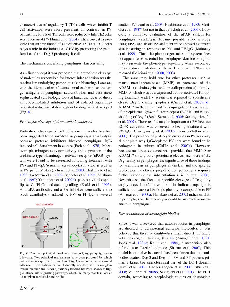
As a Wrst concept it was proposed that proteolytic cleavageof molecules responsible for intercellular adhesion was themechanism underlying pemphigus skin blistering. Later on,with the identiWcation of desmosomal cadherins as the tar-get antigens of pemphigus autoantibodies and with moresophisticated cell biologic tools at hand, the ideas of directantibody-mediated inhibition and of indirect signalling-mediated reduction of desmoglein binding were developed(Fig. 8).
Proteolytic cleavage of desmosomal cadherins
Proteolytic cleavage of cell adhesion molecules has Wrstbeen suggested to be involved in pemphigus acantholysisbecause protease inhibitors blocked pemphigus IgG-induced cell detachment in culture (Farb et al. 1978). More-over, plasminogen activator activity and expression of theurokinase-type plasminogen activator receptor (uPAR) sys-tem were found to be increased following treatment withPV- and PF-IgG/serum in keratinocytes in vitro as well asin PV patients’ skin (Feliciani et al. 2003; Hashimoto et al.1983; Lo Muzio et al. 2002; Schaefer et al. 1996; Seishimaet al. 1997; Yamamoto et al. 2007b), possibly via phospho-lipase C (PLC)-mediated signalling (Esaki et al. 1995).Anti-uPA antibodies and a PA inhibitor were suYcient toblock acantholysis induced by PV- or PF-IgG in several
studies (Feliciani et al. 2003; Hashimoto et al. 1983; Mori-oka et al. 1987) but not in that by Schuh et al. (2003). How-ever, a deWnitive evaluation of the uPAR system forpemphigus acantholysis became possible since a studyusing uPA- and tissue PA-deWcient mice showed extensiveskin blistering in response to PV- and PF-IgG (Mahoneyet al. 1999). Thus, the plasminogen activator system doesnot appear to be essential for pemphigus skin blistering butmay aggravate the phenotype, especially when secondaryinXammatory mediators such as IL-1� and TNF-� arereleased (Feliciani et al. 2000, 2003).
The same may hold true for other proteases such asmatrix metalloproteinases (MMP) or proteases of theADAM (a disintegrin and metalloproteinase) family.MMP-9, which was overexpressed but not activated follow-ing treatment with PV serum was reported to speciWcallycleave Dsg 3 during apoptosis (Cirillo et al. 2007a, d).ADAM17 on the other hand, was upregulated by activationof the epidermal growth factor receptor (EGFR) and causedshedding of Dsg 2 (Bech-Serra et al. 2006; Santiago-Josefatet al. 2007). These results may be important for PV becauseEGFR activation was observed following treatment withPV-IgG (Chernyavsky et al. 2007a; Frusic-Zlotkin et al.2006). The presence of proteolytic enzymes in PV sera mayalso explain why IgG-depleted PV sera were found to bepathogenic in culture (Cirillo et al. 2007c). However,because no direct evidence was provided that MMP-9 orADAM17 or any other proteinase cleaves members of theDsg family in pemphigus, the signiWcance of these Wndingsfor acantholysis in pemphigus is unclear and the speciWcproteolysis hypothesis proposed for pemphigus requiresfurther experimental substantiation (Cirillo et al. 2008).Nevertheless, the fact that speciWc cleavage of Dsg 1 bystaphylococcal exfoliative toxin in bullous impetigo issuYcient to cause a histologic phenotype comparable to PF(Amagai et al. 2000a; Hanakawa et al. 2002) indicates that,in principle, speciWc proteolysis could be an eVective mech-anism in pemphigus.
Direct inhibition of desmoglein binding
Since it was discovered that autoantibodies in pemphigusare directed to desmosomal adhesion molecules, it wasbelieved that these autoantibodies might directly interferewith desmoglein binding (Fig. 8) (Amagai et al. 1991;Jones et al. 1986a; Koulu et al. 1984), a mechanism alsorefered to as “steric hindrance”(Sharma et al. 2007). Thismodel is attractive because it has been shown that autoanti-bodies against Dsg 3 and Dsg 1 in PV and PF patients pri-marily target the aminoterminal part of the EC 1 domain(Futei et al. 2000; Hacker-Foegen et al. 2003; Ishii et al.2008; Muller et al. 2008b; Sekiguchi et al. 2001). The EC 1domain, according to morphologic studies on desmoglein
Fig. 8 The two principal mechanisms underlying pemphigus skinblistering. Two principal mechanisms have been proposed by whichautoantibodies speciWc for Dsg 1 and Dsg 3 could impair desmosomaladhesion. First, antibodies could directly interfere with desmogleintransinteraction (a). Second, antibody binding has been shown to trig-ger intracellular signalling pathways, which indirectly results in loss ofdesmoglein-mediated binding (b)
123
Histochem Cell Biol (2008) 130:21–54 35
transinteraction in desmosomes, is increasingly recognizedas the part of the desmosomal cadherin ectodomain, respon-sible for trans-interaction (Al-Amoudi et al. 2007; He et al.2003) and may harbour the putative transadhesive interface,based on data from the crystal structure of classical cadher-ins (Boggon et al. 2002; Overduin et al. 1995; Shapiro et al.1995). Moreover, it seems that autoantibody reactivity tothe aminoterminal parts (EC 1) of the Dsg 3 ectodomaincorrelates with high disease activity as well as epidermal ormucosal involvement in PV although the titers of theseantibodies do not show this correlation (Amagai et al. 1992;Muller et al. 2006, 2008b; Salato et al. 2005).
First functional data that anti-Dsg 3 antibodies in PVmay directly interfere with Dsg 3 binding were providedusing monoclonal antibodies derived from the active PVmouse model (Amagai et al. 2000b). AK 23, which wasdirected against the aminoterminal part of EC 1 was foundto be pathogenic and capable to induce epidermal blisteringin vivo, at least when PF-IgG or exfoliative toxin A wasadded to inactivate Dsg 1 (Shimizu et al. 2005; Tsunodaet al. 2003). Antibodies to other parts of the Dsg 3 extra-cellular domain such as AK 9 and AK 18 were ineVective toinduce blisters. Recently, by using single-molecule atomicforce microscopy (AFM), it was shown that PV-IgG as wellas AK 23 directly interfered with homophilic Dsg 3 bindingunder cell free conditions (Heupel et al. 2007) which sup-ports the hypothesis of direct inhibition of Dsg 3 binding inPV (Stanley and Amagai 2006). However, no direct inhibi-tion of Dsg 1 binding by PV-IgG and PF-IgG was detectedby AFM. These autoantibodies induced keratinocyte disso-ciation and reduced binding of both Dsg 3- and Dsg 1-coated microbeads to the surface of cultured keratinocytes,as revealed by laser trapping (Heupel et al. 2007; Waschkeet al. 2005). These data suggest that autoantobodies inter-fere with Dsg 1 binding rather by indirect, cell-dependentmechanisms.
Finally, it has to be noted that, if direct inhibition occurs,it is not possible to discriminate at present whether interfer-ence with Dsg 3 binding in PV was mediated by steric hin-drance, i.e. by blocking trans-interaction of desmogleinmolecules by the bound autoantibody, or rather by alloste-ric eVects, i.e. autoantibody-induced conformationalchanges of the Dsg 3 ectodomain, which in turn interferewith Dsg 3 transinteraction. An antibody directed againstthe putative transadhesive interface may directly inducesteric hindrance, whereas antibodies directed against otherparts of the desmoglein ectodomain could indirectly inhibitdesmoglein binding by allosteric mechanisms. The fact thatAK 18 and AK 9, which were directed to the middle andthe carboxyterminal parts of the Dsg 3 ectodomain, werenot pathogenic and did not directly interfere with Dsg 3binding suggests that these speciWc antibodies were notcapable of causing allosteric hindrance (Heupel et al. 2007;
Tsunoda et al. 2003). On the other hand, an antibodydirected against the EC2 domain, although this part of themolecule may not be involved in transinteraction, mightbe large enough to cause steric hindrance of Dsg 3 tran-sinteraction. Therefore, “direct inhibition”, instead of“steric hindrance” of desmoglein binding should be useduntil discrimination between steric and allosteric eVects ispossible.
Desmoglein compensation in pemphigus
The desmoglein compensation hypothesis was proposed toexplain the diVerent clinical phenotypes of PV and PF onthe basis of their diVerent autoantibody proWles (Amagai2003; Payne et al. 2004; Sharma et al. 2007; Shirakata et al.1998; Stanley and Amagai 2006; Udey and Stanley 1999).According to this concept, in the deep epidermis whichcontains both Dsg 1 and Dsg 3, Dsg 3 compensates for thefunctional loss of Dsg 1 induced by Dsg 1-speciWc autoanti-bodies, resulting in more superWcial blistering in PF(Fig. 9). In PV, when only Dsg 3 antibodies are present, noepidermal blistering would occur because Dsg 1 is consid-ered to compensate for autoantibody-induced loss of Dsg 3binding. However, acantholysis occurs in mucous mem-branes where Dsg 3 is assumed to be the predominantlyexpressed Dsg isoform, leading to the phenotype of muco-sal-dominant PV. When autoantibodies to Dsg 1 are alsoproduced in PV, epidermal blistering occurs. However, it isunclear why the cleavage plane is restricted to the deep epi-dermis in PV since in PF Dsg 1 autoantibodies cause super-Wcial blistering (Fig. 10). For this reason and other reasonssuch as the cases of pemphigus in which the autoantibodyproWles do not correlate with the clinical phenotype or thepresence of other desmosomal cadherin isoforms in the epi-dermis, this concept has been challenged (Amagai et al.2006; Bystryn and Rudolph 2005; Muller et al. 2002; Spin-dler et al. 2007).
Experimental support for the desmoglein compensationhypothesis in vivo was obtained in mice. It was shown thatPF-IgG were suYcient to cause skin blistering in Dsg 3-deWcient mice but not in normal mice (Mahoney et al.1999). In skin layers where Dsg 1 and Dsg 3 were found,autoantibodies against both desmogleins were required forblistering. In line with these Wndings, forced expression ofDsg 3 in the superWcial epidermis abolished the ability ofPF-IgG to induce acantholysis in mice (Wu et al. 2000). Incontrast, in human skin and in cultured human keratino-cytes in vitro, PF-IgG were eVective to induce acantholysisdespite of the presence of both Dsg 1 and Dsg 3 (Spindleret al. 2007). The discrepancy between these conXictingWndings may be explained in part by the notion that thedesmoglein compensation hypothesis is based on the fol-lowing two assumptions: (1) the expression pattern of Dsg
123

36 Histochem Cell Biol (2008) 130:21–54
3 and Dsg 1 do not substantially overlap in epidermal andmucosal layers where the cleavage plane in PV and PF islocated. (2) Dsg 1- and Dsg 3-speciWc autoantibodies onlylead to inactivation of either Dsg 1 or Dsg 3, respectively.Because of the latter, the desmoglein compensation hypo-thesis has been used to promote the idea that autoantibodiesreduce Dsg binding by direct inhibition rather than byunspeciWc proteolysis (Mahoney et al. 1999).
Regarding the distribution of Dsg 1 and Dsg 3, it isimportant to note that Dsg 3 expression patterns in speciWcepidermal layers are diVerent in mice and men. In mice,expression of Dsg 3 is restricted to the basal and immedi-ately suprabasal epidermal layer (Mahoney et al. 2006,1999). In human skin, when PV and PF were used for stain-ing, a similar staining pattern was revealed (Amagai et al.1996; Shimizu et al. 1995). In contrast, when speciWc anti-bodies or in situ hybridisation were used for Dsg 3 mappingin human epidermis, it was demonstrated that Dsg 3 is pres-ent throughout the spinous layers and thus Dsg 3 distribu-tion showed substantial overlap with expression of Dsg 1(Arnemann et al. 1993; Mahoney et al. 2006; Spindler et al.2007). However, immunostaining of human epidermisusing another monoclonal antibody detected expression ofDsg 3 in the lower epidermis only (Wu et al. 2000). In oral
mucosa, equally strong expression of Dsg 1 and Dsg 3 wasfound throughout the epithelium when speciWc antibodieswere used (Mahoney et al. 2006), whereas Dsg 1 stainingintensity was found to be much lower when PV-IgG wereused for immunstaining (Shirakata et al. 1998). Takentogether, the expression patterns of Dsg 1 and Dsg 3broadly overlap in human epidermis and appear to be iden-tical in oral mucosa.
With respect to the second assumption the desmogleincompensation is based on, i.e. selective inactivation of Dsg 1but not of Dsg 3 by Dsg 1-speciWc antibodies, it wasshown recently that both PF-IgG (only containing Dsg 1-speciWc antibodies) and PV-IgG from patients with onlyDsg 3-speciWc antibodies were equally eVective to reducebinding of Dsg 1- and Dsg 3-coated beads to the surface ofcultured keratinocytes (Heupel et al. 2007; Spindler et al.2007). These data indicate that PV-IgG and PF-IgG canreduce binding of Dsg 1 and Dsg 3, at least on the keratino-cyte cell surface. Taken together, the relevance of desmog-lein compensation for pemphigus pathogenesis in humanscannot be concluded from experiments in mice, especiallybecause distribution patterns of Dsg 1 and Dsg 3 substan-tially diVer in the two species. Therefore, alternative mod-els have to be worked out to explain the diVerent epidermal
Fig. 9 The desmoglein compensation hypothesis. Based on the diVer-ent autoantibody proWles in PV and PF together with the Wndings thatDsg 3 is present in the deep epidermis only whereas Dsg 1 is primarilyexpressed in the superWcial epidermis, the desmoglein compensationhypothesis has been proposed to explain the epidermal cleavage planestypical for PV and PF. According to this model, blistering in PF aVects
the superWcial epidermis because Dsg 3 is present in the deep epider-mis to compensate for the autoantibody-induced loss of Dsg 1 binding.In PV, epidermal involvement would occur only when autoantibodiesagainst both Dsg 1 and Dsg 3 are present because Dsg 1 is found in allepidermal layers and could compensate for loss of Dsg 3 binding whenantibodies to Dsg 3 are solely present
123
Histochem Cell Biol (2008) 130:21–54 37
cleavage planes in PV and PF. These may involve diVerentsignalling pathways required for maintenance of desmo-somal adhesion in the speciWc epidermal layers as outlinedbelow.
Signalling pathways in pemphigus and in desmosome disassembly
Since it has been shown that PV-IgG upon binding to kerat-inocytes induced a rapid transient increase of intracellularCa2+ (Seishima et al. 1995), several signalling pathwayshave been shown to be involved in pemphigus pathogenesis(Fig. 8) (Kitajima 2002; Lanza et al. 2006; Sharma et al.2007; Sitaru and Zillikens 2005). Interestingly, evidencehas been provided that transadhering non-desmosomal cad-herins, for instance Dsg 3, are involved in “outside-in” sig-nalling and that binding of pemphigus IgG interferes withthis function (Muller et al. 2008a). This can be concludedfrom experiments which showed that autoantibody bindingas well as keratinocyte separation started between desmo-somes (Sato et al. 2000; Takahashi et al. 1985) and thatnon-junctional Dsg 3 and plakoglobin were depleted Wrstbefore changes in the desmosomal fractions were present(Aoyama and Kitajima 1999; Williamson et al. 2006;Yamamoto et al. 2007a). Interestingly, to trigger Dsg-induced signalling, autoantibody-mediated cross-linking ofDsg 3 and Dsg 1 seems not to be required because monova-lent Fab fragments and single-chain variants of PV- andPF-IgG were eVective to cause skin blistering in vivo and todisrupt the desmosomal plaque in vitro (de Bruin et al.2007; Ishii et al. 2008; Payne et al. 2005; Rock et al. 1990).
Ca2+, PLC and PKC It was shown that PV-IgG caused arapid, transient phospholipase C (PLC)-dependent increaseof inositol 1,4,5 trisphosphate and of intracellular Ca2+
leading to activation of both PKC and plasminogen activa-tor (PA) (Esaki et al. 1995; Kitajima et al. 1999; Memaret al. 1996; Osada et al. 1997; Seishima et al. 1995, 1999).Because a chelator of intracellular free Ca2+ blocked kerati-nocyte dissociation in vitro and inhibitors of calmodulin,PLC and PKC were eVective to block PV-IgG-inducedacantholysis in vivo (Arredondo et al. 2005; Sanchez-Car-pintero et al. 2004), it is possible that this signalling path-way may be involved in PV acantholysis. However,because the PA system is not believed to be crucial in thisprocess, PKC signalling may contribute to PV acantholysis
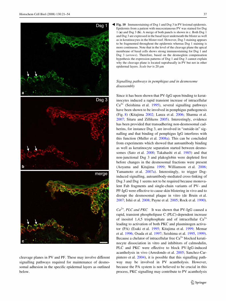
Fig. 10 Immunostaining of Dsg 1 and Dsg 3 in PV lesional epidermis.Epidermis from a patient with mucocutaneous PV was stained for Dsg1 (a) and Dsg 3 (b). A merge of both panels is shown in c. Both Dsg 1and Dsg 3 are expressed in the basal layer underneath the blister as wellas in keratinocytes in the blister roof. However, Dsg 3 staining appearsto be fragmented throughout the epidermis whereas Dsg 1 staining ismore continuous. Note that in the level of the cleavage plane the apicalmembrane of basal cells shows strong immunostaining for Dsg 1 andDsg 3 (arrows). Therefore, based on the desmoglein compensationhypothesis the expression patterns of Dsg 1 and Dsg 3 cannot explainwhy the cleavage plane is located suprabasally in PV but not in otherepidermal layers. Scale bar is 20 �m
�
123
38 Histochem Cell Biol (2008) 130:21–54
by other pathways such as phosphorylation of �-catenin inadherens junctions (Chernyavsky et al. 2007b). Thishypothesis is supported by experiments, which showed thatkeratinocyte adhesion was negatively regulated by pharma-cologic PKC activation (Kimura et al. 2007).
p38MAPK Activation of p38MAPK at present is the mostpromising signalling mechanism to be responsible for acan-tholysis in pemphigus. It has been demonstrated in vivothat p38MAPK and one of its downstream targets, heatshock protein (HSP) 25, were phosphorylated in responseto PV-IgG and PF-IgG and that pharmacologic inhibition ofp38MAPK abolished blister formation (Berkowitz et al.2006, 2007b). Similarly, p38MAPK and the human homo-log HSP 27 were found to be phosphorylated in skin lesionsof PV and PF patients (Berkowitz et al. 2007a). In culturedhuman keratinocytes, phosphorylation of p38MAPK andHSP27 occured after 30 min of exposure to PV-IgG(Berkowitz et al. 2005; Kawasaki et al. 2006). However,another study found that activity of p38MAPK was notincreased before 120 min and that activity peaked after240 min of PV-IgG treatment (Chernyavsky et al. 2007a).In the latter study it was shown that activation was medi-ated, at least in part, by Dsg 1- and/or Dsg 3-speciWc anti-bodies because Dsg depletion by siRNA reducedp38MAPK activation by 50%. Inhibition of p38MAPKblocked autoantibody-induced keratinocyte dissociation,Rho A inactivation, cytokeratin retraction and reorganiza-tion of the actin cytoskeleton (Berkowitz et al. 2005; Cher-nyavsky et al. 2007a; Waschke et al. 2006). These resultsdemonstrate that p38MAPK is involved in the mechanismsleading to acantholysis and that inhibition of p38MAPKcould be a beneWcial approach to treat pemphigus patients.Nevertheless, based on the Wnding that activation ofp38MAPK can also be a consequence of cell detachment inrat intestinal epithelium (Rosen et al. 2002), it was pro-posed that p38MAPK activation is a consequence of celldissociation (Sharma et al. 2007). However, this is unlikelybecause signiWcant pemphigus-IgG-induced acantholysisusually takes 12–24 h to occur (Berkowitz et al. 2006; Cal-delari et al. 2001; Lanza et al. 2008; Mahoney et al. 1999;Nagasaka et al. 2004; Nguyen et al. 2000c; Shu et al. 2007;Spindler et al. 2007; Takahashi et al. 1985; Tsunoda et al.2003; Waschke et al. 2005; Waschke et al. 2006; William-son et al. 2006).
The mechanisms by which p38MAPK and HSP27 leadto keratinocyte dissociation are largely unclear but mayinvolve reorganization of the keratinocyte cytoskeleton. Ithas been shown recently that serine phosphorylation ofcytokeratin 8 by p38MAPK induced cytokeratin networkdisassembly (Woll et al. 2007). Similarly, HSP27 wasfound to associate with cytokeratin Wlaments and to inhibitassembly of glial Wbrillary acidic protein (GFAP) (Perng
et al. 1999). Similarly, HSP25 was found to interact withactin Wlaments and to prevent formation of aggregates ofthermally denatured actin (Panasenko et al. 2003). How-ever, because p38MAPK-mediated phosphorylation ofHSP27 or HSP 25 promoted actin assembly and stabilizedF-actin against depolymerization in response to cytochala-sin D or heat shock (Benndorf et al. 1994; Geum et al.2002; Guay et al. 1997), one would expect that PV-IgG-induced HSP27 phosphorylation also would stabilize actinWlaments. Therefore, it is more likely that HSP27 is part ofa salvage pathway in response to PV-IgG and does notdirectly contribute to PV-IgG-induced actin reorganization.
Rho A GTPase Rho GTPases are important regulators ofthe cytoskeleton and of cell adhesion (Braga and Yap 2005;Bustelo et al. 2007; Fukata et al. 1999; JaVe and Hall2005). In the epidermis, Rac 1 was found not to be crucialfor maintaining epithelial integrity but for stem cell diVer-entiation, presumably via negative regulation of c-Mycexpression (Benitah et al. 2005; Chrostek et al. 2006).Recently, it has been shown that PV- and PF-IgG-inducedepidermal splitting, keratinocyte dissociation, as well asloss of Dsg 1 and Dsg 3 binding in vitro were accompaniedby p38MAPK-dependent inactivation of Rho A and thatspeciWc activation of Rho A by the bacterial toxin cytotoxicnecrotizing factor y (CNFy) abolished these eVects (Spin-dler et al. 2007; Waschke et al. 2006). Moreover, toxin-mediated inactivation of Rho A and Rac 1 resulted in epi-dermal splitting, keratinocyte dissociation and actin reorga-nization similar to treatment with pemphigus IgG.Especially, inactivation of Rho GTPases by toxin B causeddeep epidermal acantholysis comparable to the eVects ofPV-IgG. Based on this observation, it is tempting to specu-late that the cleavage plane in pemphigus may in part beexplained by diVerent, layer-speciWc signalling pathwaysimportant for keratinocyte cohesion.
Because cytokeratin retraction and actin reorganizationin response to pemphigus IgG were also abrogated by acti-vation of Rho A, these results suggested that Rho A couldbe involved in the regulation of desmoglein cytoskeletalanchorage (Waschke et al. 2006). This idea was supportedby Triton extraction experiments which showed that Rho Aactivation reduced Dsg 3 in the non-cytoskeleton boundfraction. Alternatively, Rho GTPases may regulate endo-cytosis of desmosomal cadherins similar to their role inE-cadherin turnover (Akhtar and Hotchin 2001; Izumi et al.2004; Kamei et al. 1999). Interestingly, endocytosis anddepletion of Dsg3 are increasingly recognized to contributeto pemphigus acantholysis (Calkins et al. 2006; Yamamotoet al. 2007a). Taken together, Rho GTPases seem to beimportant for maintenance of desmosomes and activationof Rho A could be used to develop new strategies in pem-phigus treatment.
123

Histochem Cell Biol (2008) 130:21–54 39
These data are in contrast to previous Wndings, whichsuggested that Rho GTPases are not involved in the regula-tion of desmosomal adhesion. This was concluded fromexperiments in keratinocytes which showed that Rac 1inactivation by transfection with a dominant-inactivemutant as well as inactivation of Rho A by C3 toxin for 25min was suYcient to displace E-cadherin but not desmopla-kin from cell junctions (Braga et al. 1997). These studiesled to the hypothesis that Rho GTPases are important regu-lators of keratinocyte adherens junctions but not desmo-somes (Braga and Yap 2005). However, becauseinactivation of Rho A by a cell-permeable C3 fusion toxinwas suYcient to displace Dsg 3 from cell borders after180 min, the negative results from the early study might becaused by the shorter incubation period (Waschke et al.2006).
Plakoglobin Besides its involvement in PV pathogenesis,plakoglobin is important for several aspects of desmosomaladhesion. Plakoglobin deWciency resulted in subcornealacantholysis, loss of desmosomes, and impaired cytoskele-tal anchorage of desmoplakin and desmogleins in vivo andin vitro, supporting the notion that plakoglobin is an impor-tant cytoskeletal linker and is crucial for desmosomeassembly (Bierkamp et al. 1999; Yin et al. 2005). However,at least in part, plakoglobin functions seem to be compen-sated by other desmosomal plaque proteins such as plako-philin 1 because a plakoglobin-deWcient keratinocyte cellline displayed Dsg 3 cytoskeletal anchorage and keratino-cyte aggregation similar to wild-type cells (Caldelari et al.2001). The linker function of plakoglobin seems to be regu-lated by tyrosine phosphorylation of plakoglobin becausefollowing EGFR activation, phosphorylated plakoglobinremained associated with Dsg 2 but not with desmoplakin(Gaudry et al. 2001). Moreover, plakoglobin phosphoryla-tion was shown to be required for EGFR-induced loss ofcell adhesion and to regulate binding of plakoglobin to des-moplakin (Miravet et al. 2003; Yin et al. 2005). These dataindicate that plakoglobin is required to integrate the eVectsfrom extracellular cues and from diVerent signalling path-ways in order to allow coordinated modulation of desmo-somal adhesion. In addition, plakoglobin regulates the turnover of desmosomal components because the protein levelsof desmogleins as well as of plakoglobin and desmoplakinwere decreased in plakoglobin-deWcient cells (Yin et al.2005).
With respect to pemphigus, it has been shown thatplakoglobin is critical for PV pathogenesis because kerati-nocytes from plakoglobin-deWcient mice were resistant toPV-IgG-induced keratinocyte dissociation, cytokeratin retrac-tion and disruption of the desmosomal plaque (Caldelariet al. 2001; de Bruin et al. 2007). These studies providedWrst evidence that direct inhibition of Dsg binding alone
cannot account for acantholysis in PV and raised thehypothesis that plakoglobin could be part of a receptorcomplex required to transfer the signal from autoantibody-bound Dsg 3 into the cell, a phenomenon referred to as“outside-in” signalling (Muller et al. 2008a). As outlinedabove, it has been shown that plakoglobin is involved inc-Myc repression, which is required for keratinocyte diVer-entiation and that PV-IgG by depleting noncytokeratin-anchored plakoglobin on the cell surface also led to c-Mycoverexpression (Williamson et al. 2006). However, as dis-cussed below, the role of c-Myc signalling in acantholysisis unclear at present.
EGFR The involvement of the EGFR in PV pathogenesishas been reported by showing that EGFR activity increasedafter 30 min and peaked after 60 min of PV-IgG incubation.This was followed by activation of ERK1/2, c-Jun phos-phorylation and Wnally by keratinocyte apoptosis (Cher-nyavsky et al. 2007a; Frusic-Zlotkin et al. 2006). Inhibitionof EGFR reduced EGFR signalling as well as apoptosis andacantholysis (Frusic-Zlotkin et al. 2006). This Wnding wassurprising because EGFR signalling usually is consideredto promote cell survival and proliferation (Bazley and Gul-lick 2005; Muller et al. 2008a). However, because 60 hwere required to induce acantholysis, very high concentra-tions of IgG (5 mg/ml) were used and activation ofp38MAPK was not detected in this study, the signiWcanceof these results is unclear.
EGFR could contribute to PV pathogenesis by stimu-lating desmosome disassembly. It has been shown that EGF-induced loss of keratinocyte aggregation was mediated byplakoglobin phosphorylation, which led to uncoupling ofthe desmoglein 2-plakoglobin complex from desmoplakinand thus from the cytoskeleton (Gaudry et al. 2001; Yinet al. 2005). Moreover, EGFR seems to negatively regulatethe protein levels of desmosomal cadherins, in part by pro-moting their metalloproteinase-mediated degradation, aswell as the incorporation of desmosomal cadherins into thedesmosomal plaque (Bech-Serra et al. 2006; Lorch et al.2004; Santiago-Josefat et al. 2007).
Src The tyrosine kinase Src is activated by PV-IgG within30 min and seems to contribute to EGFR and p38MAPKactivation because inhibition of Src reduced EGFR andp38MAPK phosphorylation by 45 and 30%, respectively(Chernyavsky et al. 2007a). Because Src inhibition reducedPV-IgG-induced loss of keratinocyte cohesion, cytokeratinretraction and apoptosis in vitro and inhition of tyrosinkinases blocked PV-IgG-induced acantholysis in vivo(Chernyavsky et al. 2007a; Sanchez-Carpintero et al.2004), these results indicate that Src is signiWcantlyinvolved in PV acantholysis. Besides activation ofp38MAPK and EGFR, a recent study provided another
123
40 Histochem Cell Biol (2008) 130:21–54
possible underlying mechanism: Src was shown to directlyphosphorylate p120catenin in response to PV-IgG, whichcorrelated with the degree of acantholysis (Chernyavskyet al. 2007b). Because p120catenin is involved in stabiliza-tion of classical cadherins such as E-cadherin on the cellsurface as well as in cadherin-dependent regulation of RhoA and Rac 1 activity (Alema and Salvatore 2007; Kow-alczyk and Reynolds 2004), Src-mediated phosphorylationof p120catenin may be important for PV pathogenesis.
Cholinergic receptors The cholinergic system of thehuman epidermis involves two classes of cholinergig recep-tors, the nicotinic and the muscarinic acetylcholine recep-tors (nAChR and mAChR), which in keratinocytes areinvolved in the regulation of cell–cell and cell–matrix adhe-sion as well as in cell migration (Grando 2006a). As out-lined above, it is unclear whether autoantibodies againstcholinergic receptors in pemphigus directly contribute toacantholysis. However, it was shown that cholinergic ago-nists can ameliorate PV acantholysis in vivo and in vitro(Nguyen et al. 2004b) and that local application of cholin-ergic agonists such as pilocarpine had therapeutic eVect onoral and skin lesions in PV patients (Iraji and YooseW 2006;Namazi 2004). Cholinergic agonists increased proteinlevels of Dsg 1, Dsg 3 and E-cadherin and antagonists tocholinergic receptors resulted in keratinocyte dissociation,which was paralleled by phosphorylation of these mole-cules (Grando 2006a). Recently, a mechanism by whichsignalling from cholinergic receptors could directly inter-fere with PV-IgG-induced eVects was demonstrated. TheM1 mAChR agonist pilocarpine inhibited PV-IgG-inducedacantholysis by reducing serine phosphorylation of �-cate-nin and Src-mediated tyrosine phosphorylation ofp120catenin by activation of the speciWc protein phospha-tases (Chernyavsky et al. 2007b). Similarly, an agonist ofthe � 7 nAChR reduced p120catenin phosphorylation bothon the level of Src activation as well as by activation of thetyrosine phosphatase. Moreover, it was shown that M3 andM4 receptors can lead to activation of Rho A and that car-bachol, which activates � 3 and � 7 nAChR, activates RhoA, Rac 1 and Cdc42 (Chernyavsky et al. 2004a, b; Ruiz-Velasco et al. 2002), which also might be involved in theinhibition of PV-IgG-induced acantholysis. Taken together,these data indicate that activation of cholinergic receptorscould provide a promising strategy to treat PV patients.
Regulation of cell cycle and gene expression Recent datasupport the hypothesis that PV-IgG change expression pat-terns of molecules involved in cell cycle regulation and thatthese molecules might be involved in acantholysis. As out-lined above, PV-IgG-induced plakoglobin depletionresulted in c-Myc overexpression in cultured keratinocytes(Muller et al. 2008a; Williamson et al. 2006). Accordingly,
c-Myc overexpression was found in the epidermis of PVpatients, but interestingly not in PF skin, indicating that thismechanism may only be important for PV (Williamsonet al. 2007, 2006). Because pharmacologic inhibition ofc-Myc as well as of plakoglobin degradation by blockingGSK 3 abrogated pemphigus-IgG-induced skin blistering,these results indicate that this mechanism could also beimportant to induce acantholysis. However, the Wndingsthat c-Myc overexpression was found after 24 h, whereasPV-IgG-induced loss of cell aggregation was observed asearly as after 12 h and that c-Myc overexpression wasabsent in PF skin indicate that c-Myc overexpression likelycontributes to ongoing proliferation of keratinocytes in PV,whereas it seems not to be essential for acantholysis. There-fore, other mechanisms must exist in addition to inducekeratinocyte dissociation. These mechanisms may, amongstothers, involve expression of genes diVerent from c-Myc,which is suggested by the fact that plakoglobin deWciencyresulted in protection of keratinocytes from apoptosis, pos-sibly via overexpression of the anti-apoptotic molecule Bcl-XL (Dusek et al. 2007a).
Similar to c-Myc, cyclin-dependent kinase 2 (cdk2),another kinase involved in the regulation of cell cycle pro-gression, seems to be involved in keratinocyte proliferationand acantholysis in pemphigus (Lanza et al. 2008). PVserum increased protein levels of cdk2 and PV serum-induced acantholysis in vivo was abolished by pharmaco-logic inhibition of cdk2. Moreover, siRNA-mediated down-regulation of cdk2 blocked cell dissociation in culturedkeratinocytes indicating that cdk2 may be important for PVacantholysis in vivo and in vitro. However, at present, it isunclear how continuing keratinocyte proliferation in conse-quence to increased c-Myc and cdk2 signalling contributesto acantholysis in PV.
One explanation is that reduced expression of desmo-somal components in hyperproliferating cells may fosterthe loss of keratinocyte cohesion in pemphigus. PV-IgG donot only induce direct signalling eVects on cell junctionsand the cytoskeleton but also change gene expression pat-terns, which may also contribute to pemphigus pathogene-sis. It has been shown by DNA microarray that PV-IgGwithin 8 h down-regulated transcription of 198 genesincluding genes encoding for adhesion molecules includingDsg 3 and desmoplakin, cytoskeletal proteins such as diVer-ent cytokeratins as well as molecules involved in cell cycleregulation such as p53 and cyclin D2 in cultured keratino-cytes and that expression of some of these genes was anta-gonistically regulated by methylprednisolone (Nguyen et al.2004a). When PV serum was used for 24 h, the expressionof even more genes was altered. In cultured human kerati-nocytes, transcription of 231 genes was decreased includingDsc 2 and plakophilin 3 whereas transcription of 329 geneswas increased. In vivo, expression of 1114 genes was
123
Histochem Cell Biol (2008) 130:21–54 41
reduced whereas transcription of 349 other genes wasinceased (Lanza et al. 2008). Pharmacologic inhibition ofcdk2 blunted the eVect of PV-IgG on the expression ofmost of the genes including desmoplakin and also blockedPV-IgG-induced acantholysis. Thus, it is conceivable thataltered desmosome formation may be involved in themechanisms underlying keratinocyte dissociation inpemphigus.
Apoptosis Apoptosis has been detected in skin lesions andin perilesional skin of PV and PF patients (Gniadecki et al.1998; Puviani et al. 2003; Wang et al. 2004a) and hall-marks of apoptosis such as DNA fragmentation, increasedexpression of pro-apoptotic molecules Fas, FasL, Bax, p53,depetion of anti-apoptotic Bcl-2 and FLIPL as well as acti-vation of caspases 1, 3 and 8 have been observed followingtreatment of cultured keratinocytes with PV-IgG or PVserum (Arredondo et al. 2005; Baroni et al. 2004; Cher-nyavsky et al. 2007a; Frusic-Zlotkin et al. 2005, 2006; Pel-acho et al. 2004; Puviani et al. 2003; Wang et al. 2004a, b).Hence, compelling evidence has been provided for the pres-ence of programmed cell death in PV although the pheno-type of acantholytic cells is diVerent from cells undergoingapoptosis (Arredondo et al. 2005). However, for severalexperiments prolonged incubation times of 48–72 h (Arre-dondo et al. 2005; Baroni et al. 2004; Frusic-Zlotkin et al.2005, 2006; Wang et al. 2004a, b) were required whereas inmost studies from the literature, acantholysis was clearlypresent after 18–24 h (Berkowitz et al. 2006; Caldelariet al. 2001; Lanza et al. 2008; Mahoney et al. 1999; Naga-saka et al. 2004; Nguyen et al. 2000c; Shu et al. 2007; Spin-dler et al. 2007; Takahashi et al. 1985; Tsunoda et al. 2003;Waschke et al. 2005; Waschke et al. 2006; Williamsonet al. 2006). In another study, apoptosis was detected byTUNEL reactivity starting after 6 h of PV-IgG treatment(Chernyavsky et al. 2007a). However, in this study, loss ofintercellular adhesion was present after 120 min and thusbefore onset of apoptosis. Therefore, it is conceivable thatapoptosis may be an event parallel to or in consequence ofacantholysis. On the other hand, it was reported that acti-vated caspase 3 cleaves Dsg 3 (Weiske et al. 2001) and thatcaspase and calpain inhibitors can block PV-IgG-inducedacantholysis in keratinocyte monolayers and in skin organculture (Arredondo et al. 2005; Wang et al. 2004a, b;Weiske et al. 2001). Taken together, it is unclear at presentto which extent PV-IgG-induced acantholysis is caused byapoptosis.
Targets of signalling pathways in pemphigus
Pemphigus is a desmosomal disease because pathogenicautoantibodies are directed against Dsg 1 and Dsg 3. More-over, these antibodies result in the depletion of desmosomal
components from the cell surface, alterations of desmo-somal plaques and a loss of desmosomes (Aoyama and Kit-ajima 1999; Aoyama et al. 1999; Calkins et al. 2006; deBruin et al. 2007; Sato et al. 2000; Shu et al. 2007; Was-chke et al. 2006; Williamson et al. 2006; Yamamoto et al.2007a). However, it is unclear at present whether desmo-somes are the primary targets of autoantibodies or whethercomponents outside of desmosomes cause disassembly ofdesmosomes via mechanisms involving adherens junctionsor the cytoskeleton, which Wnally results in acantholysis.The latter hypothesis is supported by the observation thatintercellular spaces between desmosomes widen beforedesmosomes separate (Takahashi et al. 1985).
EVects on desmosomes and adherens junctions It is wellestablished that pemphigus autoantibodies lead to depletionof desmosomal components from desmosomes as well asfrom the cell surface. Within 30–60 min, PV-IgG inducedinternalization and lysosomal degradation of cytoskeleton-unbound Dsg 3 together with plakoglobin (Aoyama andKitajima 1999; Aoyama et al. 1999; Calkins et al. 2006;Sato et al. 2000; Williamson et al. 2006; Yamamoto et al.2007a). After 24 h, Dsg 3 but not Dsg 2, plakoglobin ordesmoplakin was also depleted from the cytoskeletal frac-tions leading to reduced total cellular Dsg 3 levels (Calkinset al. 2006; Yamamoto et al. 2007a). This eVect was medi-ated by Dsg 3-speciWc antibodies because monoclonal Dsg3 antibodies such as AK23 were eVective to induce Dsg 3depletion and this eVect correlated with the pathogenicactivity of these autoantibodies in vivo and in vitro (Shuet al. 2007; Yamamoto et al. 2007a). However, no strongcorrelation between Dsg 3 depletion and keratinocyte dis-sociation was observed. Dsg 3 depletion was observed inDJM-1 cells at 40% conXuence as well as in normal humankeratinocytes. In contrast, in conXuent HaCaT monolayers,a reduction of Dsg 3 half-life but no depletion of total cellu-lar Dsg 3 was detected (Cirillo et al. 2006; Waschke et al.2006). Because depletion of Dsg 3 was Wrst found in para-llel with serine phosphorylation of Dsg 3, it was suggestedthat phosphorylation might be required for Dsg 3 degrada-tion (Aoyama and Kitajima 1999; Aoyama et al. 1999;Bystryn and Rodriguez 1978; Rodriguez and Bystryn1977). However, phosphorylation of Dsg 3 was observed insome studies but not in others, which might be due to diVer-ent cell culture models indicating that Dsg phosphorylationis not necessary for acantholysis (Chernyavsky et al.2007b; Nguyen et al. 2004a).
Some evidence exists that depletion of Dsg 1 may alsooccur in pemphigus. However, studies to systematicallyinvestigate Dsg 1 turnover in pemphigus are lacking. Dsg 1internalization and decreased immunostaining of Dsg 1have been observed following treatment with PF-IgG/-serum and PV serum (Cirillo et al. 2007b; Waschke et al.
123
42 Histochem Cell Biol (2008) 130:21–54
2005). This is in line with previous Wndings from the litera-ture, which demonstrated that PF-IgG did not bind to kera-tinocytes in the lower epidermis of PF patients but stronglylabelled basal keratinocytes in the epidermis of healthy sub-jects suggesting that in PF the lower epidermis wasdepleted from Dsg 1 (Bystryn and Rodriguez 1978; Rodri-guez and Bystryn 1977). Taken together, it is unclear atpresent to which extent Dsg depletion from desmosomescauses antibody-mediated acantholysis. However, even ifdepletion of desmosomal components occured secondary tokeratinocyte dissociation, it likely would further aggravatethe loss of keratinocyte cohesion.
The involvement of adherens junctions in pemphiguspathogenesis is unclear at present. It has been shown thatformation of adherens junctions is a prerequisite for desmo-some assembly and �-catenin-deWcient keratinocytes hadreduced numbers of desmosmes (Vasioukhin et al. 2000).Because Rho GTPases are known to primarily regulateadhesion mediated by classical cadherins in adherens junc-tions whereas the requirement of Rho A and Rac 1 activityfor maintenance of desmosomal adhesion was reported justrecently (Braga and Yap 2005; Fukata et al. 1999; Spindleret al. 2007; Waschke et al. 2006), it was suggested thatpemphigus IgG-induced inactivation of Rho A causes dis-assembly of desmosomes via destabilization of adherensjunctions (Sharma et al. 2007). Although alterations ofadherens junctions in response to PV-IgG were negligiblecompared to the eVects on desmosomes (Calkins et al.2006; de Bruin et al. 2007; Muller et al. 2008a; Waschkeet al. 2006), this scenario cannot be completely ruled out atpresent. In fact it was reported recently that PV-IgG lead tophosphorylation of �-catenin and p120catenin by PKC andSrc, respectively (Chernyavsky et al. 2007b). However,more studies are required to address the question how cate-nin phosphorylation in adherens junctions is associatedwith keratinocyte dissociation in pemphigus.
EVects on the cytoskeleton Alterations of the cytoskele-ton have been described both in pemphigus skin lesions aswell as in keratinocytes exposed to pemphigus autoantibod-ies,. These eVects include dysorganization of cytokeratinWlaments, the actin cytoskeleton as well as of microtubules.
First, it has been described that cytokeratin Wlamentsbecome detached from cell junctions and accumulate in theperinuclear region, a phenomenon commonly referred to as“keratin retraction” (Berkowitz et al. 2005; Caldelari et al.2001; Calkins et al. 2006; Chernyavsky et al. 2007a; Jinbuet al. 1992; Kitajima et al. 1987; Waschke et al. 2006; Wil-gram et al. 1961, 1964; Williamson et al. 2006). It wasshown that cytokeratin retraction could be detected as earlyas 4–6 h after addition of PV-IgG, i.e. when Wrst alterationsof desmosomes became visible, and was fully establishedafter 24 h (Berkowitz et al. 2005; Calkins et al. 2006;
Chernyavsky et al. 2007a). However, because no cytokera-tin Wlament reorganization was observed before cell detach-ment and the desmosomes of acantholytic cells remainedanchored to cytokeratin Wlament bundles (Kitajima et al.1986; Shimizu et al. 2004; Spindler et al. 2007), it appearsthat cytokeratin retraction is not required for acantholysisbut may be a secondary event.
Recent studies demonstrated that acantholysis in pem-phigus was paralleled by alterations of actin Wlamentsincluding increased stress Wber formation as well as frag-mentation of actin Wlament bundles. Moreover, pharmaco-logical inhibition of pemphigus IgG-induced acantholysisalso was eVective to reverse the eVects on the cytoskeleton(Berkowitz et al. 2005; Spindler et al. 2007; Waschke et al.2005; Waschke et al. 2006). Similarly, microtubule distri-bution was found to be altered by pemphigus IgG (Kitajimaet al. 1986). Taken together, pemphigus IgG cause pro-found cytoskeletal reorganization but it is unclear at presentwhether these eVects contribute to acantholysis or are trig-gered consequent to keratinocyte dissociation. Neverthe-less, it is likely that alterations in the cytoskeleton mayaccount for the changes in cell shape during keratinocytedissociation, which seem to start between existing desmo-somes (Bystryn and Grando 2006; Takahashi et al. 1985).
The role of the diVerent mechanisms in blister formation
As outlined above, both direct and indirect mechanismsWnally leading to a loss of desmoglein-mediated adhesionhave been found to be involved in pemphigus acantholysis.At present, compelling evidence indicates that acantholysisin PV and in PF is initiated by cellular signalling pathwaysrather than by direct inhibition of Dsg binding (Fig. 11).This was Wrst shown using plakoglobin-deWcient keratino-cytes, which were resistant against PV-IgG-induced acan-tholysis and was further supported by the Wndings thatinhibition of p38MAPK and activation of Rho A is suY-cient to abolish PV-IgG and PF-IgG-induced skin blistering(Berkowitz et al. 2005, 2006, 2007b; Caldelari et al. 2001;Waschke et al. 2006). Moreover, low temperature-abro-gated keratinocyte dissociation but not antibody binding tothe keratinocyte cell surface indicating that direct inhibitionof cadherin transinteraction by autoantibody binding is notsuYcient to cause acantholysis (Calkins et al. 2006). Ifextracellular autoantibody-mediated direct inhibition ofDsg binding would be the primary cause of acantholysis, itis hard to imagine how this process should be blocked bymodiWed intracellular signalling or low temperature. It ispossible that antibody-mediated direct inhibition of Dsgbinding may contribute to trigger cellular signalling events(Muller et al. 2008a; Sharma et al. 2007; Tsunoda et al.2003). However, because PF-IgG were shown to inducecellular signalling events but not to directly reduce Dsg 1
123

Histochem Cell Biol (2008) 130:21–54 43
binding, it seems that direct inhibition of Dsg transinterac-tion is not essential to alter Dsg-mediated signalling (Heu-pel et al. 2007; Waschke et al. 2005).
It is tempting to speculate that the clinical phenotype ofPV may be more severe compared to PF because additionalmechanisms could be involved in PV (Fig. 11). Indeed,some mechanisms have only been found to contribute toacantholysis in PV but not in PF. For instance, direct inhibi-tion of Dsg 3 transinteraction was found in PV but not in PFunder conditions where antibodies caused acantholysis(Heupel et al. 2007). Therefore, it is possible that directinhibition as a second pathway to reduce Dsg 3 binding inPV may explain the more severe phenotype of PV. Simi-larly, depletion of Dsg 3 has been convincingly shown in
PV whereas depletion of Dsg 1 in both PV and PF is lessclear (Yamamoto et al. 2007a). Moreover, altered plakoglo-bin signalling leading to c-Myc overexpression was foundin PV patients’s skin but not in PF (Williamson et al. 2007,2006). Other pathways have been investigated for PV onlywhereas data for PF are lacking. As outlined above, inhibi-tors of calmodulin, PKC, EGFR, cdk2 as well as of tyrosinkinases have been shown to inhibit PV-IgG-induced acan-tholysis.
Finally, it has to be emphasized that desmoglein proteo-lysis, mechanical stress as well as secondary changes forexample in the extracellular Ca2+ concentration might con-tribute to skin blistering. For instance, acantholysis mayfoster skin blistering by derangement of the epidermal Ca2+
Fig. 11 The mechanisms in-volved in pemphigus acantholy-sis. Accumulating evidence indicates that PV-IgG and PF-IgG initiate keratinocyte dissociation via intracellular signalling pathways including p38 MAPK, Rho A and plakoglobin (PV only). In addition, other mechanisms such as direct inhibition of Dsg 3 binding and Dsg 3 depletion from desmosomes as well as other signalling events seem to contribute to PV pathogenesis, whereas their role for acantholysis in PF is unclear. These mechanisms may account for the more severe clinical phenotype of PV compared to PF. PLC phospholipase C, PKC protein kinase C, cdk 2 cyclin-dependent kinase 2, EGFR epidermal growth factor receptor
123
44 Histochem Cell Biol (2008) 130:21–54
gradient, which in turn would result in loss of desmosomalcadherin binding, especially when the tight junctions of thegranular layer are aVected.
Concluding remarks
The major goal for the future is to elucidate the primary sig-nalling pathways responsible for the diverse eVects of pem-phigus IgG such as inhibition of desmoglein binding,depletion of desmosomal components, loss of desmosomes,reorganization of the cytoskeleton and Wnally the inductionof acantholysis. Other pathways will be identiWed to repre-sent salvage mechanisms to rescue keratinocyte cohesion(Grando 2006b). Moreover, future experiments may revealhow the diVerent signalling pathways are involved in theregulation of desmosomal adhesion in the speciWc epider-mal layers. This knowledge may elucidate why the clea-vage planes in PV and PF usually are diVerent. The secondgoal is to characterize whether acantholysis in pemphigus iscaused by mechanisms directly targeting desmosomes orwhether cellular signalling pathways regulate desmosomaladhesion via reorganisation of the cytoskeleton or via othertypes of cell contacts such as adherens junctions.
Acknowledgments The author is grateful to Detlev Drenckhahn forhelpful comments and for critically reading the manuscript as well asto Michael Christof for Wgure design. Photographs of pemphiguspatients were generously contributed by D. Zillikens and E. Schmidt(both Department of Dermatology, University of Lübeck). Specialthanks to the members of the PTF (Wolfgang-Moritz Heupel, VolkerSpindler, Peter Engerer, Judith Gutberlet, Seraj Khan) for inspiringdiscussion. The author’s work was supported by grants from the Deut-sche Forschungsgemeinschaft (SFB 487, TP B5 to Detlev Dren-ckhahn) and the IZKF Würzburg (TP A-51).
References
Aberle H, Bierkamp C, Torchard D, Serova O, Wagner T, Natt E,Wirsching J, Heidkamper C, Montagna M, Lynch HT et al (1995)The human plakoglobin gene localizes on chromosome 17q21and is subjected to loss of heterozygosity in breast and ovariancancers. Proc Natl Acad Sci USA 92:6384–6388
Ahmed AR, Spigelman Z, Cavacini LA, Posner MR (2006) Treatmentof pemphigus vulgaris with rituximab and intravenous immuneglobulin. New Engl J Med 355:1772–1779
Akat K, Bleck CK, Lee YM, Haselmann-Weiss U, Kartenbeck J(2008) Characterization of a novel type of adherens junction inmeningiomas and the derived cell line HBL-52. Cell Tissue Res331:401–412
Akhtar N, Hotchin NA (2001) RAC1 regulates adherens junctionsthrough endocytosis of E-cadherin. Molec Biol Cell 12:847–862
Al-Amoudi A, Diez DC, Betts MJ, Frangakis AS (2007) The moleculararchitecture of cadherins in native epidermal desmosomes. Nature450:832–837
Alema S, Salvatore AM (2007) p120 catenin and phosphorylation:mechanisms and traits of an unresolved issue. Biochim BiophysActa 1773:47–58
Amagai M (2003) Desmoglein as a target in autoimmunity and infec-tion. J Am Acad Dermatol 48:244–252
Amagai M, Klaus-Kovtun V, Stanley JR (1991) Autoantibodiesagainst a novel epithelial cadherin in pemphigus vulgaris, a dis-ease of cell adhesion. Cell 67:869–877
Amagai M, Karpati S, Prussick R, Klaus-Kovtun V, Stanley JR (1992)Autoantibodies against the amino-terminal cadherin-like bindingdomain of pemphigus vulgaris antigen are pathogenic. J Clin In-vest 90:919–926
Amagai M, Hashimoto T, Shimizu N, Nishikawa T (1994a) Absorp-tion of pathogenic autoantibodies by the extracellular domain ofpemphigus vulgaris antigen (Dsg3) produced by baculovirus.J Clin Invest 94:59–67
Amagai M, Karpati S, Klaus-Kovtun V, Udey MC, Stanley JR (1994b)Extracellular domain of pemphigus vulgaris antigen (desmoglein3) mediates weak homophilic adhesion. J Invest Dermatol103:609–615
Amagai M, Hashimoto T, Green KJ, Shimizu N, Nishikawa T (1995)Antigen-speciWc immunoadsorption of pathogenic autoantibodiesin pemphigus foliaceus. J Invest Dermatol 104:895–901
Amagai M, Koch PJ, Nishikawa T, Stanley JR (1996) Pemphigus vul-garis antigen (desmoglein 3) is localized in the lower epidermis,the site of blister formation in patients. J Invest Dermatol106:351–355
Amagai M, Nishikawa T, Nousari HC, Anhalt GJ, Hashimoto T (1998)Antibodies against desmoglein 3 (pemphigus vulgaris antigen)are present in sera from patients with paraneoplastic pemphigusand cause acantholysis in vivo in neonatal mice. J Clin Invest102:775–782
Amagai M, Tsunoda K, Zillikens D, Nagai T, Nishikawa T (1999) Theclinical phenotype of pemphigus is deWned by the anti-desmog-lein autoantibody proWle. J Am Acad Dermatol 40:167–170
Amagai M, Matsuyoshi N, Wang ZH, Andl C, Stanley JR (2000a) Tox-in in bullous impetigo and staphylococcal scalded-skin syndrometargets desmoglein 1. Nat Med 6:1275–1277
Amagai M, Tsunoda K, Suzuki H, Nishifuji K, Koyasu S, NishikawaT (2000b) Use of autoantigen-knockout mice in developing an ac-tive autoimmune disease model for pemphigus. J Clin Invest105:625–631
Amagai M, Yamaguchi T, Hanakawa Y, Nishifuji K, Sugai M, StanleyJR (2002) Staphylococcal exfoliative toxin B speciWcally cleavesdesmoglein 1. J Invest Dermatol 118:845–850
Amagai M, Ahmed AR, Kitajima Y, Bystryn JC, Milner Y, GniadeckiR, Hertl M, Pincelli C, Fridkis-Hareli M, Aoyama Y, Frusic-Zlot-kin M, Muller E, David M, Mimouni D, Vind-Kezunovic D,Michel B, Mahoney M, Grando S (2006) Are desmoglein autoan-tibodies essential for the immunopathogenesis of pemphigus vul-garis, or just ‘witnesses of disease’? Exp Dermatol 15:815
Anastasiadis PZ, Reynolds AB (2001) Regulation of Rho GTPases byp120-catenin. Curr Opin Cell Biol 13:604–610
Angst BD, Khan LU, Severs NJ, Whitely K, Rothery S, Thompson RP,Magee AI, Gourdie RG (1997) Dissociated spatial patterning ofgap junctions and cell adhesion junctions during postnatal diVer-entiation of ventricular myocardium. Circ Res 80:88–94
Anhalt GJ, Labib RS, Voorhees JJ, Beals TF, Diaz LA (1982) Induc-tion of pemphigus in neonatal mice by passive transfer of IgGfrom patients with the disease. New Engl J Med 306:1189–1196
Anhalt GJ, Patel HP, Labib RS, Diaz LA, Proud D (1986) Dexameth-asone inhibits plasminogen activator activity in experimentalpemphigus in vivo but does not block acantholysis. J Immunol136:113–117
Aoki V, Millikan RC, Rivitti EA, Hans-Filho G, Eaton DP, Warren SJ,Li N, Hilario-Vargas J, HoVmann RG, Diaz LA (2004) Environ-mental risk factors in endemic pemphigus foliaceus (fogo selva-gem). J Invest Dermatol Symp Proc/Soc Invest Dermatol Inc9:34–40
123

Histochem Cell Biol (2008) 130:21–54 45
Aoyama Y, Kitajima Y (1999) Pemphigus vulgaris-IgG causes a rapiddepletion of desmoglein 3 (Dsg3) from the Triton X-100 solublepools, leading to the formation of Dsg3-depleted desmosomes ina human squamous carcinoma cell line, DJM-1 cells. J Invest Der-matol 112:67–71
Aoyama Y, Owada MK, Kitajima Y (1999) A pathogenic autoanti-body, pemphigus vulgaris-IgG, induces phosphorylation of de-smoglein 3, and its dissociation from plakoglobin in culturedkeratinocytes. Eur J Immunol 29:2233–2240
Armstrong DK, McKenna KE, Purkis PE, Green KJ, Eady RA, LeighIM, Hughes AE (1999) HaploinsuYciency of desmoplakin causesa striate subtype of palmoplantar keratoderma. Hum Mol Genet8:143–148
Arnemann J, Spurr NK, Wheeler GN, Parker AE, Buxton RS (1991)Chromosomal assignment of the human genes coding for the ma-jor proteins of the desmosome junction, desmoglein DGI (DSG),desmocollins DGII/III (DSC), desmoplakins DPI/II (DSP), andplakoglobin DPIII (JUP). Genomics 10:640–645
Arnemann J, Sullivan KH, Magee AI, King IA, Buxton RS (1993)StratiWcation-related expression of isoforms of the desmosomalcadherins in human epidermis. J Cell Sci 104(Pt 3):741–750
Arredondo J, Chernyavsky AI, Karaouni A, Grando SA (2005) Novelmechanisms of target cell death and survival and of therapeuticaction of IVIg in Pemphigus. Am J Pathol 167:1531–1544
Asimaki A, Syrris P, Wichter T, Matthias P, SaYtz JE, McKenna WJ(2007) A novel dominant mutation in plakoglobin causes arrhyth-mogenic right ventricular cardiomyopathy. Am J Hum Genet81:964–973
Baroni A, Buommino E, Paoletti I, Orlando M, Ruocco E, Ruocco V(2004) Pemphigus serum and captopril induce heat shock protein70 and inducible nitric oxide synthase overexpression, triggeringapoptosis in human keratinocytes. Br J Dermatol 150:1070–1080
Baumgartner W, Hinterdorfer P, Ness W, Raab A, Vestweber D, Schin-dler H, Drenckhahn D (2000) Cadherin interaction probed byatomic force microscopy. Proc Natl Acad Sci USA 97:4005–4010
Baumgartner W, Golenhofen N, Grundhofer N, Wiegand J, Dren-ckhahn D (2003) Ca2+ dependency of N-cadherin functionprobed by laser tweezer and atomic force microscopy. J Neurosci23:11008–11014
Baykal C, Azizlerli G, Thoma-Uszynski S, Hertl M (2002) Pemphigusvulgaris localized to the nose and cheeks. J Am Acad Dermatol47:875–880
Bazley LA, Gullick WJ (2005) The epidermal growth factor receptorfamily. Endocr Relat Cancer 12(Suppl 1):S17–S27
Bazzi H, Christiano AM (2007) Broken hearts, woolly hair, and tat-tered skin: when desmosomal adhesion goes awry. Curr Opin CellBiol 19:515–520
Bech-Serra JJ, Santiago-Josefat B, Esselens C, Saftig P, Baselga J, Ar-ribas J, Canals F (2006) Proteomic identiWcation of desmoglein 2and activated leukocyte cell adhesion molecule as substrates ofADAM17 and ADAM10 by diVerence gel electrophoresis. MolCell Biol 26:5086–5095
Benitah SA, Frye M, Glogauer M, Watt FM (2005) Stem cell depletionthrough epidermal deletion of Rac1. Science (New York, NY)309:933–935
Benndorf R, Hayess K, Ryazantsev S, Wieske M, Behlke J, Lutsch G(1994) Phosphorylation and supramolecular organization of mu-rine small heat shock protein HSP25 abolish its actin polymeriza-tion-inhibiting activity. J Biol Chem 269:20780–20784
Berkowitz P, Hu P, Liu Z, Diaz LA, Enghild JJ, Chua MP, RubensteinDS (2005) Desmosome signaling. Inhibition of p38MAPK pre-vents pemphigus vulgaris IgG-induced cytoskeleton reorganiza-tion. J Biol Chem 280:23778–23784
Berkowitz P, Hu P, Warren S, Liu Z, Diaz LA, Rubenstein DS (2006)p38MAPK inhibition prevents disease in pemphigus vulgarismice. Proc Natl Acad Sci USA 103:12855–12860
Berkowitz P, Diaz LA, Hall RP, Rubenstein DS (2007a) Induction ofp38MAPK and HSP27 phosphorylation in pemphigus patientskin. J Invest Dermatol 128(3):738–740
Berkowitz P, Liu Z, Diaz LA, Rubenstein DS (2007b) Autoantibodiesin the autoimmune disease pemphigus foliaceus induce blisteringin vivo via p38MAPK-dependent signalling. J Invest Dermatol127(Suppl):34 (abstract 201)
Beutner EH, Jordon RE (1964) Demonstration of skin antibodies insera of pemphigus vulgaris patients by indirect immunoXuores-cent staining. Proc Soc Exp Biol Med 117:505–510
Bhol K, Natarajan K, Nagarwalla N, Mohimen A, Aoki V, Ahmed AR(1995) Correlation of peptide speciWcity and IgG subclass withpathogenic and nonpathogenic autoantibodies in pemphigus vul-garis: a model for autoimmunity. Proc Natl Acad Sci USA92:5239–5243
Bierkamp C, McLaughlin KJ, Schwarz H, Huber O, Kemler R (1996)Embryonic heart and skin defects in mice lacking plakoglobin.Dev Biol 180:780–785
Bierkamp C, Schwarz H, Huber O, Kemler R (1999) Desmosomallocalization of beta-catenin in the skin of plakoglobin null-mutantmice. Development (Cambridge, England) 126:371–381
Bizzozero G (1864) Delle cellule cigliate, del reticulo Malpighianid’ell epiderme. Ann Univ Med 190
Blaschuk OW, Sullivan R, David S, Pouliot Y (1990) IdentiWcation ofa cadherin cell adhesion recognition sequence. Dev Biol139:227–229
Boggon TJ, Murray J, Chappuis-Flament S, Wong E, Gumbiner BM,Shapiro L (2002) C-cadherin ectodomain structure and implica-tions for cell adhesion mechanisms. Science (New York, NY296:1308–1313
Bonne S, van Hengel J, van Roy F (1998) Chromosomal mapping ofhuman armadillo genes belonging to the p120(ctn)/plakophilinsubfamily. Genomics 51:452–454
Bonne S, Gilbert B, Hatzfeld M, Chen X, Green KJ, van Roy F (2003)DeWning desmosomal plakophilin-3 interactions. J Cell Biol161:403–416
Bornslaeger EA, Corcoran CM, Stappenbeck TS, Green KJ (1996)Breaking the connection: displacement of the desmosomal plaqueprotein desmoplakin from cell–cell interfaces disrupts anchorageof intermediate Wlament bundles and alters intercellular junctionassembly. J Cell Biol 134:985–1001
Bornslaeger EA, Godsel LM, Corcoran CM, Park JK, Hatzfeld M,Kowalczyk AP, Green KJ (2001) Plakophilin 1 interferes withplakoglobin binding to desmoplakin, yet together with plakoglo-bin promotes clustering of desmosomal plaque complexes at cell–cell borders. J Cell Sci 114:727–738
Borrmann CM, Grund C, Kuhn C, Hofmann I, PieperhoV S, FrankeWW (2006) The area composita of adhering junctions connectingheart muscle cells of vertebrates. II. Colocalizations of desmo-somal and fascia adhaerens molecules in the intercalated disk.Euro J Cell Biol 85:469–485
Braga VM, Yap AS (2005) The challenges of abundance: epithelialjunctions and small GTPase signalling. Curr Opin Cell Biol17:466–474
Braga VM, Machesky LM, Hall A, Hotchin NA (1997) The smallGTPases Rho and Rac are required for the establishment of cad-herin-dependent cell–cell contacts. J Cell Biol 137:1421–1431
Brennan D, Hu Y, Joubeh S, Choi YW, Whitaker-Menezes D, O’BrienT, Uitto J, Rodeck U, Mahoney MG (2007) Suprabasal Dsg2expression in transgenic mouse skin confers a hyperproliferativeand apoptosis-resistant phenotype to keratinocytes. J Cell Sci120:758–771
Burdett ID, Sullivan KH (2002) Desmosome assembly in MDCK cells:transport of precursors to the cell surface occurs by two phases ofvesicular traYc and involves major changes in centrosome andGolgi location during a Ca(2+) shift. Exp Cell Res 276:296–309
123
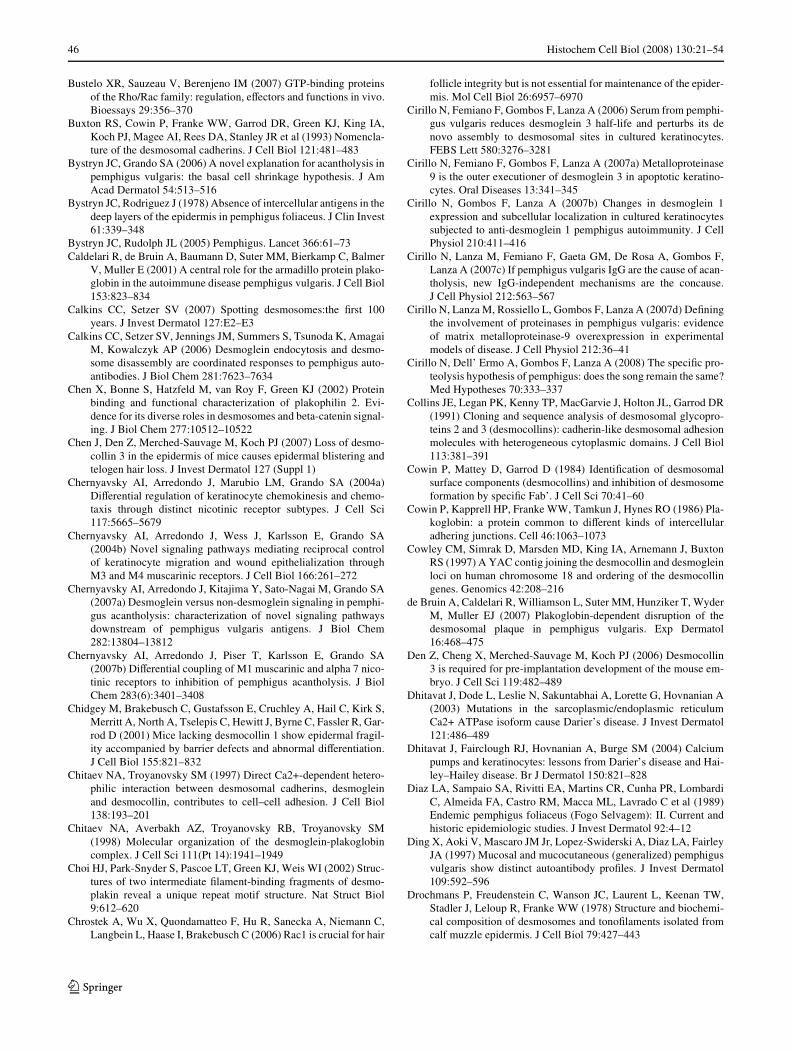
46 Histochem Cell Biol (2008) 130:21–54
Bustelo XR, Sauzeau V, Berenjeno IM (2007) GTP-binding proteinsof the Rho/Rac family: regulation, eVectors and functions in vivo.Bioessays 29:356–370
Buxton RS, Cowin P, Franke WW, Garrod DR, Green KJ, King IA,Koch PJ, Magee AI, Rees DA, Stanley JR et al (1993) Nomencla-ture of the desmosomal cadherins. J Cell Biol 121:481–483
Bystryn JC, Grando SA (2006) A novel explanation for acantholysis inpemphigus vulgaris: the basal cell shrinkage hypothesis. J AmAcad Dermatol 54:513–516
Bystryn JC, Rodriguez J (1978) Absence of intercellular antigens in thedeep layers of the epidermis in pemphigus foliaceus. J Clin Invest61:339–348
Bystryn JC, Rudolph JL (2005) Pemphigus. Lancet 366:61–73Caldelari R, de Bruin A, Baumann D, Suter MM, Bierkamp C, Balmer
V, Muller E (2001) A central role for the armadillo protein plako-globin in the autoimmune disease pemphigus vulgaris. J Cell Biol153:823–834
Calkins CC, Setzer SV (2007) Spotting desmosomes:the Wrst 100years. J Invest Dermatol 127:E2–E3
Calkins CC, Setzer SV, Jennings JM, Summers S, Tsunoda K, AmagaiM, Kowalczyk AP (2006) Desmoglein endocytosis and desmo-some disassembly are coordinated responses to pemphigus auto-antibodies. J Biol Chem 281:7623–7634
Chen X, Bonne S, Hatzfeld M, van Roy F, Green KJ (2002) Proteinbinding and functional characterization of plakophilin 2. Evi-dence for its diverse roles in desmosomes and beta-catenin signal-ing. J Biol Chem 277:10512–10522
Chen J, Den Z, Merched-Sauvage M, Koch PJ (2007) Loss of desmo-collin 3 in the epidermis of mice causes epidermal blistering andtelogen hair loss. J Invest Dermatol 127 (Suppl 1)
Chernyavsky AI, Arredondo J, Marubio LM, Grando SA (2004a)DiVerential regulation of keratinocyte chemokinesis and chemo-taxis through distinct nicotinic receptor subtypes. J Cell Sci117:5665–5679
Chernyavsky AI, Arredondo J, Wess J, Karlsson E, Grando SA(2004b) Novel signaling pathways mediating reciprocal controlof keratinocyte migration and wound epithelialization throughM3 and M4 muscarinic receptors. J Cell Biol 166:261–272
Chernyavsky AI, Arredondo J, Kitajima Y, Sato-Nagai M, Grando SA(2007a) Desmoglein versus non-desmoglein signaling in pemphi-gus acantholysis: characterization of novel signaling pathwaysdownstream of pemphigus vulgaris antigens. J Biol Chem282:13804–13812
Chernyavsky AI, Arredondo J, Piser T, Karlsson E, Grando SA(2007b) DiVerential coupling of M1 muscarinic and alpha 7 nico-tinic receptors to inhibition of pemphigus acantholysis. J BiolChem 283(6):3401–3408
Chidgey M, Brakebusch C, Gustafsson E, Cruchley A, Hail C, Kirk S,Merritt A, North A, Tselepis C, Hewitt J, Byrne C, Fassler R, Gar-rod D (2001) Mice lacking desmocollin 1 show epidermal fragil-ity accompanied by barrier defects and abnormal diVerentiation.J Cell Biol 155:821–832
Chitaev NA, Troyanovsky SM (1997) Direct Ca2+-dependent hetero-philic interaction between desmosomal cadherins, desmogleinand desmocollin, contributes to cell–cell adhesion. J Cell Biol138:193–201
Chitaev NA, Averbakh AZ, Troyanovsky RB, Troyanovsky SM(1998) Molecular organization of the desmoglein-plakoglobincomplex. J Cell Sci 111(Pt 14):1941–1949
Choi HJ, Park-Snyder S, Pascoe LT, Green KJ, Weis WI (2002) Struc-tures of two intermediate Wlament-binding fragments of desmo-plakin reveal a unique repeat motif structure. Nat Struct Biol9:612–620
Chrostek A, Wu X, Quondamatteo F, Hu R, Sanecka A, Niemann C,Langbein L, Haase I, Brakebusch C (2006) Rac1 is crucial for hair
follicle integrity but is not essential for maintenance of the epider-mis. Mol Cell Biol 26:6957–6970
Cirillo N, Femiano F, Gombos F, Lanza A (2006) Serum from pemphi-gus vulgaris reduces desmoglein 3 half-life and perturbs its denovo assembly to desmosomal sites in cultured keratinocytes.FEBS Lett 580:3276–3281
Cirillo N, Femiano F, Gombos F, Lanza A (2007a) Metalloproteinase9 is the outer executioner of desmoglein 3 in apoptotic keratino-cytes. Oral Diseases 13:341–345
Cirillo N, Gombos F, Lanza A (2007b) Changes in desmoglein 1expression and subcellular localization in cultured keratinocytessubjected to anti-desmoglein 1 pemphigus autoimmunity. J CellPhysiol 210:411–416
Cirillo N, Lanza M, Femiano F, Gaeta GM, De Rosa A, Gombos F,Lanza A (2007c) If pemphigus vulgaris IgG are the cause of acan-tholysis, new IgG-independent mechanisms are the concause.J Cell Physiol 212:563–567
Cirillo N, Lanza M, Rossiello L, Gombos F, Lanza A (2007d) DeWningthe involvement of proteinases in pemphigus vulgaris: evidenceof matrix metalloproteinase-9 overexpression in experimentalmodels of disease. J Cell Physiol 212:36–41
Cirillo N, Dell’ Ermo A, Gombos F, Lanza A (2008) The speciWc pro-teolysis hypothesis of pemphigus: does the song remain the same?Med Hypotheses 70:333–337
Collins JE, Legan PK, Kenny TP, MacGarvie J, Holton JL, Garrod DR(1991) Cloning and sequence analysis of desmosomal glycopro-teins 2 and 3 (desmocollins): cadherin-like desmosomal adhesionmolecules with heterogeneous cytoplasmic domains. J Cell Biol113:381–391
Cowin P, Mattey D, Garrod D (1984) IdentiWcation of desmosomalsurface components (desmocollins) and inhibition of desmosomeformation by speciWc Fab’. J Cell Sci 70:41–60
Cowin P, Kapprell HP, Franke WW, Tamkun J, Hynes RO (1986) Pla-koglobin: a protein common to diVerent kinds of intercellularadhering junctions. Cell 46:1063–1073
Cowley CM, Simrak D, Marsden MD, King IA, Arnemann J, BuxtonRS (1997) A YAC contig joining the desmocollin and desmogleinloci on human chromosome 18 and ordering of the desmocollingenes. Genomics 42:208–216
de Bruin A, Caldelari R, Williamson L, Suter MM, Hunziker T, WyderM, Muller EJ (2007) Plakoglobin-dependent disruption of thedesmosomal plaque in pemphigus vulgaris. Exp Dermatol16:468–475
Den Z, Cheng X, Merched-Sauvage M, Koch PJ (2006) Desmocollin3 is required for pre-implantation development of the mouse em-bryo. J Cell Sci 119:482–489
Dhitavat J, Dode L, Leslie N, Sakuntabhai A, Lorette G, Hovnanian A(2003) Mutations in the sarcoplasmic/endoplasmic reticulumCa2+ ATPase isoform cause Darier’s disease. J Invest Dermatol121:486–489
Dhitavat J, Fairclough RJ, Hovnanian A, Burge SM (2004) Calciumpumps and keratinocytes: lessons from Darier’s disease and Hai-ley–Hailey disease. Br J Dermatol 150:821–828
Diaz LA, Sampaio SA, Rivitti EA, Martins CR, Cunha PR, LombardiC, Almeida FA, Castro RM, Macca ML, Lavrado C et al (1989)Endemic pemphigus foliaceus (Fogo Selvagem): II. Current andhistoric epidemiologic studies. J Invest Dermatol 92:4–12
Ding X, Aoki V, Mascaro JM Jr, Lopez-Swiderski A, Diaz LA, FairleyJA (1997) Mucosal and mucocutaneous (generalized) pemphigusvulgaris show distinct autoantibody proWles. J Invest Dermatol109:592–596
Drochmans P, Freudenstein C, Wanson JC, Laurent L, Keenan TW,Stadler J, Leloup R, Franke WW (1978) Structure and biochemi-cal composition of desmosomes and tonoWlaments isolated fromcalf muzzle epidermis. J Cell Biol 79:427–443
123
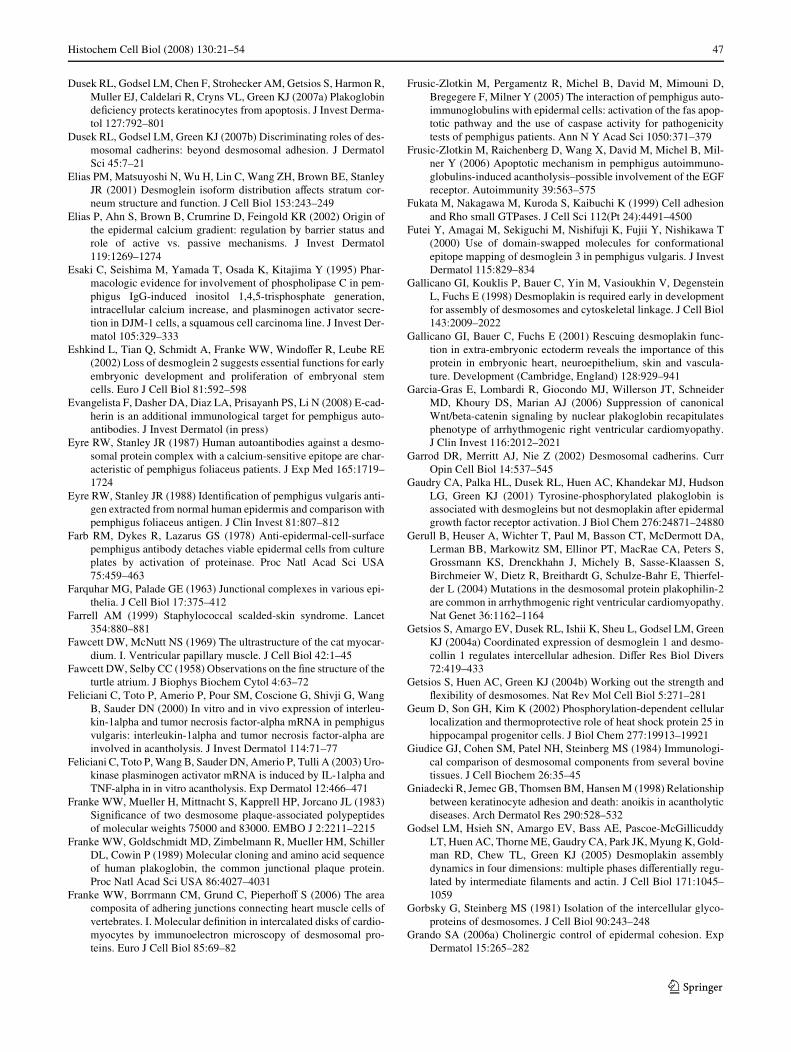
Histochem Cell Biol (2008) 130:21–54 47
Dusek RL, Godsel LM, Chen F, Strohecker AM, Getsios S, Harmon R,Muller EJ, Caldelari R, Cryns VL, Green KJ (2007a) PlakoglobindeWciency protects keratinocytes from apoptosis. J Invest Derma-tol 127:792–801
Dusek RL, Godsel LM, Green KJ (2007b) Discriminating roles of des-mosomal cadherins: beyond desmosomal adhesion. J DermatolSci 45:7–21
Elias PM, Matsuyoshi N, Wu H, Lin C, Wang ZH, Brown BE, StanleyJR (2001) Desmoglein isoform distribution aVects stratum cor-neum structure and function. J Cell Biol 153:243–249
Elias P, Ahn S, Brown B, Crumrine D, Feingold KR (2002) Origin ofthe epidermal calcium gradient: regulation by barrier status androle of active vs. passive mechanisms. J Invest Dermatol119:1269–1274
Esaki C, Seishima M, Yamada T, Osada K, Kitajima Y (1995) Phar-macologic evidence for involvement of phospholipase C in pem-phigus IgG-induced inositol 1,4,5-trisphosphate generation,intracellular calcium increase, and plasminogen activator secre-tion in DJM-1 cells, a squamous cell carcinoma line. J Invest Der-matol 105:329–333
Eshkind L, Tian Q, Schmidt A, Franke WW, WindoVer R, Leube RE(2002) Loss of desmoglein 2 suggests essential functions for earlyembryonic development and proliferation of embryonal stemcells. Euro J Cell Biol 81:592–598
Evangelista F, Dasher DA, Diaz LA, Prisayanh PS, Li N (2008) E-cad-herin is an additional immunological target for pemphigus auto-antibodies. J Invest Dermatol (in press)
Eyre RW, Stanley JR (1987) Human autoantibodies against a desmo-somal protein complex with a calcium-sensitive epitope are char-acteristic of pemphigus foliaceus patients. J Exp Med 165:1719–1724
Eyre RW, Stanley JR (1988) IdentiWcation of pemphigus vulgaris anti-gen extracted from normal human epidermis and comparison withpemphigus foliaceus antigen. J Clin Invest 81:807–812
Farb RM, Dykes R, Lazarus GS (1978) Anti-epidermal-cell-surfacepemphigus antibody detaches viable epidermal cells from cultureplates by activation of proteinase. Proc Natl Acad Sci USA75:459–463
Farquhar MG, Palade GE (1963) Junctional complexes in various epi-thelia. J Cell Biol 17:375–412
Farrell AM (1999) Staphylococcal scalded-skin syndrome. Lancet354:880–881
Fawcett DW, McNutt NS (1969) The ultrastructure of the cat myocar-dium. I. Ventricular papillary muscle. J Cell Biol 42:1–45
Fawcett DW, Selby CC (1958) Observations on the Wne structure of theturtle atrium. J Biophys Biochem Cytol 4:63–72
Feliciani C, Toto P, Amerio P, Pour SM, Coscione G, Shivji G, WangB, Sauder DN (2000) In vitro and in vivo expression of interleu-kin-1alpha and tumor necrosis factor-alpha mRNA in pemphigusvulgaris: interleukin-1alpha and tumor necrosis factor-alpha areinvolved in acantholysis. J Invest Dermatol 114:71–77
Feliciani C, Toto P, Wang B, Sauder DN, Amerio P, Tulli A (2003) Uro-kinase plasminogen activator mRNA is induced by IL-1alpha andTNF-alpha in in vitro acantholysis. Exp Dermatol 12:466–471
Franke WW, Mueller H, Mittnacht S, Kapprell HP, Jorcano JL (1983)SigniWcance of two desmosome plaque-associated polypeptidesof molecular weights 75000 and 83000. EMBO J 2:2211–2215
Franke WW, Goldschmidt MD, Zimbelmann R, Mueller HM, SchillerDL, Cowin P (1989) Molecular cloning and amino acid sequenceof human plakoglobin, the common junctional plaque protein.Proc Natl Acad Sci USA 86:4027–4031
Franke WW, Borrmann CM, Grund C, PieperhoV S (2006) The areacomposita of adhering junctions connecting heart muscle cells ofvertebrates. I. Molecular deWnition in intercalated disks of cardio-myocytes by immunoelectron microscopy of desmosomal pro-teins. Euro J Cell Biol 85:69–82
Frusic-Zlotkin M, Pergamentz R, Michel B, David M, Mimouni D,Bregegere F, Milner Y (2005) The interaction of pemphigus auto-immunoglobulins with epidermal cells: activation of the fas apop-totic pathway and the use of caspase activity for pathogenicitytests of pemphigus patients. Ann N Y Acad Sci 1050:371–379
Frusic-Zlotkin M, Raichenberg D, Wang X, David M, Michel B, Mil-ner Y (2006) Apoptotic mechanism in pemphigus autoimmuno-globulins-induced acantholysis–possible involvement of the EGFreceptor. Autoimmunity 39:563–575
Fukata M, Nakagawa M, Kuroda S, Kaibuchi K (1999) Cell adhesionand Rho small GTPases. J Cell Sci 112(Pt 24):4491–4500
Futei Y, Amagai M, Sekiguchi M, Nishifuji K, Fujii Y, Nishikawa T(2000) Use of domain-swapped molecules for conformationalepitope mapping of desmoglein 3 in pemphigus vulgaris. J InvestDermatol 115:829–834
Gallicano GI, Kouklis P, Bauer C, Yin M, Vasioukhin V, DegensteinL, Fuchs E (1998) Desmoplakin is required early in developmentfor assembly of desmosomes and cytoskeletal linkage. J Cell Biol143:2009–2022
Gallicano GI, Bauer C, Fuchs E (2001) Rescuing desmoplakin func-tion in extra-embryonic ectoderm reveals the importance of thisprotein in embryonic heart, neuroepithelium, skin and vascula-ture. Development (Cambridge, England) 128:929–941
Garcia-Gras E, Lombardi R, Giocondo MJ, Willerson JT, SchneiderMD, Khoury DS, Marian AJ (2006) Suppression of canonicalWnt/beta-catenin signaling by nuclear plakoglobin recapitulatesphenotype of arrhythmogenic right ventricular cardiomyopathy.J Clin Invest 116:2012–2021
Garrod DR, Merritt AJ, Nie Z (2002) Desmosomal cadherins. CurrOpin Cell Biol 14:537–545
Gaudry CA, Palka HL, Dusek RL, Huen AC, Khandekar MJ, HudsonLG, Green KJ (2001) Tyrosine-phosphorylated plakoglobin isassociated with desmogleins but not desmoplakin after epidermalgrowth factor receptor activation. J Biol Chem 276:24871–24880
Gerull B, Heuser A, Wichter T, Paul M, Basson CT, McDermott DA,Lerman BB, Markowitz SM, Ellinor PT, MacRae CA, Peters S,Grossmann KS, Drenckhahn J, Michely B, Sasse-Klaassen S,Birchmeier W, Dietz R, Breithardt G, Schulze-Bahr E, Thierfel-der L (2004) Mutations in the desmosomal protein plakophilin-2are common in arrhythmogenic right ventricular cardiomyopathy.Nat Genet 36:1162–1164
Getsios S, Amargo EV, Dusek RL, Ishii K, Sheu L, Godsel LM, GreenKJ (2004a) Coordinated expression of desmoglein 1 and desmo-collin 1 regulates intercellular adhesion. DiVer Res Biol Divers72:419–433
Getsios S, Huen AC, Green KJ (2004b) Working out the strength andXexibility of desmosomes. Nat Rev Mol Cell Biol 5:271–281
Geum D, Son GH, Kim K (2002) Phosphorylation-dependent cellularlocalization and thermoprotective role of heat shock protein 25 inhippocampal progenitor cells. J Biol Chem 277:19913–19921
Giudice GJ, Cohen SM, Patel NH, Steinberg MS (1984) Immunologi-cal comparison of desmosomal components from several bovinetissues. J Cell Biochem 26:35–45
Gniadecki R, Jemec GB, Thomsen BM, Hansen M (1998) Relationshipbetween keratinocyte adhesion and death: anoikis in acantholyticdiseases. Arch Dermatol Res 290:528–532
Godsel LM, Hsieh SN, Amargo EV, Bass AE, Pascoe-McGillicuddyLT, Huen AC, Thorne ME, Gaudry CA, Park JK, Myung K, Gold-man RD, Chew TL, Green KJ (2005) Desmoplakin assemblydynamics in four dimensions: multiple phases diVerentially regu-lated by intermediate Wlaments and actin. J Cell Biol 171:1045–1059
Gorbsky G, Steinberg MS (1981) Isolation of the intercellular glyco-proteins of desmosomes. J Cell Biol 90:243–248
Grando SA (2006a) Cholinergic control of epidermal cohesion. ExpDermatol 15:265–282
123
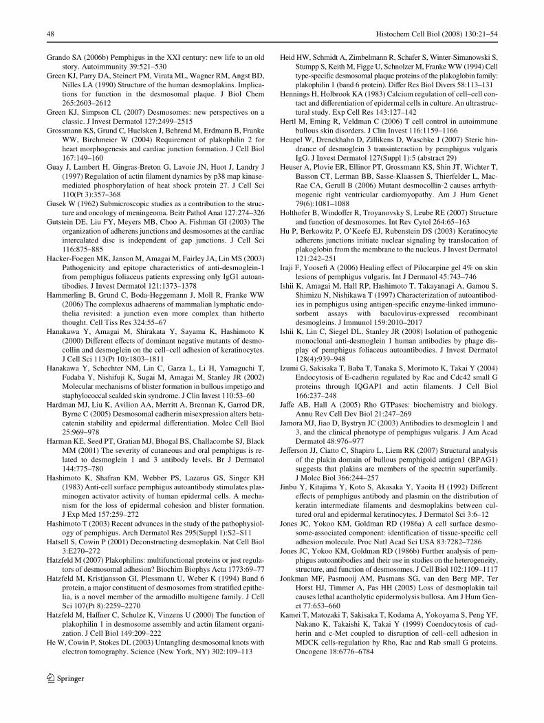
48 Histochem Cell Biol (2008) 130:21–54
Grando SA (2006b) Pemphigus in the XXI century: new life to an oldstory. Autoimmunity 39:521–530
Green KJ, Parry DA, Steinert PM, Virata ML, Wagner RM, Angst BD,Nilles LA (1990) Structure of the human desmoplakins. Implica-tions for function in the desmosomal plaque. J Biol Chem265:2603–2612
Green KJ, Simpson CL (2007) Desmosomes: new perspectives on aclassic. J Invest Dermatol 127:2499–2515
Grossmann KS, Grund C, Huelsken J, Behrend M, Erdmann B, FrankeWW, Birchmeier W (2004) Requirement of plakophilin 2 forheart morphogenesis and cardiac junction formation. J Cell Biol167:149–160
Guay J, Lambert H, Gingras-Breton G, Lavoie JN, Huot J, Landry J(1997) Regulation of actin Wlament dynamics by p38 map kinase-mediated phosphorylation of heat shock protein 27. J Cell Sci110(Pt 3):357–368
Gusek W (1962) Submicroscopic studies as a contribution to the struc-ture and oncology of meningeoma. Beitr Pathol Anat 127:274–326
Gutstein DE, Liu FY, Meyers MB, Choo A, Fishman GI (2003) Theorganization of adherens junctions and desmosomes at the cardiacintercalated disc is independent of gap junctions. J Cell Sci116:875–885
Hacker-Foegen MK, Janson M, Amagai M, Fairley JA, Lin MS (2003)Pathogenicity and epitope characteristics of anti-desmoglein-1from pemphigus foliaceus patients expressing only IgG1 autoan-tibodies. J Invest Dermatol 121:1373–1378
Hammerling B, Grund C, Boda-Heggemann J, Moll R, Franke WW(2006) The complexus adhaerens of mammalian lymphatic endo-thelia revisited: a junction even more complex than hithertothought. Cell Tiss Res 324:55–67
Hanakawa Y, Amagai M, Shirakata Y, Sayama K, Hashimoto K(2000) DiVerent eVects of dominant negative mutants of desmo-collin and desmoglein on the cell–cell adhesion of keratinocytes.J Cell Sci 113(Pt 10):1803–1811
Hanakawa Y, Schechter NM, Lin C, Garza L, Li H, Yamaguchi T,Fudaba Y, Nishifuji K, Sugai M, Amagai M, Stanley JR (2002)Molecular mechanisms of blister formation in bullous impetigo andstaphylococcal scalded skin syndrome. J Clin Invest 110:53–60
Hardman MJ, Liu K, Avilion AA, Merritt A, Brennan K, Garrod DR,Byrne C (2005) Desmosomal cadherin misexpression alters beta-catenin stability and epidermal diVerentiation. Molec Cell Biol25:969–978
Harman KE, Seed PT, Gratian MJ, Bhogal BS, Challacombe SJ, BlackMM (2001) The severity of cutaneous and oral pemphigus is re-lated to desmoglein 1 and 3 antibody levels. Br J Dermatol144:775–780
Hashimoto K, Shafran KM, Webber PS, Lazarus GS, Singer KH(1983) Anti-cell surface pemphigus autoantibody stimulates plas-minogen activator activity of human epidermal cells. A mecha-nism for the loss of epidermal cohesion and blister formation.J Exp Med 157:259–272
Hashimoto T (2003) Recent advances in the study of the pathophysiol-ogy of pemphigus. Arch Dermatol Res 295(Suppl 1):S2–S11
Hatsell S, Cowin P (2001) Deconstructing desmoplakin. Nat Cell Biol3:E270–272
Hatzfeld M (2007) Plakophilins: multifunctional proteins or just regula-tors of desmosomal adhesion? Biochim Biophys Acta 1773:69–77
Hatzfeld M, Kristjansson GI, Plessmann U, Weber K (1994) Band 6protein, a major constituent of desmosomes from stratiWed epithe-lia, is a novel member of the armadillo multigene family. J CellSci 107(Pt 8):2259–2270
Hatzfeld M, HaVner C, Schulze K, Vinzens U (2000) The function ofplakophilin 1 in desmosome assembly and actin Wlament organi-zation. J Cell Biol 149:209–222
He W, Cowin P, Stokes DL (2003) Untangling desmosomal knots withelectron tomography. Science (New York, NY) 302:109–113
Heid HW, Schmidt A, Zimbelmann R, Schafer S, Winter-Simanowski S,Stumpp S, Keith M, Figge U, Schnolzer M, Franke WW (1994) Celltype-speciWc desmosomal plaque proteins of the plakoglobin family:plakophilin 1 (band 6 protein). DiVer Res Biol Divers 58:113–131
Hennings H, Holbrook KA (1983) Calcium regulation of cell–cell con-tact and diVerentiation of epidermal cells in culture. An ultrastruc-tural study. Exp Cell Res 143:127–142
Hertl M, Eming R, Veldman C (2006) T cell control in autoimmunebullous skin disorders. J Clin Invest 116:1159–1166
Heupel W, Drenckhahn D, Zillikens D, Waschke J (2007) Steric hin-drance of desmoglein 3 transinteraction by pemphigus vulgarisIgG. J Invest Dermatol 127(Suppl 1):5 (abstract 29)
Heuser A, Plovie ER, Ellinor PT, Grossmann KS, Shin JT, Wichter T,Basson CT, Lerman BB, Sasse-Klaassen S, Thierfelder L, Mac-Rae CA, Gerull B (2006) Mutant desmocollin-2 causes arrhyth-mogenic right ventricular cardiomyopathy. Am J Hum Genet79(6):1081–1088
Holthofer B, WindoVer R, Troyanovsky S, Leube RE (2007) Structureand function of desmosomes. Int Rev Cytol 264:65–163
Hu P, Berkowitz P, O’Keefe EJ, Rubenstein DS (2003) Keratinocyteadherens junctions initiate nuclear signaling by translocation ofplakoglobin from the membrane to the nucleus. J Invest Dermatol121:242–251
Iraji F, YooseW A (2006) Healing eVect of Pilocarpine gel 4% on skinlesions of pemphigus vulgaris. Int J Dermatol 45:743–746
Ishii K, Amagai M, Hall RP, Hashimoto T, Takayanagi A, Gamou S,Shimizu N, Nishikawa T (1997) Characterization of autoantibod-ies in pemphigus using antigen-speciWc enzyme-linked immuno-sorbent assays with baculovirus-expressed recombinantdesmogleins. J Immunol 159:2010–2017
Ishii K, Lin C, Siegel DL, Stanley JR (2008) Isolation of pathogenicmonoclonal anti-desmoglein 1 human antibodies by phage dis-play of pemphigus foliaceus autoantibodies. J Invest Dermatol128(4):939–948
Izumi G, Sakisaka T, Baba T, Tanaka S, Morimoto K, Takai Y (2004)Endocytosis of E-cadherin regulated by Rac and Cdc42 small Gproteins through IQGAP1 and actin Wlaments. J Cell Biol166:237–248
JaVe AB, Hall A (2005) Rho GTPases: biochemistry and biology.Annu Rev Cell Dev Biol 21:247–269
Jamora MJ, Jiao D, Bystryn JC (2003) Antibodies to desmoglein 1 and3, and the clinical phenotype of pemphigus vulgaris. J Am AcadDermatol 48:976–977
JeVerson JJ, Ciatto C, Shapiro L, Liem RK (2007) Structural analysisof the plakin domain of bullous pemphigoid antigen1 (BPAG1)suggests that plakins are members of the spectrin superfamily.J Molec Biol 366:244–257
Jinbu Y, Kitajima Y, Koto S, Akasaka Y, Yaoita H (1992) DiVerenteVects of pemphigus antibody and plasmin on the distribution ofkeratin intermediate Wlaments and desmoplakins between cul-tured oral and epidermal keratinocytes. J Dermatol Sci 3:6–12
Jones JC, Yokoo KM, Goldman RD (1986a) A cell surface desmo-some-associated component: identiWcation of tissue-speciWc celladhesion molecule. Proc Natl Acad Sci USA 83:7282–7286
Jones JC, Yokoo KM, Goldman RD (1986b) Further analysis of pem-phigus autoantibodies and their use in studies on the heterogeneity,structure, and function of desmosomes. J Cell Biol 102:1109–1117
Jonkman MF, Pasmooij AM, Pasmans SG, van den Berg MP, TerHorst HJ, Timmer A, Pas HH (2005) Loss of desmoplakin tailcauses lethal acantholytic epidermolysis bullosa. Am J Hum Gen-et 77:653–660
Kamei T, Matozaki T, Sakisaka T, Kodama A, Yokoyama S, Peng YF,Nakano K, Takaishi K, Takai Y (1999) Coendocytosis of cad-herin and c-Met coupled to disruption of cell–cell adhesion inMDCK cells-regulation by Rho, Rac and Rab small G proteins.Oncogene 18:6776–6784
123
Histochem Cell Biol (2008) 130:21–54 49
Karpati S, Amagai M, Prussick R, Cehrs K, Stanley JR (1993) Pemphi-gus vulgaris antigen, a desmoglein type of cadherin, is localizedwithin keratinocyte desmosomes. J Cell Biol 122:409–415
Kawasaki Y, Aoyama Y, Tsunoda K, Amagai M, Kitajima Y (2006)Pathogenic monoclonal antibody against desmoglein 3 augmentsdesmoglein 3 and p38 MAPK phosphorylation in human squa-mous carcinoma cell line. Autoimmunity 39:587–590
Kelly DE (1966) Fine structure of desmosomes, hemidesmosomes, andan adepidermal globular layer in developing newt epidermis.J Cell Biol 28:51–72
Kimura TE, Merritt AJ, Garrod DR (2007) Calcium-independent des-mosomes of keratinocytes are hyper-adhesive. J Invest Dermatol127:775–781
King IA, Sullivan KH, Bennett R Jr, Buxton RS (1995) The desmocol-lins of human foreskin epidermis: identiWcation and chromosomalassignment of a third gene and expression patterns of the threeisoforms. J Invest Dermatol 105:314–321
Kitajima Y (2002) Mechanisms of desmosome assembly and disas-sembly. Clin Exp Dermatol 27:684–690
Kitajima Y, Inoue S, Yaoita H (1986) EVects of pemphigus antibody onthe organization of microtubules and keratin-intermediate Wlamentsin cultured human keratinocytes. Br J Dermatol 114:171–179
Kitajima Y, Inoue S, Yaoita H (1987) EVects of pemphigus antibodyon the regeneration of cell–cell contact in keratinocyte culturesgrown in low to normal Ca++ concentration. J Invest Dermatol89:167–171
Kitajima Y, Aoyama Y, Seishima M (1999) Transmembrane signalingfor adhesive regulation of desmosomes and hemidesmosomes,and for cell–cell datachment induced by pemphigus IgG in cul-tured keratinocytes: involvement of protein kinase C. J InvestDermatol Symp Proc/Soc Invest Dermatol Inc 4:137–144
Kljuic A, Bazzi H, Sundberg JP, Martinez-Mir A, O’Shaughnessy R,Mahoney MG, Levy M, Montagutelli X, Ahmad W, Aita VM,Gordon D, Uitto J, Whiting D, Ott J, Fischer S, Gilliam TC, Jah-oda CA, Morris RJ, Panteleyev AA, Nguyen VT, Christiano AM(2003) Desmoglein 4 in hair follicle diVerentiation and epidermaladhesion: evidence from inherited hypotrichosis and acquiredpemphigus vulgaris. Cell 113:249–260
Koch PJ, Walsh MJ, Schmelz M, Goldschmidt MD, Zimbelmann R,Franke WW (1990) IdentiWcation of desmoglein, a constitutivedesmosomal glycoprotein, as a member of the cadherin family ofcell adhesion molecules. Euro J Cell Biol 53:1–12
Koch PJ, Goldschmidt MD, Walsh MJ, Zimbelmann R, Franke WW(1991a) Complete amino acid sequence of the epidermal desmog-lein precursor polypeptide and identiWcation of a second type ofdesmoglein gene. Euro J Cell Biol 55:200–208
Koch PJ, Goldschmidt MD, Walsh MJ, Zimbelmann R, Schmelz M,Franke WW (1991b) Amino acid sequence of bovine muzzle epi-thelial desmocollin derived from cloned cDNA: a novel subtypeof desmosomal cadherins. DiVer Res Biol Divers 47:29–36
Koch PJ, Mahoney MG, Cotsarelis G, Rothenberger K, Lavker RM,Stanley JR (1998) Desmoglein 3 anchors telogen hair in the folli-cle. J Cell Sci 111(Pt 17):2529–2537
Kodama S, Ikeda S, Asahara T, Kishida M, Kikuchi A (1999) Axin di-rectly interacts with plakoglobin and regulates its stability. J BiolChem 274:27682–27688
Koeser J, Troyanovsky SM, Grund C, Franke WW (2003) De novo for-mation of desmosomes in cultured cells upon transfection ofgenes encoding speciWc desmosomal components. Exp Cell Res285:114–130
Kolligs FT, Kolligs B, Hajra KM, Hu G, Tani M, Cho KR, Fearon ER(2000) Gamma-catenin is regulated by the APC tumor suppressorand its oncogenic activity is distinct from that of beta-catenin.Genes Dev 14:1319–1331
Kolly C, Zakher A, Strauss C, Suter MM, Muller EJ (2007) Keratino-cyte transcriptional regulation of the human c-Myc promoter
occurs via a novel Lef/Tcf binding element distinct from neoplas-tic cells. FEBS Lett 581:1969–1976
Korman NJ, Eyre RW, Klaus-Kovtun V, Stanley JR (1989) Demon-stration of an adhering-junction molecule (plakoglobin) in the au-toantigens of pemphigus foliaceus and pemphigus vulgaris. NewEngl J Med 321:631–635
Kottke MD, Delva E, Kowalczyk AP (2006) The desmosome: cell sci-ence lessons from human diseases. J Cell Sci 119:797–806
Koulu L, Kusumi A, Steinberg MS, Klaus-Kovtun V, Stanley JR(1984) Human autoantibodies against a desmosomal core proteinin pemphigus foliaceus. J Exp Med 160:1509–1518
Kowalczyk AP, Reynolds AB (2004) Protecting your tail: regulation ofcadherin degradation by p120-catenin. Curr Opin Cell Biol16:522–527
Kowalczyk AP, Borgwardt JE, Green KJ (1996) Analysis of desmo-somal cadherin-adhesive function and stoichiometry of desmo-somal cadherin-plakoglobin complexes. J Invest Dermatol107:293–300
Kowalczyk AP, Bornslaeger EA, Borgwardt JE, Palka HL, Dhaliwal AS,Corcoran CM, Denning MF, Green KJ (1997) The amino-terminaldomain of desmoplakin binds to plakoglobin and clusters desmo-somal cadherin-plakoglobin complexes. J Cell Biol 139:773–784
Kowalczyk AP, Hatzfeld M, Bornslaeger EA, Kopp DS, BorgwardtJE, Corcoran CM, Settler A, Green KJ (1999) The head domainof plakophilin-1 binds to desmoplakin and enhances its recruit-ment to desmosomes. Implications for cutaneous disease. J BiolChem 274:18145–18148
Kowalczyk AP, Navarro P, Dejana E, Bornslaeger EA, Green KJ,Kopp DS, Borgwardt JE (1998) VE-cadherin and desmoplakinare assembled into dermal microvascular endothelial intercellularjunctions: a pivotal role for plakoglobin in the recruitment of des-moplakin to intercellular junctions. J Cell Sci 111(Pt 20):3045–3057
Lanza A, Cirillo N, Femiano F, Gombos F (2006) How does acanthol-ysis occur in pemphigus vulgaris: a critical review. J Cutan Pathol33:401–412
Lanza A, Cirillo N, Rossiello R, Rienzo M, Cutillo L, Casamassimi A,de Nigris F, Schiano C, Rossiello L, Femiano F, Gombos F,Napoli C (2008) Evidence of key role of CDK-2 overexpressionin pemphigus vulgaris. J Biol Chem. doi:10.1074/jbc.M702186200
Lever WF (1953) Pemphigus. Medicine 32:2–132Lo Muzio L, Pannone G, Staibano S, Mignogna MD, Rubini C, Farro-
nato G, Ferrari F, Nocini PF, De Rosa G (2002) Strict correlationbetween uPAR and plakoglobin expression in pemphigus vulga-ris. J Cutan Pathol 29:540–548
Lorch JH, Klessner J, Park JK, Getsios S, Wu YL, Stack MS, Green KJ(2004) Epidermal growth factor receptor inhibition promotes des-mosome assembly and strengthens intercellular adhesion in squa-mous cell carcinoma cells. J Biol Chem 279:37191–37200
Lyell A (1983) The staphylococcal scalded skin syndrome in historicalperspective: emergence of dermopathic strains of Staphylococcusaureus and discovery of the epidermolytic toxin. A review ofevents up to 1970. J Am Acad Dermatol 9:285–294
Maeda O, Usami N, Kondo M, Takahashi M, Goto H, Shimokata K,Kusugami K, Sekido Y (2004) Plakoglobin (gamma-catenin) hasTCF/LEF family-dependent transcriptional activity in beta-cate-nin-deWcient cell line. Oncogene 23:964–972
Mahoney MG, Wang Z, Rothenberger K, Koch PJ, Amagai M, StanleyJR (1999) Explanations for the clinical and microscopic localiza-tion of lesions in pemphigus foliaceus and vulgaris. J Clin Invest103:461–468
Mahoney MG, Hu Y, Brennan D, Bazzi H, Christiano AM, Wahl JKIII (2006) Delineation of diversiWed desmoglein distribution instratiWed squamous epithelia: implications in diseases. Exp Der-matol 15:101–109
123
50 Histochem Cell Biol (2008) 130:21–54
Marcozzi C, Burdett ID, Buxton RS, Magee AI (1998) Coexpressionof both types of desmosomal cadherin and plakoglobin confersstrong intercellular adhesion. J Cell Sci 111(Pt 4):495–509
Mathur M, Goodwin L, Cowin P (1994) Interactions of the cytoplas-mic domain of the desmosomal cadherin Dsg1 with plakoglobin.J Biol Chem 269:14075–14080
McGrath JA (2005) Inherited disorders of desmosomes. Austr J Der-matol 46:221–229
McGrath JA, McMillan JR, Shemanko CS, Runswick SK, Leigh IM,Lane EB, Garrod DR, Eady RA (1997) Mutations in the plakophi-lin 1 gene result in ectodermal dysplasia/skin fragility syndrome.Nat Genet 17:240–244
McKoy G, Protonotarios N, Crosby A, Tsatsopoulou A, AnastasakisA, Coonar A, Norman M, Baboonian C, JeVery S, McKenna WJ(2000) IdentiWcation of a deletion in plakoglobin in arrhythmo-genic right ventricular cardiomyopathy with palmoplantar kerato-derma and woolly hair (Naxos disease). Lancet 355:2119–2124
McMillan JR, Shimizu H (2001) Desmosomes: structure and functionin normal and diseased epidermis. J Dermatol 28:291–298
McNutt NS, Fawcett DW (1969) The ultrastructure of the cat myocar-dium. II. Atrial muscle. J Cell Biol 42:46–67
Melish ME, Glasgow LA (1970) The staphylococcal scalded-skin syn-drome. New Engl J Med 282:1114–1119
Memar O, Christensen B, Rajaraman S, Goldblum R, Tyring SK,Brysk MM, McCormick DJ, el-Zaim H, Fan JL, Prabhakar BS(1996) Induction of blister-causing antibodies by a recombinantfull-length, but not the extracellular, domain of the pemphigusvulgaris antigen (desmoglein 3). J Immunol 157:3171–3177
Menon GK, Elias PM (1991) Ultrastructural localization of calcium inpsoriatic and normal human epidermis. Arch Dermatol 127:57–63
Merritt AJ, Berika MY, Zhai W, Kirk SE, Ji B, Hardman MJ, GarrodDR (2002) Suprabasal desmoglein 3 expression in the epidermisof transgenic mice results in hyperproliferation and abnormaldiVerentiation. Molec Cell Biol 22:5846–5858
Mertens C, Kuhn C, Franke WW (1996) Plakophilins 2a and 2b: con-stitutive proteins of dual location in the karyoplasm and the des-mosomal plaque. J Cell Biol 135:1009–1025
Mertens C, Kuhn C, Moll R, Schwetlick I, Franke WW (1999) Desmo-somal plakophilin 2 as a diVerentiation marker in normal andmalignant tissues. DiVer Res Biol Divers 64:277–290
Mertens C, Hofmann I, Wang Z, Teichmann M, Sepehri Chong S, Sch-nolzer M, Franke WW (2001) Nuclear particles containing RNApolymerase III complexes associated with the junctional plaqueprotein plakophilin 2. Proc Natl Acad Sci USA 98:7795–7800
Milingou M, Wood P, Masouye I, McLean WH, Borradori L (2006)Focal palmoplantar keratoderma caused by an autosomal domi-nant inherited mutation in the desmoglein 1 gene. Dermatology(Basel, Switzerland) 212:117–122
Miranda KC, Joseph SR, Yap AS, Teasdale RD, Stow JL (2003) Con-textual binding of p120ctn to E-cadherin at the basolateral plasmamembrane in polarized epithelia. J Biol Chem 278:43480–43488
Miravet S, Piedra J, Castano J, Raurell I, Franci C, Dunach M, Garciade Herreros A (2003) Tyrosine phosphorylation of plakoglobincauses contrary eVects on its association with desmosomes andadherens junction components and modulates beta-catenin-medi-ated transcription. Molec Cell Biol 23:7391–7402
Miyagawa S, Amagai M, Iida T, Yamamoto Y, Nishikawa T, Shirai T(1999) Late development of antidesmoglein 1 antibodies in pem-phigus vulgaris: correlation with disease progression. Br J Der-matol 141:1084–1087
Moll R, Franke WW (1982) Intermediate Wlaments and their interac-tion with membranes. The desmosome-cytokeratin Wlament com-plex and epithelial diVerentiation. Pathol Res Pract 175:146–161
Moll R, Cowin P, Kapprell HP, Franke WW (1986) Desmosomal pro-teins: new markers for identiWcation and classiWcation of tumors.Lab Invest J Tech Methods Pathol 54:4–25
Morioka S, Lazarus GS, Jensen PJ (1987) Involvement of urokinase-type plasminogen activator in acantholysis induced by pemphigusIgG. J Invest Dermatol 89:474–477
Mueller H, Franke WW (1983) Biochemical and immunological char-acterization of desmoplakins I and II, the major polypeptides ofthe desmosomal plaque. J Molec Biol 163:647–671
Muller E, Kernland K, Caldelari R, Wyder M, Balmer V, Hunziker T(2002) Unusual pemphigus phenotype in the presence of a Dsg1and Dsg3 autoantibody proWle. J Invest Dermatol 118:551–555
Muller R, Svoboda V, Wenzel E, Gebert S, Hunzelmann N, MullerHH, Hertl M (2006) IgG reactivity against non-conformationalNH-terminal epitopes of the desmoglein 3 ectodomain relates toclinical activity and phenotype of pemphigus vulgaris. Exp Der-matol 15:606–614
Muller EJ, Williamson L, Kolly C, Suter MM (2008a) Outside-in sig-naling through integrins and cadherins: a central mechanism tocontrol epidermal growth and diVerentiation? J Invest Dermatol128:501–516
Muller R, Svoboda V, Wenzel E, Muller HH, Hertl M (2008b) IgGagainst extracellular subdomains of desmoglein 3 relates to clini-cal phenotype of pemphigus vulgaris. Exp Dermatol 17:35–43
Nagasaka T, Nishifuji K, Ota T, Whittock NV, Amagai M (2004)DeWning the pathogenic involvement of desmoglein 4 in pemphi-gus and staphylococcal scalded skin syndrome. J Clin Invest114:1484–1492
Namazi MR (2004) Practice pearl: gargling with cholinergic ophthal-mic drops for treating the oral lesions of pemphigus vulgaris.J Drugs Dermatol 3:484–485
Nguyen VT, Ndoye A, Grando SA (2000a) Novel human alpha9 ace-tylcholine receptor regulating keratinocyte adhesion is targetedby Pemphigus vulgaris autoimmunity. Am J Pathol 157:1377–1391
Nguyen VT, Ndoye A, Grando SA (2000b) Pemphigus vulgaris anti-body identiWes pemphaxin. A novel keratinocyte annexin-likemolecule binding acetylcholine. J Biol Chem 275:29466–29476
Nguyen VT, Ndoye A, Shultz LD, Pittelkow MR, Grando SA (2000c)Antibodies against keratinocyte antigens other than desmogleins1 and 3 can induce pemphigus vulgaris-like lesions. J Clin Invest106:1467–1479
Nguyen VT, Arredondo J, Chernyavsky AI, Kitajima Y, Pittelkow M,Grando SA (2004a) Pemphigus vulgaris IgG and methylprednis-olone exhibit reciprocal eVects on keratinocytes. J Biol Chem279:2135–2146
Nguyen VT, Arredondo J, Chernyavsky AI, Pittelkow MR, KitajimaY, Grando SA (2004b) Pemphigus vulgaris acantholysis amelio-rated by cholinergic agonists. Arch Dermatol 140:327–334
Nollet F, Kools P, van Roy F (2000) Phylogenetic analysis of the cad-herin superfamily allows identiWcation of six major subfamiliesbesides several solitary members. J Molec Biol 299:551–572
Norgett EE, Hatsell SJ, Carvajal-Huerta L, Cabezas JC, Common J,Purkis PE, Whittock N, Leigh IM, Stevens HP, Kelsell DP (2000)Recessive mutation in desmoplakin disrupts desmoplakin-inter-mediate Wlament interactions and causes dilated cardiomyopathy,woolly hair and keratoderma. Hum Molec Genet 9:2761–2766
North AJ, Bardsley WG, Hyam J, Bornslaeger EA, Cordingley HC,Trinnaman B, Hatzfeld M, Green KJ, Magee AI, Garrod DR(1999) Molecular map of the desmosomal plaque. J Cell Sci112(Pt 23):4325–4336
Odland GF (1958) The Wne structure of the interrelationship of cells inthe human epidermis. J Biophys Biochem Cytol 4:529–538
Osada K, Seishima M, Kitajima Y (1997) Pemphigus IgG activates andtranslocates protein kinase C from the cytosol to the particulate/cytoskeleton fractions in human keratinocytes. J Invest Dermatol108:482–487
Overduin M, Harvey TS, Bagby S, Tong KI, Yau P, Takeichi M, IkuraM (1995) Solution structure of the epithelial cadherin domain
123

Histochem Cell Biol (2008) 130:21–54 51
responsible for selective cell adhesion. Science (New York, NY)267:386–389
Panasenko OO, Kim MV, Marston SB, Gusev NB (2003) Interactionof the small heat shock protein with molecular mass 25 kDa(hsp25) with actin. Euro J Biochem/FEBS 270:892–901
Pasdar M, Nelson WJ (1988) Kinetics of desmosome assembly in Ma-din–Darby canine kidney epithelial cells: temporal and spatialregulation of desmoplakin organization and stabilization uponcell–cell contact. II. Morphological analysis. J Cell Biol 106:687–695
Pasdar M, Nelson WJ (1989) Regulation of desmosome assembly inepithelial cells: kinetics of synthesis, transport, and stabilizationof desmoglein I, a major protein of the membrane core domain.J Cell Biol 109:163–177
Pasdar M, Li Z, Chlumecky V (1995) Plakoglobin: kinetics of synthe-sis, phosphorylation, stability, and interactions with desmogleinand E-cadherin. Cell Motility Cytoskeleton 32:258–272
Payne AS, Hanakawa Y, Amagai M, Stanley JR (2004) Desmosomesand disease: pemphigus and bullous impetigo. Curr Opin CellBiol 16:536–543
Payne AS, Ishii K, Kacir S, Lin C, Li H, Hanakawa Y, Tsunoda K,Amagai M, Stanley JR, Siegel DL (2005) Genetic and functionalcharacterization of human pemphigus vulgaris monoclonal auto-antibodies isolated by phage display. J Clin Invest 115:888–899
Pelacho B, Natal C, Espana A, Sanchez-Carpintero I, Iraburu MJ, Lo-pez-Zabalza MJ (2004) Pemphigus vulgaris autoantibodies in-duce apoptosis in HaCaT keratinocytes. FEBS Lett 566:6–10
Perng MD, Cairns L, van den Ijssel P, Prescott A, Hutcheson AM,Quinlan RA (1999) Intermediate Wlament interactions can be al-tered by HSP27 and alphaB-crystallin. J Cell Sci 112(Pt13):2099–2112
Pertz O, Bozic D, Koch AW, Fauser C, Brancaccio A, Engel J (1999)A new crystal structure, Ca2+ dependence and mutational analy-sis reveal molecular details of E-cadherin homoassociation.EMBO J 18:1738–1747
PieperhoV S, Franke WW (2007) The area composita of adhering junc-tions connecting heart muscle cells of vertebrates—IV: coales-cence and amalgamation of desmosomal and adhaerens junctioncomponents—late processes in mammalian heart development.Euro J Cell Biol 86:377–391
PieperhoV S, Schumacher H, Franke WW (2008) The area compositaof adhering junctions connecting heart muscle cells of verte-brates. V. The importance of plakophilin-2 demonstrated by smallinterference RNA-mediated knockdown in cultured rat cardio-myocytes. Euro J Cell Biol (in press)
Pilichou K, Nava A, Basso C, BeVagna G, Bauce B, Lorenzon A, FrigoG, Vettori A, Valente M, Towbin J, Thiene G, Danieli GA, Ramp-azzo A (2006) Mutations in desmoglein-2 gene are associatedwith arrhythmogenic right ventricular cardiomyopathy. Circula-tion 113:1171–1179
Porter KR (1956) Observations on the Wne structure of animal epider-mis. In: Proceedings of the International Congress on ElectronMicroscopy. Royal Microscopical Society, London, p 539
Puviani M, Marconi A, Cozzani E, Pincelli C (2003) Fas ligand inpemphigus sera induces keratinocyte apoptosis through the acti-vation of caspase-8. J Invest Dermatol 120:164–167
Rampazzo A, Nava A, Malacrida S, BeVagna G, Bauce B, Rossi V,Zimbello R, Simionati B, Basso C, Thiene G, Towbin JA, DanieliGA (2002) Mutation in human desmoplakin domain binding toplakoglobin causes a dominant form of arrhythmogenic right ven-tricular cardiomyopathy. Am J Hum Genet 71:1200–1206
Rickman L, Simrak D, Stevens HP, Hunt DM, King IA, Bryant SP,Eady RA, Leigh IM, Arnemann J, Magee AI, Kelsell DP, BuxtonRS (1999) N-terminal deletion in a desmosomal cadherin causesthe autosomal dominant skin disease striate palmoplantar kerato-derma. Hum Molec Genet 8:971–976
Rock B, Labib RS, Diaz LA (1990) Monovalent Fab’ immunoglobulinfragments from endemic pemphigus foliaceus autoantibodiesreproduce the human disease in neonatal Balb/c mice. J Clin In-vest 85:296–299
Rock B, Martins CR, TheoWlopoulos AN, Balderas RS, Anhalt GJ,Labib RS, Futamura S, Rivitti EA, Diaz LA (1989) The pathogeniceVect of IgG4 autoantibodies in endemic pemphigus foliaceus(fogo selvagem). New Engl J Med 320:1463–1469
Rodriguez J, Bystryn JC (1977) Pemphigus foliaceus associated withabsence of intercellular antigens in lower layers of epidermis.Arch Dermatol 113:1696–1699
Rosen K, Shi W, Calabretta B, Filmus J (2002) Cell detachment trig-gers p38 mitogen-activated protein kinase-dependent overexpres-sion of Fas ligand. A novel mechanism of Anoikis of intestinalepithelial cells. J Biol Chem 277:46123–46130
Ruiz P, Brinkmann V, Ledermann B, Behrend M, Grund C, Thalham-mer C, Vogel F, Birchmeier C, Gunthert U, Franke WW, Birch-meier W (1996) Targeted mutation of plakoglobin in mice revealsessential functions of desmosomes in the embryonic heart. J CellBiol 135:215–225
Ruiz-Velasco R, Lanning CC, Williams CL (2002) The activation ofRac1 by M3 muscarinic acetylcholine receptors involves thetranslocation of Rac1 and IQGAP1 to cell junctions and changesin the composition of protein complexes containing Rac1, IQ-GAP1, and actin. J Biol Chem 277:33081–33091
Runswick SK, O’Hare MJ, Jones L, Streuli CH, Garrod DR (2001)Desmosomal adhesion regulates epithelial morphogenesis andcell positioning. Nat Cell Biol 3:823–830
Sakuntabhai A, Ruiz-Perez V, Carter S, Jacobsen N, Burge S, Monk S,Smith M, Munro CS, O’Donovan M, Craddock N, KucherlapatiR, Rees JL, Owen M, Lathrop GM, Monaco AP, Strachan T,Hovnanian A (1999) Mutations in ATP2A2, encoding a Ca2+pump, cause Darier disease. Nat Genet 21:271–277
Salato VK, Hacker-Foegen MK, Lazarova Z, Fairley JA, Lin MS(2005) Role of intramolecular epitope spreading in pemphigusvulgaris. Clin Immunol (Orlando, FL) 116:54–64
Sanchez-Carpintero I, Espana A, Pelacho B, Lopez Moratalla N, Ru-benstein DS, Diaz LA, Lopez-Zabalza MJ (2004) In vivo block-ade of pemphigus vulgaris acantholysis by inhibition ofintracellular signal transduction cascades. Br J Dermatol151:565–570
Santiago-Josefat B, Esselens C, Bech-Serra JJ, Arribas J (2007) Post-transcriptional up-regulation of ADAM17 upon epidermal growthfactor receptor activation and in breast tumors. J Biol Chem282:8325–8331
Sato M, Aoyama Y, Kitajima Y (2000) Assembly pathway of desmog-lein 3 to desmosomes and its perturbation by pemphigus vulgaris-IgG in cultured keratinocytes, as revealed by time-lapsed labelingimmunoelectron microscopy. Lab Invest J Tech Methods Pathol80:1583–1592
Schaefer BM, Jaeger CJ, Kramer MD (1996) Plasminogen activatorsystem in pemphigus vulgaris. Br J Dermatol 135:726–732
Schafer S, Koch PJ, Franke WW (1994) IdentiWcation of the ubiqui-tous human desmoglein, Dsg2, and the expression catalogue ofthe desmoglein subfamily of desmosomal cadherins. Exp CellRes 211:391–399
SchaVer J (1920) Vorlesungen über Histologie und Histogenese. WienSchiltz JR, Michel B (1976) Production of epidermal acantholysis in
normal human skin in vitro by the IgG fraction from pemphigusserum. J Invest Dermatol 67:254–260
Schmelz M, Franke WW (1993) Complexus adhaerentes, a new groupof desmoplakin-containing junctions in endothelial cells: the syn-desmos connecting retothelial cells of lymph nodes. Euro J CellBiol 61:274–289
Schmelz M, Duden R, Cowin P, Franke WW (1986a) A constitutivetransmembrane glycoprotein of Mr 165,000 (desmoglein) in
123
52 Histochem Cell Biol (2008) 130:21–54
epidermal and non-epidermal desmosomes. I. Biochemical iden-tiWcation of the polypeptide. Euro J Cell Biol 42:177–183
Schmelz M, Duden R, Cowin P, Franke WW (1986b) A constitutivetransmembrane glycoprotein of Mr 165,000 (desmoglein) in epi-dermal and non-epidermal desmosomes. II. Immunolocalizationand microinjection studies. Euro J Cell Biol 42:184–199
Schmelz M, Moll R, Kuhn C, Franke WW (1994) Complexus adhaer-entes, a new group of desmoplakin-containing junctions in endo-thelial cells: II. DiVerent types of lymphatic vessels. DiVer ResBiol Divers 57:97–117
Schmidt A, Langbein L, Rode M, Pratzel S, Zimbelmann R, FrankeWW (1997) Plakophilins 1a and 1b: widespread nuclear proteinsrecruited in speciWc epithelial cells as desmosomal plaque compo-nents. Cell Tissue Res 290:481–499
Schmidt A, Langbein L, Pratzel S, Rode M, Rackwitz HR, Franke WW(1999) Plakophilin 3—a novel cell-type-speciWc desmosomalplaque protein. DiVer Res Biol Divers 64:291–306
Schmidt E, Brocker EB, Zillikens D (2000) [Pemphigus. Loss of des-mosomal cell–cell contact]. Der Hautarzt; Zeitschrift DermatolVenerologie verwandte Gebiete 51:309–318
Schuh T, Besch R, Braungart E, Flaig MJ, Douwes K, Sander CA,Magdolen V, Probst C, Wosikowski K, Degitz K (2003) Proteaseinhibitors prevent plasminogen-mediated, but not pemphigus vul-garis-induced, acantholysis in human epidermis. Biol Chem384:311–315
Schwarz MA, Owaribe K, Kartenbeck J, Franke WW (1990) Desmo-somes and hemidesmosomes: constitutive molecular compo-nents. Annu Rev Cell Biol 6:461–491
Seishima M, Esaki C, Osada K, Mori S, Hashimoto T, Kitajima Y(1995) Pemphigus IgG, but not bullous pemphigoid IgG, causes atransient increase in intracellular calcium and inositol 1,4,5-tri-phosphate in DJM-1 cells, a squamous cell carcinoma line. J In-vest Dermatol 104:33–37
Seishima M, Satoh S, Nojiri M, Osada K, Kitajima Y (1997) Pemphi-gus IgG induces expression of urokinase plasminogen activatorreceptor on the cell surface of cultured keratinocytes. J InvestDermatol 109:650–655
Seishima M, Iwasaki-Bessho Y, Itoh Y, Nozawa Y, Amagai M, Kitaj-ima Y (1999) Phosphatidylcholine-speciWc phospholipase C, butnot phospholipase D, is involved in pemphigus IgG-induced sig-nal transduction. Arch Dermatol Res 291:606–613
Sekiguchi M, Futei Y, Fujii Y, Iwasaki T, Nishikawa T, Amagai M(2001) Dominant autoimmune epitopes recognized by pemphigusantibodies map to the N-terminal adhesive region of desmogleins.J Immunol 167:5439–5448
Shapiro L, Fannon AM, Kwong PD, Thompson A, Lehmann MS, Gru-bel G, Legrand JF, Als-Nielsen J, Colman DR, Hendrickson WA(1995) Structural basis of cell–cell adhesion by cadherins. Nature374:327–337
Sharma P, Mao X, Payne AS (2007) Beyond steric hindrance: the roleof adhesion signaling pathways in the pathogenesis of pemphigus.J Dermatol Sci 48:1–14
Sheu HM, Kitajima Y, Yaoita H (1989) Involvement of protein kinaseC in translocation of desmoplakins from cytosol to plasma mem-brane during desmosome formation in human squamous cell car-cinoma cells grown in low to normal calcium concentration. ExpCell Res 185:176–190
Shimada T, Kawazato H, Yasuda A, Ono N, Sueda K (2004) Cytoarchi-tecture and intercalated disks of the working myocardium and theconduction system in the mammalian heart. Anat Rec 280:940–951
Shimanovich I, Nitschke M, Rose C, Grabbe J, Zillikens D (2008)Treatment of severe pemphigus with protein A immunoadsorp-tion, rituximab and intravenous immunoglobulins. Br J Dermatol158:382–388
Shimizu H, Masunaga T, Ishiko A, Kikuchi A, Hashimoto T, Nishika-wa T (1995) Pemphigus vulgaris and pemphigus foliaceus sera
show an inversely graded binding pattern to extracellular regionsof desmosomes in diVerent layers of human epidermis. J InvestDermatol 105:153–159
Shimizu A, Ishiko A, Ota T, Tsunoda K, Amagai M, Nishikawa T(2004) IgG binds to desmoglein 3 in desmosomes and causes adesmosomal split without keratin retraction in a pemphigusmouse model. J Invest Dermatol 122:1145–1153
Shimizu A, Ishiko A, Ota T, Saito H, Oka H, Tsunoda K, Amagai M,Nishikawa T (2005) In vivo ultrastructural localization of the de-smoglein 3 adhesive interface to the desmosome mid-line. J In-vest Dermatol 124:984–989
Shirakata Y, Amagai M, Hanakawa Y, Nishikawa T, Hashimoto K(1998) Lack of mucosal involvement in pemphigus foliaceus maybe due to low expression of desmoglein 1. J Invest Dermatol110:76–78
Shu E, Yamamoto Y, Aoyama Y, Kitajima Y (2007) Intraperitonealinjection of pemphigus vulgaris-IgG into mouse depletes epider-mal keratinocytes of desmoglein 3 associated with generation ofacantholysis. Arch Dermatol Res 299:165–167
Sitaru C, Zillikens D (2005) Mechanisms of blister induction by auto-antibodies. Exp Dermatol 14:861–875
Skerrow CJ, Matoltsy AG (1974) Chemical characterization of iso-lated epidermal desmosomes. J Cell Biol 63:524–530
Smith EA, Fuchs E (1998) DeWning the interactions between interme-diate Wlaments and desmosomes. J Cell Biol 141:1229–1241
Sobolik-Delmaire T, KataWasz D, Wahl JK 3rd (2006) Carboxyl termi-nus of Plakophilin-1 recruits it to plasma membrane, whereasamino terminus recruits desmoplakin and promotes desmosomeassembly. J Biol Chem 281:16962–16970
Sonnenberg A, Liem RK (2007) Plakins in development and disease.Exp Cell Res 313:2189–2203
Spaeth S, Riechers R, Borradori L, Zillikens D, Budinger L, Hertl M(2001) IgG, IgA and IgE autoantibodies against the ectodomain ofdesmoglein 3 in active pemphigus vulgaris. Br J Dermatol144:1183–1188
Spindler V, Drenckhahn D, Zillikens D, Waschke J (2007) PemphigusIgG causes skin splitting in the presence of both desmoglein 1 anddesmoglein 3. Am J Pathol 171:906–916
Stanley JR, Amagai M (2006) Pemphigus, bullous impetigo, and thestaphylococcal scalded-skin syndrome. New Engl J Med355:1800–1810
Stanley JR, Koulu L, Thivolet C (1984) Distinction between epidermalantigens binding pemphigus vulgaris and pemphigus foliaceusautoantibodies. J Clin Invest 74:313–320
Stappenbeck TS, Green KJ (1992) The desmoplakin carboxyl terminuscoaligns with and speciWcally disrupts intermediate Wlament net-works when expressed in cultured cells. J Cell Biol 116:1197–1209
Swartzendruber DC (1965) Desmosomes in germinal centers of mousespleen. Exp Cell Res 40:429–432
Syed SE, Trinnaman B, Martin S, Major S, Hutchinson J, Magee AI(2002) Molecular interactions between desmosomal cadherins.Biochem J 362:317–327
Syrris P, Ward D, Asimaki A, Sen-Chowdhry S, Ebrahim HY, EvansA, Hitomi N, Norman M, Pantazis A, Shaw AL, Elliott PM, McK-enna WJ (2006) Clinical expression of plakophilin-2 mutations infamilial arrhythmogenic right ventricular cardiomyopathy. Circu-lation 113:356–364
Takahashi Y, Patel HP, Labib RS, Diaz LA, Anhalt GJ (1985) Experi-mentally induced pemphigus vulgaris in neonatal BALB/c mice:a time-course study of clinical, immunologic, ultrastructural, andcytochemical changes. J Invest Dermatol 84:41–46
Troyanovsky SM, Eshkind LG, Troyanovsky RB, Leube RE, FrankeWW (1993) Contributions of cytoplasmic domains of desmo-somal cadherins to desmosome assembly and intermediate Wla-ment anchorage. Cell 72:561–574
123
Histochem Cell Biol (2008) 130:21–54 53
Troyanovsky SM, Troyanovsky RB, Eshkind LG, Krutovskikh VA,Leube RE, Franke WW (1994a) IdentiWcation of the plakoglo-bin-binding domain in desmoglein and its role in plaqueassembly and intermediate Wlament anchorage. J Cell Biol127:151–160
Troyanovsky SM, Troyanovsky RB, Eshkind LG, Leube RE, FrankeWW (1994b) IdentiWcation of amino acid sequence motifs in des-mocollin, a desmosomal glycoprotein, that are required for plako-globin binding and plaque formation. Proc Natl Acad Sci USA91:10790–10794
Troyanovsky RB, Chitaev NA, Troyanovsky SM (1996) Cadherinbinding sites of plakoglobin: localization, speciWcity and role intargeting to adhering junctions. J Cell Sci 109(Pt 13):3069–3078
Tselepis C, Chidgey M, North A, Garrod D (1998) Desmosomal adhe-sion inhibits invasive behavior. Proc Natl Acad Sci USA95:8064–8069
Tsunoda K, Ota T, Aoki M, Yamada T, Nagai T, Nakagawa T, KoyasuS, Nishikawa T, Amagai M (2003) Induction of pemphigus phe-notype by a mouse monoclonal antibody against the amino-termi-nal adhesive interface of desmoglein 3. J Immunol 170:2170–2178
Udey MC, Stanley JR (1999) Pemphigus–diseases of antidesmosomalautoimmunity. Jama 282:572–576
van Tintelen JP, Hofstra RM, Wiesfeld AC, van den Berg MP, HauerRN, Jongbloed JD (2007) Molecular genetics of arrhythmogenicright ventricular cardiomyopathy: emerging horizon? Curr OpinCardiol 22:185–192
Vasioukhin V, Bauer C, Yin M, Fuchs E (2000) Directed actin poly-merization is the driving force for epithelial cell–cell adhesion.Cell 100:209–219
Vasioukhin V, Bowers E, Bauer C, Degenstein L, Fuchs E (2001) Des-moplakin is essential in epidermal sheet formation. Nat Cell Biol3:1076–1085
Veldman CM, Gebhard KL, Uter W, Wassmuth R, Grotzinger J,Schultz E, Hertl M (2004) T cell recognition of desmoglein 3 pep-tides in patients with pemphigus vulgaris and healthy individuals.J Immunol 172:3883–3892
Wahl JK III (2005) A role for plakophilin-1 in the initiation of desmo-some assembly. J Cell Biochem 96:390–403
Wahl JK, Sacco PA, McGranahan-Sadler TM, Sauppe LM, WheelockMJ, Johnson KR (1996) Plakoglobin domains that deWne its asso-ciation with the desmosomal cadherins and the classical cadher-ins: identiWcation of unique and shared domains. J Cell Sci 109(Pt5):1143–1154
Wan H, Dopping-Hepenstal PJ, Gratian MJ, Stone MG, Zhu G, PurkisPE, South AP, Keane F, Armstrong DK, Buxton RS, McGrath JA,Eady RA (2004) Striate palmoplantar keratoderma arising fromdesmoplakin and desmoglein 1 mutations is associated with con-trasting perturbations of desmosomes and the keratin Wlament net-work. Br J Dermatol 150:878–891
Wang X, Bregegere F, Frusic-Zlotkin M, Feinmesser M, Michel B,Milner Y (2004a) Possible apoptotic mechanism in epidermal cellacantholysis induced by pemphigus vulgaris autoimmunoglobu-lins. Apoptosis 9:131–143
Wang X, Bregegere F, Soroka Y, Frusic-Zlotkin M, Milner Y (2004b)Replicative senescence enhances apoptosis induced by pemphi-gus autoimmune antibodies in human keratinocytes. FEBS Lett567:281–286
Waschke J, Bruggeman P, Baumgartner W, Zillikens D, DrenckhahnD (2005) Pemphigus foliaceus IgG causes dissociation of de-smoglein 1-containing junctions without blocking desmoglein 1transinteraction. J Clin Invest 115:3157–3165
Waschke J, Spindler V, Bruggeman P, Zillikens D, Schmidt G, Dren-ckhahn D (2006) Inhibition of Rho A activity causes pemphigusskin blistering. J Cell Biol 175:721–727
Waschke J, Menendez-Castro C, Bruggeman P, Koob R, Amagai M,Gruber HJ, Drenckhahn D, Baumgartner W (2007) Imaging andforce spectroscopy on desmoglein 1 using atomic force micros-copy reveal multivalent Ca(2+)-dependent, low-aYnity trans-interaction. J Membrane Biol 216:83–92
Weiske J, Schoneberg T, Schroder W, Hatzfeld M, Tauber R, Huber O(2001) The fate of desmosomal proteins in apoptotic cells. J BiolChem 276:41175–41181
Wendeler MW, Drenckhahn D, Gessner R, Baumgartner W (2007)Intestinal LI-cadherin acts as a Ca2+-dependent adhesion switch.J Molec Biol 370:220–230
Whittock NV, Bower C (2003) Genetic evidence for a novel humandesmosomal cadherin, desmoglein 4. J Invest Dermatol 120:523–530
Whittock NV, Ashton GH, Dopping-Hepenstal PJ, Gratian MJ, KeaneFM, Eady RA, McGrath JA (1999) Striate palmoplantar kerato-derma resulting from desmoplakin haploinsuYciency. J InvestDermatol 113:940–946
Whittock NV, Wan H, Morley SM, Garzon MC, Kristal L, Hyde P,McLean WH, Pulkkinen L, Uitto J, Christiano AM, Eady RA,McGrath JA (2002) Compound heterozygosity for non-sense andmis-sense mutations in desmoplakin underlies skin fragility/woolly hair syndrome. J Invest Dermatol 118:232–238
Wilgram GF, CaulWeld JB, Lever WF (1961) An electron microscopicstudy of acantholysis in pemphigus vulgaris. J Invest Dermatol36:373–382
Wilgram GF, CaulWeld JB, Madgic EB (1964) An Electron Micro-scopic Study of Acantholysis and Dyskeratosis in Pemphigus Fo-liaceus: with a Special Note on Peculiar Intracytoplasmic Bodies.J Invest Dermatol 43:287–299
Williamson L, Raess NA, Caldelari R, Zakher A, de Bruin A, PosthausH, Bolli R, Hunziker T, Suter MM, Muller EJ (2006) Pemphigusvulgaris identiWes plakoglobin as key suppressor of c-Myc in theskin. EMBO J 25:3298–3309
Williamson L, Hunziker T, Suter MM, Muller EJ (2007) Nuclearc-Myc: a molecular marker for early stage pemphigus vulgaris.J Invest Dermatol 127:1549–1555
WindoVer R, Borchert-Stuhltrager M, Leube RE (2002) Desmosomes:interconnected calcium-dependent structures of remarkable sta-bility with signiWcant integral membrane protein turnover. J CellSci 115:1717–1732
Woll S, WindoVer R, Leube RE (2007) p38 MAPK-dependent shapingof the keratin cytoskeleton in cultured cells. J Cell Biol 177:795–807
Wu H, Wang ZH, Yan A, Lyle S, Fakharzadeh S, Wahl JK, WheelockMJ, Ishikawa H, Uitto J, Amagai M, Stanley JR (2000) Protectionagainst pemphigus foliaceus by desmoglein 3 in neonates. NewEngl J Med 343:31–35
Yamamoto Y, Aoyama Y, Shu E, Tsunoda K, Amagai M, Kitajima Y(2007a) Anti-desmoglein 3 (Dsg3) monoclonal antibodies depletedesmosomes of Dsg3 and diVer in their Dsg3-depleting activitiesrelated to pathogenicity. J Biol Chem 282:17866–17876
Yamamoto Y, Aoyama Y, Shu E, Tsunoda K, Amagai M, Kitajima Y(2007b) No activation of urokinase plasminogen activator by anti-desmoglein 3 monoclonal IgG antibodies in cultured humankeratinocytes. J Dermatol Sci 47:119–125
Yancey KB (2005) The pathophysiology of autoimmune blistering dis-eases. J Clin Invest 115:825–828
Yeh SW, Ahmed B, Sami N, Razzaque Ahmed A (2003) Blistering dis-orders: diagnosis and treatment. Dermatol Therapy 16:214–223
Yin T, Green KJ (2004) Regulation of desmosome assembly and adhe-sion. Semin Cell Dev Biol 15:665–677
Yin T, Getsios S, Caldelari R, Godsel LM, Kowalczyk AP, Muller EJ,Green KJ (2005) Mechanisms of plakoglobin-dependent adhe-sion: desmosome-speciWc functions in assembly and regulation
123

54 Histochem Cell Biol (2008) 130:21–54
by epidermal growth factor receptor. J Biol Chem 280:40355–40363
Yoshida K, Takae Y, Saito H, Oka H, Tanikawa A, Amagai M, Nishik-awa T (2005) Cutaneous type pemphigus vulgaris: a rare clinicalphenotype of pemphigus. J Am Acad Dermatol 52:839–845
Zagorodniuk I, Weltfriend S, Shtruminger L, Sprecher E, Kogan O,Pollack S, Bergman R (2005) A comparison of anti-desmogleinantibodies and indirect immunoXuorescence in the serodiagnosisof pemphigus vulgaris. Int J Dermatol 44:541–544
123
![Oral Manifestations of Pemphigus Vulgaris: Clinical ... · bullous pemphigus, and paraneoplastic pemphigus [4]. The differential diagnosis includes other dermatological diseases with](https://img.pdfslide.net/doc/110x75/5cbb138688c9930c5f8bb27d/oral-manifestations-of-pemphigus-vulgaris-clinical-bullous-pemphigus-and.jpg)