Embed Size (px)
Citation preview
The BRASSINOSTEROID INSENSITIVE1–LIKE3 SignalosomeComplex Regulates Arabidopsis Root DevelopmentC W OPEN
Norma Fàbregas,a Na Li,b Sjef Boeren,b Tara E. Nash,c Michael B. Goshe,d Steven D. Clouse,c Sacco de Vries,b,2
and Ana I. Caño-Delgadoa,2,3
a Department of Molecular Genetics, Centre de Recerca en Agrigenòmica, 08193 Barcelona, SpainbDepartment of Biochemistry, Wageningen University, 6703 HA Wageningen, The NetherlandscDepartment of Horticultural Science, North Carolina State University, Raleigh, North Carolina 27695dDepartment of Molecular and Structural Biochemistry, Raleigh, North Carolina 27695
Brassinosteroid (BR) hormones are primarily perceived at the cell surface by the leucine-rich repeat receptor-like kinaseBRASSINOSTEROID INSENSITIVE1 (BRI1). In Arabidopsis thaliana, BRI1 has two close homologs, BRI1-LIKE1 (BRL1) and BRL3,respectively, which are expressed in the vascular tissues and regulate shoot vascular development. Here, we identify novelcomponents of the BRL3 receptor complex in planta by immunoprecipitation and mass spectrometry analysis. Whereas BRI1ASSOCIATED KINASE1 (BAK1) and several other known BRI1 interactors coimmunoprecipitated with BRL3, no evidence wasfound of a direct interaction between BRI1 and BRL3. In addition, we confirmed that BAK1 interacts with the BRL1 receptor bycoimmunoprecipitation and fluorescence microscopy analysis. Importantly, genetic analysis of brl1 brl3 bak1-3 triple mutantsrevealed that BAK1, BRL1, and BRL3 signaling modulate root growth and development by contributing to the cellular activitiesof provascular and quiescent center cells. This provides functional relevance to the observed protein–protein interactions of theBRL3 signalosome. Overall, our study demonstrates that cell-specific BR receptor complexes can be assembled to performdifferent cellular activities during plant root growth, while highlighting that immunoprecipitation of leucine-rich repeat receptorkinases in plants is a powerful approach for unveiling signaling mechanisms with cellular resolution in plant development.
INTRODUCTION
Plant steroid hormones brassinosteroids (BRs) are perceivedby the plasma membrane–localized BRASSINOSTEROID IN-SENSITIVE1 (BRI1; Li and Chory, 1997). BRI1 is one of the best-characterized leucine-rich repeat (LRR) receptor-like kinase (RLK)proteins in plants. Brassinolide (BL) binding occurs at the BRI1extracellular domain. This domain consists of 25 LRRs interruptedby a 70–amino acid island domain between the 21st and 22ndLRR, which creates a surface pocket for ligand binding (Wanget al., 2001; Kinoshita et al., 2005; Hothorn et al., 2011; She et al.,2011). Ligand-mediated BRI1 receptor activation results in mutualtransphosphorylation events with one or several of the SOMATICEMBRYOGENESIS RECEPTOR KINASE (SERK) coreceptors, oneof which is SERK3/BRI1 ASSOCIATED KINASE1 (BAK1) (Li et al.,
2002; Russinova et al., 2004; Wang et al., 2005; Karlova et al.,2006; Wang et al., 2008). Recent evidence suggests that SERKcoreceptors are essential for BRI1-mediated signaling (Gou et al.,2012). Downstream of BRI1 and SERKs, members of the BRAS-SINOSTEROID SIGNALING KINASE (BSK) cytoplasmic kinasefamily are subject to BRI1-mediated phosphorylation (Tang et al.,2008), and subsequently the signal is transmitted to BRI1-EMSSUPPRESSOR1 (BES1) and BRASSINAZOLE-RESISTANT1 (BZR1)transcription factors (Wang et al., 2002; Yin et al., 2005).Many of the components of the BRI1 pathway have been iden-
tified using forward or reverse genetic approaches (Wang et al.,2012), while structural studies of the extracellular domain of BRI1confirmed the behavior of BRI1 mutant alleles (Hothorn et al.,2011; She et al., 2011). As an alternative approach, we previouslyemployed immunoprecipitation (IP) of the green fluorescent protein(GFP)–tagged SERK1 coreceptor (Karlova et al., 2006; Smaczniaket al., 2012). This resulted in the identification of BRI1 as well asBAK1, suggesting that at least in part, protein–protein interactionsmirror the genetic evidence.In Arabidopsis thaliana, there are two closely related members
of the small BRI1-like family, BRASSINOSTEROID RECEPTOR-LIKE1 (BRL1) and BRL3, respectively, that share the overall struc-ture of BRI1, including the ligand binding island domain, and canbind to BL with higher (BRL1) or similar (BRL3) binding affinity asthe main BRI1 receptor (Caño-Delgado et al., 2004; Kinoshitaet al., 2005). While BRI1 is expressed in most if not all cells(Friedrichsen et al., 2000), the expression of BRL1 and BRL3 isenriched in the vascular tissues. The analysis of the bri1 brl1 brl3mutant in the inflorescence stem suggested a redundant role
1 Current address: Department of Molecular Genetics, Centre deRecerca en Agrigenòmica, Consejo Superior de InvestigacionesCientificas, Campus Universitat Autónoma de Barcelona, Bellaterra(Cerdanyola del Vallés), 08193 Barcelona, Spain.2 These authors contributed equally to this work.3 Address correspondence to [email protected] author responsible for distribution of materials integral to the findingspresented in this article in accordance with the policy described in theInstructions for Authors (www.plantcell.org) is: Ana I. Caño-Delgado ([email protected]).C Some figures in this article are displayed in color online but in black andwhite in the print edition.W Online version contains Web-only data.OPENArticles can be viewed online without a subscription.www.plantcell.org/cgi/doi/10.1105/tpc.113.114462
This article is a Plant Cell Advance Online Publication. The date of its first appearance online is the official date of publication. The article has been
edited and the authors have corrected proofs, but minor changes could be made before the final version is published. Posting this version online
reduces the time to publication by several weeks.
The Plant Cell Preview, www.aspb.org ã 2013 American Society of Plant Biologists. All rights reserved. 1 of 12

with BRI1 for these two receptors in regulating cell proliferationduring vascular bundle patterning (Caño-Delgado et al., 2004).Since then, the discrete localization of BRLs together with thedramatic phenotype of triple BR receptor mutants has hamperedthe identification of novel specific roles for BRL receptors inplant growth and development. In this study, it was of interest tounravel the composition of the BRL3 receptor complex and itscontribution to plant growth and development.
Here, we report the identification of proteins associated withBRL3 receptors using IP and liquid chromatography–tandemmass spectrometry (LC/MS/MS) techniques. We found that theBRL3 complex contains the BAK1 coreceptor and several otherpreviously described BR-signaling components. BRs regulatethe normal cell cycle progression of root meristematic cells,including that of the rarely dividing quiescent center (QC) cells,during root growth (González-García et al., 2011), yet a role forBRLs in the root had not hitherto been reported. The geneticanalysis of brl1 brl3 mutants in combination with bak1-3 con-firms a novel cell-specific role for the BRL3 complex in regu-lating QC cell renewal in response to BRs. Our study unveilsthe functional relevance of BR receptor complexes in regu-lating BR-mediated responses with cellular resolution in plantdevelopment.
RESULTS
Protein Expression of BRI1-Like Family Members
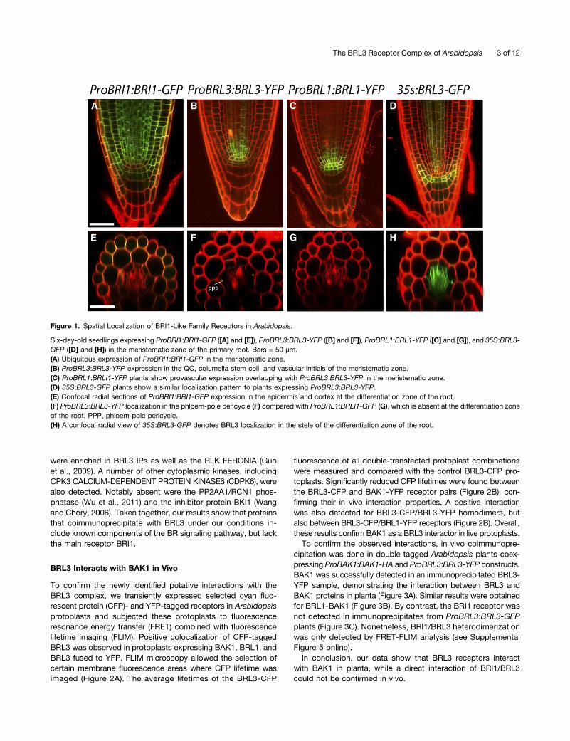
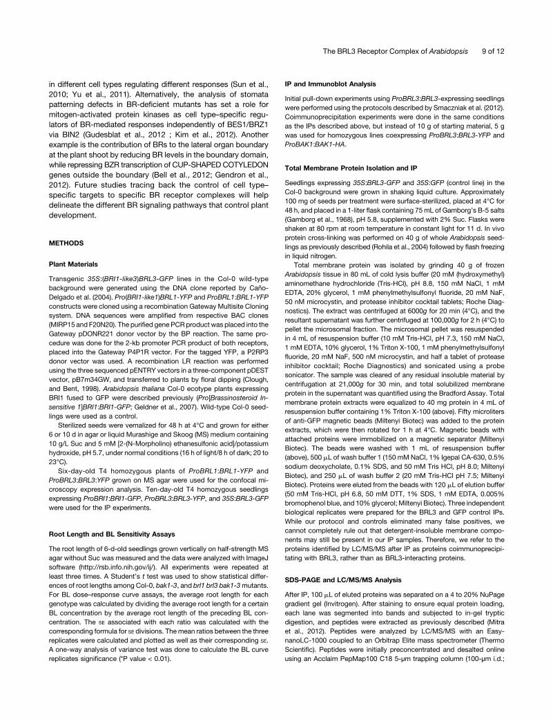
In Arabidopsis, expression of BRL1 and BRL3 is enriched in thevascular tissues, whereas that of BRI1 appears in most plantcells (Caño-Delgado et al., 2004). To reveal the localization ofBRL1 and BRL3 receptors, the full-length genomic sequences ofBRL1 and BRL3 were fused to yellow fluorescent protein (YFP)under the control of the native promoters consisting of a region2 kb upstream of the start codon for BRL1 and BRL3 (ProBRL1:BRL1-YFP and ProBRL3:BRL3-YFP, respectively). Localizationof these receptor fusions in stable T4 homozygous plants wascompared with that of ProBRI1:BRI1-GFP plants, a constructpreviously shown to complement bri1 null mutants (Geldneret al., 2007). Root analysis of 6-d-old plants revealed the pres-ence of BRI1-GFP in all cell files of the root apical meristem(Figure 1A), similar to that reported (Friedrichsen et al., 2000;Geldner et al., 2007; Wilma van Esse et al., 2011). Further up inthe meristem, confocal microscopy of a transverse sectionshowed a predominant BRI1 localization at the outer cell files(epidermis/cortex) at the differentiation zone (Figure 1E). Incontrast with BRI1, the localization pattern for BRL1 and BRL3was specific to a few cell files. At the root apex BRL1 and BRL3are similarly localized at the QC, columella stem cells, and agroup of provascular cells, including vascular initials cells lo-cated right above the QC (Figures 1B and 1C). Transverse viewat the differentiation zone of the root showed the presence ofBRL3 at the phloem-pole pericycle cells (Figure 1F), whereasBRL1 was absent from these cells (Figure 1G).
Since the expression of BRL1 and BRL3 receptors under theirnative promoters was much lower than that of ProBRI1:BRI1-GFP lines, additional 35S:BRL3-GFP–overexpressing plants
were established in order to increase the amount of BRL3protein in the plant (Figures 1D and 1H). The analysis of 35S:BRL3-GFP plants revealed a localization pattern similar tothe ProBRL3:BRL3-YFP native lines (Figures 1B and 1D),suggesting an additional spatial control at the protein levelfor BRL3. At the elongation and differentiation zone of theroot, a stele-specific localization was found in 35S:BRL3-GFPplants (Figure 1H).In mature plant organs, BRL1 and BRL3 were predominantly
localized at leaf veins and associated with phloem tissues inthe vascular bundles of the shoot inflorescence stem (seeSupplemental Figure 1 online). While 35S:BRL3-GFP plantsshowed a strong localization in the vascular tissues, GFP wasalso present at the epidermis where BRI1 is predominantly lo-calized (see Supplemental Figure 2 online).
Identification of Proteins That Coimmunoprecipitatewith BRL3
Previously identified BRI1 interactors have been essential forunderstanding BR signal transduction in the plant. Here, we usean alternative approach, to identify a native GFP-tagged plantreceptor complex from young seedlings directly by IP using anti-GFP antibodies immobilized on beads (see Methods). In pre-liminary experiments, wild-type Columbia-0 (Col-0) seedlingswere used as a control to detect proteins that bind nonspecificallyto anti-GFP beads. Three independent biological replicates wereperformed for each tagged receptor and wild-type complex pu-rified in pairs. The resulting peptides were analyzed by LC/MS/MS as previously described (Smaczniak et al., 2012). First, theProBRL3:BRL3-YFP line was used. In this line, BRL3-YFP wasundetectable in total protein extracts analyzed directly by im-munoblot, but successfully enriched after IP (see SupplementalFigure 3A online). Peptide measurement by LC/MS/MS of nativeBRL3 IPs only yielded a few proteins besides the BRL3 bait,including BRL1, DET3, and clathrin binding protein At4g18060(see Supplemental Table 1 online). To increase the sensitivity ofour approach, 35S:BRL3-GFP lines were subsequently used forIP (Figures 1C, 1D, 1G, and 1H; see Supplemental Figures 1Aand 4 online). Furthermore, a revised procedure (see Methods)that employs protein cross-linking, microsomal membrane iso-lation, and complete solubilization after ultracentrifugation, inaddition to removal of putative GFP-only coimmunoprecipitatedproteins by comparison with a 35S:GFP-expressing control ratherthan the wild type, was employed to minimize false positives.Using this protocol and LC/MS/MS analysis combined with strin-gent peptide and protein scoring parameters, a total of 128 BRL3coimmunoprecipitated proteins were significantly enriched (seeSupplemental Data Set 1 online).Previously described components of BR signaling, such as
BSK1 and BSK3 (Kim et al., 2011), were found among theproteins identified in BRL3 IPs (Table 1). Indeed, the coreceptorBAK1 (Li et al., 2002; Nam and Li, 2002) was also purified asa highly significant interactor of BRL3. Interestingly, BRI1 itselfwas not detected among the proteins that coimmunoprecipi-tate with BRL3 using our experimental conditions.The VHA-A2 (Dettmer et al., 2006) and AHA2 ATPases (related
to the AHA1 plasma membrane ATPase; Witthöft et al., 2011)
2 of 12 The Plant Cell
were enriched in BRL3 IPs as well as the RLK FERONIA (Guoet al., 2009). A number of other cytoplasmic kinases, includingCPK3 CALCIUM-DEPENDENT PROTEIN KINASE6 (CDPK6), werealso detected. Notably absent were the PP2AA1/RCN1 phos-phatase (Wu et al., 2011) and the inhibitor protein BKI1 (Wangand Chory, 2006). Taken together, our results show that proteinsthat coimmunoprecipitate with BRL3 under our conditions in-clude known components of the BR signaling pathway, but lackthe main receptor BRI1.
BRL3 Interacts with BAK1 in Vivo
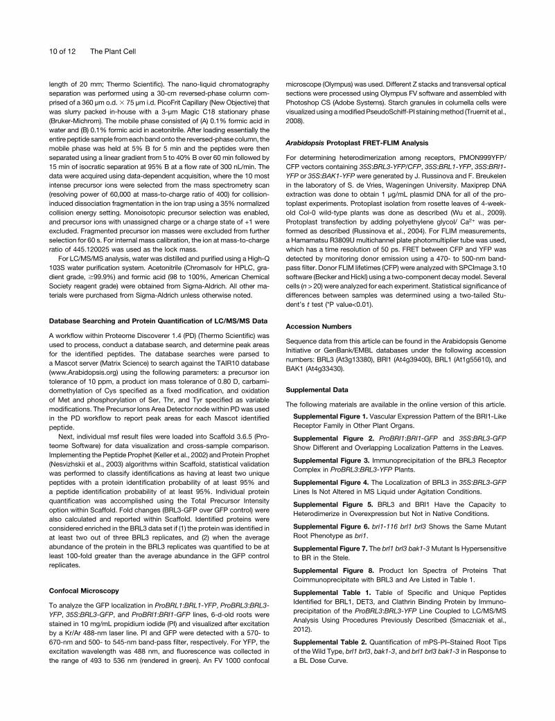
To confirm the newly identified putative interactions with theBRL3 complex, we transiently expressed selected cyan fluo-rescent protein (CFP)- and YFP-tagged receptors in Arabidopsisprotoplasts and subjected these protoplasts to fluorescenceresonance energy transfer (FRET) combined with fluorescencelifetime imaging (FLIM). Positive colocalization of CFP-taggedBRL3 was observed in protoplasts expressing BAK1, BRL1, andBRL3 fused to YFP. FLIM microscopy allowed the selection ofcertain membrane fluorescence areas where CFP lifetime wasimaged (Figure 2A). The average lifetimes of the BRL3-CFP
fluorescence of all double-transfected protoplast combinationswere measured and compared with the control BRL3-CFP pro-toplasts. Significantly reduced CFP lifetimes were found betweenthe BRL3-CFP and BAK1-YFP receptor pairs (Figure 2B), con-firming their in vivo interaction properties. A positive interactionwas also detected for BRL3-CFP/BRL3-YFP homodimers, butalso between BRL3-CFP/BRL1-YFP receptors (Figure 2B). Overall,these results confirm BAK1 as a BRL3 interactor in live protoplasts.To confirm the observed interactions, in vivo coimmunopre-
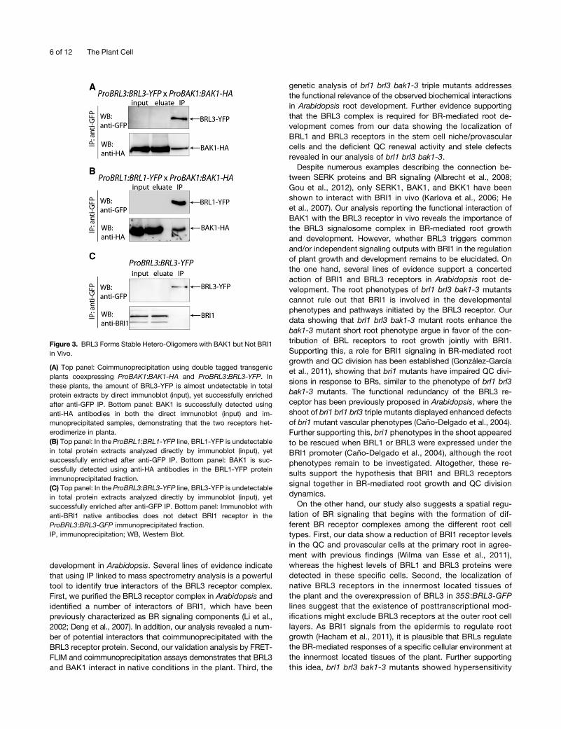
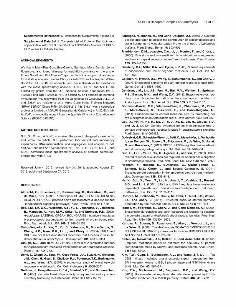
cipitation was done in double tagged Arabidopsis plants coex-pressing ProBAK1:BAK1-HA and ProBRL3:BRL3-YFP constructs.BAK1 was successfully detected in an immunoprecipitated BRL3-YFP sample, demonstrating the interaction between BRL3 andBAK1 proteins in planta (Figure 3A). Similar results were obtainedfor BRL1-BAK1 (Figure 3B). By contrast, the BRI1 receptor wasnot detected in immunoprecipitates from ProBRL3:BRL3-GFPplants (Figure 3C). Nonetheless, BRI1/BRL3 heterodimerizationwas only detected by FRET-FLIM analysis (see SupplementalFigure 5 online).In conclusion, our data show that BRL3 receptors interact
with BAK1 in planta, while a direct interaction of BRI1/BRL3could not be confirmed in vivo.
Figure 1. Spatial Localization of BRI1-Like Family Receptors in Arabidopsis.
Six-day-old seedlings expressing ProBRI1:BRI1-GFP ([A] and [E]), ProBRL3:BRL3-YFP ([B] and [F]), ProBRL1:BRL1-YFP ([C] and [G]), and 35S:BRL3-GFP ([D] and [H]) in the meristematic zone of the primary root. Bars = 50 µm.(A) Ubiquitous expression of ProBRI1:BRI1-GFP in the meristematic zone.(B) ProBRL3:BRL3-YFP expression in the QC, columella stem cell, and vascular initials of the meristematic zone.(C) ProBRL1:BRLI1-YFP plants show provascular expression overlapping with ProBRL3:BRL3-YFP in the meristematic zone.(D) 35S:BRL3-GFP plants show a similar localization pattern to plants expressing ProBRL3:BRL3-YFP.(E) Confocal radial sections of ProBRI1:BRI1-GFP expression in the epidermis and cortex at the differentiation zone of the root.(F) ProBRL3:BRL3-YFP localization in the phloem-pole pericycle (F) compared with ProBRL1:BRLI1-GFP (G), which is absent at the differentiation zoneof the root. PPP, phloem-pole pericycle.(H) A confocal radial view of 35S:BRL3-GFP denotes BRL3 localization in the stele of the differentiation zone of the root.
The BRL3 Receptor Complex of Arabidopsis 3 of 12

The BRL1/BRL3/BAK1 Receptor Complex Accounts forRoot Growth and QC Organization
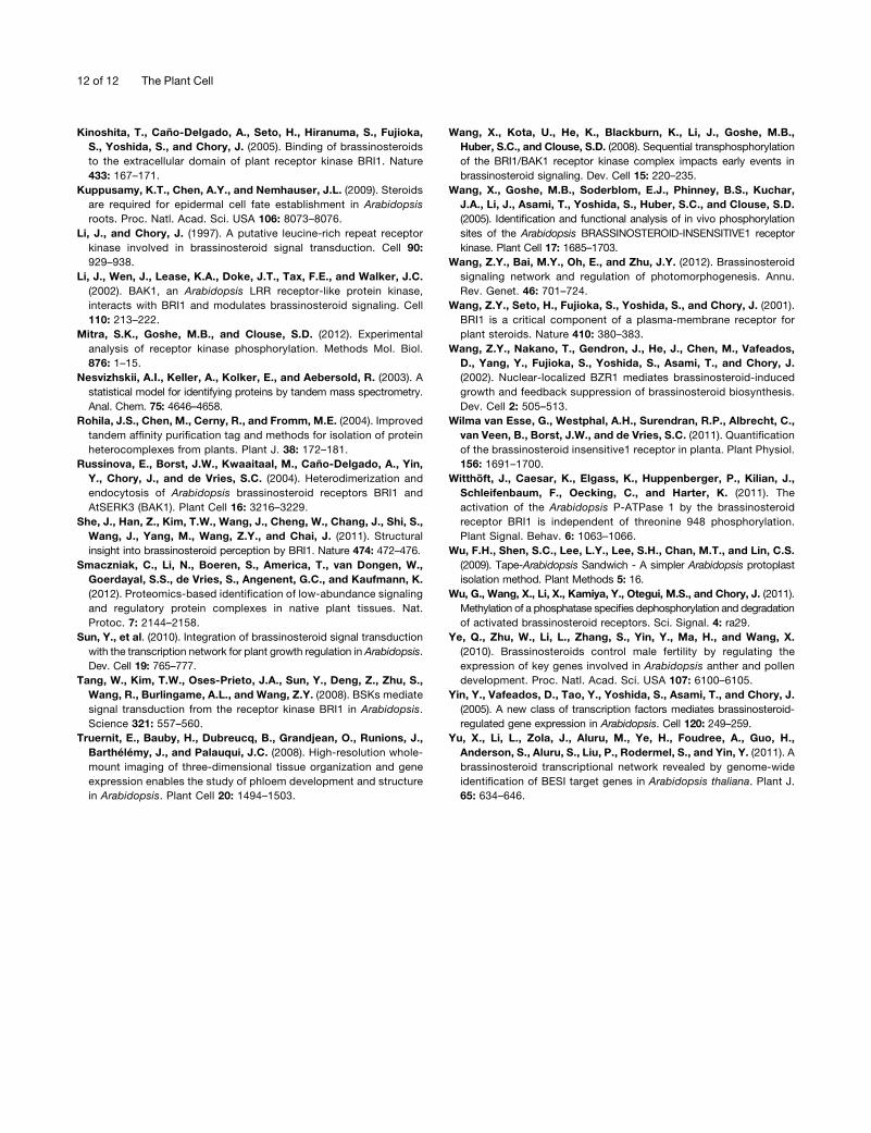
BRI1-mediated signaling regulates normal cell cycle progressionof root meristematic cells, including the rarely divided QC cellsduring root growth (González-García et al., 2011), yet a role forBRLs in the root has not hitherto been reported. To addressthe biological relevance of the observed BRL1 and BRL3 in-teractions with BAK1, we performed a genetic analysis usingmultiple combinations of BRI1-like receptors and the BAK1coreceptor. Root length analysis of 6-d-old seedlings showedthat bak1-3 roots are significantly shorter than Col-0 wild-typeplants (Figures 4A and 4B), in agreement with previous reports(Nam and Li, 2002; Albrecht et al., 2008), whereas brl1 brl3
double mutant roots were of similar length as wild-type ones.Strikingly, brl1 brl3 bak1-3 triple mutants enhanced the bak1-3short root phenotype (Figures 4A and 4B), supporting the notionthat biochemical interaction between BRL1/BRL3 and BAK1 isrequired for BR-mediated root growth. By contrast, the roots ofbri1-301 brl1 brl3 (Figures 4A and 4B) and bri1-116 brl1 brl3 (seeSupplemental Figure 6 online) were of similar length as those oftheir respective bri1 parents.BR sensitivity of brl1 brl3 bak1-3 mutants was analyzed in
a dose–response curve. Increasing BL concentrations signifi-cantly reduced the root length of wild-type plants (Figure 4C; inagreement with González-García et al., 2011). At 0.1 nM BL,a 20% reduction of root growth was observed in the wild-type plants that was not observed in brl1 brl3, bak1-3, and
Figure 2. FRET-FLIM Validation Analysis in Arabidopsis Protoplasts.
(A) and (B) FRET combined with FLIM in Arabidopsis protoplasts transfected with BRL3-CFP alone or coexpressing BRL3-CFP with BAK1-YFP, BRL3-YFP, and BRL1-YFP.(A) Top panel: Fluorescence intensity images show expression along the entire membrane. Bottom panel: Fluorescence lifetime images show t lifetimefor CFP represented by a colored scale. Bars = 10 µm.(B) Graphical representation for significantly reduced CFP t lifetimes between the BRL3-CFP and BAK1-YFP receptor pairs confirming their in vivointeraction. Reduced lifetime also significant for protoplasts coexpressing BRL3 with BRL3 and BRL1 membrane proteins. Results are the average ofthree independent replicate experiments 6 SE (n = 45). Student’s t test indicates that differences are statistically significant between BRL3-CFP andBAK1-YFP as well as between BRL3-CFP homodimers and BRL1-YFP heterodimers (*P value <0.01).[See online article for color version of this figure.]
4 of 12 The Plant Cell
brl1 brl3 bak1-3 (Figure 4C). In response to 1 nM BL, the rootlength of brl1 brl3 and bak1-3 was similar to that of the wildtype, whereas in the same conditions, brl1 brl3 bak1-3 showeda significantly reduced sensitivity to BR-mediated root short-ening (Figure 4C).
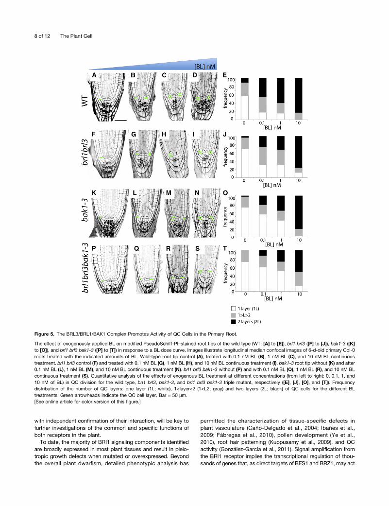
A previous mutant analysis showed that BRs are required tomaintain quiescence at the root stem cell niche (González-García et al., 2011). Since BRL receptors localized to these cells(Figures 1B and 1C), we asked whether the BRL3 complex isnecessary to preserve the competence of QC cells to divide atthe root apex. Confocal microscopy and quantitative analysis ofQC cells in a wild-type population showed that 58% of theplants had one layer of QC cells (1L), whereas 10% had twolayers (2L; Figures 5A and 5E). The remaining 32% of the rootsanalyzed exhibited an intermediary phenotype, where only someof the QC cells appeared divided (Figures 5A and 5E). Thistransition state was referred to as (1<L<2). Analysis of QC or-ganization was studied in different mutant combinations. Com-pared with wild-type plants (Figures 5A and 5E), a reduction inthe frequency of QC division was observed in brl1 brl3 (Figures5F and 5J), bak1-3 (Figures 5K and 5O), and brl1 brl3 bak1-3(Figures 5P and 5T) mutants, showing 70% of the plants withone layer of QC cells. Strikingly, treatment with 0.1 nM BL en-hanced the observed insensibility to BL in brl1 brl3 doublemutants (25% 2L; Figures 5G and 5J) and bak1-3 mutant(20% 2L; Figures 5L and 5O) compared with the wild type(45% 2L; Figures 5B and 5E) in the QC cells. This phenotypewas even stronger in the brl1 brl3 bak1-3 triple mutants (13% 2L;Figures 5Q and 5T), in agreement with our previous results in theBL dose–response curve.
Similar to our results obtained for the BL dose–responsecurve in roots (Figure 4C), this phenotype was dose specific and1 nM BL promoted QC division in wild-type plants (60% 2L;Figures 5C and 5E), whereas the QC cells of brl1 brl3 and bak1-3plants retained some insensitivity to this hormone concentration(35% 2L; Figures 5H, 5J, 5M, and 5O). This insensitivity wasstronger in brl1 brl3 bak1-3 mutants treated with 1 nM BL (28%2L; Figures 5R and 5T) and was confirmed when these plantswere treated with 10 nM BL (Figures 5S and 5T). In agreement,brl1 brl3 bak1-3 mutants showed an increased number of plantswith one QC layer compared with the remaining genotypes forall BL concentrations analyzed (Figures 5E, 5J, 5O, and 5T). Thecomplete quantitative analysis for three independent biologicalreplicates is shown in Supplemental Supplemental Table 2 on-line. Furthermore, we observed that the brl1 brl3 bak1-3mutantsshowed hypersensitivity to BR in the stele when compared withbak1 or brl1 brl3 mutants (see Supplemental Figure 7 online).These results unveil a concerted action of these BR receptors inthe QC and provascular cells and provide biological significancefor the observed biochemical interactions. In conclusion, ouranalysis shows that BRLs regulate BR-mediated responses ofa specific cellular environment at the innermost located tissuesof the plant.
DISCUSSION
Our study provides biochemical and genetic evidence for theinteraction of the BR receptors BRL3 and BRL1 and the cor-eceptor BAK1 in vivo, while demonstrating that the BRL3 sig-nalosome complex is required for normal root growth and
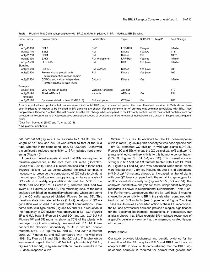
Table 1. Proteins That Coimmunoprecipitate with BRL3 and Are Implicated in BRI1-Mediated BR Signaling
Gene Locus Protein Name Localization Type BZR1/BES1 Targeta Fold Change
BRsAt3g13380 BRL3 PMb LRR-RLK Yes/yes InfiniteAt4g00710 BSK3 PM Kinase Yes/low 118At4g35230 BSK1 PM Kinase Yes 1037At4g33430 BAK1 PM, endosome LRR-RLK Yes/yes InfiniteAt3g51550 FERONIA PM RLK Yes (low) Infinite
SignalingAt4g23650 CDPK6, PM, cytosol Kinase Yes (low) 205At1g63500 Protein kinase protein with
tetratricopeptide repeat domainPM Kinase Yes (low) 160
At3g57530 CDPKW and calcium-dependentprotein kinase 32 (CDPK32)
Cytosol Kinase Yes Infinite
TransportAt2g21410 VHA-A2 proton pump Vacuole, tonoplast ATPase 110At4g30190 AHA2 ATPase 2 Vacuole ATPase Yes 254TraffickingAt3g60190 Dynamin-related protein 1E (DRP1E) PM, cell plate GTPase Yes 208
A summary of selected proteins that coimmunoprecipitate with BRL3. Only proteins that passed the cutoff threshold described in Methods and havebeen implicated or known to be involved in BR signaling are shown. For the complete list of proteins that coimmunoprecipitate with BRL3, seeSupplemental Data Set 1 online. The last column lists the fold change when compared to the GFP-only control. Infinite means that peptides were notdetected in the control sample. Representative product ion spectra of peptides identified for each of these proteins are shown in Supplemental Figure 8online.aData from Sun et al. (2010) and Yu et al. (2011).bPM, plasma membrane.
The BRL3 Receptor Complex of Arabidopsis 5 of 12
development in Arabidopsis. Several lines of evidence indicatethat using IP linked to mass spectrometry analysis is a powerfultool to identify true interactors of the BRL3 receptor complex.First, we purified the BRL3 receptor complex in Arabidopsis andidentified a number of interactors of BRI1, which have beenpreviously characterized as BR signaling components (Li et al.,2002; Deng et al., 2007). In addition, our analysis revealed a num-ber of potential interactors that coimmunoprecipitated with theBRL3 receptor protein. Second, our validation analysis by FRET-FLIM and coimmunoprecipitation assays demonstrates that BRL3and BAK1 interact in native conditions in the plant. Third, the
genetic analysis of brl1 brl3 bak1-3 triple mutants addressesthe functional relevance of the observed biochemical interactionsin Arabidopsis root development. Further evidence supportingthat the BRL3 complex is required for BR-mediated root de-velopment comes from our data showing the localization ofBRL1 and BRL3 receptors in the stem cell niche/provascularcells and the deficient QC renewal activity and stele defectsrevealed in our analysis of brl1 brl3 bak1-3.Despite numerous examples describing the connection be-
tween SERK proteins and BR signaling (Albrecht et al., 2008;Gou et al., 2012), only SERK1, BAK1, and BKK1 have beenshown to interact with BRI1 in vivo (Karlova et al., 2006; Heet al., 2007). Our analysis reporting the functional interaction ofBAK1 with the BRL3 receptor in vivo reveals the importance ofthe BRL3 signalosome complex in BR-mediated root growthand development. However, whether BRL3 triggers commonand/or independent signaling outputs with BRI1 in the regulationof plant growth and development remains to be elucidated. Onthe one hand, several lines of evidence support a concertedaction of BRI1 and BRL3 receptors in Arabidopsis root de-velopment. The root phenotypes of brl1 brl3 bak1-3 mutantscannot rule out that BRI1 is involved in the developmentalphenotypes and pathways initiated by the BRL3 receptor. Ourdata showing that brl1 brl3 bak1-3 mutant roots enhance thebak1-3 mutant short root phenotype argue in favor of the con-tribution of BRL receptors to root growth jointly with BRI1.Supporting this, a role for BRI1 signaling in BR-mediated rootgrowth and QC division has been established (González-Garcíaet al., 2011), showing that bri1 mutants have impaired QC divi-sions in response to BRs, similar to the phenotype of brl1 brl3bak1-3 mutants. The functional redundancy of the BRL3 re-ceptor has been previously proposed in Arabidopsis, where theshoot of bri1 brl1 brl3 triple mutants displayed enhanced defectsof bri1 mutant vascular phenotypes (Caño-Delgado et al., 2004).Further supporting this, bri1 phenotypes in the shoot appearedto be rescued when BRL1 or BRL3 were expressed under theBRI1 promoter (Caño-Delgado et al., 2004), although the rootphenotypes remain to be investigated. Altogether, these re-sults support the hypothesis that BRI1 and BRL3 receptorssignal together in BR-mediated root growth and QC divisiondynamics.On the other hand, our study also suggests a spatial regu-
lation of BR signaling that begins with the formation of dif-ferent BR receptor complexes among the different root celltypes. First, our data show a reduction of BRI1 receptor levelsin the QC and provascular cells at the primary root in agree-ment with previous findings (Wilma van Esse et al., 2011),whereas the highest levels of BRL1 and BRL3 proteins weredetected in these specific cells. Second, the localization ofnative BRL3 receptors in the innermost located tissues ofthe plant and the overexpression of BRL3 in 35S:BRL3-GFPlines suggest that the existence of posttranscriptional mod-ifications might exclude BRL3 receptors at the outer root celllayers. As BRI1 signals from the epidermis to regulate rootgrowth (Hacham et al., 2011), it is plausible that BRLs regulatethe BR-mediated responses of a specific cellular environment atthe innermost located tissues of the plant. Further supportingthis idea, brl1 brl3 bak1-3 mutants showed hypersensitivity
Figure 3. BRL3 Forms Stable Hetero-Oligomers with BAK1 but Not BRI1in Vivo.
(A) Top panel: Coimmunoprecipitation using double tagged transgenicplants coexpressing ProBAK1:BAK1-HA and ProBRL3:BRL3-YFP. Inthese plants, the amount of BRL3-YFP is almost undetectable in totalprotein extracts by direct immunoblot (input), yet successfully enrichedafter anti-GFP IP. Bottom panel: BAK1 is successfully detected usinganti-HA antibodies in both the direct immunoblot (input) and im-munoprecipitated samples, demonstrating that the two receptors het-erodimerize in planta.(B) Top panel: In the ProBRL1:BRL1-YFP line, BRL1-YFP is undetectablein total protein extracts analyzed directly by immunoblot (input), yetsuccessfully enriched after anti-GFP IP. Bottom panel: BAK1 is suc-cessfully detected using anti-HA antibodies in the BRL1-YFP proteinimmunoprecipitated fraction.(C) Top panel: In the ProBRL3:BRL3-YFP line, BRL3-YFP is undetectablein total protein extracts analyzed directly by immunoblot (input), yetsuccessfully enriched after anti-GFP IP. Bottom panel: Immunoblot withanti-BRI1 native antibodies does not detect BRI1 receptor in theProBRL3:BRL3-GFP immunoprecipitated fraction.IP, immunoprecipitation; WB, Western Blot.
6 of 12 The Plant Cell

to BRs in the stele when compared with bak1 and/or brl1 brl3mutants.
Finally, the lack of BRI1-specific peptides in BRL3 coimmu-noprecipitates subjected to mass spectrometry analysis togetherwith the inability to detect native BRI1 receptors in coimmuno-precipitation of ProBRL3:BRL3-YFP lines are consistent withthe formation of distinct BR receptor complexes, although nega-tive mass spectrometry and coimmunoprecipitation results cannotbe considered conclusive. Considering the complementary
localization of BRI1 an BRL3 receptors in the root, good can-didates for BRL3-specific interactors might be proteins thatcoimmunoprecipitate with BRL3 that appeared enriched in thestem cell niche, including BAK1, PIN FORMED7, ATP-bindingcassette-36, and an uncharacterized LRR-RLK (At1g53440), aswell as proteins enriched in provascular/stele tissues, such asan uncharacterized LRR-RLK (At2g37050) and an ATP-bindingcassette-2 transporter. Future identification and comparison ofproteins that coimmunoprecipitate with BRI1 and BRL3, along
Figure 4. The Root and BR Insensitivity Phenotypes of the brl1 brl3 bak1-3 Triple Mutant Are More Severe Than Those of bak1-3.
(A) Phenotype of 6-d-old wild-type (WT), bak1-3, brl1 brl3, brl1 brl3 bak1-3, bri1-301, and bri1-301 brl1 brl3 mutants. Bar = 0.5 mm.(B) Root length assay of 6-d-old wild-type, bak1-3, brl1 brl3, brl1 brl3 bak1-3, bri1-301, and bri1-301 brl1 brl3 seedlings. Results are average 6 SE (n =90). Student’s t test indicates that root length differences are statistically significant between Col-0 wild type and bak1-3 as well as between bak1-3 andbrl1 brl3 bak1-3 (*P value < 0.01).(C) Dose–response curve of exogenous BL treatments (0.001, 0.01, 0.1, 1, and 10 nM) in 6-d-old seedlings. Ratios of root shortening were calculated foreach BL concentration and represented in the graph. Results are the average of three independent replicate experiments 6 SE (n = 85). Student’s t testindicates that brl1 brl3 bak1-3 is statistically less sensitive to BL when compared with Col-0 wild type at 0.1 and 1 nM of BL (*P value < 0.01).[See online article for color version of this figure.]
The BRL3 Receptor Complex of Arabidopsis 7 of 12
with independent confirmation of their interaction, will be key tofurther investigations of the common and specific functions ofboth receptors in the plant.
To date, the majority of BRI1 signaling components identifiedare broadly expressed in most plant tissues and result in pleio-tropic growth defects when mutated or overexpressed. Beyondthe overall plant dwarfism, detailed phenotypic analysis has
permitted the characterization of tissue-specific defects inplant vasculature (Caño-Delgado et al., 2004; Ibañes et al.,2009; Fàbregas et al., 2010), pollen development (Ye et al.,2010), root hair patterning (Kuppusamy et al., 2009), and QCactivity (González-García et al., 2011). Signal amplification fromthe BRI1 receptor implies the transcriptional regulation of thou-sands of genes that, as direct targets of BES1 and BRZ1, may act
Figure 5. The BRL3/BRL1/BAK1 Complex Promotes Activity of QC Cells in the Primary Root.
The effect of exogenously applied BL on modified PseudoSchiff-PI–stained root tips of the wild type (WT; [A] to [E]), brl1 brl3 ([F] to [J]), bak1-3 ([K]to [O]), and brl1 brl3 bak1-3 ([P] to [T]) in response to a BL dose curve. Images illustrate longitudinal median confocal images of 6-d-old primary Col-0roots treated with the indicated amounts of BL. Wild-type root tip control (A), treated with 0.1 nM BL (B), 1 nM BL (C), and 10 nM BL continuoustreatment. brl1 brl3 control (F) and treated with 0.1 nM BL (G), 1 nM BL (H), and 10 nM BL continuous treatment (I). bak1-3 root tip without (K) and after0.1 nM BL (L), 1 nM BL (M), and 10 nM BL continuous treatment (N). brl1 brl3 bak1-3 without (P) and with 0.1 nM BL (Q), 1 nM BL (R), and 10 nM BLcontinuous treatment (S). Quantitative analysis of the effects of exogenous BL treatment at different concentrations (from left to right: 0, 0.1, 1, and10 nM of BL) in QC division for the wild type, brl1 brl3, bak1-3, and brl1 brl3 bak1-3 triple mutant, respectively ([E], [J], [O], and [T]). Frequencydistribution of the number of QC layers: one layer (1L; white), 1<layer<2 (1<L2; gray) and two layers (2L; black) of QC cells for the different BLtreatments. Green arrowheads indicate the QC cell layer. Bar = 50 µm.[See online article for color version of this figure.]
8 of 12 The Plant Cell
in different cell types regulating different responses (Sun et al.,2010; Yu et al., 2011). Alternatively, the analysis of stomatapatterning defects in BR-deficient mutants has set a role formitogen-activated protein kinases as cell type–specific regu-lators of BR-mediated responses independently of BES1/BRZ1via BIN2 (Gudesblat et al., 2012 ; Kim et al., 2012). Anotherexample is the contribution of BRs to the lateral organ boundaryat the plant shoot by reducing BR levels in the boundary domain,while repressing BZR transcription of CUP-SHAPED COTYLEDONgenes outside the boundary (Bell et al., 2012; Gendron et al.,2012). Future studies tracing back the control of cell type–specific targets to specific BR receptor complexes will helpdelineate the different BR signaling pathways that control plantdevelopment.
METHODS
Plant Materials
Transgenic 35S:(BRI1-like3)BRL3-GFP lines in the Col-0 wild-typebackground were generated using the DNA clone reported by Caño-Delgado et al. (2004). Pro(BRI1-like1)BRL1-YFP and ProBRL1:BRL1-YFPconstructs were cloned using a recombination Gateway Multisite Cloningsystem. DNA sequences were amplified from respective BAC clones(MIRP15 and F20N20). The purified gene PCRproduct was placed into theGateway pDONR221 donor vector by the BP reaction. The same pro-cedure was done for the 2-kb promoter PCR product of both receptors,placed into the Gateway P4P1R vector. For the tagged YFP, a P2RP3donor vector was used. A recombination LR reaction was performedusing the three sequenced pENTRY vectors in a three-component pDESTvector, pB7m34GW, and transferred to plants by floral dipping (Clough,and Bent, 1998). Arabidopsis thaliana Col-0 ecotype plants expressingBRI1 fused to GFP were described previously (Pro[Brassinosteroid In-sensitive 1]BRI1:BRI1-GFP; Geldner et al., 2007). Wild-type Col-0 seed-lings were used as a control.
Sterilized seeds were vernalized for 48 h at 4°C and grown for either6 or 10 d in agar or liquid Murashige and Skoog (MS) medium containing10 g/L Suc and 5 mM [2-(N-Morpholino) ethanesulfonic acid]/potassiumhydroxide, pH 5.7, under normal conditions (16 h of light/8 h of dark; 20 to23°C).
Six-day-old T4 homozygous plants of ProBRL1:BRL1-YFP andProBRL3:BRL3:YFP grown on MS agar were used for the confocal mi-croscopy expression analysis. Ten-day-old T4 homozygous seedlingsexpressing ProBRI1:BRI1-GFP, ProBRL3:BRL3-YFP, and 35S:BRL3-GFPwere used for the IP experiments.
Root Length and BL Sensitivity Assays
The root length of 6-d-old seedlings grown vertically on half-strength MSagar without Suc was measured and the data were analyzed with ImageJsoftware (http://rsb.info.nih.gov/ij/). All experiments were repeated atleast three times. A Student’s t test was used to show statistical differ-ences of root lengths among Col-0, bak1-3, and brl1 brl3 bak1-3mutants.For BL dose–response curve assays, the average root length for eachgenotype was calculated by dividing the average root length for a certainBL concentration by the average root length of the preceding BL con-centration. The SE associated with each ratio was calculated with thecorresponding formula for SE divisions. Themean ratios between the threereplicates were calculated and plotted as well as their corresponding SE.A one-way analysis of variance test was done to calculate the BL curvereplicates significance (*P value < 0.01).
IP and Immunoblot Analysis
Initial pull-down experiments using ProBRL3:BRL3-expressing seedlingswere performed using the protocols described by Smaczniak et al. (2012).Coimmunoprecipitation experiments were done in the same conditionsas the IPs described above, but instead of 10 g of starting material, 5 gwas used for homozygous lines coexpressing ProBRL3:BRL3-YFP andProBAK1:BAK1-HA.
Total Membrane Protein Isolation and IP
Seedlings expressing 35S:BRL3-GFP and 35S:GFP (control line) in theCol-0 background were grown in shaking liquid culture. Approximately100 mg of seeds per treatment were surface-sterilized, placed at 4°C for48 h, and placed in a 1-liter flask containing 75 mL of Gamborg’s B-5 salts(Gamborg et al., 1968), pH 5.8, supplemented with 2% Suc. Flasks wereshaken at 80 rpm at room temperature in constant light for 11 d. In vivoprotein cross-linking was performed on 40 g of whole Arabidopsis seed-lings as previously described (Rohila et al., 2004) followed by flash freezingin liquid nitrogen.
Total membrane protein was isolated by grinding 40 g of frozenArabidopsis tissue in 80 mL of cold lysis buffer (20 mM (hydroxymethyl)aminomethane hydrochloride (Tris-HCl), pH 8.8, 150 mM NaCl, 1 mMEDTA, 20% glycerol, 1 mM phenylmethylsulfonyl fluoride, 20 mM NaF,50 nM microcystin, and protease inhibitor cocktail tablets; Roche Diag-nostics). The extract was centrifuged at 6000g for 20 min (4°C), and theresultant supernatant was further centrifuged at 100,000g for 2 h (4°C) topellet the microsomal fraction. The microsomal pellet was resuspendedin 4 mL of resuspension buffer (10 mM Tris-HCl, pH 7.3, 150 mM NaCl,1 mM EDTA, 10% glycerol, 1% Triton X-100, 1 mM phenylmethylsulfonylfluoride, 20 mM NaF, 500 nM microcystin, and half a tablet of proteaseinhibitor cocktail; Roche Diagnostics) and sonicated using a probesonicator. The sample was cleared of any residual insoluble material bycentrifugation at 21,000g for 30 min, and total solubilized membraneprotein in the supernatant was quantified using the Bradford Assay. Totalmembrane protein extracts were equalized to 40 mg protein in 4 mL ofresuspension buffer containing 1% Triton X-100 (above). Fifty microlitersof anti-GFP magnetic beads (Miltenyi Biotec) was added to the proteinextracts, which were then rotated for 1 h at 4°C. Magnetic beads withattached proteins were immobilized on a magnetic separator (MiltenyiBiotec). The beads were washed with 1 mL of resuspension buffer(above), 500 mL of wash buffer 1 (150 mM NaCl, 1% Igepal CA-630, 0.5%sodium deoxycholate, 0.1% SDS, and 50 mM Tris HCl, pH 8.0; MiltenyiBiotec), and 250 mL of wash buffer 2 (20 mM Tris-HCl pH 7.5; MiltenyiBiotec). Proteins were eluted from the beads with 120 mL of elution buffer(50 mM Tris-HCl, pH 6.8, 50 mM DTT, 1% SDS, 1 mM EDTA, 0.005%bromophenol blue, and 10% glycerol; Miltenyi Biotec). Three independentbiological replicates were prepared for the BRL3 and GFP control IPs.While our protocol and controls eliminated many false positives, wecannot completely rule out that detergent-insoluble membrane compo-nents may still be present in our IP samples. Therefore, we refer to theproteins identified by LC/MS/MS after IP as proteins coimmunoprecipi-tating with BRL3, rather than as BRL3-interacting proteins.
SDS-PAGE and LC/MS/MS Analysis
After IP, 100 mL of eluted proteins was separated on a 4 to 20% NuPagegradient gel (Invitrogen). After staining to ensure equal protein loading,each lane was segmented into bands and subjected to in-gel trypticdigestion, and peptides were extracted as previously described (Mitraet al., 2012). Peptides were analyzed by LC/MS/MS with an Easy-nanoLC-1000 coupled to an Orbitrap Elite mass spectrometer (ThermoScientific). Peptides were initially preconcentrated and desalted onlineusing an Acclaim PepMap100 C18 5-µm trapping column (100-µm i.d.;
The BRL3 Receptor Complex of Arabidopsis 9 of 12
length of 20 mm; Thermo Scientific). The nano-liquid chromatographyseparation was performed using a 30-cm reversed-phase column com-prised of a 360 µm o.d.3 75 µm i.d. PicoFrit Capillary (New Objective) thatwas slurry packed in-house with a 3-µm Magic C18 stationary phase(Bruker-Michrom). The mobile phase consisted of (A) 0.1% formic acid inwater and (B) 0.1% formic acid in acetonitrile. After loading essentially theentire peptide sample fromeachbandonto the reversed-phase column, themobile phase was held at 5% B for 5 min and the peptides were thenseparated using a linear gradient from 5 to 40% B over 60 min followed by15 min of isocratic separation at 95% B at a flow rate of 300 nL/min. Thedata were acquired using data-dependent acquisition, where the 10 mostintense precursor ions were selected from the mass spectrometry scan(resolving power of 60,000 at mass-to-charge ratio of 400) for collision-induced dissociation fragmentation in the ion trap using a 35% normalizedcollision energy setting. Monoisotopic precursor selection was enabled,and precursor ions with unassigned charge or a charge state of +1 wereexcluded. Fragmented precursor ion masses were excluded from furtherselection for 60 s. For internal mass calibration, the ion at mass-to-chargeratio of 445.120025 was used as the lock mass.
For LC/MS/MS analysis, water was distilled and purified using a High-Q103S water purification system. Acetonitrile (Chromasolv for HPLC, gra-dient grade, $99.9%) and formic acid (98 to 100%, American ChemicalSociety reagent grade) were obtained from Sigma-Aldrich. All other ma-terials were purchased from Sigma-Aldrich unless otherwise noted.
Database Searching and Protein Quantification of LC/MS/MS Data
A workflow within Proteome Discoverer 1.4 (PD) (Thermo Scientific) wasused to process, conduct a database search, and determine peak areasfor the identified peptides. The database searches were parsed toa Mascot server (Matrix Science) to search against the TAIR10 database(www.Arabidopsis.org) using the following parameters: a precursor iontolerance of 10 ppm, a product ion mass tolerance of 0.80 D, carbami-domethylation of Cys specified as a fixed modification, and oxidationof Met and phosphorylation of Ser, Thr, and Tyr specified as variablemodifications. The Precursor Ions Area Detector node within PDwas usedin the PD workflow to report peak areas for each Mascot identifiedpeptide.
Next, individual msf result files were loaded into Scaffold 3.6.5 (Pro-teome Software) for data visualization and cross-sample comparison.Implementing the Peptide Prophet (Keller et al., 2002) and Protein Prophet(Nesvizhskii et al., 2003) algorithms within Scaffold, statistical validationwas performed to classify identifications as having at least two uniquepeptides with a protein identification probability of at least 95% anda peptide identification probability of at least 95%. Individual proteinquantification was accomplished using the Total Precursor Intensityoption within Scaffold. Fold changes (BRL3-GFP over GFP control) werealso calculated and reported within Scaffold. Identified proteins wereconsidered enriched in the BRL3 data set if (1) the protein was identified inat least two out of three BRL3 replicates, and (2) when the averageabundance of the protein in the BRL3 replicates was quantified to be atleast 100-fold greater than the average abundance in the GFP controlreplicates.
Confocal Microscopy
To analyze the GFP localization in ProBRL1:BRL1-YFP, ProBRL3:BRL3-YFP, 35S:BRL3-GFP, and ProBRI1:BRI1-GFP lines, 6-d-old roots werestained in 10 mg/mL propidium iodide (PI) and visualized after excitationby a Kr/Ar 488-nm laser line. PI and GFP were detected with a 570- to670-nm and 500- to 545-nm band-pass filter, respectively. For YFP, theexcitation wavelength was 488 nm, and fluorescence was collected inthe range of 493 to 536 nm (rendered in green). An FV 1000 confocal
microscope (Olympus) was used. Different Z stacks and transversal opticalsections were processed using Olympus FV software and assembled withPhotoshop CS (Adobe Systems). Starch granules in columella cells werevisualized using amodifiedPseudoSchiff-PI stainingmethod (Truernit et al.,2008).
Arabidopsis Protoplast FRET-FLIM Analysis
For determining heterodimerization among receptors, PMON999YFP/CFP vectors containing 35S:BRL3-YFP/CFP, 35S:BRL1-YFP, 35S:BRI1-YFP or 35S:BAK1-YFP were generated by J. Russinova and F. Breukelenin the laboratory of S. de Vries, Wageningen University. Maxiprep DNAextraction was done to obtain 1 µg/mL plasmid DNA for all of the pro-toplast experiments. Protoplast isolation from rosette leaves of 4-week-old Col-0 wild-type plants was done as described (Wu et al., 2009).Protoplast transfection by adding polyethylene glycol/ Ca2+ was per-formed as described (Russinova et al., 2004). For FLIM measurements,a Hamamatsu R3809U multichannel plate photomultiplier tube was used,which has a time resolution of 50 ps. FRET between CFP and YFP wasdetected by monitoring donor emission using a 470- to 500-nm band-pass filter. Donor FLIM lifetimes (CFP) were analyzed with SPCImage 3.10software (Becker and Hickl) using a two-component decaymodel. Severalcells (n > 20) were analyzed for each experiment. Statistical significance ofdifferences between samples was determined using a two-tailed Stu-dent’s t test (*P value<0.01).
Accession Numbers
Sequence data from this article can be found in the Arabidopsis GenomeInitiative or GenBank/EMBL databases under the following accessionnumbers: BRL3 (At3g13380), BRl1 (At4g39400), BRL1 (At1g55610), andBAK1 (At4g33430).
Supplemental Data
The following materials are available in the online version of this article.
Supplemental Figure 1. Vascular Expression Pattern of the BRI1-LikeReceptor Family in Other Plant Organs.
Supplemental Figure 2. ProBRI1:BRI1-GFP and 35S:BRL3-GFPShow Different and Overlapping Localization Patterns in the Leaves.
Supplemental Figure 3. Immunoprecipitation of the BRL3 ReceptorComplex in ProBRL3:BRL3-YFP Plants.
Supplemental Figure 4. The Localization of BRL3 in 35S:BRL3-GFPLines Is Not Altered in MS Liquid under Agitation Conditions.
Supplemental Figure 5. BRL3 and BRI1 Have the Capacity toHeterodimerize in Overexpression but Not in Native Conditions.
Supplemental Figure 6. bri1-116 brl1 brl3 Shows the Same MutantRoot Phenotype as bri1.
Supplemental Figure 7. The brl1 brl3 bak1-3Mutant Is Hypersensitiveto BR in the Stele.
Supplemental Figure 8. Product Ion Spectra of Proteins ThatCoimmunoprecipitate with BRL3 and Are Listed in Table 1.
Supplemental Table 1. Table of Specific and Unique PeptidesIdentified for BRL1, DET3, and Clathrin Binding Protein by Immuno-precipitation of the ProBRL3:BRL3-YFP Line Coupled to LC/MS/MSAnalysis Using Procedures Previously Described (Smaczniak et al.,2012).
Supplemental Table 2. Quantification of mPS-PI–Stained Root Tipsof the Wild Type, brl1 brl3, bak1-3, and brl1 brl3 bak1-3 in Response toa BL Dose Curve.
10 of 12 The Plant Cell
Supplemental References 1. References for Supplemental Figures 1-8.
Supplemental Data Set 1. Complete List of Proteins That Coimmu-noprecipitate with BRL3, Identified by LC/MS/MS Analysis of BRL3-GFP versus GFP-Only Control.
ACKNOWLEDGMENTS
We thank Mary-Paz González-García, Santiago Mora-García, JennyRussinova, and Josep Vilarrasa for insightful comments on the article,Emma Sudrià and Eloi Franco-Trepat for technical support, Juan Vegasfor statistical analysis, Joanne Chory for anti-BRI1 antibodies, Jan WillemBorst for FRET-FLIM experiments, and Kevin Blackburn for assistancewith the mass spectrometry analysis. S.D.C., T.E.N., and M.B.G. arefunded by grants from the U.S. National Science Foundation (MCB-1021363 and DBI-1126244). N.F. is funded by an Formació de personalInvestigador PhD fellowship from the Generalitat de Catalunya. A.I.C.-D.and S.d.V. are recipients of a Marie-Curie Initial Training Network“BRAVISSIMO” (Grant PITN-GA-2008-215118). S.d.V. was a sabbaticalprofessor funded by AGAUR (Generalitat de Catalunya) in A.I.C.-D.’s lab.A.I.C.-D. is funded by a grant from the Spanish Ministry of Education andScience (BIO2010/00505).
AUTHOR CONTRIBUTIONS
N.F., S.d.V., and A.I.C.-D. conceived the project, designed experiments,and wrote the article. N.F. performed biochemical and microscopyexperiments, DNA manipulation, and segregation and analysis of brl1brl3 bak1 and bri1 brl1 brl3 mutants. N.F., N.L., S.B., T.E.N., M.B.G., andS.D.C. performed mass spectrometry analysis of proteins coimmuno-precipitated with BRL3.
Received June 4, 2013; revised July 31, 2013; accepted August 27,2013; published September 24, 2013.
REFERENCES
Albrecht, C., Russinova, E., Kemmerling, B., Kwaaitaal, M., andde Vries, S.C. (2008). Arabidopsis SOMATIC EMBRYOGENESISRECEPTOR KINASE proteins serve brassinosteroid-dependent and-independent signaling pathways. Plant Physiol. 148: 611–619.
Bell, E.M., Lin, W.C., Husbands, A.Y., Yu, L., Jaganatha, V., Jablonska,B., Mangeon, A., Neff, M.M., Girke, T., and Springer, P.S. (2012).Arabidopsis LATERAL ORGAN BOUNDARIES negatively regulatesbrassinosteroid accumulation to limit growth in organ boundaries.Proc. Natl. Acad. Sci. USA 109: 21146–21151.
Caño-Delgado, A., Yin, Y., Yu, C., Vafeados, D., Mora-García, S.,Cheng, J.C., Nam, K.H., Li, J., and Chory, J. (2004). BRL1 andBRL3 are novel brassinosteroid receptors that function in vasculardifferentiation in Arabidopsis. Development 131: 5341–5351.
Clough, S.J., and Bent, A.F. (1998). Floral dip: A simplified methodfor Agrobacterium-mediated transformation of Arabidopsis thaliana.Plant J. 16: 735–743.
Deng, Z., Zhang, X., Tang, W., Oses-Prieto, J.A., Suzuki, N., Gendron,J.M., Chen, H., Guan, S., Chalkley, R.J., Peterman, T.K., Burlingame,A.L., and Wang, Z.Y. (2007). A proteomics study of brassinosteroidresponse in Arabidopsis. Mol. Cell. Proteomics 6: 2058–2071.
Dettmer, J., Hong-Hermesdorf, A., Stierhof, Y.D., and Schumacher,K. (2006). Vacuolar H+-ATPase activity is required for endocytic andsecretory trafficking in Arabidopsis. Plant Cell 18: 715–730.
Fàbregas, N., Ibañes, M., and Caño-Delgado, A.I. (2010). A systemsbiology approach to dissect the contribution of brassinosteroid andauxin hormones to vascular patterning in the shoot of Arabidopsisthaliana. Plant Signal. Behav. 5: 903–906.
Friedrichsen, D.M., Joazeiro, C.A., Li, J., Hunter, T., and Chory, J.(2000). Brassinosteroid-insensitive-1 is a ubiquitously expressedleucine-rich repeat receptor serine/threonine kinase. Plant Physiol.123: 1247–1256.
Gamborg, O.L., Miller, R.A., and Ojima, K. (1968). Nutrient requirementsof suspension cultures of soybean root cells. Exp. Cell Res. 50:151–158.
Geldner, N., Hyman, D.L., Wang, X., Schumacher, K., and Chory, J.(2007). Endosomal signaling of plant steroid receptor kinase BRI1.Genes Dev. 21: 1598–1602.
Gendron, J.M., Liu, J.S., Fan, M., Bai, M.Y., Wenkel, S., Springer,P.S., Barton, M.K., and Wang, Z.Y. (2012). Brassinosteroids reg-ulate organ boundary formation in the shoot apical meristem ofArabidopsis. Proc. Natl. Acad. Sci. USA 109: 21152–21157.
González-García, M.P., Vilarrasa-Blasi, J., Zhiponova, M., Divol,F., Mora-García, S., Russinova, E., and Caño-Delgado, A.I.(2011). Brassinosteroids control meristem size by promoting cellcycle progression in Arabidopsis roots. Development 138: 849–859.
Gou, X., Yin, H., He, K., Du, J., Yi, J., Xu, S., Lin, H., Clouse, S.D.,and Li, J. (2012). Genetic evidence for an indispensable role ofsomatic embryogenesis receptor kinases in brassinosteroid signaling.PLoS Genet. 8: e1002452.
Gudesblat, G.E., Schneider-Pizo�n, J., Betti, C., Mayerhofer, J., Vanhoutte,I., van Dongen, W., Boeren, S., Zhiponova, M., de Vries, S., Jonak,C., and Russinova, E. (2012). SPEECHLESS integrates brassinosteroidand stomata signalling pathways. Nat. Cell Biol. 14: 548–554.
Guo, H., Li, L., Ye, H., Yu, X., Algreen, A., and Yin, Y. (2009). Threerelated receptor-like kinases are required for optimal cell elongationin Arabidopsis thaliana. Proc. Natl. Acad. Sci. USA 106: 7648–7653.
Hacham, Y., Holland, N., Butterfield, C., Ubeda-Tomas, S.,Bennett, M.J., Chory, J., and Savaldi-Goldstein, S. (2011).Brassinosteroid perception in the epidermis controls root meristemsize. Development 138: 839–848.
He, K., Gou, X., Yuan, T., Lin, H., Asami, T., Yoshida, S., Russell,S.D., and Li, J. (2007). BAK1 and BKK1 regulate brassinosteroid-dependent growth and brassinosteroid-independent cell-deathpathways. Curr. Biol. 17: 1109–1115.
Hothorn, M., Belkhadir, Y., Dreux, M., Dabi, T., Noel, J.P., Wilson,I.A., and Chory, J. (2011). Structural basis of steroid hormoneperception by the receptor kinase BRI1. Nature 474: 467–471.
Ibañes, M., Fàbregas, N., Chory, J., and Caño-Delgado, A.I. (2009).Brassinosteroid signaling and auxin transport are required to establishthe periodic pattern of Arabidopsis shoot vascular bundles. Proc. Natl.Acad. Sci. USA 106: 13630–13635.
Karlova, R., Boeren, S., Russinova, E., Aker, J., Vervoort, J., andde Vries, S. (2006). The Arabidopsis SOMATIC EMBRYOGENESISRECEPTOR-LIKE KINASE1 protein complex includes BRASSINOSTEROID-INSENSITIVE1. Plant Cell 18: 626–638.
Keller, A., Nesvizhskii, A.I., Kolker, E., and Aebersold, R. (2002).Empirical statistical model to estimate the accuracy of peptideidentifications made by MS/MS and database search. Anal. Chem.74: 5383–5392.
Kim, T.W., Guan, S., Burlingame, A.L., and Wang, Z.Y. (2011). TheCDG1 kinase mediates brassinosteroid signal transduction fromBRI1 receptor kinase to BSU1 phosphatase and GSK3-like kinaseBIN2. Mol. Cell 43: 561–571.
Kim, T.W., Michniewicz, M., Bergmann, D.C., and Wang, Z.Y.(2012). Brassinosteroid regulates stomatal development by GSK3-mediated inhibition of a MAPK pathway. Nature 482: 419–422.
The BRL3 Receptor Complex of Arabidopsis 11 of 12
Kinoshita, T., Caño-Delgado, A., Seto, H., Hiranuma, S., Fujioka,S., Yoshida, S., and Chory, J. (2005). Binding of brassinosteroidsto the extracellular domain of plant receptor kinase BRI1. Nature433: 167–171.
Kuppusamy, K.T., Chen, A.Y., and Nemhauser, J.L. (2009). Steroidsare required for epidermal cell fate establishment in Arabidopsisroots. Proc. Natl. Acad. Sci. USA 106: 8073–8076.
Li, J., and Chory, J. (1997). A putative leucine-rich repeat receptorkinase involved in brassinosteroid signal transduction. Cell 90:929–938.
Li, J., Wen, J., Lease, K.A., Doke, J.T., Tax, F.E., and Walker, J.C.(2002). BAK1, an Arabidopsis LRR receptor-like protein kinase,interacts with BRI1 and modulates brassinosteroid signaling. Cell110: 213–222.
Mitra, S.K., Goshe, M.B., and Clouse, S.D. (2012). Experimentalanalysis of receptor kinase phosphorylation. Methods Mol. Biol.876: 1–15.
Nesvizhskii, A.I., Keller, A., Kolker, E., and Aebersold, R. (2003). Astatistical model for identifying proteins by tandem mass spectrometry.Anal. Chem. 75: 4646–4658.
Rohila, J.S., Chen, M., Cerny, R., and Fromm, M.E. (2004). Improvedtandem affinity purification tag and methods for isolation of proteinheterocomplexes from plants. Plant J. 38: 172–181.
Russinova, E., Borst, J.W., Kwaaitaal, M., Caño-Delgado, A., Yin,Y., Chory, J., and de Vries, S.C. (2004). Heterodimerization andendocytosis of Arabidopsis brassinosteroid receptors BRI1 andAtSERK3 (BAK1). Plant Cell 16: 3216–3229.
She, J., Han, Z., Kim, T.W., Wang, J., Cheng, W., Chang, J., Shi, S.,Wang, J., Yang, M., Wang, Z.Y., and Chai, J. (2011). Structuralinsight into brassinosteroid perception by BRI1. Nature 474: 472–476.
Smaczniak, C., Li, N., Boeren, S., America, T., van Dongen, W.,Goerdayal, S.S., de Vries, S., Angenent, G.C., and Kaufmann, K.(2012). Proteomics-based identification of low-abundance signalingand regulatory protein complexes in native plant tissues. Nat.Protoc. 7: 2144–2158.
Sun, Y., et al. (2010). Integration of brassinosteroid signal transductionwith the transcription network for plant growth regulation in Arabidopsis.Dev. Cell 19: 765–777.
Tang, W., Kim, T.W., Oses-Prieto, J.A., Sun, Y., Deng, Z., Zhu, S.,Wang, R., Burlingame, A.L., and Wang, Z.Y. (2008). BSKs mediatesignal transduction from the receptor kinase BRI1 in Arabidopsis.Science 321: 557–560.
Truernit, E., Bauby, H., Dubreucq, B., Grandjean, O., Runions, J.,Barthélémy, J., and Palauqui, J.C. (2008). High-resolution whole-mount imaging of three-dimensional tissue organization and geneexpression enables the study of phloem development and structurein Arabidopsis. Plant Cell 20: 1494–1503.
Wang, X., Kota, U., He, K., Blackburn, K., Li, J., Goshe, M.B.,Huber, S.C., and Clouse, S.D. (2008). Sequential transphosphorylationof the BRI1/BAK1 receptor kinase complex impacts early events inbrassinosteroid signaling. Dev. Cell 15: 220–235.
Wang, X., Goshe, M.B., Soderblom, E.J., Phinney, B.S., Kuchar,J.A., Li, J., Asami, T., Yoshida, S., Huber, S.C., and Clouse, S.D.(2005). Identification and functional analysis of in vivo phosphorylationsites of the Arabidopsis BRASSINOSTEROID-INSENSITIVE1 receptorkinase. Plant Cell 17: 1685–1703.
Wang, Z.Y., Bai, M.Y., Oh, E., and Zhu, J.Y. (2012). Brassinosteroidsignaling network and regulation of photomorphogenesis. Annu.Rev. Genet. 46: 701–724.
Wang, Z.Y., Seto, H., Fujioka, S., Yoshida, S., and Chory, J. (2001).BRI1 is a critical component of a plasma-membrane receptor forplant steroids. Nature 410: 380–383.
Wang, Z.Y., Nakano, T., Gendron, J., He, J., Chen, M., Vafeados,D., Yang, Y., Fujioka, S., Yoshida, S., Asami, T., and Chory, J.(2002). Nuclear-localized BZR1 mediates brassinosteroid-inducedgrowth and feedback suppression of brassinosteroid biosynthesis.Dev. Cell 2: 505–513.
Wilma van Esse, G., Westphal, A.H., Surendran, R.P., Albrecht, C.,van Veen, B., Borst, J.W., and de Vries, S.C. (2011). Quantificationof the brassinosteroid insensitive1 receptor in planta. Plant Physiol.156: 1691–1700.
Witthöft, J., Caesar, K., Elgass, K., Huppenberger, P., Kilian, J.,Schleifenbaum, F., Oecking, C., and Harter, K. (2011). Theactivation of the Arabidopsis P-ATPase 1 by the brassinosteroidreceptor BRI1 is independent of threonine 948 phosphorylation.Plant Signal. Behav. 6: 1063–1066.
Wu, F.H., Shen, S.C., Lee, L.Y., Lee, S.H., Chan, M.T., and Lin, C.S.(2009). Tape-Arabidopsis Sandwich - A simpler Arabidopsis protoplastisolation method. Plant Methods 5: 16.
Wu, G., Wang, X., Li, X., Kamiya, Y., Otegui, M.S., and Chory, J. (2011).Methylation of a phosphatase specifies dephosphorylation and degradationof activated brassinosteroid receptors. Sci. Signal. 4: ra29.
Ye, Q., Zhu, W., Li, L., Zhang, S., Yin, Y., Ma, H., and Wang, X.(2010). Brassinosteroids control male fertility by regulating theexpression of key genes involved in Arabidopsis anther and pollendevelopment. Proc. Natl. Acad. Sci. USA 107: 6100–6105.
Yin, Y., Vafeados, D., Tao, Y., Yoshida, S., Asami, T., and Chory, J.(2005). A new class of transcription factors mediates brassinosteroid-regulated gene expression in Arabidopsis. Cell 120: 249–259.
Yu, X., Li, L., Zola, J., Aluru, M., Ye, H., Foudree, A., Guo, H.,Anderson, S., Aluru, S., Liu, P., Rodermel, S., and Yin, Y. (2011). Abrassinosteroid transcriptional network revealed by genome-wideidentification of BESI target genes in Arabidopsis thaliana. Plant J.65: 634–646.
12 of 12 The Plant Cell
DOI 10.1105/tpc.113.114462; originally published online September 24, 2013;Plant Cell
and Ana I. Caño-DelgadoNorma Fàbregas, Na Li, Sjef Boeren, Tara E. Nash, Michael B. Goshe, Steven D. Clouse, Sacco de Vries
Root DevelopmentArabidopsisLIKE3 Signalosome Complex Regulates −The BRASSINOSTEROID INSENSITIVE1
This information is current as of February 5, 2016
Supplemental Data http://www.plantcell.org/content/suppl/2013/09/13/tpc.113.114462.DC1.html
Permissions https://www.copyright.com/ccc/openurl.do?sid=pd_hw1532298X&issn=1532298X&WT.mc_id=pd_hw1532298X
eTOCs http://www.plantcell.org/cgi/alerts/ctmain
Sign up for eTOCs at:
CiteTrack Alerts http://www.plantcell.org/cgi/alerts/ctmain
Sign up for CiteTrack Alerts at:
Subscription Information http://www.aspb.org/publications/subscriptions.cfm
is available at:Plant Physiology and The Plant CellSubscription Information for
ADVANCING THE SCIENCE OF PLANT BIOLOGY © American Society of Plant Biologists