Embed Size (px)
Citation preview
polymers
Review
Plant Secondary Metabolite-Derived Polymers:A Potential Approach to Develop Antimicrobial Films
Ahmed Al-Jumaili 1,2, Avishek Kumar 1, Kateryna Bazaka 1,3 and Mohan V. Jacob 1,* ID
1 Electronics Materials Lab, College of Science and Engineering, James Cook University, Townsville,QLD 4811, Australia; [email protected] (A.A.-J.); [email protected] (A.K.);[email protected] (K.B.)
2 Physics Department, College of Science, Ramadi, Anbar University, Ramadi 11, Iraq3 School of Chemistry, Physics, Mechanical Engineering, Queensland University of Technology, Brisbane,
QLD 4000, Australia* Correspondence: [email protected]; Tel.: +61-7-4781-4379
Received: 4 April 2018; Accepted: 2 May 2018; Published: 10 May 2018�����������������
Abstract: The persistent issue of bacterial and fungal colonization of artificial implantable materialsand the decreasing efficacy of conventional systemic antibiotics used to treat implant-associatedinfections has led to the development of a wide range of antifouling and antibacterial strategies.This article reviews one such strategy where inherently biologically active renewable resources,i.e., plant secondary metabolites (PSMs) and their naturally occurring combinations (i.e., essentialoils) are used for surface functionalization and synthesis of polymer thin films. With a distinctmode of antibacterial activity, broad spectrum of action, and diversity of available chemistries,plant secondary metabolites present an attractive alternative to conventional antibiotics. However,their conversion from liquid to solid phase without a significant loss of activity is not trivial. Usingselected examples, this article shows how plasma techniques provide a sufficiently flexible andchemically reactive environment to enable the synthesis of biologically-active polymer coatings fromvolatile renewable resources.
Keywords: volatile renewable resources; microbial infection; plant secondary metabolites;antimicrobial essential oils; biologically-active polymers; plasma-assisted technique
1. Introduction
In 1963, Lieutenant W. Sanborn was the first to systematically relate surface contamination tothe transmission of microorganisms [1]. Later, numerous studies have confirmed the attachment andproliferation of microbial cells on artificial surfaces, such as that of medical devices [2,3]. In spite ofsignificant progress in the development of antibacterial and antifouling surfaces, microbial adhesionand the resulting development of a thick sessile layer, i.e., the biofilm, on the surfaces of syntheticimplants remains a major issue with their clinical use [4]. Therapeutic statistics have demonstratedthat approximately 80% of worldwide surgical site associated-infections may relate to microscopicbiofilm formation [5]. Further, owing to microbial infection, and the subsequent failure of medicaldevices, there has been a significant increase in the number of revision surgeries [6,7]. In the UnitedStates alone, approximately 17 million new biofilm-related infections are reported annually, leading toapproximately 550,000 fatalities each year [8].
The emergence of bacteria that are resistant to typically used antibiotics is now well recognized [9,10].The most serious problem caused by antibiotic resistance is that some pathogenic bacteria have nowbecome resistant to virtually all standard antibiotics [11,12]. Significant examples are methicillin-resistantStaphylococcus aureus (MRSA), vancomycin-resistant Enterococcus (VRE), multi-drug-resistant Mycobacterium
Polymers 2018, 10, 515; doi:10.3390/polym10050515 www.mdpi.com/journal/polymers

Polymers 2018, 10, 515 2 of 24
tuberculosis (MDR-TB), and Klebsiella pneumoniae carbapenemase-producing bacteria [13]. Moreover, today,MRSA, a leading cause of most common hospital infections, and Neisseria gonorrhoeae, the pathogenresponsible for gonorrhea, are almost resistant to benzyl penicillin, while in the past, these pathogenswere highly susceptible to the drug [14]. The impact of microbial resistance can be diminishedconsiderably through reduced antibiotic consumption.
Renewable resources have attracted some research attention as precursors for developing tailoredbioactive polymers that are capable of minimizing the rate of bacterial adhesion and biofilm growth inhealthcare facilities. Within the therapeutic arsenal of naturally-available alternatives that have beenexplored, plant secondary metabolites (PSMs), such as essential oils and herb extracts, have revealedrelatively powerful broad-spectrum antibacterial activities [15,16]. Good examples of currently usedPSMs are tea tree (Melaleuca alternifolia), geranium, zataria, and cinnamon oils that have showninherent bactericidal performance in their liquid and/or vapor form toward important pathogenicmicrobes. Due to the presence of a large number of active molecules within a single essential oilor plant extract, their antimicrobial pathway is not fully understood and cannot be attributed to aparticular mechanism [17]. However, the pharmaceutical, cosmetic, and food industries have recentlypaid great attention to bioactive PSMs, by way of the usage of natural additives as a substitute forsynthetic preservatives [18]. Indeed, PSMs are a relatively low-cost renewable resource available incommercial quantities, with limited toxicity, and potentially, different biocidal mechanisms to syntheticantibiotics, which make them an appropriate precursor for “green” functional polymeric materials.On the other hand, using PSMs for surface functionalization through immobilization or synthesisof coatings without loss of functionality is challenging, in part due to the issues with solubility andvolatility of these precursors. The plasma-assisted technique overcomes these challenges, allowingthe fabrication of a polymerized 3D matrix from renewable precursors with control over its surfaceproperties and chemical functionality. Under appropriate fabrication conditions, plasma-enabledsynthesis may help preserve/retain the inherent antimicrobial functionality of PSMs within the solidpolymer-like thin films. Plasma polymers of PSMs (PP-PSMs) have several advantages includinglow cytotoxicity, long-term stability, and a reduced risk of developing microbial resistance. Theseadvantageous properties render PP-PSMs a suitable candidate for bioactive coating applications.
Thus, the focus of this article is on:
• The challenge of bacterial adhesion, biofilms formation, and medical device-associated infections.• The retention of inherent antimicrobial activity of sustainable monomers, e.g., plant secondary
metabolites within solid polymers with the aim of applying them as bioactive coatings.
2. Microbial Contamination
Global production of medical devices and associated materials is an industry worth over $180billion, and is expanding swiftly [19]. Microbial contamination of these biomaterials is a serious andwidespread problem facing current health systems, because it often leads to devastating infections andthe failure of the affected device. Adhesion of planktonic microorganisms (e.g., bacteria and fungi)to surfaces is the first stage during surface colonization, followed by the subsequent formation ofbiofilms which provide an ideal environment for the microbial community to flourish and effectivelyevade treatment. An active biofilm can be up to 1000 times more resistant to an antimicrobialtreatment than planktonic bacteria of the same species [20,21]. Biofilms act as a nidus for systemicpathogenic infections, including dental cavities, periodontal disease, pneumonia associated withcystic fibrosis, otitis media, osteomyelitis, bacterial prostatitis, native valve endocarditis, meloidosis,and musculoskeletal infections [22,23]. Thus, a thorough understanding of the mechanisms by whichmicroorganisms attach to the substrate, and the structure and dynamics of biofilm formation isnecessary to develop bio-active coatings that reduce or prevent medical device-associated infections.
Polymers 2018, 10, 515 3 of 24
2.1. Bacterial Adhesion
Bacterial cells are essentially capable of attaching to all natural and artificial surfaces [24]. Yet ithas been assumed that bacteria favorably stick to rougher surfaces for three reasons: (i) A highersurface area available for attachment; (ii) protection from shear forces; and (iii) chemical changes thatcause preferential physico-chemical interactions [25]. Also, there is consensus among scientists thatthe solid–liquid interface between a surface and an aqueous medium (e.g., water and blood) providesa suitable environment for the adhesion and propagation of bacteria [26].
Before the first microorganism reaches the surface, water, salt ions, or proteins that exist in theenvironment will adhere to the substrate because of the nature of the attachment, which is dependenton the properties of the material [27] and the chemistry of the environment. Consequently, a singlelayer of organic macromolecules called a ‘conditioning film’ is formed [28]. The characteristicsof conditioning films in turn significantly influence the surface colonization. As the bacterial cellapproaches the surface (a few nanometers), the initial stage of adhesion is governed by a numberof physico-chemical effects, which include long-range and short-range forces. The long range forcesinclude gravitational, van der Waals, and electrostatic interactions, while the short range forces includehydrogen bonding, dipole–dipole, ionic, and hydrophobic interactions [21,29]. The initial microbialattachment is considered reversible, as the cell will attach to the conditioning film not the surfaceitself. During adhesion to the surface, various bacteria can transiently produce flagella that renderthem very motile. Depending on the species, microorganisms may have appendages such as fimbriae,or polymeric fibers, also called pili or curli, which enhance attachment to surfaces [30]. For example,the curli fibres of E. coli are 4–6 nm wide unbranched filaments, having a distinctive morphology thatcan be easily detected by electron microscopy [31]. If the microorganisms are not immediately removedfrom the surface, they can anchor themselves more permanently by producing a large amount offibrous glycocalyx that performs the role of ’cement‘ to attach cells to the targeted surface [32].
2.2. Biofilm Formation
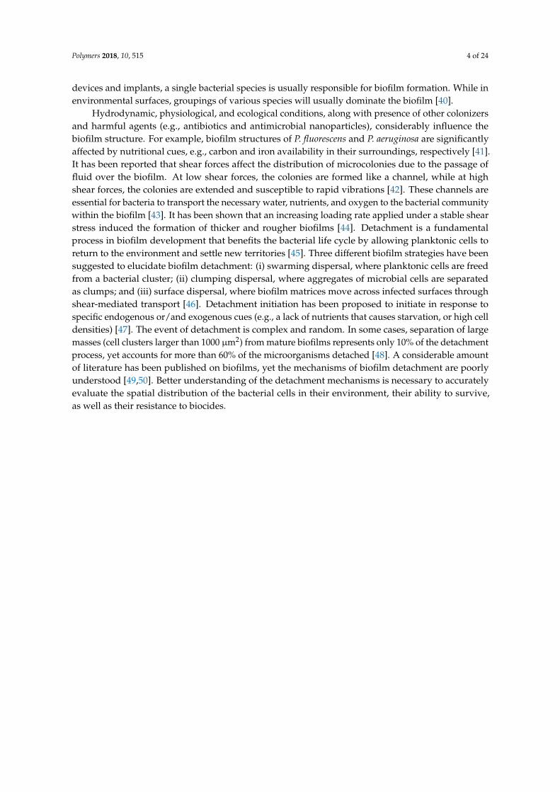
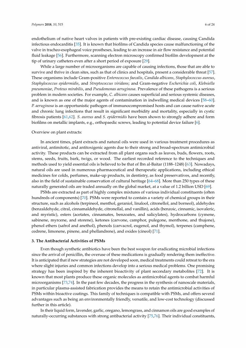
After adhering to solid surfaces, the next step of permanent attachment is growing a bacterial“sanctuary”, which is the biofilm. Biofilm formation is a four stage process which includes:(i) irreversible attachment; (ii) early development; (iii) maturation; and (iv) detachment or dispersal ofcells, as seen Figure 1 [29]. In the case of irreversible adhesion, major changes occur in gene/proteinexpression of microbial cells. It has been shown conclusively that bacteria secrete a highly hydratedlayer (biofilm) that provides a shield against host defense system and antibiotics, and strengthens theattachment of the microorganisms to the surface. Early steps of biofilm formation are controlled byphysical adsorption processes and evolution dynamics of planktonic pathogens [33].
A biofilm cluster consists of accumulations of extracellular polymeric substances (EPS), primarilypolysaccharides, proteins, nucleic acids, and lipids [34,35]. Typically, a viable biofilm involves threeorganic layers. The first layer is attached to the surface of the tissue or biomaterial, the second layeris called the “biofilm base”, which holds the bacterial aggregation, and the third layer, known asthe “surface film”, performs as an outer layer where planktonic organisms are released [6]. Biofilmarchitecture is heterogeneous both in space and time. The thickness of a biofilm varies dependingon the microbial species. For example, the mean thickness of a P. aeruginosa biofilm is about 24 µm,while S. epidermidis has a mean biofilm thickness of 32.3 µm; thickness can reach more than 400 µmin some species [36]. Active biofilms are highly hydrated, with 50%–90% of the overall area at eachsectioning depth comprising EPS and liquid [37]. Direct microscopic observation has shown thatbiofilm clusters accumulate a large quantity of pathogens within a small area, with microorganism celldensities on an infected surface reaching 106 cells/cm2 [38]. Microorganisms communicate with eachother inside a biofilm by producing chemotactic particles or pheromones, in a process called “quorumsensing” [39]. Biofilm sanctuaries can include a single infectious species or multiple infectious species,as well as non-pathogenic microorganisms which nevertheless can produce substances that wouldbenefit the survival and proliferation of the pathogenic species. In the case of the infection of medical
Polymers 2018, 10, 515 4 of 24
devices and implants, a single bacterial species is usually responsible for biofilm formation. While inenvironmental surfaces, groupings of various species will usually dominate the biofilm [40].
Hydrodynamic, physiological, and ecological conditions, along with presence of other colonizersand harmful agents (e.g., antibiotics and antimicrobial nanoparticles), considerably influence thebiofilm structure. For example, biofilm structures of P. fluorescens and P. aeruginosa are significantlyaffected by nutritional cues, e.g., carbon and iron availability in their surroundings, respectively [41].It has been reported that shear forces affect the distribution of microcolonies due to the passage offluid over the biofilm. At low shear forces, the colonies are formed like a channel, while at highshear forces, the colonies are extended and susceptible to rapid vibrations [42]. These channels areessential for bacteria to transport the necessary water, nutrients, and oxygen to the bacterial communitywithin the biofilm [43]. It has been shown that an increasing loading rate applied under a stable shearstress induced the formation of thicker and rougher biofilms [44]. Detachment is a fundamentalprocess in biofilm development that benefits the bacterial life cycle by allowing planktonic cells toreturn to the environment and settle new territories [45]. Three different biofilm strategies have beensuggested to elucidate biofilm detachment: (i) swarming dispersal, where planktonic cells are freedfrom a bacterial cluster; (ii) clumping dispersal, where aggregates of microbial cells are separatedas clumps; and (iii) surface dispersal, where biofilm matrices move across infected surfaces throughshear-mediated transport [46]. Detachment initiation has been proposed to initiate in response tospecific endogenous or/and exogenous cues (e.g., a lack of nutrients that causes starvation, or high celldensities) [47]. The event of detachment is complex and random. In some cases, separation of largemasses (cell clusters larger than 1000 µm2) from mature biofilms represents only 10% of the detachmentprocess, yet accounts for more than 60% of the microorganisms detached [48]. A considerable amountof literature has been published on biofilms, yet the mechanisms of biofilm detachment are poorlyunderstood [49,50]. Better understanding of the detachment mechanisms is necessary to accuratelyevaluate the spatial distribution of the bacterial cells in their environment, their ability to survive,as well as their resistance to biocides.
Polymers 2018, 10, 515 5 of 24Polymers 2018, 10, x FOR PEER REVIEW 5 of 23
Figure 1. Schematic of the lifecycle of P. aeruginosa grown in glucose media. Images of inverted fluorescence microscopy with 400× magnification present stages of biofilm development. In stage I, planktonic bacteria attach to a solid surface. In stage II, the attachment becomes irreversible. Stage III elucidates the microcolony foundation. Stage IV illustrates the biofilm maturation and growth of the three-dimensional bacterial sanctuaries. In stage V, dispersion occurs and free planktonic cells are released from the cluster biofilm to colonize new locations. Images characterize a 250 × 250 µm2 field. Reproduced from [51].
2.3. The Impact of Biofilm Formation in the Healthcare Environment
Microbial infections related to bacterial attachment and biofilm formation have been detected on various medical devices including prosthetic heart valves, orthopedic implants, intravascular catheters, artificial hearts, left ventricular assist devices, cardiac pacemakers, vascular prostheses, cerebrospinal fluid shunts, urinary catheters, ocular prostheses, contact lenses, and intrauterine contraceptive devices [52]. The three most common device-related infections are central line-associated bloodstream infection, ventilator-associated pneumonia (VAP), and Foley catheter-associated urinary tract infection (UTI) [53]. Studies have shown that 60–70% of nosocomial infections are associated with some type of an implanted medical device [54]. More specifically, the Centre for Disease Control and Prevention in the USA reported that of the infections in medical devices, 32% are urinary tract infections, 22% are surgical site infections, 15% can be attributed to pneumonia and lung infections, and 14% constitute bloodstream infections [55]. Microorganisms also form biofilms on the damaged vascular endothelium of native heart valves in patients with pre-existing cardiac disease, causing Candida infectious endocarditis [35]. It is known that biofilms of Candida species
Figure 1. Schematic of the lifecycle of P. aeruginosa grown in glucose media. Images of invertedfluorescence microscopy with 400× magnification present stages of biofilm development. In stage I,planktonic bacteria attach to a solid surface. In stage II, the attachment becomes irreversible. StageIII elucidates the microcolony foundation. Stage IV illustrates the biofilm maturation and growth ofthe three-dimensional bacterial sanctuaries. In stage V, dispersion occurs and free planktonic cells arereleased from the cluster biofilm to colonize new locations. Images characterize a 250 × 250 µm2 field.Reproduced from [51].
2.3. The Impact of Biofilm Formation in the Healthcare Environment
Microbial infections related to bacterial attachment and biofilm formation have been detectedon various medical devices including prosthetic heart valves, orthopedic implants, intravascularcatheters, artificial hearts, left ventricular assist devices, cardiac pacemakers, vascular prostheses,cerebrospinal fluid shunts, urinary catheters, ocular prostheses, contact lenses, and intrauterinecontraceptive devices [52]. The three most common device-related infections are central line-associatedbloodstream infection, ventilator-associated pneumonia (VAP), and Foley catheter-associated urinarytract infection (UTI) [53]. Studies have shown that 60–70% of nosocomial infections are associated withsome type of an implanted medical device [54]. More specifically, the Centre for Disease Control andPrevention in the USA reported that of the infections in medical devices, 32% are urinary tract infections,22% are surgical site infections, 15% can be attributed to pneumonia and lung infections, and 14%constitute bloodstream infections [55]. Microorganisms also form biofilms on the damaged vascular
Polymers 2018, 10, 515 6 of 24
endothelium of native heart valves in patients with pre-existing cardiac disease, causing Candidainfectious endocarditis [35]. It is known that biofilms of Candida species cause malfunctioning of thevalve in tracheo-esophageal voice prostheses, leading to an increase in air flow resistance and potentialfluid leakage [56]. Furthermore, scanning electron microscopy confirmed biofilm development at thetip of urinary catheters even after a short period of exposure [29].
While a large number of microorganisms are capable of causing infections, those that are able tosurvive and thrive in clean sites, such as that of clinics and hospitals, present a considerable threat [57].These organisms include Gram-positive Enterococcus faecalis, Candida albicans, Staphylococcus aureus,Staphylococcus epidermidis, and Streptococcus viridans; and Gram-negative Escherichia coli, Klebsiellapneumoniae, Proteus mirabilis, and Pseudomonas aeruginosa. Prevalence of these pathogens is a seriousproblem in modern societies. For example, C. albicans causes superficial and serious systemic diseases,and is known as one of the major agents of contamination in indwelling medical devices [58–60].P. aeruginosa is an opportunistic pathogen of immunocompromised hosts and can cause native acuteand chronic lung infections that result in significant morbidity and mortality, especially in cysticfibrosis patients [61,62]. S. aureus and S. epidermidis have been shown to strongly adhere and formbiofilms on metallic implants, e.g., orthopaedic screws, leading to potential device failure [6].
Overview on plant extracts:
In ancient times, plant extracts and natural oils were used in various treatment procedures asantiviral, antimitotic, and antitoxigenic agents due to their strong and broad-spectrum antimicrobialactivity. These products can be extracted from all plant organs such as leaves, buds, flowers, roots,stems, seeds, fruits, bark, twigs, or wood. The earliest recorded reference to the techniques andmethods used to yield essential oils is believed to be that of Ibn al-Baitar (1188–1248) [63]. Nowadays,natural oils are used in numerous pharmaceutical and therapeutic applications, including ethicalmedicines for colds, perfumes, make-up products, in dentistry, as food preservatives, and recently,also in the field of sustainable conservation of cultural heritage [64–68]. More than 250 types of thesenaturally generated oils are traded annually on the global market, at a value of 1.2 billion USD [69].
PSMs are extracted as part of highly complex mixtures of various individual constituents (oftenhundreds of components) [70]. PSMs were reported to contain a variety of chemical groups in theirstructure, such as alcohols (terpineol, menthol, geraniol, linalool, citronellol, and borneol), aldehydes(benzaldehyde, citral, cinnamaldehyde, citronellal, and vanillin), acids (benzoic, cinnamic, isovaleric,and myristic), esters (acetates, cinnamates, benzoates, and salicylates), hydrocarbons (cymene,sabinene, myrcene, and storene), ketones (carvone, camphor, pulegone, menthone, and thujone),phenol ethers (safrol and anethol), phenols (carvacrol, eugenol, and thymol), terpenes (camphene,cedrene, limonene, pinene, and phellandrene), and oxides (cineol) [71].
3. The Antibacterial Activities of PSMs
Even though synthetic antibiotics have been the best weapon for eradicating microbial infectionssince the arrival of penicillin, the overuse of these medications is gradually rendering them ineffective.It is anticipated that if new strategies are not developed soon, medical treatments could retreat to the erawhere slight injuries and common infections develop into a serious medical problems. One promisingstrategy has been inspired by the inherent bioactivity of plant secondary metabolites [72]. It isknown that most plants produce these organic molecules as antimicrobial agents to combat harmfulmicroorganisms [73,74]. In the past few decades, the progress in the synthesis of nanoscale materials,in particular plasma-assisted fabrication provides the means to retain the antimicrobial activities ofPSMs within bioactive coatings. This family of techniques is compatible with PSMs, and offers severaladvantages such as being an environmentally friendly, versatile, and low-cost technology (discussedfurther in this article).
In their liquid form, lavender, garlic, oregano, lemongrass, and cinnamon oils are good examples ofnaturally-occurring substances with strong antibacterial activity [75,76]. Their individual constituents,
Polymers 2018, 10, 515 7 of 24
e.g., citronellol and geraniol are aromatic acyclic monoterpene alcohols that are very powerfulbactericides [77–80]. Terpinene-4-ol, a major component of tea tree oil, is a broad-spectrum nonspecificbiocide well-known as a natural agent against microbial species such as E. coli, P. aeruginosa,Acinetobacter baumannii, and several drug-resistant bacteria (e.g., MRSA) [81]. A number of PSMs havebeen used against cancer cells, whereas others are currently used in food preservation [82,83]. In theirvapor phase, a number of PSMs have demonstrated strong antibacterial activities [84,85]. So far, thereare thousands of natural oils currently known. Among them, 300 oils are important and commonlyused in the pharmaceutical, food, sanitary, agronomic, perfume, and cosmetic productions [86].
3.1. The Antibacterial Mechanisms of PSMs
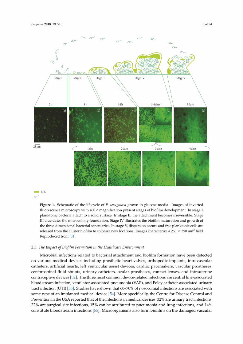
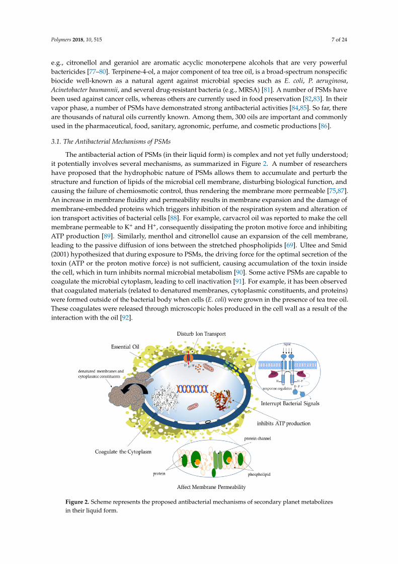
The antibacterial action of PSMs (in their liquid form) is complex and not yet fully understood;it potentially involves several mechanisms, as summarized in Figure 2. A number of researchershave proposed that the hydrophobic nature of PSMs allows them to accumulate and perturb thestructure and function of lipids of the microbial cell membrane, disturbing biological function, andcausing the failure of chemiosmotic control, thus rendering the membrane more permeable [75,87].An increase in membrane fluidity and permeability results in membrane expansion and the damage ofmembrane-embedded proteins which triggers inhibition of the respiration system and alteration ofion transport activities of bacterial cells [88]. For example, carvacrol oil was reported to make the cellmembrane permeable to K+ and H+, consequently dissipating the proton motive force and inhibitingATP production [89]. Similarly, menthol and citronellol cause an expansion of the cell membrane,leading to the passive diffusion of ions between the stretched phospholipids [69]. Ultee and Smid(2001) hypothesized that during exposure to PSMs, the driving force for the optimal secretion of thetoxin (ATP or the proton motive force) is not sufficient, causing accumulation of the toxin insidethe cell, which in turn inhibits normal microbial metabolism [90]. Some active PSMs are capable tocoagulate the microbial cytoplasm, leading to cell inactivation [91]. For example, it has been observedthat coagulated materials (related to denatured membranes, cytoplasmic constituents, and proteins)were formed outside of the bacterial body when cells (E. coli) were grown in the presence of tea tree oil.These coagulates were released through microscopic holes produced in the cell wall as a result of theinteraction with the oil [92].
Polymers 2018, 10, x FOR PEER REVIEW 7 of 23
nonspecific biocide well-known as a natural agent against microbial species such as E. coli, P. aeruginosa, Acinetobacter baumannii, and several drug-resistant bacteria (e.g., MRSA) [81]. A number of PSMs have been used against cancer cells, whereas others are currently used in food preservation [82,83]. In their vapor phase, a number of PSMs have demonstrated strong antibacterial activities [84,85]. So far, there are thousands of natural oils currently known. Among them, 300 oils are important and commonly used in the pharmaceutical, food, sanitary, agronomic, perfume, and cosmetic productions [86].
3.1. The Antibacterial Mechanisms of PSMs
The antibacterial action of PSMs (in their liquid form) is complex and not yet fully understood; it potentially involves several mechanisms, as summarized in Figure 2. A number of researchers have proposed that the hydrophobic nature of PSMs allows them to accumulate and perturb the structure and function of lipids of the microbial cell membrane, disturbing biological function, and causing the failure of chemiosmotic control, thus rendering the membrane more permeable [75,87]. An increase in membrane fluidity and permeability results in membrane expansion and the damage of membrane-embedded proteins which triggers inhibition of the respiration system and alteration of ion transport activities of bacterial cells [88]. For example, carvacrol oil was reported to make the cell membrane permeable to K+ and H+, consequently dissipating the proton motive force and inhibiting ATP production [89]. Similarly, menthol and citronellol cause an expansion of the cell membrane, leading to the passive diffusion of ions between the stretched phospholipids [69]. Ultee and Smid (2001) hypothesized that during exposure to PSMs, the driving force for the optimal secretion of the toxin (ATP or the proton motive force) is not sufficient, causing accumulation of the toxin inside the cell, which in turn inhibits normal microbial metabolism [90]. Some active PSMs are capable to coagulate the microbial cytoplasm, leading to cell inactivation [91]. For example, it has been observed that coagulated materials (related to denatured membranes, cytoplasmic constituents, and proteins) were formed outside of the bacterial body when cells (E. coli) were grown in the presence of tea tree oil. These coagulates were released through microscopic holes produced in the cell wall as a result of the interaction with the oil [92].
Figure 2. Scheme represents the proposed antibacterial mechanisms of secondary planet metabolizes in their liquid form.
Figure 2. Scheme represents the proposed antibacterial mechanisms of secondary planet metabolizesin their liquid form.

Polymers 2018, 10, 515 8 of 24
Exposure to PSMs can lead to the reduction in enzymatic activities, loss of turgor pressure,changes in DNA synthesis and inhibition of different metabolic functions [89]. Moreover, some oils,such as rose, geranium, lavender, and rosemary have been shown to inhibit cell–cell communication,affecting the quorum sensing (QS) network in the bacterial community [93]. The QS system is vitalfor bacterial growth, and hence, any interference with the sensing network may reduce pathogenicity,biofilm formation, and antibiotic resistance during infection events.
The antimicrobial performance of PSMs is linked to their chemical structure, particularly thepresence of –OH functional groups [94]. Each compound may reveal a different biocidal mechanismtoward microorganisms [69]. The bioactivity of several active oils is associated with the presence ofphenolic groups. For example, the antimicrobial efficacy of clove, thyme, and oregano oils is relatedto the presence of phenol-containing eugenol, thymol, and carvacrol, respectively [95]. However,other findings indicate that the components present in high quantities in the oil are not necessarilyresponsible for the entire biological activity of a PSM. The antibacterial performance of these complexmixtures relies on a variety of synergistic effects of different sub-components in the oil. Furthermore,it can also be attributed to the presence of other components that may be effective even in smallquantities [96,97]. In the case of essential oils containing a high percentage of phenolic compounds (e.g.,carvacrol, thymol), it can be assumed that their bactericidal action would be similar to other phenolicgroups, e.g., by way of the disturbance of the membrane, disorder the proton motive force, electron flow,and coagulation of cell contents [87]. In the case of complex mixtures, where numerous active moleculesare present, potential synergistic and antagonistic influences, as well as minor compounds that canhave an important contribution to the oil’s activity, need to be considered [64,98]. It is important to statethat the biocidal mechanisms of PSMs are dissimilar from currently used synthetic antibiotics, whichshould minimize the likelihood of the development of microbial cross-drug resistance [99].
3.2. Sustainable Polymers from Bioactive Essential Oils
The ecological concerns of current petroleum processing, along with the economic recession,depleting reserves, and political aspects, have led to increased interest in the production of sustainablepolymers derived from renewable resources [100,101]. These eco-friendly polymers can be derivedfrom a wide range of possible precursor materials, including oxygen-rich monomers (e.g., carboxylicacids), hydrocarbon-rich monomers (fatty acids, terpenes, and vegetable oils), and non-hydrocarbonmonomers (carbon dioxide) [102]. So far, polymers derived from essential oils, vegetable oils,bio-ethanol, cellulose, fats, resins, naturally occurring polysaccharides, microbial syntheses, and othernatural ingredients have been widely used for a variety of applications [103–108]. Essential oils,in particular, are renewable in nature, relatively inexpensive, available in commercial quantities,and display minimal toxicity compared to many conventionally-used precursors, which make them anappropriate precursor for “green” functional materials. Among them, terpenes (major components ina large number of essential oils) have received considerable attention. Their structure contains one ormore carbon–carbon double bonds, showing a carbon skeleton of isoprene. The abundance of doublebonds allows for cationic and radical polymerization of terpenes, along with epoxidation as a path tobiodegradable oxygenated polymers [109]. Cationic polymerization has been generally accepted to bethe most appropriate kind of chain reaction for these monomers [110]. However, essential oils havenot been widely applied to the production of bioactive polymers due to limitations associated withfabrication systems and oil properties [111,112]. These limitations include challenges in controlling thesurface chemistry and morphology of the synthesized materials, and solubility and/or volatility of thenatural monomers.
Recent technological advances in the field of controlled polymerization, catalysis,nanoencapsulation, and effective organic functionalization, give great potential for the application ofessential oils in manufacturing of sustainable polymers with innovative designs and characteristics.This allows the fabrication of organic films with good control over film thickness, physico-chemicalproperties, and, importantly, biological functionality. For instance, it was possible to successfully

Polymers 2018, 10, 515 9 of 24
engineer antibacterial UV-cured networks by using a thiol-ene route with covalent immobilizationof natural terpenes (linalool and a trithiol) as antibacterial agents, without employing any organicsolvent. These bio-based materials exhibited attractive thermal properties, were not affected by waterpenetration under high moisture conditions, and displayed strong inhibition of microorganisms [113].Chen et al., (2012) developed the reversible transfer polymerization approach to design a series ofcationic rosin-containing methacrylate bioactive-copolymers. The antibacterial activities of theserosin-containing copolymers were found to be dependent on both the degree of quaternization of therosin group, the molecular weight of copolymers, and the conformation of hydrophobic group [114].Furthermore, a cinnamon essential oil/cyclodextrin integrated into a polylactic acid nanofilm made byelectrospinning and co-precipitation showed strong antimicrobial activity [115].
Several studies have been carried out in order to incorporate active essential oils into selectedpolymers through applying emulsification or homogenization methods, where ultra-fine emulsionsof oils are formed containing polymer at the continuous aqueous phase. Upon drying, lipid dropletsremain incorporated into the polymer structure. The releasing rate of the embedded-oils from filmsis subject to multiple factors, such as electrostatic interactions between the oil and the polymerchains, osmosis, structural variations induced by the presence of the oil, as well as environmentalcircumstances [116]. Remarkably, a small fraction of an incorporated essential oil within a polymerstructure is sufficient to achieve the desired antimicrobial properties. For example, quince seedmucilage films containing a low percentage (1.5–2%) of oregano essential oil were reported to bevery effective against several microorganisms, including S. aureus, E. coli, and S. putrefaciens [117].Other findings showed that inactive chitosan films were transformed into bioactive materials when asmall quantity (~1–2%) of extract from two endemic herbs (Thymus moroderi or Thymus piperella) wereintegrated within the films [118].
Encapsulation of oils has been developed as one such technology that has great potential toimprove the physical stability of the active components, protecting them from degradation dueto environmental aspects (e.g., oxygen, light, moisture, and pH) [119]. Among the nanometricencapsulation structures currently being used, nanoemulsions are mainly utilized due to the possibilityof formulation with natural components and the compatibility with industrially scalable manufacturingprocesses by high pressure homogenization [120]. Nanoemulsions are defined as emulsions withultra-small droplet sizes of approximately 100 nm. At this scale, there is a potential of enhancingphysico-chemical properties and stability of the active compound. In addition, the oil bioactivitycan be considerably increased, since significant increases in the surface area per unit of mass can beachieved, improving the passive mechanisms of cell absorption, which again allows for the reductionof the oil quantity required to ensure antimicrobial action [121]. The encapsulated essential oils arepromising antimicrobial agents for biodegradable/edible coatings in food packaging industries toinhibit pathogenic microorganisms [122]. It has been reported that the encapsulation in nanoemulsionformulation of a terpenes mixture and limonene increased the antimicrobial performance of the purecompounds against various microorganisms such as E. coli and Saccharomyces cerevisiae, throughincreases of transport mechanisms in the membrane of the target cell [120]. Mohammadi et al., (2015)also encapsulated Zataria multiflora essential oil in chitosan nanoparticles (average size of 125–175 nm)and reported a controlled and sustained release of essential oil for 40 days, along with a superiorantifungal performance in comparison with the un-encapsulated oil [123]. Moreover, films with 1.5%nanocomposite marjoram oil diminished the numbers of E. coli, S. aureus, and Listeria monocytogenespopulations with respect to the control of up to 4.52, 5.80, and 6.33 log, respectively [124]. Similarly,introduction of carvacrol nanoemulsions into modified chitosan have led to the development of abioactive film, which was active against Gram-negative pathogenic bacteria [125].
It is worth mentioning that, in many cases, the vapor phase of essential oils exhibits stronginhibitive performance against pathogens, even more effective than direct application [126,127].For instance, Avila-Sosa et al. (2012) found that chitosan films incorporating cinnamon or Mexican
Polymers 2018, 10, 515 10 of 24
oregano essential oils can inhibit fungi by vapor contact at lower oil concentrations than those requiredfor amaranth and starch polymeric coatings [116].
3.3. Plasma-Assisted Fabrication of PSMs
Among fabrication techniques, cold plasma polymerization is a multipurpose approach that is arelatively fast and low-cost method for fabricating coatings from a wide array of natural precursors,including those that do not usually polymerize by conventional methods, and do not require furtherchemical or physical processing (e.g., annealing and catalysts) [128]. From a processing point ofview, many PSMs are compatible with plasma polymerization, which is in essence a chemical vapordeposition process enhanced by the catalytic activity of plasma, because they are highly volatile atroom temperature no external heat or carrier gas are required to deliver the precursor macromoleculesto the fabrication zone.
Introduction of PSMs molecules, in vapor phase, into a highly reactive plasma field triggers awide range of reactions including fragmentation, oligomerization, rearrangement, and polymerization.The degree of dissociation is highly dependent on the amount of energy provided into the plasmasystem and the pressure in the chamber. Fragmentation is initiated by active electrons rather thanthermal excitation or chemical reactions, creating a unique mixture of chemically diverse species(e.g., unsaturated bonds, ions, neutrals, and free radicals), which may not be reachable under otherconditions [129]. It is believed that weakly ionized plasma and relatively low substrate temperatureduring deposition promotes condensation and adsorption of non-excited species, which help toincrease the proportion of non/partially-fragmented precursor molecules on the substrate [130].The recombination of the reactive species and precursor molecules may lead to the formation of theorganic thin layer (polymer) on the surface of a given substrate. Due to the diversity of functionalgroups and reactive species, the polymer can be formed in several ways, involving free-radicals andinduced-polymerization of fragments containing unsaturated carbon–carbon bonds; recombinationfragment/recombination is initiated by the plasma-generated and surface-attached reactive ions [131].The formed polymer is often highly branched and highly cross-linked (amorphous), comprising largequantities of trapped free radicals in its structure [132].
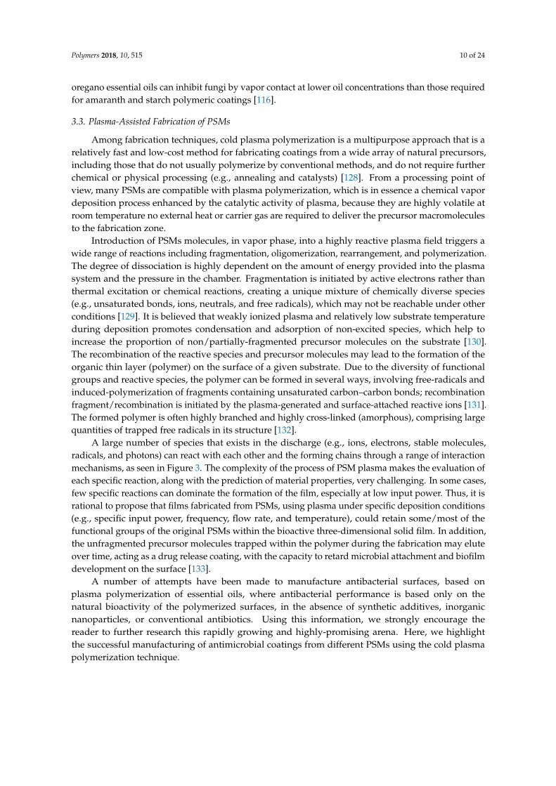
A large number of species that exists in the discharge (e.g., ions, electrons, stable molecules,radicals, and photons) can react with each other and the forming chains through a range of interactionmechanisms, as seen in Figure 3. The complexity of the process of PSM plasma makes the evaluation ofeach specific reaction, along with the prediction of material properties, very challenging. In some cases,few specific reactions can dominate the formation of the film, especially at low input power. Thus, it isrational to propose that films fabricated from PSMs, using plasma under specific deposition conditions(e.g., specific input power, frequency, flow rate, and temperature), could retain some/most of thefunctional groups of the original PSMs within the bioactive three-dimensional solid film. In addition,the unfragmented precursor molecules trapped within the polymer during the fabrication may eluteover time, acting as a drug release coating, with the capacity to retard microbial attachment and biofilmdevelopment on the surface [133].
A number of attempts have been made to manufacture antibacterial surfaces, based onplasma polymerization of essential oils, where antibacterial performance is based only on thenatural bioactivity of the polymerized surfaces, in the absence of synthetic additives, inorganicnanoparticles, or conventional antibiotics. Using this information, we strongly encourage thereader to further research this rapidly growing and highly-promising arena. Here, we highlightthe successful manufacturing of antimicrobial coatings from different PSMs using the cold plasmapolymerization technique.

Polymers 2018, 10, 515 11 of 24
3.3.1. Terpinen-4-ol
Terpinen-4-ol is a monocyclic terpene alcohol that is an active component of tea tree oil.Terpinen-4-ol has demonstrated powerful antimicrobial and anti-inflammatory properties [134,135].Upon interaction with microorganisms, cyclic terpene hydrocarbons have been shown to accumulatein the cell membrane. This disturbs membrane integrity, triggering an increased passive flux ofprotons through the membrane and dissipation of the proton motive force [136]. Bazaka et al.(2011) prepared plasma polymerized coatings derived from terpinen-4-ol at various input powerlevels, showing a considerable potential in minimizing bacterial attachment and metabolic activity ofS. aureus and P. aeruginosa. Fabrication at a low input power level, 10 W, resulted in a partial retentionof biologically-active groups of the original precursor, which led to significant antimicrobial andantibiofouling activities of the terpenol-derived coatings [137]. Confocal laser scanning microscopyevidently showed that around 90% of S. aureus cells retained on the films of the 10 W substrata werenon-viable, in comparison to that retained on the surface of 25 W films [138,139]. However, whenfabricated at higher input power (25 W), these films lost their biocidal activity, and promoted adhesionand proliferation of tested bacterial cells and biofilm development. In a recent report, the decrease inantibacterial activity with increasing radio frequency (RF) energy was also observed in the plasmapolymerization of polyterpenol films [140].
3.3.2. Carvone
Carvone is found in various essential oils, such as caraway, spearmint, and dill. This PSM hasshown a variety of antiproliferative effects with regards to microbial cells; likely due to the presenceof a monoterpene group in its structure [141,142]. In addition, carvone and its related compoundswere shown to be potential chemopreventive agents, due to their ability to induce increased activityof detoxifying enzymes. The α,β-unsaturated ketone system in carvone is generally expected tobe responsible for the high enzyme-inducing action [143]. Recently, Chan et al., (2016) fabricatedpolymer coatings resultant from plasma polymerization of carvone [144]. At an input power of 10 W,carvone polymerized coatings demonstrated almost equal antimicrobial performance against bothGram-negative and Gram-positive bacteria (86% decrease in E. coli and 84% reduction in S. aureus),with no cytotoxic effect towards primary human endothelial cells. In addition, these coatings weresmooth, highly cross-linked hydrocarbons, with low fractions of carboxyl, hydroxyl, and amine-amidefunctionalities. Although the carvone surfaces reduce bacterial adhesion, it was observed that somecells were damaged and died after attaching to the surface. The scanning electron microscope(SEM) images clearly exhibited membrane distortion, pore creation, and membrane rupture ofmicroorganisms attached on the surface of plasma polymers of carvone.
3.3.3. Eucalyptol
Eucalyptol, a major component of eucalyptus oil and a minor component of tea treeoil, is a saturated monoterpene known by a variety of synonyms, such as 1,8-cineole,1,8-epoxy-p-menthane, and cajeputol. This PSM has been demonstrated to retain strong biologicalactivities, including anti-inflammatory, antifungal, antibiofilm, and antiseptic properties towarda range of bacteria [145–148]. The retention of the natural bio-active groups of the 1,8-cineoleoil was also achieved using plasma polymerization. Fabricated at 20 W, moderate hydrophobiccoatings were achieved, with the ability to reduce the attachment of E. coli and S. aureus cells by98% and 64%, respectively, compared to unmodified glass. In addition, the 1,8-cineole plasma filmsresisted biofilm formation after 5 days of incubation in the presence of bacterial cells. The polymersurface and any products that may be released from the film were also found not to be cytotoxic tomammalian cells [149]. In the same way, Mann and Fisher (2017) used a range of applied RF powers(P = 50–150 W) and H2O(v) plasma-treatment during the plasma fabrication of 1,8-cineole polymers.The fabricated films retained some antimicrobial behaviors characteristic of the precursor, in addition
Polymers 2018, 10, 515 12 of 24
to the desired properties, such as being highly adherent to the substrate, conformal, and with smoothsurfaces. The in vitro studies showed that E. coli were largely nonviable and unable to colonize theplasma–cineole surface over the 5 day biofilm development assay period. The biofilm coverage onthese surfaces was significantly lower (<10%) than the glass control [150].
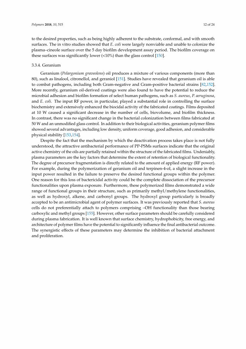
3.3.4. Geranium
Geranium (Pelargonium graveolens) oil produces a mixture of various components (more than80), such as linalool, citronellol, and geraniol [151]. Studies have revealed that geranium oil is ableto combat pathogens, including both Gram-negative and Gram-positive bacterial strains [82,152].More recently, geranium oil-derived coatings were also found to have the potential to reduce themicrobial adhesion and biofilm formation of select human pathogens, such as S. aureus, P. aeruginosa,and E. coli. The input RF power, in particular, played a substantial role in controlling the surfacebiochemistry and extensively enhanced the biocidal activity of the fabricated coatings. Films depositedat 10 W caused a significant decrease in the number of cells, biovolume, and biofilm thickness.In contrast, there was no significant change in the bacterial colonization between films fabricated at50 W and an unmodified glass control. In addition to their biological activities, geranium polymer filmsshowed several advantages, including low density, uniform coverage, good adhesion, and considerablephysical stability [153,154].
Despite the fact that the mechanism by which the deactivation process takes place is not fullyunderstood, the attractive antibacterial performance of PP-PSMs surfaces indicate that the originalactive chemistry of the oils are partially retained within the structure of the fabricated films. Undeniably,plasma parameters are the key factors that determine the extent of retention of biological functionality.The degree of precursor fragmentation is directly related to the amount of applied energy (RF power).For example, during the polymerization of geranium oil and terpinen-4-ol, a slight increase in theinput power resulted in the failure to preserve the desired functional groups within the polymer.One reason for this loss of bactericidal activity could be the complete dissociation of the precursorfunctionalities upon plasma exposure. Furthermore, these polymerized films demonstrated a widerange of functional groups in their structure, such as primarily methyl/methylene functionalities,as well as hydroxyl, alkene, and carbonyl groups. The hydroxyl group particularly is broadlyaccepted to be an antimicrobial agent of polymer surfaces. It was previously reported that S. aureuscells do not preferentially attach to polymers comprising –OH functionality than those bearingcarboxylic and methyl groups [155]. However, other surface parameters should be carefully consideredduring plasma fabrication. It is well known that surface chemistry, hydrophobicity, free energy, andarchitecture of polymer films have the potential to significantly influence the final antibacterial outcome.The synergistic effects of these parameters may determine the inhibition of bacterial attachmentand proliferation.

Polymers 2018, 10, 515 13 of 24
Polymers 2018, 10, x FOR PEER REVIEW 12 of 23
adhesion and biofilm formation of select human pathogens, such as S. aureus, P. aeruginosa, and E. coli. The input RF power, in particular, played a substantial role in controlling the surface biochemistry and extensively enhanced the biocidal activity of the fabricated coatings. Films deposited at 10 W caused a significant decrease in the number of cells, biovolume, and biofilm thickness. In contrast, there was no significant change in the bacterial colonization between films fabricated at 50 W and an unmodified glass control. In addition to their biological activities, geranium polymer films showed several advantages, including low density, uniform coverage, good adhesion, and considerable physical stability [153,154].
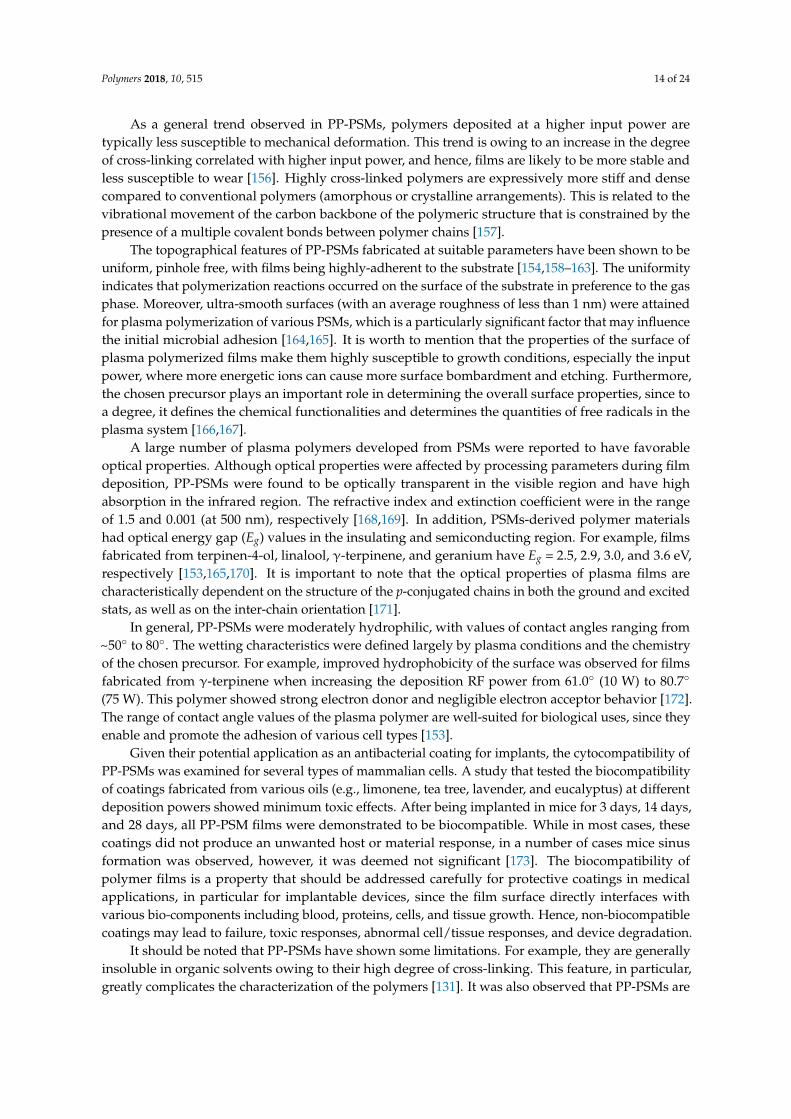
Figure 3. Representative examples of plasma polymerization of plant secondary metabolites, where retention of the antimicrobial activity was achieved. As soon as a bioactive secondary plant metabolite (or an essential oil) is placed under low pressure, the molecules gain sufficient kinetic energy to separate and begin independently moving towards the glow region within the deposition chamber. Exposure of the molecules to the highly reactive plasma initiates various chemical reactions, such as bonds fragmentation, oligomerization, and polymerization. At the chosen plasma parameters, the process allows for the preservation of active functional groups of PSMs within the cross-linked solid polymeric films. Direct observations of SEM demonstrated the powerful antimicrobial performance of geranium, terpenen-4-ol, and carvone films in contact with different pathogens. The antimicrobial activities of these films included antibiofouling effects and/or bactericidal actions (e.g., membrane distortion, pores creation, and membrane damage). The SEM images are reproduced with permission from [137,144,153].
Figure 3. Representative examples of plasma polymerization of plant secondary metabolites, whereretention of the antimicrobial activity was achieved. As soon as a bioactive secondary plant metabolite(or an essential oil) is placed under low pressure, the molecules gain sufficient kinetic energy to separateand begin independently moving towards the glow region within the deposition chamber. Exposureof the molecules to the highly reactive plasma initiates various chemical reactions, such as bondsfragmentation, oligomerization, and polymerization. At the chosen plasma parameters, the processallows for the preservation of active functional groups of PSMs within the cross-linked solid polymericfilms. Direct observations of SEM demonstrated the powerful antimicrobial performance of geranium,terpenen-4-ol, and carvone films in contact with different pathogens. The antimicrobial activities ofthese films included antibiofouling effects and/or bactericidal actions (e.g., membrane distortion, porescreation, and membrane damage). The SEM images are reproduced with permission from [137,144,153].
3.4. Properties of PSM-Derived Polymers
For a successful polymeric antibacterial coating to satisfy the requirements of biomedicalapplications, the material should possess a range of specific biological, physical, and chemicalproperties. Films fabricated from PSMs display a wide range of desired properties, including opticaltransparency, moderate hydrophilicity, relatively high degradation temperature, low post-annealingretention, and good biocompatibility, forming simple, useful, and versatile bioactive coatings. Hence,a brief description of some important physico-chemical characteristics of PP-PSMs fabricated at a lowinput power (below 100 W) is provided below.
Polymers 2018, 10, 515 14 of 24
As a general trend observed in PP-PSMs, polymers deposited at a higher input power aretypically less susceptible to mechanical deformation. This trend is owing to an increase in the degreeof cross-linking correlated with higher input power, and hence, films are likely to be more stable andless susceptible to wear [156]. Highly cross-linked polymers are expressively more stiff and densecompared to conventional polymers (amorphous or crystalline arrangements). This is related to thevibrational movement of the carbon backbone of the polymeric structure that is constrained by thepresence of a multiple covalent bonds between polymer chains [157].
The topographical features of PP-PSMs fabricated at suitable parameters have been shown to beuniform, pinhole free, with films being highly-adherent to the substrate [154,158–163]. The uniformityindicates that polymerization reactions occurred on the surface of the substrate in preference to the gasphase. Moreover, ultra-smooth surfaces (with an average roughness of less than 1 nm) were attainedfor plasma polymerization of various PSMs, which is a particularly significant factor that may influencethe initial microbial adhesion [164,165]. It is worth to mention that the properties of the surface ofplasma polymerized films make them highly susceptible to growth conditions, especially the inputpower, where more energetic ions can cause more surface bombardment and etching. Furthermore,the chosen precursor plays an important role in determining the overall surface properties, since toa degree, it defines the chemical functionalities and determines the quantities of free radicals in theplasma system [166,167].
A large number of plasma polymers developed from PSMs were reported to have favorableoptical properties. Although optical properties were affected by processing parameters during filmdeposition, PP-PSMs were found to be optically transparent in the visible region and have highabsorption in the infrared region. The refractive index and extinction coefficient were in the rangeof 1.5 and 0.001 (at 500 nm), respectively [168,169]. In addition, PSMs-derived polymer materialshad optical energy gap (Eg) values in the insulating and semiconducting region. For example, filmsfabricated from terpinen-4-ol, linalool, γ-terpinene, and geranium have Eg = 2.5, 2.9, 3.0, and 3.6 eV,respectively [153,165,170]. It is important to note that the optical properties of plasma films arecharacteristically dependent on the structure of the p-conjugated chains in both the ground and excitedstats, as well as on the inter-chain orientation [171].
In general, PP-PSMs were moderately hydrophilic, with values of contact angles ranging from~50◦ to 80◦. The wetting characteristics were defined largely by plasma conditions and the chemistryof the chosen precursor. For example, improved hydrophobicity of the surface was observed for filmsfabricated from γ-terpinene when increasing the deposition RF power from 61.0◦ (10 W) to 80.7◦
(75 W). This polymer showed strong electron donor and negligible electron acceptor behavior [172].The range of contact angle values of the plasma polymer are well-suited for biological uses, since theyenable and promote the adhesion of various cell types [153].
Given their potential application as an antibacterial coating for implants, the cytocompatibility ofPP-PSMs was examined for several types of mammalian cells. A study that tested the biocompatibilityof coatings fabricated from various oils (e.g., limonene, tea tree, lavender, and eucalyptus) at differentdeposition powers showed minimum toxic effects. After being implanted in mice for 3 days, 14 days,and 28 days, all PP-PSM films were demonstrated to be biocompatible. While in most cases, thesecoatings did not produce an unwanted host or material response, in a number of cases mice sinusformation was observed, however, it was deemed not significant [173]. The biocompatibility ofpolymer films is a property that should be addressed carefully for protective coatings in medicalapplications, in particular for implantable devices, since the film surface directly interfaces withvarious bio-components including blood, proteins, cells, and tissue growth. Hence, non-biocompatiblecoatings may lead to failure, toxic responses, abnormal cell/tissue responses, and device degradation.
It should be noted that PP-PSMs have shown some limitations. For example, they are generallyinsoluble in organic solvents owing to their high degree of cross-linking. This feature, in particular,greatly complicates the characterization of the polymers [131]. It was also observed that PP-PSMs are
Polymers 2018, 10, 515 15 of 24
highly susceptible to changes brought by the chemical composition of the medium (e.g., the aqueoussolution and body fluid) that may affect their operation in some applications [174].
Essential oils variations:
Generally, it is accepted that the chemical composition of essential oils varies by plant health,growth stage, climate, edaphic factors, and harvest time. On the other hand, the degradation kinetics ofthese oils, due to external factors (e.g., temperature, light, and atmospheric oxygen exposure, presenceof impurities), should be thoroughly taken into account [175]. For example, pure cinnamaldehyde wasreported to decompose to benzaldehyde at temperatures approaching 60 ◦C. But, once it combinedwith eugenol or cinnamon leaf oil, cinnamaldehyde remained stable at 200 ◦C [176]. The molecularstructures of natural oils have a substantial effect on the degree of degradation. Compounds rich inallylic hydrogen atoms could be potential targets for autoxidation, where hydrogen atom abstractionis giving rise to resonance-stabilized radicals, which are highly preferable due to their lower activationenergy [177]. Furthermore, essential oil components are generally known to easily convert into eachother (through processes such as isomerization, oxidation, cyclization, or dehydrogenation reactions),because of their structural relationship within the same chemical group [175,177].
It is important to mention that several essential oils (e.g., tea tree, lavender, and terpenene-4-ol)have shown some irritation and allergies in users (via inhalation or direct contact) [178–180].The allergic reactions typically arise from certain components such as benzyl alcohol, cinnamylalcohol, iso-eugenol, eugenol, hydroxycitronellal, geraniol, and various others constituents [181–183].However, sensitive symptoms due to essential oils can range from relatively minor incidences ofirritation and sensitization, to contact dermatitis and the most serious anaphylactic reaction, thus,should be well considered [184,185].
4. Challenges
In the scientific and manufacturing field, replication or reproduction of consistent systematicresults is the key to success [186]. A major issue of plasma techniques is the constancy of theresult, particularly across different plasma systems, due to differences in processing parameters(e.g., power, pressure, temperature, flow rate, and tube geometry). For example, changes in the designof plasma equipment can affect the flow dynamics of vapors through the system, and the profile of theplasma discharge zone, which could potentially alter the nature, homogeneity, and density of the gasphase species inside the reactor. Indeed, this problem becomes more obvious during the fabricationof functional coatings from PSMs, where retention of certain chemical moieties is essential [187].To minimize the variation of films produced across different plasma systems, a scaling factor route canbe applied that takes into account both the actual energy consumed in the active plasma field, and thedifferences in the geometry of the utilized reactors [188].
Another concern comes from the varying properties of the renewable precursor. It is welldocumented that essential oil composition is very complex and depends on multiple interactingfactors. In addition, De Masi et al. (2006) reported that the chemical compounds of essential oils werefound to be extremely variable in various cultivars/genotypes of the same plant species and theythese differences were not necessarily correlated with genetic relationships [189]. Additionally, the oilquality and biological activity can be affected storage conditions, e.g., temperature [190]. The potentialto obtain biopolymer films with consistent properties regardless of the base material source, method,or time of harvest is important for successful integration into industrialized processes.
As mentioned previously, typical plasma polymerization of PSMs (continuous mode) yields thefragmentation of large quantities of precursor molecules. The random recombination of fragments,radicals, and atoms renders the chemical structure and configuration completely irregular. In fact,the density of desired functional groups remain relatively insufficient even if the used fabricationpower is low. Pulsed-plasma polymerization can address this issue. This technique offers a sequenceof on-periods (a few µs-long periods during which fragmentation takes place) and off-periods (µs to
Polymers 2018, 10, 515 16 of 24
ms-long periods during which recombination and polymerization occurs), where the resultant polymershould consist of more chemically regular structures than those of the continuous mode [191]. The ideais to further reduce the degree of dissociation/fragmentation of the precursor molecules, and hencethe off-period reactions contributing more non-fragmented functionalities into the formed polymer.To date, the pulsed-plasma polymerization has not been used for the synthesis of antibacterialsurfaces from PSMs. We highly encourage researchers to explore and expand the usage of thepulsed-plasma method, where the optimization of the desired functionality will essentially include theincreasing/decreasing the off-period in pulsed polymerization.
5. Conclusions
A better understanding of the way to preserve/retain the bioactivity of essential oils within athin film is critical for the development of a wide range of bactericidal coatings suitable for medicaldevices. The aforementioned polymer materials that were derived from renewable resources present apromising approach toward producing antimicrobial and biocompatible materials and tissue contactcoatings. However, information on the long-term performance of plasma polymerized PSMs thin filmsrequires further exploration. Also, although a small number of systematic studies showed promisingantimicrobial activity using encapsulating essential oils, further research in this direction is warranted.
Funding: This research received no external funding.
Acknowledgments: Ahmed Al-Jumaili acknowledges the post graduate scholarship offered by the Ministry ofHigher Education and Scientific Research, Iraq, and is grateful to JCUPRS for the financial support.
Conflicts of Interest: The authors declare no conflicts of interest.
References
1. Sanborn, L.W.R. The relation of surface contamination to the transmission of disease. Am. J. Public HealthNations Health 1963, 53, 1278–1283. [CrossRef] [PubMed]
2. Dancer, S.J. Importance of the environment in meticillin-resistant Staphylococcus aureus acquisition: The casefor hospital cleaning. Lancet Infect. Dis. 2008, 8, 101–113. [CrossRef]
3. Knetsch, M.L.W.; Koole, L.H. New strategies in the development of antimicrobial coatings: The example ofincreasing usage of silver and silver nanoparticles. Polymers 2011, 3, 340–366. [CrossRef]
4. Wu, S.; Liu, X.; Yeung, A.; Yeung, K.W.; Kao, R.Y.; Wu, G.; Hu, T.; Xu, Z.; Chu, P.K. Plasma-modifiedbiomaterials for self-antimicrobial applications. ACS Appl. Mater. Interfaces 2011, 3, 2851–2860. [CrossRef][PubMed]
5. Edmiston, C.E., Jr.; McBain, A.J.; Roberts, C.; Leaper, D. Clinical and microbiological aspects ofbiofilm-associated surgical site infections. In Biofilm-Based Healthcare-Associated Infections; Springer:International Publishing: Cham, Switzerland, 2015; pp. 47–67.
6. Veerachamy, S.; Yarlagadda, T.; Manivasagam, G.; Yarlagadda, P.K. Bacterial Adherence and BiofilmFormation on Medical Implants: A Review. Proc. Inst. Mech. Eng. Part H J. Eng. Med. 2014, 228, 1083–1099.[CrossRef] [PubMed]
7. Batoni, G.; Maisetta, G.; Esin, S. Antimicrobial peptides and their interaction with biofilms of medicallyrelevant bacteria. Biochim. Biophys. Acta Biomembr. 2016, 1858, 1044–1060. [CrossRef] [PubMed]
8. Joseph, R.; Naugolny, A.; Feldman, M.; Herzog, I.M.; Fridman, M.; Cohen, Y. Cationic pillararenes potentlyinhibit biofilm formation without affecting bacterial growth and viability. J. Am. Chem. Soc. 2016, 138,754–757. [CrossRef] [PubMed]
9. Mingeot-Leclercq, M.-P.; Décout, J.-L. Bacterial lipid membranes as promising targets to fight antimicrobialresistance, molecular foundations and illustration through the renewal of aminoglycoside antibiotics andemergence of amphiphilic aminoglycosides. MedChemComm 2016, 7, 586–611. [CrossRef]
10. Chopra, I.; Roberts, M. Tetracycline antibiotics: Mode of action, applications, molecular biology, andepidemiology of bacterial resistance. Microbiol. Mol. Biol. Rev. 2001, 65, 232–260. [CrossRef] [PubMed]
11. Woon, S.-A.; Fisher, D. Antimicrobial agents–optimising the ecological balance. BMC Med. 2016, 14, 114.[CrossRef] [PubMed]
Polymers 2018, 10, 515 17 of 24
12. Mathur, S.; Singh, R. Antibiotic resistance in food lactic acid bacteria—A review. Int. J. Food Microbiol. 2005,105, 281–295. [CrossRef] [PubMed]
13. Willers, C.; Wentzel, J.F.; Plessis, L.H.d.; Gouws, C.; Hamman, J.H. Efflux as a mechanism of antimicrobialdrug resistance in clinical relevant microorganisms: The role of efflux inhibitors. Expert Opin. Ther. Targets2017, 21, 23–36. [CrossRef] [PubMed]
14. Russo, A.; Concia, E.; Cristini, F.; de Rosa, F.G.; Esposito, S.; Menichetti, F.; Petrosillo, N.; Tumbarello, M.;Venditti, M.; Viale, P.; et al. Current and future trends in antibiotic therapy of acute bacterial skin andskin-structure infections. Clin. Microbiol. Infect. 2016, 22, S27–S36. [CrossRef]
15. Hasan, J.; Crawford, R.J.; Ivanova, E.P. Antibacterial surfaces: The quest for a new generation of biomaterials.Trends Biotechnol. 2013, 31, 295–304. [CrossRef] [PubMed]
16. O’Bryan, C.A.; Pendleton, S.J.; Crandall, P.G.; Ricke, S.C. Potential of plant essential oils and their componentsin animal agriculture–in vitro studies on antibacterial mode of action. Front. Vet. Sci. 2015, 2, 35. [CrossRef][PubMed]
17. Nazzaro, F.; Fratianni, F.; De Martino, L.; Coppola, R.; De Feo, V. Effect of essential oils on pathogenicbacteria. Pharmaceuticals 2013, 6, 1451–1474. [CrossRef] [PubMed]
18. Murbach Teles Andrade, B.F.; Nunes Barbosa, L.; da Silva Probst, I.; Fernandes Júnior, A. Antimicrobialactivity of essential oils. J. Essent. Oil Res. 2014, 26, 34–40. [CrossRef]
19. Altenstetter, C. Global and local dynamics: The regulation of medical technologies in the European Union,Japan and the United States. Presented to panel 6E Context and Regulatory Design, Third Biennial Conference‘Regulation in the Age of Crisis’, Dublin, Ireland, 17–19 June 2010.
20. Méndez-Vilas, A.; Díaz, J. Microscopy: Science, Technology, Applications and Education; Formatex ResearchCenter: Badajoz, Spain, 2010.
21. Hetrick, E.M.; Schoenfisch, M.H. Reducing implant-related infections: Active release strategies. Chem. Soc. Rev.2006, 35, 780–789. [CrossRef] [PubMed]
22. Chandki, R.; Banthia, P.; Banthia, R. Biofilms: A microbial home. J. Indian Soc. Periodontol. 2011, 15, 111–114.[PubMed]
23. De la Fuente-Núñez, C.; Reffuveille, F.; Fernández, L.; Hancock, R.E.W. Bacterial biofilm development as amulticellular adaptation: Antibiotic resistance and new therapeutic strategies. Curr. Opin. Microbiol. 2013,16, 580–589. [CrossRef] [PubMed]
24. Chen, Y.; Harapanahalli, A.K.; Busscher, H.J.; Norde, W.; van der Mei, H.C. Nanoscale cell wall deformationimpacts long-range bacterial adhesion forces on surfaces. Appl. Environ. Microbiol. 2014, 80, 637–643.[CrossRef] [PubMed]
25. Scheuerman, T.R.; Camper, A.K.; Hamilton, M.A. Effects of substratum topography on bacterial adhesion.J. Colloid Interface Sci. 1998, 208, 23–33. [CrossRef] [PubMed]
26. Donlan, R.M. Biofilms: Microbial life on surfaces. Emerg. Infect. Dis. 2002, 8, 881–890. [CrossRef] [PubMed]27. Crawford, R.J.; Ivanova, E.P. Superhydrophobic Surfaces; Elsevier: New York, NY, USA, 2015.28. Boland, T.; Latour, R.A.; Stutzenberger, F.J. Molecular basis of bacterial adhesion. In Handbook of Bacterial
Adhesion; Springer: Berlin/Heidelberg, Germany, 2000; pp. 29–41.29. Basak, S.; Rajurkar, M.N.; Attal, R.O.; Mallick, S.K. Biofilms: A challenge to medical fraternity in infection
control. Infect. Control 2013, 57. [CrossRef]30. Anselme, K.; Davidson, P.; Popa, A.M.; Giazzon, M.; Liley, M.; Ploux, L. The interaction of cells and bacteria
with surfaces structured at the nanometre scale. Acta Biomater. 2010, 6, 3824–3846. [CrossRef] [PubMed]31. Epstein, E.A.; Reizian, M.A.; Chapman, M.R. Spatial clustering of the curlin secretion lipoprotein requires
curli fiber assembly. J. Bacteriol. 2009, 191, 608–615. [CrossRef] [PubMed]32. Fletcher, M.; Savage, D.C. Bacterial Adhesion: Mechanisms and Physiological Significance; Springer Science &
Business Media: Berlin/Heidelberg, Germany, 2013.33. Moss, J.A.; Nocker, A.; Lepo, J.E.; Snyder, R.A. Stability and change in estuarine biofilm bacterial community
diversity. Appl. Environ. Microbiol. 2006, 72, 5679–5688. [CrossRef] [PubMed]34. Flemming, H.-C.; Wingender, J. The biofilm matrix. Nat. Rev. Microbiol. 2010, 8, 623–633. [CrossRef]
[PubMed]35. Douglas, L.J. Candida biofilms and their role in infection. Trends Microbiol. 2003, 11, 30–36. [CrossRef]36. Ma, L.; Conover, M.; Lu, H.; Parsek, M.R.; Bayles, K.; Wozniak, D.J. Assembly and development of the
Pseudomonas aeruginosa biofilm matrix. PLoS Pathog. 2009, 5, e1000354. [CrossRef] [PubMed]

Polymers 2018, 10, 515 18 of 24
37. Karimi, A.; Karig, D.; Kumar, A.; Ardekani, A. Interplay of physical mechanisms and biofilm processes:Review of microfluidic methods. Lab Chip 2015, 15, 23–42. [CrossRef] [PubMed]
38. Sharafat, I.; Saeed, D.K.; Yasmin, S.; Imran, A.; Zafar, Z.; Hameed, A.; Ali, N. Interactive effect of trivalentiron on activated sludge digestion and biofilm structure in attached growth reactor of waste tire rubber.Environ. Technol. 2017, 1–37. [CrossRef] [PubMed]
39. Hassan, A.; Usman, J.; Kaleem, F.; Omair, M.; Khalid, A.; Iqbal, M. Evaluation of different detection methodsof biofilm formation in the clinical isolates. Braz. J. Infect. Dis. 2011, 15, 305–311. [CrossRef]
40. Nguyen, S.H.; Webb, H.K.; Crawford, R.J.; Ivanova, E.P. Natural antibacterial surfaces. In AntibacterialSurfaces; Springer: Berlin/Heidelberg, Germany, 2015; pp. 9–26.
41. Jackson, D.W.; Suzuki, K.; Oakford, L.; Simecka, J.W.; Hart, M.E.; Romeo, T. Biofilm formation and dispersalunder the influence of the global regulator csra of Escherichia coli. J. Bacteriol. 2002, 184, 290–301. [CrossRef][PubMed]
42. Socransky, S.S.; Haffajee, A.D. Dental biofilms: Difficult therapeutic targets. Periodontology 2000 2002, 28,12–55. [CrossRef] [PubMed]
43. Donlan, R.M. Biofilm formation: A clinically relevant microbiological process. Clin. Infect. Dis. 2001, 33,1387–1392. [CrossRef] [PubMed]
44. Derlon, N.; Coufort-Saudejaud, C.; Queinnec, I.; Paul, E. Growth limiting conditions and denitrificationgovern extent and frequency of volume detachment of biofilms. Chem. Eng. J. 2013, 218, 368–375. [CrossRef]
45. Barraud, N.; Hassett, D.J.; Hwang, S.-H.; Rice, S.A.; Kjelleberg, S.; Webb, J.S. Involvement of nitric oxide inbiofilm dispersal of Pseudomonas aeruginosa. J. Bacteriol. 2006, 188, 7344–7353. [CrossRef] [PubMed]
46. Hall-Stoodley, L.; Costerton, J.W.; Stoodley, P. Bacterial biofilms: From the natural environment to infectiousdiseases. Nat. Rev. Microbiol. 2004, 2, 95–108. [CrossRef] [PubMed]
47. Alexander, S.-A.; Schiesser, C.H. Heteroorganic molecules and bacterial biofilms: Controlling biodeteriorationof cultural heritage. Org. Chem. 2017, 180–222.
48. Fish, K.E.; Osborn, A.M.; Boxall, J. Characterising and understanding the impact of microbial biofilms andthe extracellular polymeric substance (EPS) matrix in drinking water distribution systems. Environ. Sci.Water Res. Technol. 2016, 2, 614–630. [CrossRef]
49. Kaplan, J.Á. Biofilm dispersal: Mechanisms, clinical implications, and potential therapeutic uses. J. Dent. Res.2010, 89, 205–218. [CrossRef] [PubMed]
50. Parsek, M.R. Controlling the connections of cells to the biofilm matrix. J. Bacteriol. 2016, 198, 12–14. [CrossRef][PubMed]
51. Rasamiravaka, T.; Labtani, Q.; Duez, P.; El Jaziri, M. The formation of biofilms by Pseudomonas aeruginosa:A review of the natural and synthetic compounds interfering with control mechanisms. BioMed Res. Int.2015, 2015, 1–17. [CrossRef] [PubMed]
52. Bryers, J.D. Medical biofilms. Biotechnol. Bioeng. 2008, 100, 1–18. [CrossRef] [PubMed]53. Device-Related Infections. Available online: http://www.infectioncontroltoday.com/articles/2006/11/
device-related-infections.aspx (accessed on 3 May 2018).54. Pradeep, K.S.; Easwer, H.; Maya, N.A. Multiple drug resistant bacterial biofilms on implanted catheters—A
reservoir of infection. J. Assoc. Phys. India 2013, 61, 702–707.55. Klevens, R.M.; Edwards, J.R.; Gaynes, R.; System, N.N.I.S. The impact of antimicrobial-resistant, health
care–associated infections on mortality in the United States. Clin. Infect. Dis. 2008, 47, 927–930. [CrossRef][PubMed]
56. Coenye, T.; De Prijck, K.; Nailis, H.; Nelis, H.J. Prevention of Candida albicans biofilm formation. Open Mycol.J. 2011, 5, 9–20. [CrossRef]
57. Deorukhkar, S.C.; Saini, S. Why Candida species have emerged as important nosocomial pathogens? Int. J.Curr. Microbiol. Appl. Sci. 2016, 5, 533–545. [CrossRef]
58. Sanclement, J.A.; Webster, P.; Thomas, J.; Ramadan, H.H. Bacterial biofilms in surgical specimens of patientswith chronic rhinosinusitis. Laryngoscope 2005, 115, 578–582. [CrossRef] [PubMed]
59. Ramage, G.; Martínez, J.P.; López-Ribot, J.L. Candida biofilms on implanted biomaterials: A clinicallysignificant problem. FEMS Yeast Res. 2006, 6, 979–986. [CrossRef] [PubMed]
60. Jabra-Rizk, M.A.; Kong, E.F.; Tsui, C.; Nguyen, M.H.; Clancy, C.J.; Fidel, P.L.; Noverr, M. Candida albicanspathogenesis: Fitting within the host-microbe damage response framework. Infect. Immun. 2016, 84, 2724–2739.[CrossRef] [PubMed]
Polymers 2018, 10, 515 19 of 24
61. Alhede, M.; Kragh, K.N.; Qvortrup, K.; Allesen-Holm, M.; van Gennip, M.; Christensen, L.D.; Jensen, P.Ø.;Nielsen, A.K.; Parsek, M.; Wozniak, D. Phenotypes of non-attached Pseudomonas aeruginosa aggregatesresemble surface attached biofilm. PLoS ONE 2011, 6, e27943. [CrossRef] [PubMed]
62. Wagner, V.E.; Iglewski, B.H. P. aeruginosa biofilms in CF infection. Clin. Rev. Allergy Immunol. 2008, 35,124–134. [CrossRef] [PubMed]
63. Firenzuoli, F.; Jaitak, V.; Horvath, G.; Bassolé, I.H.N.; Setzer, W.N.; Gori, L. Essential oils: New perspectivesin human health and wellness. Evid.-Based Complement. Altern. Med. 2014, 2014, 1–2. [CrossRef] [PubMed]
64. Wang, W.; Li, N.; Luo, M.; Zu, Y.; Efferth, T. Antibacterial Activity and Anticancer Activity of Rosmarinusofficinalis L. Essential Oil Compared to That of Its Main Components. Molecules 2012, 17, 2704–2713.[CrossRef] [PubMed]
65. Inouye, S.; Takizawa, T.; Yamaguchi, H. Antibacterial activity of essential oils and their major constituentsagainst respiratory tract pathogens by gaseous contact. J. Antimicrob. Chemother. 2001, 47, 565–573. [CrossRef][PubMed]
66. Stupar, M.; Grbic, M.L.; Džamic, A.; Unkovic, N.; Ristic, M.; Jelikic, A.; Vukojevic, J. Antifungal activity ofselected essential oils and biocide benzalkonium chloride against the fungi isolated from cultural heritageobjects. S. Afr. J. Bot. 2014, 93, 118–124. [CrossRef]
67. Rotolo, V.; Barresi, G.; Di Carlo, E.; Giordano, A.; Lombardo, G.; Crimi, E.; Costa, E.; Bruno, M.; Palla, F. Plantextracts as green potential strategies to control the biodeterioration of cultural heritage. Int. J. Conserv. Sci.2016, 2, 839–846.
68. Borrego, S.; Valdés, O.; Vivar, I.; Lavin, P.; Guiamet, P.; Battistoni, P.; Gómez de Saravia, S.; Borges, P. Essentialoils of plants as biocides against microorganisms isolated from cuban and argentine documentary heritage.ISRN Microbiol. 2012, 2012, 1–7. [CrossRef] [PubMed]
69. Swamy, M.K.; Akhtar, M.S.; Sinniah, U.R. Antimicrobial properties of plant essential oils against humanpathogens and their mode of action: An updated review. Evid.-Based Complement. Altern. Med. 2016, 2016,1–21. [CrossRef] [PubMed]
70. Calo, J.R.; Crandall, P.G.; O’Bryan, C.A.; Ricke, S.C. Essential oils as antimicrobials in food systems—Areview. Food Control 2015, 54, 111–119. [CrossRef]
71. Eze, U.A. In vitro antimicrobial activity of essential oils from the lamiaceae and rutaceae plant familiesagainst β lactamse-producing clinical isolates of Moraxella Catarrhalis. EC Pharm. Sci. 2016, 2, 325–337.
72. Kateryna, B.; Olha, B.; Igor, L.; Shuyan, X.; Elena, P.I.; Michael, K.; Kostya, O. Plasma-potentiated smallmolecules—Possible alternative to antibiotics? Nano Futur. 2017, 1, 025002.
73. Silva, N.; Fernandes Júnior, A. Biological properties of medicinal plants: A review of their antimicrobialactivity. J. Venom. Anim. Toxins Incl. Trop. Dis. 2010, 16, 402–413. [CrossRef]
74. Hammami, I.; Triki, M.A.; Rebai, A. Chemical compositions, antibacterial and antioxidant activities ofessential oil and various extracts of Geranium sanguineum L. Flowers. Sch. Res. Libr. 2011, 3, 135–144.
75. Hui, L.; He, L.; Huan, L.; XiaoLan, L.; AiGuo, Z. Chemical composition of Lavender essential oil and itsantioxidant activity and inhibition against rhinitis-related bacteria. Afr. J. Microbiol. Res. 2010, 4, 309–313.
76. Glinel, K.; Thebault, P.; Humblot, V.; Pradier, C.-M.; Jouenne, T. Antibacterial surfaces developed frombio-inspired approaches. Acta Biomater. 2012, 8, 1670–1684. [CrossRef] [PubMed]
77. Hierro, I.; Valero, A.; Perez, P.; Gonzalez, P.; Cabo, M.; Montilla, M.; Navarro, M. Action of differentmonoterpenic compounds against Anisakis simplex s.l. L3 larvae. Phytomedicine 2004, 11, 77–82. [CrossRef][PubMed]
78. Bigos, M.; Wasiela, M.; Kalemba, D.; Sienkiewicz, M. Antimicrobial activity of geranium oil against clinicalstrains of Staphylococcus aureus. Molecules 2012, 17, 10276–10291. [CrossRef] [PubMed]
79. Friedman, M.; Henika, P.R.; Levin, C.E.; Mandrell, R.E. Antibacterial activities of plant essential oils andtheir components against Escherichia coli O157:H7 and Salmonella enterica in apple juice. J. Agric. Food Chem.2004, 52, 6042–6048. [CrossRef] [PubMed]
80. Tisserand, R.; Young, R. Essential Oil Safety: A Guide for Health Care Professionals; Elsevier Health Sciences:New York, NY, USA, 2013.
81. Sun, L.-M.; Zhang, C.-L.; Li, P. Characterization, antibiofilm, and mechanism of action of novel peg-stabilizedlipid nanoparticles loaded with terpinen-4-ol. J. Agric. Food Chem. 2012, 60, 6150–6156. [CrossRef] [PubMed]
82. Prabuseenivasan, S.; Jayakumar, M.; Ignacimuthu, S. In vitro antibacterial activity of some plant essentialoils. BMC Complement. Altern. Med. 2006, 6, 39. [CrossRef] [PubMed]
Polymers 2018, 10, 515 20 of 24
83. Faid, M.; Bakhy, K.; Anchad, M.; Tantaoui-Elaraki, A. Almond paste: Physicochemical and microbiologicalcharacterization and preservation with sorbic acid and cinnamon. J. Food Prot. 1995, 58, 547–550. [CrossRef]
84. Maruzzella, J.C.; Sicurella, N.A. Antibacterial activity of essential oil vapors. J. Pharm. Sci. 1960, 49, 692–694.[CrossRef]
85. Maruzzella, J.C.; Chiaramonte, J.S.; Garofalo, M.M. Effects of vapors of aromatic chemicals on fungi. J. Pharm. Sci.1961, 50, 665–668. [CrossRef]
86. Akthar, M.S.; Degaga, B.; Azam, T. Antimicrobial activity of essential oils extracted from medicinal plantsagainst the pathogenic microorganisms: A review. Issues Biol. Sci. Pharm. Res. 2014, 2, 1–7.
87. Burt, S. Essential oils: Their antibacterial properties and potential applications in foods—A review. Int. J.Food Microbiol. 2004, 94, 223–253. [CrossRef] [PubMed]
88. Devi, K.P.; Nisha, S.A.; Sakthivel, R.; Pandian, S.K. Eugenol (an essential oil of clove) acts as an antibacterialagent against salmonella typhi by disrupting the cellular membrane. J. Ethnopharmacol. 2010, 130, 107–115.[CrossRef] [PubMed]
89. Ultee, A.; Kets, E.; Smid, E. Mechanisms of action of carvacrol on the food-borne pathogen Bacillus cereus.Appl. Environ. Microbiol. 1999, 65, 4606–4610. [PubMed]
90. Ultee, A.; Smid, E.J. Influence of Carvacrol on growth and toxin production by Bacillus cereus. Int. J. FoodMicrobiol. 2001, 64, 373–378. [CrossRef]
91. Raut, J.S.; Karuppayil, S.M. A status review on the medicinal properties of essential oils. Ind. Crops Prod.2014, 62, 250–264. [CrossRef]
92. Gustafson, J.; Liew, Y.; Chew, S.; Markham, J.; Bell, H.; Wyllie, S.; Warmington, J. Effects of tea tree oil onEscherichia coli. Lett. Appl. Microbiol. 1998, 26, 194–198. [CrossRef] [PubMed]
93. Szabó, M.Á.; Varga, G.Z.; Hohmann, J.; Schelz, Z.; Szegedi, E.; Amaral, L.; Molnár, J. Inhibition ofquorum-sensing signals by essential oils. Phytother. Res. 2010, 24, 782–786. [CrossRef] [PubMed]
94. Christaki, E.; Bonos, E.; Giannenas, I.; Florou-Paneri, P. Aromatic plants as a source of bioactive compounds.Agriculture 2012, 2, 228–243. [CrossRef]
95. Ben Arfa, A.; Combes, S.; Preziosi-Belloy, L.; Gontard, N.; Chalier, P. Antimicrobial activity of Carvacrolrelated to its chemical structure. Lett. Appl. Microbiol. 2006, 43, 149–154. [CrossRef] [PubMed]
96. Bassolé, I.H.N.; Juliani, H.R. Essential oils in combination and their antimicrobial properties. Molecules 2012,17, 3989–4006. [CrossRef] [PubMed]
97. Trombetta, D.; Castelli, F.; Sarpietro, M.G.; Venuti, V.; Cristani, M.; Daniele, C.; Saija, A.; Mazzanti, G.;Bisignano, G. Mechanisms of antibacterial action of three monoterpenes. Antimicrob. Agents Chemother. 2005,49, 2474–2478. [CrossRef] [PubMed]
98. Cavar, S.; Maksimovic, M.; Vidic, D.; Paric, A. Chemical composition and antioxidant and antimicrobialactivity of essential oil of Artemisia annua L. from Bosnia. Ind. Crops Prod. 2012, 37, 479–485. [CrossRef]
99. Stoica, P.; Chifiriuc, M.C.; Râpă, M.; Bleotu, C.; Lungu, L.; Vlad, G.; Grigore, R.; Bertes, teanu, S, .;Stavropoulou, E.; Lazăr, V. Fabrication, characterization and bioevaluation of novel antimicrobial compositesbased on polycaprolactone, chitosan and essential oils. Rom. Biotechnol. Lett. 2015, 20, 10521–10535.
100. Balat, M.; Balat, M. Political, economic and environmental impacts of biomass-based hydrogen. Int. J. Hydrog.Energy 2009, 34, 3589–3603. [CrossRef]
101. Balzani, V.; Armaroli, N. Energy for a Sustainable World: From the Oil Age to a Sun-Powered Future; John Wiley& Sons: Hoboken, NJ, USA, 2010.
102. Winnacker, M.; Rieger, B. Recent progress in sustainable polymers obtained from cyclic terpenes: Synthesis,properties, and application potential. ChemSusChem 2015, 8, 2455–2471. [CrossRef] [PubMed]
103. Saggiorato, A.G.; Gaio, I.; Treichel, H.; de Oliveira, D.; Cichoski, A.J.; Cansian, R.L. Antifungal activity of basilessential oil (Ocimum basilicum L.): Evaluation in vitro and on an italian-type sausage surface. Food BioprocessTechnol. 2012, 5, 378–384. [CrossRef]
104. Teramoto, Y.; Nishio, Y. Cellulose diacetate-graft-poly(lactic acid)s: Synthesis of wide-ranging compositionsand their thermal and mechanical properties. Polymer 2003, 44, 2701–2709. [CrossRef]
105. Arrieta, M.P.; López, J.; Hernández, A.; Rayón, E. Ternary PLA–PHB–Limonene blends intended forbiodegradable food packaging applications. Eur. Polym. J. 2014, 50, 255–270. [CrossRef]
106. Robertson, M.L.; Paxton, J.M.; Hillmyer, M.A. Tough blends of polylactide and castor oil. ACS Appl. Mater.Interfaces 2011, 3, 3402–3410. [CrossRef] [PubMed]
107. Morschbacker, A. Bio-ethanol based ethylene. Polym. Rev. 2009, 49, 79–84. [CrossRef]
Polymers 2018, 10, 515 21 of 24
108. Mathers, R.T.; Damodaran, K.; Rendos, M.G.; Lavrich, M.S. Functional hyperbranched polymers usingring-opening metathesis polymerization of dicyclopentadiene with monoterpenes. Macromolecules 2009, 42,1512–1518. [CrossRef]
109. Quilter, H.C.; Hutchby, M.; Davidson, M.G.; Jones, M.D. Polymerisation of a terpene-derived lactone:A bio-based alternative to ε-caprolactone. Polym. Chem. 2017, 8, 833–837. [CrossRef]
110. Gandini, A. The irruption of polymers from renewable resources on the scene of macromolecular scienceand technology. Green Chem. 2011, 13, 1061–1083. [CrossRef]
111. Wilbon, P.A.; Chu, F.; Tang, C. Progress in renewable polymers from natural terpenes, terpenoids, and rosin.Macromol. Rapid Commun. 2013, 34, 8–37. [CrossRef] [PubMed]
112. Shit, S.C.; Shah, P.M. Edible polymers: Challenges and opportunities. J. Polym. 2014, 2014, 1–13. [CrossRef]113. Modjinou, T.; Versace, D.-L.; Abbad-Andallousi, S.; Bousserrhine, N.; Babinot, J.; Langlois, V.; Renard, E.
Antibacterial networks based on isosorbide and linalool by photoinitiated process. ACS Sustain. Chem. Eng.2015, 3, 1094–1100. [CrossRef]
114. Chen, Y.; Wilbon, P.A.; Chen, Y.P.; Zhou, J.; Nagarkatti, M.; Wang, C.; Chu, F.; Decho, A.W.; Tang, C.Amphipathic antibacterial agents using cationic methacrylic polymers with natural rosin as pendant group.RSC Adv. 2012, 2, 10275–10282. [CrossRef]
115. Wen, P.; Zhu, D.-H.; Feng, K.; Liu, F.-J.; Lou, W.-Y.; Li, N.; Zong, M.-H.; Wu, H. Fabrication of electrospunpolylactic acid nanofilm incorporating cinnamon essential oil/β-cyclodextrin inclusion complex forantimicrobial packaging. Food Chem. 2016, 196, 996–1004. [CrossRef] [PubMed]
116. Avila-Sosa, R.; Palou, E.; Jiménez Munguía, M.T.; Nevárez-Moorillón, G.V.; Navarro Cruz, A.R.;López-Malo, A. Antifungal activity by vapor contact of essential oils added to amaranth, chitosan, or starchedible films. Int. J. Food Microbiol. 2012, 153, 66–72. [CrossRef] [PubMed]
117. Jouki, M.; Yazdi, F.T.; Mortazavi, S.A.; Koocheki, A. Quince seed mucilage films incorporated with oreganoessential oil: Physical, thermal, barrier, antioxidant and antibacterial properties. Food Hydrocoll. 2014, 36,9–19. [CrossRef]
118. Ruiz-Navajas, Y.; Viuda-Martos, M.; Sendra, E.; Perez-Alvarez, J.A.; Fernández-López, J. In vitro antibacterialand antioxidant properties of chitosan edible films incorporated with thymus moroderi or thymus piperellaessential oils. Food Control 2013, 30, 386–392. [CrossRef]
119. Quirós-Sauceda, A.E.; Ayala-Zavala, J.F.; Olivas, G.I.; González-Aguilar, G.A. Edible coatings asencapsulating matrices for bioactive compounds: A review. J. Food Sci. Technol. 2014, 51, 1674–1685.[CrossRef] [PubMed]
120. Donsì, F.; Annunziata, M.; Sessa, M.; Ferrari, G. Nanoencapsulation of essential oils to enhance theirantimicrobial activity in foods. LWT Food Sci. Technol. 2011, 44, 1908–1914. [CrossRef]
121. Sessa, M.; Ferrari, G.; Donsì, F. Novel edible coating containing essential oil nanoemulsions to prolong theshelf life of vegetable products. Chem. Eng. Trans. 2015, 43, 55–60.
122. Peng, Y.; Li, Y. Combined effects of two kinds of essential oils on physical, mechanical and structuralproperties of chitosan films. Food Hydrocoll. 2014, 36, 287–293. [CrossRef]
123. Mohammadi, A.; Hashemi, M.; Hosseini, S.M. Nanoencapsulation of zataria multiflora essential oilpreparation and characterization with enhanced antifungal activity for controlling Botrytis cinerea, thecausal agent of gray mould disease. Innov. Food Sci. Emerg. Technol. 2015, 28, 73–80. [CrossRef]
124. Alboofetileh, M.; Rezaei, M.; Hosseini, H.; Abdollahi, M. Antimicrobial activity of alginate/claynanocomposite films enriched with essential oils against three common foodborne pathogens. Food Control2014, 36, 1–7. [CrossRef]
125. Severino, R.; Ferrari, G.; Vu, K.D.; Donsì, F.; Salmieri, S.; Lacroix, M. Antimicrobial effects of modifiedchitosan based coating containing nanoemulsion of essential oils, modified atmosphere packaging andgamma irradiation against Escherichia coli O157:H7 and Salmonella typhimurium on green beans. Food Control2015, 50, 215–222. [CrossRef]
126. Goñi, P.; López, P.; Sánchez, C.; Gómez-Lus, R.; Becerril, R.; Nerín, C. Antimicrobial activity in the vapourphase of a combination of cinnamon and clove essential oils. Food Chem. 2009, 116, 982–989. [CrossRef]
127. López, P.; Sánchez, C.; Batlle, R.; Nerín, C. Solid- and vapor-phase antimicrobial activities of six essential oils:Susceptibility of selected foodborne bacterial and fungal strains. J. Agric. Food Chem. 2005, 53, 6939–6946.[CrossRef] [PubMed]
Polymers 2018, 10, 515 22 of 24
128. Jacob, M.V.; Easton, C.D.; Anderson, L.J.; Bazaka, K. RF plasma polymerised thin films from natural resources.Int. J. Mod. Phys. Conf. Ser. 2014, 32, 1–10. [CrossRef]
129. Kumar, A.; Grant, D.S.; Bazaka, K.; Jacob, M.V. Tailoring terpenoid plasma polymer properties by controllingthe substrate temperature during pecvd. J. Appl. Polym. Sci. 2018, 135, 45771. [CrossRef]
130. Lopez, G.P.; Ratner, B.D. Substrate temperature effects on film chemistry in plasma deposition of organics. 1.Nonpolymerizable precursors. Langmuir 1991, 7, 766–773. [CrossRef]
131. Shi, F.F. Recent advances in polymer thin films prepared by plasma polymerization synthesis, structuralcharacterization, properties and applications. Surf. Coat. Technol. 1996, 82, 1–15. [CrossRef]
132. Bazaka, K.; Jacob, M.V.; Crawford, R.J.; Ivanova, E.P. Efficient surface modification of biomaterial to preventbiofilm formation and the attachment of microorganisms. Appl. Microbiol. Biotechnol. 2012, 95, 299–311.[CrossRef] [PubMed]
133. Bazaka, K.; Jacob, M.; Chrzanowski, W.; Ostrikov, K. Anti-bacterial surfaces: Natural agents, mechanisms ofaction, and plasma surface modification. RSC Adv. 2015, 5, 48739–48759. [CrossRef]
134. Brady, A.; Loughlin, R.; Gilpin, D.; Kearney, P.; Tunney, M. In vitro activity of tea-tree oil against clinicalskin isolates of meticillin-resistant and-sensitive Staphylococcus aureus and coagulase-negative staphylococcigrowing planktonically and as biofilms. J. Med. Microbiol. 2006, 55, 1375–1380. [CrossRef] [PubMed]
135. Mondello, F.; De Bernardis, F.; Girolamo, A.; Salvatore, G.; Cassone, A. In vitro and in vivo activity of teatree oil against azole-susceptible and-resistant human pathogenic yeasts. J. Antimicrob. Chemother. 2003, 51,1223–1229. [CrossRef] [PubMed]
136. Carson, C.F.; Mee, B.J.; Riley, T.V. Mechanism of action of Melaleuca alternifolia (tea tree) oil on Staphylococcusaureus determined by time-kill, lysis, leakage, and salt tolerance assays and electron microscopy. Antimicrob.Agents Chemother. 2002, 46, 1914–1920. [CrossRef] [PubMed]
137. Bazaka, K.; Jacob, M.V.; Truong, V.K.; Crawford, R.J.; Ivanova, E.P. The effect of polyterpenol thin filmsurfaces on bacterial viability and adhesion. Polymers 2011, 3, 388–404. [CrossRef]
138. Bazaka, K.; Jacob, M.; Truong, V.; Wang, F.; Pushpamali, W.; Wang, J.; Ellis, A.; Berndt, C.; Crawford, R.;Ivanova, E. Effect of plasma-enhanced chemical vapour deposition on the retention of antibacterial activityof terpinen-4-ol. Biomacromolecules 2010, 11, 2016. [CrossRef] [PubMed]
139. Bazaka, K.; Jacob, M.V.; Ivanova, E.P. A study of a retention of antimicrobial activity by plasma polymerizedterpinen-4-ol thin films. In Materials Science Forum; Trans Tech Publ: Zürich, Switzerland, 2010; pp. 2261–2264.
140. Bayram, O.; Simsek, O. Investigation of the effect of RF energy on optical, morphological, chemical andantibacterial properties of polyterpenol thin films obtained by RF-PECVD technique. J. Mater. Sci. Mater.Electron. 2018, 29, 6586–6593. [CrossRef]
141. Aggarwal, K.K.; Khanuja, S.P.S.; Ahmad, A.; Santha Kumar, T.R.; Gupta, V.K.; Kumar, S. Antimicrobialactivity profiles of the two enantiomers of limonene and carvone isolated from the oils of Mentha spicata andAnethum sowa. Flavour Fragr. J. 2002, 17, 59–63. [CrossRef]
142. McGeady, P.; Wansley, D.L.; Logan, D.A. Carvone and perillaldehyde interfere with the serum-inducedformation of filamentous structures in Candida albicans at substantially lower concentrations than thosecausing significant inhibition of growth. J. Nat. Prod. 2002, 65, 953–955. [CrossRef] [PubMed]
143. De Carvalho, C.C.C.R.; da Fonseca, M.M.R. Carvone: Why and how should one bother to produce thisterpene. Food Chem. 2006, 95, 413–422. [CrossRef]
144. Chan, Y.W.; Siow, K.S.; Ng, P.Y.; Gires, U.; Majlis, B.Y. Plasma polymerized carvone as an antibacterial andbiocompatible coating. Mater. Sci. Eng. C 2016, 68, 861–871. [CrossRef] [PubMed]
145. Juergens, U.; Dethlefsen, U.; Steinkamp, G.; Gillissen, A.; Repges, R.; Vetter, H. Anti-inflammatory activity of1,8-cineol (Eucalyptol) in bronchial asthma: A double-blind placebo-controlled trial. Respir. Med. 2003, 97,250–256. [CrossRef] [PubMed]
146. Dalleau, S.; Cateau, E.; Bergès, T.; Berjeaud, J.-M.; Imbert, C. In vitro activity of terpenes against Candidabiofilms. Int. J. Antimicrob. Agents 2008, 31, 572–576. [CrossRef] [PubMed]
147. Serafino, A.; Vallebona, P.S.; Andreola, F.; Zonfrillo, M.; Mercuri, L.; Federici, M.; Rasi, G.; Garaci, E.;Pierimarchi, P. Stimulatory effect of eucalyptus essential oil on innate cell-mediated immune response.BMC Immunol. 2008, 9, 17. [CrossRef] [PubMed]
148. Juergens, U.R.; Engelen, T.; Racké, K.; Stöber, M.; Gillissen, A.; Vetter, H. Inhibitory activity of 1,8-cineol(eucalyptol) on cytokine production in cultured human lymphocytes and monocytes. Pulm. Pharmacol. Ther.2004, 17, 281–287. [CrossRef] [PubMed]
Polymers 2018, 10, 515 23 of 24
149. Pegalajar-Jurado, A.; Easton, C.D.; Styan, K.E.; McArthur, S.L. Antibacterial activity studies of plasmapolymerised cineole films. J. Mater. Chem. B 2014, 2, 4993–5002. [CrossRef]
150. Mann, M.N.; Fisher, E.R. Investigation of antibacterial 1,8-cineole-derived thin films formed viaplasma-enhanced chemical vapor deposition. ACS Appl. Mater. Interfaces 2017, 9, 36548–36560. [CrossRef][PubMed]
151. Jalali-Heravi, M.; Zekavat, B.; Sereshti, H. Characterization of essential oil components of iranian geraniumoil using gas chromatography–mass spectrometry combined with chemometric resolution techniques.J. Chromatogr. A 2006, 1114, 154–163. [CrossRef] [PubMed]
152. Edwards-Jones, V.; Buck, R.; Shawcross, S.G.; Dawson, M.M.; Dunn, K. The effect of essential oils onmethicillin-resistant Staphylococcus aureus using a dressing model. Burns 2004, 30, 772–777. [CrossRef][PubMed]
153. Al-Jumaili, A.; Bazaka, K.; Jacob, M.V. Retention of antibacterial activity in geranium plasma polymer thinfilms. Nanomaterials 2017, 7, 270. [CrossRef] [PubMed]
154. Al-Jumaili, A.; Alancherry, S.; Bazaka, K.; Jacob, M.V. The electrical properties of plasma-deposited thin filmsderived from Pelargonium graveolens. Electronics 2017, 6, 86. [CrossRef]
155. Tegoulia, V.A.; Cooper, S.L. Staphylococcus aureus adhesion to self-assembled monolayers: Effect of surfacechemistry and fibrinogen presence. Colloids Surf. B Biointerfaces 2002, 24, 217–228. [CrossRef]
156. Bazaka, K.; Jacob, M.V. Nanotribological and nanomechanical properties of plasma-polymerizedpolyterpenol thin films. J. Mater. Res. 2011, 26, 2952–2961. [CrossRef]
157. Bae, I.; Jung, C.; Cho, S.; Song, Y.; Boo, J. Characterization of organic polymer thin films deposited using thePECVD method. J. Korean Phys. Soc. 2007, 50, 1854–1857. [CrossRef]
158. Bazaka, K.; Jacob, M.V. Effects of iodine doping on optoelectronic and chemical properties of polyterpenolthin films. Nanomaterials 2017, 7, 11. [CrossRef] [PubMed]
159. Vasilev, K.; Griesser, S.S.; Griesser, H.J. Antibacterial surfaces and coatings produced by plasma techniques.Plasma Process. Polym. 2011, 8, 1010–1023. [CrossRef]
160. Jampala, S.N.; Sarmadi, M.; Somers, E.; Wong, A.; Denes, F. Plasma-enhanced synthesis of bactericidalquaternary ammonium thin layers on stainless steel and cellulose surfaces. Langmuir 2008, 24, 8583–8591.[CrossRef] [PubMed]
161. Barnes, L.; Cooper, I. Biomaterials and Medical Device-Associated Infections; Elsevier: New York, NY, USA, 2014.162. Lischer, S.; Körner, E.; Balazs, D.J.; Shen, D.; Wick, P.; Grieder, K.; Haas, D.; Heuberger, M.; Hegemann, D.
Antibacterial burst-release from minimal Ag-containing plasma polymer coatings. J. R. Soc. Interface 2011, 8,1019–1030. [CrossRef] [PubMed]
163. Bayram, O. Determination of the optical and chemical properties of aniline doped plasma polymerized cineolethin films synthesized at various RF powers. J. Mater. Sci. Mater. Electron. 2018, 29, 8564–8570. [CrossRef]
164. Bazaka, K.; Ahmad, J.; Oelgemöller, M.; Uddin, A.; Jacob, M.V. Photostability of plasma polymerizedγ-terpinene thin films for encapsulation of OPV. Sci. Rep. 2017, 7, 45599. [CrossRef] [PubMed]
165. Bazaka, K.; Jacob, M. Synthesis of radio frequency plasma polymerized non-synthetic terpinen-4-ol thinfilms. Mater. Lett. 2009, 63, 1594–1597. [CrossRef]
166. Yasuda, H. Plasma Polymerization; Academic Press: Cambridge, MA, USA, 2012.167. Ahmad, J.; Bazaka, K.; Whittle, J.D.; Michelmore, A.; Jacob, M.V. Structural characterization of γ-terpinene
thin films using mass spectroscopy and X-ray photoelectron spectroscopy. Plasma Process. Polym. 2015, 12,1085–1094. [CrossRef]
168. Easton, C.; Jacob, M. Optical characterisation of radio frequency plasma polymerised Lavandula angustifoliaessential oil thin films. Thin Solid Films 2009, 517, 4402–4407. [CrossRef]
169. Taguchi, D.; Manaka, T.; Iwamoto, M.; Bazaka, K.; Jacob, M.V. Analyzing hysteresis behaviorof capacitance–voltage characteristics of Izo/C60/Pentacene/Au diodes with a hole-transportelectron-blocking polyterpenol layer by electric-field-induced optical second-harmonic generationmeasurement. Chem. Phys. Lett. 2013, 572, 150–153. [CrossRef]
170. Ahmad, J.; Bazaka, K.; Jacob, M. Optical and surface characterization of radio frequency plasma polymerized1-isopropyl-4-methyl-1,4-cyclohexadiene thin films. Electronics 2014, 3, 266–281. [CrossRef]
171. Kim, M.; Cho, S.; Han, J.; Hong, B.; Kim, Y.; Yang, S.; Boo, J.-H. High-rate deposition of plasma polymerizedthin films using PECVD method and characterization of their optical properties. Surf. Coat. Technol. 2003,169, 595–599. [CrossRef]
Polymers 2018, 10, 515 24 of 24
172. Ahmad, J.; Bazaka, K.; Oelgemöller, M.; Jacob, M.V. Wetting, solubility and chemical characteristics ofplasma-polymerized 1-isopropyl-4-methyl-1, 4-cyclohexadiene thin films. Coatings 2014, 4, 527–552. [CrossRef]
173. Jacob, M.; Bazaka, K. Fabrication of Electronic Materials from Australian Essential Oils; Australian Government:Australia; Rural Industries Research and Development Corporation: Research and Development Corporation,Canberra, 2010.
174. Bazaka, K.; Jacob, M.V.; Crawford, R.J.; Ivanova, E.P. Plasma-assisted surface modification of organicbiopolymers to prevent bacterial attachment. Acta Biomater. 2011, 7, 2015–2028. [CrossRef] [PubMed]
175. Turek, C.; Stintzing, F.C. Stability of essential oils: A review. Compr. Rev. Food Sci. Food Saf. 2013, 12, 40–53.[CrossRef]
176. Friedman, M.; Kozukue, N.; Harden, L.A. Cinnamaldehyde content in foods determined by gaschromatography−mass spectrometry. J. Agric. Food Chem. 2000, 48, 5702–5709. [CrossRef] [PubMed]
177. Bäcktorp, C.; Wass, J.T.J.; Panas, I.; Sköld, M.; Börje, A.; Nyman, G. Theoretical investigation of linalooloxidation. J. Phys. Chem. A 2006, 110, 12204–12212. [CrossRef] [PubMed]
178. Selvaag, E.; Holm, J.Ø.; Thune, P. Allergic contact dermatitis in an aroma therapist with multiplesensitizations to essential oils. Contact Dermat. 1995, 33, 354–355. [CrossRef]
179. Bleasel, N.; Tate, B.; Rademaker, M. Allergic contact dermatitis following exposure to essential oils. Aust. J.Dermatol. 2002, 43, 211–213. [CrossRef]
180. Edris, A.E. Pharmaceutical and therapeutic potentials of essential oils and their individual volatileconstituents: A review. Phytother. Res. 2007, 21, 308–323. [CrossRef] [PubMed]
181. Uter, W.; Schmidt, E.; Geier, J.; Lessmann, H.; Schnuch, A.; Frosch, P. Contact allergy to essential oils:Current patch test results (2000–2008) from the information network of departments of dermatology (IVDK).Contact Dermat. 2010, 63, 277–283. [CrossRef] [PubMed]
182. Brand, C.; Grimbaldeston, M.; Gamble, J.; Drew, J.; Finlay-Jones, J.; Hart, P. Tea tree oil reduces the swellingassociated with the efferent phase of a contact hypersensitivity response. Inflamm. Res. 2002, 51, 236–244.[CrossRef] [PubMed]
183. Herman, A.; Tambor, K.; Herman, A. Linalool affects the antimicrobial efficacy of essential oils. Curr. Microbiol.2016, 72, 165–172. [CrossRef] [PubMed]
184. Karbach, J.; Ebenezer, S.; Warnke, P.; Behrens, E.; Al-Nawas, B. Antimicrobial effect of Australian antibacterialessential oils as alternative to common antiseptic solutions against clinically relevant oral pathogens. Clin. Lab.2015, 61, 61–68. [CrossRef] [PubMed]
185. Maddocks-Jennings, W. Critical incident: Idiosyncratic allergic reactions to essential oils. Complement. Ther.Nurs. Midwifery 2004, 10, 58–60. [CrossRef]
186. Al-Jumaili, A.; Alancherry, S.; Bazaka, K.; Jacob, M. Review on the antimicrobial properties of carbonnanostructures. Materials 2017, 10, 1066. [CrossRef] [PubMed]
187. Kumar, A.; Grant, D.; Alancherry, S.; Al-Jumaili, A.; Bazaka, K.; Jacob, M.V. Plasma polymerization:Electronics and biomedical application. In Plasma Science and Technology for Emerging Economies; Springer:Berlin/Heidelberg, Germany, 2017; pp. 593–657.
188. Hegemann, D.; Hossain, M.M.; Körner, E.; Balazs, D.J. Macroscopic description of plasma polymerization.Plasma Process. Polym. 2007, 4, 229–238. [CrossRef]
189. De Masi, L.; Siviero, P.; Esposito, C.; Castaldo, D.; Siano, F.; Laratta, B. Assessment of agronomic, chemicaland genetic variability in common basil (Ocimum basilicum L.). Eur. Food Res. Technol. 2006, 223, 273–281.[CrossRef]
190. Mastromatteo, M.; Lucera, A.; Sinigaglia, M.; Corbo, M.R. Combined effects of thymol, carvacrol andtemperature on the quality of non conventional poultry patties. Meat Sci. 2009, 83, 246–254. [CrossRef][PubMed]
191. Friedrich, J. Pulsed-plasma polymerization. In The Plasma Chemistry of Polymer Surfaces; Wiley-VCH VerlagGmbH & Co. KGaA: Weinheim, Germany, 2012; pp. 377–456.
© 2018 by the authors. Licensee MDPI, Basel, Switzerland. This article is an open accessarticle distributed under the terms and conditions of the Creative Commons Attribution(CC BY) license (http://creativecommons.org/licenses/by/4.0/).