Embed Size (px)
Citation preview

REVIEW
Plant Surfaces: Structures and Functions for BiomimeticInnovations
Wilhelm Barthlott1 . Matthias Mail1,2 . Bharat Bhushan3 . Kerstin Koch4
Received: 21 October 2016 / Accepted: 4 December 2016 / Published online: 4 January 2017
� The Author(s) 2016. This article is published with open access at Springerlink.com
Abstract An overview of plant surface structures and their evolution is presented. It combines surface chemistry and
architecture with their functions and refers to possible biomimetic applications. Within some 3.5 billion years biological
species evolved highly complex multifunctional surfaces for interacting with their environments: some 10 million living
prototypes (i.e., estimated number of existing plants and animals) for engineers. The complexity of the hierarchical
structures and their functionality in biological organisms surpasses all abiotic natural surfaces: even superhydrophobicity is
restricted in nature to living organisms and was probably a key evolutionary step with the invasion of terrestrial habitats
some 350–450 million years ago in plants and insects. Special attention should be paid to the fact that global environmental
change implies a dramatic loss of species and with it the biological role models. Plants, the dominating group of organisms
on our planet, are sessile organisms with large multifunctional surfaces and thus exhibit particular intriguing features.
Superhydrophilicity and superhydrophobicity are focal points in this work. We estimate that superhydrophobic plant leaves
(e.g., grasses) comprise in total an area of around 250 million km2, which is about 50% of the total surface of our planet. A
survey of structures and functions based on own examinations of almost 20,000 species is provided, for further references
we refer to Barthlott et al. (Philos. Trans. R. Soc. A 374: 20160191, 1). A basic difference exists between aquatic non-
vascular and land-living vascular plants; the latter exhibit a particular intriguing surface chemistry and architecture. The
diversity of features is described in detail according to their hierarchical structural order. The first underlying and essential
feature is the polymer cuticle superimposed by epicuticular wax and the curvature of single cells up to complex multi-
cellular structures. A descriptive terminology for this diversity is provided. Simplified, the functions of plant surface
characteristics may be grouped into six categories: (1) mechanical properties, (2) influence on reflection and absorption of
spectral radiation, (3) reduction of water loss or increase of water uptake, moisture harvesting, (4) adhesion and non-
adhesion (lotus effect, insect trapping), (5) drag and turbulence increase, or (6) air retention under water for drag reduction
or gas exchange (Salvinia effect). This list is far from complete. A short overview of the history of bionics and the
impressive spectrum of existing and anticipated biomimetic applications are provided. The major challenge for engineers
and materials scientists, the durability of the fragile nanocoatings, is also discussed.
Keywords Bionics � Superhydrophobicity � Hierarchical structuring � Lotus effect � Salvinia effect � Evolution
& Wilhelm Barthlott
1 Nees Institute for Biodiversity of Plants, Rheinische
Friedrich-Wilhelms University of Bonn, Venusbergweg 22,
53115 Bonn, Germany
2 Institute of Crop Science and Resource Conservation
(INRES) – Horticultural Science, Rheinische Friedrich-
Wilhelms University of Bonn, Auf dem Hugel 6,
53121 Bonn, Germany
3 Nanoprobe Laboratory for Bio & Nanotechnology and
Biomimetics, The Ohio State University, 201 W. 19th
Avenue, Columbus, OH 43210-1142, USA
4 Faculty of Life Sciences, Rhine-Waal University of Applied
Sciences, Marie Curie-Straße 1, 47533 Kleve, Germany
123
Nano-Micro Lett. (2017) 9:23
DOI 10.1007/s40820-016-0125-1
1 Introduction
Surfaces define the boundaries for the well-structured
world of solids, and it is surfaces that define their inter-
actions. They play crucial roles in environmental interac-
tions. This is of particular importance for sessile organisms
with large functional surfaces: plants. Green plants cover
the terrestrial biomes of our planet and—not surprisingly—
show a stunning diversity of hierarchical surface structures
which has been revealed with the help of scanning electron
microscopy techniques (SEM) first employed in the 1970s
(survey in Ref. [2]). It is even possible to examine the
hierarchical surface structures at the macroscopic scale, as
illustrated in two of the giants in the plant kingdom; the
Saguaro cactus (Fig. 1a) and the Titan Arum (Fig. 1b). On
the other hand, the details of structures like wax crystals on
their surface (Figs. 4h and 7) are only revealed by scanning
electron microscopes.
Pollen and spores exhibit particularly refined hierarchi-
cal structures; they are distinctive from all other plant
surfaces like leaves. The functional properties of pollen
(e.g., the pollen of Cucurbita pepo, Fig. 2a) are associated
with the attachment and detachment to the pollinating
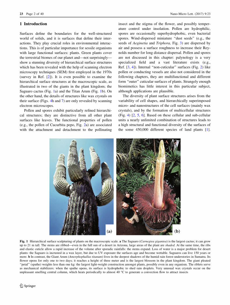
Fig. 1 Hierarchical surface sculpturing of plants on the macroscopic scale. a The Saguaro (Carnegiea gigantea) is the largest cactus; it can grow
up to 21 m tall. The stems are ribbed—even in the full sun of a desert in Arizona, large areas of the plant are shaded. At the same time, the ribs
and elastic cuticle allow a rapid increase of the volume after sporadic rainfalls: the stems expand. Loss of water is a major problem for desert
plants: the Saguaro is incrusted in a wax layer, but due to UV exposure the surfaces age and become wettable. Saguaros can live 150 years or
more. b In contrast, the Giant Arum (Amorphophallus titanum) lives in the deepest shadows of the humid rain forest understories in Sumatra. Its
flower opens for only one to two days; it reaches a height of three meter and is the largest blossom in the plant kingdom. The giant pleated
‘‘petal’’ (spathe) weights less than one kg: the largest light-weight construction amongst plants, possibly even in any organism. The riblets serve
as mechanical stabilizers: when the spathe opens, its surface is hydrophobic to shed rain droplets. Very unusual wax crystals occur on the
unpleasant smelling central column, which heats periodically to almost 40 �C to generate a convection flow to attract insects
insect and the stigma of the flower, and possibly temper-
ature control under insolation. Pollen are hydrophilic,
spores are occasionally superhydrophobic, even bacterial
spores. Wind-dispersed miniature ‘‘dust seeds’’ (e.g., the
seeds of Aeginetia and Triphora, Fig. 3) are dispersed by
air and possess a surface roughness to increase their Rey-
nolds number for long distance dispersal. Pollen and spores
are not discussed in this chapter: palynology is a very
specialized field and a vast literature exists (e.g.,
Ref. [3, 4]). Internal ‘‘non-cuticular’’ surfaces (Fig. 2) like
pollen or conducting vessels are also not considered in the
following chapters, they are multifunctional and different
form ‘‘outer’’ cuticular surfaces of plants. Strangely enough
biomimetics has little interest in this particular subject,
although applications are plausible.
The diversity of plant surface structures arises from the
variability of cell shapes, and hierarchically superimposed
micro- and nanostructures of the cell surfaces (mainly wax
crystals), and by the formation of multicellular structures
(Fig. 4) [2, 5, 6]. Based on these cellular and sub-cellular
units a nearly unlimited combination of structures leads to
a high structural and functional diversity of the surfaces of
the some 450,000 different species of land plants [1].
23 Page 2 of 40 Nano-Micro Lett. (2017) 9:23
123

Superhydrophobicity is one of the most remarkable fea-
tures of many plant surfaces; most families of higher plants
[1] include many species where the entirety or part (e.g.,
only lower side of the leaves) of the assimilating leaf
surface is superhydrophobic. Grasses (Fig. 5a, b) with a
few exceptions (e.g., Maize), are superhydrophobic and are
with around 12,000 species, one of the largest plant fami-
lies which dominates the largest ecosystems of our globe.
As is the case in many plants, often only the young leaves
are superhydrophobic (Fig. 5b), older ones may become
wettable: superhydrophobicity is often an instable state in
plant surfaces (compare also the Saguaro, Fig. 1a)—as is
also seen in technical surfaces. Some grasses (like Elymus
arenarius with a static contact angle of 161�) exhibit
similar properties to lotus leaves [7]. One square meter of a
meadow may exhibit a minimum of four square meters of
leaf surfaces (compare, e.g., Ref. [8]). According to FAO
assessments [9], grasslands (e.g., steps, savannahs, wheat,
and rice fields) cover some 52.5 million square kilometers.
A rough calculation indicates that at least 250 million
square kilometers (with other plant families included,
possibly much more) are superhydrophobic: this means
more than half of the total surface area of our whole planet.
In all actuality, the dimensions are probably considerably
higher and might equal the total surface of our planet.
There is a basic and obvious difference in surface
structure and function between aquatic and terrestrial
plants. In terrestrial (vascular) plants an epidermis is pre-
sent, as the specialized outermost cell layer with a cuticle
of the primary tissues of all leaves and several other organs
it plays an important role in environmental interactions and
surface structuring. A simplified model presented in Fig. 6
shows a layered stratification of the outermost part of
epidermis cells. Starting with the outside, one finds a
highly functional thin outermost layer, the polymer cuticle
with its superimposed waxes. This outermost layer covers
nearly all aerial tissues of land-living plants as a continuous
extracellular membrane, but is absent in roots. One of the
most important attributes of the cuticle is its function as a
transpiration barrier, which enables plants to overcome the
Fig. 2 Internal (‘‘non-cuticular’’) functional surfaces are usually hydrophilic, two examples from a squash or pumpkin (Cucurbita pepo) are
illustrated: a pollen grain, its surface functions are connected with attachment and detachment to the pollinating insect and the stigma of the
pumpkin flower, and possibly temperature control under insolation. In wind-dispersed pollen, these structures might also increase the Reynolds-
numbers, b a vessel-element of the same plant exhibiting complex spiral and perforated structures to transport water within the plant. The
structural elements of internal surfaces fundamentally differ from the outer cuticular surfaces (compare, e.g., Figs. 3, 7, 14, and 15)
Fig. 3 SEM micrographs of two seed surfaces with concave cell sculpturing. The miniature seeds of both species: a indica and b Triphora
trianthophora are optimized for seed dispersal by wind. They are hydrophobic and float for a short time in water (compare Aeginetia in Fig. 27):
the concave sculpture of the non-living cells can be interpreted as a shrinkage deformation during seed maturing and drying. The bands which
form an inner network in (a), and the surface pattern in (b) are built by cellulose. All these features are light-weight constructions and generate
high Reynolds-numbers to prolong the floating time
Nano-Micro Lett. (2017) 9:23 Page 3 of 40 23
123

100 μm
20 μm
100 μm
20 μm
(b)(a)
(d)(c)
(f)(e)
(h)(g)
Iridescent leaf of Elaphoglossum wurdackii with a smooth surface
Velvety leaves of a Dahlia flower with convex, structured cells
Hairy leaves of Leucadendron argenteum with straight hairs
Waxy leaves of Eucalyptus macrocarpa with wax threads
23 Page 4 of 40 Nano-Micro Lett. (2017) 9:23
123

physical and physiological problems connected to an
ambient environment, such as desiccation.
The cuticle is basically a biopolymer made of polyester
called cutin, impregnated with integrated (intracuticular)
waxes. Additionally, waxes on the cuticle surface (epicu-
ticular waxes) play an important role in surface structuring
at the sub-cellular scale. They occur in different mor-
phologies, show a large variability in their chemistry, and
are able to self-assemble into three-dimensional crystals.
Intracuticular waxes function as the main transport barrier
to reduce the loss of water and small molecules such as
ions from inside of the cell, and also for reducing the
uptake of liquids and molecules from the outside. Epicu-
ticular waxes form the boundary layer for many interac-
tions with the plants environment, like wettability or
spectral reflection (see Sect. 6). The next layer (Fig. 6) is
the pectin layer. It connects the cuticle to the much thicker
underlying cellulose wall, which is built by single cellulose
fibrils. Pectin is not always formed as a layer, but in some
species, especially during the early ontogeny of the cuticle,
a layered structure has been shown by transmission
electron microscopy (TEM). Additionally polysaccharides,
not shown in this schematic, are integrated into the cellu-
lose wall. The last layer shown is the plasma membrane,
which separates the living, water-containing compartment
cell from the outer non-living part of the epidermis.
We focus on superhydrophobic and superhydrophilic
surfaces, which are of particular importance for biomimetic
applications (e.g., self-cleaning: lotus effect). Superhy-
drophilicity means, a droplet imposed on a surface
‘‘spreads’’ instantly and a contact angle cannot even be
measured, e.g., in the leaves of Ruellia [10]. In contrast, on
a superhydrophobic surface water remains as an almost
bFig. 4 Macroscopic optical appearance of plant surfaces and their
surface micro-structures. (a) The leaves of Elaphoglossum wurdackii
appear glossy because of a flat surface structure shown in (b), their
iridescence is caused by thin layers within the cuticle. In (c) the
flower petals of Dahlia appear velvety due to the convex microstruc-
ture of the epidermis cells, shown in (d). In e the silvery appearance
of the Leucadendron argenteum leaves is caused by a dense layer of
light reflecting hairs (f). In (g) the leaf and flower bud surfaces of
Eucalyptus macrocarpa appear white or bluish, caused by a dense
covering with thread-like wax crystals (h)
Cuticle
Cutin withintracuticular waxPectine
Epicuticular wax
Cell wall
Plasma membrane
Fig. 6 A simplified model of the stratification of the outermost layers
of a plant epidermal cell. The schematics shows the outermost wax
layer in its most common form, as composite of three-dimensional
waxes with an underlying wax film. Below this layer is the cuticula,
made of a cutin network and integrated waxes. The cuticula is
connected with the underlying cellulose wall by a pectin layer. Below
the cell wall, the plasma membrane is shown. This membrane
separates the water-containing living part of the cells from the
outermost non-living outer cell wall and cuticle, as shown in the
schematic
Fig. 5 Plant surfaces exposed to the air are very often superhydrophobic. Like the leaves (a) of the reed (Phragmites australis) and the grassland
in the background, or the surfaces of the floating fern (Salvinia natans) in the foreground left in the Oder national park, Germany. Dew droplets
in the early morning roll-off of the superhydrophobic grass leaves (b). Since grasslands alone forms the largest terrestrial ecosystems (ca. 52.5
million km2) of our planet, we estimate there are at least about 250 million km2 of superhydrophobic leaf surfaces, which equal about 50% of the
earth’s total surface. But all plant roots are superhydrophilic like the surfaces of water plants, illustrated in (c): The Madagascar Laceleaf
(Aponogeton madagascariensis) additionally exhibits a grid of a lattice-like network to reduce the flow resistance. Source a kindly provided by
Pierre Ibisch
Nano-Micro Lett. (2017) 9:23 Page 5 of 40 23
123

globular droplet with a contact angle of more than 150�.The SEM micrographs presented were largely taken from
our archive of almost 220,000 SEM micrographs at the
University of Bonn which has been built up as a result of
over four decades of research on biological surfaces
(compare Ref. [1]) by the first author and his collaborators.
Biomimetics and bionics (which we consider here as
synonymous) are surmised to be modern scientific fields;
despite the evidence that inspiration from living organisms is
as old as mankind. The magnificent 17,000-year-old pale-
olithic paintings in the caves of Lascaux are bioinspired—
like the Cadillac tail fins in the 1960s. Bio-inspiration in the
sense of non-functional ‘‘biodecoration’’ is an inspiration for
art and design into modern times [11, 12]. Early attempts to
copy mechanical functions were not particularly success-
ful—Ovid’s story of Daedalus and Icarus and Leonardo da
Vincis design of flying machines and other devices did not
translate into technical success stories.
Historically, the dream of flying and the use of the
strange phenomena ‘‘electricity’’ were the two fundamental
forces for the foundation of what we call today bionics or
biomimetics. The construction of an electric battery based
on observations of the Torpedo fish (today we call it
Electric Ray) by Alessandro Volta in 1800 was the first
milestone [11] of bionics. And Icarus dream was realized
with the first well-documented, repeated, and successful
flights by Otto Lilienthal from 1894 onwards; his design
was based on his analysis of the flight of birds. The term
‘‘Biotechnik’’ (usually abbreviated in German as ‘‘Bionik’’)
for the new field was coined by Raul France in 1920 [13]
and finally rediscovered under the influence of cybernetics
under the name ‘‘Bionics’’ [14] and ‘‘Biomimetics’’
between 1960 and 1964; the misleading term ‘‘Biomi-
micry’’ arose in 1982 (for a historical survey see Ref. [11]).
Surfaces came surprisingly late into the focus of bionics:
The Swiss engineer George de Mestral observed in 1941
the way that the burrs (Arctium) clung to his trousers and
his dog—in 1958, he developed the bionic hook-and-loop
fastener under the trade mark Velcro�. Starting with the
discovery of hierarchically structured superhydrophobic
lotus-surfaces [2, 15, 16] and the drag-reducing shark skin
[17, 18], biomimetic surface technologies (e.g., lotus-,
shark-, gecko-, moth eye-, and salvinia-effect) became a
most important field [1, 19, 20]. The publication of the
‘‘Lotus Effect�’’ in 1997 [15] led to a change of paradigms
in surface technologies [1]. Biological role models provide
an extraordinary diversity for innovative surface tech-
nologies, which are described for plants in the following
chapters primarily under the view of biologists.
This paper is completely based on our Sect. 3.6 ‘‘Plant
Surfaces: Structures and Functions for Biomimetic Appli-
cations’’ in the 4th edition of B. Bhushan, Handbook of
Nanotechnology (Springer 2017) [21].
2 Chemistry of Plant Surfaces
Here, we consider only the surfaces of higher or vascular
plants (Tracheophyta). Primarily aquatic plants (from uni-
cellular algae to seaweeds, see Sect. 8) lack a cuticle and
have very differing superhydrophilic surfaces. For biomi-
metic applications, vascular plants are most important. In
land or vascular plants, waxes from monomolecular layers
to thick crusts or 3D-crystals, form the boundary layer of
the surface (Fig. 7). They are sometimes visible as a white
or bluish coloration of leaves and fruits, as in wheat or
cabbage, grapes, or plums. These colorations are caused by
reflection of parts of the visible light spectrum by a dense
coverage of three-dimensional (3D) wax structures. The
fan palm Copernicia prunifera, the natural source of car-
nauba wax, has massive crusts of epicuticular wax,
weighing several mg cm-2. Carnauba wax is commercially
used, e.g., for car and furniture polishes, medical products,
and candy. Even when there is not a bluish coloration
visible, three-dimensional waxes are often present. In
plants, three-dimensional waxes are responsible for several
surface functions. Waxes are not only an essential part of
the plant cuticle, but can also be found in fungi, lichens,
and animals. Waxes occur as filling material within the
basic cutin network (intracuticular), and are also found on
top of the cuticle (epicuticular). The epicuticular waxes
occur in very differing morphologies, all of which are
crystalline and thus self-assembling (Fig. 7) (survey in Ref.
[22]). However, waxes of different plants, and also waxes
of different parts of a plant, vary in their morphology and
chemical composition—they are absent in roots. In general,
plant waxes are mixtures of long-chain hydrocarbons and
their derivatives, and in some species they also contain
cyclic compounds. Because of the strong correlation
between the wax crystal morphology and their chemical
composition, some waxes, such as the nonacosan-10-ol
tubules of the Lotus leaves and many other plants, have
been named after their main wax constitution [23].
2.1 Chemical Composition of Wax
The term ‘‘wax’’ is used for a variety of biogenic products
that contain fatty materials of various kinds. Well-known
examples are bees wax, paraffin, and carnauba wax from
wax palms (Copernicia prunifera). Plant waxes are mix-
tures of aliphatic hydrocarbons and their derivatives, with
carbon chain lengths between 20 and 40, and in the case
of esters (two connected chains) about 60 atoms. Several
reviews have addressed the chemical composition of plant
waxes [24–26]. The chemical composition of plant waxes
is highly variable amongst different plant species, or
within the organs of one species (e.g., upper or lower side
23 Page 6 of 40 Nano-Micro Lett. (2017) 9:23
123

of leaves) and even during organ development [27]. The
main component classes are primary and secondary alco-
hols, ketones, fatty acids, and aldehydes. Alkanes are very
common in plant waxes, but usually occur in low con-
centrations. Other compounds are more rarely found in
plant waxes, but in those waxes where they occur, they
may be the dominant compound. The most common wax
compounds and their typical chain length are shown in
Table 1. Examples of commonly found waxes and their
major compounds are presented in Table 2. For some of
those waxes, it has been shown that their dominant
compounds crystallize in the same morphology as the
complete wax mixture. Examples are the primary alcohols
and the b-diketone waxes found on different parts of
wheat plants [28]. However, an increasing number of
publications report the discovery of new wax components
and a long list of rare and uncommon ingredients, such as
methyl-branched aliphatics [29]. Environmental factors,
such as temperature or light intensity, influence the
quantity of waxes and their chemical composition
[30–32].
Many or even most plant ‘‘waxes’’ do not match the
chemical definition of true waxes and they are usually
complex mixtures of differing compounds. For example,
triterpenoids are cyclic hydrocarbons, which occur in
high concentrations in the epicuticular coatings of grapes
(Vitis vinifera) [30]. Other plant waxes contain polymeric
components such as polymerized aldehydes which are
only slightly soluble in chloroform [33, 34]. It should be
noted that nearly all the existing data of the chemical
composition of plant waxes are based on solvent-ex-
tracted waxes. These are mixtures of epicuticular and
intracuticular waxes, which may be chemically different,
as shown for the waxes of Prunus laurocerasus by Jetter
and Schaffer [27] and by Wen et al. [35], for Taxus
baccata. The development of more selective methods of
Euphorbia resinifera: layer and platelets Crassula ovata: crusts with cracks Aloe striata: crusts with plates, C=cuticula
Thalictrum flavum glaucum: tubules Eucalyptus gunnii: β-diketone tubules Aristolochia albida: plates
Sassafras albidum: ridged rodllets Musa spp.: aggregated rodlets Convallaria majalis: plates
(g) (h) (i)
(d) (e) (f)
(a) (b) (c)2 μm 100 μm 3 μmC
10 μm2 μm2 μm
5 μm 10 μm 10 μm
Fig. 7 SEM micrographs of epicuticular waxes: in (a) waxes on a leaf of Euphorbia resinifera have been particularly removed to show the
composite structure of a basal wax layer with three-dimensional wax platelets on it. In (b) a wax crust with fissures on a leaf of Crassula ovata is
shown. A cross section through the periclinal wall of Aloe striata (c) shows the cuticle (indicated by C) and a wax layer (indicated by an arrow)
with wax platelets on top. In (d) nonacosanol tubules on Thalictrum flavum glaucum leaves and (e) ß-diketone wax tubules on Eucalyptus gunnii
leaves are shown. In (f) wax platelets on Aristolochia albida leaf and in (g) transversely ridged rodlets on a leaf of Sassafras albidum are shown.
In (h) longitudinally aggregated wax threads form large aggregated rodlets on the lower side of the leaves of Musa species (spp.). In Convallaria
majalis leaves, shown in (i), wax platelets are arranged in a pattern, similar to magnetic field lines, around the stomata. Thin wax films are not
visible in SEM, but are present below and between the three-dimensional waxes shown here
Nano-Micro Lett. (2017) 9:23 Page 7 of 40 23
123

wax sampling allows selective removal of the epicutic-
ular waxes and their analysis separately from the intra-
cuticular wax fractions [27, 36].
Epicuticular wax structures usually occur in the size
ranging from 0.2 to 100 lm (Fig. 7); thus, the appropriate
microscopic techniques for investigation of their
Table 1 The most common
chemical compounds in plant
waxes and their spectrum of
chain length
Chain length
1 Aliphatic compounds
1.1 In waxes frequently existing, but mostly as minor compounds
Alkanes CH3–(CH2)n–CH3 Odd C19–C37
Primary alcoholsa CH3–(CH2)n–CH2–OH Even C12–C36
Esters CH3–(CH2)n–C0–0–(CH2)m–CH3 Even C30–C60
Fatty acids CH3–(CH2)n–COOH Even C12–C36
Aldehydes CH3–(CH2)n–CHO Even C14––C34
1.2 In waxes rarely existing, but if, than as major wax compounds
Ketones e.g., palmitones CH3–(CH2)n–CO–(CH2)m–CH3 Odd C25–C33
ß–diketones CH3–(CH2)n–CO–CH2–CO–(CH2)m–CH3 Odd C27–C35
Sec. alcohols e.g., nonacosan-10-ol CH3–(CH2)n–CH2OH–(CH2)m–CH3 Odd C21–C33
2 Cyclic Compounds
Flavonoids
e.g., Quercetin
Triterpene e.g., ß-Amyrin
a Primary alcohols are common minor constitutions in waxes, but can occur as major compounds in the wax, e.g.,of grasses, eucalypts, clover, and other legumes [26]. Further examples of occurrence are given in Table 2
Table 2 Common wax types in plant species and their major chemical compounds
Wax type Species Dominating chemical compound(s)
Films Hedera helix Prim. alcohols, aldehydes
Films Magnolia grandiflora Fatty acids C24–C30, prim. alcohols C24–C28
Films Prunus laurocerasus Alkanes C29, C31
Crust Crassula ovata Aldehydes C30, C32, alkane C31
Diketone tubules Eucalyptus globulus Beta-diketones C33
Diketone tubules Leymus arenarius Beta-diketone C31, hydroxy-beta-diketone C31
Nonacosanol tubules Ginkgo biloba Sec. alcohol C29
Nonacosanol tubules Nelumbo nucifera Sec. alkanediols C29
Nonacosanol tubules Thalictrum flavum glaucum Sec. alcohol C29
Nonacosanol tubules Tropaeolum majus Sec. alcohol C29
Nonacosanol tubules Tulipa gesneriana Sec. alcohol C29
Platelets Convallaria majalis Prim. alcohol C26, C28, aldehydes
Platelets Euphorbia myrsinites Prim. alcohol C26, aldehydes
Platelets Galanthus nivalis Prim. alcohol C26
Platelets Iris germanica Prim. alcohol C26
Platelets Triticum aestivum Prim. alcohol C28
Transversely ridged rodlets Aristolochia tomentosa Ketones
Transversely ridged rodlets Gypsophila acutifolia Alkanes C31
Transversely ridged rodlets Liriodendron chinense Ketones
Longitudinal ridged rodlets Benincasa hispida Triterpenol acetates
With exception of the fruit surface of Benincasa hispida, data represent the waxes on the leaves of the species. All references for the chemical
data are listed in [42] and examples of the wax types here listed are shown in Fig. 7
23 Page 8 of 40 Nano-Micro Lett. (2017) 9:23
123

morphology are SEM and low pressure- or environmental
SEM. Several SEM investigations showed that most of the
epicuticular waxes form three-dimensional structures, with
great variations of their morphologies. Comprehensive
overviews of the terminology and micromorphology of
epicuticular waxes are given by Barthlott et al. [22], Jeffree
[26], and in Ref. [37]. The comprehensive classification of
Barthlott et al. [22], which we follow here, includes 23
different wax types. It is based on chemical and morpho-
logical features and also considers orientation of single
crystals on the surface and the orientation of the waxes to
each other (pattern formation). In this classification, the
wax morphologies include thin films and several three-di-
mensional structures such as crusts, platelets, filaments,
rods, and tubules which have a hollow center. Morpho-
logical sub-types are, for example, entire and non-entire
wax platelets. A further sub-classification is based on the
arrangement of the crystals, e.g., whether they are ran-
domly distributed, in clusters, in parallel orientation, or in
specific arrangements around stomata, as the ‘‘Convallar-
ia’’ type (Fig. 7h). The most common wax morphologies
are introduced in the following section and are shown in
Table 2.
Probably all terrestrial plant surfaces are covered by thin
(in the extreme monomolecular) wax films, the three-di-
mensional wax crystals appear on underlying wax film as
shown in Fig. 7a for the waxes of Euphorbia resinifera and
has been reported for several species [22, 38–41]. Wax
films are often incorrectly referred to as ‘‘amorphous’’ [42].
On several plant surfaces, wax films are limited to a few
molecular layers which are hardly visible in the SEM. By
mechanical isolation of the epicuticular 3D waxes, e.g.,
freezing in glycerol, the waxes can be removed from the
cuticle, and transferred onto a smooth artificial substrate for
microscopic investigations [36]. With this method, the
remnants of the wax film can be detected, and the film
thicknesses can be determined. Wax film formation has
been investigated on a living plant surface by atomic force
microscopy (AFM, shown in Fig. 8) [40, 43]. Such inves-
tigations show that wax films are composed of several
monomolecular layers, with thicknesses up to several
hundred nanometers. In the following, these relatively thin
wax films (\0.5 lm) are called two-dimensional (2D)
waxes, and the thicker wax layers (0.5–1 lm) and wax
crusts ([1 lm) are called three-dimensional (3D) waxes.
Wax crusts are often found in succulent plants, as on the
leaves of Crassula ovata, shown in Fig. 7b. Such a mul-
tilayered assembly of waxes is detectable by a cross section
through the epidermis, as shown in Fig. 7c for Aloe striata.
Three-dimensional waxes occur in different morpholo-
gies. Most common are tubules, platelets, rodlets, and
longitudinally aggregated rodlets shown in Fig. 7d–i.
Wax tubules are hollow structures, which can be dis-
tinguished chemically and morphologically. The first type,
called nonacosanol tubules, contains large amounts of
asymmetrical secondary alcohols, predominantly nona-
cosan-10-ol and its homologues and to a certain degree also
asymmetrical diols [23, 44, 45]. Nonacosan-10-ol is the
most common ‘‘waxy’’ coating of all major vascular plant
groups and was evolved with the conquest of land some
450 million years ago, a phylogenetic tree is provided by
Ref. [1]. The nonacosanol tubules are usually 0.3–1.1 lm
long and 0.1–0.2 lm wide. The second type of tubules
contains high amounts of ß-diketones, such as hentriacon-
tane-14,16-dione [46]. This particular kind of wax tubule is
characteristic for many grasses (Poaceae) and also occurs
in various other groups [47]. Figure 7e shows that the ß-
diketone tubules are two to five times longer than the
nonacosanol tubules shown in Fig. 7d. Their length reaches
from 2 to 5 lm, and diameters vary between 0.2 and
0.3 lm.
Platelets, as shown in Fig. 7f, are the very common wax
structures found in all major groups of plants. Following
the terminology of Barthlott et al. [22], waxes are termed
platelets when flat crystals are connected with their narrow
side to the surface. Platelets can be further differentiated by
their outline into, e.g., entire or undulated ones. Platelets
vary considerably in shape, chemical composition, and
spatial pattern. For platelets, only limited information
about the connection between morphology and chemical
composition is available. In some species, wax platelets are
dominated by high amounts of a single chemical com-
pound, which can be primary alcohols, alkanes, aldehydes,
esters, secondary alcohols, or flavonoids [26]. In contrast to
platelets, plates are polygonal crystalloids with distinct
edges and are attached to the surface at varying angles.
The morphology of three-dimensional wax structures is
not necessarily determined by the dominating chemical
Fig. 8 FM experimental set-up for long-term investigations of wax
crystallization on a living plant surface. The tip of the leaf of a
snowdrop (Galanthus nivalis) has been fixed on the specimen holder
with a drop of two-compound glue. Existing waxes have been
removed, and the rebuilding (self-healing) of the wax was studied
over several hours. Appropriate scan conditions for living plant
surfaces are given in the text, and the method of wax removal is
described in detail in [40]
Nano-Micro Lett. (2017) 9:23 Page 9 of 40 23
123

compound or compound class. One example of wax crys-
tals determined by a minor component of a complex mix-
ture is the transversely ridged rodlets, shown in Fig. 7g,
which contain high amounts of hentriacontane-16-one
(palmitone) [48]. Wax rodlets are massive sculptures which
are irregular, polygonal, triangular, or circular in their cross
sections. They have a distinct longitudinal axis, with a
length/width ratio usually not exceeding 50:1. In addition,
rodlets may have a variable diameter along the length of
their axis. More complex structures are the longitudinally
ridged rodlets, as those found on banana leaves (Musa
species), shown in Fig. 7h. These waxes consist exclu-
sively of aliphatic compounds, with high amounts of wax
esters and less of hydrocarbons, aldehydes, primary alco-
hols, and fatty acids. The origin of these wax aggregates is
still not clear, and so far all attempts to recrystallize these
wax types have failed. As a consequence of that, it is
assumed that their origin is connected to structural prop-
erties of the underlying plant cuticle.
Brassica oleracea is known to have very complex wax
crystal morphology, several cultivars form several different
wax types, and where several different wax morphologies
can occur on the same cell surface [30]. Why the different
three-dimensional wax morphologies co-exist on the sur-
face of a single cell is unknown, as is whether these dif-
ferent morphologies are built up by phase separation of
different compounds or if they are formed by the same
compound.
The last example in Fig. 7i represents plant surfaces on
which waxes are arranged in a specific pattern. Examples
are parallel rows of longitudinally aligned platelets, with
the orientation extending over several cells (e.g., in Con-
vallaria majalis, shown in Fig. 7i), or rosettes, in which the
arrangements of platelets are more or less in radially
assembled clusters. In particular, the parallel orientation of
platelets on the leaves of several plant species leads to the
question of how the orientation is controlled by the plant. It
is assumed that the cutin network functions as a template
for the growth of the three-dimensional wax crystals, but
there is still a lack of information about the molecular
structure of the cuticle, so this question is still unanswered.
Certain surface wax morphologies and their orientation
patterns are characteristic for certain groups of plants; thus,
patterns and the morphology of plant waxes have been used
in plant systematics. Barthlott et al. [47] provide an over-
view of the existence of the most important wax types in
plants, based on SEM analysis of at least 13,000 species,
representing all major groups of vascular plants.
2.2 Chemical Heterogeneities
Surfaces of a particular plant species may exhibit chemical
heterogeneities in the classical sense, the best example are
the superhydrophilic pinning anchor cells on top of each
superhydrophobic trichome of Salvinia molesta (Fig. 28):
In a broader sense, all organism have chemically hetero-
geneous surfaces: root surfaces differ dramatically from
leaf surfaces. And within one leaf, the upper side differs
from the underside. In leaves of Quercus robur, contact
angles range from 30� to 130� depending on the part of the
leaves where wettability was determined [7].
The aquatic watermilfoil Myriophyllum brasiliense is—
like all submersed water plants—superhydrophilic, a con-
tact angle of the leaves cannot be determined. However, as
soon a flowering shoot approaches the water level, wax
crystals are generated and the new leaves outside of the
water exhibit a contact angle of 162� like in a lotus leaf [7].
2.3 Crystallinity
All aliphatic plant surface waxes have a crystalline order.
The classical definition for crystals implies a periodic
structure in three dimensions, but with the increasing
importance of liquid crystals and the detection of qua-
sicrystals, it has become necessary to extend the definition,
so that certain less periodic and helical structures, as found
for some waxes, were included [49].
The crystal structure of the epicuticular waxes can be
examined by electron diffraction (ED), nuclear mass res-
onance (NMR) spectroscopy, and X-ray powder diffraction
(XRD). ED with the TEM provides the structure informa-
tion of single wax crystals of less than 1 lm size, as shown
in Fig. 9a, b, for a single wax platelet. However, even with
a low-intensity imaging system, the crystal structure is
rapidly destroyed by the electron beam intensity. There-
fore, XRD is useful for determining the crystal symmetry,
as well as providing information about different types of
disorder. Very thin mono- or bi-molecular layers of waxes,
as shown in Fig. 9c, d, are of course not periodic in three
dimensions, but form two-dimensional crystals at the
molecular level. As mentioned before, in addition the
planar wax structures, such as films and platelets, many
natural plant waxes develop irregular three-dimensional
morphologies, or structures such as threads and tubules
with a large extension in one direction. These morpho-
logically different waxes were found to occur in three
different crystal structures. The majority of waxes exhibit
an orthorhombic structure, which is the most common for
pure aliphatic compounds. Tubules containing mainly
secondary alcohols show diffraction reflections of a tri-
clinic phase, with a relatively large disorder, and ß-dike-
tone tubules show a hexagonal structure [42].
Self-assembly of waxes is an inherent result of the
crystalline nature. That different wax morphologies on
plant surfaces originate by self-assembly of the wax
molecules has been shown by the recrystallization of
23 Page 10 of 40 Nano-Micro Lett. (2017) 9:23
123

waxes, which were isolated from plant surfaces
[28, 40, 45, 50–52]. In these studies, most waxes recrys-
tallized in their original morphology, as found on the plant
surfaces.
Self-assembly processes resulting in nano- and micro-
structures are found in nature, as well as in engineering.
They are the basis for highly efficient ways of structuring
surfaces down to the molecular level. Self-assembly is a
general process of structuring in which atoms, molecules,
particles, or other building units interact and self-organize
to form well-defined structures. The processes of self-
assembly in molecular systems are determined by five
characteristics: the components, interactions, reversibility,
environment, and mass transport with agitation [53]. The
most important driving forces are weak and non-covalent
intermolecular interactions, such as Van der Waals and
Coulomb interactions, hydrophobic interactions, and
hydrogen bonds. During self-assembly, their interactions
start from a less-ordered state, e.g., dissolved waxes in a
solution, to a final more-ordered state, a crystal [54, 55].
Environmental factors such as temperature, solvent, and
substrate might influence the self-assembly process, and in
the case of waxes, their morphology.
The most suitable microscopy technique for studying the
self-assembly process of waxes under environmental con-
ditions is atomic force microscopy (AFM) because it
combines sufficient resolving power to image nanostruc-
tures with the ability to work at STP (standard temperature
and pressure) with living plant material (Fig. 8). Self-
assembly of waxes has been studied directly on plant sur-
faces, as well as the recrystallization of waxes and single
wax compounds on artificial surfaces. However, AFM is
not suitable for all plant surfaces. Within a leaf surface,
large structures such as hairs with dimensions of several
tens of micrometers can emerge out of the epidermis and
pose a barrier against the surface scanning probe. Addi-
tionally, high aspect ratio structures caused by cell surface
structures might cause artifacts in the resulting images.
Species with smooth or slightly convex cell surface
sculptures are most appropriate for AFM investigations.
The process of wax regeneration occurs over several hours;
thus the loss of water from inside the plant has to be
minimized to reduce the specimen drift by material
shrinking during investigation. This precondition limits the
range of specimens for AFM with a small specimen
chamber, because the sizes and shapes of the leaves must
allow them to be mounted in the AFM without cutting
them. An experimental set-up where the complete plant is
placed close to the AFM and a leaf is fixed on the AFM
specimen holder is shown in Fig. 8. The leaf was fixed at
its lower side to the specimen holder with a drop of a two-
compound glue, and waxes on the upper leaf side were
removed by embedding them into a drop of water soluble
glue. After hardening, the glue and the embedded waxes
were removed from the leaf surface and the process of wax
regeneration was studied. Temperature increase in long-
term investigations, caused by the laser beam on top of the
cantilever, induces expansion of the water in the leaf,
resulting in a drift of the specimen. To minimize this,
reflective cantilevers must be used, and the laser beam
intensity should be reduced by integrating an attenuation
filter above the cantilever [40]. However, the waxes
themselves are fragile; thus appropriate scan conditions at
scan sizes of 3–20 lm are tapping mode and scan rates of
0.7–2 Hz, encompassing 256 lines per image and a set-
point near the upper limit to minimize the interaction
between tip and sample. Figure 10 shows the regeneration
of a wax film on a leaf of snowdrop (Galanthus nivalis) by
AFM of a wax platelet Electron diffraction AFM of alkane layer
Model of alkane layer
(c)
(d)(b)(a)
Fig. 9 The layered and crystalline structure of alkane waxes is demonstrated by an AFM map of a single wax platelet (a) and the corresponding
electron diffraction pattern, shown in (b). In (a) the steps visible on the crystal surface are caused by a perpendicular orientation of the molecules.
Such steps can be monomolecular, e.g., for alkanes, or in some waxes bilayers are formed by polar molecules of primary alcohols. The AFM map
of recrystallized alkanes (c) and the model shown in (d) demonstrate the layered orthorhombic wax structure
Nano-Micro Lett. (2017) 9:23 Page 11 of 40 23
123

formation of a multilayered wax film and the growth of
three-dimensional wax platelets. This and further investi-
gations show that the growth of the three-dimensional wax
crystals occurs by apical accumulation of new wax mole-
cules on only one side of the crystal. The regeneration of
the wax film results in a multilayered crystalline coverage
on the plant cuticle. The time needed to regenerate waxes
shows large variations depending on the species, with some
species never regenerating the wax that was removed. In
these plants, wax synthesis seems to be inactive when
leaves are mature [56].
Alternatively, self-assembly of plant waxes can be
studied by recrystallization of the waxes on artificial sub-
strates (Fig. 11). Based on those studies, the formation of
wax tubules and platelets has been described in detail. Wax
platelets, characteristic for wheat leaves (Triticum aes-
tivum), are constructed from the primary alcohol octa-
cosan-1-ol [28]. Crystallization of the wax mixture isolated
from the leaves and of pure octacosan-1-ol on different
artificial substrates showed a substrate-dependent growth.
On a non-polar, crystalline substrate (highly ordered pyr-
olytic graphite, HOPG), platelets grow with a vertical
orientation to the substrates, whereas on a polar surface,
such as mica, crystals grow horizontally to the substrate
surface. On amorphous polar glass only amorphous wax
layers grow. This substrate dependence demonstrates epi-
taxial control of crystal growth depending on the orienta-
tion and order of the first layers of molecules adhering on
the substrate surface. Octacosan-1-ol forms ordered bilayer
structures on the substrate. In these, the first layers of
molecules lie flat on non-polar substrates, but stand upright
(perpendicular) on crystalline polar surfaces. The grown
platelet morphology results from an anisotropic crystal
growth, caused by a faster parallel assembly of the mole-
cules at the length side of already existing molecules than
at the ends of the molecules [57]. AFM micrographs in
Fig. 12 and schematics of the molecule orientation
demonstrate the differences of growth on polar and non-
polar substrates for octacosan-1-ol molecules. In both
cases, flat crystals with different orientations grow. Crys-
tals grown horizontal to the substrate surface are called
plates (Fig. 12a, b), those grown perpendicular to the
substrate surface are termed platelets (Fig. 12c, d). The
substrates on which the crystals grow influence the crystal
morphology and their orientation. This fact can be used to
create different kinds of nano- and micro-patterns on
technical surfaces [28, 52, 58, 59]. In summary, substrates
can have a direct influence on the self-assembly processes
of wax crystals, and can function as a template on the
molecular level. In this case, the substrate organizes the
assembly of the molecules in a specific spatial arrangement
[60, 61]. Such a template effect was reported for wax
platelets formed by primary alcohols [28]. On HOPG
substrate, the spatial pattern of the reassembled wax pla-
telets strictly followed the hexagonal symmetry of the
crystalline substrate. However, the cutin matrix of the
2 μm 2 μm 2 μm 2 μm
13 min
200 nm
Growth direction1 μm
33 min 67 min 80 min
20 min
33 min
55 min
74 min
Overlaped profiles of the growing crystalshown in the AFM figures above
Fig. 10 AFM maps and a series of profile lines, taken from repeated scans during the crystal growth on a leaf of Snowdrop (Galanthus nivalis).
The first AFM map represents the wax regeneration within 13 min; the last map was taken after 80 min after wax removal. The white arrows
mark the same position of the crystal as the black arrow marks in the profile figure. In the figure below, the outlines of the growing crystal have
been overlapped to demonstrate, that the extension is occurring at the distal end of the growing crystal and that at this time the growth in height is
limited to a few nanometers. Outlines have been taken from four AFM scans: 20, 33, 55, and 74 min after the wax regeneration process began.
The experimental set-up is shown in Fig. 8
23 Page 12 of 40 Nano-Micro Lett. (2017) 9:23
123

cuticle, which acts as a substrate in plant surfaces, is
assumed to be amorphous, and an epitaxial growth on an
amorphous substrate seems paradoxical.
An example of wax crystals composed of more than one
compound is the transversely ridged rodlets. These waxes
can be recrystallized from the total wax mixture, but not
from individual compounds such as alkanes or palmitones.
For these waxes, it is assumed that their morphologies are
also formed by a self-assembly-based crystallization pro-
cess, but the presence of minor amounts of other com-
pounds is required as an additive for crystal growth [48].
The origin of wax tubules, shown schematically in
Fig. 13a and in SEM micrographs in Fig. 13b, has been
debated for a long time. Several observations, such as
spiral lines on the surfaces of some nonacosanol tubules
[51], led to the assumption that tubules arise from a
twisting or folding of a platelet-like precursor form.
Recrystallization experiments with nonacosan-10-ol waxes
showed that these tubules grow perpendicular to the sub-
strate surface when recrystallized on HOPG. This vertical
orientation of the tubules allows a detailed study of the
growth process by AFM and shows that the building of
nonacosan-10-ol tubules from Lotus (Nelumbo nucifera)
and Nasturtium (Tropaeolum majus) leaves is based on a
continuous growth of a small circular precursor structure
by supplementation of the wax on top of it [52]. The AFM
micrographs shown in Fig. 13c–g, are consecutive AFM
images of growing tubules, made during the tubule for-
mation process. The terminal ends of growing tubules are
asymmetric in height. This asymmetry seems to be caused
by an accumulation of new wax molecules at edges found
at the terminal end of the tubules and indicates a helical
10 μm
Hierarchical structure with 0.12 μg mm-2
Hierarchical structure with 0.20 μg mm-2
Hierarchical structure with 0.40 μg mm-2
2 μm
10 μm 2 μm
10 μm 2 μm
Fig. 11 Biomimetic superhydrophobic hierarchically structured technical surfaces. The silicon replica with pillars of 14 lm in diameter and
30 lm in height with 23 lm pitch, covered by self-assembled alkanes (hexatriacontane). From top to bottom an increase in crystal density is
shown. Highest water repellence and lowest hysteresis has been found for the structures given in the middle line, where 20 lg cm-2
hexatriacontane was applied on the surfaces. These surfaces have been used for detailed study of wetting and adhesion (from Ref. [59])
Nano-Micro Lett. (2017) 9:23 Page 13 of 40 23
123
growth mechanism for the tubules. The pure nonacosan-10-
ol alcohol, the dominating compound of wax tubules, can
crystallize in different forms [39, 45, 51]. Here, Jetter and
Riederer [45] show that a range of alkanediols, present in
the waxes of many secondary alcohol tube-forming spe-
cies, also have tube-forming capability.
Chemical analysis of the leaf waxes of Lotus and Nas-
turtium (Tropaeolum majus) shows that waxes of both
species are composed of a mixture of aliphatic compounds,
with nonacosan-10-ol (a secondary alcohol) and nona-
cosandiols (an C29 alkane with two alcohol groups) as their
main components [52]. These compounds have been sep-
arated from the rest of the wax compounds and used for
recrystallization experiments. It could be shown with
mixtures of nonacosan-10-ol and nonacosandiols compo-
nents that a minimum amount of two percent of nona-
cosandiols support tubules formation [23].
Analysis of wax chemistry, crystalline order, and their
self-assembly has led to a better understanding of the
molecular architecture of three-dimensional waxes [62].
Based on these data, a model of nonacosan-10-ol tubule
structure has been developed, as shown in Fig. 13a. Here it
is assumed that the lateral oxygen atoms at the side of the
straight molecules hinder the formation of the normal,
densely packed, orthorhombic structure and require addi-
tional space, causing a local disorder between the mole-
cules and cause a spiral growth, leading to the tubule form.
Glands or glandular trichomes may produce very par-
ticular substances, they can be found on approximately
30% of all vascular plants [63]. Multicellular glands
include salt glands, nectaries, or the adhesive-secreting
glands of some carnivorous plants [64]. Secretion and
accumulation of toxic compounds at the plant surface
allows direct contact with insects, pathogens, and herbi-
vores, and might therefore be an effective defense strategy
[64]. The exudates of glands are, for example, terpenoids,
nicotine, alkaloids, or flavonoids. The exudates of some
ferns and angiosperms, in particular several members of the
Primulaceae, are composed of flavonoids [65, 66]. These
flavonoid exudates or ‘‘farina’’ are morphologically similar
to waxes, but are chemically distinct from plant waxes.
Other glandular trichomes, such as the glands of the car-
nivorous plants of the genera Drosera (sundew) and Pin-
guicula (butterworts) secrete adhesives and enzymes to
trap and digest small insects like mosquitoes and fruit flies.
Chemical heterogeneities are implied by the presence of
other glands. The definition of this phenomenon depends
on the scale: all biological surfaces show chemical
heterogeneities, the most common case are leaves with a
hydrophilic upper side and a superhydrophobic lower side.
On a much smaller scale, the trichomes of certain Salvinia
species are most remarkable for hydrophilic islands within
a superhydrophobic surface, the Salvinia paradox (Fig. 28)
[1, 67].
AFMSEM
4 nm
Anisotropic growth
4 nm
(a) (b)
(c) (d)
polar substrate plates
Anisotropic growth
non-polar substrate platelets
780 nm
980 nm 2 μm
2 μm
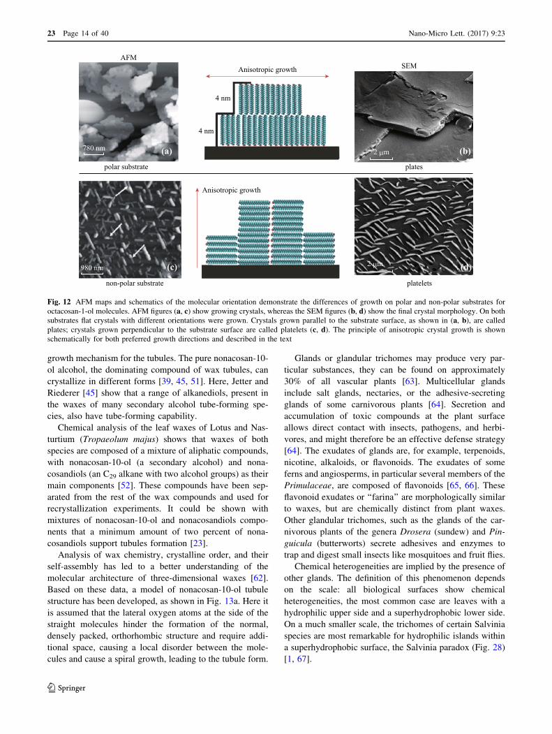
Fig. 12 AFM maps and schematics of the molecular orientation demonstrate the differences of growth on polar and non-polar substrates for
octacosan-1-ol molecules. AFM figures (a, c) show growing crystals, whereas the SEM figures (b, d) show the final crystal morphology. On both
substrates flat crystals with different orientations were grown. Crystals grown parallel to the substrate surface, as shown in (a, b), are called
plates; crystals grown perpendicular to the substrate surface are called platelets (c, d). The principle of anisotropic crystal growth is shown
schematically for both preferred growth directions and described in the text
23 Page 14 of 40 Nano-Micro Lett. (2017) 9:23
123

3 Structuring of Plant Surfaces: HierarchicalArchitecture between Nano-and Macrostructures
3.1 The Cuticle
Primarily water plants (from unicellular algae to giant
seaweeds) lack a cuticle; this particular polymer layer is
restricted to higher plants (see Sect. 8).
The cuticle covers leaves, flowers, stems, fruits, and seeds
and serves as a protective continuous layer covering the pri-
mary organs of all vascular plants and mosses. But in roots and
secondary structures (e.g., bark) a cuticle is not present. The
cuticle is a hydrophobic composite material, composed of a
polymer called cutin and integrated and superimposed lipids
called ‘‘waxes’’ (see Sect. 2). The cuticle network is formed by
cutin, a polyester-like biopolymer composed of hydroxyl and
hydroxyepoxy fatty acids, and sometimes also by cutan,
which is built by polymethylene chains. Non-lipid compounds
of the cuticle are cellulose, pectin, phenols, and proteins.
Large differences in the chemical composition and
microstructure of the cuticle have been found by comparing
different species and different developmental stages. Chemi-
cal composition, microstructure, and biosynthesis of the
cuticle have been reviewed by several authors [26, 68–75].
3.2 Hierarchical Sculpturing
The cuticle and molecular wax films are the foundation
of the surface. Additional levels of ‘‘hierarchical sculp-
turing’’ (or less precise ‘‘structuring’’) are formed by wax
crystals (Fig. 7), the form of single cells (Fig. 18),
composed of sculptures like simple or multicellular hairs
(Fig. 14), the form and curvature of whole organs likes
leaves (Fig. 27b, c) up to the gross morphological levels
visible from larger distances like the riblets of the Titan
arum flower (Amorphophallus, Fig. 1). The concept of
hierarchical sculpturing of plant surfaces and a coherent
terminology were introduced by Ref. [2] and used in
many publications (e.g., Ref. [5, 76]). Based on the
comprehensive survey by Ref. [1], we have revised this
concept starting with a ‘‘flat’’ surface as first level, fol-
lowed by a sequence of 4 or more superimposed ‘‘micro-
architectural’’ hierarchical levels. The term sculpture
(‘‘architecture’’) or sculptural seems more appropriate for
three-dimensional features (discussed previously in Ref.
[2]). The terms structure or structural also includes
chemistry and chemical heterogeneities, we prefer thus to
use the term ‘‘sculpture’’ for the micro-architectural
elements within the following four levels:
110 nm
110 nm
7x4 nm
2x4 nm1 μm
2 μm
200 nm 200 nm 200 nm 200 nm 200 nm
(b)
(g)(e)(d)(c) (f)
(a)Wax tubules model SEM of wax tubules on a leaf
Fig. 13 A model and SEM micrograph of the molecular order of nonacosan-10-ol tubules and AFM analysis of their self-assembly. Based on
SEM characterization, chemical analysis, single compound crystallization, and crystallographic data, a model of the nonacosanol tubules has
been developed (a). Original nonacosan-10-ol tubules are shown in the SEM micrograph (b) for Thalictrum flavum glaucum leaves. Consecutive
AFM figures of tubule formation (nonacosan-10-ol wax from Tropaeolum majus) were made after applying a wax solution on HOPG. After
65 min (c) the waxes mainly formed curved rodlets, which were horizontally arranged to the substrate. The same area of the HOPG substrate
shows that waxes start to form circles (d–g) and after 223 min (g) the rodlets initially observed were dissolved and short tubules were formed
Nano-Micro Lett. (2017) 9:23 Page 15 of 40 23
123

3.3 First Sculptural Level
Flat surfaces defined by their hydrophilic or hydrophobic
chemistry on the scale of the resolution of a scanning
electron microscope (SEM). Flat is a relative category that
depends on the scale. Here, we limit the definition of flat to
surfaces that feature structures of usually less than 10 nm
in height. Flat surfaces are rarely found in plants and ani-
mals, e.g., the leaves of Rubber Figs (Ficus elastic).
3.4 Second Sculptural Level
Cell surfaces are covered with structures between 50 nm
and 20 lm. Structures at this level on plants are usually
formed by epicuticular wax crystals (e.g., Fig. 7) which
may exhibit a large spectrum of shapes like rodlets,
platelets, or tubules. They may exceed 200 lm in height
(e.g., Strelitzia, Copernicia, Benincasa).
The main elements of the second sculptural level in
plants are (i) epicuticular wax crystals, the diversity of
waxes was discussed in the preceding chapter on the
chemistry of surfaces, (ii) cuticular folds (Fig. 15), (iii) sub-
cuticular inclusions (Fig. 16). Functionally, a superimpo-
sition of cuticular folds and wax crystals is not necessary:
Structuring on a specific level is performed by one group of
elements, which seems to be a basic law [2]. The data
gathered by investigating thousands of plant surfaces con-
stitute the rule that, e.g., wax crystals and cuticular folds
exclude each other. These frequently found morphological
modifications of the outermost cell walls are known to
influence the second hierarchical level. They are schemat-
ically shown in Fig. 17. In the first case, shown in Fig. 17a,
200 μm 200 μm 200 μm
50 μm200 μm200 μm
100 μm 100 μm 400 μm(h)(g) (i)
(e)(d) (f)
(b)(a) (c)
Salvinia minima: multicellular hairsLavandula angustifolia: hairs and glandsCistus symphytifolius: hairs and glands
Virola surinamensis: starshaped trichome
Phaseolus vulgaris: hairs for climbingKalanchoe tomenosa: simple hairsLeucadendron argenteum: simple hairs
Caiophora coronaria (d) and Cynoglossum officinale (e): hairs with barbed hooks
Fig. 14 SEM micrographs of hairs and glands on plant surfaces. a A dense layer of straight, unbranched hairs almost orientated parallel to the
leaf surface on Leucadendron argenteum. b The unbranched hairs of Kalanchoe tomentosa are orientated upright. c The shoot surface of a bean
shoot Phaseolus vulgaris with terminal hooks to facilitate climbing. d Single hairs on a leaf of Caiophora coronaria and e those on the seed
surface of Cynoglossum officinale are characterized by terminal and lateral barbed hooks. f The star-like trichome on the leaf of Virola
surinamensis has a flat surface in contrast to the convex epidermal cells with epicuticular wax crystalloids. g Simple branched star-like hairs and
two morphological different glands (arrows) on the leaf of Cistus symphytifolius, and h multiple ramified hairs and short-stalked glands (arrow)
on a leaf of Lavandula angustifolia. i The four trichomes of Salvinia minima originate from a common base, in contrast to S. molesta (Fig. 28) the
tips are free and do not show the eggbeater shape
23 Page 16 of 40 Nano-Micro Lett. (2017) 9:23
123
the surface structure is induced by concavities of the cell
wall which lead to coves and folding of the surface. The
second kind of structuring originates by sub-cuticular
inserts of mineral crystals, such as silicon oxides (Fig. 17b).
The third kind of surface structuring results from the folding
of the cuticle itself (Fig. 17c). Additionally, on many plants,
waxes on top of the cuticle lead to surface structuring as
shown in Fig. 17d. Waxes and their structural diversity
have already been introduced; thus, cuticular folds and sub-
cuticular are introduced in the next chapter.
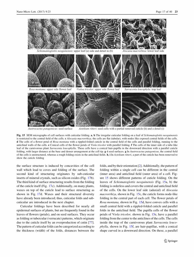
Cuticular foldings have been described for nearly all
epidermal surfaces of plants, but are frequently found in the
leaves of flowers (petals), and on seed surfaces. They occur
as folding or tubercular (verrucate) patterns, which originate
due to the cuticle itself by an overproduction of cutin [77].
The pattern of cuticular folds can be categorized according to
the thickness (width) of the folds, distances between the
folds, and by their orientation [2]. Additionally, the pattern of
folding within a single cell can be different in the central
(inner area) and anticlinal field (outer area) of a cell. Fig-
ure 15 shows different patterns of cuticle folding. On the
leaves of Schismatoglottis neoguinensis (Fig. 15a, b) the
folding is orderless and covers the central and anticlinal field
of the cells. On the lower leaf side (adaxial) of Alocasia
macrorrhiza, shown in Fig. 15c, the cuticle forms node-like
folding in the central part of each cell. The flower petals of
Rosa montana, shown in Fig. 15d, have convex cells with a
small central field with a rippled-folded cuticle and parallel
folds in the anticlinal field. The papilla cells of the flower
petals of Viola tricolor, shown in Fig. 15e, have a parallel
folding from the center to the anticlines of the cells. The cells
inside the trap of the carnivorous plant Sarracenia leuco-
phylla, shown in Fig. 15f, are hair-papillae, with a conical
shape curved in a downward direction. On these, a parallel
Schismantoglottis neoguinensis: upper leaf (a) side and detail in (b) Alocasia macrorhiza: lower leaf side
20 μm 10 μm 20 μm
10 μm 10 μm 10 μm
50 μm 20 μm 3 μm
(a) (b) (c)
(d) (e) (f)
(g) (h) (i)Austrocactus patagonicus: seed surface
Rosa montana: upper side flower leaf Violoa tricolor: upper side flower leaf Sarracenia leucophyla: trap leaf
Aztekium ritteri: seed cells with a partial removed cuticle (h) and a detail (i)
Fig. 15 SEM micrographs of cell surfaces with cuticular folding. a, b The irregular cuticular folding on a leaf of Schismatoglottis neoguinensis
is restricted to the central field of the cells. c Alocasia macrorrhiza, the cells are flat (tabular), with nodes like exposed central fields of the cells.
d The cells of a flower petal of Rosa montana with a rippled-folded cuticle in the central field of the cells and parallel folding, running to the
anticlinal walls of the cells. e Conical cells of the flower petals of Viola tricolor with parallel folding. f The cells of the inner side of a tube-like
leaf of the carnivorous plant Sarracenia leucophylla. These cells have a conical hair-papilla in the downward direction with a parallel cuticle
folding, with larger distance at the base and denser arrangement at the cell tip. g–i seed surfaces. g In Austrocactus patagonicus, the central field
of the cells is unstructured, whereas a rough folding exists in the anticlinal fields. h, i In Aztekium ritteri, a part of the cuticle has been removed to
show the cuticle folding
Nano-Micro Lett. (2017) 9:23 Page 17 of 40 23
123

cuticle folding exists with larger distance at the base and a
denser arrangement at the cell tip. The seed surface of Aus-
trocactus patagonicus, shown in Fig. 15g, has cupular
formed cells with unstructured central fields and broad par-
allel folds in the anticlinal fields. A high-magnification SEM
micrograph of the seed surface of Aztekium ritteri, shown in
Fig. 15h, i, shows a partially removed cuticle and demon-
strates that the origin of surface folding is caused by the
cuticle itself.
Some micro-structures on epidermis cells arise from
sub-cuticular inserts of mineral crystals, as indicated in
Fig. 17b. These sub-cuticular inserts can be solid crystals
of silicon dioxide, as shown in Fig. 16a, b for tin plant or
horse tail (Equisetum) plants. Calcium oxalate crystals are
also frequently found in plants, and verification of silicon
or calcium presence can be made simply by energy dis-
persive X-rays (EDX) analysis, included in SEM. Silicon
(Si) is a bioactive element associated with beneficial effects
on mechanical and physiological properties of plants. It is a
common element found in plants and occurs as monosilic
acid or in the polymerized form as phytoliths (SiO2–nH2O)
[78]. In plants, Silica tends to crystallize in the form of
silica in cell walls, cell lumina, at intercellular spaces and
in the sub-cuticular layer [79]. Recently Ensikat et al. [80]
investigated calcium apatite, a material which plays a
crucial role in animals, in the complex trichomes of the
family Loasaceae (Fig. 16c–e). Calcium oxalate crystals
have been reported for more than 250 plant families [81];
they are deposed within the living tissue.
3.5 Third Sculptural Level
Unicellular (multicellular in certain hair types in plants)
structures usually caused by particular shapes of the outer
cell wall which may vary from convex to papillose cells
and ultimately to hairs, which may be unicellular or mul-
ticellular (for a terminology see Ref. [2]); dimensions range
from about 2 lm to several centimeters, i.e., in trichomes,
(hairs). Structures of the second level may be superimposed
to structures of the third level (e.g., Fig. 22). To understand
this level, often a thorough microscopic analysis is essen-
tial, as the description and terminology for this diversity
are complex.
The outlines of cells. The description of plant micro- and
nanostructures requires the use of some basic uniform
terms, for example, to describe the outline of a single
epidermis cell. Several variations are known and intro-
duced in detail by Barthlott and Ehler [2], Barthlott [76],
and Koch et al. [82, 83]. In the following, a brief intro-
duction is given.
The boundary walls between two adjacent epidermal
cells are called anticlinal walls, whereas the outer wall
forming the cell surface is called the periclinal wall. The
primary sculpture of a single cell encompasses the outline,
including the shape and relief of the anticlines and curva-
ture of the outer periclinal wall. There are two basic forms
of cells, the tetragonal and polygonal form, both of which
can have a uniform length of their sides or be elongated.
Additionally, the course of the anticlinal walls can be
straight or uneven. It is assumed that the outline of anti-
clines has an influence on the mechanical stability of the
epidermis tissue, but experimental evidence for this
hypothesis is not available. The cell sculptures or curvature
of the outer epidermis wall (periclinal wall) can be tabular
(flat), convex (arced to the outside), or concave (arced to
the inside), and have a large influence on the surface
roughness at the micrometer scale. Additionally, only the
central area of a cell can form a convex outgrowth and
30 μm 10 μm(a)
(c)
(d) (e)
(b)
40 μm
100 μm100 μm
Fig. 16 Cell surface structuring by sub-cuticular silicon dioxide and apatite insertions. a, b SEM micrographs of the horsetail (Equisetum
arvense). b A detail of (a) shows that the stomata and their surrounding cells have a micro-pattern of small enhanced spots, formed by sub-
cuticular inserts of silicon-oxide crystals. c, d, e Complex glochidia hooked (e) and stinging (d) hairs are shown on the leaf surface in the flower
nettle Loasa. The trichomes of Loasaceae are unique and mineralized by apatite. c, e from Ref. [80]
23 Page 18 of 40 Nano-Micro Lett. (2017) 9:23
123

form a papilla or hair-like structure. The convex cell type is
the most common cell type of epidermal surfaces, often
found on flower leaves, stems, and leaves [84]. These cell
morphologies originate by the expansion of the outer side
(periclinal wall) of the epidermis cells. They can be divided
into several sub-types, depending on the outline of the
epidermis cells and their aspect ratio (width to height),
which determines their designation. In Fig. 18, a schematic
of different convex cell outlines and their designations is
given. The terminology is based on the cell outline and
aspect ratios (ß = width/height) of the cells and includes:
convex (ß C 3/1), hemispherical (ß * 2/1), cupola (ß\ 3/
2), conical (ß[ 3/2), papilla (ß\ 3/2 and[ 1/2), hair-
papilla (ß\ 1/3 and[ 1/6), and hairs (ß\ 1/7). In these,
hairs are built by the outgrowth of a single surface cell.
Hairs are often named trichome (gr.: trichoma).
The leaf surfaces of Leucadendron argenteum and
Kalanchoe tomentosa, shown in the SEM micrographs in
Figs. 14a, b, are two representative surfaces with hairs.
Hairs can decrease, but also increase the loss of water and
influence the wettability of the surfaces [85]. The wide
spectrum of functions of plant hairs has been reviewed by
Wagner et al. [64], and more recently by Martin and Glover
[84]. With respect to their functions, it is important to notice
that hairs can be glandular or non-glandular (non-secreting),
dead or living, and hairs can also be built up by several cells
(multicellular), which are introduced later. Unicellular tri-
chomes can be found on the aerial surfaces of most flower-
plants (angiosperms), some conifers (gymnosperms) and on
some mosses (bryophytes) [64]. Many plants of dry habitats
show a dense cover of dead, air-filled hairs to reflect the
visible light, which makes the surfaces appear white. The
structures of hairs are often more complex; thus, the
definition based on the aspect ratio fits well only for simple,
undivided hairs. On the shoots of common beans (Phaseo-
lus vulgaris), hairs form hooks to get better adhesion for
climbing (Fig. 14c) and in Caiophora coronaria (Fig. 14d)
and Cynoglossum officinale (Fig. 14e) the hairs have lateral
barbed hooks. The stellate hairs of Virola surinamensis
differ by having completely smooth surfaces from the other
epidermal cells covered by a dense layer of wax crystals
(Fig. 14f). Further trichomes are the simple or double-
branched hairs and secretion glands on the leaves of Cistus
symphytifolius and Lavandula angustifolia as shown in
Fig. 14g, h. These complex hair structures require a more
differentiated description than the aspect ratio used for
simple hairs [86, 87]. The sizes and morphologies of tri-
chomes are often species specific, making some trichomes
useful as morphological features in plant systematics [87].
Deformation induced by water loss of dead-desiccated cells
can leads to concave cell morphologies and other complex
modifications (Fig. 3). This is characteristic for seed coats
and can result in most complex hierarchical sculpturing
formed in cacti [88] or orchids [89].
3.6 Fourth Sculptural Level
Multicellular structures caused by specific arrangement
patterns of several of epidermal cells. There is a wide
variety of possibilities for this group of structuring. Mul-
ticellular hairs are common in all groups of vascular plants,
apart from conifers.
Particularly interesting forms occur in the floating ferns of
the genus Salvinia. Within this genus, morphologically dif-
ferent kinds of water-repellent (superhydrophobic) hairs
exist [90], which in some species (S. auriculata, S. molesta)
Wax Wax
Wax
Cell wall
Cell wallCell wall
Cell wall
PCM
PM
PM
CM
Wax
P
P
P
(b)(a)
(d)(c)
Fig. 17 Schematic of cross sections through plant epidermis cells show different sources leading to micro-structuring of cell surfaces. In (a) the
surface profile is induced by coves of the underlying cell wall, in (b) by insertion of sub-cuticular minerals, in (c) by folding of the cuticle and in
(d) by waxes, which are located on top of the cuticle (epicuticular wax). Wax epicuticular waxes, CM cuticular membrane, P pectin, PM plasma
membrane (modified after Ref. [2])
Nano-Micro Lett. (2017) 9:23 Page 19 of 40 23
123
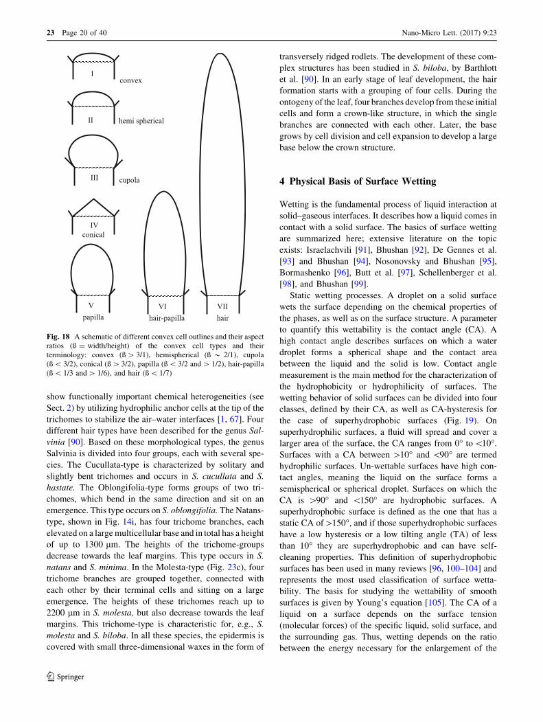
show functionally important chemical heterogeneities (see
Sect. 2) by utilizing hydrophilic anchor cells at the tip of the
trichomes to stabilize the air–water interfaces [1, 67]. Four
different hair types have been described for the genus Sal-
vinia [90]. Based on these morphological types, the genus
Salvinia is divided into four groups, each with several spe-
cies. The Cucullata-type is characterized by solitary and
slightly bent trichomes and occurs in S. cucullata and S.
hastate. The Oblongifolia-type forms groups of two tri-
chomes, which bend in the same direction and sit on an
emergence. This type occurs on S. oblongifolia. The Natans-
type, shown in Fig. 14i, has four trichome branches, each
elevated on a large multicellular base and in total has a height
of up to 1300 lm. The heights of the trichome-groups
decrease towards the leaf margins. This type occurs in S.
natans and S. minima. In the Molesta-type (Fig. 23c), four
trichome branches are grouped together, connected with
each other by their terminal cells and sitting on a large
emergence. The heights of these trichomes reach up to
2200 lm in S. molesta, but also decrease towards the leaf
margins. This trichome-type is characteristic for, e.g., S.
molesta and S. biloba. In all these species, the epidermis is
covered with small three-dimensional waxes in the form of
transversely ridged rodlets. The development of these com-
plex structures has been studied in S. biloba, by Barthlott
et al. [90]. In an early stage of leaf development, the hair
formation starts with a grouping of four cells. During the
ontogeny of the leaf, four branches develop from these initial
cells and form a crown-like structure, in which the single
branches are connected with each other. Later, the base
grows by cell division and cell expansion to develop a large
base below the crown structure.
4 Physical Basis of Surface Wetting
Wetting is the fundamental process of liquid interaction at
solid–gaseous interfaces. It describes how a liquid comes in
contact with a solid surface. The basics of surface wetting
are summarized here; extensive literature on the topic
exists: Israelachvili [91], Bhushan [92], De Gennes et al.
[93] and Bhushan [94], Nosonovsky and Bhushan [95],
Bormashenko [96], Butt et al. [97], Schellenberger et al.
[98], and Bhushan [99].
Static wetting processes. A droplet on a solid surface
wets the surface depending on the chemical properties of
the phases, as well as on the surface structure. A parameter
to quantify this wettability is the contact angle (CA). A
high contact angle describes surfaces on which a water
droplet forms a spherical shape and the contact area
between the liquid and the solid is low. Contact angle
measurement is the main method for the characterization of
the hydrophobicity or hydrophilicity of surfaces. The
wetting behavior of solid surfaces can be divided into four
classes, defined by their CA, as well as CA-hysteresis for
the case of superhydrophobic surfaces (Fig. 19). On
superhydrophilic surfaces, a fluid will spread and cover a
larger area of the surface, the CA ranges from 0� to\10�.Surfaces with a CA between [10� and \90� are termed
hydrophilic surfaces. Un-wettable surfaces have high con-
tact angles, meaning the liquid on the surface forms a
semispherical or spherical droplet. Surfaces on which the
CA is [90� and \150� are hydrophobic surfaces. A
superhydrophobic surface is defined as the one that has a
static CA of[150�, and if those superhydrophobic surfaces
have a low hysteresis or a low tilting angle (TA) of less
than 10� they are superhydrophobic and can have self-
cleaning properties. This definition of superhydrophobic
surfaces has been used in many reviews [96, 100–104] and
represents the most used classification of surface wetta-
bility. The basis for studying the wettability of smooth
surfaces is given by Young’s equation [105]. The CA of a
liquid on a surface depends on the surface tension
(molecular forces) of the specific liquid, solid surface, and
the surrounding gas. Thus, wetting depends on the ratio
between the energy necessary for the enlargement of the
I
II
III
IV
V VI VII
convex
hairhair-papillapapilla
conical
cupola
hemi spherical
Fig. 18 A schematic of different convex cell outlines and their aspect
ratios (ß = width/height) of the convex cell types and their
terminology: convex (ß[ 3/1), hemispherical (ß * 2/1), cupola
(ß\ 3/2), conical (ß[ 3/2), papilla (ß\ 3/2 and[ 1/2), hair-papilla
(ß\ 1/3 and[ 1/6), and hair (ß\ 1/7)
23 Page 20 of 40 Nano-Micro Lett. (2017) 9:23
123

surface and the gain of energy due to adsorption (Fig. 19)
[91, 106, 107]. The basis for studying equilibrium wetting
on rough surfaces was established many years ago by
Wenzel [108], Cassie and Baxter [109, 110]. The Wenzel
equation expresses a general amplification of the wetta-
bility induced by roughness and applies to a CA where
droplets are in equilibrium, but not to advancing and
receding angles of a droplet on a rough solid surface that
give rise to contact angle hysteresis (CAH). Hysteresis is
responsible for the sticking of liquids on a surface, and is
the difference of an advancing and receding angle of a
moving droplet (CAH = CAadv – CArec, shown in Fig. 19).
If a droplet moves over a solid surface, the CA at the front
of the droplet (advancing CA) is greater than that at the
back of the droplet (receding CA). However, if the droplet
rolls with little resistance, the contact angle hysteresis is
small. Advancing and receding contact angles can also be
determined when additional liquid is added to a sessile
drop and the contact line advances; if liquid is removed
from the drop, the CA decreases to a receding value before
the contact retreats.
On water-repellent surfaces, a droplet applied starts to
roll-off the surface when it is tilted to a specific angle. This
tilt angle (TA) is simply defined as the tilting angle of a
surface on which an applied drop of water starts to move.
Low TA (\10�) is characteristic for superhydrophobic and
self-cleaning surfaces. If the droplet is in Cassie–Baxter
stage [109], with air trapped between the surface and the
applied water droplet, the real contact between the droplet
and the surface is very small compared to wettable sur-
faces, on which an applied drop of water tends to spread,
and contact angle is low. In intermediate wetting stages
[111], high contact angles correspond with an increased
contact between the surface and the water droplet applied.
This phenomenon has also been described as petal-effect
[107, 112] because of its occurrence in the flower leaves
(petals) of some roses.
Dynamic interactions are bouncing, splashing, and
spreading of droplets. The impact behavior of falling drops
has been subject to studies for several liquids, such as
water, ethanol, emulsions, and various types of structured
and unstructured, solid and liquid surfaces [113, 114]. In
addition to the physicochemical surface properties and
structures, other factors determine the impact behavior of
falling drops: their velocity, size, surface tension, and
viscosity [115–117]. Falling drops can impact without any
breakup or impact with a splash or splashing, in which the
impacting drop releases smaller droplets flying away from
the point of impact. Roughly two different types of
splashing on incompliant surfaces are described, the
‘‘prompt splash’’ and the ‘‘corona splash’’. The ‘‘corona
splash’’ consists of relatively large droplets and is the
outcome of breaking up fingers developing at a flattening
drop’s rim. In ‘‘prompt splash’’ very small and fast droplets
are generated when the advancing lamella of a spreading
drop is disturbed by rough surface structures higher than a
specific fraction of the lamella, causing the lamella to
rupture locally and release small splash droplets [118].
Dynamic interactions between water droplets and the
surfaces are well researched in the different types of
splash-phenomena described, e.g., in Rioboo et al. [119],
Yarin [113], Xu et al. [118], Motzkus et al. [120], Gilet and
Bourouiba [121], and Koch and Grichnik [122]. More
recently in focus came the contact times between droplets
and surfaces: the time span a drop impacting and bouncing
on a hydrophobic surface is in contact with the surface. The
contact time as well as the shape of the rebound droplet
strongly depends on the structure and the chemistry of the
surface. A reduction of this contact time is of advantage in
several applications, e.g., anti-icing, self-cleaning, or spray
cooling [123]. But also in biological surfaces, the contact
time might be important.
Dynamic wetting processes and their control may play
an underestimated role in the interaction between plant
surfaces and rain, the maintenance of air layers under
water, or pesticide applications.
5 Superhydrophilic and Superhydrophobic PlantSurfaces
The wettability of plant surfaces plays a crucial role in their
interaction with the environment. A physical constraint of
water plants is that they must be hydrophilic, in land plants,
Liquid
Liquid
θ
θrec.θ
adv.
AirContact angle
γLA
γSAγSLSolid surface
Young’s equation: γSA−γSL = γLAcosθ
α α: Tilt angle
Tilt angle and hysteresis
Hysteresis=θadvancing−θreceding
Solid surface
Fig. 19 The schematic shows the wetting of a solid surface. cLA, cLS,
and cSA are the interfacial tensions at the boundaries between liquid
(L), solid (S), and air (A), which determine the CA of an applied
water droplet and is described by Young‘s equation. The hysteresis of
a water droplet on a tilted surface represents the adhesion of the liquid
on the surface and can be determined by measuring the tilting angle or
the advanced and receding angle of a water droplet
Nano-Micro Lett. (2017) 9:23 Page 21 of 40 23
123

the whole range from superhydrophilic to superhydropho-
bic occurs (Fig. 20).
Hydrophilic and superhydrophilic surfaces (contact
angle—if measurable—between 0� and \90�) are known
from all aquatic plants and many surfaces of land plants
which usually have a papillate cell morphology and
cuticular folds, but also from leaves with flat, tabular cells.
Surfaces covered by wax may become hydrophilic with the
erosion of theses layers (e.g., Carnegiea, Fig. 1a), a very
common process in many plants (see below). Modified wax
layers may even become superhydrophilic like in the
Atacama desert cactus Copiapoa cinerea adapted to fog
harvesting. Hydrophilic behavior in flower petals of the
daisy family (Asteraceae) and their polymer replica was
analyzed in detail by Koch et al. [124] and provides
information for biomimetic applications (see Sect. 7).
Superhydrophobicity was discovered in terrestrial
plants, and is one of the main and most obvious charac-
teristics of most vascular non-aquatic plants [1]. Superhy-
drophobic surfaces cover hundreds of millions of square
kilometers on our planet’s surface (see Sect. 1). One pre-
condition is always a hydrophobic chemistry of the surface
and hierarchical sculpturing on two to several levels
[1, 2, 7, 19]—these are the only two essentials to lead to
superhydrophobicity in organisms. In plants, it is gener-
ated—with only a few exceptions—by 3D wax crystalloids
of the morphological and chemical diversity previously
described and illustrated (Fig. 7). Superhydrophobicity in
these cases is caused merely by a sculpturing on the second
level. For reasons of stability under dynamic conditions
and to minimize mechanical damage, an additional third
hierarchical level (usually convex cell surfaces) is essen-
tial—the best example are the Lotus leaves (Fig. 21) with
contact angles[160� and a tilting angle of less than 4� [15]
(survey of recent literature in Ref. [1]). The various func-
tions of superhydrophobicity are discussed in Sect. 6.
However, there is a phenomenon that ‘‘hairy’’ surfaces with
cellular trichomes only covered by non-structured molec-
ular films also commonly cause this effect. The classical
examples are the superhydrophobic leaves of the Ladys
Mantle (Alchemilla) analyzed in detail by Otten and Her-
minghaus [125]. This is so obvious and often observed, that
the names ‘‘Ladys Mantle’’ as well as ‘‘Alchemilla’’ refer
to the superhydrophobic properties. The ‘‘Alchemilla-
type’’ of superhydrophobicity also occurs in the feathers of
many birds.
A list of extremely superhydrophobic plant surfaces was
provided by Neinhuis and Barthlott [7]. Amongst the
extremes—all measured with the same equipment under
the same conditions—are plants from most different rela-
tionships. Examples are Indian Cress (160� Tropaeolum),
grasses (161� Elymus), Ginkgo trees (161� Gingko), Lotus
(162� Nelumbo, Fig. 22c), Californian Poppy (162�Eschscholtzia), Aroids (164� Colocasia, Fig. 22a), and
Euphorbia myrsinites (162�, Fig. 22b). Some mono-
cotyledons of the family Alstroemeriaceae (‘‘Peruvian
Lilies’’) exhibit extremely high contact angles caused by
parallelly oriented wax platelets; the extreme was
Hairs and glands
Convex cells +2-D wax
Papilla cells +2-D wax
Tabular cells +2-D wax
Hydrophilicstructures
Cuticular folds +2-D wax
Convex cells +2-D wax
Convex cells +some 3-D wax
Hydrophobicstructures
Structure and wetting behavior of plant surfaces
Hairs +3-D wax
Tabular cells +3-D wax*
Convex cells +many 3-D wax
Papilla cells +3-D wax
Superhydrophobicstructures
Porous cells
Papilla cells
Hairs
Superhydrophilicstructures
Fig. 20 Four groups of plant surfaces wettability and the possible surface structures and structure combinations. The drawings used for the four
groups are correlated to specific contact angles. Both the hydrophobic and superhydrophobic surfaces can be built by convex cells with three-
dimensional waxes on it, but only a dense layer of wax crystals results in superhydrophobicity
23 Page 22 of 40 Nano-Micro Lett. (2017) 9:23
123

measured in a climbing Bomarea reaching a static contact
angle of 169� [126].
Superhydrophobicity is usually not very persistent in
plant surfaces. Leaves, with a limited life span, do usually
not last longer than one year. Persistent leathery leaves
(like in many Mediterranean and tropical climates) are
usually hydrophilic—but they may start to be hydrophobic
in their earliest developmental stages. This dynamics can
be measured in the persistent leaves of Welwitschia mir-
abilis, a single leaf growing over two centuries from its
base. They start being superhydrophobic exhibiting tubular
nonacosan-crystals which eroded after the first year and the
(a) (b) (c)5 μm10 μm5 μm
Colocasia esculenta Euphorbia myrsinites Nelumbo nucifera
Fig. 22 Extremely superhydrophobic leaf surfaces of (a) Colocasia esculenta, (Contact angle 164�) Euphorbia myrsinites (Contact angle 162�),and Lotus Nelumbo nucifera (Contact angle 162�), data from [7]. Leaves of all three species are characterized by convex (a, b) to papillose
(c) cells, covered by three-dimensional wax crystals
(a)
The lotus plant (Nelumbo nucifera) (a): removement of dirt particles by water (b, c)
SEM micrographes of the lotus leaf (d) show the papillose cells (e) and wax tubules (f) on it
(b)
(e) (f)
(c)
(d)20 μm 10 μm 1 μm
Fig. 21 Superhydrophobic and self-cleaning surface of Lotus (Nelumbo nucifera). A flowering plant of Lotus (a), a lotus leaf contaminated with
clay (b) and removal of the adhering particles by water (c). The SEM micrographs (d–f) show the lotus leaf surface in different magnifications:
(d) The papilla epidermis, (e) single cell papilla, and (f) the epicuticular nonacosan-10-ol tubules on the cell surface
Nano-Micro Lett. (2017) 9:23 Page 23 of 40 23
123

leaf becomes wettable [1]. Plums are covered by a
mechanically delicate whitish wax cover—touching it with
a finger is enough to destroy the structure and thus the
‘‘glaucous’’ color. An extreme provides the chemically
complex brilliant white waxy coating of the succulent
desert plants of the genus Dudleya (e.g., D. brittonii): it
only takes a rain drop to destroy the instable coating [66].
Dudleya is not self-healing, in contrast to other plants,
where a wax cover destroyed may regenerate within hours.
Water plants are superhydrophilic, but when they rise
above the water level they exhibit their old evolutionary
potential, e.g., the flowering stalks of the watermilfoil
Myriophyllum become superhydrophobic like the unique
flowers of Nymphoides [1]. Floating species like Salvinia
or Pistia are usually superhydrophobic. A striking example
is illustrated in Fig. 23b: the grasshopper Paulinia acumi-
nate feeds exclusively on Salvinia and optical mimics its
color and surface (camouflage)—but it also is superhy-
drophobic based on wax crystals, the same adaption to its
semiaquatic habitat as its host plant [1, 127].
Only few surfaces like lotus leaves have a very
stable superhydrophobicity. As indicated in the introduc-
tion, superhydrophobicity usually disappears in aging bio-
logical surfaces and they become wettable (compare,
Fig. 1a).
6 Functional Diversity of Plant Surfaces
The surface of plants is the critical interface for the inter-
action with the environment and fulfills many and most
different functions (Fig. 24). Aspects and literature have
been summarized by [1, 70, 71, 76, 128–130]. The surfaces
are usually multifunctional and each attempt to categorize
a bc d
e
f
g
Fig. 24 Schematic of the most important functions of the plant
boundary layer on a hydrophobic micro-structured surface: a Trans-
port barrier: limitation of uncontrolled water loss/leaching from
interior and foliar uptake, b surface wettability, c anti-adhesive, self-
cleaning properties (lotus effect): reduction of contamination,
pathogen attack, and reduction of attachment/locomotion of insects,
d signaling: cues for host-pathogens/insect recognition and epidermal
cell development, e change of optical properties, f mechanical
properties: resistance against mechanical stress and maintenance of
physiological integrity, g reduction of surface temperature by
increasing turbulent air flow over the boundary air layer (modified
after Ref. [75])
1 cm
400 μm 20 μm 10 μm
(a)
(c) (d) (e)
(b)
Fig. 23 Air-retaining surfaces of the floating fern Salvinia. a A water droplet on the upper leaf side of Salvinia biloba and b a mimicking
grasshopper Paulinia acuminata feeding on the leaf, even the grasshopper has a wax surface and is superhydrophobic. The SEM micrographs (c–
e) of the leaf surface of Salvinia show the multicellular hairs c, and in higher magnification (d) the epidermis cells, and the wax rodlets (e) on the
epidermis cells
23 Page 24 of 40 Nano-Micro Lett. (2017) 9:23
123

them is thus unrewarding [83, 131]. We simplify this in the
following seven categories.
6.1 Mechanical Properties
The cuticle itself is a highly sophisticated chemically
stable layer which serves as an elastic mechanical protec-
tive structure [74, 128]. The shape of a ripe tomato is
maintained to a high degree by its thin cuticle. Hierarchical
surface structures fulfill various mechanical tasks; they can
reduce the ability of insects to walk (e.g., fish-hooked hairs
on bean leaves against aphids, trapping surfaces in insec-
tivorous plants or flowers of Arum) or increase the ability
of insects to walk, as is the case in flowers and even orient
the visitors with cuticular folds [132]. Mechanochemical
defense trichomes are common like in nettles (Urtica) or in
Loasaceae (Fig. 14d).
6.2 Attachment
Attachment mechanisms are most common in zoochorous
fruits like burrs; complicated hooked spines, and hairs are
evolved in many groups (Asteraceae, Krameriaceae,
Pedaliaceae) [133]. A particularly refined attachment
mechanism with extractable cellulose threads occurs in the
seeds of the orchid Chiloschista [89]. Attachment plays a
role in the interaction between pollen and its disperser and
the stigma, where it becomes deposed. Climbing plants like
Ivy (Hedera) or some highly specialized water plants that
are growing on rocks in currents (e.g., Podostemaceae)
exhibit attachment devices by glue-like adhesives, which
are not really surface phenomena.
6.3 Reflection, Absorption, and Transmission
of Spectral Radiation
Reflection, absorption, and transmission of spectral radia-
tion are of crucial importance for light harvesting and
temperature control under insolation; colors as well as light
reflection play an important role in the interaction between
flowers and their pollinators.
6.3.1 Light Management
Absorption of light for photosynthesis is the precondition
for plant life. Leaves need to collect electromagnetic
radiation through photosynthesis while flowers need to
harvest radiation in order to intensify their coloration to be
more attractive for pollinators. In these processes, the
architecture of the plant surfaces plays an important role.
While self-cleaning surfaces often combine convex- or
concave-shaped epidermal cells with water-repellent 3D
waxes, light-harvesting leaf surfaces often possess only
convexly shaped epidermal cells. Specifically plants under
low-light conditions reduce the loss of light due to specular
surface reflection by increasing the transmittance of energy
via multiple reflections between the surface structures
[134]. An extreme example is the ‘‘luminescent moss’’
Schistostega living in caves with batteries of spherical cells
collecting the faintest light-like lenses [135]. However,
convexly shaped cells combined with a cuticular folding on
top are well known for petal surfaces, especially in
angiosperms [2]. These cuticular folds were thought to
reduce the surface reflection [136], act as a specific optical
signal for pollinators [2] and cause iridescence generated
through diffraction gratings [137].
6.3.2 Coloration Signals
The intriguing interaction between flowers and their pol-
linators is the reason for the evolution of most refined
coloration signals (e.g., Lloyd and Barett [138]). The
spectrum comprised virtually all grades of reflections (from
black to white) and all visible colors, even metallic-mirror
like surfaces have been evolved in the orchid Ophrys
speculum, thin layer iridescences like in our example
(Fig. 4a). To intensify their color signal, some flowers use
refined surface structures. The surface architecture of the
flowers of the so called ‘‘Johnny-jump-up’’ Viola tricolor is
an excellent model for this color intensification process.
Viola possess extreme papillated epidermal cells (aspect
ratio about 2.93) covered with a fine cuticular folding
(width about 0.26 lm). This surface topography acts as a
light-trap for incident light by reducing the specular
reflection on the surface. Via this surface design more light
passes through the cell wall. Furthermore, the extremely
steep cell walls (papilla tip angle about 26�) cause an
optimized scattering of the light within the petal (scattering
angle of about 170�). This allows the absorption of more
light by the pigments, which further results in the intensi-
fication of the color signal. Such a surface is highly
interesting for biomimetic applications, for example, in
solar panel development. Flowers also use optical signals
in the ultraviolet range, not visible to the human eye, but
visible to visiting bees: Bidens ferulifolia in Fig. 25 pro-
vides an impressive example. Large databases on ultravi-
olet reflection of flowers are published [139–141]. Usually
it is assumed that the reflection is determined only by
chemical compounds (flavonoids) within the living cells
(vacuoles), but we have recently shown that UV-patterns
are also supported by the surface structure. Another phe-
nomenon caused by surface structures is iridescence of
some leaves like in the fern Elaphoglossum (Fig. 4a). This
phenomenon seems to occur mostly under low-light
intensities of tropical rain forest understories, but is also
known in some flowering plants, e.g., the petals of some
Nano-Micro Lett. (2017) 9:23 Page 25 of 40 23
123
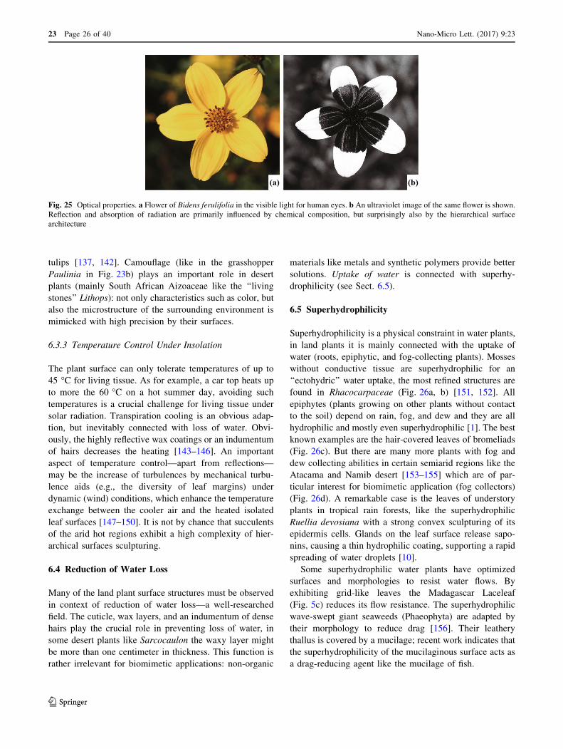
tulips [137, 142]. Camouflage (like in the grasshopper
Paulinia in Fig. 23b) plays an important role in desert
plants (mainly South African Aizoaceae like the ‘‘living
stones’’ Lithops): not only characteristics such as color, but
also the microstructure of the surrounding environment is
mimicked with high precision by their surfaces.
6.3.3 Temperature Control Under Insolation
The plant surface can only tolerate temperatures of up to
45 �C for living tissue. As for example, a car top heats up
to more the 60 �C on a hot summer day, avoiding such
temperatures is a crucial challenge for living tissue under
solar radiation. Transpiration cooling is an obvious adap-
tion, but inevitably connected with loss of water. Obvi-
ously, the highly reflective wax coatings or an indumentum
of hairs decreases the heating [143–146]. An important
aspect of temperature control—apart from reflections—
may be the increase of turbulences by mechanical turbu-
lence aids (e.g., the diversity of leaf margins) under
dynamic (wind) conditions, which enhance the temperature
exchange between the cooler air and the heated isolated
leaf surfaces [147–150]. It is not by chance that succulents
of the arid hot regions exhibit a high complexity of hier-
archical surfaces sculpturing.
6.4 Reduction of Water Loss
Many of the land plant surface structures must be observed
in context of reduction of water loss—a well-researched
field. The cuticle, wax layers, and an indumentum of dense
hairs play the crucial role in preventing loss of water, in
some desert plants like Sarcocaulon the waxy layer might
be more than one centimeter in thickness. This function is
rather irrelevant for biomimetic applications: non-organic
materials like metals and synthetic polymers provide better
solutions. Uptake of water is connected with superhy-
drophilicity (see Sect. 6.5).
6.5 Superhydrophilicity
Superhydrophilicity is a physical constraint in water plants,
in land plants it is mainly connected with the uptake of
water (roots, epiphytic, and fog-collecting plants). Mosses
without conductive tissue are superhydrophilic for an
‘‘ectohydric’’ water uptake, the most refined structures are
found in Rhacocarpaceae (Fig. 26a, b) [151, 152]. All
epiphytes (plants growing on other plants without contact
to the soil) depend on rain, fog, and dew and they are all
hydrophilic and mostly even superhydrophilic [1]. The best
known examples are the hair-covered leaves of bromeliads
(Fig. 26c). But there are many more plants with fog and
dew collecting abilities in certain semiarid regions like the
Atacama and Namib desert [153–155] which are of par-
ticular interest for biomimetic application (fog collectors)
(Fig. 26d). A remarkable case is the leaves of understory
plants in tropical rain forests, like the superhydrophilic
Ruellia devosiana with a strong convex sculpturing of its
epidermis cells. Glands on the leaf surface release sapo-
nins, causing a thin hydrophilic coating, supporting a rapid
spreading of water droplets [10].
Some superhydrophilic water plants have optimized
surfaces and morphologies to resist water flows. By
exhibiting grid-like leaves the Madagascar Laceleaf
(Fig. 5c) reduces its flow resistance. The superhydrophilic
wave-swept giant seaweeds (Phaeophyta) are adapted by
their morphology to reduce drag [156]. Their leathery
thallus is covered by a mucilage; recent work indicates that
the superhydrophilicity of the mucilaginous surface acts as
a drag-reducing agent like the mucilage of fish.
(a) (b)
Fig. 25 Optical properties. a Flower of Bidens ferulifolia in the visible light for human eyes. b An ultraviolet image of the same flower is shown.
Reflection and absorption of radiation are primarily influenced by chemical composition, but surprisingly also by the hierarchical surface
architecture
23 Page 26 of 40 Nano-Micro Lett. (2017) 9:23
123

6.6 Superhydrophobicity
Superhydrophobicity is one of the most obvious charac-
teristics of many land plant surfaces and may serve dif-
ferent functions. In particular, the structures for air
retention under water are highly complex (e.g., Salvinia,
Fig. 23); and they are often combined with a superimposed
compartmentation [1] of the whole organ surfaces from the
microscopic to the macroscopic level (Fig. 27).
(i) Unwettability per se to avoid wetting for mechan-
ical reasons: thin laminar leaves become too
heavy when wetted. This is probably an important
determination leading to the adaptation of super-
hydrophobicity in larger leaves in rain forests
(e.g., Cibotium [1])—an extreme provides the
Titan Arum Amorphophallus (Fig. 1b).
(ii) Reduction of adhesion for insects—usually con-
nected with superhydrophobicity—is caused by
epicuticular wax crystals in insect-catching plants
(e.g., Sarracenia, Nepenthes) or flowers which
trap their pollinators (Ceropegia, Aristolochia) or
to avoid unwanted nectar thieves (Fritillaria,
(a) (b) (c)
Fig. 27 Compartmentation of surfaces generating small functional units for air retention: (a) in microscopic dimension in the seed of Aeginetia
indica illustrated in Fig. 3 for temporary floating. A particularly refined compartmentation system (b, c) exhibits Salvinia cucullata in a
macroscopic dimension: the superhydrophobic leaves (diameter ca. 1.5 cm) form hood-like compartments (b), submersed in water, (c) each leaf
holds a very stable large air bubble
200 μm 10 μm
500 μm
(a) (b)
(c) (d)
Fig. 26 a The water absorbing pores of a Sphagnum squarrosum moss. b The water adsorbing porous surface of the moss Rhacocarpus
purpurascens. c The epiphytic growing Spanish moss (Tillandsia usneoides). d Water droplets in the needles of the Canary Pine (Pinus
canariensis) harvesting fog out of the trade wind clouds
Nano-Micro Lett. (2017) 9:23 Page 27 of 40 23
123
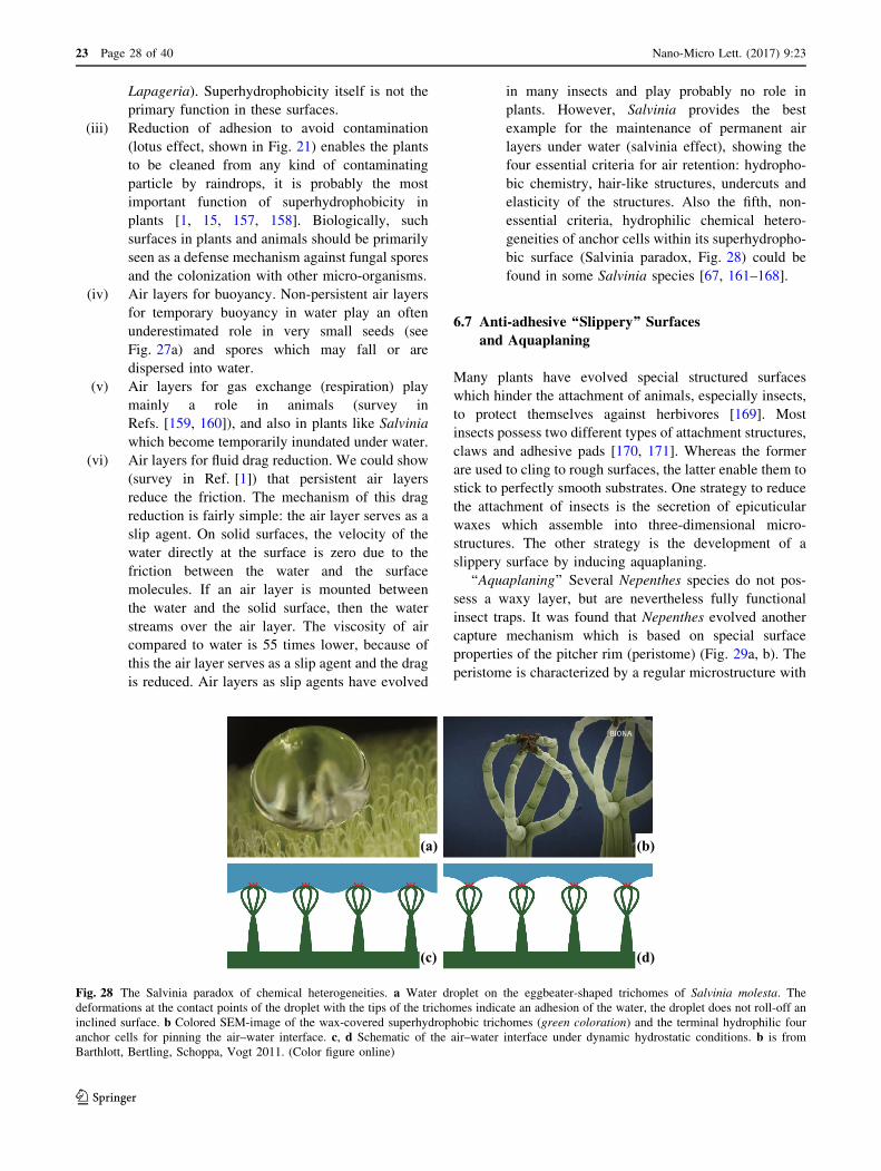
Lapageria). Superhydrophobicity itself is not the
primary function in these surfaces.
(iii) Reduction of adhesion to avoid contamination
(lotus effect, shown in Fig. 21) enables the plants
to be cleaned from any kind of contaminating
particle by raindrops, it is probably the most
important function of superhydrophobicity in
plants [1, 15, 157, 158]. Biologically, such
surfaces in plants and animals should be primarily
seen as a defense mechanism against fungal spores
and the colonization with other micro-organisms.
(iv) Air layers for buoyancy. Non-persistent air layers
for temporary buoyancy in water play an often
underestimated role in very small seeds (see
Fig. 27a) and spores which may fall or are
dispersed into water.
(v) Air layers for gas exchange (respiration) play
mainly a role in animals (survey in
Refs. [159, 160]), and also in plants like Salvinia
which become temporarily inundated under water.
(vi) Air layers for fluid drag reduction. We could show
(survey in Ref. [1]) that persistent air layers
reduce the friction. The mechanism of this drag
reduction is fairly simple: the air layer serves as a
slip agent. On solid surfaces, the velocity of the
water directly at the surface is zero due to the
friction between the water and the surface
molecules. If an air layer is mounted between
the water and the solid surface, then the water
streams over the air layer. The viscosity of air
compared to water is 55 times lower, because of
this the air layer serves as a slip agent and the drag
is reduced. Air layers as slip agents have evolved
in many insects and play probably no role in
plants. However, Salvinia provides the best
example for the maintenance of permanent air
layers under water (salvinia effect), showing the
four essential criteria for air retention: hydropho-
bic chemistry, hair-like structures, undercuts and
elasticity of the structures. Also the fifth, non-
essential criteria, hydrophilic chemical hetero-
geneities of anchor cells within its superhydropho-
bic surface (Salvinia paradox, Fig. 28) could be
found in some Salvinia species [67, 161–168].
6.7 Anti-adhesive ‘‘Slippery’’ Surfaces
and Aquaplaning
Many plants have evolved special structured surfaces
which hinder the attachment of animals, especially insects,
to protect themselves against herbivores [169]. Most
insects possess two different types of attachment structures,
claws and adhesive pads [170, 171]. Whereas the former
are used to cling to rough surfaces, the latter enable them to
stick to perfectly smooth substrates. One strategy to reduce
the attachment of insects is the secretion of epicuticular
waxes which assemble into three-dimensional micro-
structures. The other strategy is the development of a
slippery surface by inducing aquaplaning.
‘‘Aquaplaning’’ Several Nepenthes species do not pos-
sess a waxy layer, but are nevertheless fully functional
insect traps. It was found that Nepenthes evolved another
capture mechanism which is based on special surface
properties of the pitcher rim (peristome) (Fig. 29a, b). The
peristome is characterized by a regular microstructure with
(a) (b)
(c) (d)
Fig. 28 The Salvinia paradox of chemical heterogeneities. a Water droplet on the eggbeater-shaped trichomes of Salvinia molesta. The
deformations at the contact points of the droplet with the tips of the trichomes indicate an adhesion of the water, the droplet does not roll-off an
inclined surface. b Colored SEM-image of the wax-covered superhydrophobic trichomes (green coloration) and the terminal hydrophilic four
anchor cells for pinning the air–water interface. c, d Schematic of the air–water interface under dynamic hydrostatic conditions. b is from
Barthlott, Bertling, Schoppa, Vogt 2011. (Color figure online)
23 Page 28 of 40 Nano-Micro Lett. (2017) 9:23
123

radial ridges of smooth overlapping epidermal cells, which
form a series of steps towards the pitcher inside. The
peristome ridges (Fig. 29c) mostly extend into tooth-like
structures at the inner edge, in between which large glands
(extrafloral nectaries) are situated. Secretion of nectar by
the glands attracts small insects, and also leads to a
hydrophilic coverage of the surface. The plant surface
microstructure combined with hydrophilic surface chem-
istry renders the pitcher rim completely wettable. Water
droplets spread rapidly and form homogeneous thin films,
which make the peristome extremely slippery for insects.
When the peristome is wet, the fluid films prevent the
insects’ tarsal adhesive pads from making close contact
with the surface, similar to the aquaplaning of a car tire on
a wet road. In addition, the anisotropic microstructure of
the peristome surface allows interlocking of claws only
while the insect is running towards the pitcher inside, but
not on the way out [172]. Under natural conditions the
slippery water films are caused by rain, condensation, and
nectar secretion. In contrast to this, dry peristomes are not
slippery for insects. This weather-dependent variation of
peristome slipperiness leads to an intermittent and
unpredictable activation of Nepenthes pitcher traps, which
might make the evolution of specific avoidance behaviors
more difficult [173].
7 Biomimetic Application
Bionics or biomimetics describes the processes in which
structures and concepts evolved by living organisms are
taken and implemented into technologies (surveys in
[1, 11, 19]). Bionics or biomimetics is an old field (historical
survey in Ref. [11]), but functional surfaces came surpris-
ingly very late to bionic applications. The first example was
the hook-and-loop fastener by the Swiss engineer Georges de
Mestral in the 1950s, popularly known as Velcro�. It is based
on plant surfaces (burrs). The drag-reducing riblets of the
shark skin were analyzed in the 1980s [17] and together with
the self-cleaning lotus-surfaces [2, 15] ushered in a new era
of biomimetic applications—including the swimming com-
petition in Olympic games (see Sect. 7.6).
‘‘Surfaces’’ in engineering are thin boundary layers, and
they are exposed to the harsh physicochemical and
Peristom
100 μm
20 μm
100 μm1 cm(a) (b) (e)
(d)
(c)
Fig. 29 Pitcher traps of the carnivorous plant Nepenthes alata. In (a) the complete pitchers trap of N. alata and in (b) a longitudinal cut through
the trap are shown. c Parallel ridges of the hydrophilic peristome. The arrow indicates the direction toward the inside of the pitcher. d The waxy
and slippery surface inside the trap with inactive stomata. e Glands located in the digestive zone at the lower part of the trap are shown. c was
kindly provided by Holger Bohn
Nano-Micro Lett. (2017) 9:23 Page 29 of 40 23
123

mechanical influences of the environment. Thin layers, and
in particular, the biomimetic interesting hierarchical
structures, have a restricted stability and durability.
Nanocoatings play a most important and increasing eco-
nomic role (compare Ref. [174]). We live—in the literal
sense of Mark Twain’s novel—in ‘‘the gilded age’’ of
surface applications. The bulk of a technical solid is usu-
ally stable, but every homemaker knows the difference
between the durability of a silver spoon in contrast to a
silvered utensil. A medieval iron pan could still be used in
the kitchen over the next centuries—but a polymer-coated
anti-adhesive pan lasts a few years. The extremely long
lifespan of spectacle glasses is today limited by the
sophisticated and convenient anti-reflex coatings. In col-
loquial language, there is an obvious difference between
‘‘solid’’ and ‘‘superficial.’’ We need coating technologies—
but durability and persistence are major technical chal-
lenges for engineers and material scientists in a world of
decreasing natural resources which must be used
sustainably.
Section 7 is organized along using the same scheme as
in Sect. 6 on plant functionality.
7.1 Mechanical Properties
Mechanical Properties play a very important role in bio-
mimetic composite materials—but are nearly negligible for
surfaces. Technical materials available today (polymers,
metals, etc.) have often superior mechanical properties.
7.2 Attachment
Attachment mechanisms of plants were the first biomimetic
surface application and have been around since 1958 (see
above) with the Velcro� hook-and-loop fasteners. Plants,
in contrast to animals (e.g., geckos) are only a limited
source of inspirations for attachment mechanisms.
7.3 Reflection, Transmission, and Absorption
of Spectral Radiation
Reflection, transmission, and absorption of spectral radia-
tion are of great importance for biomimetics. The appli-
cations concentrate on light harvesting by bioinspired
surfaces of solar panels. An appropriate model for such
surfaces is the petals of Viola tricolor and it is related
species (see Sect. 6.3). They show a highly reduced
reflection of light, an optimized scattering within the
petals; additionally, they are superhydrophobic, showing a
contact angle of 169� [175]. Temperature control under
insolation (e.g., car tops, roofs) is another field of interest.
Coloration technologies have always been bioinspired (like
indigo and purple as ancient dyes used for millennia).
Flowers exhibit sophisticated structures to intensify colors
by hierarchical structuring. However, most inspirations are
provided by animal coloration, like the iridescence of
peacock feathers or butterfly (Morpho) wings. Butterflies
also provide ultrablack colorations [176] like the Gaboon
viper [177]. It is not by chance that the darkest technical
black (Vantablack) is produced by carbon nanotubes.
7.4 Reduction of Water Loss
Reduction of water loss is essential for terrestrial plants—
but like the many mechanical properties (see Sect. 7.1) of
minor importance for biomimetic applications. Technical
materials like an aluminum foil prevent loss better than a
cuticle.
7.5 Superhydrophilicity
Superhydrophilicity has recently become relevant for
technical applications, which are most diverse. Biomimetic
fog-collecting meshes and other devices will probably play
an increasing role in certain arid regions (e.g., Chile,
Namibia), and it was shown that a biomimetic superhy-
drophilic hierarchical structuring increases the collection
efficiency [154, 155, 178]. Koch et al. [124] have shown
that the hydrophilic Gazania petal structures provide a
model for the design of microfluidic devices for small
volume liquid transport by capillary forces; beneficial in
both fog harvesting and microfluidic devices.
Evidently, superhydrophilic surfaces are wettable, but
the water film evaporates very fast and leads to the fast
drying ‘‘no-drop’’ glasses (e.g., Alltop�) and a sophisti-
cated facade paint (StoColor Dryonic�). Drag reduction by
mucilaginous surfaces seen in fish also plays a technical
role; we have seen evidence in seaweed surfaces that plants
have also evolved a similar technology.
The form and dynamics of bouncing droplets [93, 179]
are influenced by surface chemistry and structure: super-
hydrophilic surfaces may prevent splashing, an important
effect for biomedical applications (hygiene) or agriculture
[180]. In plants, this phenomenon was analyzed in Cala-
thea zebrina, a hydrophilic tropical understory plant, and
its corresponding polymer replica (Fig. 30) by [122]. The
anti-splashing effect is promising for biomimetic
applications.
7.6 Superhydrophobicity
Superhydrophobicity, the most obvious feature of many
plant surfaces, has an increasing and most diverse role in
biomimetics as early as the 1990s (survey in Ref. [1]).
Superhydrophobicity does not exist in abiotic nature and is
one of the most important biomimetic applications under
23 Page 30 of 40 Nano-Micro Lett. (2017) 9:23
123

different aspects. Superhydrophobicity is generated in
many plants by nonacosan-10-ol tubules with a diameter of
110 nm (Fig. 13). It could be shown that carbon nanotubes
can also be used to generate superhydrophobic, even air-
retaining surfaces.
– Pure water repellency (umbrellas to textile building) is
usually applied by sprays providing a sculpturing by
nanoparticles on, e.g., textiles (which are intrinsically
hierarchically structured). It is obvious, that dry
conditions further reduce corrosion, and anti-corrosion
coatings are the focus of technical applications [181].
This also applies for anti-icing properties. A whole
range of products and techniques are available, mostly
coatings or sprays, the product names often indicate
their function (e.g., ‘NeverWet’). Dry surfaces may
allow new dimensions in textile-based architecture
owing to weight reduction during rainfall. Anti-ice and
anti-frost performance is increasing in importance
[182, 183].
– Self-cleaning Lotus Effect� surfaces are farout most
important [1] and a vast literature exists since 1997 (see
Ref. [19, 52, 184–186]). It is also one of the most
important applications in facade paints, with the added
advantage that they remain functional for several
decades.
The lotus effect is evolved in plants predominantly as a
defense mechanism against pathogens: the attachment, e.g.,
of fungal spores is dramatically reduced [187], and no
water films are available as a precondition for a
colonization or the formation of biofilms. This has an
unexpected application: in commercial agriculture all
plants treated with pesticides by spray application surfac-
tants have to be added. It was shown that the application of
the solution containing only the surfactant (without the
active ingredient) increases the chances of infection sig-
nificant [187–192]. This phenomenon will hopefully lead
to the development of a new generation of less aggressive,
bioinspired surfactants for pesticide application.
– Superhydrophobic plants surfaces, like those of Salvi-
nia, have a striking capacity to collect and adsorb
spilled oil from water [193], a basis for biomimetic
materials. Hydrophobic sand coating can be used to
control deep drainage in tailings [194].
– Air-retaining Salvinia� Effect surfaces are a recent
development, only prototypes exist [1, 195, 196]. Fluid
drag reduction of up to 30% in a hydrodynamic water
channel has been measured [197]. The main application
is seen in ship hulls: container ships transport about
80% of the global goods, and a reduction of the fuel
(oil) consumption of 125 million tons and of 395
million tons of CO2 is estimated. A novel technology
using air-retaining grids has recently been introduced
and can be combined with the existing-refined micro-
bubble technologies: micro-bubbles adhere to the grid-
surface and the air layer under the grid can be
regenerated. It is obvious that a permanent intact air
layer prevents biofouling. Oil pollution is an unfortu-
nate and unavoidable problem in our oceans. One
possible solution is to create a hydrophobic barrier at
(a) (b)
(c) (d)
20 μm
25 μm 25 μm
Fig. 30 a Calathea zebrina leaves with their light and dark green pattern. b SEM micrograph of the leaf surface (tilt angle 45�). SEM of the
replicated dark leaf areas (c) and bright leaf areas (d) in side view (90� tilt angle). The visible color differences in the leaf are mirrored in the
different shapes of the cell papillae of the light and dark surfaces areas. (Color figure online)
Nano-Micro Lett. (2017) 9:23 Page 31 of 40 23
123

the waterline of the ship to separate the ship hull from
the water interface to avoid oil from creeping into the
structures and suppressing the air.
Biomimetic drag-reducing boat surfaces based on shark
skin riblets have been applied in America’s Cup contests in
1987 and 2010; the Speedo Fastskin� swimming suits
introduced in 2000 were adopted by almost all gold medal
winners in the Olympic Games in Beijing 2008 (Fig. 31).
But riblet structures reduce the friction only about 3%, the
more complex Salvinia� Effect technologies about 30%.
Swimwear covering the body almost totally was banned in
2009; however, there is some evidence that in the Olympic
Games in Rio 2016 a refined technology of bionic swim-
ming suits combining superhydrophobic salvinia effect
areas with shark riblet areas may have been applied.
Underwater persistent air layers will probably fulfill many
other functions like gas exchange (‘‘plastron’’ in biology)
or even refined sensory functions like in the backswimmer
Notonecta [198].
7.7 Other Applications
As indicated in the preceding chapter, there are additional
functions. Like the ‘‘aquaplaning’’ effect of the surfaces of
the carnivorous Nepenthes plants which produce water
films and become slippery to trap insects [172, 199]. In
particular slippery liquid-infused surfaces (SLIPS) are of
focal interest (e.g., Ref. [200, 201]).
8 Living Prototypes: Evolution of Plant Surfacesand Biodiversity
The often used phrase ‘‘inspired by nature’’ for biomimetics is
misleading: a nuclear reactor is inspired by natural nuclear
fission or the nuclear fusion in our sun. Thus a nuclear reactor
is inspired by nature—but not bionic. Biomimetics is exclu-
sively based on living organisms and the evolution of some 10
million living prototypes (survey in Ref. [1]). Superhy-
drophobic surfaces occur exclusively in living organisms,
which evolved hierarchical structured superhydrophobic
surfaces based on a very limited selection of molecules. There
is no evidence for the occurrence of superhydrophobic sur-
faces in anorganic nature (man-made technical products are
excluded). Surfaces are the crucial interface between an
organism and its environment—3.5 billion years of mutation
and selection have created a stunning diversity in an estimated
10 million different species of plants and animals (survey in
Ref. [1]). The two basic phases with contrasting physical
constraints are life in water and life outside of water.
8.1 Water Plants
Non-vascular primary water plants evolved in water
(originally the oceans) a couple of trillion years ago and
have no true conductive tissue. The groups are phyloge-
netically very different non-related clades; they comprise
microscopic unicellular algae up to seaweeds which might
reach a height of more than 45 m in Macrocystis. The
(a) (b)
Fig. 31 Some Olympic records today are also a triumph of bionics. Mark Spitz (a) wore his famous pre-bionic swimsuit in the 1972 Olympics in
Munich. In contrast, biomimetic swimsuits covering almost the entire body were popular after 2000; the suits were based on the biomimetic drag-
reducing shark riblet technology. At the World Swimming Championship in July 2009 in Rome almost all world records were broken using full-
body biomimetic polyurethane swimsuits. Subsequently, the length of swimwear was limited by the World Swimming Federation FINA in
December 2009. But almost full-body swimwear is back today, e.g., the swimming suit of Nada Higl (b): somewhat smaller than in 2009, but still
covering large parts of the swimmers. Today, it seems to be a high-tech combination of chemically heterogenic surfaces: shark riblets (drag
reduction about 3%) and superhydrophobic salvinia effect (drag reduction about 30%) areas. For divers, drag reduction plays no role—the divers
in Rio de Janeiro 2016 were still dressed like Mark Spitz in 1972. Sources a kindly provided by the National Portrait Gallery, Smithsonian
Institution, b from Wikipedia Commons
23 Page 32 of 40 Nano-Micro Lett. (2017) 9:23
123
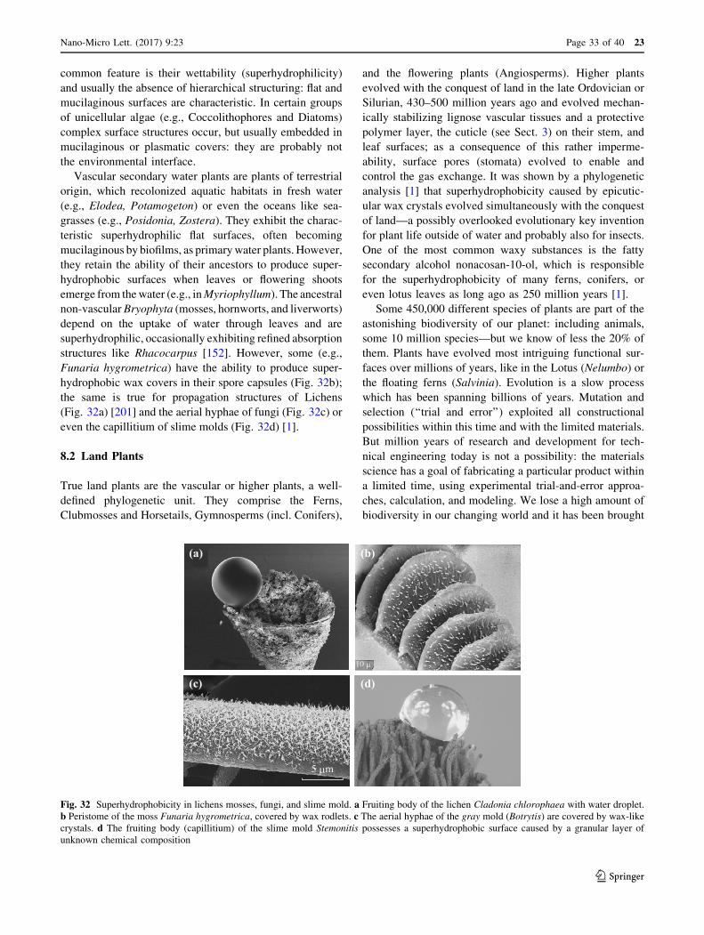
common feature is their wettability (superhydrophilicity)
and usually the absence of hierarchical structuring: flat and
mucilaginous surfaces are characteristic. In certain groups
of unicellular algae (e.g., Coccolithophores and Diatoms)
complex surface structures occur, but usually embedded in
mucilaginous or plasmatic covers: they are probably not
the environmental interface.
Vascular secondary water plants are plants of terrestrial
origin, which recolonized aquatic habitats in fresh water
(e.g., Elodea, Potamogeton) or even the oceans like sea-
grasses (e.g., Posidonia, Zostera). They exhibit the charac-
teristic superhydrophilic flat surfaces, often becoming
mucilaginous by biofilms, as primary water plants. However,
they retain the ability of their ancestors to produce super-
hydrophobic surfaces when leaves or flowering shoots
emerge from the water (e.g., inMyriophyllum). The ancestral
non-vascular Bryophyta (mosses, hornworts, and liverworts)
depend on the uptake of water through leaves and are
superhydrophilic, occasionally exhibiting refined absorption
structures like Rhacocarpus [152]. However, some (e.g.,
Funaria hygrometrica) have the ability to produce super-
hydrophobic wax covers in their spore capsules (Fig. 32b);
the same is true for propagation structures of Lichens
(Fig. 32a) [201] and the aerial hyphae of fungi (Fig. 32c) or
even the capillitium of slime molds (Fig. 32d) [1].
8.2 Land Plants
True land plants are the vascular or higher plants, a well-
defined phylogenetic unit. They comprise the Ferns,
Clubmosses and Horsetails, Gymnosperms (incl. Conifers),
and the flowering plants (Angiosperms). Higher plants
evolved with the conquest of land in the late Ordovician or
Silurian, 430–500 million years ago and evolved mechan-
ically stabilizing lignose vascular tissues and a protective
polymer layer, the cuticle (see Sect. 3) on their stem, and
leaf surfaces; as a consequence of this rather imperme-
ability, surface pores (stomata) evolved to enable and
control the gas exchange. It was shown by a phylogenetic
analysis [1] that superhydrophobicity caused by epicutic-
ular wax crystals evolved simultaneously with the conquest
of land—a possibly overlooked evolutionary key invention
for plant life outside of water and probably also for insects.
One of the most common waxy substances is the fatty
secondary alcohol nonacosan-10-ol, which is responsible
for the superhydrophobicity of many ferns, conifers, or
even lotus leaves as long ago as 250 million years [1].
Some 450,000 different species of plants are part of the
astonishing biodiversity of our planet: including animals,
some 10 million species—but we know of less the 20% of
them. Plants have evolved most intriguing functional sur-
faces over millions of years, like in the Lotus (Nelumbo) or
the floating ferns (Salvinia). Evolution is a slow process
which has been spanning billions of years. Mutation and
selection (‘‘trial and error’’) exploited all constructional
possibilities within this time and with the limited materials.
But million years of research and development for tech-
nical engineering today is not a possibility: the materials
science has a goal of fabricating a particular product within
a limited time, using experimental trial-and-error approa-
ches, calculation, and modeling. We lose a high amount of
biodiversity in our changing world and it has been brought
(a) (b)
(c) (d)
5 μm
10 μ
Fig. 32 Superhydrophobicity in lichens mosses, fungi, and slime mold. a Fruiting body of the lichen Cladonia chlorophaea with water droplet.
b Peristome of the moss Funaria hygrometrica, covered by wax rodlets. c The aerial hyphae of the gray mold (Botrytis) are covered by wax-like
crystals. d The fruiting body (capillitium) of the slime mold Stemonitis possesses a superhydrophobic surface caused by a granular layer of
unknown chemical composition
Nano-Micro Lett. (2017) 9:23 Page 33 of 40 23
123

to our attention [11, 203] that this also means the loss of
biological role models, the ‘‘living prototypes’’ for
engineers.
9 Conclusions
The diversity of plant surface structures is a result of
several billions of years of evolutionary processes. Plants
evolved a stunningly high diversity of surfaces and func-
tionality for their interaction with the environment—the
self-cleaning properties of Lotus is only one example.
Millions of years of mutation and selection, trial and error:
free information for engineers and materials scientists.
Bionics is an old field of research and development
starting around 1800—but surfaces played a surprisingly
late role for biomimetic applications, the only exception is
the hook-and-loop fasteners (‘‘Velcro�’’) in the 1950s
based on burrs. The publication of the lotus effect in 1997
[15] created awareness by engineers and materials scien-
tists, terms like ‘‘superhydrophobicity’’ came into use in
the last two decades and opened a new era in surface
technologies (survey in Ref. [1]). Surfaces play an
increasing roll, the global market for nanocoatings is esti-
mated to reach 14.2 billion US dollars by 2019 [173].
Biological surfaces have provided a remarkable number
of innovations in the last three decades. Surface tech-
nologies have been largely influenced by research on bio-
logical interfaces and came rather late into focus of
technical innovations. All data indicate we are only in the
beginning of a new era of biologically inspired surface
technologies.
Understanding biological surfaces is crucial—we have
shown that we are still in the beginning of this process. But
we are also in the beginning of a dramatic loss of biodi-
versity in the Anthropocene. Some 10 million different
species (possible biological role models) exist—We lose a
high amount of biodiversity in our changing world and it
has been brought to our attention [11, 202] that this also
means the loss of biological role models, the ‘‘living pro-
totypes’’ for engineers. Bionics is another intrinsic value to
the diversity of life which should be treasured.
Acknowledgements This survey is based on over three decades of
research on surfaces by a large working group with ever changing
members: first and foremost, we acknowledge all the students who
had successful theses in this field. Their names can be found in the
papers quoted in the references. We acknowledge the discussions with
colleagues and friends; in particular, we would like to thank Horst
Bleckmann (Bonn), Alfred Leder, Martin Brede (both Rostock),
Thomas Schimmel, Stefan Walheim, Markus Moosmann, Torsten
Scherer (all KIT Karlsruhe), Walter Erdelen (former UNESCO Paris),
Anna-Julia Schulte (Euskirchen), Daud Rafiqpoor, Birte Bohnlein,
Peter Hager (all Bonn), Gerhard Gottsberger (Ulm), Stanislav Gorb
(Kiel), Georg Noga (Bonn), and Maximilian Weigend (Bonn).
Technical assistance was provided over the years by Hans-Jurgen
Ensikat, the late Wolfgang Roden, Alexandra Runge, Bernd Haesel-
ing, and Danica Christensen. Our work in Bionics and Biodiversity
was supported by the Deutsche Bundesstiftung Umwelt DBU, the
German Research Council DFG, the Federal Ministry for Science and
Education BMBF, and the Academy of Science and Literature in
Mainz.
Open Access This article is distributed under the terms of the
Creative Commons Attribution 4.0 International License (http://crea
tivecommons.org/licenses/by/4.0/), which permits unrestricted use,
distribution, and reproduction in any medium, provided you give
appropriate credit to the original author(s) and the source, provide a
link to the Creative Commons license, and indicate if changes were
made.
References
1. W. Barthlott, M. Mail, C. Neinhuis, Superhydrophobic hierar-
chically structured surfaces in biology: evolution, structural
principles and biomimetic applications. Philos. Trans. R. Soc. A
374(2073), 20160191 (2016). doi:10.1098/rsta.2016.0191
2. W. Barthlott, N. Ehler, Raster-Elektronenmikroskopie der Epi-
dermis-Oberflachen von Spermatophyten. Trop. Subtrop.
Pflanzenwelt 19, 1–105 (1977)
3. S. Blackmore, K. Ferguson (eds.), in Pollen and Sres: Form and
Function, vol. 12. Linnean Society Symposium Series (Aca-
demic Press, London, 1986)
4. S.N. Agashe, E. Caulton, Pollen and Spores: Application with
Special Emphasis on Aerobiology and Allergy (Taylor Francis
Inc., New York, 2009)
5. K. Koch, B. Bhushan, W. Barthlott, Multifunctional plant sur-
faces and smart materials, in Springer Handbook of Nanotech-
nology, 3rd edn., ed. by B. Bhushan (Springer, Heidelberg,
2010), pp. 1399–1436
6. B. Bhushan, Springer Handbook of Nanotechnology, 3rd edn.
(Springer, Heidelberg, 2010)
7. C. Neinhuis, W. Barthlott, Characterization and distribution of
water-repellent, self-cleaning plant surfaces. Ann. Bot. Lond.
79, 667–677 (1997). doi:10.1006/anbo.1997.0400
8. S. Porembski, B. Martens-Aly, W. Barthlott, Surface/volume-
rations of plants with special consideration of succulents. Beitr.
Biol. Pflanzen 66, 189–209 (1992)
9. J.M. Suttie, S.G. Reynolds, C. Batello, Grasslands of the World
(Food and Agricultural Organisations of the UN, Rome, 2005)
10. K. Koch, I.C. Blecher, G. Konig, S. Kehraus, W. Barthlott, The
superhydrophilic and superoleophilic leaf surface of Ruellia
devosiana (Acanthaceae): a biological model for spreading of
water and oil on surfaces. Funct. Plant Biol. 36, 339–350 (2009).
doi:10.1071/FP08295
11. W. Barthlott, D. Rafiqpoor, W. Erdelen, Bionics and Biodiver-
sity- Bio-Inspired Technical Innovation for a Sustainable Future,
in Biomimetic Research for Architecture and Building Con-
struction-Biological Design and Integrative Structures, ed. by J.
Knippers, K. Nickel, T. Speck (Springer, Berlin, 2016)
12. R.L. Ripley, B. Bhushan, Bioarchitecture: bioinspired art and
architecture—a perspective. Philos. Trans. R. Soc. A 374,
20160192 (2016). doi:10.1098/rsta.2016.0192
13. R.H. France, Die Pflanze als Erfinder. Stuttgart, Germany:
Franckh’sche Verlagshandlung (Engl. edition: Plants as inven-
tors. London: Simpkin and Marshall, 1920)
14. J.C. Robinette (ed.), Living Prototypes–the Key to New Tech-
nology, in Proceeding of the Symposium. (Wright Air Devel-
opment Division, 13–15 September 1960)
23 Page 34 of 40 Nano-Micro Lett. (2017) 9:23
123

15. W. Barthlott, C. Neinhuis, Purity of the sacred lotus, or escape
from contamination in biological surfaces. Planta 202, 1–8
(1997). doi:10.1007/s004250050096
16. H.C. von Baeyer, The Lotus effect. The Sciences 40(1), 12–15
(2000). doi:10.1002/j.2326-1951.2000.tb03461.x
17. D.W. Bechert, A. Dinkelacker, W.-E. Reif, On the fluid dynamic
of the shark skin. Bull. Am. Phys. Soc. (1983). doi:10.1002/
cben.201400033
18. D.W. Bechert, M. Bruse, W. Hage, R. Meyer, Fluid mechanics
of biological surfaces and their technological application.
Naturwissenschaften 87, 157–171 (2000). doi:10.1007/
s001140050696
19. B. Bhushan, Biomimetics-Bioinspired Hierarchical-Structured
Surfaces for Green Science and Technology, 2nd edn. (Springer,
Heidelberg, 2016)
20. P. Forbes, The Gecko’s Foot (Fourth Estate, London, 2005)
21. W. Barthlott, M. Mail, B. Bhushan, K. Koch, Plant Surfaces:
Structures and Functions for Biomimetic Applications, in
Springer Handbook of Nanotechnology, 4th edn., ed. by B.
Bhushan (Springer, Heidelberg, 2017)
22. W. Barthlott, C. Neinhuis, D. Cutler, F. Ditsch, I. Meusel, I.
Theisen, H. Wilhelmi, Classification and terminology of plant
epicuticular waxes. Bot. J. Linnean Soc. 126, 237–260 (1998).
doi:10.1111/j.1095-8339.1998.tb02529.x
23. A. Dommisse, J. Wirtz, K. Koch, K. Wandelt, W. Barthlott, T.
Kolter, Synthesis of Snonacosan-10-ol, the main component of
plant surface tubular wax crystals. Eur. J. Org. Chem. 2007,
3508–3511 (2007). doi:10.1002/ejoc.200700262
24. M. Riederer, C. Markstadter, Cuticular Waxes: A Critical
Assessment of Current Knowledge, in Plant Cuticles an Inte-
grated Functional Approach, ed. by G. Kerstiens (University
Scientific, Oxford, 1996), pp. 189–200
25. L. Kunst, A.L. Samuels, Biosynthesis and secretion of plant
cuticular wax. Prog. Lip. Res. 42, 51–80 (2003). doi:10.1016/
S0163-7827(02)00045-0
26. C.E. Jeffree, The Fine Structure of the Plant Cuticle, in Biology
of the Plant Cuticle, ed. by M. Riederer, C. Muller (Blackwell,
Oxford, 2006), pp. 11–125
27. R. Jetter, S. Schaffer, Chemical composition of the Prunus
laurocerasus leaf surface. Dynamic changes of the epicuticular
wax film during leaf development. Plant Phys. 126, 1725–1737
(2001). doi:10.1104/pp.126.4.1725
28. K. Koch, W. Barthlott, S. Koch, A. Hommes, K. Wandelt, W.
Mamdouh, S. De-Feyter, P. Broekmann, Structural analysis of
wheat wax (Triticum aestivum, c.v. ‘Naturastar’ L.): from the
molecular level to three dimensional crystals. Planta 223,
258–270 (2005). doi:10.1007/s00425-005-0081-3
29. R. Jetter, L. Kunst, A.L. Samuels, Composition of Plant Cutic-
ular Waxes, in Biology of the Plant Cuticle, in Annual Plant
Reviews, ed. by M. Riederer, C. Muller (Blackwell, Oxford,
2006), pp. 145–175
30. E.A. Baker, Chemistry and Morphology of Plant Epicuticular
Waxes, in The Plant Cuticle, ed. by D.F. Cutler, K.L. Alvin,
C.E. Price (Academic Press, London, 1982), pp. 139–165
31. T. Shepherd, D.W. Griffiths, The effects of stress on plant
cuticular waxes. New Phytol. 171, 469–499 (2006). doi:10.1111/
j.1469-8137.2006.01826.x
32. K. Koch, K.D. Hartmann, L. Schreiber, W. Barthlott, C. Nein-
huis, Influence of air humidity on epicuticular wax chemical
composition, morphology and wettability of leaf surfaces.
Environ. Exp. Bot. 56, 1–9 (2006). doi:10.1016/j.envexpbot.
2004.09.013
33. C. Markstadter, W. Federle, R. Jetter, M. Riederer, B. Holl-
dobler, Chemical composition of the slippery epicuticular wax
blooms on Macaranga Thouars. (Euphorbiaceae) ant-plants.
Chemoecology 10, 33–40 (2000). doi:10.1007/s000490050005
34. M. Riedel, A. Eichner, R. Jetter, Slippery surfaces of carnivo-
rous plants: composition of epicuticular wax crystals in
Nepenthes alata Blanco pitchers. Planta 218, 87–97 (2003).
doi:10.1007/s00425-003-1075-7
35. M. Wen, C. Buschhaus, R. Jetter, Nanotubules on plant surfaces:
chemical composition of epicuticular wax crystals on needles of
Taxus baccata L. Phytochemistry 67, 1808–1817 (2007). doi:10.
1016/j.phytochem.2006.01.018
36. H. Ensikat, C. Neinhuis, W. Barthlott, Direct access to plant
epicuticular wax crystals by a new mechanical isolation
method. Int. J. Plant Sci. 161, 143–148 (2000). doi:10.1086/
314234
37. D. Frolich, W. Barthlott, Die mikromorphologie der epicuticu-
laren wachse und das system der monocotylen. Trop. Subtrop.
Pflanzenwelt 63, 1–135 (1988)
38. N.D. Hallam, B.E. Juniper, The Anatomy of the Leaf surface, in
The Ecology of Leaf Surface Micro-organisms, ed. by T.F.
Preece, C.H. Dickinson (Academic Press, London, 1971),
pp. 3–37
39. C.E. Jeffree, The Cuticle, Epicuticular Waxes and Trichomes of
Plants, with Reference to Their Structure, Functions and Evo-
lution, in Insects and the Plant Surface, ed. by B.E. Juniper, R.
Southwood (Edward Arnold, London, 1986), pp. 23–63
40. K. Koch, C. Neinhuis, H.J. Ensikat, W. Barthlott, Self-assembly
of epicuticular waxes on living plant surfaces imaged by atomic
force microscopy (AFM). J. Exp. Bot. 55, 711–718 (2004).
doi:10.1093/jxb/erh077
41. K. Koch, H.J. Ensikat, The hydrophobic coatings of plant sur-
faces: epicuticular wax crystals and their morphologies, crys-
tallinity and molecular self-assembly. Micron 39, 759–772
(2008). doi:10.1016/j.micron.2007.11.010
42. H.J. Ensikat, B. Boese, W. Mader, W. Barthlott, K. Koch,
Crystallinity of plant epicuticular waxes: electron and X-ray
diffraction studies. Chem. Phys. Lipids 144, 45–59 (2006).
doi:10.1016/j.chemphyslip.2006.06.016
43. K. Koch, A. Dommisse, C. Neinhuis, W. Barthlott, Self-
assembly of Epicuticular Waxes on Living Plant Surfaces by
Atomic Force Microscopy, in Scanning Tunneling Microscopy/
Spectroscopy and Related Techniques, ed. by P.M. Koenraad,
M. Kemerink (American Institute of Physics, Melville, 2003),
pp. 457–460
44. P.J. Holloway, C.E. Jeffree, E.A. Baker, Structural determina-
tion of secondary alcohols from plant epicuticular waxes. Phy-
tochemisty 15, 1768–1770 (1976). doi:10.1016/S0031-
9422(00)97477-6
45. R. Jetter, M. Riederer, In vitro reconstitution of epicuticular wax
crystals: formation of tubular aggregates by long chain sec-
ondary alkanediols. Bot. Acta 108, 111–120 (1995). doi:10.
1111/j.1438-8677.1995.tb00840.x
46. I. Meusel, C. Neinhuis, C. Markstadter, W. Barthlott, Chemical
composition and recrystallization of epicuticular waxes: coiled
rodlets and tubules. Plant Biol. 2, 462–470 (2000). doi:10.1055/
s-2000-5961
47. W. Barthlott, I. Theisen, T. Borsch, C. Neinhuis, Epicuticular
Waxes and Vascular Plant Systematics: Integrating Micromor-
phological and Chemical Data, in Deep Morphology: Toward a
Renaissance of Morphology in Plant Systematics, ed. by T.F.
Stuessy, V. Mayer, E. Horandl (Reg. Veg. Gantner Verlag,
Ruggell, 2003), pp. 457–460
48. I. Meusel, C. Neinhuis, C. Markstadter, W. Barthlott,
Ultrastructure, chemical composition, and recrystallization of
epicuticular waxes: transversely ridged rodlets. Can. J. Bot. 77,
706–720 (1999). doi:10.1139/cjb-77-5-706
49. International Union of Crystallography, Report of the Executive
Committee for 1991. Acta Crystalogr. A 48, 922–946 (1992).
doi:10.1107/S0108767392008328
Nano-Micro Lett. (2017) 9:23 Page 35 of 40 23
123

50. C.E. Jeffree, E.A. Baker, P.J. Holloway, Ultrastructure and
recrystallization of plant epicuticular waxes. New Physiol. 75,
539–549 (1975). doi:10.1111/j.1469-8137.1975.tb01417.x
51. R. Jetter, M. Riederer, Epicuticular crystals of nonacosan-10-ol:
in vitro reconstitution and factors influencing crystal habits.
Planta 195, 257–270 (1994). doi:10.1007/BF00199686
52. K. Koch, A. Dommisse, W. Barthlott, Chemistry and crystal
growth of plant wax tubules of Lotus (Nelumbo nucifera) and
Nasturtium (Tropaeolum majus) leaves on technical substrates.
Crys. Growth Des. 6, 2571–2578 (2006). doi:10.1021/
cg060035w
53. G.M. Whitesides, M. Boncheva, Beyond molecules: self-
assembly of mesoscopic and macroscopic components. Proc.
Nat. Acad. Sci. USA 99(8), 4769–4774 (2002). doi:10.1073/
pnas.082065899
54. J. Zhang, W. Zhong-Lin, J. Liu, C. Shaowei, G. Liu, Self-
assembled Nanostructures (Kluwer Academic Publishers, New
York, 2003)
55. N. Boden, P.J.B. Edwards, K.W. Jolley, C. Neinhuis, Self-
assembly and Self-organization in Micellar Liquid Crystals, in
Structure and Dynamics of Strongly Interacting Colloids and
Supermolecular Aggregates in Solutions, ed. by S.H. Chen, J.S.
Huang, P. Tartaglia (Kluwer Academic Publishers, Dordrecht,
1992)
56. C. Neinhuis, K. Koch, W. Barthlott, Movement and regeneration
of epicuticular waxes through plant cuticles. Planta 213,
427–434 (2001). doi:10.1007/s004250100530
57. D. Dorset, Development of lamellar structures in natural waxes-
an electron diffraction investigation. J. Phys. D 32, 1276–1280
(1999). doi:10.1088/0022-3727/32/11/315
58. B. Bhushan, K. Koch, Y.C. Jung, Biomimetic hierarchical
structure for self-cleaning. Appl. Phys. Lett. 93, 093101 (2008).
doi:10.1063/1.2976635
59. B. Bhushan, K. Koch, Y.C. Jung, Nanostructures for superhy-
drophobicity and low adhesion. Soft Matter 4, 1799–1804
(2008). doi:10.1039/b808146h
60. S. De Feyter, F.C. De Schryver, Self-assembly at the liquid/solid
interface: STM reveals. J. Phys. Chem. B 109, 4290–4302
(2005). doi:10.1021/jp045298k
61. F.C. Meldrum, S. Ludwigs, Template-directed control of crystal
morphologies. Macromol. Biosci. 7, 152–162 (2007). doi:10.
1002/mabi.200600191
62. K. Koch, A. Dommisse, A. Niemietz, W. Barthlott, K. Wandelt,
Nanostructure of epicuticular plant waxes: self-assembly of wax
tubules. Surf. Sci. 603, 1961–1968 (2009). doi:10.1016/j.susc.
2009.03.019
63. A. Fahn, Structure and function of secretory cells. Adv. Bot.
Res. 31, 37–75 (2000). doi:10.1016/S0065-2296(00)31006-0
64. G.J. Wagner, E. Wang, R.W. Shephers, New approaches for
studying and exploiting an old protuberance, the Plant Tri-
chome. Ann. Bot. 93, 3–11 (2004). doi:10.1093/aob/mch011
65. E. Wollenweber, The distribution and chemical constituents of
the farinose exudates in gymnogrammoid ferns. Am. Fern J. 68,
13–28 (1978). doi:10.2307/1546411
66. W. Barthlott, E. Wollenweber, Zur Feinstruktur, Chemie und
taxonomischen Signifikanz epicuticularer Wachse und ahnlicherSekrete. Tropische und subtropische Pflanzenwelt 32, 7–67
(1981)
67. W. Barthlott, T. Schimmel, S. Wiersch, K. Koch, M. Brede
et al., The Salvinia paradox: superhydrophobic surfaces with
hydrophilic pins for air retention under water. Adv. Mater. 22,
2325–2328 (2010). doi:10.1002/adma.200904411
68. P.J. Holloway, Plant Cuticles: Physicochemical Characteristics
and Biosynthesis, in Air Pollution and the Leaf Cuticle, ed. by
K.E. Percy, J.N. Cape, R. Jagels, C.J. Simpson (Springer, Berlin,
1994), pp. 1–13
69. P.E. Kolattukudy, Plant cuticle and suberin. eLS (2001). doi:10.
1038/npg.els.0001920
70. J.T. Martin, B.E. Juniper, The Cuticles of Plants (Edward
Arnold, London, 1970)
71. D.F. Cutler, K.L. Alvin, C.E. Price, The Plant Cuticle (Aca-
demic Press, London, 1982)
72. G. Kerstiens, Plant Cuticles: An Integrated Functional
Approach (Bios Scient Pub, Oxford, 1996)
73. M. Riederer, C. Muller, Biology of the Plant Cuticle (Blackwell,
Oxford Pub, Oxford, 2006)
74. H. Bargel, C. Neinhuis, Tomato (Lycopersicon esculentum
Mill.) fruit growth and ripening as related to the biomechanical
properties of fruit skin and isolated cuticle. J. Exp. Bot. 56,
1049–1060 (2005). doi:10.1093/jxb/eri098
75. H. Bargel, K. Koch, Z. Cerman, C. Neinhuis, Structure-function
relationships of the plant cuticle and cuticular waxes-a smart
material? Funct. Plant Biol. 33, 893–910 (2006). doi:10.1071/
FP06139
76. W. Barthlott, Scanning Electron Microscopy of the Epidermal
Surface in Plants, in Application of the Scanning EM in Tax-
onomy and Functional Morphology, Systematics associations’
special volume, ed. by D. Claugher (Clarendon Press, Oxford,
1990), pp. 69–94
77. W. Barthlott, Morphogenese und mikromorphologie komplexer
cuticular-faltungsmuster an bluten-trichomen von antirrhinum
L. (Scrophulariaceae). Ber. Dt. Bot. Ges. 93, 379–390 (1980)
78. L.H.P. Jones, K.A. Handreck, Silica in soils, plants, and animals.
Adv. Agron. 19, 107–149 (1967). doi:10.1016/S0065-
2113(08)60734-8
79. A.G. Sangster, M.J. Hudson, H.J. Tubb, Silicon Deposition in
Higher Plants, in Silicon in Agriculture, ed. by L.E. Datnoff,
G.H. Snyder, G.H. Korndorfer (Elsevier, Amsterdam, 2001),
pp. 85–114
80. H.-J. Ensikat, T. Geisler-Wierwille, M. Weigend, A first report
of hydroxylated apatite as structural biomineral in Loasaceae-
plants teeth against herbivores. Sci. Rep. 6, 26073 (2016).
doi:10.1038/srep26073
81. R.K. Saeedur, Calcium Oxalate in Biological Systems (CRC
Press, Boca Raton, 1995)
82. K. Koch, B. Bhushan, W. Barthlott, Diversity of structure,
morphology and wetting of plant surfaces. Soft Matter 4,
1943–1963 (2008). doi:10.1039/b804854a
83. K. Koch, B. Bhushan, W. Barthlott, Multifunctional surface
structures of plants: an inspiration for biomimetics. Prog. Mater
Sci. 54, 137–178 (2009). doi:10.1016/j.pmatsci2008.07.003
84. C. Martin, B.J. Glover, Functional aspects of cell patterning in
aerial epidermis. Curr. Opin. Plant Biol. 10, 70–82 (2007).
doi:10.1016/j.pbi.2006.11.004
85. C.A. Brewer, W.K. Smith, T.C. Vogelmann, Functional inter-
action between leaf trichomes, leaf wettability and the optical
properties of water droplets. Plant, Cell Environ. 14, 955–962
(1991). doi:10.1111/j.1365-3040.1991.tb00965.x
86. E. Rodriguez, P.L. Healey, I. Mehta, Biology and Chemistry of
Plant Trichomes (Plenum Press, New York, 1984)
87. H.D. Behnke, Plant Trichomes-structure and Ultrastructure:
General Terminology, Taxonomic Applications, and Aspects of
Trichome Bacterial Interaction in Leaf Tips of Dioscorea, in
Biology and Chemistry of Plant Trichomes, ed. by E. Rodriguez,
P.L. Healey, I. Mehta (Plenum Press, New York, 1984),
pp. 1–21
88. W. Barthlott, D. Hunt, Seed-Diversity in Cactaceae Subfam.
Cactoideae, in Succulent Plant Research, vol. 5, ed. by D. Hundt
(Milborne Port, Sherborne, 2000)
89. W. Barthlott, B. Große-Veldmann, N. Korotkova, Orchid seed
diversity: a scanning electron microscopy survey. Englera 32,
1–244 (2014)
23 Page 36 of 40 Nano-Micro Lett. (2017) 9:23
123

90. W. Barthlott, S. Wiersch, Z. Colic, K. Koch, Classification of
trichome types within species of the water fern Salvinia, and
ontogeny of the egg-beater trichomes. Botany-Botanique 87,
830–836 (2009). doi:10.1139/B09-048
91. J.N. Israelachvili, Intermolecular and Surface Forces, 2nd edn.
(Academic Press, London, 1992)
92. B. Bhushan, Introduction to Tribology, 2nd edn. (Wiley, New
York, 2013)
93. P.G. De Gennes, F. Brochard-Wyart, D. Quere, Capillarity and
Wetting Phenomena: Drops, Bubbles, Pearls, Waves (Springer,
New York, 2004)
94. B. Bhushan, Y.C. Jung, Wetting, adhesion and friction of
superhydrophobic and hydrophilic leaves and fabricated
micro/nanopatterned surfaces. J. Phys.: Condens. Matter 20,
225010 (2008). doi:10.1088/0953-8984/20/22/225010
95. M. Nosonovsky, B. Bhushan, Green Tribology, Biomimetics,
Energy Conservation and Sustainability (Springer, Berlin, 2012)
96. E.Y. Bormashenko, Wetting of Real Surfaces, in De Gruyter
Studies in Mathematical Physics, vol. 19 (Verlag Walter de
Gruyter, 2013), pp. 187
97. H.J. Butt, K. Graf, M. Kappl, Physics and Chemistry of inter-
faces, 3rd edn. (Wiley-VCH, Weinheim, 2013)
98. F. Schellenberger, N. Encinas, D. Vollmer, H.-J. Butt, How
water advances on superhydrophobic surfaces. Phys. Rev. Lett.
116, 096101 (2016). doi:10.1103/PhysRevLett.116.096101
99. B. Bhushan, Nanotribology and Nanomechanics-an Introduc-
tion, 3rd edn. (Springer, Heidelberg, 2011)
100. C.W. Extrand, Model for contact angle and hysteresis on rough
and ultraphobic surfaces. Langmuir 18, 7991–7999 (2002).
doi:10.1021/la025769z
101. Y.C. Jung, B. Bhushan, Wetting behavior during evaporation
and condensation of water microdroplets on superhydrophobic
patterned surfaces. J. Microsc. 229, 127–140 (2008). doi:10.
1111/j.1365-2818.2007.01875.x
102. B. Bhushan, Y.C. Jung, Wetting study of patterned surfaces for
superhydrophobicity. Ultramicroscopy 107, 1033–1041 (2007).
doi:10.1016/j.ultramic.2007.05.002
103. P. Roach, N.J. Shirtcliffe, M.I. Newton, Progress in superhy-
drophobic surface development. Soft Matter 4, 224–240 (2008).
doi:10.1039/B712575P
104. X. Zhang, F. Shi, J. Niu, Y. Jiang, Z. Wang, Superhydrophobic
surfaces: from structural control to functional application.
J. Mater. Chem. 18, 62–633 (2008). doi:10.1039/B711226B
105. C.W. Extrand, Origins of wetting. Langmuir 32(31), 7697–7706
(2016). doi:10.1021/acs.langmuir.6b01935
106. A.V. Adamson, Physical Chemistry of Surfaces (Wiley, New
York, 1990)
107. M. Nosonovsky, B. Bhushan, Lotus Versus Rose: Biomimetic
Surface Effects, in Green Tribology, Biomimetics, Energy
Conservation and Sustainability, ed. by M. Nosonovsky, B.
Bhushan (Springer, Berlin, 2012), pp. 25–40
108. R.N. Wenzel, Resistance of solid surfaces to wetting by
water. Ind. Eng. Chem. 28, 988 (1936). doi:10.1021/ie50320a024
109. A.B.D. Cassie, S. Baxter, Wettability of porous surfaces.
Trans. Faraday Soc. 40, 546–551 (1944). doi:10.1039/
tf9444000546
110. A. Marmur, Wetting on hydrophobic rough surfaces: to be
heterogeneous or not to be? Langmuir 19, 8343–8348 (2003).
doi:10.1021/la0344682
111. E.Y. Bormashenko, Wetting transitions on biomimetic surfaces.
Philos. Trans. R. Soc. A 368, 4695–4711 (2010). doi:10.1098/
rsta.2010.0121
112. L. Feng, Y. Zhang, J. Xi, Y. Zhu, N. Wang, F. Xia, L. Jiang,
Petal effect: a superhydrophobic state with high adhesive force.
Langmuir 24, 4114–4119 (2008). doi:10.1021/la703821h
113. A.-L. Yarin, Drop impact dynamics: splashing, spreading,
receding, bouncing. Ann. Rev. Fluid Mech. 38, 159–192 (2006).
doi:10.1146/annurev.fluid.38.050304.092144
114. C. Josserand, S.T. Thoroddsen, Drop impact on a solid surface.
Ann. Rev. Fluid Mech. 48, 365–391 (2015). doi:10.1146/
annurev-fluid-122414-034401
115. J. Fukai, Y. Shiiba, T. Yamamoto, O. Miyatake, D. Poulikakos,
C.M. Megaridis, Z. Zhao, Wetting effects on the spreading of a
liquid droplet surface: experiment and modeling colliding with a
flat. Phys. Fluids 7, 236–247 (1995). doi:10.1063/1.868622
116. C. Mundo, M. Sommerfeld, C. Tropea, Droplet-wall collisions:
experimental studies of the deformation and breakup process.
Int. J. Multiph. Flow 21, 151–173 (1995). doi:10.1016/0301-
9322(94)00069-V
117. T. Mao, D. Kuhn, H. Tran, Spread and rebound of liquid dro-
plets upon impact on flat surfaces. AIChE J. 43, 2169–2179
(1997). doi:10.1002/aic.690430903
118. L. Xu, L. Barcos, S.R. Nagel, Splashing of liquids: interplay of
surface roughness with surrounding gas. Phys. Rev. E 76,
066311 (2007). doi:10.1103/PhysRevE.76.066311
119. R. Rioboo, C. Tropea, M. Marengo, Outcomes from a drop
impact on solid surfaces. At. Sprays 11, 155–165 (2001). doi:10.
1615/AtomizSpr.v11.i2.40
120. C. Motzkus, F. Gensdarmes, E. Gehin, Study of the coalescence/
splash threshold of droplet impact on liquid films and its rele-
vance in assessing airborne particle release. J. Colloid Interface
Sci. 362, 540–552 (2011). doi:10.1016/j.jcis.2011.06.031
121. T. Gilet, L. Bourouiba, Rain-induced ejection of pathogens from
leaves: revisiting the hypothesis of splash-on-film using high-
speed visualization. Integr. Comp. Biol. 54(6), 974–984 (2014).
doi:10.1093/icb/icu116
122. K. Koch, R. Grichnik, Influence of surface structure and
chemistry on water droplet splashing. Philos. Trans. R. Soc. A
374, 20160183 (2016). doi:10.1098/rsta.2016.0183
123. Y. Liu, L. Moevius, X. Xu, T. Qian, J.M. Yeomans, Z. Wang,
Pancake bouncing on superhydrophobic surfaces. Nat. Phys.
10(7), 515–519 (2014). doi:10.1038/nphys2980
124. K. Koch, M. Bennemann, H.F. Bohn, D.C. Albach, W. Barthlott,
Surface microstructures of daisy florets (Asteraceae) and char-
acterization of their anisotropic wetting. Bioinspir. Biomim. 8,
036005 (2013). doi:10.1088/1748-3182/8/3/036005
125. A. Otten, S. Herminghaus, How plants keep dry: a physicist’s
point of view. Langmuir 20, 2405–2408 (2004). doi:10.1021/
la034961d
126. H.-J. Ensikat, P. Ditsche-Kuru, C. Neinhuis, W. Barthlott,
Superhydrophobicity in perfection: the outstanding properties of
the lotus leaf. Beilstein J. Nanotechnol. 2, 152–161 (2011).
doi:10.3762/bjnano.2.19
127. W. Barthlott, K. Riede, M. Wolter, Mimicry and ultrastructural
analogy between the semi-aquatic grasshopper Paulinia acumi-
nata (Orthoptera: Pauliniidae) and its foodplant, the water-fern
Salvinia auriculata (Filicateae: Salviniaceae). Amazoniana 13,
47–58 (1994)
128. H. Bargel, W. Barthlott, K. Koch, L. Schreiber, C. Neinhuis,
Plant Cuticles: Multifunctional Interfaces Between Plant and
Environment, in The Evolution of Plant Physiology, ed. by A.R.
Hemsley, I. Poole (Academic Press, London, 2003),
pp. 171–194
129. L. Schreiber, J. Schonherr, Water and Solute Permeability of
Plant Cuticles (Springer, Heidelberg, 2009)
130. K. Koch, W. Barthlott, Superhydrophobic and superhydrophilic
plant surfaces: an inspiration for biomimetic materials. Philos.
Trans. R. Soc. A 367, 1487–1509 (2009). doi:10.1098/rsta.2009.
002
131. W. Barthlott, Epidermal and seed surface characters of plants:
systematic applicability and some evolutionary aspects. Nordic
Nano-Micro Lett. (2017) 9:23 Page 37 of 40 23
123

J. Bot. 1, 345–355 (1981). doi:10.1111/j.1756-1051.1981.
tb00704.x
132. P.G. Kevan, M.A. Lanet, Flower petal microtexture is a tactile
cue for bees. Proc. Nat. Acad. Sci. USA 82, 4750–4752 (1985).
doi:10.1073/pnas.82.14.4750
133. S.N. Gorb, Functional surfaces in biology: adhesion related
phenomena, vol. 1 (Springer Science & Business Media, Berlin,
2009)
134. F. Exner, S. Exner, Die physikalischen Grundlagen der
Blutenfarbungen. Sitzungsber. Kais. Akad. Wiss. Wien, Math.-
nat. Kl 119, 191–245 (1910)
135. Y. Toda, Physiological studies on Schistostega osmundacea
(Dicks.) Mohr. J. Coll. Sci. Imp. Univ. Tokyo 40(5), 1–30
(1918)
136. Q. Kay, H. Daoud, C. Stirton, Pigment distribution, light
reflection and cell structure in petals. Bot. J. Linnean Soc. 83,
57–84 (1981)
137. H.M. Whitney, M. Kolle, P. Andrew, L. Chittka, U. Steiner, B.
Glover, Floral iridescence, produced by diffractive optics, acts
as cue for animal pollinators. Science 323, 130–133 (2009).
doi:10.1126/science.1166256
138. D.G. Lloyd, S.C.H. Barret, Floral Biology-Studies on Floral
Evolution in Animal-pollinated Plants (Springer, Berlin,
1995)
139. B. Burr, D. Rosen, W. Barthlott, Untersuchungen zur Ultravi-
olettreflexion von Angiospermenbluten. III. Dilleniidae und
Asteridae S. I. Trop. Subtrop. Pflanzenwelt 93 (Akad. Wiss. Lit.
Mainz, F. Steiner Verlag, Stuttgart, 1995), 186 pp
140. B. Burr, W. Barthlott, Untersuchungen zur Ultraviolettreflexion
von Angiospermenbluten. II. Magnoliidae, Ranunculidae,
Hamamelididae, Caryophyllidae, Rosidae. Trop. subtrop.
Pflanzenwelt 87 (Akad. Wiss. Lit. Mainz, F. Steiner Verlag,
Stuttgart, 1993), 193 pp
141. N. Biedinger, W. Barthlott, Untersuchungen zur Ultraviolettre-
flexion von Angiospermenbluten. I. Monocotyledoneae. Trop.
Subtrop. Pflanzenwelt 86 (Akad. Wiss. Lit. Mainz, F. Steiner
Verlag, Stuttgart, 1993), 122 pp
142. H. Whitney, M. Kolle, R. Alvarez-Fernandez, U. Steiner, B.
Glover, Contributions of iridescence to floral patterning. Com-
mun. Integr. Biol. 2(3), 230–232 (2009)
143. S. Robinson, C.E. Lovelock, C.B. Osmond, Wax as a mecha-
nism for protection against photoinhibition: a study of Cotyle-
don orbiculata. Bot. Acta 106, 307–312 (1993). doi:10.1111/j.
1438-8677.1993.tb00753.x
144. C. Muller, M. Riederer, Plant surface properties in chemical
ecology. Chem. Ecol. 3, 2621–2651 (2005). doi:10.1007/
s10886-005-7617-7
145. J.R. Ehleringer, O. Bjorkman, Pubescence and leaf spectral
characteristics in a desert shrub Encelia farinosa. Oecologia 36,
151–162 (1978). doi:10.1007/BF00349805
146. M.G. Holmes, D.R. Keiller, Effects of pubescence and waxes on
the reflectance of leaves in the ultraviolet and photosynthetic
wavebands: a comparison of a range of species. Plant Cell
Environ. 25, 85–93 (2002). doi:10.1046/j.1365-3040.2002.
00779.x
147. D.M. Gates, Energy Exchange and Transpiration, in Water and
Plant Life, ed. by O.L. Lange, L. Kappen, E.D. Schulze
(Springer, New York, 1976), pp. 137–147
148. P.H. Schuepp, Model experiments on free convection heat and
mass transfer of leaves and plant elements. Boundary-Layer
Meteorol. 3, 454–457 (1973). doi:10.1007/BF01034988
149. P.H. Schuepp, Leaf boundary layers. New Phytol. 125, 477–507
(1993). doi:10.1111/j.1469-8137.1993.tb03898.x
150. H.G. Jones, E. Rotenberg, Radiation and Temperature Regula-
tion in Plants, in Encyclopedia of Life Science, ed. by A.M.
Hetherington (Wiley, New York, 2001), pp. 1–8
151. W. Barthlott, W. Schultze-Motel, Zur feinstruktur der blatto-
berflachen und systematischen stellung der laubmoosgattung
rhacocarpus und anderer hedwigiaceae. Willdenowia 11, 3–11
(1981)
152. H.G. Edelmann, C. Neinhuis, M. Jarvis, B. Evans, E. Fischer,
W. Barthlott, Ultrastructure and chemistry of the cell wall of the
moss Rhacocarpus purpurascens (Rhacocarpaceae): a puzzling
architecture among plants. Planta 206, 315–321 (1998). doi:10.
1007/s004250050406
153. F.T. Malik, R.M. Clement, D.T. Gethin, W. Krawszik, A.R.
Parker, Nature’s moisture harvesters: a comparative review.
Bioinspir. Biomim. 9, 031002 (2014). doi:10.1088/1748-3182/9/
3/031002
154. M.A.K. Azad, D. Ellerbrok, W. Barthlott, K. Koch, Fog col-
lecting biomimetic surfaces: influence of microstructure and
wettability. Bioinspir. Biomim. 10, 016004 (2015). doi:10.1088/
1748-3190/10/1/016004
155. M.A.K. Azad, W. Barthlott, K. Koch, Hierarchical surface
architecture of plants as an inspiration for biomimetic fog col-
lectors. Langmuir 31(48), 13172–13179 (2015). doi:10.1021/
acs.langmuir.5b02430
156. P.T. Martone, L. Kost, M. Boller, Drag reduction in wave-swept
macroalgae: alternative stratgies and new predictions. Am.
J. Bot. 99(5), 806–815 (2012). doi:10.3732/ajb.1100541
157. W. Barthlott, Die Selbstreinigungsfahigkeit Pflanzlicher Ober-
flachen Durch Epicuticularwachse, in Klima- und Umwelt-
forschung an der Universitat Bonn, ed. by H. Koch (Rheinische
Friedrich-Wilhelms-Universitat Bonn, Bonn, 1992),
pp. 117–120
158. C.W. Extrand, S.I. Moon, Repellency of the lotus leaf: contact
angles, drop retention, and sliding angles. Langmuir 30,
8791–8797 (2014). doi:10.1021/la5019482
159. A. Balmert, H.F. Bohn, P. Ditsche-Kuru, W. Barthlott, Dry
under water: comparative morphology and functional aspects of
air-retaining insect surfaces. J. Morphol. 272, 442–451 (2011).
doi:10.1002/jmor.10921
160. R.S. Seymour, P.G.D. Matthews, Physical gills in diving insects
and spiders: theory and experiment. J. Exp. Biol. 216, 164–170
(2013). doi:10.1242/jeb.070276
161. K. Koch, H.F. Bohn, W. Barthlott, Hierarchical sculpturing of
plant surfaces and superhydrophobicity. Part of the ‘Langmuir
25th year: wetting and superhydrophobicity’ special issue’.
Langmuir 25, 14116–14120 (2009). doi:10.1021/la9017322
162. M. Amabili, A. Giacomello, S. Meloni, C.M. Casciola, Unrav-
eling the Salvinia paradox: design principles for submerged
superhydrophobicity. Adv. Mater. Interfaces 2, 1500248 (2015).
doi:10.1002/admi.201500248
163. P. Ditsche, E. Gorb, M. Mayser, S. Gorb, T. Schimmel, W.
Barthlott, Elasticity of the hair cover in air-retaining Salvinia
surfaces. Appl. Phys. A 121, 505–511 (2015). doi:10.1007/
s00339-015-9439-y
164. D. Gandyra, S. Walheim, S. Gorb, W. Barthlott, T. Schimmel,
The capillary adhesion technique: a versatile method for deter-
mining the liquid adhesion force and sample stiffness. Beilstein
J. Nanotchnol. 6, 11–18 (2015). doi:10.3762/bjnano.6.2
165. W. Konrad, C. Apeltauer, J. Frauendiener, W. Barthlott, A.
Roth-Nebelsick, Applying methods from differential geometry
to devise stable and persistent air layers attached to objects
immersed in water. J. Bion. Eng. 6, 350–356 (2009). doi:10.
1016/S1672-6529(08)60133-X
166. M.J. Mayser, W. Barthlott, Layers of air in the water beneath the
floating fern Salvinia are exposed to fluctuations in pressure.
Integr. Comp. Biol. 54, 1001–1007 (2014). doi:10.1093/icb/
icu072
167. M.J. Mayser, H.F. Bohn, M. Reker, W. Barthlott, Measuring air
layer volumes retained by submerged floating-ferns Salvinia and
23 Page 38 of 40 Nano-Micro Lett. (2017) 9:23
123

biomimetic superhydrophobic surfaces. Beilstein J. Nanotech-
nol. 5, 812–821 (2014). doi:10.3762/bjnano.5.93
168. A. Solga, Z. Cerman, B.F. Striffler, M. Spaeth, W. Barthlott, The
dream of staying clean: lotus and biomimetic surfaces. Bioinspir.
Biomim. 2, 126–134 (2007). doi:10.1088/1748-3182/2/4/S02
169. S.D. Eigenbrode, Plant Surface Waxes and insect Behaviour, in
Plant Cuticles: An Integrated Functional Approach, ed. by G.
Kerstiens (BIOS Scientific Publishers, Oxford, 1996),
pp. 201–222
170. R.G. Beutel, S.N. Gorb, Ultrastructure of attachment special-
izations of hexapods (Arthropoda): evolutionary patterns infer-
red from a revised ordinal phylogeny. J. Zool Syst. 39(4),
177–207 (2001). doi:10.1046/j.1439-0469.2001.00155.x
171. S. Gorb, Attachment Devices of Insect Cuticle (Springer Science
& Business Media, Berlin, 2001)
172. H.F. Bohn, W. Federle, Insect aquaplaning: nepenthes pitcher
plants capture prey with the peristome, a fully wettable water-
lubricated anisotropic surface. Proc. Nat. Acad. Sci. USA
101(39), 14138–14143 (2004). doi:10.1073/pnas.0405885101
173. U. Bauer, H.F. Bohn, W. Federle, Harmless nectar source or
deadly trap: nepenthes pitchers are activated by rain, conden-
sation and nectar. Proc. R. Soc. B 275, 259–265 (2008). doi:10.
1098/rspb.2007.1402
174. J. Gould, Learning from natures best. Nature 519, S2–S3 (2015).
doi:10.1038/519S2a
175. A.J. Schulte, D.M. Droste, K. Koch, W. Barthlott, Hierarchically
structured superhydrophobic flowers with low hysteresis of the
wild pansy (Viola tricolor)-new design principles for biomimetic
materials. Beilstein J. Nanotechnol. 2, 228–236 (2011). doi:10.
3762/bjnano.2.27
176. M. Hopkin, Butterflies boast ultrablack wings: insects use
optical trick to get the blackest black out of dark pigments. Nat.
News (2004). doi:10.1038/news040126-4
177. M. Spinner, S.N. Gorb, A. Balmert, H. Bleckmann, G. Westhoff,
Non-contaminating camouflage: multifunctional skin microor-
namentation in the West African Gaboon viper (Bitis rhino-
ceros). PLoS ONE 9, e91087 (2014). doi:10.1371/journal.pone.
0091087
178. M.A.K. Azad, Fog Collection on Plant Surfaces and Biomimetic
Applications, (Dissertation, University of Bonn, 2016)
179. M. Nosonovsky, B. Bhushan, Energy transitions in superhy-
drophobicity: low adhesion, easy flow and bouncing. J. Phys.:
Condens. Matter 20, 395005 (2008). doi:10.1088/0953-8984/20/
39/395005
180. G. Zouridakis, J.E. Moore, J. Maitland, Biomedical Technology
and Devices, 2nd edn. (CRC Press, Boca Raton, 2013)
181. C.J. Weng, C.H. Chang, C.W. Peng, S.W. Chen, J.M. Yeh, C.L.
Hsu, Y. Wei, Advanced anticorrosive coatings prepared from
the mimicked Xanthosoma sagittifolium-leaf-like electroactive
epoxy with synergistic effects of superhydrophobicity and redox
catalytic capability. Chem. Mater. 23(8), 2075–2083 (2011).
doi:10.1021/cm1030377
182. S. Farhadi, M. Farzaneh, S.A. Kulinich, Anti-icing performance
of superhydrophobic surfaces. Appl. Surf. Sci. 257, 6264–6269
(2011). doi:10.1016/j.apsusc.2011.02.057
183. L. Cao, A.K. Jones, V.K. Sikka, J. Wu, D. Gao, Anti-icing
superhydrophobic coatings. Langmuir 25, 12444–12448 (2009).
doi:10.1021/la902882b
184. Y.Y. Yan, N. Gao, W. Barthlott, Mimicking natural superhy-
drophobic surfaces and grasping the wetting process: a review
on recent progress in preparing superhydrophobic surfaces. Adv.
Colloid Interface Sci. 169, 80–105 (2011). doi:10.1016/j.cis.
2011.08.005
185. R. Furstner, W. Barthlott, C. Neinhuis, P. Walzel, Wetting and
self-cleaning properties of artificial superhydrophobic surfaces.
Langmuir 21, 956–961 (2005). doi:10.1021/la0401011
186. K. Koch, B. Bhushan, Y.C. Jung, W. Barthlott, Fabrication of
artificial Lotus leaves and significance of hierarchical structure
for superhydrophobicity and low adhesion. Soft Matter 5(7),
1386–1393 (2009). doi:10.1039/b818940d
187. M. Schwab, G. Noga, W. Barthlott, Bedeutung der epicuticu-
larwachse fur die pathogenabwehr am beispiel von botrytis
cinerea-infektionen bei kohlrabi und erbse. Gartenbauwis-
senschaft 60, 102–109 (1995)
188. G.J. Noga, M. Knoche, M. Wolter, W. Barthlott, Changes in leaf
micro-morphology induced by surfactant application. Angew.
Bot. 61, 521–528 (1987)
189. M. Wolter, W. Barthlott, M. Knoche, G.J. Noga, Concentration
effects and regeneration of epicuticular waxes after treatment
with Triton-X-100 surfactant. Angew. Bot. 62(1–2), 53–62
(1988)
190. G. Noga, M. Wolter, W. Barthlott, W. Petry, Quantitative
evaluation of epicuticular wax alterations as induced by sur-
factant treatment. Angew. Botanik 65, 239–252 (1991)
191. G. Noga, M. Knoche, M. Wolter, The impact of Triton X-100
surfactant on leaf micromorphology. HortScience 23(3), 808
(1988)
192. C. Neinhuis, M. Wolter, W. Barthlott, Epicuticular wax of
Brassica oleracea: changes in microstructure and ability to be
contaminated of leaf surfaces after application of Triton X-100.
J. Plant Dis. Prot. 99, 542–549 (1992)
193. C. Zeiger, I.C.R. da Silva, M. Mail, M.N. Kavalenka, W.
Barthlott, H. Holscher, Microstructures of superhydrophobic
plant leaves-inspiration for efficient oil spill cleanup materials.
Bioinspir. Biomim. 11(5), 056003 (2016). doi:10.1088/1748-
3190/11/5/056003
194. M.A. Salem, W. Al-Zayadneh, H.F. Schulze, A.J. Cheruth,
Effect of nanohydrophobic sand layer on Bermudagrass (Cyn-
odon spp.) in urban landscaping. Urban Water J. 11, 167–173
(2013). doi:10.1080/1573062X.2013.768684
195. J.-E. Melskotte, M. Brede, A. Ott, M. Mayser, W. Barthlott, A.
Leder, Kunstliche Luft Haltende Oberflachen zur Reibungsre-
duktion am Schiff/artificial Air Retaining Surfaces for Drag
reduction on Shiphulls, in Lasermethoden in der
Stromungsmesstechnik, vol. 39, ed. by M. Brede, B. Ruck, D.
Dopheide (Deutsche Gesellschaft fur Laser-Anemometrie
GALA e.V, Karlsruhe, 2012), pp. 1–6
196. O. Tricinci, T. Terencio, B. Mazzolai, N.M. Pugno, F. Greco, V.
Mattoli, 3D micropatterned surfaces inspired by Salvinia
molesta via direct laser lithography. ACS Appl. Mater. Inter-
faces 7(46), 25560–25567 (2015). doi:10.1021/acsami.5b07722
197. J.-E. Melskotte, M. Brede, A. Wolter, W. Barthlott, A. Leder,
Schleppversuche an Kunstlichen, Luft Haltenden Oberflachen
zur Reibungsreduktion am Schiff, in Lasermethoden in der
Stromungsmesstechnik, 21. Fachtagung, 3–5. September 2013,
ed. by C.J. Kahler et al. (Munchen, Germany, 2013) 53, 1–7
198. P. Ditsche-Kuru, E.S. Schneider, J.-E. Melskotte, M. Brede, A.
Leder, W. Barthlott, Superhydrophobic surfaces of the water
bug Notonecta glauca: a model for friction reduction and air
retention. Beilstein J. Nanotechnol. 2, 137–144 (2011). doi:10.
3762/bjnano.2.17
199. W. Federle, U. Maschwitz, B. Fiala, M. Riederer, B. Holldobler,
Slippery ant-plants and skilful climbers: selection and protection
of specific ant partners by epicuticular wax blooms in Macar-
anga (Euphorbiaceae). Oeco 112, 217–224 (1997). doi:10.1007/
s004420050303
200. P. Kim, M.J. Kreder, J. Alvarenga, J. Aizenberg, Hierarchical or
not? effect of the length scale and hierarchy of the surface
roughness on omniphobicity of lubricant-infused substrates.
Nano Lett. 13(4), 1793–1799 (2013). doi:10.1021/nl4003969
201. T.S. Wong, S.H. Kang, S.K. Tang, E.J. Smythe, B.D. Hatton, A.
Grinthal, J. Aizenberg, Bioinspired self-repairing slippery
Nano-Micro Lett. (2017) 9:23 Page 39 of 40 23
123

surfaces with pressure-stable omniphobicity. Nature 477,
443–447 (2011). doi:10.1038/nature10447
202. C.A.E. Hamlett, N.J. Shirtcliffe, F.B. Pyatt, M.I. Newton, G.
McHale, K. Koch, Passive water control at the surface of a
superhydrophobic lichen. Planta 234(6), 1267–1274 (2011).
doi:10.1007/s00425-011-1475-z
203. W. Barthlott, W. Erdelen, M.D. Rafiqpoor, Biodiversity and
Technical Innovations: Bionics, in Concept and Value in Bio-
diversity. Routledge Studies in Biodiversity Politics and Man-
agement, ed. by D. Lanzerath, M. Friele, D. Dopheide
(Routledge, New York, 2014), pp. 300–315
23 Page 40 of 40 Nano-Micro Lett. (2017) 9:23
123


![REVIEW Open Access Biomimetic patterned surfaces for ...hibits high friction/wear due to its soft nature [7]. In the past, various chemical modifications of surfaces such as boundary](https://img.pdfslide.net/doc/110x75/5faf0bacf29365493373f346/review-open-access-biomimetic-patterned-surfaces-for-hibits-high-frictionwear.jpg)