Embed Size (px)
Citation preview
Plasma proteins
J. Švarcová, T. Popelková, B. Sopko
Plasma proteins
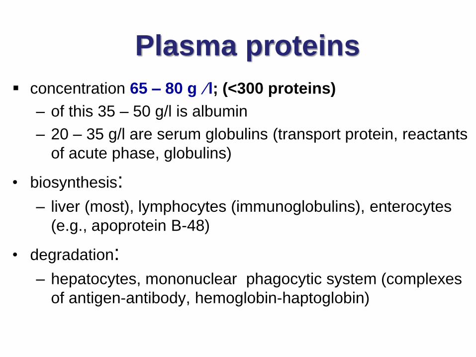
▪ concentration 65 – 80 g l; (<300 proteins)
– of this 35 – 50 g/l is albumin
– 20 – 35 g/l are serum globulins (transport protein, reactants
of acute phase, globulins)
• biosynthesis: – liver (most), lymphocytes (immunoglobulins), enterocytes
(e.g., apoprotein B-48)
• degradation: – hepatocytes, mononuclear phagocytic system (complexes
of antigen-antibody, hemoglobin-haptoglobin)
Plasma proteins
Types of plasma proteins
1. Albumin
2. Globulins a-globulins : a1 a a2-globulins
b-globulins: b1 a b2-globulins
g-globulins
3. Fibrinogen
Under different conditions (pathological conditions, age, pregnancy etc.) the protein levels depart from the usual range.
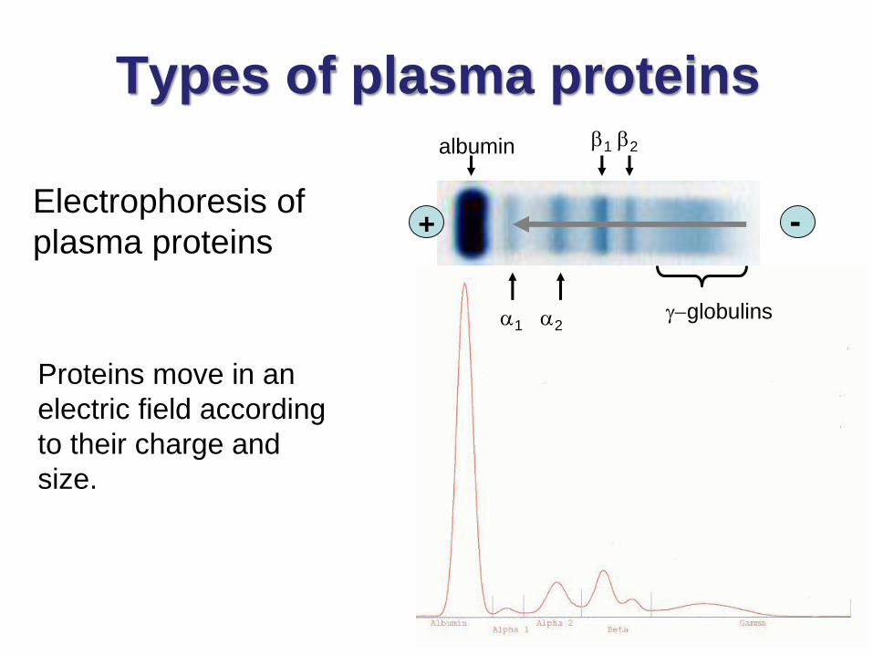
Electrophoresis of
plasma proteins
Types of plasma proteins
Proteins move in an
electric field according
to their charge and
size.
b1 b2
+ -
albumin
a1 a2g-globulins
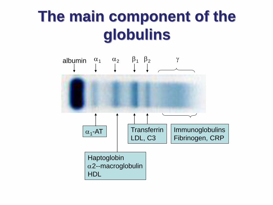
The main component of the
globulins
a1-AT
Haptoglobin
a2--macroglobulin
HDL
Transferrin
LDL, C3
Immunoglobulins
Fibrinogen, CRP
a1 a2albumin b1 b2 g
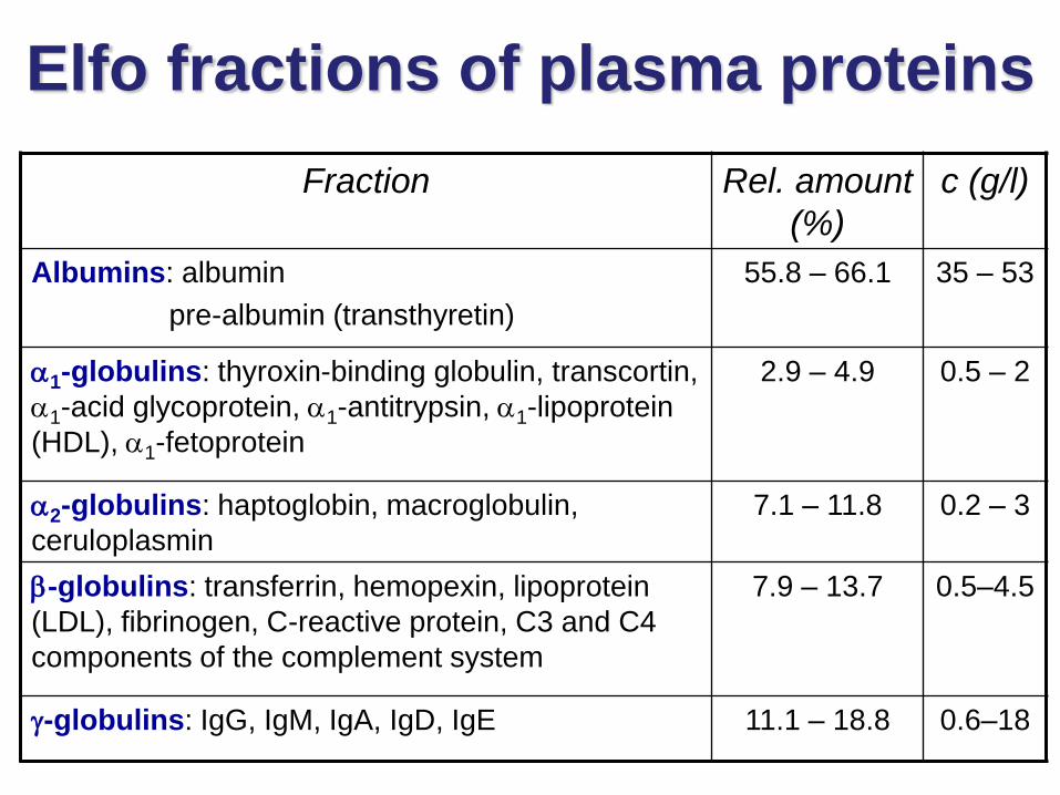
Elfo fractions of plasma proteins
Fraction Rel. amount
(%)
c (g/l)
Albumins: albumin
pre-albumin (transthyretin)
55.8 – 66.1 35 – 53
a1-globulins: thyroxin-binding globulin, transcortin,
a1-acid glycoprotein, a1-antitrypsin, a1-lipoprotein
(HDL), a1-fetoprotein
2.9 – 4.9 0.5 – 2
a2-globulins: haptoglobin, macroglobulin,
ceruloplasmin
7.1 – 11.8 0.2 – 3
b-globulins: transferrin, hemopexin, lipoprotein
(LDL), fibrinogen, C-reactive protein, C3 and C4
components of the complement system
7.9 – 13.7 0.5–4.5
g-globulins: IgG, IgM, IgA, IgD, IgE 11.1 – 18.8 0.6–18
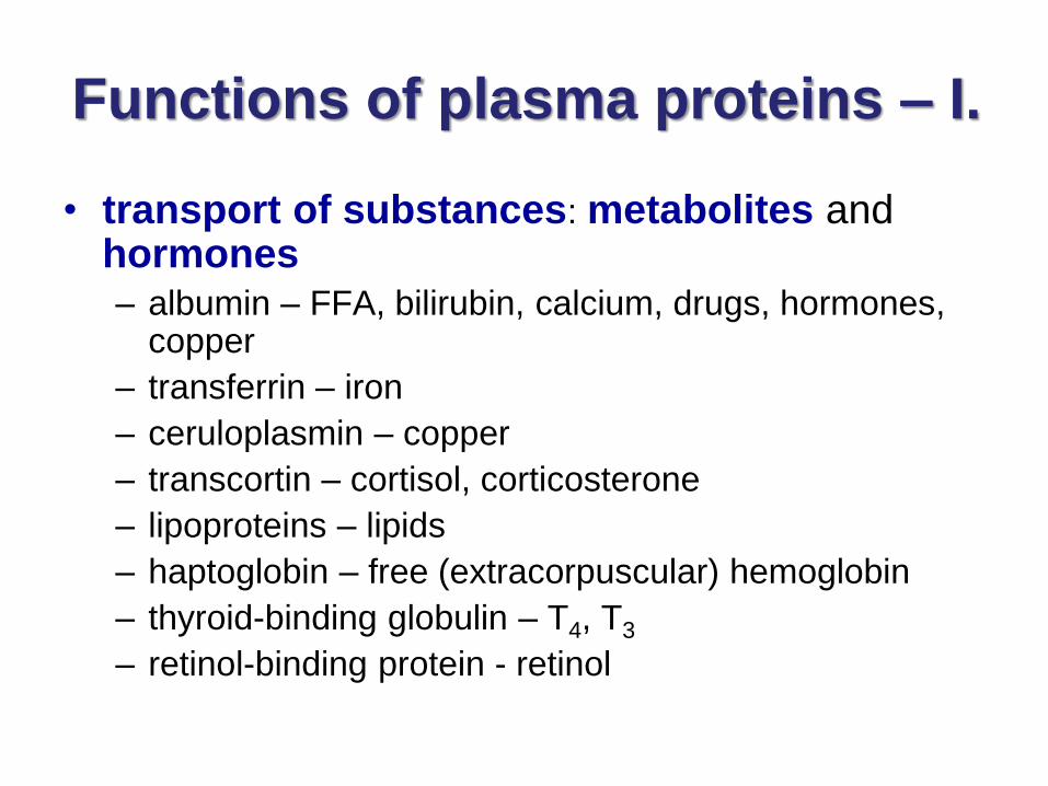
Functions of plasma proteins – I.
• transport of substances: metabolites and hormones– albumin – FFA, bilirubin, calcium, drugs, hormones,
copper
– transferrin – iron
– ceruloplasmin – copper
– transcortin – cortisol, corticosterone
– lipoproteins – lipids
– haptoglobin – free (extracorpuscular) hemoglobin
– thyroid-binding globulin – T4, T3
– retinol-binding protein - retinol
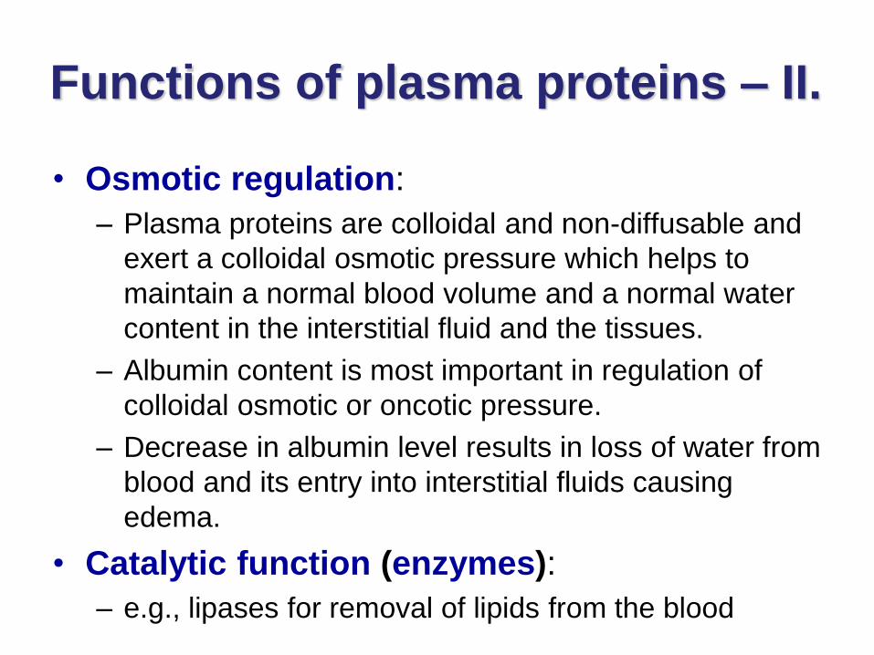
Functions of plasma proteins – II.
• Osmotic regulation:
– Plasma proteins are colloidal and non-diffusable and
exert a colloidal osmotic pressure which helps to
maintain a normal blood volume and a normal water
content in the interstitial fluid and the tissues.
– Albumin content is most important in regulation of
colloidal osmotic or oncotic pressure.
– Decrease in albumin level results in loss of water from
blood and its entry into interstitial fluids causing
edema.
• Catalytic function (enzymes):
– e.g., lipases for removal of lipids from the blood
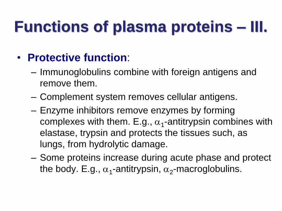
Functions of plasma proteins – III.
• Protective function:
– Immunoglobulins combine with foreign antigens and
remove them.
– Complement system removes cellular antigens.
– Enzyme inhibitors remove enzymes by forming
complexes with them. E.g., a1-antitrypsin combines with
elastase, trypsin and protects the tissues such, as
lungs, from hydrolytic damage.
– Some proteins increase during acute phase and protect
the body. E.g., a1-antitrypsin, a2-macroglobulins.
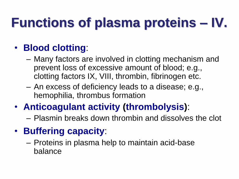
Functions of plasma proteins – IV.
• Blood clotting:– Many factors are involved in clotting mechanism and
prevent loss of excessive amount of blood; e.g., clotting factors IX, VIII, thrombin, fibrinogen etc.
– An excess of deficiency leads to a disease; e.g., hemophilia, thrombus formation
• Anticoagulant activity (thrombolysis):– Plasmin breaks down thrombin and dissolves the clot
• Buffering capacity:
– Proteins in plasma help to maintain acid-base balance
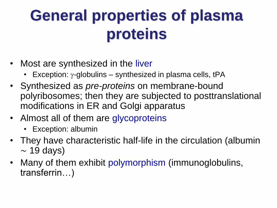
General properties of plasma
proteins
• Most are synthesized in the liver• Exception: g-globulins – synthesized in plasma cells, tPA
• Synthesized as pre-proteins on membrane-bound polyribosomes; then they are subjected to posttranslational modifications in ER and Golgi apparatus
• Almost all of them are glycoproteins• Exception: albumin
• They have characteristic half-life in the circulation (albumin ∼ 19 days)
• Many of them exhibit polymorphism (immunoglobulins, transferrin…)
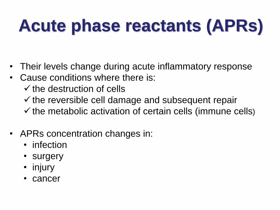
Acute phase reactants (APRs)
• Their levels change during acute inflammatory response
• Cause conditions where there is:
✓ the destruction of cells
✓ the reversible cell damage and subsequent repair
✓ the metabolic activation of certain cells (immune cells)
• APRs concentration changes in:
• infection
• surgery
• injury
• cancer
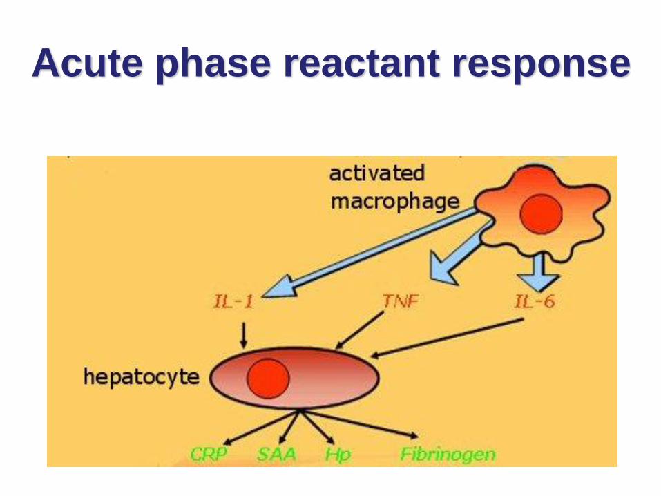
Acute phase reactant response
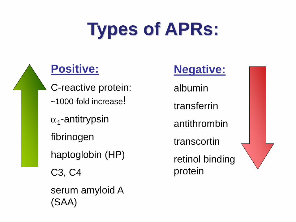
Types of APRs:
Negative:
albumin
transferrin
antithrombin
transcortin
retinol binding
protein
Positive:
C-reactive protein:
~1000-fold increase!
a1-antitrypsin
fibrinogen
haptoglobin (HP)
C3, C4
serum amyloid A
(SAA)
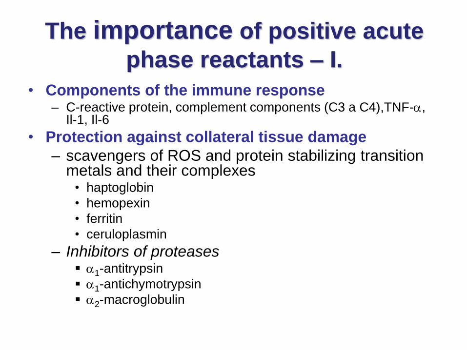
The importance of positive acute
phase reactants – I.
• Components of the immune response– C-reactive protein, complement components (C3 a C4),TNF-a,
Il-1, Il-6
• Protection against collateral tissue damage
– scavengers of ROS and protein stabilizing transition metals and their complexes
• haptoglobin
• hemopexin
• ferritin
• ceruloplasmin
– Inhibitors of proteases▪ a1-antitrypsin
▪ a1-antichymotrypsin
▪ a2-macroglobulin
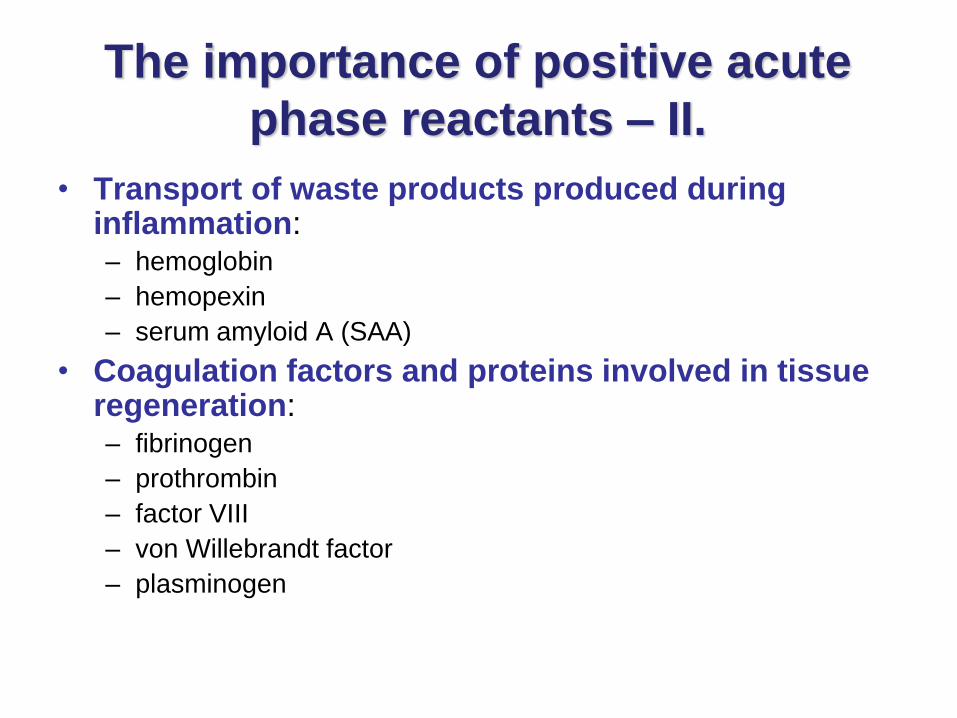
The importance of positive acute
phase reactants – II.
• Transport of waste products produced during inflammation:– hemoglobin
– hemopexin
– serum amyloid A (SAA)
• Coagulation factors and proteins involved in tissue regeneration:– fibrinogen
– prothrombin
– factor VIII
– von Willebrandt factor
– plasminogen
The importance of negative acute
phase reactants
• The criterion for determining inflammation (decrease inflammation)
– transcortin (corticosteroid-binding protein) – binds cortisol
• The criterion for protein synthesis in the liver
Albumin
• Concentration in plasma: 45 gl
• 60% of the total plasma protein
• Functions:– maintenance of plasma oncotic pressure
(values lower than 20 g leads to edema) – protein reserve, the source of amino acids
– transport of:• steroid hormones
• free fatty acids
• bilirubin
• drugs (sulfonamides, aspirin)
• Ca2+
• Cu2+
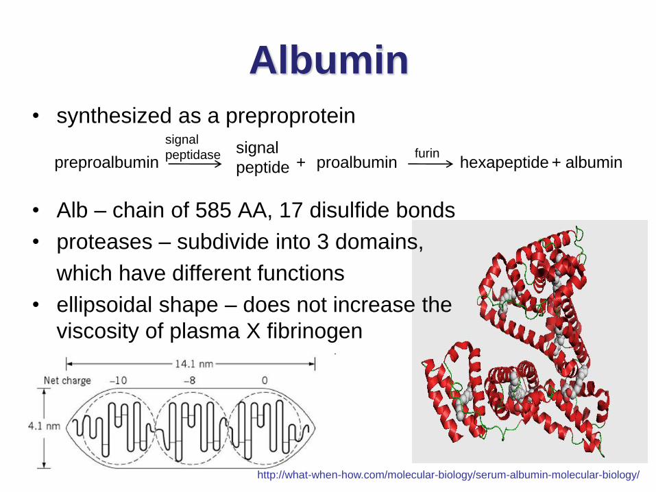
Albumin
• synthesized as a preproprotein
• Alb – chain of 585 AA, 17 disulfide bonds
• proteases – subdivide into 3 domains,
which have different functions
• ellipsoidal shape – does not increase the
viscosity of plasma X fibrinogen
http://what-when-how.com/molecular-biology/serum-albumin-molecular-biology/
preproalbumin proalbuminsignal
peptide hexapeptide albumin+ +furin
signal
peptidase
Causes of Albumin Deficiency
• Liver diseases (cirrhosis) – decrease in the ratio of
albumin to globulins
• Protein malnutrition
• Excessive excretion by kidneys (renal disease)
• Mutation causing analbuminemia (affects splicing)
a1-antitrypsin
• Main globulin of a1 fraction (90 %)
• is synthesized in the liver in hepatocytes and macrophages
• glycoprotein, highly polymorphous (≈75 forms)
• Functions:– Main plasma inhibitor of serine proteases (trypsin,
elastase...)
– during the acute phase increases inhibition of degradation of connective tissue by elastase
– deficiency proteolytic lung damage (emphysema)
Transferrin
• Transferrin is a β-globulin
• Binds free iron in serum
• Normally it is about one third saturated with iron
• Transferrin levels are decreased in:
– liver disease (e.g. cirrhosis)
– Chronic infections
– Nephrosis
– Congenital atransferrinaemia
• Increased serum transferrin levels occur during increased
transferrin synthesis caused as a result of iron deficiency
anemia
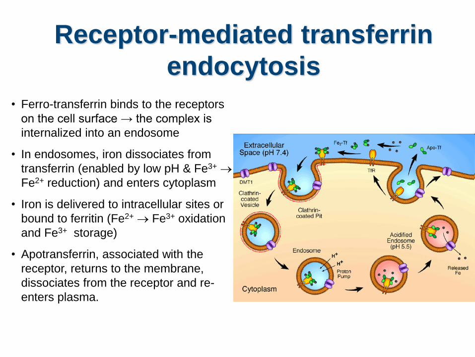
Receptor-mediated transferrin
endocytosis
• Ferro-transferrin binds to the receptors
on the cell surface → the complex is
internalized into an endosome
• In endosomes, iron dissociates from
transferrin (enabled by low pH & Fe3+ →
Fe2+ reduction) and enters cytoplasm
• Iron is delivered to intracellular sites or
bound to ferritin (Fe2+ → Fe3+ oxidation
and Fe3+ storage)
• Apotransferrin, associated with the
receptor, returns to the membrane,
dissociates from the receptor and re-
enters plasma.
Transferrin
• Free Fe2+ ions are toxic for organism – catalyses Fenton reaction (formation of highly toxic OH radical)
H2O2 + Fe2+ → OH- + ˙OH + Fe3+
• Transferrin with other plasma proteins that bind iron or heme, acts as an antioxidant (prevents ROS)
• Causes of decline in transferrin :
– burns, infections, malignant processes and liver and kidney diseases
• Cause of relative transferrin excess:
Iron-deficiency anemia
Ferritin
• Intracellular protein; only small portion in plasma
• 24 subunits surround 3000 - 4500 ions of Fe3+
• Function: stores iron that can be called upon for use when
needed
• Primary hemochromatosis – genetic disorder characterized
by increased absorption of iron from the intestine
accumulated iron damages organs such as the liver, skin,
heart, and pancreas. Concentration of ferritin is elevated.
Cerruloplasmin
• Conc. in plasma: 300 mgl
• Functions:
– carries 90% of copper in plasma (copper – cofactor for a
variety of enzymes)
1 molecule binds 6 atoms of copper
binds copper more tightly than albumin that carries other
10% of plasma copper albumin may be more
important in copper transport (donates copper to tissues
more readily)
Haptoglobin (Hp)
• a2- globulin, tetramer a2b2 chains
• Exists in 3 polymorphic forms
• Functions:
– binds free hemoglobin and delivers it to the
reticuloendothelial cells
– complex Hb-Hp is too large to pass through glomerulus
prevention of loss of free Hb (and Fe)
– Free Hb passes through glomerulus, enters tubules and
tends to precipitate therein kidney damage
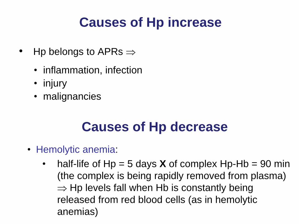
• Hp belongs to APRs
• inflammation, infection
• injury
• malignancies
Causes of Hp increase
Causes of Hp decrease
• Hemolytic anemia:
• half-life of Hp = 5 days X of complex Hp-Hb = 90 min
(the complex is being rapidly removed from plasma)
Hp levels fall when Hb is constantly being
released from red blood cells (as in hemolytic
anemias)
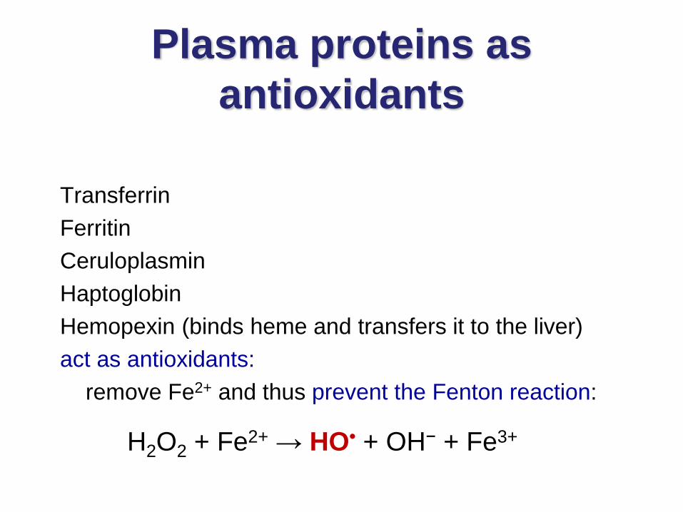
Plasma proteins as
antioxidants
Transferrin
Ferritin
Ceruloplasmin
Haptoglobin
Hemopexin (binds heme and transfers it to the liver)
act as antioxidants:
remove Fe2+ and thus prevent the Fenton reaction:
H2O2 + Fe2+ → HO• + OH− + Fe3+

C-reactive protein (CRP)
• Belongs to b2-globulin, the levels of which rise in
response to inflammation
• Acute-phase reactant
• Its physiological role is to bind to phosphocholine
expressed on the surface of dead or dying cells (and
some types of bacteria)
• plasma concentration levels of CRP rapidly increase
within 2 hours of acute insult, reaching a peak at 48
hours (bacterial, viral, fungal infection, rheumatic
diseases, malignity, tissue necrosis)
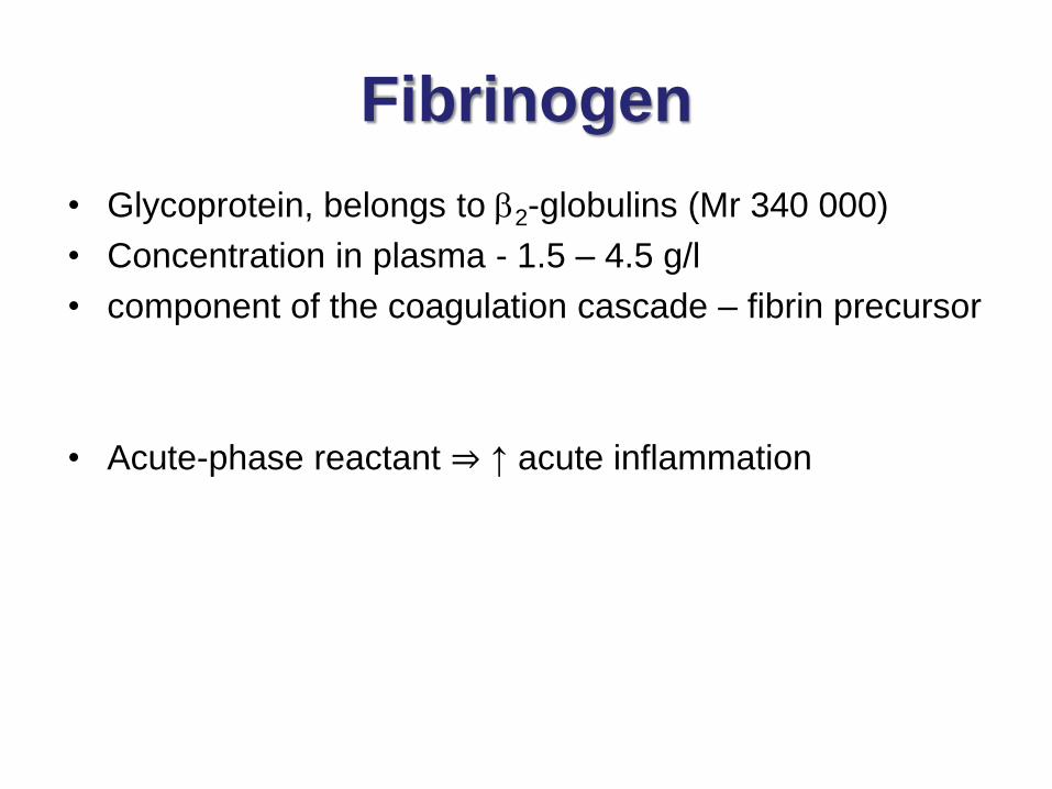
Fibrinogen
• Glycoprotein, belongs to b2-globulins (Mr 340 000)
• Concentration in plasma - 1.5 – 4.5 g/l
• component of the coagulation cascade – fibrin precursor
• Acute-phase reactant ⇒ ↑ acute inflammation
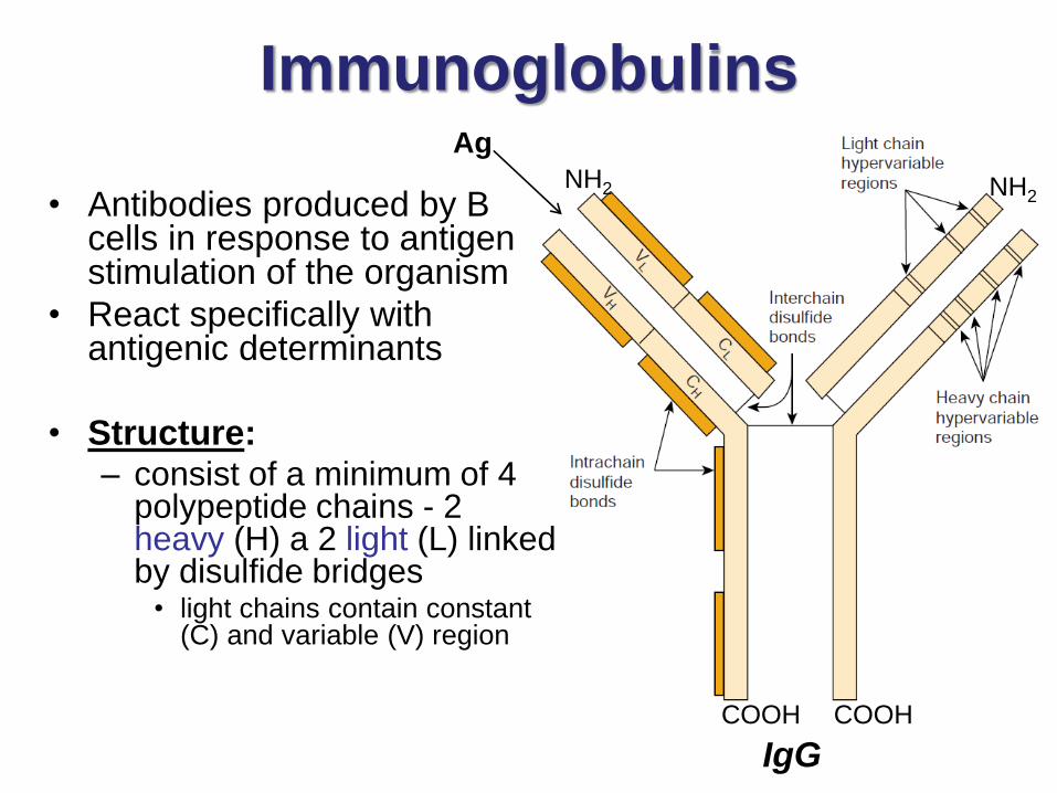
Immunoglobulins
• Antibodies produced by B cells in response to antigen stimulation of the organism
• React specifically with antigenic determinants
• Structure:– consist of a minimum of 4
polypeptide chains - 2 heavy (H) a 2 light (L) linked by disulfide bridges
• light chains contain constant (C) and variable (V) region
COOH
NH2
Ag
COOH
NH2
IgG
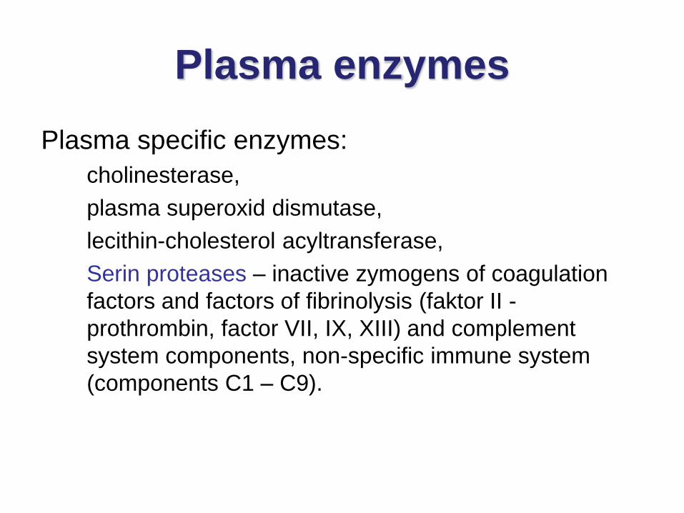
Plasma enzymes
Plasma specific enzymes:
cholinesterase,
plasma superoxid dismutase,
lecithin-cholesterol acyltransferase,
Serin proteases – inactive zymogens of coagulation
factors and factors of fibrinolysis (faktor II -
prothrombin, factor VII, IX, XIII) and complement
system components, non-specific immune system
(components C1 – C9).
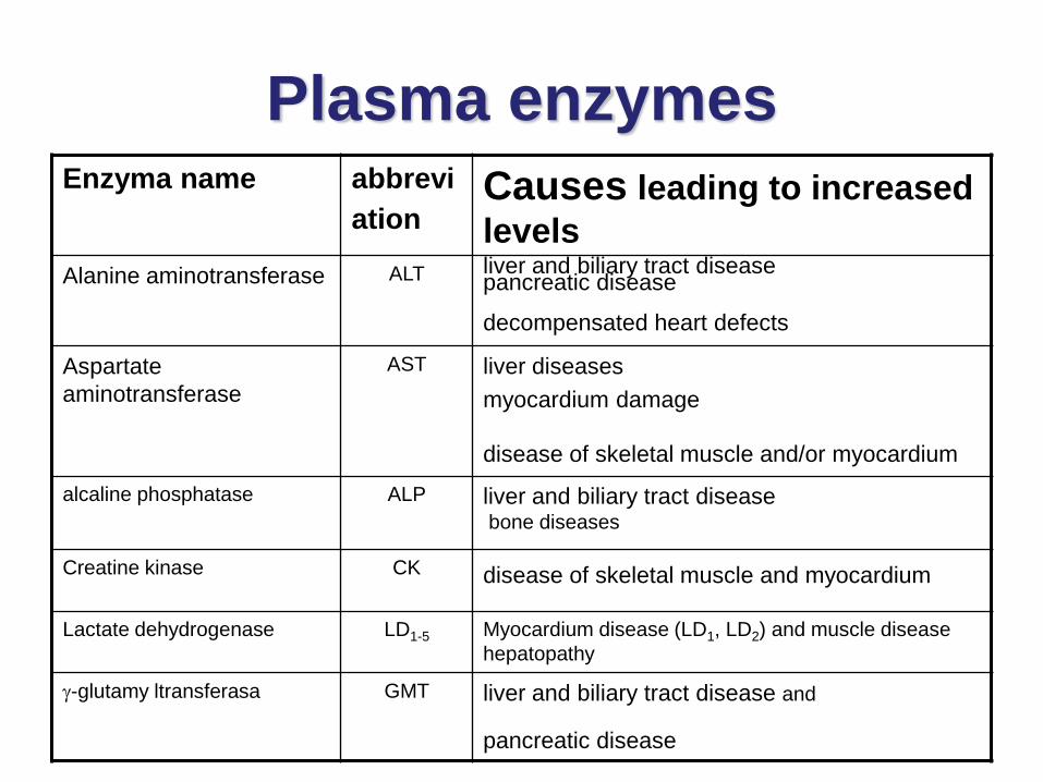
Plasma enzymes Enzyma name abbrevi
ationCauses leading to increased
levels
Alanine aminotransferase ALT liver and biliary tract diseasepancreatic disease
decompensated heart defects
Aspartate
aminotransferase
AST liver diseases
myocardium damage
disease of skeletal muscle and/or myocardium
alcaline phosphatase ALP liver and biliary tract diseasebone diseases
Creatine kinase CK disease of skeletal muscle and myocardium
Lactate dehydrogenase LD1-5 Myocardium disease (LD1, LD2) and muscle disease
hepatopathy
g-glutamy ltransferasa GMT liver and biliary tract disease and
pancreatic disease
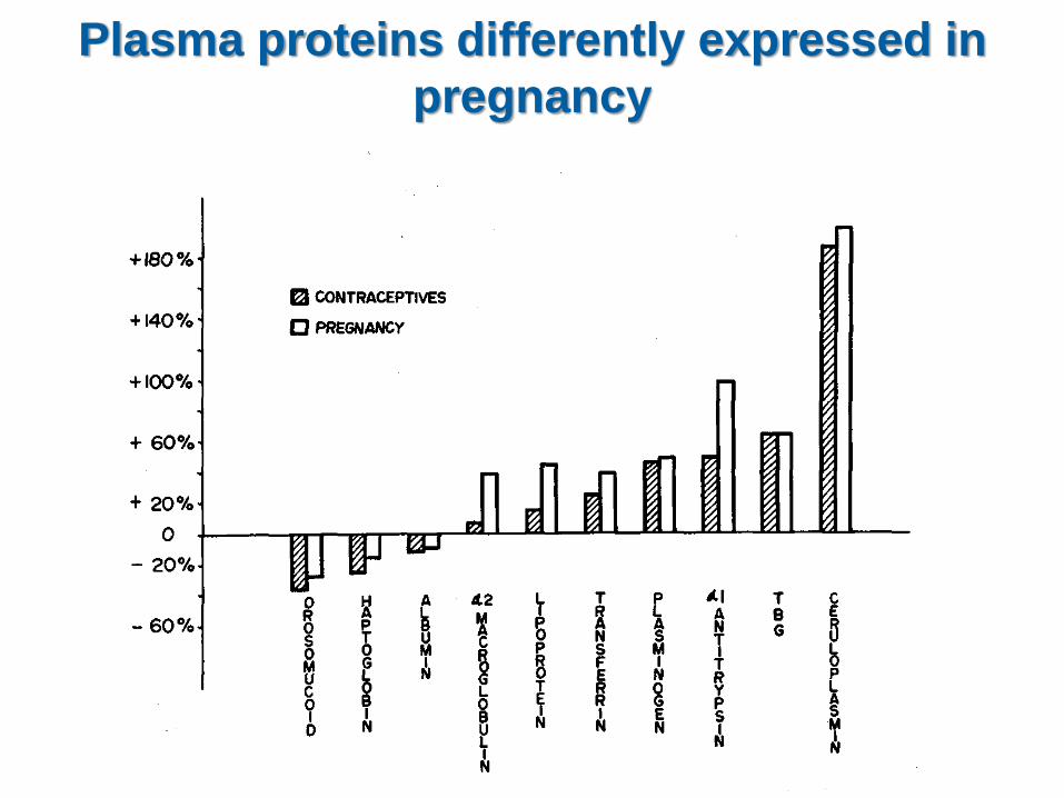
Plasma proteins differently expressed in
pregnancy
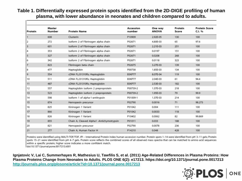
Table 1. Differentially expressed protein spots identified from the 2D-DIGE profiling of human
plasma, with lower abundance in neonates and children compared to adults.
Ignjatovic V, Lai C, Summerhayes R, Mathesius U, Tawfilis S, et al. (2011) Age-Related Differences in Plasma Proteins: How
Plasma Proteins Change from Neonates to Adults. PLOS ONE 6(2): e17213. https://doi.org/10.1371/journal.pone.0017213
http://journals.plos.org/plosone/article?id=10.1371/journal.pone.0017213
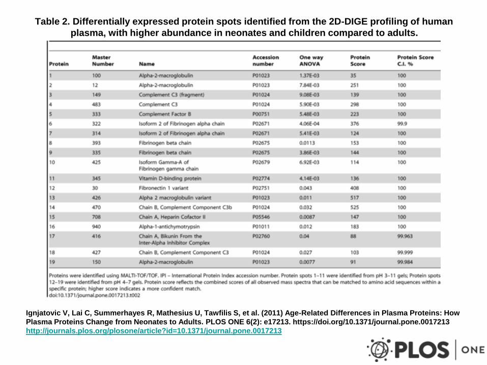
Table 2. Differentially expressed protein spots identified from the 2D-DIGE profiling of human
plasma, with higher abundance in neonates and children compared to adults.
Ignjatovic V, Lai C, Summerhayes R, Mathesius U, Tawfilis S, et al. (2011) Age-Related Differences in Plasma Proteins: How
Plasma Proteins Change from Neonates to Adults. PLOS ONE 6(2): e17213. https://doi.org/10.1371/journal.pone.0017213
http://journals.plos.org/plosone/article?id=10.1371/journal.pone.0017213
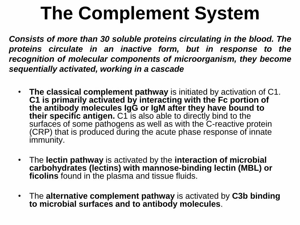
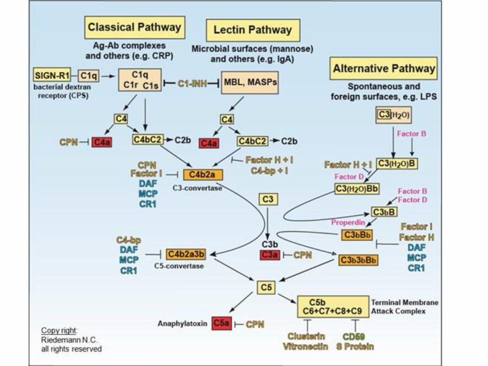
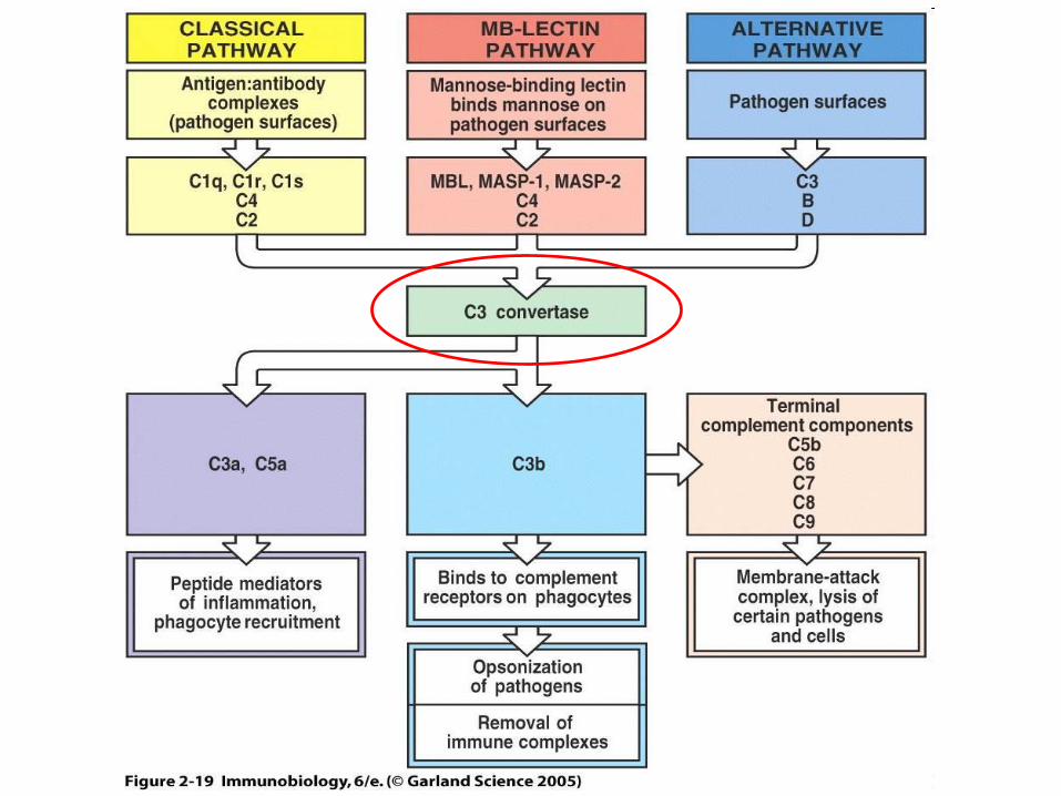
The Complement System
• The classical complement pathway is initiated by activation of C1. C1 is primarily activated by interacting with the Fc portion of the antibody molecules IgG or IgM after they have bound to their specific antigen. C1 is also able to directly bind to the surfaces of some pathogens as well as with the C-reactive protein (CRP) that is produced during the acute phase response of innate immunity.
• The lectin pathway is activated by the interaction of microbial carbohydrates (lectins) with mannose-binding lectin (MBL) or ficolins found in the plasma and tissue fluids.
• The alternative complement pathway is activated by C3b binding to microbial surfaces and to antibody molecules.
Consists of more than 30 soluble proteins circulating in the blood. The
proteins circulate in an inactive form, but in response to the
recognition of molecular components of microorganism, they become
sequentially activated, working in a cascade
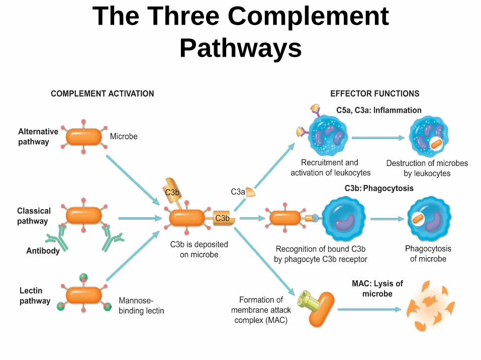
The Three Complement
Pathways