Embed Size (px)
Citation preview
M Sentis
LP3 J Herman S Noel L Mercadier A Kabashin
P Blandin
CiNAM W Marine
LHuC T Itina
INRS (Canada) JC Kieffer and co-authors
INFLPR (Roumanie) I Mihalescu M Dinescu
Et beaucoup drsquoautres
Aix-Marseille University CNRS LP3 UMR 7341 13288 Marseille
France
Plasmas-Laser et applications
vers le biomeacutedical
Summary
Few words on pulsed laser matter interaction
Laser Plasma produced at moderate intensity
bull Bio thin films produced by PLD
bull Nanoclusters for imaging and therapy
bull LIBS for biomedical applications
Laser Plasma X-ray source
bull Phase contrast imaging
Laser Plasma Proton source
bullProton therapy
bullIsotopes for TEP
~1010
Wcm2
~1022
Wcm2
NGC 2009
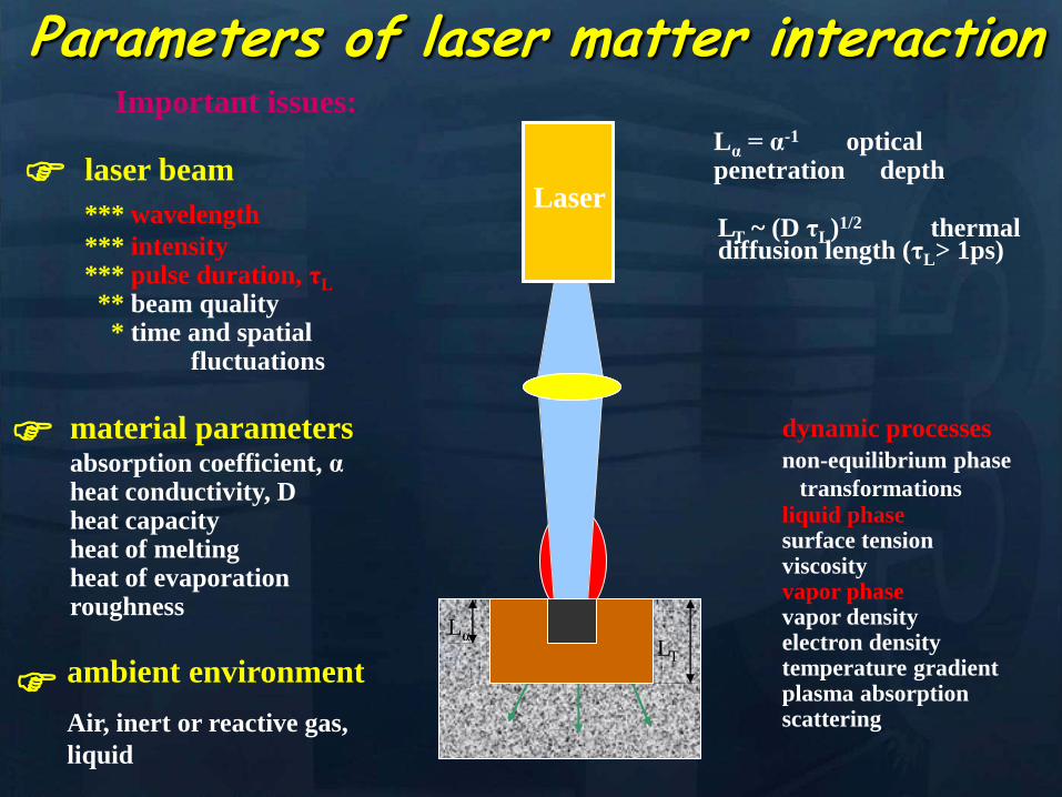
Parameters of laser matter interaction
laser beam
wavelength
intensity pulse duration τL
beam quality time and spatial fluctuations
Important issues
dynamic processes
non-equilibrium phase
transformations liquid phase surface tension viscosity vapor phase vapor density electron density temperature gradient plasma absorption scattering
LT
LT ~ (D τL)12 thermal diffusion length (τLgt 1ps)
material parameters absorption coefficient α heat conductivity D heat capacity heat of melting heat of evaporation roughness
ambient environment
Air inert or reactive gas
liquid
Lα = α-1 optical penetration depth
Laser
Lα
Les reacutegimes drsquointeraction laser-matiegravere
Production de
proton
1022 Wcm2 103 Wcm2
Diffusion
X-ray
emission
laser
Production X
I = E(S x t)
E = energie
t= dureacutee de lrsquoimpulsion
S = tache focale
Ablation
S
1 fs = 10-15s - 1 ps = 10-12s
Focalisation sur des tregraves petites dimensions (surface min ~ sup2)
rarr Conseacutequence de cette concentration dans lrsquoespace
Densiteacutes de Puissance eacutenormes
Ordre de grandeur laser 10 W agrave λ = 500 nm (vert)
densiteacute de puissance max au waist (=Puissancesurface) =
10(0510-6)sup2 = 4 GWcm2
Lentille focale f
Diamegravetre D
Diamegravetre au waist (=col
en franccedilais)
Φasymp λ f πD Ordre de grandeur si f ~ D rarr Φ ~ λ
Proprieacuteteacute de focalisation de lrsquoeacutemission LASER
Les Lasers une course vers les tregraves hautes intensiteacutes
Zetta 1021
Exa 1018
Peta 1015
Tera1012
Giga 109
Strickland amp Mourou Opt Comm 56 219 (1985)
t fs oscillateur
t
compresseur
t amplificateur
t eacutetireur
1985 Le concept
Quelques grandeurs caracteacuteristiques
10 - 14
10 - 11
10 - 12
10 - 13
10 - 10
Thermalisation eacutelectronique
Relaxation eacutelectron - phonon
Diffusion Thermique
fusion
Ablation
non thermique
Thermique
temps (s)
Zepto 10-21
Atto 10-18
Femto 10-15
Pico 10-12
Nano 10-9
Sources laser cw impulsionnels (ms micros) courts (ns)ultra-courts (psfs as)
NGC 2009
Surface absorption Thermal conduction
Surface melting Vaporization
Plasma ignition Explosive phase change
Plasma absorption Self-regulating heat transfer
Collisional acceleration Plume splitting Ambient interpenetration
Rapid cooling Condensation Plume deceleration
0
ns
Plume detachment Stagnation and collapse
m s
Absorption reflection
Heat transfer
Thermodynamics (phase change)
Plasma breakdown
Shock waves (gas)
Stress waves (solid)
Laser-plasma interactions
Gas dynamic expansion
Atomic amp molecular processes
A wide range of physics is involved in
laser-matter interactions
wP =(4pe2neme)12
nc (cm-3) = 11 x 1021 (1 mmLaser)2
How is a laser plasma formed
Laser Induced Plasma in gas and
liquids at moderate intensities
I = 10 11 to 10 14 Wcm2
bull Biomedical thin films by P LD in gaseous media
bull Nanoclusters produced by laser ablation for medical imaging and therapy
bull LIBS for biomedical applications
ns to fs laser pulse
duration
Laser Induced Plasma in Gas PLD
CCD PMT
spectrometer
Target
Substrates
or Faraday
cup
laser beam
Pulsed Laser Deposition
CCD PMT
spectrometer
Target
Substrates
or Faraday
cup
laser beam
Pulsed Laser Deposition
Target Just about anything (metals semiconductorshellip)
Laser Typically excimer (UV 10 nanosecond pulses)
Vacuum Atmospheres to ultrahigh vacuum
Processes in PLD
Laser pulse
Processes in PLD
e- e-
e-
e- e-
e-
e-
e-
e-
e- e-
e-
e-
e-
Electronic excitation
Processes in PLD
e- e-
e-
e- e-
e-
e-
e-
e-
e- e-
e-
e-
e-
Energy relaxation to lattice (~1 ps)
lattice
Processes in PLD
Heat diffusion (over microseconds)
lattice
Processes in PLD
Melting (tens of ns) Evaporation Plasma
Formation (microseconds) Resolidification
lattice
Processes in PLD
lattice
If laser pulse is long (ns) or
repetition rate is high laser may
continue interactions
Processes in Pulsed Laser Deposition
1 Absorption of laser pulse in material
Qab=(1-R)Ioe-aL
(metals absorption depths ~ 10 nm depends on )
2 Relaxation of energy (~ 1 ps) (electron-phonon interaction)
3 Heat transfer Melting and Evaporation
when electrons and lattice at thermal equilibrium (long pulses)
use heat conduction equation
(or heat diffusion model)
abp QTKt
TC
)(
At peak of laser pulse temperatures on target can reach
gt105 K (gt 40 eV)
Electric Fields gt 105 Vcm also high magnetic fields
Plasma Temperatures 3000-5000 K
Ablated Species with energies 1 ndash100 eV
Initially Incredibly Non-Equilibrium
Why biomaterials
- repair and reconstruction of parts of the musculo-skeletal system of vertebrates
- minimal biological requirement biocompatibility associated with the absence of
any adverse effect (non-toxic and non-allergic)
Other requests
- resistance to physiological fluids
- should not interfere with the bodyrsquos natural immunity system
- withstand mechanical stress during whole lifetime
- manufacturability in any appropriate shape
Possible classification
-a) biologically inactive (inert) alumina zirconia stainless steel CoCrNi
CoCrMo titanium titanium alloys carbon latex PE PMMA hellip
-b) porous Calcium Phosphates (CaPs) CaP-coated metal
-c) bioactive dense calcium phosphate ceramics bioactive glasses bioactive
glass-ceramics bioactive composites hellip
-d) Resorbable tricalcium phosphate calciumaliminate polylactic acid poly-L-
acetate
Our option biocompatible porous and bioactive CaP
Acronym
Chemical formula Compound name CaP
ratio
HA Ca10(PO4)6(OH)2 Hydroxylapatite 167
FA Ca10(PO4)6F2 Fluorapatite 167
CDHA Ca10x(HPO4)x(PO4)6x(OH)2x (0x2) Calcium-deficient Hydroxylapatite 133-167
BA Ca83(PO4)43 (CO3-HPO4)17(OH)03
BA=carbonated CDHA (x=17)
Biological apatite 138-193
Mn-CHA HA with (04-2) Mn2+ and (2-6) CO32- Mn2+ doped carbonated hydroxylapatite 151- 165
OHA Ca10(PO4)6(OH)22xOxٱx (0x1) Oxyhydroxylapatite 167
OA Ca10O(PO4)6 Oxyapatite 167
MCPM Ca(H2PO4)2middotH2O Monocalcium phosphate monohydrate 05
MCPA Ca(H2PO4)2 Monocalcium phosphate anhydrate 05
DCPD CaHPO4middot2H2O Dicalcium phosphate dihydrate (Brushite) 1
DCPA CaHPO4 Dicalcium phosphate anhydrate 1
OCP Ca8(HPO4)2(PO4)4middot5H2O Octacalcium phosphate 133
a-TCP Ca3(PO4)2 (monoclinic) Tricalcium phosphate (phase a) 15
-TCP Ca3(PO4)2 (rhombohedral) Tricalcium phosphate (phase Whitlockite) 15
TTCP Ca4O(PO4)2 Tetracalcium phosphate 2
a-DCP Ca2P2O7 (orthorhombic) Dicalcium phosphate (phase a) 1
-DCP Ca2P2O7 (tetragonal) Dicalcium phosphate (phase ) 1
ACP Cax(PO4)y nH2O (Amorphous Calcium pyrophosphate) 12-22
Bone
Resorbable CaP
CaP coating
Ti
Implant
Alternative solution Biomimetic coatings for metallic implants
Main deficiency of CaPs brittle in bulk
HA molecule Ca10(PO4)6OH2
Projection in the
(001) base plan of
the hydroxyapatite
unit cell (hexagonal
structure)
How difficult is to deposit CaPs (1)
- very complex molecules
How difficult is to deposit CaPs (2)
Crystal structure of OCP projected down the c-axis
Alternating apatite- and
hydrated- layers (100)
planes
The region with shaded
atoms the ldquoapatitic layerrdquo
is very similar to HA
The zone containing 10
water molecules is the
ldquohydrated layerrdquo
H atoms are omitted for
clarity
Octacalcium phosphate
(Ca8(HPO4)2(PO4)4middot5H2O)
Mesures par spectroscopie optique
drsquoemission sur le plasma issu de lrsquoirradiation
laser 248 nm drsquoune cible drsquoHA pure
Croissance impulsion par impulsion
Preacutesence des ions de
bull calcium (Ca I et Ca II)
bull oxygegravene (O I et O II) et
bull phosphore (P II)
Eacutemissions dues agrave des espegraveces moleacuteculaires preacutesentes dans le plasma
Couche mince drsquohydroxyapatite obtenue par
ablation laser
Goutelettes
CaP = 18 CaP = 21
CaP = 17 Trois reacutegions
typiques
i Reacutegion majoritaire lisse
ii Agglomeacuteration de gouttelettes sub-micromeacutetriques
iii Gouttelettes micromeacutetriques
Human primary osteoblasts (hOB) were cultured on OCP coated-Ti
Mn-CHA coatedndashTi HA coated-Ti Ti control (polystyrene)
hOB response SEM micrographs
- on bare Ti (a) after 7 days (b) after 21 days
Elongated rod-like morphology
Bioactivity tests
- OCP coatings (c) after 7 days (d) after 21 days
- In time cells spread and expand with flattened polyhedral-morphology
- Numerous cytoplasmatic extensions
hOB response SEM micrographs
- Mn-CHA coatings (e) after 7 days (f) after 21 days
- In time cells spread and expand with flattened polyhedral-morphology
- Less cytoplasmatic extensions
hOB response SEM micrographs
Biocompatible metallic thin films by PLD
Alloys NiTinol NiTi
NiTi characteristics ndash Super-Elastic Property
ndash Radiopaque
ndash Shape-Memory Effect
NiTi biomedical applications ndash Tweezers for removing foreign objects via small incisions
ndash Anchors for tendon fixation
ndash Stents for cardiovascular applications
ndash Dentistry - Orthodontic wires which no not need to be retightened and adjusted
Deposition chamber Multitarget system
PULSED LASER DEPOSITION (PLD)
SEM
Nitinol 30 sequences Nitinol 20 sequences
Cytotoxicity evaluation on microorganism culture
(a) E coli reference cell culture
(b) E coli cell culture on Ti surface
(c) E coli cell culture on NiTi surface
(d) S cerevisiae references cell culture
(e) S cerevisiae cell culture Ti surface
(f) S cerevisiae cell culture on NiTi surface
(g) SEM on E coli cell
culture on NiTi surface
(h) SEM on S cerevisiae cell culture on
NiTi surface E coli and S cerevisiae cell
cultures on the nitinol thin films
o Biocompatible
o Low adherence of microorganism on NiTi surface NiTi alloy does not stimulate the development at genetic level for specific genes which are involved in adhesion processes for microorganism cells
o Low release of Ni ions in solutions
CONCLUSIONS
MAPLE Matrix Assisted Pulsed Laser Evaporation
bull No need for UHV
bull Matrix prepared outside the
chamber
bull High flexibility
You can use everything
you can dissolve in a
solvent
MAPLE of polymers blends with Ag
nanoparticles
Controlled release of antibacterial agents
MAPLE deposition of PEGPLGA films with incorporated silver nanoparticles (AgNPs)
0 5 10 15 20 25 30000
005
010
015
020
025
Nu
mb
er o
f E
C
oli
X109
Time (hours)
Killing activity
0 5 10 15 20 25 30 3500
05
10
15
20
25
Bact
eria
l ce
ll n
um
ber
X109
Time (hours)
Delay of bacterial growth
Antibacterial efficiency against E Coli
Direct writing by femtosecond laser
pulses
Ormosil scaffolds for tissue engineering
50 lines ormosil sample
Distance between lines =
100 μm
30x30 lines ormosil
sample Distance between
lines = 50 μm
3 layers 30x30 lines ormosil
sample Distance between
lines = 100 μm
Scaffold functionalization
Protein deposition on the scaffolds by MAPLE
- Lysozyme antibacterial antitumor uses
- Fibrinogen biocompatible protein
Fibroblast cells grown on a polymeric grid with 100 microm distance between lines
Spectroscopie drsquoun panache plasma
laser femtoseconde
Laser
t =400 ns Spot 35x35 micromsup2 Cu
Z mm
Expeacuteriences reacutealiseacutees par (Grojo et al LP3Marseille)
nanoparticules
atoms
031 Jcm-2
Formation de nano agreacutegats
AFM
(Pereira et al )
TEM
(Kabashin et al)
Eacutetude des meacutecanismes de la formation drsquoagreacutegats
Couplage
- Direct Simulation Monte Carlo
- Dynamique Moleacuteculaire
Information deacutetailleacutee
-Continu du panache
-Distribution en taille des agreacutegats
K Gouriet thegravese (2008)
Simulation par Dynamique Moleacuteculaire (MD)
=gtdistribution drsquoagreacutegats
Cluster size N
No
rmaliz
ed
Yie
ld
100
101
102
103
104
105
10-9
10-8
10-7
10-6
10-5
10-4
10-3
10-2
10-1
100
N-426
N-122
t=1 ns
τ = 15 ps F ~ 2 Fth 4010 nm t =1 ns
Zhigilei et al (Virginia University)
Target applications of mobile nanomaterials
Bioimaging
Optical imaging deals with
visualization of biological objects
or tissues in order to detect
pathogens or follow the delivery
of drugs etc nanoparticles are
uses as contrast agents
In vitro
Cellular
imaging
In vivo Cancer
detection drug delivery
etc
Light-induced
therapies
Cancer therapy nanoparticles are used
as photosensitizers to produce local
targeted destruction of cancer cells
Photodynamic
therapy
Light-induced
hyperthermia
Diagnosis and Sensing A Diseases can be diagnosed through the (simultaneous) detection of a (set of) biomolecule(s) characteristic to a specific disease type and stage (biomarkers)
Huffman Nanomedicine and Nanobiotechnology Vol 1 1 2009
D
molecular signature of sick cell of infecting agent
(eg an antibody)
Cell membrane
Nanoparticle
Coating molecule specifically attracted to the molecular signature
C A nanoparticle can be functionalized in such a way that specifically targets a biomarker Thus the detection of the nanoparticle is linked to the detection of the biomarker and to the diagnosis of a disease
B Each cell type has unique molecular signatures that differentiate healthy and sick tissues Similarly an infection can be diagnosed by detecting the distinctive molecular signature of the infecting agent
Laser ablation of material from a solid target and production of
nanoparticlesnanostructures
Production of nanoclusters by laser
ablation and their subsequent cooling in
ambient medium Gaseous ambience
Nanoclusters can be
deposited on a
substrate forming a
nanostructured film
Aqueous ambience
Nanoclusters
crystallize and form
colloidal solution
NPs produced in gaseous ambience
0 20 40 60 80 100 120 140
0
2
4
6
8
Heig
ht
[nm
]
[nm]
01 1 102
3
4
5
6
7
Mean
part
icle
heig
ht
(nm
)
Helium pressure (Torr)
Silicon nanocrystal size AFM
Gas pressure decreases Nanocrystal size decreases
AFM of isolated laser-
ablated Si particles on
HOPG
Minimal particle size is 2-3 nm (from height measurements)
001Torr
1Torr
01Torr
10Torr
100Torr
NGC 2009
Photoluminescence of size selected nanoclusters
0 1 2 3 4 5 6
39 Jcm2
Num
ber
of
clust
ers
Size (nm)
30 Jcm2
14 Jcm2
0 1 2 3 4 5 6
He 4 Torr
10 Jcm2
15 20 25 30 35 40
0
500
1000
He 4 Torr
39 Jcmsup2
30 Jcmsup2
14 Jcmsup2
10 Jcmsup2
Inte
nsi
ty o
f P
L (
ua
)Photon energy (eV)
Silicon nanoclusters
Medical Imaging A Optical properties of nanoparticles depend greatly on its structure Particularly the color (wavelength) emitted by a quantum dot (a semiconductor nanoparticle) depends on its diameter
C The quantum dots (QD) can be injected to a
subject and then be detected by exciting them to emit light
Source Department of immunology University of Toronto
Solutions of CdSe QDrsquos of different diameter
CdSe nanoparticle (QD) structure
Source Laurence Livermore Laboratories
Imaging of QDrsquos targeted on cellular structures
Nano Letters 2008 Vol 8 pp3887-3892
B
In vivo cancer targeting and imaging with
semiconductor quantum dots
Gao et al Nat Biotech 22 969 - 976 (2004)
NPs produced in aqueous ambience
Advantages of laser ablation-based nanofabrication
compared to chemical methods
Disadvantage of chemical synthesis contaminationhelliphelliphellip
In particular chemical method for the fabrication of gold nanoparticles
Reduction of chloroauric acid (HAuCl4) with citrate in water
Control size by adding a stabilizing agent (thiol- (-SH) containing molecules)
Contamination impurities Cl- on surface non-biocompatible surfactantshellip
Problems in imaging
applications (especially
in vivo)
Problems in field-
enhanced applications (eg contaminants can
provide false Raman signals)
Focalized laser beam
Liquid medium
Target
Plasma
Laser ablation in liquids a way to avoid residual
contamination
- Laser energy can be transmitted through a liquid to ablate a solid
target the nanomaterial is released to the solution
- The process can be performed without any dirty chemical products in
clean liquid environment (deionized water PBS solution) no
contamination
Experiment
Ablation of gold final colloidal solutions
-Solution are ranging from deep wine red to pink and purple
- Color related to the size dependant surface plasmon resonance
Size distribution of Au nanoparticles prepared in
distilled water
0 50 100 150 200 250 300
0
200
400
600
800
1000
Rela
tive a
bundance (
arb
units)
Particle size (nm)
0 10 20 30 40 50 60 70 80 90 100
0
200
400
600
800
1000
Re
lative
Ab
un
da
nce
(a
rb u
nits)
Particle Size (nm) 0 2 4 6 8 10
0
200
400
600
800
1000
Re
lative
Ab
un
da
nce
(a
rb u
nits)
Particle Size (nm)
F = 20F0 Jcm2 F = 5F0 Jcm2 F = F0 Jcm2
-Size and dispersion of nanoparticles can be controlled by laser fluence
- Shape of craters on target at high F suggests the presence of cavitation phenomena
4 plusmn 15 nm
A V Kabashin M Meunier J Appl Phys 94 7941 (2003)
In water Very dispersed population of nanoparticles
Dextran (MW=6000gmol) Quenches the growth of the nanoparticles 2-4nm nanoparticles are produced (230-1850 atoms) Very fast dextran-gold interaction
Influence of the concentration
Dextran 6k
In pure water
300mJ
Ablation in biopolymers (dextran PEG)
Au OH
O
-
Dextran (C6H10O5)n
Nanoparticles react with OH
groups of biopolymers
Gold Nanoparticles vs Alzheimer
A Alzheimer and other degenerative diseases are caused my the clustering of amyloidal beta (Aβ) protein
D Gold nanoparticles can be functionalized to specifically attach to aggregates of this protein (amyloidosis)
Functionalized nanoparticle
Source wwwinternetchemistrycom
Chemical structure of Aβ-protein
Source wwwthefutureofthingscom
C
B
Alzheimerrsquos brain Healthy brain
Source Berkeley Lab
Gold Nanoparticles vs Alzheimer A The functionalized gold nanoparticles selectively attach to the aggregate of amyloidal protein The microwaves of certain frequency are irradiated on the sample Resonance with the gold nanoparticles increases the local temperature and destroy the aggregate
Nanoletters 2006 Vol 6 pp110-115
Before irradiation After irradiation
LIBS for biomedical applications in vivo analysis of tissue for real-time
analysis
real-time diagnosis of pathogen
presence in human fluids (bloodurine
CSF sputum)
71 35
ICCD
spectromegravetre
Time and space
Emission spectroscopie
LIBS (Laser Induced Breakdown Spectroscopy)
Principle of sample identification screening applications based on discriminant analysis here for warning when healthy tooth material is targeted during laser drilling
BMC Oral Health 2001 1 1 Ota Samek1 Helmut H Telle2 and David CS Beddows
Laser-induced breakdown spectroscopy a tool for real-time in vitro and in vivo identification of carious teeth
Sensitive Detection of Epithelial Ovarian Cancer Biomarkers
Using Tag-Laser Induced Breakdown Spectroscopy
Yuri Markushin and Noureddine Melikechi
Optical Science Center for Applied Research Delaware State University USA
X rays produced by laser plasmas
I = ~1016 agrave 1018 Wcm2
nc cm-3
1111021 mm 2
Thot Ilas2
1 3
Ilas2
3 4
Hot electrons
(characteristic lines)
X-ray
emission
laser
X-rays
Line emission Fast electrons knock
free core electrons
from the inner shell
Bremsstrahlung electrons deflected by nucleus
Laser-generated hot electrons produce x-rays by two dominant mechanisms
ne
critical surface (cf cathode)
conventional X-ray tube
Kmetec ldquoMeV X-ray generation with a femtosecond laserrdquo Phys Rev Lett 68 1527 (1992)
Rousse ldquoEfficient Ka X-ray source from fs-laser-produced plasmasrdquo Phys Rev E 50 2200 (1994)
(typical recombination lifetimes several fs)
Hou et al Appl Phys Lett 84 2259 (2004)
Tightly focused fs lasers create the worldrsquos smallest
hard x-ray source (~4 microm) for precision imaging
x-ray source size is ~ 4 larger than laser focus
because of lateral heat transport Bowes Opt Lett 31 116 (2006)
2004 premiegravere deacutemonstration drsquoimagerie avec contraste de phase avec une source X creacutee par laser
Source
Rayon non diffracteacute
Rayon diffracteacute
Lcoh 2R1
eacutechantillon
Absorption vs contraste de phase
dsource-object dobject-detector
X-ray
Source
Object
Detector
position
Dsource
Intensity
interface
dsource-object dobject-detector
X-ray
Source
Object
Detector
position
Fresnel Diffraction
Dsource
Intensity
interface
enhancement interface
R Toth et al
Rev Sci Instrum 76
083701 (2005)
Imagerie par contraste de phase
R Toth et al Rev Sci Instrum
76 083701 (2005)
copy INRS
bull Meilleure reacutesolution (5microm)
bull dose plus faible
bull meilleur contraste (phase)
bull lrsquoeacutenergie X peut ecirctre ajusteacutee
aux besoins de lrsquoimagerie
La reacutefeacuterence mondiale ALLS
(INRS- Montreacuteal) 200TW + Puissance moyenne 50W
47J 23fs 10Hz
in a 8microm spot
Sans miroir deacuteformable
microCT
Scan duration avec
50W10Hz (360 images)
2 min (mouse scan)
courtesy Prof Dr Oswald Willi U Duumlselldorf
MeV Protons produced by laser CR39 or RCF 1010 to 1013 H+
courtesy Prof Don Umstadter U Nebraska-Lincoln
Target Normal Sheath Acceleration hot electrons traversing
target electrostatically accelerate impurity hydrogen ions on the rear surface
virtual
cathode
I ge 1019 Wcm2
Protons de 10MeV (INRS )
~1022 Wcm2
Avantages des particules chargeacutees ou hadrons (protons ions) Rayons X de la Radio-Theacuterapie
Le pic de Bragg
La profondeur de peacuteneacutetration deacutepend de l rsquoeacutenergie incidente des protons
la tumeur la plus superficielle = l rsquoœil 70 MeV
la plus profonde = la prostate 200 - 250 MeV
Protontheacuterapie par laser (projet SAPHIR)
CPO
Exemple de dosimeacutetrie preacutevisionnelle
(photons vs protons)
Comparaison de lrsquoeacutepargne de dose dans les tissus sains en protons (image de droite)
par rapport aux photons (IMRT Intensity Modulated Radio-Therapy image de gauche)
Les irradiations colateacuterales en radiotheacuterapie et en protontheacuterapie
Avantage consideacuterable pour le traitement de tumeurs localiseacutees pregraves d rsquoorganes sensibles (œil cerveau)
currently treating patients 5
5 next 2 years
others awaiting approval
10000 m2 $125 M facility opened in 2006
12 m diameter 250 MeV synchrotron
ldquoThere are too few physicists in the world
and they are an incredibly important part of
doing this We have one of the largest
physics departments in the world with more than 50 medical physicistsrdquo
--- Dr James D Cox head of Radiation Oncology at MD Anderson Cancer Center Houston Texas
Proton Therapy enables precise exposure of small tumors
with minimal damage to surrounding healthy tissue
250 MeV
PROTON beam
10 20
Depth in Tissue [cm]
0 0
50
100
DO
SE
(
)
6 MV
PHOTON
beam
tunable
Bragg
peak
but requires large expensive facilities
eye tumors 65 MeV protons
deep tumors gt200 MeV
Laser proton therapy could be much smaller amp cheaper Fourkal Med Phys 29 2788 (2002)
Malka Med Phys 31 1587 (2004)
On-site production of short-lived isotopes for medical imaging
Laser-generated quasi-mono-energetic electrons efficiently photo-activate materials of interest
bull High Rep rate bull Low cost bull Compact
Limitations to the widespread use of PET arise from the high costs of cyclotrons needed to
produce the short-lived radionucleotides for PET scanning Few hospitals and universities
are capable of maintaining such systems - Wikipedia -
radiotracer activation reaction half-life medical use
15O 16O (n)15O 2 minutes neuro-imaging
11C 12C(n)11C 20 minutes neuro-receptor-specific brain imaging
18F 19F(n)18F 110 minutes clinical oncology
Positron Emission Tomography
on-site
production
essential
18F PET scan of tumor 15O PET scan of human brain
Reed ldquoEfficient initiation of photonuclear reactions using quasi-monoenergetic electron beams from LWFArdquo J Appl Phys 102 073103 (2007)
TEP in situ
+ in vivo
Modegravele animal
microCT +
contraste de phase
+ in vivo
modegraveles de cancer
Comprendre
le cancer
Preacuteparer le traitement
protons 50MeV
Summary
Few words on pulsed laser matter interaction
Laser Plasma produced at moderate intensity
bull Bio thin films produced by PLD
bull Nanoclusters for imaging and therapy
bull LIBS for biomedical applications
Laser Plasma X-ray source
bull Phase contrast imaging
Laser Plasma Proton source
bullProton therapy
bullIsotopes for TEP
~1010
Wcm2
~1022
Wcm2
NGC 2009
Parameters of laser matter interaction
laser beam
wavelength
intensity pulse duration τL
beam quality time and spatial fluctuations
Important issues
dynamic processes
non-equilibrium phase
transformations liquid phase surface tension viscosity vapor phase vapor density electron density temperature gradient plasma absorption scattering
LT
LT ~ (D τL)12 thermal diffusion length (τLgt 1ps)
material parameters absorption coefficient α heat conductivity D heat capacity heat of melting heat of evaporation roughness
ambient environment
Air inert or reactive gas
liquid
Lα = α-1 optical penetration depth
Laser
Lα
Les reacutegimes drsquointeraction laser-matiegravere
Production de
proton
1022 Wcm2 103 Wcm2
Diffusion
X-ray
emission
laser
Production X
I = E(S x t)
E = energie
t= dureacutee de lrsquoimpulsion
S = tache focale
Ablation
S
1 fs = 10-15s - 1 ps = 10-12s
Focalisation sur des tregraves petites dimensions (surface min ~ sup2)
rarr Conseacutequence de cette concentration dans lrsquoespace
Densiteacutes de Puissance eacutenormes
Ordre de grandeur laser 10 W agrave λ = 500 nm (vert)
densiteacute de puissance max au waist (=Puissancesurface) =
10(0510-6)sup2 = 4 GWcm2
Lentille focale f
Diamegravetre D
Diamegravetre au waist (=col
en franccedilais)
Φasymp λ f πD Ordre de grandeur si f ~ D rarr Φ ~ λ
Proprieacuteteacute de focalisation de lrsquoeacutemission LASER
Les Lasers une course vers les tregraves hautes intensiteacutes
Zetta 1021
Exa 1018
Peta 1015
Tera1012
Giga 109
Strickland amp Mourou Opt Comm 56 219 (1985)
t fs oscillateur
t
compresseur
t amplificateur
t eacutetireur
1985 Le concept
Quelques grandeurs caracteacuteristiques
10 - 14
10 - 11
10 - 12
10 - 13
10 - 10
Thermalisation eacutelectronique
Relaxation eacutelectron - phonon
Diffusion Thermique
fusion
Ablation
non thermique
Thermique
temps (s)
Zepto 10-21
Atto 10-18
Femto 10-15
Pico 10-12
Nano 10-9
Sources laser cw impulsionnels (ms micros) courts (ns)ultra-courts (psfs as)
NGC 2009
Surface absorption Thermal conduction
Surface melting Vaporization
Plasma ignition Explosive phase change
Plasma absorption Self-regulating heat transfer
Collisional acceleration Plume splitting Ambient interpenetration
Rapid cooling Condensation Plume deceleration
0
ns
Plume detachment Stagnation and collapse
m s
Absorption reflection
Heat transfer
Thermodynamics (phase change)
Plasma breakdown
Shock waves (gas)
Stress waves (solid)
Laser-plasma interactions
Gas dynamic expansion
Atomic amp molecular processes
A wide range of physics is involved in
laser-matter interactions
wP =(4pe2neme)12
nc (cm-3) = 11 x 1021 (1 mmLaser)2
How is a laser plasma formed
Laser Induced Plasma in gas and
liquids at moderate intensities
I = 10 11 to 10 14 Wcm2
bull Biomedical thin films by P LD in gaseous media
bull Nanoclusters produced by laser ablation for medical imaging and therapy
bull LIBS for biomedical applications
ns to fs laser pulse
duration
Laser Induced Plasma in Gas PLD
CCD PMT
spectrometer
Target
Substrates
or Faraday
cup
laser beam
Pulsed Laser Deposition
CCD PMT
spectrometer
Target
Substrates
or Faraday
cup
laser beam
Pulsed Laser Deposition
Target Just about anything (metals semiconductorshellip)
Laser Typically excimer (UV 10 nanosecond pulses)
Vacuum Atmospheres to ultrahigh vacuum
Processes in PLD
Laser pulse
Processes in PLD
e- e-
e-
e- e-
e-
e-
e-
e-
e- e-
e-
e-
e-
Electronic excitation
Processes in PLD
e- e-
e-
e- e-
e-
e-
e-
e-
e- e-
e-
e-
e-
Energy relaxation to lattice (~1 ps)
lattice
Processes in PLD
Heat diffusion (over microseconds)
lattice
Processes in PLD
Melting (tens of ns) Evaporation Plasma
Formation (microseconds) Resolidification
lattice
Processes in PLD
lattice
If laser pulse is long (ns) or
repetition rate is high laser may
continue interactions
Processes in Pulsed Laser Deposition
1 Absorption of laser pulse in material
Qab=(1-R)Ioe-aL
(metals absorption depths ~ 10 nm depends on )
2 Relaxation of energy (~ 1 ps) (electron-phonon interaction)
3 Heat transfer Melting and Evaporation
when electrons and lattice at thermal equilibrium (long pulses)
use heat conduction equation
(or heat diffusion model)
abp QTKt
TC
)(
At peak of laser pulse temperatures on target can reach
gt105 K (gt 40 eV)
Electric Fields gt 105 Vcm also high magnetic fields
Plasma Temperatures 3000-5000 K
Ablated Species with energies 1 ndash100 eV
Initially Incredibly Non-Equilibrium
Why biomaterials
- repair and reconstruction of parts of the musculo-skeletal system of vertebrates
- minimal biological requirement biocompatibility associated with the absence of
any adverse effect (non-toxic and non-allergic)
Other requests
- resistance to physiological fluids
- should not interfere with the bodyrsquos natural immunity system
- withstand mechanical stress during whole lifetime
- manufacturability in any appropriate shape
Possible classification
-a) biologically inactive (inert) alumina zirconia stainless steel CoCrNi
CoCrMo titanium titanium alloys carbon latex PE PMMA hellip
-b) porous Calcium Phosphates (CaPs) CaP-coated metal
-c) bioactive dense calcium phosphate ceramics bioactive glasses bioactive
glass-ceramics bioactive composites hellip
-d) Resorbable tricalcium phosphate calciumaliminate polylactic acid poly-L-
acetate
Our option biocompatible porous and bioactive CaP
Acronym
Chemical formula Compound name CaP
ratio
HA Ca10(PO4)6(OH)2 Hydroxylapatite 167
FA Ca10(PO4)6F2 Fluorapatite 167
CDHA Ca10x(HPO4)x(PO4)6x(OH)2x (0x2) Calcium-deficient Hydroxylapatite 133-167
BA Ca83(PO4)43 (CO3-HPO4)17(OH)03
BA=carbonated CDHA (x=17)
Biological apatite 138-193
Mn-CHA HA with (04-2) Mn2+ and (2-6) CO32- Mn2+ doped carbonated hydroxylapatite 151- 165
OHA Ca10(PO4)6(OH)22xOxٱx (0x1) Oxyhydroxylapatite 167
OA Ca10O(PO4)6 Oxyapatite 167
MCPM Ca(H2PO4)2middotH2O Monocalcium phosphate monohydrate 05
MCPA Ca(H2PO4)2 Monocalcium phosphate anhydrate 05
DCPD CaHPO4middot2H2O Dicalcium phosphate dihydrate (Brushite) 1
DCPA CaHPO4 Dicalcium phosphate anhydrate 1
OCP Ca8(HPO4)2(PO4)4middot5H2O Octacalcium phosphate 133
a-TCP Ca3(PO4)2 (monoclinic) Tricalcium phosphate (phase a) 15
-TCP Ca3(PO4)2 (rhombohedral) Tricalcium phosphate (phase Whitlockite) 15
TTCP Ca4O(PO4)2 Tetracalcium phosphate 2
a-DCP Ca2P2O7 (orthorhombic) Dicalcium phosphate (phase a) 1
-DCP Ca2P2O7 (tetragonal) Dicalcium phosphate (phase ) 1
ACP Cax(PO4)y nH2O (Amorphous Calcium pyrophosphate) 12-22
Bone
Resorbable CaP
CaP coating
Ti
Implant
Alternative solution Biomimetic coatings for metallic implants
Main deficiency of CaPs brittle in bulk
HA molecule Ca10(PO4)6OH2
Projection in the
(001) base plan of
the hydroxyapatite
unit cell (hexagonal
structure)
How difficult is to deposit CaPs (1)
- very complex molecules
How difficult is to deposit CaPs (2)
Crystal structure of OCP projected down the c-axis
Alternating apatite- and
hydrated- layers (100)
planes
The region with shaded
atoms the ldquoapatitic layerrdquo
is very similar to HA
The zone containing 10
water molecules is the
ldquohydrated layerrdquo
H atoms are omitted for
clarity
Octacalcium phosphate
(Ca8(HPO4)2(PO4)4middot5H2O)
Mesures par spectroscopie optique
drsquoemission sur le plasma issu de lrsquoirradiation
laser 248 nm drsquoune cible drsquoHA pure
Croissance impulsion par impulsion
Preacutesence des ions de
bull calcium (Ca I et Ca II)
bull oxygegravene (O I et O II) et
bull phosphore (P II)
Eacutemissions dues agrave des espegraveces moleacuteculaires preacutesentes dans le plasma
Couche mince drsquohydroxyapatite obtenue par
ablation laser
Goutelettes
CaP = 18 CaP = 21
CaP = 17 Trois reacutegions
typiques
i Reacutegion majoritaire lisse
ii Agglomeacuteration de gouttelettes sub-micromeacutetriques
iii Gouttelettes micromeacutetriques
Human primary osteoblasts (hOB) were cultured on OCP coated-Ti
Mn-CHA coatedndashTi HA coated-Ti Ti control (polystyrene)
hOB response SEM micrographs
- on bare Ti (a) after 7 days (b) after 21 days
Elongated rod-like morphology
Bioactivity tests
- OCP coatings (c) after 7 days (d) after 21 days
- In time cells spread and expand with flattened polyhedral-morphology
- Numerous cytoplasmatic extensions
hOB response SEM micrographs
- Mn-CHA coatings (e) after 7 days (f) after 21 days
- In time cells spread and expand with flattened polyhedral-morphology
- Less cytoplasmatic extensions
hOB response SEM micrographs
Biocompatible metallic thin films by PLD
Alloys NiTinol NiTi
NiTi characteristics ndash Super-Elastic Property
ndash Radiopaque
ndash Shape-Memory Effect
NiTi biomedical applications ndash Tweezers for removing foreign objects via small incisions
ndash Anchors for tendon fixation
ndash Stents for cardiovascular applications
ndash Dentistry - Orthodontic wires which no not need to be retightened and adjusted
Deposition chamber Multitarget system
PULSED LASER DEPOSITION (PLD)
SEM
Nitinol 30 sequences Nitinol 20 sequences
Cytotoxicity evaluation on microorganism culture
(a) E coli reference cell culture
(b) E coli cell culture on Ti surface
(c) E coli cell culture on NiTi surface
(d) S cerevisiae references cell culture
(e) S cerevisiae cell culture Ti surface
(f) S cerevisiae cell culture on NiTi surface
(g) SEM on E coli cell
culture on NiTi surface
(h) SEM on S cerevisiae cell culture on
NiTi surface E coli and S cerevisiae cell
cultures on the nitinol thin films
o Biocompatible
o Low adherence of microorganism on NiTi surface NiTi alloy does not stimulate the development at genetic level for specific genes which are involved in adhesion processes for microorganism cells
o Low release of Ni ions in solutions
CONCLUSIONS
MAPLE Matrix Assisted Pulsed Laser Evaporation
bull No need for UHV
bull Matrix prepared outside the
chamber
bull High flexibility
You can use everything
you can dissolve in a
solvent
MAPLE of polymers blends with Ag
nanoparticles
Controlled release of antibacterial agents
MAPLE deposition of PEGPLGA films with incorporated silver nanoparticles (AgNPs)
0 5 10 15 20 25 30000
005
010
015
020
025
Nu
mb
er o
f E
C
oli
X109
Time (hours)
Killing activity
0 5 10 15 20 25 30 3500
05
10
15
20
25
Bact
eria
l ce
ll n
um
ber
X109
Time (hours)
Delay of bacterial growth
Antibacterial efficiency against E Coli
Direct writing by femtosecond laser
pulses
Ormosil scaffolds for tissue engineering
50 lines ormosil sample
Distance between lines =
100 μm
30x30 lines ormosil
sample Distance between
lines = 50 μm
3 layers 30x30 lines ormosil
sample Distance between
lines = 100 μm
Scaffold functionalization
Protein deposition on the scaffolds by MAPLE
- Lysozyme antibacterial antitumor uses
- Fibrinogen biocompatible protein
Fibroblast cells grown on a polymeric grid with 100 microm distance between lines
Spectroscopie drsquoun panache plasma
laser femtoseconde
Laser
t =400 ns Spot 35x35 micromsup2 Cu
Z mm
Expeacuteriences reacutealiseacutees par (Grojo et al LP3Marseille)
nanoparticules
atoms
031 Jcm-2
Formation de nano agreacutegats
AFM
(Pereira et al )
TEM
(Kabashin et al)
Eacutetude des meacutecanismes de la formation drsquoagreacutegats
Couplage
- Direct Simulation Monte Carlo
- Dynamique Moleacuteculaire
Information deacutetailleacutee
-Continu du panache
-Distribution en taille des agreacutegats
K Gouriet thegravese (2008)
Simulation par Dynamique Moleacuteculaire (MD)
=gtdistribution drsquoagreacutegats
Cluster size N
No
rmaliz
ed
Yie
ld
100
101
102
103
104
105
10-9
10-8
10-7
10-6
10-5
10-4
10-3
10-2
10-1
100
N-426
N-122
t=1 ns
τ = 15 ps F ~ 2 Fth 4010 nm t =1 ns
Zhigilei et al (Virginia University)
Target applications of mobile nanomaterials
Bioimaging
Optical imaging deals with
visualization of biological objects
or tissues in order to detect
pathogens or follow the delivery
of drugs etc nanoparticles are
uses as contrast agents
In vitro
Cellular
imaging
In vivo Cancer
detection drug delivery
etc
Light-induced
therapies
Cancer therapy nanoparticles are used
as photosensitizers to produce local
targeted destruction of cancer cells
Photodynamic
therapy
Light-induced
hyperthermia
Diagnosis and Sensing A Diseases can be diagnosed through the (simultaneous) detection of a (set of) biomolecule(s) characteristic to a specific disease type and stage (biomarkers)
Huffman Nanomedicine and Nanobiotechnology Vol 1 1 2009
D
molecular signature of sick cell of infecting agent
(eg an antibody)
Cell membrane
Nanoparticle
Coating molecule specifically attracted to the molecular signature
C A nanoparticle can be functionalized in such a way that specifically targets a biomarker Thus the detection of the nanoparticle is linked to the detection of the biomarker and to the diagnosis of a disease
B Each cell type has unique molecular signatures that differentiate healthy and sick tissues Similarly an infection can be diagnosed by detecting the distinctive molecular signature of the infecting agent
Laser ablation of material from a solid target and production of
nanoparticlesnanostructures
Production of nanoclusters by laser
ablation and their subsequent cooling in
ambient medium Gaseous ambience
Nanoclusters can be
deposited on a
substrate forming a
nanostructured film
Aqueous ambience
Nanoclusters
crystallize and form
colloidal solution
NPs produced in gaseous ambience
0 20 40 60 80 100 120 140
0
2
4
6
8
Heig
ht
[nm
]
[nm]
01 1 102
3
4
5
6
7
Mean
part
icle
heig
ht
(nm
)
Helium pressure (Torr)
Silicon nanocrystal size AFM
Gas pressure decreases Nanocrystal size decreases
AFM of isolated laser-
ablated Si particles on
HOPG
Minimal particle size is 2-3 nm (from height measurements)
001Torr
1Torr
01Torr
10Torr
100Torr
NGC 2009
Photoluminescence of size selected nanoclusters
0 1 2 3 4 5 6
39 Jcm2
Num
ber
of
clust
ers
Size (nm)
30 Jcm2
14 Jcm2
0 1 2 3 4 5 6
He 4 Torr
10 Jcm2
15 20 25 30 35 40
0
500
1000
He 4 Torr
39 Jcmsup2
30 Jcmsup2
14 Jcmsup2
10 Jcmsup2
Inte
nsi
ty o
f P
L (
ua
)Photon energy (eV)
Silicon nanoclusters
Medical Imaging A Optical properties of nanoparticles depend greatly on its structure Particularly the color (wavelength) emitted by a quantum dot (a semiconductor nanoparticle) depends on its diameter
C The quantum dots (QD) can be injected to a
subject and then be detected by exciting them to emit light
Source Department of immunology University of Toronto
Solutions of CdSe QDrsquos of different diameter
CdSe nanoparticle (QD) structure
Source Laurence Livermore Laboratories
Imaging of QDrsquos targeted on cellular structures
Nano Letters 2008 Vol 8 pp3887-3892
B
In vivo cancer targeting and imaging with
semiconductor quantum dots
Gao et al Nat Biotech 22 969 - 976 (2004)
NPs produced in aqueous ambience
Advantages of laser ablation-based nanofabrication
compared to chemical methods
Disadvantage of chemical synthesis contaminationhelliphelliphellip
In particular chemical method for the fabrication of gold nanoparticles
Reduction of chloroauric acid (HAuCl4) with citrate in water
Control size by adding a stabilizing agent (thiol- (-SH) containing molecules)
Contamination impurities Cl- on surface non-biocompatible surfactantshellip
Problems in imaging
applications (especially
in vivo)
Problems in field-
enhanced applications (eg contaminants can
provide false Raman signals)
Focalized laser beam
Liquid medium
Target
Plasma
Laser ablation in liquids a way to avoid residual
contamination
- Laser energy can be transmitted through a liquid to ablate a solid
target the nanomaterial is released to the solution
- The process can be performed without any dirty chemical products in
clean liquid environment (deionized water PBS solution) no
contamination
Experiment
Ablation of gold final colloidal solutions
-Solution are ranging from deep wine red to pink and purple
- Color related to the size dependant surface plasmon resonance
Size distribution of Au nanoparticles prepared in
distilled water
0 50 100 150 200 250 300
0
200
400
600
800
1000
Rela
tive a
bundance (
arb
units)
Particle size (nm)
0 10 20 30 40 50 60 70 80 90 100
0
200
400
600
800
1000
Re
lative
Ab
un
da
nce
(a
rb u
nits)
Particle Size (nm) 0 2 4 6 8 10
0
200
400
600
800
1000
Re
lative
Ab
un
da
nce
(a
rb u
nits)
Particle Size (nm)
F = 20F0 Jcm2 F = 5F0 Jcm2 F = F0 Jcm2
-Size and dispersion of nanoparticles can be controlled by laser fluence
- Shape of craters on target at high F suggests the presence of cavitation phenomena
4 plusmn 15 nm
A V Kabashin M Meunier J Appl Phys 94 7941 (2003)
In water Very dispersed population of nanoparticles
Dextran (MW=6000gmol) Quenches the growth of the nanoparticles 2-4nm nanoparticles are produced (230-1850 atoms) Very fast dextran-gold interaction
Influence of the concentration
Dextran 6k
In pure water
300mJ
Ablation in biopolymers (dextran PEG)
Au OH
O
-
Dextran (C6H10O5)n
Nanoparticles react with OH
groups of biopolymers
Gold Nanoparticles vs Alzheimer
A Alzheimer and other degenerative diseases are caused my the clustering of amyloidal beta (Aβ) protein
D Gold nanoparticles can be functionalized to specifically attach to aggregates of this protein (amyloidosis)
Functionalized nanoparticle
Source wwwinternetchemistrycom
Chemical structure of Aβ-protein
Source wwwthefutureofthingscom
C
B
Alzheimerrsquos brain Healthy brain
Source Berkeley Lab
Gold Nanoparticles vs Alzheimer A The functionalized gold nanoparticles selectively attach to the aggregate of amyloidal protein The microwaves of certain frequency are irradiated on the sample Resonance with the gold nanoparticles increases the local temperature and destroy the aggregate
Nanoletters 2006 Vol 6 pp110-115
Before irradiation After irradiation
LIBS for biomedical applications in vivo analysis of tissue for real-time
analysis
real-time diagnosis of pathogen
presence in human fluids (bloodurine
CSF sputum)
71 35
ICCD
spectromegravetre
Time and space
Emission spectroscopie
LIBS (Laser Induced Breakdown Spectroscopy)
Principle of sample identification screening applications based on discriminant analysis here for warning when healthy tooth material is targeted during laser drilling
BMC Oral Health 2001 1 1 Ota Samek1 Helmut H Telle2 and David CS Beddows
Laser-induced breakdown spectroscopy a tool for real-time in vitro and in vivo identification of carious teeth
Sensitive Detection of Epithelial Ovarian Cancer Biomarkers
Using Tag-Laser Induced Breakdown Spectroscopy
Yuri Markushin and Noureddine Melikechi
Optical Science Center for Applied Research Delaware State University USA
X rays produced by laser plasmas
I = ~1016 agrave 1018 Wcm2
nc cm-3
1111021 mm 2
Thot Ilas2
1 3
Ilas2
3 4
Hot electrons
(characteristic lines)
X-ray
emission
laser
X-rays
Line emission Fast electrons knock
free core electrons
from the inner shell
Bremsstrahlung electrons deflected by nucleus
Laser-generated hot electrons produce x-rays by two dominant mechanisms
ne
critical surface (cf cathode)
conventional X-ray tube
Kmetec ldquoMeV X-ray generation with a femtosecond laserrdquo Phys Rev Lett 68 1527 (1992)
Rousse ldquoEfficient Ka X-ray source from fs-laser-produced plasmasrdquo Phys Rev E 50 2200 (1994)
(typical recombination lifetimes several fs)
Hou et al Appl Phys Lett 84 2259 (2004)
Tightly focused fs lasers create the worldrsquos smallest
hard x-ray source (~4 microm) for precision imaging
x-ray source size is ~ 4 larger than laser focus
because of lateral heat transport Bowes Opt Lett 31 116 (2006)
2004 premiegravere deacutemonstration drsquoimagerie avec contraste de phase avec une source X creacutee par laser
Source
Rayon non diffracteacute
Rayon diffracteacute
Lcoh 2R1
eacutechantillon
Absorption vs contraste de phase
dsource-object dobject-detector
X-ray
Source
Object
Detector
position
Dsource
Intensity
interface
dsource-object dobject-detector
X-ray
Source
Object
Detector
position
Fresnel Diffraction
Dsource
Intensity
interface
enhancement interface
R Toth et al
Rev Sci Instrum 76
083701 (2005)
Imagerie par contraste de phase
R Toth et al Rev Sci Instrum
76 083701 (2005)
copy INRS
bull Meilleure reacutesolution (5microm)
bull dose plus faible
bull meilleur contraste (phase)
bull lrsquoeacutenergie X peut ecirctre ajusteacutee
aux besoins de lrsquoimagerie
La reacutefeacuterence mondiale ALLS
(INRS- Montreacuteal) 200TW + Puissance moyenne 50W
47J 23fs 10Hz
in a 8microm spot
Sans miroir deacuteformable
microCT
Scan duration avec
50W10Hz (360 images)
2 min (mouse scan)
courtesy Prof Dr Oswald Willi U Duumlselldorf
MeV Protons produced by laser CR39 or RCF 1010 to 1013 H+
courtesy Prof Don Umstadter U Nebraska-Lincoln
Target Normal Sheath Acceleration hot electrons traversing
target electrostatically accelerate impurity hydrogen ions on the rear surface
virtual
cathode
I ge 1019 Wcm2
Protons de 10MeV (INRS )
~1022 Wcm2
Avantages des particules chargeacutees ou hadrons (protons ions) Rayons X de la Radio-Theacuterapie
Le pic de Bragg
La profondeur de peacuteneacutetration deacutepend de l rsquoeacutenergie incidente des protons
la tumeur la plus superficielle = l rsquoœil 70 MeV
la plus profonde = la prostate 200 - 250 MeV
Protontheacuterapie par laser (projet SAPHIR)
CPO
Exemple de dosimeacutetrie preacutevisionnelle
(photons vs protons)
Comparaison de lrsquoeacutepargne de dose dans les tissus sains en protons (image de droite)
par rapport aux photons (IMRT Intensity Modulated Radio-Therapy image de gauche)
Les irradiations colateacuterales en radiotheacuterapie et en protontheacuterapie
Avantage consideacuterable pour le traitement de tumeurs localiseacutees pregraves d rsquoorganes sensibles (œil cerveau)
currently treating patients 5
5 next 2 years
others awaiting approval
10000 m2 $125 M facility opened in 2006
12 m diameter 250 MeV synchrotron
ldquoThere are too few physicists in the world
and they are an incredibly important part of
doing this We have one of the largest
physics departments in the world with more than 50 medical physicistsrdquo
--- Dr James D Cox head of Radiation Oncology at MD Anderson Cancer Center Houston Texas
Proton Therapy enables precise exposure of small tumors
with minimal damage to surrounding healthy tissue
250 MeV
PROTON beam
10 20
Depth in Tissue [cm]
0 0
50
100
DO
SE
(
)
6 MV
PHOTON
beam
tunable
Bragg
peak
but requires large expensive facilities
eye tumors 65 MeV protons
deep tumors gt200 MeV
Laser proton therapy could be much smaller amp cheaper Fourkal Med Phys 29 2788 (2002)
Malka Med Phys 31 1587 (2004)
On-site production of short-lived isotopes for medical imaging
Laser-generated quasi-mono-energetic electrons efficiently photo-activate materials of interest
bull High Rep rate bull Low cost bull Compact
Limitations to the widespread use of PET arise from the high costs of cyclotrons needed to
produce the short-lived radionucleotides for PET scanning Few hospitals and universities
are capable of maintaining such systems - Wikipedia -
radiotracer activation reaction half-life medical use
15O 16O (n)15O 2 minutes neuro-imaging
11C 12C(n)11C 20 minutes neuro-receptor-specific brain imaging
18F 19F(n)18F 110 minutes clinical oncology
Positron Emission Tomography
on-site
production
essential
18F PET scan of tumor 15O PET scan of human brain
Reed ldquoEfficient initiation of photonuclear reactions using quasi-monoenergetic electron beams from LWFArdquo J Appl Phys 102 073103 (2007)
TEP in situ
+ in vivo
Modegravele animal
microCT +
contraste de phase
+ in vivo
modegraveles de cancer
Comprendre
le cancer
Preacuteparer le traitement
protons 50MeV
NGC 2009
Parameters of laser matter interaction
laser beam
wavelength
intensity pulse duration τL
beam quality time and spatial fluctuations
Important issues
dynamic processes
non-equilibrium phase
transformations liquid phase surface tension viscosity vapor phase vapor density electron density temperature gradient plasma absorption scattering
LT
LT ~ (D τL)12 thermal diffusion length (τLgt 1ps)
material parameters absorption coefficient α heat conductivity D heat capacity heat of melting heat of evaporation roughness
ambient environment
Air inert or reactive gas
liquid
Lα = α-1 optical penetration depth
Laser
Lα
Les reacutegimes drsquointeraction laser-matiegravere
Production de
proton
1022 Wcm2 103 Wcm2
Diffusion
X-ray
emission
laser
Production X
I = E(S x t)
E = energie
t= dureacutee de lrsquoimpulsion
S = tache focale
Ablation
S
1 fs = 10-15s - 1 ps = 10-12s
Focalisation sur des tregraves petites dimensions (surface min ~ sup2)
rarr Conseacutequence de cette concentration dans lrsquoespace
Densiteacutes de Puissance eacutenormes
Ordre de grandeur laser 10 W agrave λ = 500 nm (vert)
densiteacute de puissance max au waist (=Puissancesurface) =
10(0510-6)sup2 = 4 GWcm2
Lentille focale f
Diamegravetre D
Diamegravetre au waist (=col
en franccedilais)
Φasymp λ f πD Ordre de grandeur si f ~ D rarr Φ ~ λ
Proprieacuteteacute de focalisation de lrsquoeacutemission LASER
Les Lasers une course vers les tregraves hautes intensiteacutes
Zetta 1021
Exa 1018
Peta 1015
Tera1012
Giga 109
Strickland amp Mourou Opt Comm 56 219 (1985)
t fs oscillateur
t
compresseur
t amplificateur
t eacutetireur
1985 Le concept
Quelques grandeurs caracteacuteristiques
10 - 14
10 - 11
10 - 12
10 - 13
10 - 10
Thermalisation eacutelectronique
Relaxation eacutelectron - phonon
Diffusion Thermique
fusion
Ablation
non thermique
Thermique
temps (s)
Zepto 10-21
Atto 10-18
Femto 10-15
Pico 10-12
Nano 10-9
Sources laser cw impulsionnels (ms micros) courts (ns)ultra-courts (psfs as)
NGC 2009
Surface absorption Thermal conduction
Surface melting Vaporization
Plasma ignition Explosive phase change
Plasma absorption Self-regulating heat transfer
Collisional acceleration Plume splitting Ambient interpenetration
Rapid cooling Condensation Plume deceleration
0
ns
Plume detachment Stagnation and collapse
m s
Absorption reflection
Heat transfer
Thermodynamics (phase change)
Plasma breakdown
Shock waves (gas)
Stress waves (solid)
Laser-plasma interactions
Gas dynamic expansion
Atomic amp molecular processes
A wide range of physics is involved in
laser-matter interactions
wP =(4pe2neme)12
nc (cm-3) = 11 x 1021 (1 mmLaser)2
How is a laser plasma formed
Laser Induced Plasma in gas and
liquids at moderate intensities
I = 10 11 to 10 14 Wcm2
bull Biomedical thin films by P LD in gaseous media
bull Nanoclusters produced by laser ablation for medical imaging and therapy
bull LIBS for biomedical applications
ns to fs laser pulse
duration
Laser Induced Plasma in Gas PLD
CCD PMT
spectrometer
Target
Substrates
or Faraday
cup
laser beam
Pulsed Laser Deposition
CCD PMT
spectrometer
Target
Substrates
or Faraday
cup
laser beam
Pulsed Laser Deposition
Target Just about anything (metals semiconductorshellip)
Laser Typically excimer (UV 10 nanosecond pulses)
Vacuum Atmospheres to ultrahigh vacuum
Processes in PLD
Laser pulse
Processes in PLD
e- e-
e-
e- e-
e-
e-
e-
e-
e- e-
e-
e-
e-
Electronic excitation
Processes in PLD
e- e-
e-
e- e-
e-
e-
e-
e-
e- e-
e-
e-
e-
Energy relaxation to lattice (~1 ps)
lattice
Processes in PLD
Heat diffusion (over microseconds)
lattice
Processes in PLD
Melting (tens of ns) Evaporation Plasma
Formation (microseconds) Resolidification
lattice
Processes in PLD
lattice
If laser pulse is long (ns) or
repetition rate is high laser may
continue interactions
Processes in Pulsed Laser Deposition
1 Absorption of laser pulse in material
Qab=(1-R)Ioe-aL
(metals absorption depths ~ 10 nm depends on )
2 Relaxation of energy (~ 1 ps) (electron-phonon interaction)
3 Heat transfer Melting and Evaporation
when electrons and lattice at thermal equilibrium (long pulses)
use heat conduction equation
(or heat diffusion model)
abp QTKt
TC
)(
At peak of laser pulse temperatures on target can reach
gt105 K (gt 40 eV)
Electric Fields gt 105 Vcm also high magnetic fields
Plasma Temperatures 3000-5000 K
Ablated Species with energies 1 ndash100 eV
Initially Incredibly Non-Equilibrium
Why biomaterials
- repair and reconstruction of parts of the musculo-skeletal system of vertebrates
- minimal biological requirement biocompatibility associated with the absence of
any adverse effect (non-toxic and non-allergic)
Other requests
- resistance to physiological fluids
- should not interfere with the bodyrsquos natural immunity system
- withstand mechanical stress during whole lifetime
- manufacturability in any appropriate shape
Possible classification
-a) biologically inactive (inert) alumina zirconia stainless steel CoCrNi
CoCrMo titanium titanium alloys carbon latex PE PMMA hellip
-b) porous Calcium Phosphates (CaPs) CaP-coated metal
-c) bioactive dense calcium phosphate ceramics bioactive glasses bioactive
glass-ceramics bioactive composites hellip
-d) Resorbable tricalcium phosphate calciumaliminate polylactic acid poly-L-
acetate
Our option biocompatible porous and bioactive CaP
Acronym
Chemical formula Compound name CaP
ratio
HA Ca10(PO4)6(OH)2 Hydroxylapatite 167
FA Ca10(PO4)6F2 Fluorapatite 167
CDHA Ca10x(HPO4)x(PO4)6x(OH)2x (0x2) Calcium-deficient Hydroxylapatite 133-167
BA Ca83(PO4)43 (CO3-HPO4)17(OH)03
BA=carbonated CDHA (x=17)
Biological apatite 138-193
Mn-CHA HA with (04-2) Mn2+ and (2-6) CO32- Mn2+ doped carbonated hydroxylapatite 151- 165
OHA Ca10(PO4)6(OH)22xOxٱx (0x1) Oxyhydroxylapatite 167
OA Ca10O(PO4)6 Oxyapatite 167
MCPM Ca(H2PO4)2middotH2O Monocalcium phosphate monohydrate 05
MCPA Ca(H2PO4)2 Monocalcium phosphate anhydrate 05
DCPD CaHPO4middot2H2O Dicalcium phosphate dihydrate (Brushite) 1
DCPA CaHPO4 Dicalcium phosphate anhydrate 1
OCP Ca8(HPO4)2(PO4)4middot5H2O Octacalcium phosphate 133
a-TCP Ca3(PO4)2 (monoclinic) Tricalcium phosphate (phase a) 15
-TCP Ca3(PO4)2 (rhombohedral) Tricalcium phosphate (phase Whitlockite) 15
TTCP Ca4O(PO4)2 Tetracalcium phosphate 2
a-DCP Ca2P2O7 (orthorhombic) Dicalcium phosphate (phase a) 1
-DCP Ca2P2O7 (tetragonal) Dicalcium phosphate (phase ) 1
ACP Cax(PO4)y nH2O (Amorphous Calcium pyrophosphate) 12-22
Bone
Resorbable CaP
CaP coating
Ti
Implant
Alternative solution Biomimetic coatings for metallic implants
Main deficiency of CaPs brittle in bulk
HA molecule Ca10(PO4)6OH2
Projection in the
(001) base plan of
the hydroxyapatite
unit cell (hexagonal
structure)
How difficult is to deposit CaPs (1)
- very complex molecules
How difficult is to deposit CaPs (2)
Crystal structure of OCP projected down the c-axis
Alternating apatite- and
hydrated- layers (100)
planes
The region with shaded
atoms the ldquoapatitic layerrdquo
is very similar to HA
The zone containing 10
water molecules is the
ldquohydrated layerrdquo
H atoms are omitted for
clarity
Octacalcium phosphate
(Ca8(HPO4)2(PO4)4middot5H2O)
Mesures par spectroscopie optique
drsquoemission sur le plasma issu de lrsquoirradiation
laser 248 nm drsquoune cible drsquoHA pure
Croissance impulsion par impulsion
Preacutesence des ions de
bull calcium (Ca I et Ca II)
bull oxygegravene (O I et O II) et
bull phosphore (P II)
Eacutemissions dues agrave des espegraveces moleacuteculaires preacutesentes dans le plasma
Couche mince drsquohydroxyapatite obtenue par
ablation laser
Goutelettes
CaP = 18 CaP = 21
CaP = 17 Trois reacutegions
typiques
i Reacutegion majoritaire lisse
ii Agglomeacuteration de gouttelettes sub-micromeacutetriques
iii Gouttelettes micromeacutetriques
Human primary osteoblasts (hOB) were cultured on OCP coated-Ti
Mn-CHA coatedndashTi HA coated-Ti Ti control (polystyrene)
hOB response SEM micrographs
- on bare Ti (a) after 7 days (b) after 21 days
Elongated rod-like morphology
Bioactivity tests
- OCP coatings (c) after 7 days (d) after 21 days
- In time cells spread and expand with flattened polyhedral-morphology
- Numerous cytoplasmatic extensions
hOB response SEM micrographs
- Mn-CHA coatings (e) after 7 days (f) after 21 days
- In time cells spread and expand with flattened polyhedral-morphology
- Less cytoplasmatic extensions
hOB response SEM micrographs
Biocompatible metallic thin films by PLD
Alloys NiTinol NiTi
NiTi characteristics ndash Super-Elastic Property
ndash Radiopaque
ndash Shape-Memory Effect
NiTi biomedical applications ndash Tweezers for removing foreign objects via small incisions
ndash Anchors for tendon fixation
ndash Stents for cardiovascular applications
ndash Dentistry - Orthodontic wires which no not need to be retightened and adjusted
Deposition chamber Multitarget system
PULSED LASER DEPOSITION (PLD)
SEM
Nitinol 30 sequences Nitinol 20 sequences
Cytotoxicity evaluation on microorganism culture
(a) E coli reference cell culture
(b) E coli cell culture on Ti surface
(c) E coli cell culture on NiTi surface
(d) S cerevisiae references cell culture
(e) S cerevisiae cell culture Ti surface
(f) S cerevisiae cell culture on NiTi surface
(g) SEM on E coli cell
culture on NiTi surface
(h) SEM on S cerevisiae cell culture on
NiTi surface E coli and S cerevisiae cell
cultures on the nitinol thin films
o Biocompatible
o Low adherence of microorganism on NiTi surface NiTi alloy does not stimulate the development at genetic level for specific genes which are involved in adhesion processes for microorganism cells
o Low release of Ni ions in solutions
CONCLUSIONS
MAPLE Matrix Assisted Pulsed Laser Evaporation
bull No need for UHV
bull Matrix prepared outside the
chamber
bull High flexibility
You can use everything
you can dissolve in a
solvent
MAPLE of polymers blends with Ag
nanoparticles
Controlled release of antibacterial agents
MAPLE deposition of PEGPLGA films with incorporated silver nanoparticles (AgNPs)
0 5 10 15 20 25 30000
005
010
015
020
025
Nu
mb
er o
f E
C
oli
X109
Time (hours)
Killing activity
0 5 10 15 20 25 30 3500
05
10
15
20
25
Bact
eria
l ce
ll n
um
ber
X109
Time (hours)
Delay of bacterial growth
Antibacterial efficiency against E Coli
Direct writing by femtosecond laser
pulses
Ormosil scaffolds for tissue engineering
50 lines ormosil sample
Distance between lines =
100 μm
30x30 lines ormosil
sample Distance between
lines = 50 μm
3 layers 30x30 lines ormosil
sample Distance between
lines = 100 μm
Scaffold functionalization
Protein deposition on the scaffolds by MAPLE
- Lysozyme antibacterial antitumor uses
- Fibrinogen biocompatible protein
Fibroblast cells grown on a polymeric grid with 100 microm distance between lines
Spectroscopie drsquoun panache plasma
laser femtoseconde
Laser
t =400 ns Spot 35x35 micromsup2 Cu
Z mm
Expeacuteriences reacutealiseacutees par (Grojo et al LP3Marseille)
nanoparticules
atoms
031 Jcm-2
Formation de nano agreacutegats
AFM
(Pereira et al )
TEM
(Kabashin et al)
Eacutetude des meacutecanismes de la formation drsquoagreacutegats
Couplage
- Direct Simulation Monte Carlo
- Dynamique Moleacuteculaire
Information deacutetailleacutee
-Continu du panache
-Distribution en taille des agreacutegats
K Gouriet thegravese (2008)
Simulation par Dynamique Moleacuteculaire (MD)
=gtdistribution drsquoagreacutegats
Cluster size N
No
rmaliz
ed
Yie
ld
100
101
102
103
104
105
10-9
10-8
10-7
10-6
10-5
10-4
10-3
10-2
10-1
100
N-426
N-122
t=1 ns
τ = 15 ps F ~ 2 Fth 4010 nm t =1 ns
Zhigilei et al (Virginia University)
Target applications of mobile nanomaterials
Bioimaging
Optical imaging deals with
visualization of biological objects
or tissues in order to detect
pathogens or follow the delivery
of drugs etc nanoparticles are
uses as contrast agents
In vitro
Cellular
imaging
In vivo Cancer
detection drug delivery
etc
Light-induced
therapies
Cancer therapy nanoparticles are used
as photosensitizers to produce local
targeted destruction of cancer cells
Photodynamic
therapy
Light-induced
hyperthermia
Diagnosis and Sensing A Diseases can be diagnosed through the (simultaneous) detection of a (set of) biomolecule(s) characteristic to a specific disease type and stage (biomarkers)
Huffman Nanomedicine and Nanobiotechnology Vol 1 1 2009
D
molecular signature of sick cell of infecting agent
(eg an antibody)
Cell membrane
Nanoparticle
Coating molecule specifically attracted to the molecular signature
C A nanoparticle can be functionalized in such a way that specifically targets a biomarker Thus the detection of the nanoparticle is linked to the detection of the biomarker and to the diagnosis of a disease
B Each cell type has unique molecular signatures that differentiate healthy and sick tissues Similarly an infection can be diagnosed by detecting the distinctive molecular signature of the infecting agent
Laser ablation of material from a solid target and production of
nanoparticlesnanostructures
Production of nanoclusters by laser
ablation and their subsequent cooling in
ambient medium Gaseous ambience
Nanoclusters can be
deposited on a
substrate forming a
nanostructured film
Aqueous ambience
Nanoclusters
crystallize and form
colloidal solution
NPs produced in gaseous ambience
0 20 40 60 80 100 120 140
0
2
4
6
8
Heig
ht
[nm
]
[nm]
01 1 102
3
4
5
6
7
Mean
part
icle
heig
ht
(nm
)
Helium pressure (Torr)
Silicon nanocrystal size AFM
Gas pressure decreases Nanocrystal size decreases
AFM of isolated laser-
ablated Si particles on
HOPG
Minimal particle size is 2-3 nm (from height measurements)
001Torr
1Torr
01Torr
10Torr
100Torr
NGC 2009
Photoluminescence of size selected nanoclusters
0 1 2 3 4 5 6
39 Jcm2
Num
ber
of
clust
ers
Size (nm)
30 Jcm2
14 Jcm2
0 1 2 3 4 5 6
He 4 Torr
10 Jcm2
15 20 25 30 35 40
0
500
1000
He 4 Torr
39 Jcmsup2
30 Jcmsup2
14 Jcmsup2
10 Jcmsup2
Inte
nsi
ty o
f P
L (
ua
)Photon energy (eV)
Silicon nanoclusters
Medical Imaging A Optical properties of nanoparticles depend greatly on its structure Particularly the color (wavelength) emitted by a quantum dot (a semiconductor nanoparticle) depends on its diameter
C The quantum dots (QD) can be injected to a
subject and then be detected by exciting them to emit light
Source Department of immunology University of Toronto
Solutions of CdSe QDrsquos of different diameter
CdSe nanoparticle (QD) structure
Source Laurence Livermore Laboratories
Imaging of QDrsquos targeted on cellular structures
Nano Letters 2008 Vol 8 pp3887-3892
B
In vivo cancer targeting and imaging with
semiconductor quantum dots
Gao et al Nat Biotech 22 969 - 976 (2004)
NPs produced in aqueous ambience
Advantages of laser ablation-based nanofabrication
compared to chemical methods
Disadvantage of chemical synthesis contaminationhelliphelliphellip
In particular chemical method for the fabrication of gold nanoparticles
Reduction of chloroauric acid (HAuCl4) with citrate in water
Control size by adding a stabilizing agent (thiol- (-SH) containing molecules)
Contamination impurities Cl- on surface non-biocompatible surfactantshellip
Problems in imaging
applications (especially
in vivo)
Problems in field-
enhanced applications (eg contaminants can
provide false Raman signals)
Focalized laser beam
Liquid medium
Target
Plasma
Laser ablation in liquids a way to avoid residual
contamination
- Laser energy can be transmitted through a liquid to ablate a solid
target the nanomaterial is released to the solution
- The process can be performed without any dirty chemical products in
clean liquid environment (deionized water PBS solution) no
contamination
Experiment
Ablation of gold final colloidal solutions
-Solution are ranging from deep wine red to pink and purple
- Color related to the size dependant surface plasmon resonance
Size distribution of Au nanoparticles prepared in
distilled water
0 50 100 150 200 250 300
0
200
400
600
800
1000
Rela
tive a
bundance (
arb
units)
Particle size (nm)
0 10 20 30 40 50 60 70 80 90 100
0
200
400
600
800
1000
Re
lative
Ab
un
da
nce
(a
rb u
nits)
Particle Size (nm) 0 2 4 6 8 10
0
200
400
600
800
1000
Re
lative
Ab
un
da
nce
(a
rb u
nits)
Particle Size (nm)
F = 20F0 Jcm2 F = 5F0 Jcm2 F = F0 Jcm2
-Size and dispersion of nanoparticles can be controlled by laser fluence
- Shape of craters on target at high F suggests the presence of cavitation phenomena
4 plusmn 15 nm
A V Kabashin M Meunier J Appl Phys 94 7941 (2003)
In water Very dispersed population of nanoparticles
Dextran (MW=6000gmol) Quenches the growth of the nanoparticles 2-4nm nanoparticles are produced (230-1850 atoms) Very fast dextran-gold interaction
Influence of the concentration
Dextran 6k
In pure water
300mJ
Ablation in biopolymers (dextran PEG)
Au OH
O
-
Dextran (C6H10O5)n
Nanoparticles react with OH
groups of biopolymers
Gold Nanoparticles vs Alzheimer
A Alzheimer and other degenerative diseases are caused my the clustering of amyloidal beta (Aβ) protein
D Gold nanoparticles can be functionalized to specifically attach to aggregates of this protein (amyloidosis)
Functionalized nanoparticle
Source wwwinternetchemistrycom
Chemical structure of Aβ-protein
Source wwwthefutureofthingscom
C
B
Alzheimerrsquos brain Healthy brain
Source Berkeley Lab
Gold Nanoparticles vs Alzheimer A The functionalized gold nanoparticles selectively attach to the aggregate of amyloidal protein The microwaves of certain frequency are irradiated on the sample Resonance with the gold nanoparticles increases the local temperature and destroy the aggregate
Nanoletters 2006 Vol 6 pp110-115
Before irradiation After irradiation
LIBS for biomedical applications in vivo analysis of tissue for real-time
analysis
real-time diagnosis of pathogen
presence in human fluids (bloodurine
CSF sputum)
71 35
ICCD
spectromegravetre
Time and space
Emission spectroscopie
LIBS (Laser Induced Breakdown Spectroscopy)
Principle of sample identification screening applications based on discriminant analysis here for warning when healthy tooth material is targeted during laser drilling
BMC Oral Health 2001 1 1 Ota Samek1 Helmut H Telle2 and David CS Beddows
Laser-induced breakdown spectroscopy a tool for real-time in vitro and in vivo identification of carious teeth
Sensitive Detection of Epithelial Ovarian Cancer Biomarkers
Using Tag-Laser Induced Breakdown Spectroscopy
Yuri Markushin and Noureddine Melikechi
Optical Science Center for Applied Research Delaware State University USA
X rays produced by laser plasmas
I = ~1016 agrave 1018 Wcm2
nc cm-3
1111021 mm 2
Thot Ilas2
1 3
Ilas2
3 4
Hot electrons
(characteristic lines)
X-ray
emission
laser
X-rays
Line emission Fast electrons knock
free core electrons
from the inner shell
Bremsstrahlung electrons deflected by nucleus
Laser-generated hot electrons produce x-rays by two dominant mechanisms
ne
critical surface (cf cathode)
conventional X-ray tube
Kmetec ldquoMeV X-ray generation with a femtosecond laserrdquo Phys Rev Lett 68 1527 (1992)
Rousse ldquoEfficient Ka X-ray source from fs-laser-produced plasmasrdquo Phys Rev E 50 2200 (1994)
(typical recombination lifetimes several fs)
Hou et al Appl Phys Lett 84 2259 (2004)
Tightly focused fs lasers create the worldrsquos smallest
hard x-ray source (~4 microm) for precision imaging
x-ray source size is ~ 4 larger than laser focus
because of lateral heat transport Bowes Opt Lett 31 116 (2006)
2004 premiegravere deacutemonstration drsquoimagerie avec contraste de phase avec une source X creacutee par laser
Source
Rayon non diffracteacute
Rayon diffracteacute
Lcoh 2R1
eacutechantillon
Absorption vs contraste de phase
dsource-object dobject-detector
X-ray
Source
Object
Detector
position
Dsource
Intensity
interface
dsource-object dobject-detector
X-ray
Source
Object
Detector
position
Fresnel Diffraction
Dsource
Intensity
interface
enhancement interface
R Toth et al
Rev Sci Instrum 76
083701 (2005)
Imagerie par contraste de phase
R Toth et al Rev Sci Instrum
76 083701 (2005)
copy INRS
bull Meilleure reacutesolution (5microm)
bull dose plus faible
bull meilleur contraste (phase)
bull lrsquoeacutenergie X peut ecirctre ajusteacutee
aux besoins de lrsquoimagerie
La reacutefeacuterence mondiale ALLS
(INRS- Montreacuteal) 200TW + Puissance moyenne 50W
47J 23fs 10Hz
in a 8microm spot
Sans miroir deacuteformable
microCT
Scan duration avec
50W10Hz (360 images)
2 min (mouse scan)
courtesy Prof Dr Oswald Willi U Duumlselldorf
MeV Protons produced by laser CR39 or RCF 1010 to 1013 H+
courtesy Prof Don Umstadter U Nebraska-Lincoln
Target Normal Sheath Acceleration hot electrons traversing
target electrostatically accelerate impurity hydrogen ions on the rear surface
virtual
cathode
I ge 1019 Wcm2
Protons de 10MeV (INRS )
~1022 Wcm2
Avantages des particules chargeacutees ou hadrons (protons ions) Rayons X de la Radio-Theacuterapie
Le pic de Bragg
La profondeur de peacuteneacutetration deacutepend de l rsquoeacutenergie incidente des protons
la tumeur la plus superficielle = l rsquoœil 70 MeV
la plus profonde = la prostate 200 - 250 MeV
Protontheacuterapie par laser (projet SAPHIR)
CPO
Exemple de dosimeacutetrie preacutevisionnelle
(photons vs protons)
Comparaison de lrsquoeacutepargne de dose dans les tissus sains en protons (image de droite)
par rapport aux photons (IMRT Intensity Modulated Radio-Therapy image de gauche)
Les irradiations colateacuterales en radiotheacuterapie et en protontheacuterapie
Avantage consideacuterable pour le traitement de tumeurs localiseacutees pregraves d rsquoorganes sensibles (œil cerveau)
currently treating patients 5
5 next 2 years
others awaiting approval
10000 m2 $125 M facility opened in 2006
12 m diameter 250 MeV synchrotron
ldquoThere are too few physicists in the world
and they are an incredibly important part of
doing this We have one of the largest
physics departments in the world with more than 50 medical physicistsrdquo
--- Dr James D Cox head of Radiation Oncology at MD Anderson Cancer Center Houston Texas
Proton Therapy enables precise exposure of small tumors
with minimal damage to surrounding healthy tissue
250 MeV
PROTON beam
10 20
Depth in Tissue [cm]
0 0
50
100
DO
SE
(
)
6 MV
PHOTON
beam
tunable
Bragg
peak
but requires large expensive facilities
eye tumors 65 MeV protons
deep tumors gt200 MeV
Laser proton therapy could be much smaller amp cheaper Fourkal Med Phys 29 2788 (2002)
Malka Med Phys 31 1587 (2004)
On-site production of short-lived isotopes for medical imaging
Laser-generated quasi-mono-energetic electrons efficiently photo-activate materials of interest
bull High Rep rate bull Low cost bull Compact
Limitations to the widespread use of PET arise from the high costs of cyclotrons needed to
produce the short-lived radionucleotides for PET scanning Few hospitals and universities
are capable of maintaining such systems - Wikipedia -
radiotracer activation reaction half-life medical use
15O 16O (n)15O 2 minutes neuro-imaging
11C 12C(n)11C 20 minutes neuro-receptor-specific brain imaging
18F 19F(n)18F 110 minutes clinical oncology
Positron Emission Tomography
on-site
production
essential
18F PET scan of tumor 15O PET scan of human brain
Reed ldquoEfficient initiation of photonuclear reactions using quasi-monoenergetic electron beams from LWFArdquo J Appl Phys 102 073103 (2007)
TEP in situ
+ in vivo
Modegravele animal
microCT +
contraste de phase
+ in vivo
modegraveles de cancer
Comprendre
le cancer
Preacuteparer le traitement
protons 50MeV
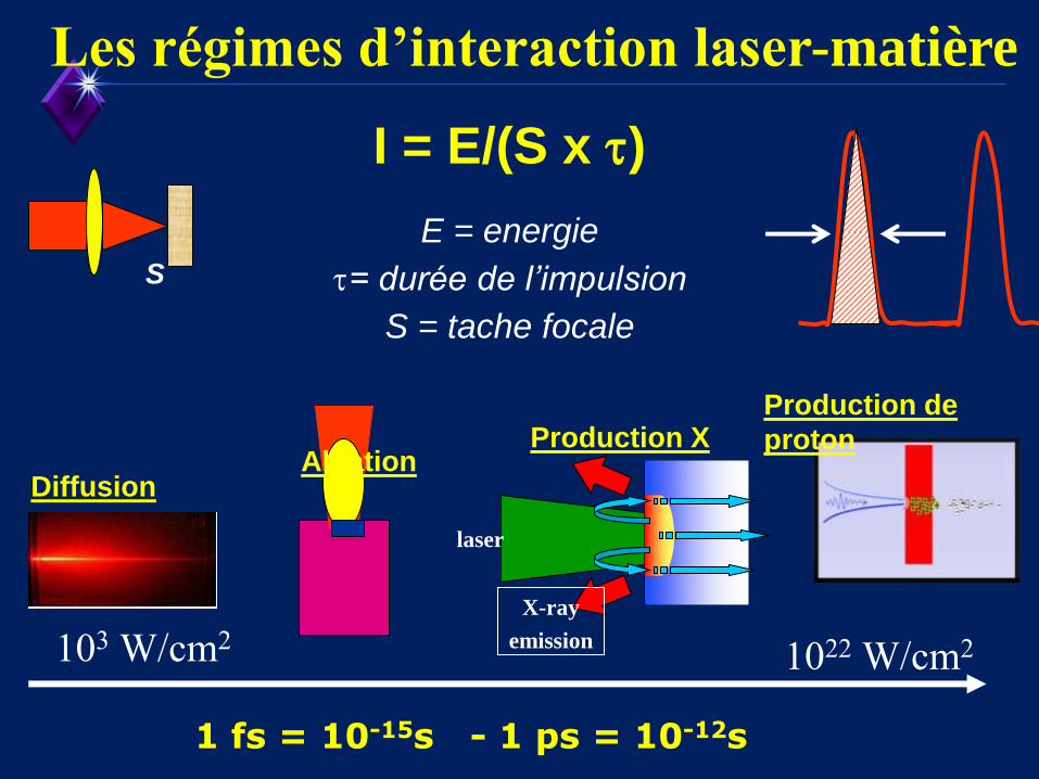
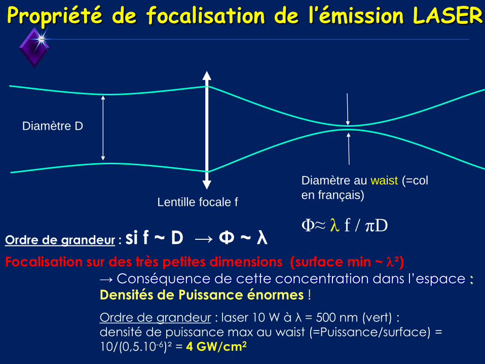
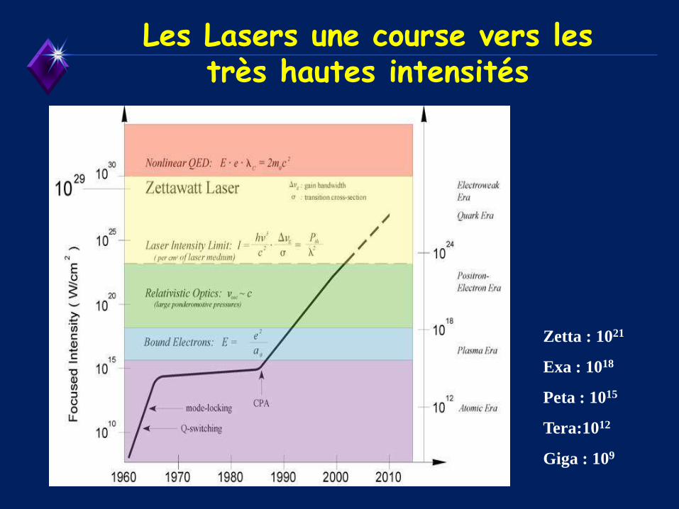
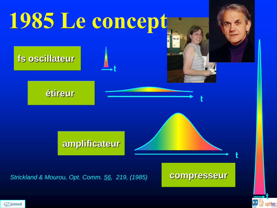
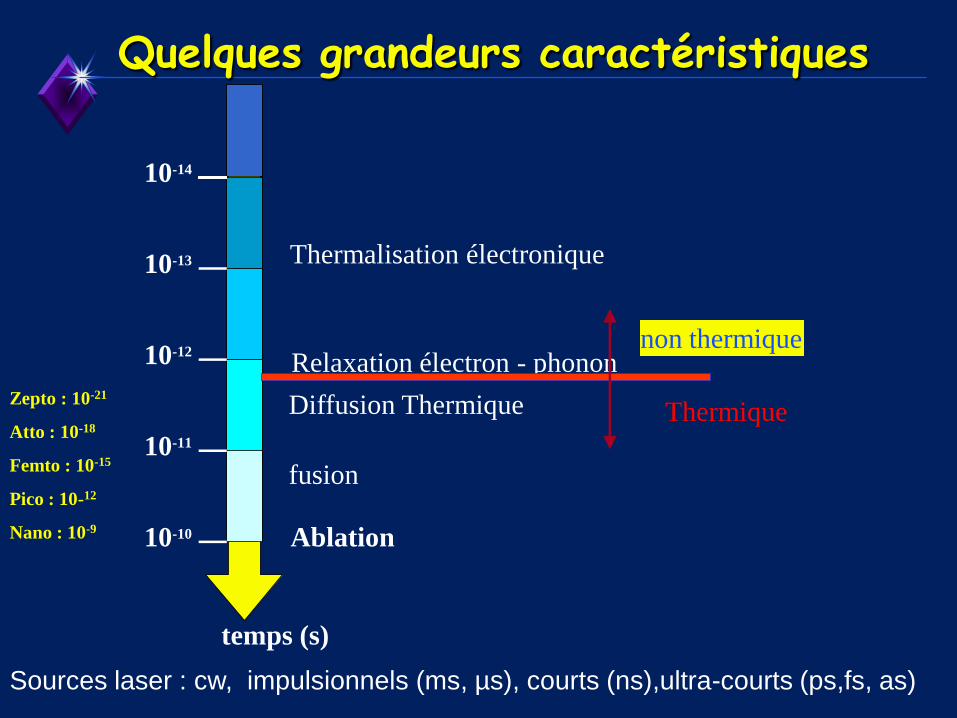
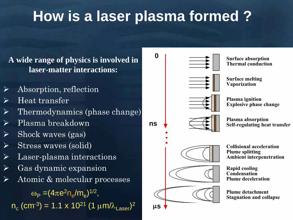
NGC 2009
Surface absorption Thermal conduction
Surface melting Vaporization
Plasma ignition Explosive phase change
Plasma absorption Self-regulating heat transfer
Collisional acceleration Plume splitting Ambient interpenetration
Rapid cooling Condensation Plume deceleration
0
ns
Plume detachment Stagnation and collapse
m s
Absorption reflection
Heat transfer
Thermodynamics (phase change)
Plasma breakdown
Shock waves (gas)
Stress waves (solid)
Laser-plasma interactions
Gas dynamic expansion
Atomic amp molecular processes
A wide range of physics is involved in
laser-matter interactions
wP =(4pe2neme)12
nc (cm-3) = 11 x 1021 (1 mmLaser)2
How is a laser plasma formed
Laser Induced Plasma in gas and
liquids at moderate intensities
I = 10 11 to 10 14 Wcm2
bull Biomedical thin films by P LD in gaseous media
bull Nanoclusters produced by laser ablation for medical imaging and therapy
bull LIBS for biomedical applications
ns to fs laser pulse
duration
Laser Induced Plasma in Gas PLD
CCD PMT
spectrometer
Target
Substrates
or Faraday
cup
laser beam
Pulsed Laser Deposition
CCD PMT
spectrometer
Target
Substrates
or Faraday
cup
laser beam
Pulsed Laser Deposition
Target Just about anything (metals semiconductorshellip)
Laser Typically excimer (UV 10 nanosecond pulses)
Vacuum Atmospheres to ultrahigh vacuum
Processes in PLD
Laser pulse
Processes in PLD
e- e-
e-
e- e-
e-
e-
e-
e-
e- e-
e-
e-
e-
Electronic excitation
Processes in PLD
e- e-
e-
e- e-
e-
e-
e-
e-
e- e-
e-
e-
e-
Energy relaxation to lattice (~1 ps)
lattice
Processes in PLD
Heat diffusion (over microseconds)
lattice
Processes in PLD
Melting (tens of ns) Evaporation Plasma
Formation (microseconds) Resolidification
lattice
Processes in PLD
lattice
If laser pulse is long (ns) or
repetition rate is high laser may
continue interactions
Processes in Pulsed Laser Deposition
1 Absorption of laser pulse in material
Qab=(1-R)Ioe-aL
(metals absorption depths ~ 10 nm depends on )
2 Relaxation of energy (~ 1 ps) (electron-phonon interaction)
3 Heat transfer Melting and Evaporation
when electrons and lattice at thermal equilibrium (long pulses)
use heat conduction equation
(or heat diffusion model)
abp QTKt
TC
)(
At peak of laser pulse temperatures on target can reach
gt105 K (gt 40 eV)
Electric Fields gt 105 Vcm also high magnetic fields
Plasma Temperatures 3000-5000 K
Ablated Species with energies 1 ndash100 eV
Initially Incredibly Non-Equilibrium
Why biomaterials
- repair and reconstruction of parts of the musculo-skeletal system of vertebrates
- minimal biological requirement biocompatibility associated with the absence of
any adverse effect (non-toxic and non-allergic)
Other requests
- resistance to physiological fluids
- should not interfere with the bodyrsquos natural immunity system
- withstand mechanical stress during whole lifetime
- manufacturability in any appropriate shape
Possible classification
-a) biologically inactive (inert) alumina zirconia stainless steel CoCrNi
CoCrMo titanium titanium alloys carbon latex PE PMMA hellip
-b) porous Calcium Phosphates (CaPs) CaP-coated metal
-c) bioactive dense calcium phosphate ceramics bioactive glasses bioactive
glass-ceramics bioactive composites hellip
-d) Resorbable tricalcium phosphate calciumaliminate polylactic acid poly-L-
acetate
Our option biocompatible porous and bioactive CaP
Acronym
Chemical formula Compound name CaP
ratio
HA Ca10(PO4)6(OH)2 Hydroxylapatite 167
FA Ca10(PO4)6F2 Fluorapatite 167
CDHA Ca10x(HPO4)x(PO4)6x(OH)2x (0x2) Calcium-deficient Hydroxylapatite 133-167
BA Ca83(PO4)43 (CO3-HPO4)17(OH)03
BA=carbonated CDHA (x=17)
Biological apatite 138-193
Mn-CHA HA with (04-2) Mn2+ and (2-6) CO32- Mn2+ doped carbonated hydroxylapatite 151- 165
OHA Ca10(PO4)6(OH)22xOxٱx (0x1) Oxyhydroxylapatite 167
OA Ca10O(PO4)6 Oxyapatite 167
MCPM Ca(H2PO4)2middotH2O Monocalcium phosphate monohydrate 05
MCPA Ca(H2PO4)2 Monocalcium phosphate anhydrate 05
DCPD CaHPO4middot2H2O Dicalcium phosphate dihydrate (Brushite) 1
DCPA CaHPO4 Dicalcium phosphate anhydrate 1
OCP Ca8(HPO4)2(PO4)4middot5H2O Octacalcium phosphate 133
a-TCP Ca3(PO4)2 (monoclinic) Tricalcium phosphate (phase a) 15
-TCP Ca3(PO4)2 (rhombohedral) Tricalcium phosphate (phase Whitlockite) 15
TTCP Ca4O(PO4)2 Tetracalcium phosphate 2
a-DCP Ca2P2O7 (orthorhombic) Dicalcium phosphate (phase a) 1
-DCP Ca2P2O7 (tetragonal) Dicalcium phosphate (phase ) 1
ACP Cax(PO4)y nH2O (Amorphous Calcium pyrophosphate) 12-22
Bone
Resorbable CaP
CaP coating
Ti
Implant
Alternative solution Biomimetic coatings for metallic implants
Main deficiency of CaPs brittle in bulk
HA molecule Ca10(PO4)6OH2
Projection in the
(001) base plan of
the hydroxyapatite
unit cell (hexagonal
structure)
How difficult is to deposit CaPs (1)
- very complex molecules
How difficult is to deposit CaPs (2)
Crystal structure of OCP projected down the c-axis
Alternating apatite- and
hydrated- layers (100)
planes
The region with shaded
atoms the ldquoapatitic layerrdquo
is very similar to HA
The zone containing 10
water molecules is the
ldquohydrated layerrdquo
H atoms are omitted for
clarity
Octacalcium phosphate
(Ca8(HPO4)2(PO4)4middot5H2O)
Mesures par spectroscopie optique
drsquoemission sur le plasma issu de lrsquoirradiation
laser 248 nm drsquoune cible drsquoHA pure
Croissance impulsion par impulsion
Preacutesence des ions de
bull calcium (Ca I et Ca II)
bull oxygegravene (O I et O II) et
bull phosphore (P II)
Eacutemissions dues agrave des espegraveces moleacuteculaires preacutesentes dans le plasma
Couche mince drsquohydroxyapatite obtenue par
ablation laser
Goutelettes
CaP = 18 CaP = 21
CaP = 17 Trois reacutegions
typiques
i Reacutegion majoritaire lisse
ii Agglomeacuteration de gouttelettes sub-micromeacutetriques
iii Gouttelettes micromeacutetriques
Human primary osteoblasts (hOB) were cultured on OCP coated-Ti
Mn-CHA coatedndashTi HA coated-Ti Ti control (polystyrene)
hOB response SEM micrographs
- on bare Ti (a) after 7 days (b) after 21 days
Elongated rod-like morphology
Bioactivity tests
- OCP coatings (c) after 7 days (d) after 21 days
- In time cells spread and expand with flattened polyhedral-morphology
- Numerous cytoplasmatic extensions
hOB response SEM micrographs
- Mn-CHA coatings (e) after 7 days (f) after 21 days
- In time cells spread and expand with flattened polyhedral-morphology
- Less cytoplasmatic extensions
hOB response SEM micrographs
Biocompatible metallic thin films by PLD
Alloys NiTinol NiTi
NiTi characteristics ndash Super-Elastic Property
ndash Radiopaque
ndash Shape-Memory Effect
NiTi biomedical applications ndash Tweezers for removing foreign objects via small incisions
ndash Anchors for tendon fixation
ndash Stents for cardiovascular applications
ndash Dentistry - Orthodontic wires which no not need to be retightened and adjusted
Deposition chamber Multitarget system
PULSED LASER DEPOSITION (PLD)
SEM
Nitinol 30 sequences Nitinol 20 sequences
Cytotoxicity evaluation on microorganism culture
(a) E coli reference cell culture
(b) E coli cell culture on Ti surface
(c) E coli cell culture on NiTi surface
(d) S cerevisiae references cell culture
(e) S cerevisiae cell culture Ti surface
(f) S cerevisiae cell culture on NiTi surface
(g) SEM on E coli cell
culture on NiTi surface
(h) SEM on S cerevisiae cell culture on
NiTi surface E coli and S cerevisiae cell
cultures on the nitinol thin films
o Biocompatible
o Low adherence of microorganism on NiTi surface NiTi alloy does not stimulate the development at genetic level for specific genes which are involved in adhesion processes for microorganism cells
o Low release of Ni ions in solutions
CONCLUSIONS
MAPLE Matrix Assisted Pulsed Laser Evaporation
bull No need for UHV
bull Matrix prepared outside the
chamber
bull High flexibility
You can use everything
you can dissolve in a
solvent
MAPLE of polymers blends with Ag
nanoparticles
Controlled release of antibacterial agents
MAPLE deposition of PEGPLGA films with incorporated silver nanoparticles (AgNPs)
0 5 10 15 20 25 30000
005
010
015
020
025
Nu
mb
er o
f E
C
oli
X109
Time (hours)
Killing activity
0 5 10 15 20 25 30 3500
05
10
15
20
25
Bact
eria
l ce
ll n
um
ber
X109
Time (hours)
Delay of bacterial growth
Antibacterial efficiency against E Coli
Direct writing by femtosecond laser
pulses
Ormosil scaffolds for tissue engineering
50 lines ormosil sample
Distance between lines =
100 μm
30x30 lines ormosil
sample Distance between
lines = 50 μm
3 layers 30x30 lines ormosil
sample Distance between
lines = 100 μm
Scaffold functionalization
Protein deposition on the scaffolds by MAPLE
- Lysozyme antibacterial antitumor uses
- Fibrinogen biocompatible protein
Fibroblast cells grown on a polymeric grid with 100 microm distance between lines
Spectroscopie drsquoun panache plasma
laser femtoseconde
Laser
t =400 ns Spot 35x35 micromsup2 Cu
Z mm
Expeacuteriences reacutealiseacutees par (Grojo et al LP3Marseille)
nanoparticules
atoms
031 Jcm-2
Formation de nano agreacutegats
AFM
(Pereira et al )
TEM
(Kabashin et al)
Eacutetude des meacutecanismes de la formation drsquoagreacutegats
Couplage
- Direct Simulation Monte Carlo
- Dynamique Moleacuteculaire
Information deacutetailleacutee
-Continu du panache
-Distribution en taille des agreacutegats
K Gouriet thegravese (2008)
Simulation par Dynamique Moleacuteculaire (MD)
=gtdistribution drsquoagreacutegats
Cluster size N
No
rmaliz
ed
Yie
ld
100
101
102
103
104
105
10-9
10-8
10-7
10-6
10-5
10-4
10-3
10-2
10-1
100
N-426
N-122
t=1 ns
τ = 15 ps F ~ 2 Fth 4010 nm t =1 ns
Zhigilei et al (Virginia University)
Target applications of mobile nanomaterials
Bioimaging
Optical imaging deals with
visualization of biological objects
or tissues in order to detect
pathogens or follow the delivery
of drugs etc nanoparticles are
uses as contrast agents
In vitro
Cellular
imaging
In vivo Cancer
detection drug delivery
etc
Light-induced
therapies
Cancer therapy nanoparticles are used
as photosensitizers to produce local
targeted destruction of cancer cells
Photodynamic
therapy
Light-induced
hyperthermia
Diagnosis and Sensing A Diseases can be diagnosed through the (simultaneous) detection of a (set of) biomolecule(s) characteristic to a specific disease type and stage (biomarkers)
Huffman Nanomedicine and Nanobiotechnology Vol 1 1 2009
D
molecular signature of sick cell of infecting agent
(eg an antibody)
Cell membrane
Nanoparticle
Coating molecule specifically attracted to the molecular signature
C A nanoparticle can be functionalized in such a way that specifically targets a biomarker Thus the detection of the nanoparticle is linked to the detection of the biomarker and to the diagnosis of a disease
B Each cell type has unique molecular signatures that differentiate healthy and sick tissues Similarly an infection can be diagnosed by detecting the distinctive molecular signature of the infecting agent
Laser ablation of material from a solid target and production of
nanoparticlesnanostructures
Production of nanoclusters by laser
ablation and their subsequent cooling in
ambient medium Gaseous ambience
Nanoclusters can be
deposited on a
substrate forming a
nanostructured film
Aqueous ambience
Nanoclusters
crystallize and form
colloidal solution
NPs produced in gaseous ambience
0 20 40 60 80 100 120 140
0
2
4
6
8
Heig
ht
[nm
]
[nm]
01 1 102
3
4
5
6
7
Mean
part
icle
heig
ht
(nm
)
Helium pressure (Torr)
Silicon nanocrystal size AFM
Gas pressure decreases Nanocrystal size decreases
AFM of isolated laser-
ablated Si particles on
HOPG
Minimal particle size is 2-3 nm (from height measurements)
001Torr
1Torr
01Torr
10Torr
100Torr
NGC 2009
Photoluminescence of size selected nanoclusters
0 1 2 3 4 5 6
39 Jcm2
Num
ber
of
clust
ers
Size (nm)
30 Jcm2
14 Jcm2
0 1 2 3 4 5 6
He 4 Torr
10 Jcm2
15 20 25 30 35 40
0
500
1000
He 4 Torr
39 Jcmsup2
30 Jcmsup2
14 Jcmsup2
10 Jcmsup2
Inte
nsi
ty o
f P
L (
ua
)Photon energy (eV)
Silicon nanoclusters
Medical Imaging A Optical properties of nanoparticles depend greatly on its structure Particularly the color (wavelength) emitted by a quantum dot (a semiconductor nanoparticle) depends on its diameter
C The quantum dots (QD) can be injected to a
subject and then be detected by exciting them to emit light
Source Department of immunology University of Toronto
Solutions of CdSe QDrsquos of different diameter
CdSe nanoparticle (QD) structure
Source Laurence Livermore Laboratories
Imaging of QDrsquos targeted on cellular structures
Nano Letters 2008 Vol 8 pp3887-3892
B
In vivo cancer targeting and imaging with
semiconductor quantum dots
Gao et al Nat Biotech 22 969 - 976 (2004)
NPs produced in aqueous ambience
Advantages of laser ablation-based nanofabrication
compared to chemical methods
Disadvantage of chemical synthesis contaminationhelliphelliphellip
In particular chemical method for the fabrication of gold nanoparticles
Reduction of chloroauric acid (HAuCl4) with citrate in water
Control size by adding a stabilizing agent (thiol- (-SH) containing molecules)
Contamination impurities Cl- on surface non-biocompatible surfactantshellip
Problems in imaging
applications (especially
in vivo)
Problems in field-
enhanced applications (eg contaminants can
provide false Raman signals)
Focalized laser beam
Liquid medium
Target
Plasma
Laser ablation in liquids a way to avoid residual
contamination
- Laser energy can be transmitted through a liquid to ablate a solid
target the nanomaterial is released to the solution
- The process can be performed without any dirty chemical products in
clean liquid environment (deionized water PBS solution) no
contamination
Experiment
Ablation of gold final colloidal solutions
-Solution are ranging from deep wine red to pink and purple
- Color related to the size dependant surface plasmon resonance
Size distribution of Au nanoparticles prepared in
distilled water
0 50 100 150 200 250 300
0
200
400
600
800
1000
Rela
tive a
bundance (
arb
units)
Particle size (nm)
0 10 20 30 40 50 60 70 80 90 100
0
200
400
600
800
1000
Re
lative
Ab
un
da
nce
(a
rb u
nits)
Particle Size (nm) 0 2 4 6 8 10
0
200
400
600
800
1000
Re
lative
Ab
un
da
nce
(a
rb u
nits)
Particle Size (nm)
F = 20F0 Jcm2 F = 5F0 Jcm2 F = F0 Jcm2
-Size and dispersion of nanoparticles can be controlled by laser fluence
- Shape of craters on target at high F suggests the presence of cavitation phenomena
4 plusmn 15 nm
A V Kabashin M Meunier J Appl Phys 94 7941 (2003)
In water Very dispersed population of nanoparticles
Dextran (MW=6000gmol) Quenches the growth of the nanoparticles 2-4nm nanoparticles are produced (230-1850 atoms) Very fast dextran-gold interaction
Influence of the concentration
Dextran 6k
In pure water
300mJ
Ablation in biopolymers (dextran PEG)
Au OH
O
-
Dextran (C6H10O5)n
Nanoparticles react with OH
groups of biopolymers
Gold Nanoparticles vs Alzheimer
A Alzheimer and other degenerative diseases are caused my the clustering of amyloidal beta (Aβ) protein
D Gold nanoparticles can be functionalized to specifically attach to aggregates of this protein (amyloidosis)
Functionalized nanoparticle
Source wwwinternetchemistrycom
Chemical structure of Aβ-protein
Source wwwthefutureofthingscom
C
B
Alzheimerrsquos brain Healthy brain
Source Berkeley Lab
Gold Nanoparticles vs Alzheimer A The functionalized gold nanoparticles selectively attach to the aggregate of amyloidal protein The microwaves of certain frequency are irradiated on the sample Resonance with the gold nanoparticles increases the local temperature and destroy the aggregate
Nanoletters 2006 Vol 6 pp110-115
Before irradiation After irradiation
LIBS for biomedical applications in vivo analysis of tissue for real-time
analysis
real-time diagnosis of pathogen
presence in human fluids (bloodurine
CSF sputum)
71 35
ICCD
spectromegravetre
Time and space
Emission spectroscopie
LIBS (Laser Induced Breakdown Spectroscopy)
Principle of sample identification screening applications based on discriminant analysis here for warning when healthy tooth material is targeted during laser drilling
BMC Oral Health 2001 1 1 Ota Samek1 Helmut H Telle2 and David CS Beddows
Laser-induced breakdown spectroscopy a tool for real-time in vitro and in vivo identification of carious teeth
Sensitive Detection of Epithelial Ovarian Cancer Biomarkers
Using Tag-Laser Induced Breakdown Spectroscopy
Yuri Markushin and Noureddine Melikechi
Optical Science Center for Applied Research Delaware State University USA
X rays produced by laser plasmas
I = ~1016 agrave 1018 Wcm2
nc cm-3
1111021 mm 2
Thot Ilas2
1 3
Ilas2
3 4
Hot electrons
(characteristic lines)
X-ray
emission
laser
X-rays
Line emission Fast electrons knock
free core electrons
from the inner shell
Bremsstrahlung electrons deflected by nucleus
Laser-generated hot electrons produce x-rays by two dominant mechanisms
ne
critical surface (cf cathode)
conventional X-ray tube
Kmetec ldquoMeV X-ray generation with a femtosecond laserrdquo Phys Rev Lett 68 1527 (1992)
Rousse ldquoEfficient Ka X-ray source from fs-laser-produced plasmasrdquo Phys Rev E 50 2200 (1994)
(typical recombination lifetimes several fs)
Hou et al Appl Phys Lett 84 2259 (2004)
Tightly focused fs lasers create the worldrsquos smallest
hard x-ray source (~4 microm) for precision imaging
x-ray source size is ~ 4 larger than laser focus
because of lateral heat transport Bowes Opt Lett 31 116 (2006)
2004 premiegravere deacutemonstration drsquoimagerie avec contraste de phase avec une source X creacutee par laser
Source
Rayon non diffracteacute
Rayon diffracteacute
Lcoh 2R1
eacutechantillon
Absorption vs contraste de phase
dsource-object dobject-detector
X-ray
Source
Object
Detector
position
Dsource
Intensity
interface
dsource-object dobject-detector
X-ray
Source
Object
Detector
position
Fresnel Diffraction
Dsource
Intensity
interface
enhancement interface
R Toth et al
Rev Sci Instrum 76
083701 (2005)
Imagerie par contraste de phase
R Toth et al Rev Sci Instrum
76 083701 (2005)
copy INRS
bull Meilleure reacutesolution (5microm)
bull dose plus faible
bull meilleur contraste (phase)
bull lrsquoeacutenergie X peut ecirctre ajusteacutee
aux besoins de lrsquoimagerie
La reacutefeacuterence mondiale ALLS
(INRS- Montreacuteal) 200TW + Puissance moyenne 50W
47J 23fs 10Hz
in a 8microm spot
Sans miroir deacuteformable
microCT
Scan duration avec
50W10Hz (360 images)
2 min (mouse scan)
courtesy Prof Dr Oswald Willi U Duumlselldorf
MeV Protons produced by laser CR39 or RCF 1010 to 1013 H+
courtesy Prof Don Umstadter U Nebraska-Lincoln
Target Normal Sheath Acceleration hot electrons traversing
target electrostatically accelerate impurity hydrogen ions on the rear surface
virtual
cathode
I ge 1019 Wcm2
Protons de 10MeV (INRS )
~1022 Wcm2
Avantages des particules chargeacutees ou hadrons (protons ions) Rayons X de la Radio-Theacuterapie
Le pic de Bragg
La profondeur de peacuteneacutetration deacutepend de l rsquoeacutenergie incidente des protons
la tumeur la plus superficielle = l rsquoœil 70 MeV
la plus profonde = la prostate 200 - 250 MeV
Protontheacuterapie par laser (projet SAPHIR)
CPO
Exemple de dosimeacutetrie preacutevisionnelle
(photons vs protons)
Comparaison de lrsquoeacutepargne de dose dans les tissus sains en protons (image de droite)
par rapport aux photons (IMRT Intensity Modulated Radio-Therapy image de gauche)
Les irradiations colateacuterales en radiotheacuterapie et en protontheacuterapie
Avantage consideacuterable pour le traitement de tumeurs localiseacutees pregraves d rsquoorganes sensibles (œil cerveau)
currently treating patients 5
5 next 2 years
others awaiting approval
10000 m2 $125 M facility opened in 2006
12 m diameter 250 MeV synchrotron
ldquoThere are too few physicists in the world
and they are an incredibly important part of
doing this We have one of the largest
physics departments in the world with more than 50 medical physicistsrdquo
--- Dr James D Cox head of Radiation Oncology at MD Anderson Cancer Center Houston Texas
Proton Therapy enables precise exposure of small tumors
with minimal damage to surrounding healthy tissue
250 MeV
PROTON beam
10 20
Depth in Tissue [cm]
0 0
50
100
DO
SE
(
)
6 MV
PHOTON
beam
tunable
Bragg
peak
but requires large expensive facilities
eye tumors 65 MeV protons
deep tumors gt200 MeV
Laser proton therapy could be much smaller amp cheaper Fourkal Med Phys 29 2788 (2002)
Malka Med Phys 31 1587 (2004)
On-site production of short-lived isotopes for medical imaging
Laser-generated quasi-mono-energetic electrons efficiently photo-activate materials of interest
bull High Rep rate bull Low cost bull Compact
Limitations to the widespread use of PET arise from the high costs of cyclotrons needed to
produce the short-lived radionucleotides for PET scanning Few hospitals and universities
are capable of maintaining such systems - Wikipedia -
radiotracer activation reaction half-life medical use
15O 16O (n)15O 2 minutes neuro-imaging
11C 12C(n)11C 20 minutes neuro-receptor-specific brain imaging
18F 19F(n)18F 110 minutes clinical oncology
Positron Emission Tomography
on-site
production
essential
18F PET scan of tumor 15O PET scan of human brain
Reed ldquoEfficient initiation of photonuclear reactions using quasi-monoenergetic electron beams from LWFArdquo J Appl Phys 102 073103 (2007)
TEP in situ
+ in vivo
Modegravele animal
microCT +
contraste de phase
+ in vivo
modegraveles de cancer
Comprendre
le cancer
Preacuteparer le traitement
protons 50MeV
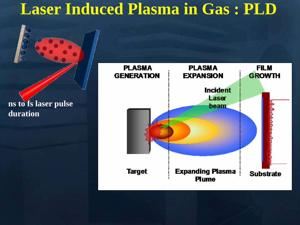
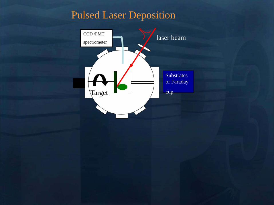
CCD PMT
spectrometer
Target
Substrates
or Faraday
cup
laser beam
Pulsed Laser Deposition
CCD PMT
spectrometer
Target
Substrates
or Faraday
cup
laser beam
Pulsed Laser Deposition
Target Just about anything (metals semiconductorshellip)
Laser Typically excimer (UV 10 nanosecond pulses)
Vacuum Atmospheres to ultrahigh vacuum
Processes in PLD
Laser pulse
Processes in PLD
e- e-
e-
e- e-
e-
e-
e-
e-
e- e-
e-
e-
e-
Electronic excitation
Processes in PLD
e- e-
e-
e- e-
e-
e-
e-
e-
e- e-
e-
e-
e-
Energy relaxation to lattice (~1 ps)
lattice
Processes in PLD
Heat diffusion (over microseconds)
lattice
Processes in PLD
Melting (tens of ns) Evaporation Plasma
Formation (microseconds) Resolidification
lattice
Processes in PLD
lattice
If laser pulse is long (ns) or
repetition rate is high laser may
continue interactions
Processes in Pulsed Laser Deposition
1 Absorption of laser pulse in material
Qab=(1-R)Ioe-aL
(metals absorption depths ~ 10 nm depends on )
2 Relaxation of energy (~ 1 ps) (electron-phonon interaction)
3 Heat transfer Melting and Evaporation
when electrons and lattice at thermal equilibrium (long pulses)
use heat conduction equation
(or heat diffusion model)
abp QTKt
TC
)(
At peak of laser pulse temperatures on target can reach
gt105 K (gt 40 eV)
Electric Fields gt 105 Vcm also high magnetic fields
Plasma Temperatures 3000-5000 K
Ablated Species with energies 1 ndash100 eV
Initially Incredibly Non-Equilibrium
Why biomaterials
- repair and reconstruction of parts of the musculo-skeletal system of vertebrates
- minimal biological requirement biocompatibility associated with the absence of
any adverse effect (non-toxic and non-allergic)
Other requests
- resistance to physiological fluids
- should not interfere with the bodyrsquos natural immunity system
- withstand mechanical stress during whole lifetime
- manufacturability in any appropriate shape
Possible classification
-a) biologically inactive (inert) alumina zirconia stainless steel CoCrNi
CoCrMo titanium titanium alloys carbon latex PE PMMA hellip
-b) porous Calcium Phosphates (CaPs) CaP-coated metal
-c) bioactive dense calcium phosphate ceramics bioactive glasses bioactive
glass-ceramics bioactive composites hellip
-d) Resorbable tricalcium phosphate calciumaliminate polylactic acid poly-L-
acetate
Our option biocompatible porous and bioactive CaP
Acronym
Chemical formula Compound name CaP
ratio
HA Ca10(PO4)6(OH)2 Hydroxylapatite 167
FA Ca10(PO4)6F2 Fluorapatite 167
CDHA Ca10x(HPO4)x(PO4)6x(OH)2x (0x2) Calcium-deficient Hydroxylapatite 133-167
BA Ca83(PO4)43 (CO3-HPO4)17(OH)03
BA=carbonated CDHA (x=17)
Biological apatite 138-193
Mn-CHA HA with (04-2) Mn2+ and (2-6) CO32- Mn2+ doped carbonated hydroxylapatite 151- 165
OHA Ca10(PO4)6(OH)22xOxٱx (0x1) Oxyhydroxylapatite 167
OA Ca10O(PO4)6 Oxyapatite 167
MCPM Ca(H2PO4)2middotH2O Monocalcium phosphate monohydrate 05
MCPA Ca(H2PO4)2 Monocalcium phosphate anhydrate 05
DCPD CaHPO4middot2H2O Dicalcium phosphate dihydrate (Brushite) 1
DCPA CaHPO4 Dicalcium phosphate anhydrate 1
OCP Ca8(HPO4)2(PO4)4middot5H2O Octacalcium phosphate 133
a-TCP Ca3(PO4)2 (monoclinic) Tricalcium phosphate (phase a) 15
-TCP Ca3(PO4)2 (rhombohedral) Tricalcium phosphate (phase Whitlockite) 15
TTCP Ca4O(PO4)2 Tetracalcium phosphate 2
a-DCP Ca2P2O7 (orthorhombic) Dicalcium phosphate (phase a) 1
-DCP Ca2P2O7 (tetragonal) Dicalcium phosphate (phase ) 1
ACP Cax(PO4)y nH2O (Amorphous Calcium pyrophosphate) 12-22
Bone
Resorbable CaP
CaP coating
Ti
Implant
Alternative solution Biomimetic coatings for metallic implants
Main deficiency of CaPs brittle in bulk
HA molecule Ca10(PO4)6OH2
Projection in the
(001) base plan of
the hydroxyapatite
unit cell (hexagonal
structure)
How difficult is to deposit CaPs (1)
- very complex molecules
How difficult is to deposit CaPs (2)
Crystal structure of OCP projected down the c-axis
Alternating apatite- and
hydrated- layers (100)
planes
The region with shaded
atoms the ldquoapatitic layerrdquo
is very similar to HA
The zone containing 10
water molecules is the
ldquohydrated layerrdquo
H atoms are omitted for
clarity
Octacalcium phosphate
(Ca8(HPO4)2(PO4)4middot5H2O)
Mesures par spectroscopie optique
drsquoemission sur le plasma issu de lrsquoirradiation
laser 248 nm drsquoune cible drsquoHA pure
Croissance impulsion par impulsion
Preacutesence des ions de
bull calcium (Ca I et Ca II)
bull oxygegravene (O I et O II) et
bull phosphore (P II)
Eacutemissions dues agrave des espegraveces moleacuteculaires preacutesentes dans le plasma
Couche mince drsquohydroxyapatite obtenue par
ablation laser
Goutelettes
CaP = 18 CaP = 21
CaP = 17 Trois reacutegions
typiques
i Reacutegion majoritaire lisse
ii Agglomeacuteration de gouttelettes sub-micromeacutetriques
iii Gouttelettes micromeacutetriques
Human primary osteoblasts (hOB) were cultured on OCP coated-Ti
Mn-CHA coatedndashTi HA coated-Ti Ti control (polystyrene)
hOB response SEM micrographs
- on bare Ti (a) after 7 days (b) after 21 days
Elongated rod-like morphology
Bioactivity tests
- OCP coatings (c) after 7 days (d) after 21 days
- In time cells spread and expand with flattened polyhedral-morphology
- Numerous cytoplasmatic extensions
hOB response SEM micrographs
- Mn-CHA coatings (e) after 7 days (f) after 21 days
- In time cells spread and expand with flattened polyhedral-morphology
- Less cytoplasmatic extensions
hOB response SEM micrographs
Biocompatible metallic thin films by PLD
Alloys NiTinol NiTi
NiTi characteristics ndash Super-Elastic Property
ndash Radiopaque
ndash Shape-Memory Effect
NiTi biomedical applications ndash Tweezers for removing foreign objects via small incisions
ndash Anchors for tendon fixation
ndash Stents for cardiovascular applications
ndash Dentistry - Orthodontic wires which no not need to be retightened and adjusted
Deposition chamber Multitarget system
PULSED LASER DEPOSITION (PLD)
SEM
Nitinol 30 sequences Nitinol 20 sequences
Cytotoxicity evaluation on microorganism culture
(a) E coli reference cell culture
(b) E coli cell culture on Ti surface
(c) E coli cell culture on NiTi surface
(d) S cerevisiae references cell culture
(e) S cerevisiae cell culture Ti surface
(f) S cerevisiae cell culture on NiTi surface
(g) SEM on E coli cell
culture on NiTi surface
(h) SEM on S cerevisiae cell culture on
NiTi surface E coli and S cerevisiae cell
cultures on the nitinol thin films
o Biocompatible
o Low adherence of microorganism on NiTi surface NiTi alloy does not stimulate the development at genetic level for specific genes which are involved in adhesion processes for microorganism cells
o Low release of Ni ions in solutions
CONCLUSIONS
MAPLE Matrix Assisted Pulsed Laser Evaporation
bull No need for UHV
bull Matrix prepared outside the
chamber
bull High flexibility
You can use everything
you can dissolve in a
solvent
MAPLE of polymers blends with Ag
nanoparticles
Controlled release of antibacterial agents
MAPLE deposition of PEGPLGA films with incorporated silver nanoparticles (AgNPs)
0 5 10 15 20 25 30000
005
010
015
020
025
Nu
mb
er o
f E
C
oli
X109
Time (hours)
Killing activity
0 5 10 15 20 25 30 3500
05
10
15
20
25
Bact
eria
l ce
ll n
um
ber
X109
Time (hours)
Delay of bacterial growth
Antibacterial efficiency against E Coli
Direct writing by femtosecond laser
pulses
Ormosil scaffolds for tissue engineering
50 lines ormosil sample
Distance between lines =
100 μm
30x30 lines ormosil
sample Distance between
lines = 50 μm
3 layers 30x30 lines ormosil
sample Distance between
lines = 100 μm
Scaffold functionalization
Protein deposition on the scaffolds by MAPLE
- Lysozyme antibacterial antitumor uses
- Fibrinogen biocompatible protein
Fibroblast cells grown on a polymeric grid with 100 microm distance between lines
Spectroscopie drsquoun panache plasma
laser femtoseconde
Laser
t =400 ns Spot 35x35 micromsup2 Cu
Z mm
Expeacuteriences reacutealiseacutees par (Grojo et al LP3Marseille)
nanoparticules
atoms
031 Jcm-2
Formation de nano agreacutegats
AFM
(Pereira et al )
TEM
(Kabashin et al)
Eacutetude des meacutecanismes de la formation drsquoagreacutegats
Couplage
- Direct Simulation Monte Carlo
- Dynamique Moleacuteculaire
Information deacutetailleacutee
-Continu du panache
-Distribution en taille des agreacutegats
K Gouriet thegravese (2008)
Simulation par Dynamique Moleacuteculaire (MD)
=gtdistribution drsquoagreacutegats
Cluster size N
No
rmaliz
ed
Yie
ld
100
101
102
103
104
105
10-9
10-8
10-7
10-6
10-5
10-4
10-3
10-2
10-1
100
N-426
N-122
t=1 ns
τ = 15 ps F ~ 2 Fth 4010 nm t =1 ns
Zhigilei et al (Virginia University)
Target applications of mobile nanomaterials
Bioimaging
Optical imaging deals with
visualization of biological objects
or tissues in order to detect
pathogens or follow the delivery
of drugs etc nanoparticles are
uses as contrast agents
In vitro
Cellular
imaging
In vivo Cancer
detection drug delivery
etc
Light-induced
therapies
Cancer therapy nanoparticles are used
as photosensitizers to produce local
targeted destruction of cancer cells
Photodynamic
therapy
Light-induced
hyperthermia
Diagnosis and Sensing A Diseases can be diagnosed through the (simultaneous) detection of a (set of) biomolecule(s) characteristic to a specific disease type and stage (biomarkers)
Huffman Nanomedicine and Nanobiotechnology Vol 1 1 2009
D
molecular signature of sick cell of infecting agent
(eg an antibody)
Cell membrane
Nanoparticle
Coating molecule specifically attracted to the molecular signature
C A nanoparticle can be functionalized in such a way that specifically targets a biomarker Thus the detection of the nanoparticle is linked to the detection of the biomarker and to the diagnosis of a disease
B Each cell type has unique molecular signatures that differentiate healthy and sick tissues Similarly an infection can be diagnosed by detecting the distinctive molecular signature of the infecting agent
Laser ablation of material from a solid target and production of
nanoparticlesnanostructures
Production of nanoclusters by laser
ablation and their subsequent cooling in
ambient medium Gaseous ambience
Nanoclusters can be
deposited on a
substrate forming a
nanostructured film
Aqueous ambience
Nanoclusters
crystallize and form
colloidal solution
NPs produced in gaseous ambience
0 20 40 60 80 100 120 140
0
2
4
6
8
Heig
ht
[nm
]
[nm]
01 1 102
3
4
5
6
7
Mean
part
icle
heig
ht
(nm
)
Helium pressure (Torr)
Silicon nanocrystal size AFM
Gas pressure decreases Nanocrystal size decreases
AFM of isolated laser-
ablated Si particles on
HOPG
Minimal particle size is 2-3 nm (from height measurements)
001Torr
1Torr
01Torr
10Torr
100Torr
NGC 2009
Photoluminescence of size selected nanoclusters
0 1 2 3 4 5 6
39 Jcm2
Num
ber
of
clust
ers
Size (nm)
30 Jcm2
14 Jcm2
0 1 2 3 4 5 6
He 4 Torr
10 Jcm2
15 20 25 30 35 40
0
500
1000
He 4 Torr
39 Jcmsup2
30 Jcmsup2
14 Jcmsup2
10 Jcmsup2
Inte
nsi
ty o
f P
L (
ua
)Photon energy (eV)
Silicon nanoclusters
Medical Imaging A Optical properties of nanoparticles depend greatly on its structure Particularly the color (wavelength) emitted by a quantum dot (a semiconductor nanoparticle) depends on its diameter
C The quantum dots (QD) can be injected to a
subject and then be detected by exciting them to emit light
Source Department of immunology University of Toronto
Solutions of CdSe QDrsquos of different diameter
CdSe nanoparticle (QD) structure
Source Laurence Livermore Laboratories
Imaging of QDrsquos targeted on cellular structures
Nano Letters 2008 Vol 8 pp3887-3892
B
In vivo cancer targeting and imaging with
semiconductor quantum dots
Gao et al Nat Biotech 22 969 - 976 (2004)
NPs produced in aqueous ambience
Advantages of laser ablation-based nanofabrication
compared to chemical methods
Disadvantage of chemical synthesis contaminationhelliphelliphellip
In particular chemical method for the fabrication of gold nanoparticles
Reduction of chloroauric acid (HAuCl4) with citrate in water
Control size by adding a stabilizing agent (thiol- (-SH) containing molecules)
Contamination impurities Cl- on surface non-biocompatible surfactantshellip
Problems in imaging
applications (especially
in vivo)
Problems in field-
enhanced applications (eg contaminants can
provide false Raman signals)
Focalized laser beam
Liquid medium
Target
Plasma
Laser ablation in liquids a way to avoid residual
contamination
- Laser energy can be transmitted through a liquid to ablate a solid
target the nanomaterial is released to the solution
- The process can be performed without any dirty chemical products in
clean liquid environment (deionized water PBS solution) no
contamination
Experiment
Ablation of gold final colloidal solutions
-Solution are ranging from deep wine red to pink and purple
- Color related to the size dependant surface plasmon resonance
Size distribution of Au nanoparticles prepared in
distilled water
0 50 100 150 200 250 300
0
200
400
600
800
1000
Rela
tive a
bundance (
arb
units)
Particle size (nm)
0 10 20 30 40 50 60 70 80 90 100
0
200
400
600
800
1000
Re
lative
Ab
un
da
nce
(a
rb u
nits)
Particle Size (nm) 0 2 4 6 8 10
0
200
400
600
800
1000
Re
lative
Ab
un
da
nce
(a
rb u
nits)
Particle Size (nm)
F = 20F0 Jcm2 F = 5F0 Jcm2 F = F0 Jcm2
-Size and dispersion of nanoparticles can be controlled by laser fluence
- Shape of craters on target at high F suggests the presence of cavitation phenomena
4 plusmn 15 nm
A V Kabashin M Meunier J Appl Phys 94 7941 (2003)
In water Very dispersed population of nanoparticles
Dextran (MW=6000gmol) Quenches the growth of the nanoparticles 2-4nm nanoparticles are produced (230-1850 atoms) Very fast dextran-gold interaction
Influence of the concentration
Dextran 6k
In pure water
300mJ
Ablation in biopolymers (dextran PEG)
Au OH
O
-
Dextran (C6H10O5)n
Nanoparticles react with OH
groups of biopolymers
Gold Nanoparticles vs Alzheimer
A Alzheimer and other degenerative diseases are caused my the clustering of amyloidal beta (Aβ) protein
D Gold nanoparticles can be functionalized to specifically attach to aggregates of this protein (amyloidosis)
Functionalized nanoparticle
Source wwwinternetchemistrycom
Chemical structure of Aβ-protein
Source wwwthefutureofthingscom
C
B
Alzheimerrsquos brain Healthy brain
Source Berkeley Lab
Gold Nanoparticles vs Alzheimer A The functionalized gold nanoparticles selectively attach to the aggregate of amyloidal protein The microwaves of certain frequency are irradiated on the sample Resonance with the gold nanoparticles increases the local temperature and destroy the aggregate
Nanoletters 2006 Vol 6 pp110-115
Before irradiation After irradiation
LIBS for biomedical applications in vivo analysis of tissue for real-time
analysis
real-time diagnosis of pathogen
presence in human fluids (bloodurine
CSF sputum)
71 35
ICCD
spectromegravetre
Time and space
Emission spectroscopie
LIBS (Laser Induced Breakdown Spectroscopy)
Principle of sample identification screening applications based on discriminant analysis here for warning when healthy tooth material is targeted during laser drilling
BMC Oral Health 2001 1 1 Ota Samek1 Helmut H Telle2 and David CS Beddows
Laser-induced breakdown spectroscopy a tool for real-time in vitro and in vivo identification of carious teeth
Sensitive Detection of Epithelial Ovarian Cancer Biomarkers
Using Tag-Laser Induced Breakdown Spectroscopy
Yuri Markushin and Noureddine Melikechi
Optical Science Center for Applied Research Delaware State University USA
X rays produced by laser plasmas
I = ~1016 agrave 1018 Wcm2
nc cm-3
1111021 mm 2
Thot Ilas2
1 3
Ilas2
3 4
Hot electrons
(characteristic lines)
X-ray
emission
laser
X-rays
Line emission Fast electrons knock
free core electrons
from the inner shell
Bremsstrahlung electrons deflected by nucleus
Laser-generated hot electrons produce x-rays by two dominant mechanisms
ne
critical surface (cf cathode)
conventional X-ray tube
Kmetec ldquoMeV X-ray generation with a femtosecond laserrdquo Phys Rev Lett 68 1527 (1992)
Rousse ldquoEfficient Ka X-ray source from fs-laser-produced plasmasrdquo Phys Rev E 50 2200 (1994)
(typical recombination lifetimes several fs)
Hou et al Appl Phys Lett 84 2259 (2004)
Tightly focused fs lasers create the worldrsquos smallest
hard x-ray source (~4 microm) for precision imaging
x-ray source size is ~ 4 larger than laser focus
because of lateral heat transport Bowes Opt Lett 31 116 (2006)
2004 premiegravere deacutemonstration drsquoimagerie avec contraste de phase avec une source X creacutee par laser
Source
Rayon non diffracteacute
Rayon diffracteacute
Lcoh 2R1
eacutechantillon
Absorption vs contraste de phase
dsource-object dobject-detector
X-ray
Source
Object
Detector
position
Dsource
Intensity
interface
dsource-object dobject-detector
X-ray
Source
Object
Detector
position
Fresnel Diffraction
Dsource
Intensity
interface
enhancement interface
R Toth et al
Rev Sci Instrum 76
083701 (2005)
Imagerie par contraste de phase
R Toth et al Rev Sci Instrum
76 083701 (2005)
copy INRS
bull Meilleure reacutesolution (5microm)
bull dose plus faible
bull meilleur contraste (phase)
bull lrsquoeacutenergie X peut ecirctre ajusteacutee
aux besoins de lrsquoimagerie
La reacutefeacuterence mondiale ALLS
(INRS- Montreacuteal) 200TW + Puissance moyenne 50W
47J 23fs 10Hz
in a 8microm spot
Sans miroir deacuteformable
microCT
Scan duration avec
50W10Hz (360 images)
2 min (mouse scan)
courtesy Prof Dr Oswald Willi U Duumlselldorf
MeV Protons produced by laser CR39 or RCF 1010 to 1013 H+
courtesy Prof Don Umstadter U Nebraska-Lincoln
Target Normal Sheath Acceleration hot electrons traversing
target electrostatically accelerate impurity hydrogen ions on the rear surface
virtual
cathode
I ge 1019 Wcm2
Protons de 10MeV (INRS )
~1022 Wcm2
Avantages des particules chargeacutees ou hadrons (protons ions) Rayons X de la Radio-Theacuterapie
Le pic de Bragg
La profondeur de peacuteneacutetration deacutepend de l rsquoeacutenergie incidente des protons
la tumeur la plus superficielle = l rsquoœil 70 MeV
la plus profonde = la prostate 200 - 250 MeV
Protontheacuterapie par laser (projet SAPHIR)
CPO
Exemple de dosimeacutetrie preacutevisionnelle
(photons vs protons)
Comparaison de lrsquoeacutepargne de dose dans les tissus sains en protons (image de droite)
par rapport aux photons (IMRT Intensity Modulated Radio-Therapy image de gauche)
Les irradiations colateacuterales en radiotheacuterapie et en protontheacuterapie
Avantage consideacuterable pour le traitement de tumeurs localiseacutees pregraves d rsquoorganes sensibles (œil cerveau)
currently treating patients 5
5 next 2 years
others awaiting approval
10000 m2 $125 M facility opened in 2006
12 m diameter 250 MeV synchrotron
ldquoThere are too few physicists in the world
and they are an incredibly important part of
doing this We have one of the largest
physics departments in the world with more than 50 medical physicistsrdquo
--- Dr James D Cox head of Radiation Oncology at MD Anderson Cancer Center Houston Texas
Proton Therapy enables precise exposure of small tumors
with minimal damage to surrounding healthy tissue
250 MeV
PROTON beam
10 20
Depth in Tissue [cm]
0 0
50
100
DO
SE
(
)
6 MV
PHOTON
beam
tunable
Bragg
peak
but requires large expensive facilities
eye tumors 65 MeV protons
deep tumors gt200 MeV
Laser proton therapy could be much smaller amp cheaper Fourkal Med Phys 29 2788 (2002)
Malka Med Phys 31 1587 (2004)
On-site production of short-lived isotopes for medical imaging
Laser-generated quasi-mono-energetic electrons efficiently photo-activate materials of interest
bull High Rep rate bull Low cost bull Compact
Limitations to the widespread use of PET arise from the high costs of cyclotrons needed to
produce the short-lived radionucleotides for PET scanning Few hospitals and universities
are capable of maintaining such systems - Wikipedia -
radiotracer activation reaction half-life medical use
15O 16O (n)15O 2 minutes neuro-imaging
11C 12C(n)11C 20 minutes neuro-receptor-specific brain imaging
18F 19F(n)18F 110 minutes clinical oncology
Positron Emission Tomography
on-site
production
essential
18F PET scan of tumor 15O PET scan of human brain
Reed ldquoEfficient initiation of photonuclear reactions using quasi-monoenergetic electron beams from LWFArdquo J Appl Phys 102 073103 (2007)
TEP in situ
+ in vivo
Modegravele animal
microCT +
contraste de phase
+ in vivo
modegraveles de cancer
Comprendre
le cancer
Preacuteparer le traitement
protons 50MeV
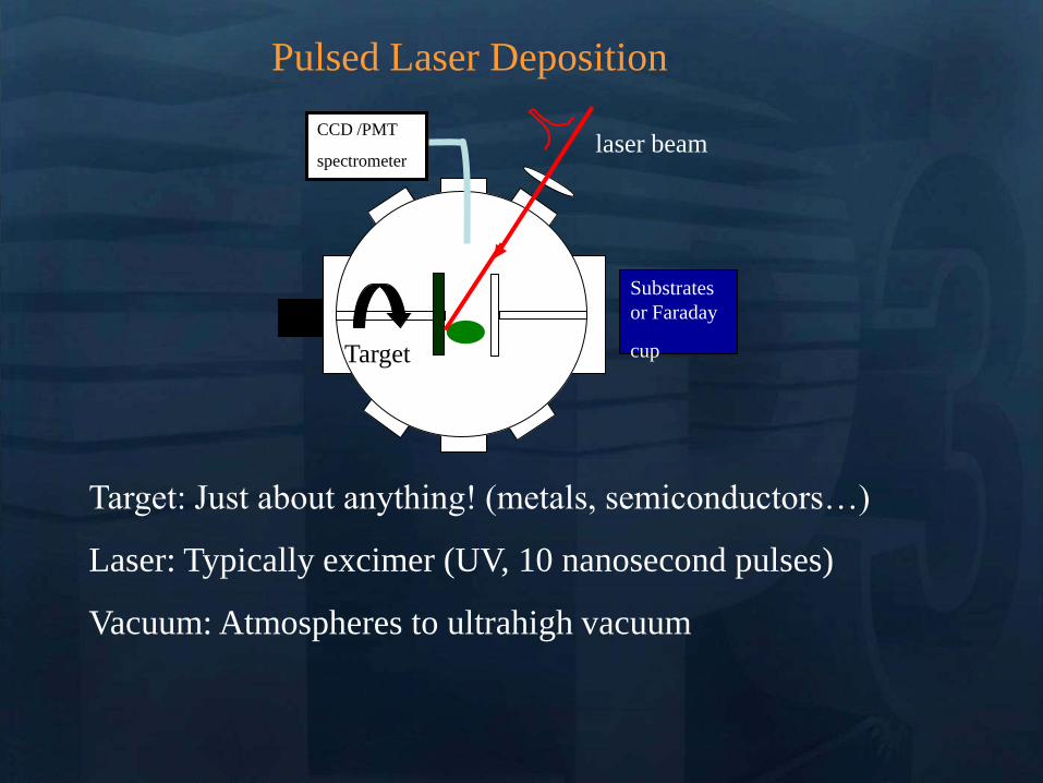
CCD PMT
spectrometer
Target
Substrates
or Faraday
cup
laser beam
Pulsed Laser Deposition
Target Just about anything (metals semiconductorshellip)
Laser Typically excimer (UV 10 nanosecond pulses)
Vacuum Atmospheres to ultrahigh vacuum
Processes in PLD
Laser pulse
Processes in PLD
e- e-
e-
e- e-
e-
e-
e-
e-
e- e-
e-
e-
e-
Electronic excitation
Processes in PLD
e- e-
e-
e- e-
e-
e-
e-
e-
e- e-
e-
e-
e-
Energy relaxation to lattice (~1 ps)
lattice
Processes in PLD
Heat diffusion (over microseconds)
lattice
Processes in PLD
Melting (tens of ns) Evaporation Plasma
Formation (microseconds) Resolidification
lattice
Processes in PLD
lattice
If laser pulse is long (ns) or
repetition rate is high laser may
continue interactions
Processes in Pulsed Laser Deposition
1 Absorption of laser pulse in material
Qab=(1-R)Ioe-aL
(metals absorption depths ~ 10 nm depends on )
2 Relaxation of energy (~ 1 ps) (electron-phonon interaction)
3 Heat transfer Melting and Evaporation
when electrons and lattice at thermal equilibrium (long pulses)
use heat conduction equation
(or heat diffusion model)
abp QTKt
TC
)(
At peak of laser pulse temperatures on target can reach
gt105 K (gt 40 eV)
Electric Fields gt 105 Vcm also high magnetic fields
Plasma Temperatures 3000-5000 K
Ablated Species with energies 1 ndash100 eV
Initially Incredibly Non-Equilibrium
Why biomaterials
- repair and reconstruction of parts of the musculo-skeletal system of vertebrates
- minimal biological requirement biocompatibility associated with the absence of
any adverse effect (non-toxic and non-allergic)
Other requests
- resistance to physiological fluids
- should not interfere with the bodyrsquos natural immunity system
- withstand mechanical stress during whole lifetime
- manufacturability in any appropriate shape
Possible classification
-a) biologically inactive (inert) alumina zirconia stainless steel CoCrNi
CoCrMo titanium titanium alloys carbon latex PE PMMA hellip
-b) porous Calcium Phosphates (CaPs) CaP-coated metal
-c) bioactive dense calcium phosphate ceramics bioactive glasses bioactive
glass-ceramics bioactive composites hellip
-d) Resorbable tricalcium phosphate calciumaliminate polylactic acid poly-L-
acetate
Our option biocompatible porous and bioactive CaP
Acronym
Chemical formula Compound name CaP
ratio
HA Ca10(PO4)6(OH)2 Hydroxylapatite 167
FA Ca10(PO4)6F2 Fluorapatite 167
CDHA Ca10x(HPO4)x(PO4)6x(OH)2x (0x2) Calcium-deficient Hydroxylapatite 133-167
BA Ca83(PO4)43 (CO3-HPO4)17(OH)03
BA=carbonated CDHA (x=17)
Biological apatite 138-193
Mn-CHA HA with (04-2) Mn2+ and (2-6) CO32- Mn2+ doped carbonated hydroxylapatite 151- 165
OHA Ca10(PO4)6(OH)22xOxٱx (0x1) Oxyhydroxylapatite 167
OA Ca10O(PO4)6 Oxyapatite 167
MCPM Ca(H2PO4)2middotH2O Monocalcium phosphate monohydrate 05
MCPA Ca(H2PO4)2 Monocalcium phosphate anhydrate 05
DCPD CaHPO4middot2H2O Dicalcium phosphate dihydrate (Brushite) 1
DCPA CaHPO4 Dicalcium phosphate anhydrate 1
OCP Ca8(HPO4)2(PO4)4middot5H2O Octacalcium phosphate 133
a-TCP Ca3(PO4)2 (monoclinic) Tricalcium phosphate (phase a) 15
-TCP Ca3(PO4)2 (rhombohedral) Tricalcium phosphate (phase Whitlockite) 15
TTCP Ca4O(PO4)2 Tetracalcium phosphate 2
a-DCP Ca2P2O7 (orthorhombic) Dicalcium phosphate (phase a) 1
-DCP Ca2P2O7 (tetragonal) Dicalcium phosphate (phase ) 1
ACP Cax(PO4)y nH2O (Amorphous Calcium pyrophosphate) 12-22
Bone
Resorbable CaP
CaP coating
Ti
Implant
Alternative solution Biomimetic coatings for metallic implants
Main deficiency of CaPs brittle in bulk
HA molecule Ca10(PO4)6OH2
Projection in the
(001) base plan of
the hydroxyapatite
unit cell (hexagonal
structure)
How difficult is to deposit CaPs (1)
- very complex molecules
How difficult is to deposit CaPs (2)
Crystal structure of OCP projected down the c-axis
Alternating apatite- and
hydrated- layers (100)
planes
The region with shaded
atoms the ldquoapatitic layerrdquo
is very similar to HA
The zone containing 10
water molecules is the
ldquohydrated layerrdquo
H atoms are omitted for
clarity
Octacalcium phosphate
(Ca8(HPO4)2(PO4)4middot5H2O)
Mesures par spectroscopie optique
drsquoemission sur le plasma issu de lrsquoirradiation
laser 248 nm drsquoune cible drsquoHA pure
Croissance impulsion par impulsion
Preacutesence des ions de
bull calcium (Ca I et Ca II)
bull oxygegravene (O I et O II) et
bull phosphore (P II)
Eacutemissions dues agrave des espegraveces moleacuteculaires preacutesentes dans le plasma
Couche mince drsquohydroxyapatite obtenue par
ablation laser
Goutelettes
CaP = 18 CaP = 21
CaP = 17 Trois reacutegions
typiques
i Reacutegion majoritaire lisse
ii Agglomeacuteration de gouttelettes sub-micromeacutetriques
iii Gouttelettes micromeacutetriques
Human primary osteoblasts (hOB) were cultured on OCP coated-Ti
Mn-CHA coatedndashTi HA coated-Ti Ti control (polystyrene)
hOB response SEM micrographs
- on bare Ti (a) after 7 days (b) after 21 days
Elongated rod-like morphology
Bioactivity tests
- OCP coatings (c) after 7 days (d) after 21 days
- In time cells spread and expand with flattened polyhedral-morphology
- Numerous cytoplasmatic extensions
hOB response SEM micrographs
- Mn-CHA coatings (e) after 7 days (f) after 21 days
- In time cells spread and expand with flattened polyhedral-morphology
- Less cytoplasmatic extensions
hOB response SEM micrographs
Biocompatible metallic thin films by PLD
Alloys NiTinol NiTi
NiTi characteristics ndash Super-Elastic Property
ndash Radiopaque
ndash Shape-Memory Effect
NiTi biomedical applications ndash Tweezers for removing foreign objects via small incisions
ndash Anchors for tendon fixation
ndash Stents for cardiovascular applications
ndash Dentistry - Orthodontic wires which no not need to be retightened and adjusted
Deposition chamber Multitarget system
PULSED LASER DEPOSITION (PLD)
SEM
Nitinol 30 sequences Nitinol 20 sequences
Cytotoxicity evaluation on microorganism culture
(a) E coli reference cell culture
(b) E coli cell culture on Ti surface
(c) E coli cell culture on NiTi surface
(d) S cerevisiae references cell culture
(e) S cerevisiae cell culture Ti surface
(f) S cerevisiae cell culture on NiTi surface
(g) SEM on E coli cell
culture on NiTi surface
(h) SEM on S cerevisiae cell culture on
NiTi surface E coli and S cerevisiae cell
cultures on the nitinol thin films
o Biocompatible
o Low adherence of microorganism on NiTi surface NiTi alloy does not stimulate the development at genetic level for specific genes which are involved in adhesion processes for microorganism cells
o Low release of Ni ions in solutions
CONCLUSIONS
MAPLE Matrix Assisted Pulsed Laser Evaporation
bull No need for UHV
bull Matrix prepared outside the
chamber
bull High flexibility
You can use everything
you can dissolve in a
solvent
MAPLE of polymers blends with Ag
nanoparticles
Controlled release of antibacterial agents
MAPLE deposition of PEGPLGA films with incorporated silver nanoparticles (AgNPs)
0 5 10 15 20 25 30000
005
010
015
020
025
Nu
mb
er o
f E
C
oli
X109
Time (hours)
Killing activity
0 5 10 15 20 25 30 3500
05
10
15
20
25
Bact
eria
l ce
ll n
um
ber
X109
Time (hours)
Delay of bacterial growth
Antibacterial efficiency against E Coli
Direct writing by femtosecond laser
pulses
Ormosil scaffolds for tissue engineering
50 lines ormosil sample
Distance between lines =
100 μm
30x30 lines ormosil
sample Distance between
lines = 50 μm
3 layers 30x30 lines ormosil
sample Distance between
lines = 100 μm
Scaffold functionalization
Protein deposition on the scaffolds by MAPLE
- Lysozyme antibacterial antitumor uses
- Fibrinogen biocompatible protein
Fibroblast cells grown on a polymeric grid with 100 microm distance between lines
Spectroscopie drsquoun panache plasma
laser femtoseconde
Laser
t =400 ns Spot 35x35 micromsup2 Cu
Z mm
Expeacuteriences reacutealiseacutees par (Grojo et al LP3Marseille)
nanoparticules
atoms
031 Jcm-2
Formation de nano agreacutegats
AFM
(Pereira et al )
TEM
(Kabashin et al)
Eacutetude des meacutecanismes de la formation drsquoagreacutegats
Couplage
- Direct Simulation Monte Carlo
- Dynamique Moleacuteculaire
Information deacutetailleacutee
-Continu du panache
-Distribution en taille des agreacutegats
K Gouriet thegravese (2008)
Simulation par Dynamique Moleacuteculaire (MD)
=gtdistribution drsquoagreacutegats
Cluster size N
No
rmaliz
ed
Yie
ld
100
101
102
103
104
105
10-9
10-8
10-7
10-6
10-5
10-4
10-3
10-2
10-1
100
N-426
N-122
t=1 ns
τ = 15 ps F ~ 2 Fth 4010 nm t =1 ns
Zhigilei et al (Virginia University)
Target applications of mobile nanomaterials
Bioimaging
Optical imaging deals with
visualization of biological objects
or tissues in order to detect
pathogens or follow the delivery
of drugs etc nanoparticles are
uses as contrast agents
In vitro
Cellular
imaging
In vivo Cancer
detection drug delivery
etc
Light-induced
therapies
Cancer therapy nanoparticles are used
as photosensitizers to produce local
targeted destruction of cancer cells
Photodynamic
therapy
Light-induced
hyperthermia
Diagnosis and Sensing A Diseases can be diagnosed through the (simultaneous) detection of a (set of) biomolecule(s) characteristic to a specific disease type and stage (biomarkers)
Huffman Nanomedicine and Nanobiotechnology Vol 1 1 2009
D
molecular signature of sick cell of infecting agent
(eg an antibody)
Cell membrane
Nanoparticle
Coating molecule specifically attracted to the molecular signature
C A nanoparticle can be functionalized in such a way that specifically targets a biomarker Thus the detection of the nanoparticle is linked to the detection of the biomarker and to the diagnosis of a disease
B Each cell type has unique molecular signatures that differentiate healthy and sick tissues Similarly an infection can be diagnosed by detecting the distinctive molecular signature of the infecting agent
Laser ablation of material from a solid target and production of
nanoparticlesnanostructures
Production of nanoclusters by laser
ablation and their subsequent cooling in
ambient medium Gaseous ambience
Nanoclusters can be
deposited on a
substrate forming a
nanostructured film
Aqueous ambience
Nanoclusters
crystallize and form
colloidal solution
NPs produced in gaseous ambience
0 20 40 60 80 100 120 140
0
2
4
6
8
Heig
ht
[nm
]
[nm]
01 1 102
3
4
5
6
7
Mean
part
icle
heig
ht
(nm
)
Helium pressure (Torr)
Silicon nanocrystal size AFM
Gas pressure decreases Nanocrystal size decreases
AFM of isolated laser-
ablated Si particles on
HOPG
Minimal particle size is 2-3 nm (from height measurements)
001Torr
1Torr
01Torr
10Torr
100Torr
NGC 2009
Photoluminescence of size selected nanoclusters
0 1 2 3 4 5 6
39 Jcm2
Num
ber
of
clust
ers
Size (nm)
30 Jcm2
14 Jcm2
0 1 2 3 4 5 6
He 4 Torr
10 Jcm2
15 20 25 30 35 40
0
500
1000
He 4 Torr
39 Jcmsup2
30 Jcmsup2
14 Jcmsup2
10 Jcmsup2
Inte
nsi
ty o
f P
L (
ua
)Photon energy (eV)
Silicon nanoclusters
Medical Imaging A Optical properties of nanoparticles depend greatly on its structure Particularly the color (wavelength) emitted by a quantum dot (a semiconductor nanoparticle) depends on its diameter
C The quantum dots (QD) can be injected to a
subject and then be detected by exciting them to emit light
Source Department of immunology University of Toronto
Solutions of CdSe QDrsquos of different diameter
CdSe nanoparticle (QD) structure
Source Laurence Livermore Laboratories
Imaging of QDrsquos targeted on cellular structures
Nano Letters 2008 Vol 8 pp3887-3892
B
In vivo cancer targeting and imaging with
semiconductor quantum dots
Gao et al Nat Biotech 22 969 - 976 (2004)
NPs produced in aqueous ambience
Advantages of laser ablation-based nanofabrication
compared to chemical methods
Disadvantage of chemical synthesis contaminationhelliphelliphellip
In particular chemical method for the fabrication of gold nanoparticles
Reduction of chloroauric acid (HAuCl4) with citrate in water
Control size by adding a stabilizing agent (thiol- (-SH) containing molecules)
Contamination impurities Cl- on surface non-biocompatible surfactantshellip
Problems in imaging
applications (especially
in vivo)
Problems in field-
enhanced applications (eg contaminants can
provide false Raman signals)
Focalized laser beam
Liquid medium
Target
Plasma
Laser ablation in liquids a way to avoid residual
contamination
- Laser energy can be transmitted through a liquid to ablate a solid
target the nanomaterial is released to the solution
- The process can be performed without any dirty chemical products in
clean liquid environment (deionized water PBS solution) no
contamination
Experiment
Ablation of gold final colloidal solutions
-Solution are ranging from deep wine red to pink and purple
- Color related to the size dependant surface plasmon resonance
Size distribution of Au nanoparticles prepared in
distilled water
0 50 100 150 200 250 300
0
200
400
600
800
1000
Rela
tive a
bundance (
arb
units)
Particle size (nm)
0 10 20 30 40 50 60 70 80 90 100
0
200
400
600
800
1000
Re
lative
Ab
un
da
nce
(a
rb u
nits)
Particle Size (nm) 0 2 4 6 8 10
0
200
400
600
800
1000
Re
lative
Ab
un
da
nce
(a
rb u
nits)
Particle Size (nm)
F = 20F0 Jcm2 F = 5F0 Jcm2 F = F0 Jcm2
-Size and dispersion of nanoparticles can be controlled by laser fluence
- Shape of craters on target at high F suggests the presence of cavitation phenomena
4 plusmn 15 nm
A V Kabashin M Meunier J Appl Phys 94 7941 (2003)
In water Very dispersed population of nanoparticles
Dextran (MW=6000gmol) Quenches the growth of the nanoparticles 2-4nm nanoparticles are produced (230-1850 atoms) Very fast dextran-gold interaction
Influence of the concentration
Dextran 6k
In pure water
300mJ
Ablation in biopolymers (dextran PEG)
Au OH
O
-
Dextran (C6H10O5)n
Nanoparticles react with OH
groups of biopolymers
Gold Nanoparticles vs Alzheimer
A Alzheimer and other degenerative diseases are caused my the clustering of amyloidal beta (Aβ) protein
D Gold nanoparticles can be functionalized to specifically attach to aggregates of this protein (amyloidosis)
Functionalized nanoparticle
Source wwwinternetchemistrycom
Chemical structure of Aβ-protein
Source wwwthefutureofthingscom
C
B
Alzheimerrsquos brain Healthy brain
Source Berkeley Lab
Gold Nanoparticles vs Alzheimer A The functionalized gold nanoparticles selectively attach to the aggregate of amyloidal protein The microwaves of certain frequency are irradiated on the sample Resonance with the gold nanoparticles increases the local temperature and destroy the aggregate
Nanoletters 2006 Vol 6 pp110-115
Before irradiation After irradiation
LIBS for biomedical applications in vivo analysis of tissue for real-time
analysis
real-time diagnosis of pathogen
presence in human fluids (bloodurine
CSF sputum)
71 35
ICCD
spectromegravetre
Time and space
Emission spectroscopie
LIBS (Laser Induced Breakdown Spectroscopy)
Principle of sample identification screening applications based on discriminant analysis here for warning when healthy tooth material is targeted during laser drilling
BMC Oral Health 2001 1 1 Ota Samek1 Helmut H Telle2 and David CS Beddows
Laser-induced breakdown spectroscopy a tool for real-time in vitro and in vivo identification of carious teeth
Sensitive Detection of Epithelial Ovarian Cancer Biomarkers
Using Tag-Laser Induced Breakdown Spectroscopy
Yuri Markushin and Noureddine Melikechi
Optical Science Center for Applied Research Delaware State University USA
X rays produced by laser plasmas
I = ~1016 agrave 1018 Wcm2
nc cm-3
1111021 mm 2
Thot Ilas2
1 3
Ilas2
3 4
Hot electrons
(characteristic lines)
X-ray
emission
laser
X-rays
Line emission Fast electrons knock
free core electrons
from the inner shell
Bremsstrahlung electrons deflected by nucleus
Laser-generated hot electrons produce x-rays by two dominant mechanisms
ne
critical surface (cf cathode)
conventional X-ray tube
Kmetec ldquoMeV X-ray generation with a femtosecond laserrdquo Phys Rev Lett 68 1527 (1992)
Rousse ldquoEfficient Ka X-ray source from fs-laser-produced plasmasrdquo Phys Rev E 50 2200 (1994)
(typical recombination lifetimes several fs)
Hou et al Appl Phys Lett 84 2259 (2004)
Tightly focused fs lasers create the worldrsquos smallest
hard x-ray source (~4 microm) for precision imaging
x-ray source size is ~ 4 larger than laser focus
because of lateral heat transport Bowes Opt Lett 31 116 (2006)
2004 premiegravere deacutemonstration drsquoimagerie avec contraste de phase avec une source X creacutee par laser
Source
Rayon non diffracteacute
Rayon diffracteacute
Lcoh 2R1
eacutechantillon
Absorption vs contraste de phase
dsource-object dobject-detector
X-ray
Source
Object
Detector
position
Dsource
Intensity
interface
dsource-object dobject-detector
X-ray
Source
Object
Detector
position
Fresnel Diffraction
Dsource
Intensity
interface
enhancement interface
R Toth et al
Rev Sci Instrum 76
083701 (2005)
Imagerie par contraste de phase
R Toth et al Rev Sci Instrum
76 083701 (2005)
copy INRS
bull Meilleure reacutesolution (5microm)
bull dose plus faible
bull meilleur contraste (phase)
bull lrsquoeacutenergie X peut ecirctre ajusteacutee
aux besoins de lrsquoimagerie
La reacutefeacuterence mondiale ALLS
(INRS- Montreacuteal) 200TW + Puissance moyenne 50W
47J 23fs 10Hz
in a 8microm spot
Sans miroir deacuteformable
microCT
Scan duration avec
50W10Hz (360 images)
2 min (mouse scan)
courtesy Prof Dr Oswald Willi U Duumlselldorf
MeV Protons produced by laser CR39 or RCF 1010 to 1013 H+
courtesy Prof Don Umstadter U Nebraska-Lincoln
Target Normal Sheath Acceleration hot electrons traversing
target electrostatically accelerate impurity hydrogen ions on the rear surface
virtual
cathode
I ge 1019 Wcm2
Protons de 10MeV (INRS )
~1022 Wcm2
Avantages des particules chargeacutees ou hadrons (protons ions) Rayons X de la Radio-Theacuterapie
Le pic de Bragg
La profondeur de peacuteneacutetration deacutepend de l rsquoeacutenergie incidente des protons
la tumeur la plus superficielle = l rsquoœil 70 MeV
la plus profonde = la prostate 200 - 250 MeV
Protontheacuterapie par laser (projet SAPHIR)
CPO
Exemple de dosimeacutetrie preacutevisionnelle
(photons vs protons)
Comparaison de lrsquoeacutepargne de dose dans les tissus sains en protons (image de droite)
par rapport aux photons (IMRT Intensity Modulated Radio-Therapy image de gauche)
Les irradiations colateacuterales en radiotheacuterapie et en protontheacuterapie
Avantage consideacuterable pour le traitement de tumeurs localiseacutees pregraves d rsquoorganes sensibles (œil cerveau)
currently treating patients 5
5 next 2 years
others awaiting approval
10000 m2 $125 M facility opened in 2006
12 m diameter 250 MeV synchrotron
ldquoThere are too few physicists in the world
and they are an incredibly important part of
doing this We have one of the largest
physics departments in the world with more than 50 medical physicistsrdquo
--- Dr James D Cox head of Radiation Oncology at MD Anderson Cancer Center Houston Texas
Proton Therapy enables precise exposure of small tumors
with minimal damage to surrounding healthy tissue
250 MeV
PROTON beam
10 20
Depth in Tissue [cm]
0 0
50
100
DO
SE
(
)
6 MV
PHOTON
beam
tunable
Bragg
peak
but requires large expensive facilities
eye tumors 65 MeV protons
deep tumors gt200 MeV
Laser proton therapy could be much smaller amp cheaper Fourkal Med Phys 29 2788 (2002)
Malka Med Phys 31 1587 (2004)
On-site production of short-lived isotopes for medical imaging
Laser-generated quasi-mono-energetic electrons efficiently photo-activate materials of interest
bull High Rep rate bull Low cost bull Compact
Limitations to the widespread use of PET arise from the high costs of cyclotrons needed to
produce the short-lived radionucleotides for PET scanning Few hospitals and universities
are capable of maintaining such systems - Wikipedia -
radiotracer activation reaction half-life medical use
15O 16O (n)15O 2 minutes neuro-imaging
11C 12C(n)11C 20 minutes neuro-receptor-specific brain imaging
18F 19F(n)18F 110 minutes clinical oncology
Positron Emission Tomography
on-site
production
essential
18F PET scan of tumor 15O PET scan of human brain
Reed ldquoEfficient initiation of photonuclear reactions using quasi-monoenergetic electron beams from LWFArdquo J Appl Phys 102 073103 (2007)
TEP in situ
+ in vivo
Modegravele animal
microCT +
contraste de phase
+ in vivo
modegraveles de cancer
Comprendre
le cancer
Preacuteparer le traitement
protons 50MeV
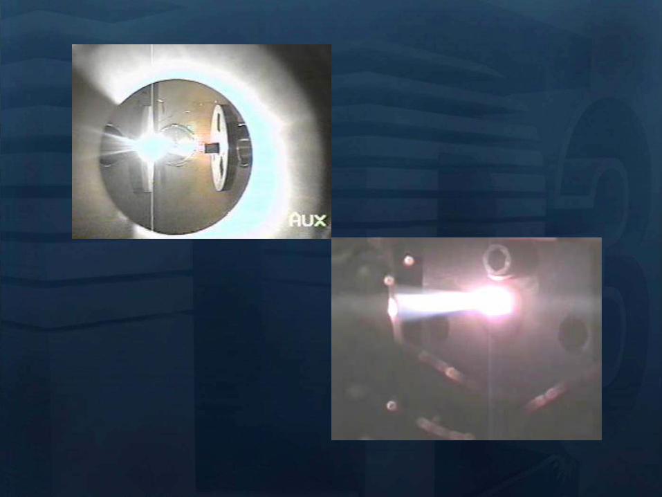
Processes in PLD
Laser pulse
Processes in PLD
e- e-
e-
e- e-
e-
e-
e-
e-
e- e-
e-
e-
e-
Electronic excitation
Processes in PLD
e- e-
e-
e- e-
e-
e-
e-
e-
e- e-
e-
e-
e-
Energy relaxation to lattice (~1 ps)
lattice
Processes in PLD
Heat diffusion (over microseconds)
lattice
Processes in PLD
Melting (tens of ns) Evaporation Plasma
Formation (microseconds) Resolidification
lattice
Processes in PLD
lattice
If laser pulse is long (ns) or
repetition rate is high laser may
continue interactions
Processes in Pulsed Laser Deposition
1 Absorption of laser pulse in material
Qab=(1-R)Ioe-aL
(metals absorption depths ~ 10 nm depends on )
2 Relaxation of energy (~ 1 ps) (electron-phonon interaction)
3 Heat transfer Melting and Evaporation
when electrons and lattice at thermal equilibrium (long pulses)
use heat conduction equation
(or heat diffusion model)
abp QTKt
TC
)(
At peak of laser pulse temperatures on target can reach
gt105 K (gt 40 eV)
Electric Fields gt 105 Vcm also high magnetic fields
Plasma Temperatures 3000-5000 K
Ablated Species with energies 1 ndash100 eV
Initially Incredibly Non-Equilibrium
Why biomaterials
- repair and reconstruction of parts of the musculo-skeletal system of vertebrates
- minimal biological requirement biocompatibility associated with the absence of
any adverse effect (non-toxic and non-allergic)
Other requests
- resistance to physiological fluids
- should not interfere with the bodyrsquos natural immunity system
- withstand mechanical stress during whole lifetime
- manufacturability in any appropriate shape
Possible classification
-a) biologically inactive (inert) alumina zirconia stainless steel CoCrNi
CoCrMo titanium titanium alloys carbon latex PE PMMA hellip
-b) porous Calcium Phosphates (CaPs) CaP-coated metal
-c) bioactive dense calcium phosphate ceramics bioactive glasses bioactive
glass-ceramics bioactive composites hellip
-d) Resorbable tricalcium phosphate calciumaliminate polylactic acid poly-L-
acetate
Our option biocompatible porous and bioactive CaP
Acronym
Chemical formula Compound name CaP
ratio
HA Ca10(PO4)6(OH)2 Hydroxylapatite 167
FA Ca10(PO4)6F2 Fluorapatite 167
CDHA Ca10x(HPO4)x(PO4)6x(OH)2x (0x2) Calcium-deficient Hydroxylapatite 133-167
BA Ca83(PO4)43 (CO3-HPO4)17(OH)03
BA=carbonated CDHA (x=17)
Biological apatite 138-193
Mn-CHA HA with (04-2) Mn2+ and (2-6) CO32- Mn2+ doped carbonated hydroxylapatite 151- 165
OHA Ca10(PO4)6(OH)22xOxٱx (0x1) Oxyhydroxylapatite 167
OA Ca10O(PO4)6 Oxyapatite 167
MCPM Ca(H2PO4)2middotH2O Monocalcium phosphate monohydrate 05
MCPA Ca(H2PO4)2 Monocalcium phosphate anhydrate 05
DCPD CaHPO4middot2H2O Dicalcium phosphate dihydrate (Brushite) 1
DCPA CaHPO4 Dicalcium phosphate anhydrate 1
OCP Ca8(HPO4)2(PO4)4middot5H2O Octacalcium phosphate 133
a-TCP Ca3(PO4)2 (monoclinic) Tricalcium phosphate (phase a) 15
-TCP Ca3(PO4)2 (rhombohedral) Tricalcium phosphate (phase Whitlockite) 15
TTCP Ca4O(PO4)2 Tetracalcium phosphate 2
a-DCP Ca2P2O7 (orthorhombic) Dicalcium phosphate (phase a) 1
-DCP Ca2P2O7 (tetragonal) Dicalcium phosphate (phase ) 1
ACP Cax(PO4)y nH2O (Amorphous Calcium pyrophosphate) 12-22
Bone
Resorbable CaP
CaP coating
Ti
Implant
Alternative solution Biomimetic coatings for metallic implants
Main deficiency of CaPs brittle in bulk
HA molecule Ca10(PO4)6OH2
Projection in the
(001) base plan of
the hydroxyapatite
unit cell (hexagonal
structure)
How difficult is to deposit CaPs (1)
- very complex molecules
How difficult is to deposit CaPs (2)
Crystal structure of OCP projected down the c-axis
Alternating apatite- and
hydrated- layers (100)
planes
The region with shaded
atoms the ldquoapatitic layerrdquo
is very similar to HA
The zone containing 10
water molecules is the
ldquohydrated layerrdquo
H atoms are omitted for
clarity
Octacalcium phosphate
(Ca8(HPO4)2(PO4)4middot5H2O)
Mesures par spectroscopie optique
drsquoemission sur le plasma issu de lrsquoirradiation
laser 248 nm drsquoune cible drsquoHA pure
Croissance impulsion par impulsion
Preacutesence des ions de
bull calcium (Ca I et Ca II)
bull oxygegravene (O I et O II) et
bull phosphore (P II)
Eacutemissions dues agrave des espegraveces moleacuteculaires preacutesentes dans le plasma
Couche mince drsquohydroxyapatite obtenue par
ablation laser
Goutelettes
CaP = 18 CaP = 21
CaP = 17 Trois reacutegions
typiques
i Reacutegion majoritaire lisse
ii Agglomeacuteration de gouttelettes sub-micromeacutetriques
iii Gouttelettes micromeacutetriques
Human primary osteoblasts (hOB) were cultured on OCP coated-Ti
Mn-CHA coatedndashTi HA coated-Ti Ti control (polystyrene)
hOB response SEM micrographs
- on bare Ti (a) after 7 days (b) after 21 days
Elongated rod-like morphology
Bioactivity tests
- OCP coatings (c) after 7 days (d) after 21 days
- In time cells spread and expand with flattened polyhedral-morphology
- Numerous cytoplasmatic extensions
hOB response SEM micrographs
- Mn-CHA coatings (e) after 7 days (f) after 21 days
- In time cells spread and expand with flattened polyhedral-morphology
- Less cytoplasmatic extensions
hOB response SEM micrographs
Biocompatible metallic thin films by PLD
Alloys NiTinol NiTi
NiTi characteristics ndash Super-Elastic Property
ndash Radiopaque
ndash Shape-Memory Effect
NiTi biomedical applications ndash Tweezers for removing foreign objects via small incisions
ndash Anchors for tendon fixation
ndash Stents for cardiovascular applications
ndash Dentistry - Orthodontic wires which no not need to be retightened and adjusted
Deposition chamber Multitarget system
PULSED LASER DEPOSITION (PLD)
SEM
Nitinol 30 sequences Nitinol 20 sequences
Cytotoxicity evaluation on microorganism culture
(a) E coli reference cell culture
(b) E coli cell culture on Ti surface
(c) E coli cell culture on NiTi surface
(d) S cerevisiae references cell culture
(e) S cerevisiae cell culture Ti surface
(f) S cerevisiae cell culture on NiTi surface
(g) SEM on E coli cell
culture on NiTi surface
(h) SEM on S cerevisiae cell culture on
NiTi surface E coli and S cerevisiae cell
cultures on the nitinol thin films
o Biocompatible
o Low adherence of microorganism on NiTi surface NiTi alloy does not stimulate the development at genetic level for specific genes which are involved in adhesion processes for microorganism cells
o Low release of Ni ions in solutions
CONCLUSIONS
MAPLE Matrix Assisted Pulsed Laser Evaporation
bull No need for UHV
bull Matrix prepared outside the
chamber
bull High flexibility
You can use everything
you can dissolve in a
solvent
MAPLE of polymers blends with Ag
nanoparticles
Controlled release of antibacterial agents
MAPLE deposition of PEGPLGA films with incorporated silver nanoparticles (AgNPs)
0 5 10 15 20 25 30000
005
010
015
020
025
Nu
mb
er o
f E
C
oli
X109
Time (hours)
Killing activity
0 5 10 15 20 25 30 3500
05
10
15
20
25
Bact
eria
l ce
ll n
um
ber
X109
Time (hours)
Delay of bacterial growth
Antibacterial efficiency against E Coli
Direct writing by femtosecond laser
pulses
Ormosil scaffolds for tissue engineering
50 lines ormosil sample
Distance between lines =
100 μm
30x30 lines ormosil
sample Distance between
lines = 50 μm
3 layers 30x30 lines ormosil
sample Distance between
lines = 100 μm
Scaffold functionalization
Protein deposition on the scaffolds by MAPLE
- Lysozyme antibacterial antitumor uses
- Fibrinogen biocompatible protein
Fibroblast cells grown on a polymeric grid with 100 microm distance between lines
Spectroscopie drsquoun panache plasma
laser femtoseconde
Laser
t =400 ns Spot 35x35 micromsup2 Cu
Z mm
Expeacuteriences reacutealiseacutees par (Grojo et al LP3Marseille)
nanoparticules
atoms
031 Jcm-2
Formation de nano agreacutegats
AFM
(Pereira et al )
TEM
(Kabashin et al)
Eacutetude des meacutecanismes de la formation drsquoagreacutegats
Couplage
- Direct Simulation Monte Carlo
- Dynamique Moleacuteculaire
Information deacutetailleacutee
-Continu du panache
-Distribution en taille des agreacutegats
K Gouriet thegravese (2008)
Simulation par Dynamique Moleacuteculaire (MD)
=gtdistribution drsquoagreacutegats
Cluster size N
No
rmaliz
ed
Yie
ld
100
101
102
103
104
105
10-9
10-8
10-7
10-6
10-5
10-4
10-3
10-2
10-1
100
N-426
N-122
t=1 ns
τ = 15 ps F ~ 2 Fth 4010 nm t =1 ns
Zhigilei et al (Virginia University)
Target applications of mobile nanomaterials
Bioimaging
Optical imaging deals with
visualization of biological objects
or tissues in order to detect
pathogens or follow the delivery
of drugs etc nanoparticles are
uses as contrast agents
In vitro
Cellular
imaging
In vivo Cancer
detection drug delivery
etc
Light-induced
therapies
Cancer therapy nanoparticles are used
as photosensitizers to produce local
targeted destruction of cancer cells
Photodynamic
therapy
Light-induced
hyperthermia
Diagnosis and Sensing A Diseases can be diagnosed through the (simultaneous) detection of a (set of) biomolecule(s) characteristic to a specific disease type and stage (biomarkers)
Huffman Nanomedicine and Nanobiotechnology Vol 1 1 2009
D
molecular signature of sick cell of infecting agent
(eg an antibody)
Cell membrane
Nanoparticle
Coating molecule specifically attracted to the molecular signature
C A nanoparticle can be functionalized in such a way that specifically targets a biomarker Thus the detection of the nanoparticle is linked to the detection of the biomarker and to the diagnosis of a disease
B Each cell type has unique molecular signatures that differentiate healthy and sick tissues Similarly an infection can be diagnosed by detecting the distinctive molecular signature of the infecting agent
Laser ablation of material from a solid target and production of
nanoparticlesnanostructures
Production of nanoclusters by laser
ablation and their subsequent cooling in
ambient medium Gaseous ambience
Nanoclusters can be
deposited on a
substrate forming a
nanostructured film
Aqueous ambience
Nanoclusters
crystallize and form
colloidal solution
NPs produced in gaseous ambience
0 20 40 60 80 100 120 140
0
2
4
6
8
Heig
ht
[nm
]
[nm]
01 1 102
3
4
5
6
7
Mean
part
icle
heig
ht
(nm
)
Helium pressure (Torr)
Silicon nanocrystal size AFM
Gas pressure decreases Nanocrystal size decreases
AFM of isolated laser-
ablated Si particles on
HOPG
Minimal particle size is 2-3 nm (from height measurements)
001Torr
1Torr
01Torr
10Torr
100Torr
NGC 2009
Photoluminescence of size selected nanoclusters
0 1 2 3 4 5 6
39 Jcm2
Num
ber
of
clust
ers
Size (nm)
30 Jcm2
14 Jcm2
0 1 2 3 4 5 6
He 4 Torr
10 Jcm2
15 20 25 30 35 40
0
500
1000
He 4 Torr
39 Jcmsup2
30 Jcmsup2
14 Jcmsup2
10 Jcmsup2
Inte
nsi
ty o
f P
L (
ua
)Photon energy (eV)
Silicon nanoclusters
Medical Imaging A Optical properties of nanoparticles depend greatly on its structure Particularly the color (wavelength) emitted by a quantum dot (a semiconductor nanoparticle) depends on its diameter
C The quantum dots (QD) can be injected to a
subject and then be detected by exciting them to emit light
Source Department of immunology University of Toronto
Solutions of CdSe QDrsquos of different diameter
CdSe nanoparticle (QD) structure
Source Laurence Livermore Laboratories
Imaging of QDrsquos targeted on cellular structures
Nano Letters 2008 Vol 8 pp3887-3892
B
In vivo cancer targeting and imaging with
semiconductor quantum dots
Gao et al Nat Biotech 22 969 - 976 (2004)
NPs produced in aqueous ambience
Advantages of laser ablation-based nanofabrication
compared to chemical methods
Disadvantage of chemical synthesis contaminationhelliphelliphellip
In particular chemical method for the fabrication of gold nanoparticles
Reduction of chloroauric acid (HAuCl4) with citrate in water
Control size by adding a stabilizing agent (thiol- (-SH) containing molecules)
Contamination impurities Cl- on surface non-biocompatible surfactantshellip
Problems in imaging
applications (especially
in vivo)
Problems in field-
enhanced applications (eg contaminants can
provide false Raman signals)
Focalized laser beam
Liquid medium
Target
Plasma
Laser ablation in liquids a way to avoid residual
contamination
- Laser energy can be transmitted through a liquid to ablate a solid
target the nanomaterial is released to the solution
- The process can be performed without any dirty chemical products in
clean liquid environment (deionized water PBS solution) no
contamination
Experiment
Ablation of gold final colloidal solutions
-Solution are ranging from deep wine red to pink and purple
- Color related to the size dependant surface plasmon resonance
Size distribution of Au nanoparticles prepared in
distilled water
0 50 100 150 200 250 300
0
200
400
600
800
1000
Rela
tive a
bundance (
arb
units)
Particle size (nm)
0 10 20 30 40 50 60 70 80 90 100
0
200
400
600
800
1000
Re
lative
Ab
un
da
nce
(a
rb u
nits)
Particle Size (nm) 0 2 4 6 8 10
0
200
400
600
800
1000
Re
lative
Ab
un
da
nce
(a
rb u
nits)
Particle Size (nm)
F = 20F0 Jcm2 F = 5F0 Jcm2 F = F0 Jcm2
-Size and dispersion of nanoparticles can be controlled by laser fluence
- Shape of craters on target at high F suggests the presence of cavitation phenomena
4 plusmn 15 nm
A V Kabashin M Meunier J Appl Phys 94 7941 (2003)
In water Very dispersed population of nanoparticles
Dextran (MW=6000gmol) Quenches the growth of the nanoparticles 2-4nm nanoparticles are produced (230-1850 atoms) Very fast dextran-gold interaction
Influence of the concentration
Dextran 6k
In pure water
300mJ
Ablation in biopolymers (dextran PEG)
Au OH
O
-
Dextran (C6H10O5)n
Nanoparticles react with OH
groups of biopolymers
Gold Nanoparticles vs Alzheimer
A Alzheimer and other degenerative diseases are caused my the clustering of amyloidal beta (Aβ) protein
D Gold nanoparticles can be functionalized to specifically attach to aggregates of this protein (amyloidosis)
Functionalized nanoparticle
Source wwwinternetchemistrycom
Chemical structure of Aβ-protein
Source wwwthefutureofthingscom
C
B
Alzheimerrsquos brain Healthy brain
Source Berkeley Lab
Gold Nanoparticles vs Alzheimer A The functionalized gold nanoparticles selectively attach to the aggregate of amyloidal protein The microwaves of certain frequency are irradiated on the sample Resonance with the gold nanoparticles increases the local temperature and destroy the aggregate
Nanoletters 2006 Vol 6 pp110-115
Before irradiation After irradiation
LIBS for biomedical applications in vivo analysis of tissue for real-time
analysis
real-time diagnosis of pathogen
presence in human fluids (bloodurine
CSF sputum)
71 35
ICCD
spectromegravetre
Time and space
Emission spectroscopie
LIBS (Laser Induced Breakdown Spectroscopy)
Principle of sample identification screening applications based on discriminant analysis here for warning when healthy tooth material is targeted during laser drilling
BMC Oral Health 2001 1 1 Ota Samek1 Helmut H Telle2 and David CS Beddows
Laser-induced breakdown spectroscopy a tool for real-time in vitro and in vivo identification of carious teeth
Sensitive Detection of Epithelial Ovarian Cancer Biomarkers
Using Tag-Laser Induced Breakdown Spectroscopy
Yuri Markushin and Noureddine Melikechi
Optical Science Center for Applied Research Delaware State University USA
X rays produced by laser plasmas
I = ~1016 agrave 1018 Wcm2
nc cm-3
1111021 mm 2
Thot Ilas2
1 3
Ilas2
3 4
Hot electrons
(characteristic lines)
X-ray
emission
laser
X-rays
Line emission Fast electrons knock
free core electrons
from the inner shell
Bremsstrahlung electrons deflected by nucleus
Laser-generated hot electrons produce x-rays by two dominant mechanisms
ne
critical surface (cf cathode)
conventional X-ray tube
Kmetec ldquoMeV X-ray generation with a femtosecond laserrdquo Phys Rev Lett 68 1527 (1992)
Rousse ldquoEfficient Ka X-ray source from fs-laser-produced plasmasrdquo Phys Rev E 50 2200 (1994)
(typical recombination lifetimes several fs)
Hou et al Appl Phys Lett 84 2259 (2004)
Tightly focused fs lasers create the worldrsquos smallest
hard x-ray source (~4 microm) for precision imaging
x-ray source size is ~ 4 larger than laser focus
because of lateral heat transport Bowes Opt Lett 31 116 (2006)
2004 premiegravere deacutemonstration drsquoimagerie avec contraste de phase avec une source X creacutee par laser
Source
Rayon non diffracteacute
Rayon diffracteacute
Lcoh 2R1
eacutechantillon
Absorption vs contraste de phase
dsource-object dobject-detector
X-ray
Source
Object
Detector
position
Dsource
Intensity
interface
dsource-object dobject-detector
X-ray
Source
Object
Detector
position
Fresnel Diffraction
Dsource
Intensity
interface
enhancement interface
R Toth et al
Rev Sci Instrum 76
083701 (2005)
Imagerie par contraste de phase
R Toth et al Rev Sci Instrum
76 083701 (2005)
copy INRS
bull Meilleure reacutesolution (5microm)
bull dose plus faible
bull meilleur contraste (phase)
bull lrsquoeacutenergie X peut ecirctre ajusteacutee
aux besoins de lrsquoimagerie
La reacutefeacuterence mondiale ALLS
(INRS- Montreacuteal) 200TW + Puissance moyenne 50W
47J 23fs 10Hz
in a 8microm spot
Sans miroir deacuteformable
microCT
Scan duration avec
50W10Hz (360 images)
2 min (mouse scan)
courtesy Prof Dr Oswald Willi U Duumlselldorf
MeV Protons produced by laser CR39 or RCF 1010 to 1013 H+
courtesy Prof Don Umstadter U Nebraska-Lincoln
Target Normal Sheath Acceleration hot electrons traversing
target electrostatically accelerate impurity hydrogen ions on the rear surface
virtual
cathode
I ge 1019 Wcm2
Protons de 10MeV (INRS )
~1022 Wcm2
Avantages des particules chargeacutees ou hadrons (protons ions) Rayons X de la Radio-Theacuterapie
Le pic de Bragg
La profondeur de peacuteneacutetration deacutepend de l rsquoeacutenergie incidente des protons
la tumeur la plus superficielle = l rsquoœil 70 MeV
la plus profonde = la prostate 200 - 250 MeV
Protontheacuterapie par laser (projet SAPHIR)
CPO
Exemple de dosimeacutetrie preacutevisionnelle
(photons vs protons)
Comparaison de lrsquoeacutepargne de dose dans les tissus sains en protons (image de droite)
par rapport aux photons (IMRT Intensity Modulated Radio-Therapy image de gauche)
Les irradiations colateacuterales en radiotheacuterapie et en protontheacuterapie
Avantage consideacuterable pour le traitement de tumeurs localiseacutees pregraves d rsquoorganes sensibles (œil cerveau)
currently treating patients 5
5 next 2 years
others awaiting approval
10000 m2 $125 M facility opened in 2006
12 m diameter 250 MeV synchrotron
ldquoThere are too few physicists in the world
and they are an incredibly important part of
doing this We have one of the largest
physics departments in the world with more than 50 medical physicistsrdquo
--- Dr James D Cox head of Radiation Oncology at MD Anderson Cancer Center Houston Texas
Proton Therapy enables precise exposure of small tumors
with minimal damage to surrounding healthy tissue
250 MeV
PROTON beam
10 20
Depth in Tissue [cm]
0 0
50
100
DO
SE
(
)
6 MV
PHOTON
beam
tunable
Bragg
peak
but requires large expensive facilities
eye tumors 65 MeV protons
deep tumors gt200 MeV
Laser proton therapy could be much smaller amp cheaper Fourkal Med Phys 29 2788 (2002)
Malka Med Phys 31 1587 (2004)
On-site production of short-lived isotopes for medical imaging
Laser-generated quasi-mono-energetic electrons efficiently photo-activate materials of interest
bull High Rep rate bull Low cost bull Compact
Limitations to the widespread use of PET arise from the high costs of cyclotrons needed to
produce the short-lived radionucleotides for PET scanning Few hospitals and universities
are capable of maintaining such systems - Wikipedia -
radiotracer activation reaction half-life medical use
15O 16O (n)15O 2 minutes neuro-imaging
11C 12C(n)11C 20 minutes neuro-receptor-specific brain imaging
18F 19F(n)18F 110 minutes clinical oncology
Positron Emission Tomography
on-site
production
essential
18F PET scan of tumor 15O PET scan of human brain
Reed ldquoEfficient initiation of photonuclear reactions using quasi-monoenergetic electron beams from LWFArdquo J Appl Phys 102 073103 (2007)
TEP in situ
+ in vivo
Modegravele animal
microCT +
contraste de phase
+ in vivo
modegraveles de cancer
Comprendre
le cancer
Preacuteparer le traitement
protons 50MeV
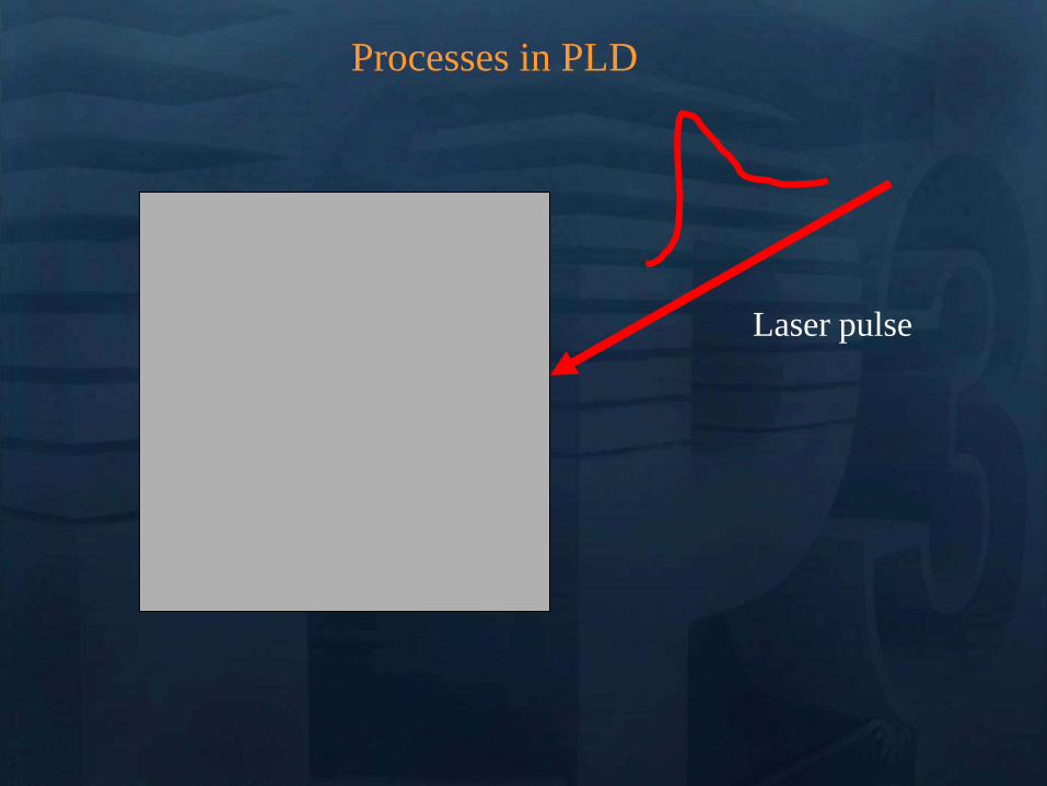
Processes in PLD
e- e-
e-
e- e-
e-
e-
e-
e-
e- e-
e-
e-
e-
Electronic excitation
Processes in PLD
e- e-
e-
e- e-
e-
e-
e-
e-
e- e-
e-
e-
e-
Energy relaxation to lattice (~1 ps)
lattice
Processes in PLD
Heat diffusion (over microseconds)
lattice
Processes in PLD
Melting (tens of ns) Evaporation Plasma
Formation (microseconds) Resolidification
lattice
Processes in PLD
lattice
If laser pulse is long (ns) or
repetition rate is high laser may
continue interactions
Processes in Pulsed Laser Deposition
1 Absorption of laser pulse in material
Qab=(1-R)Ioe-aL
(metals absorption depths ~ 10 nm depends on )
2 Relaxation of energy (~ 1 ps) (electron-phonon interaction)
3 Heat transfer Melting and Evaporation
when electrons and lattice at thermal equilibrium (long pulses)
use heat conduction equation
(or heat diffusion model)
abp QTKt
TC
)(
At peak of laser pulse temperatures on target can reach
gt105 K (gt 40 eV)
Electric Fields gt 105 Vcm also high magnetic fields
Plasma Temperatures 3000-5000 K
Ablated Species with energies 1 ndash100 eV
Initially Incredibly Non-Equilibrium
Why biomaterials
- repair and reconstruction of parts of the musculo-skeletal system of vertebrates
- minimal biological requirement biocompatibility associated with the absence of
any adverse effect (non-toxic and non-allergic)
Other requests
- resistance to physiological fluids
- should not interfere with the bodyrsquos natural immunity system
- withstand mechanical stress during whole lifetime
- manufacturability in any appropriate shape
Possible classification
-a) biologically inactive (inert) alumina zirconia stainless steel CoCrNi
CoCrMo titanium titanium alloys carbon latex PE PMMA hellip
-b) porous Calcium Phosphates (CaPs) CaP-coated metal
-c) bioactive dense calcium phosphate ceramics bioactive glasses bioactive
glass-ceramics bioactive composites hellip
-d) Resorbable tricalcium phosphate calciumaliminate polylactic acid poly-L-
acetate
Our option biocompatible porous and bioactive CaP
Acronym
Chemical formula Compound name CaP
ratio
HA Ca10(PO4)6(OH)2 Hydroxylapatite 167
FA Ca10(PO4)6F2 Fluorapatite 167
CDHA Ca10x(HPO4)x(PO4)6x(OH)2x (0x2) Calcium-deficient Hydroxylapatite 133-167
BA Ca83(PO4)43 (CO3-HPO4)17(OH)03
BA=carbonated CDHA (x=17)
Biological apatite 138-193
Mn-CHA HA with (04-2) Mn2+ and (2-6) CO32- Mn2+ doped carbonated hydroxylapatite 151- 165
OHA Ca10(PO4)6(OH)22xOxٱx (0x1) Oxyhydroxylapatite 167
OA Ca10O(PO4)6 Oxyapatite 167
MCPM Ca(H2PO4)2middotH2O Monocalcium phosphate monohydrate 05
MCPA Ca(H2PO4)2 Monocalcium phosphate anhydrate 05
DCPD CaHPO4middot2H2O Dicalcium phosphate dihydrate (Brushite) 1
DCPA CaHPO4 Dicalcium phosphate anhydrate 1
OCP Ca8(HPO4)2(PO4)4middot5H2O Octacalcium phosphate 133
a-TCP Ca3(PO4)2 (monoclinic) Tricalcium phosphate (phase a) 15
-TCP Ca3(PO4)2 (rhombohedral) Tricalcium phosphate (phase Whitlockite) 15
TTCP Ca4O(PO4)2 Tetracalcium phosphate 2
a-DCP Ca2P2O7 (orthorhombic) Dicalcium phosphate (phase a) 1
-DCP Ca2P2O7 (tetragonal) Dicalcium phosphate (phase ) 1
ACP Cax(PO4)y nH2O (Amorphous Calcium pyrophosphate) 12-22
Bone
Resorbable CaP
CaP coating
Ti
Implant
Alternative solution Biomimetic coatings for metallic implants
Main deficiency of CaPs brittle in bulk
HA molecule Ca10(PO4)6OH2
Projection in the
(001) base plan of
the hydroxyapatite
unit cell (hexagonal
structure)
How difficult is to deposit CaPs (1)
- very complex molecules
How difficult is to deposit CaPs (2)
Crystal structure of OCP projected down the c-axis
Alternating apatite- and
hydrated- layers (100)
planes
The region with shaded
atoms the ldquoapatitic layerrdquo
is very similar to HA
The zone containing 10
water molecules is the
ldquohydrated layerrdquo
H atoms are omitted for
clarity
Octacalcium phosphate
(Ca8(HPO4)2(PO4)4middot5H2O)
Mesures par spectroscopie optique
drsquoemission sur le plasma issu de lrsquoirradiation
laser 248 nm drsquoune cible drsquoHA pure
Croissance impulsion par impulsion
Preacutesence des ions de
bull calcium (Ca I et Ca II)
bull oxygegravene (O I et O II) et
bull phosphore (P II)
Eacutemissions dues agrave des espegraveces moleacuteculaires preacutesentes dans le plasma
Couche mince drsquohydroxyapatite obtenue par
ablation laser
Goutelettes
CaP = 18 CaP = 21
CaP = 17 Trois reacutegions
typiques
i Reacutegion majoritaire lisse
ii Agglomeacuteration de gouttelettes sub-micromeacutetriques
iii Gouttelettes micromeacutetriques
Human primary osteoblasts (hOB) were cultured on OCP coated-Ti
Mn-CHA coatedndashTi HA coated-Ti Ti control (polystyrene)
hOB response SEM micrographs
- on bare Ti (a) after 7 days (b) after 21 days
Elongated rod-like morphology
Bioactivity tests
- OCP coatings (c) after 7 days (d) after 21 days
- In time cells spread and expand with flattened polyhedral-morphology
- Numerous cytoplasmatic extensions
hOB response SEM micrographs
- Mn-CHA coatings (e) after 7 days (f) after 21 days
- In time cells spread and expand with flattened polyhedral-morphology
- Less cytoplasmatic extensions
hOB response SEM micrographs
Biocompatible metallic thin films by PLD
Alloys NiTinol NiTi
NiTi characteristics ndash Super-Elastic Property
ndash Radiopaque
ndash Shape-Memory Effect
NiTi biomedical applications ndash Tweezers for removing foreign objects via small incisions
ndash Anchors for tendon fixation
ndash Stents for cardiovascular applications
ndash Dentistry - Orthodontic wires which no not need to be retightened and adjusted
Deposition chamber Multitarget system
PULSED LASER DEPOSITION (PLD)
SEM
Nitinol 30 sequences Nitinol 20 sequences
Cytotoxicity evaluation on microorganism culture
(a) E coli reference cell culture
(b) E coli cell culture on Ti surface
(c) E coli cell culture on NiTi surface
(d) S cerevisiae references cell culture
(e) S cerevisiae cell culture Ti surface
(f) S cerevisiae cell culture on NiTi surface
(g) SEM on E coli cell
culture on NiTi surface
(h) SEM on S cerevisiae cell culture on
NiTi surface E coli and S cerevisiae cell
cultures on the nitinol thin films
o Biocompatible
o Low adherence of microorganism on NiTi surface NiTi alloy does not stimulate the development at genetic level for specific genes which are involved in adhesion processes for microorganism cells
o Low release of Ni ions in solutions
CONCLUSIONS
MAPLE Matrix Assisted Pulsed Laser Evaporation
bull No need for UHV
bull Matrix prepared outside the
chamber
bull High flexibility
You can use everything
you can dissolve in a
solvent
MAPLE of polymers blends with Ag
nanoparticles
Controlled release of antibacterial agents
MAPLE deposition of PEGPLGA films with incorporated silver nanoparticles (AgNPs)
0 5 10 15 20 25 30000
005
010
015
020
025
Nu
mb
er o
f E
C
oli
X109
Time (hours)
Killing activity
0 5 10 15 20 25 30 3500
05
10
15
20
25
Bact
eria
l ce
ll n
um
ber
X109
Time (hours)
Delay of bacterial growth
Antibacterial efficiency against E Coli
Direct writing by femtosecond laser
pulses
Ormosil scaffolds for tissue engineering
50 lines ormosil sample
Distance between lines =
100 μm
30x30 lines ormosil
sample Distance between
lines = 50 μm
3 layers 30x30 lines ormosil
sample Distance between
lines = 100 μm
Scaffold functionalization
Protein deposition on the scaffolds by MAPLE
- Lysozyme antibacterial antitumor uses
- Fibrinogen biocompatible protein
Fibroblast cells grown on a polymeric grid with 100 microm distance between lines
Spectroscopie drsquoun panache plasma
laser femtoseconde
Laser
t =400 ns Spot 35x35 micromsup2 Cu
Z mm
Expeacuteriences reacutealiseacutees par (Grojo et al LP3Marseille)
nanoparticules
atoms
031 Jcm-2
Formation de nano agreacutegats
AFM
(Pereira et al )
TEM
(Kabashin et al)
Eacutetude des meacutecanismes de la formation drsquoagreacutegats
Couplage
- Direct Simulation Monte Carlo
- Dynamique Moleacuteculaire
Information deacutetailleacutee
-Continu du panache
-Distribution en taille des agreacutegats
K Gouriet thegravese (2008)
Simulation par Dynamique Moleacuteculaire (MD)
=gtdistribution drsquoagreacutegats
Cluster size N
No
rmaliz
ed
Yie
ld
100
101
102
103
104
105
10-9
10-8
10-7
10-6
10-5
10-4
10-3
10-2
10-1
100
N-426
N-122
t=1 ns
τ = 15 ps F ~ 2 Fth 4010 nm t =1 ns
Zhigilei et al (Virginia University)
Target applications of mobile nanomaterials
Bioimaging
Optical imaging deals with
visualization of biological objects
or tissues in order to detect
pathogens or follow the delivery
of drugs etc nanoparticles are
uses as contrast agents
In vitro
Cellular
imaging
In vivo Cancer
detection drug delivery
etc
Light-induced
therapies
Cancer therapy nanoparticles are used
as photosensitizers to produce local
targeted destruction of cancer cells
Photodynamic
therapy
Light-induced
hyperthermia
Diagnosis and Sensing A Diseases can be diagnosed through the (simultaneous) detection of a (set of) biomolecule(s) characteristic to a specific disease type and stage (biomarkers)
Huffman Nanomedicine and Nanobiotechnology Vol 1 1 2009
D
molecular signature of sick cell of infecting agent
(eg an antibody)
Cell membrane
Nanoparticle
Coating molecule specifically attracted to the molecular signature
C A nanoparticle can be functionalized in such a way that specifically targets a biomarker Thus the detection of the nanoparticle is linked to the detection of the biomarker and to the diagnosis of a disease
B Each cell type has unique molecular signatures that differentiate healthy and sick tissues Similarly an infection can be diagnosed by detecting the distinctive molecular signature of the infecting agent
Laser ablation of material from a solid target and production of
nanoparticlesnanostructures
Production of nanoclusters by laser
ablation and their subsequent cooling in
ambient medium Gaseous ambience
Nanoclusters can be
deposited on a
substrate forming a
nanostructured film
Aqueous ambience
Nanoclusters
crystallize and form
colloidal solution
NPs produced in gaseous ambience
0 20 40 60 80 100 120 140
0
2
4
6
8
Heig
ht
[nm
]
[nm]
01 1 102
3
4
5
6
7
Mean
part
icle
heig
ht
(nm
)
Helium pressure (Torr)
Silicon nanocrystal size AFM
Gas pressure decreases Nanocrystal size decreases
AFM of isolated laser-
ablated Si particles on
HOPG
Minimal particle size is 2-3 nm (from height measurements)
001Torr
1Torr
01Torr
10Torr
100Torr
NGC 2009
Photoluminescence of size selected nanoclusters
0 1 2 3 4 5 6
39 Jcm2
Num
ber
of
clust
ers
Size (nm)
30 Jcm2
14 Jcm2
0 1 2 3 4 5 6
He 4 Torr
10 Jcm2
15 20 25 30 35 40
0
500
1000
He 4 Torr
39 Jcmsup2
30 Jcmsup2
14 Jcmsup2
10 Jcmsup2
Inte
nsi
ty o
f P
L (
ua
)Photon energy (eV)
Silicon nanoclusters
Medical Imaging A Optical properties of nanoparticles depend greatly on its structure Particularly the color (wavelength) emitted by a quantum dot (a semiconductor nanoparticle) depends on its diameter
C The quantum dots (QD) can be injected to a
subject and then be detected by exciting them to emit light
Source Department of immunology University of Toronto
Solutions of CdSe QDrsquos of different diameter
CdSe nanoparticle (QD) structure
Source Laurence Livermore Laboratories
Imaging of QDrsquos targeted on cellular structures
Nano Letters 2008 Vol 8 pp3887-3892
B
In vivo cancer targeting and imaging with
semiconductor quantum dots
Gao et al Nat Biotech 22 969 - 976 (2004)
NPs produced in aqueous ambience
Advantages of laser ablation-based nanofabrication
compared to chemical methods
Disadvantage of chemical synthesis contaminationhelliphelliphellip
In particular chemical method for the fabrication of gold nanoparticles
Reduction of chloroauric acid (HAuCl4) with citrate in water
Control size by adding a stabilizing agent (thiol- (-SH) containing molecules)
Contamination impurities Cl- on surface non-biocompatible surfactantshellip
Problems in imaging
applications (especially
in vivo)
Problems in field-
enhanced applications (eg contaminants can
provide false Raman signals)
Focalized laser beam
Liquid medium
Target
Plasma
Laser ablation in liquids a way to avoid residual
contamination
- Laser energy can be transmitted through a liquid to ablate a solid
target the nanomaterial is released to the solution
- The process can be performed without any dirty chemical products in
clean liquid environment (deionized water PBS solution) no
contamination
Experiment
Ablation of gold final colloidal solutions
-Solution are ranging from deep wine red to pink and purple
- Color related to the size dependant surface plasmon resonance
Size distribution of Au nanoparticles prepared in
distilled water
0 50 100 150 200 250 300
0
200
400
600
800
1000
Rela
tive a
bundance (
arb
units)
Particle size (nm)
0 10 20 30 40 50 60 70 80 90 100
0
200
400
600
800
1000
Re
lative
Ab
un
da
nce
(a
rb u
nits)
Particle Size (nm) 0 2 4 6 8 10
0
200
400
600
800
1000
Re
lative
Ab
un
da
nce
(a
rb u
nits)
Particle Size (nm)
F = 20F0 Jcm2 F = 5F0 Jcm2 F = F0 Jcm2
-Size and dispersion of nanoparticles can be controlled by laser fluence
- Shape of craters on target at high F suggests the presence of cavitation phenomena
4 plusmn 15 nm
A V Kabashin M Meunier J Appl Phys 94 7941 (2003)
In water Very dispersed population of nanoparticles
Dextran (MW=6000gmol) Quenches the growth of the nanoparticles 2-4nm nanoparticles are produced (230-1850 atoms) Very fast dextran-gold interaction
Influence of the concentration
Dextran 6k
In pure water
300mJ
Ablation in biopolymers (dextran PEG)
Au OH
O
-
Dextran (C6H10O5)n
Nanoparticles react with OH
groups of biopolymers
Gold Nanoparticles vs Alzheimer
A Alzheimer and other degenerative diseases are caused my the clustering of amyloidal beta (Aβ) protein
D Gold nanoparticles can be functionalized to specifically attach to aggregates of this protein (amyloidosis)
Functionalized nanoparticle
Source wwwinternetchemistrycom
Chemical structure of Aβ-protein
Source wwwthefutureofthingscom
C
B
Alzheimerrsquos brain Healthy brain
Source Berkeley Lab
Gold Nanoparticles vs Alzheimer A The functionalized gold nanoparticles selectively attach to the aggregate of amyloidal protein The microwaves of certain frequency are irradiated on the sample Resonance with the gold nanoparticles increases the local temperature and destroy the aggregate
Nanoletters 2006 Vol 6 pp110-115
Before irradiation After irradiation
LIBS for biomedical applications in vivo analysis of tissue for real-time
analysis
real-time diagnosis of pathogen
presence in human fluids (bloodurine
CSF sputum)
71 35
ICCD
spectromegravetre
Time and space
Emission spectroscopie
LIBS (Laser Induced Breakdown Spectroscopy)
Principle of sample identification screening applications based on discriminant analysis here for warning when healthy tooth material is targeted during laser drilling
BMC Oral Health 2001 1 1 Ota Samek1 Helmut H Telle2 and David CS Beddows
Laser-induced breakdown spectroscopy a tool for real-time in vitro and in vivo identification of carious teeth
Sensitive Detection of Epithelial Ovarian Cancer Biomarkers
Using Tag-Laser Induced Breakdown Spectroscopy
Yuri Markushin and Noureddine Melikechi
Optical Science Center for Applied Research Delaware State University USA
X rays produced by laser plasmas
I = ~1016 agrave 1018 Wcm2
nc cm-3
1111021 mm 2
Thot Ilas2
1 3
Ilas2
3 4
Hot electrons
(characteristic lines)
X-ray
emission
laser
X-rays
Line emission Fast electrons knock
free core electrons
from the inner shell
Bremsstrahlung electrons deflected by nucleus
Laser-generated hot electrons produce x-rays by two dominant mechanisms
ne
critical surface (cf cathode)
conventional X-ray tube
Kmetec ldquoMeV X-ray generation with a femtosecond laserrdquo Phys Rev Lett 68 1527 (1992)
Rousse ldquoEfficient Ka X-ray source from fs-laser-produced plasmasrdquo Phys Rev E 50 2200 (1994)
(typical recombination lifetimes several fs)
Hou et al Appl Phys Lett 84 2259 (2004)
Tightly focused fs lasers create the worldrsquos smallest
hard x-ray source (~4 microm) for precision imaging
x-ray source size is ~ 4 larger than laser focus
because of lateral heat transport Bowes Opt Lett 31 116 (2006)
2004 premiegravere deacutemonstration drsquoimagerie avec contraste de phase avec une source X creacutee par laser
Source
Rayon non diffracteacute
Rayon diffracteacute
Lcoh 2R1
eacutechantillon
Absorption vs contraste de phase
dsource-object dobject-detector
X-ray
Source
Object
Detector
position
Dsource
Intensity
interface
dsource-object dobject-detector
X-ray
Source
Object
Detector
position
Fresnel Diffraction
Dsource
Intensity
interface
enhancement interface
R Toth et al
Rev Sci Instrum 76
083701 (2005)
Imagerie par contraste de phase
R Toth et al Rev Sci Instrum
76 083701 (2005)
copy INRS
bull Meilleure reacutesolution (5microm)
bull dose plus faible
bull meilleur contraste (phase)
bull lrsquoeacutenergie X peut ecirctre ajusteacutee
aux besoins de lrsquoimagerie
La reacutefeacuterence mondiale ALLS
(INRS- Montreacuteal) 200TW + Puissance moyenne 50W
47J 23fs 10Hz
in a 8microm spot
Sans miroir deacuteformable
microCT
Scan duration avec
50W10Hz (360 images)
2 min (mouse scan)
courtesy Prof Dr Oswald Willi U Duumlselldorf
MeV Protons produced by laser CR39 or RCF 1010 to 1013 H+
courtesy Prof Don Umstadter U Nebraska-Lincoln
Target Normal Sheath Acceleration hot electrons traversing
target electrostatically accelerate impurity hydrogen ions on the rear surface
virtual
cathode
I ge 1019 Wcm2
Protons de 10MeV (INRS )
~1022 Wcm2
Avantages des particules chargeacutees ou hadrons (protons ions) Rayons X de la Radio-Theacuterapie
Le pic de Bragg
La profondeur de peacuteneacutetration deacutepend de l rsquoeacutenergie incidente des protons
la tumeur la plus superficielle = l rsquoœil 70 MeV
la plus profonde = la prostate 200 - 250 MeV
Protontheacuterapie par laser (projet SAPHIR)
CPO
Exemple de dosimeacutetrie preacutevisionnelle
(photons vs protons)
Comparaison de lrsquoeacutepargne de dose dans les tissus sains en protons (image de droite)
par rapport aux photons (IMRT Intensity Modulated Radio-Therapy image de gauche)
Les irradiations colateacuterales en radiotheacuterapie et en protontheacuterapie
Avantage consideacuterable pour le traitement de tumeurs localiseacutees pregraves d rsquoorganes sensibles (œil cerveau)
currently treating patients 5
5 next 2 years
others awaiting approval
10000 m2 $125 M facility opened in 2006
12 m diameter 250 MeV synchrotron
ldquoThere are too few physicists in the world
and they are an incredibly important part of
doing this We have one of the largest
physics departments in the world with more than 50 medical physicistsrdquo
--- Dr James D Cox head of Radiation Oncology at MD Anderson Cancer Center Houston Texas
Proton Therapy enables precise exposure of small tumors
with minimal damage to surrounding healthy tissue
250 MeV
PROTON beam
10 20
Depth in Tissue [cm]
0 0
50
100
DO
SE
(
)
6 MV
PHOTON
beam
tunable
Bragg
peak
but requires large expensive facilities
eye tumors 65 MeV protons
deep tumors gt200 MeV
Laser proton therapy could be much smaller amp cheaper Fourkal Med Phys 29 2788 (2002)
Malka Med Phys 31 1587 (2004)
On-site production of short-lived isotopes for medical imaging
Laser-generated quasi-mono-energetic electrons efficiently photo-activate materials of interest
bull High Rep rate bull Low cost bull Compact
Limitations to the widespread use of PET arise from the high costs of cyclotrons needed to
produce the short-lived radionucleotides for PET scanning Few hospitals and universities
are capable of maintaining such systems - Wikipedia -
radiotracer activation reaction half-life medical use
15O 16O (n)15O 2 minutes neuro-imaging
11C 12C(n)11C 20 minutes neuro-receptor-specific brain imaging
18F 19F(n)18F 110 minutes clinical oncology
Positron Emission Tomography
on-site
production
essential
18F PET scan of tumor 15O PET scan of human brain
Reed ldquoEfficient initiation of photonuclear reactions using quasi-monoenergetic electron beams from LWFArdquo J Appl Phys 102 073103 (2007)
TEP in situ
+ in vivo
Modegravele animal
microCT +
contraste de phase
+ in vivo
modegraveles de cancer
Comprendre
le cancer
Preacuteparer le traitement
protons 50MeV
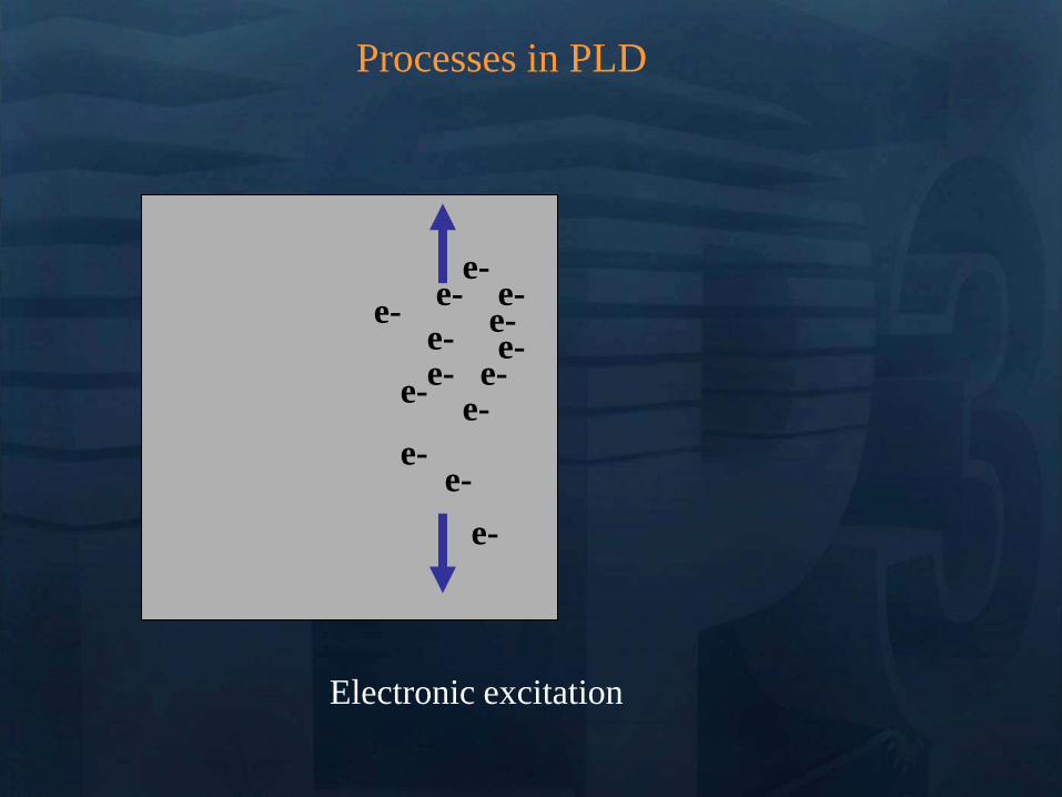
Processes in PLD
e- e-
e-
e- e-
e-
e-
e-
e-
e- e-
e-
e-
e-
Energy relaxation to lattice (~1 ps)
lattice
Processes in PLD
Heat diffusion (over microseconds)
lattice
Processes in PLD
Melting (tens of ns) Evaporation Plasma
Formation (microseconds) Resolidification
lattice
Processes in PLD
lattice
If laser pulse is long (ns) or
repetition rate is high laser may
continue interactions
Processes in Pulsed Laser Deposition
1 Absorption of laser pulse in material
Qab=(1-R)Ioe-aL
(metals absorption depths ~ 10 nm depends on )
2 Relaxation of energy (~ 1 ps) (electron-phonon interaction)
3 Heat transfer Melting and Evaporation
when electrons and lattice at thermal equilibrium (long pulses)
use heat conduction equation
(or heat diffusion model)
abp QTKt
TC
)(
At peak of laser pulse temperatures on target can reach
gt105 K (gt 40 eV)
Electric Fields gt 105 Vcm also high magnetic fields
Plasma Temperatures 3000-5000 K
Ablated Species with energies 1 ndash100 eV
Initially Incredibly Non-Equilibrium
Why biomaterials
- repair and reconstruction of parts of the musculo-skeletal system of vertebrates
- minimal biological requirement biocompatibility associated with the absence of
any adverse effect (non-toxic and non-allergic)
Other requests
- resistance to physiological fluids
- should not interfere with the bodyrsquos natural immunity system
- withstand mechanical stress during whole lifetime
- manufacturability in any appropriate shape
Possible classification
-a) biologically inactive (inert) alumina zirconia stainless steel CoCrNi
CoCrMo titanium titanium alloys carbon latex PE PMMA hellip
-b) porous Calcium Phosphates (CaPs) CaP-coated metal
-c) bioactive dense calcium phosphate ceramics bioactive glasses bioactive
glass-ceramics bioactive composites hellip
-d) Resorbable tricalcium phosphate calciumaliminate polylactic acid poly-L-
acetate
Our option biocompatible porous and bioactive CaP
Acronym
Chemical formula Compound name CaP
ratio
HA Ca10(PO4)6(OH)2 Hydroxylapatite 167
FA Ca10(PO4)6F2 Fluorapatite 167
CDHA Ca10x(HPO4)x(PO4)6x(OH)2x (0x2) Calcium-deficient Hydroxylapatite 133-167
BA Ca83(PO4)43 (CO3-HPO4)17(OH)03
BA=carbonated CDHA (x=17)
Biological apatite 138-193
Mn-CHA HA with (04-2) Mn2+ and (2-6) CO32- Mn2+ doped carbonated hydroxylapatite 151- 165
OHA Ca10(PO4)6(OH)22xOxٱx (0x1) Oxyhydroxylapatite 167
OA Ca10O(PO4)6 Oxyapatite 167
MCPM Ca(H2PO4)2middotH2O Monocalcium phosphate monohydrate 05
MCPA Ca(H2PO4)2 Monocalcium phosphate anhydrate 05
DCPD CaHPO4middot2H2O Dicalcium phosphate dihydrate (Brushite) 1
DCPA CaHPO4 Dicalcium phosphate anhydrate 1
OCP Ca8(HPO4)2(PO4)4middot5H2O Octacalcium phosphate 133
a-TCP Ca3(PO4)2 (monoclinic) Tricalcium phosphate (phase a) 15
-TCP Ca3(PO4)2 (rhombohedral) Tricalcium phosphate (phase Whitlockite) 15
TTCP Ca4O(PO4)2 Tetracalcium phosphate 2
a-DCP Ca2P2O7 (orthorhombic) Dicalcium phosphate (phase a) 1
-DCP Ca2P2O7 (tetragonal) Dicalcium phosphate (phase ) 1
ACP Cax(PO4)y nH2O (Amorphous Calcium pyrophosphate) 12-22
Bone
Resorbable CaP
CaP coating
Ti
Implant
Alternative solution Biomimetic coatings for metallic implants
Main deficiency of CaPs brittle in bulk
HA molecule Ca10(PO4)6OH2
Projection in the
(001) base plan of
the hydroxyapatite
unit cell (hexagonal
structure)
How difficult is to deposit CaPs (1)
- very complex molecules
How difficult is to deposit CaPs (2)
Crystal structure of OCP projected down the c-axis
Alternating apatite- and
hydrated- layers (100)
planes
The region with shaded
atoms the ldquoapatitic layerrdquo
is very similar to HA
The zone containing 10
water molecules is the
ldquohydrated layerrdquo
H atoms are omitted for
clarity
Octacalcium phosphate
(Ca8(HPO4)2(PO4)4middot5H2O)
Mesures par spectroscopie optique
drsquoemission sur le plasma issu de lrsquoirradiation
laser 248 nm drsquoune cible drsquoHA pure
Croissance impulsion par impulsion
Preacutesence des ions de
bull calcium (Ca I et Ca II)
bull oxygegravene (O I et O II) et
bull phosphore (P II)
Eacutemissions dues agrave des espegraveces moleacuteculaires preacutesentes dans le plasma
Couche mince drsquohydroxyapatite obtenue par
ablation laser
Goutelettes
CaP = 18 CaP = 21
CaP = 17 Trois reacutegions
typiques
i Reacutegion majoritaire lisse
ii Agglomeacuteration de gouttelettes sub-micromeacutetriques
iii Gouttelettes micromeacutetriques
Human primary osteoblasts (hOB) were cultured on OCP coated-Ti
Mn-CHA coatedndashTi HA coated-Ti Ti control (polystyrene)
hOB response SEM micrographs
- on bare Ti (a) after 7 days (b) after 21 days
Elongated rod-like morphology
Bioactivity tests
- OCP coatings (c) after 7 days (d) after 21 days
- In time cells spread and expand with flattened polyhedral-morphology
- Numerous cytoplasmatic extensions
hOB response SEM micrographs
- Mn-CHA coatings (e) after 7 days (f) after 21 days
- In time cells spread and expand with flattened polyhedral-morphology
- Less cytoplasmatic extensions
hOB response SEM micrographs
Biocompatible metallic thin films by PLD
Alloys NiTinol NiTi
NiTi characteristics ndash Super-Elastic Property
ndash Radiopaque
ndash Shape-Memory Effect
NiTi biomedical applications ndash Tweezers for removing foreign objects via small incisions
ndash Anchors for tendon fixation
ndash Stents for cardiovascular applications
ndash Dentistry - Orthodontic wires which no not need to be retightened and adjusted
Deposition chamber Multitarget system
PULSED LASER DEPOSITION (PLD)
SEM
Nitinol 30 sequences Nitinol 20 sequences
Cytotoxicity evaluation on microorganism culture
(a) E coli reference cell culture
(b) E coli cell culture on Ti surface
(c) E coli cell culture on NiTi surface
(d) S cerevisiae references cell culture
(e) S cerevisiae cell culture Ti surface
(f) S cerevisiae cell culture on NiTi surface
(g) SEM on E coli cell
culture on NiTi surface
(h) SEM on S cerevisiae cell culture on
NiTi surface E coli and S cerevisiae cell
cultures on the nitinol thin films
o Biocompatible
o Low adherence of microorganism on NiTi surface NiTi alloy does not stimulate the development at genetic level for specific genes which are involved in adhesion processes for microorganism cells
o Low release of Ni ions in solutions
CONCLUSIONS
MAPLE Matrix Assisted Pulsed Laser Evaporation
bull No need for UHV
bull Matrix prepared outside the
chamber
bull High flexibility
You can use everything
you can dissolve in a
solvent
MAPLE of polymers blends with Ag
nanoparticles
Controlled release of antibacterial agents
MAPLE deposition of PEGPLGA films with incorporated silver nanoparticles (AgNPs)
0 5 10 15 20 25 30000
005
010
015
020
025
Nu
mb
er o
f E
C
oli
X109
Time (hours)
Killing activity
0 5 10 15 20 25 30 3500
05
10
15
20
25
Bact
eria
l ce
ll n
um
ber
X109
Time (hours)
Delay of bacterial growth
Antibacterial efficiency against E Coli
Direct writing by femtosecond laser
pulses
Ormosil scaffolds for tissue engineering
50 lines ormosil sample
Distance between lines =
100 μm
30x30 lines ormosil
sample Distance between
lines = 50 μm
3 layers 30x30 lines ormosil
sample Distance between
lines = 100 μm
Scaffold functionalization
Protein deposition on the scaffolds by MAPLE
- Lysozyme antibacterial antitumor uses
- Fibrinogen biocompatible protein
Fibroblast cells grown on a polymeric grid with 100 microm distance between lines
Spectroscopie drsquoun panache plasma
laser femtoseconde
Laser
t =400 ns Spot 35x35 micromsup2 Cu
Z mm
Expeacuteriences reacutealiseacutees par (Grojo et al LP3Marseille)
nanoparticules
atoms
031 Jcm-2
Formation de nano agreacutegats
AFM
(Pereira et al )
TEM
(Kabashin et al)
Eacutetude des meacutecanismes de la formation drsquoagreacutegats
Couplage
- Direct Simulation Monte Carlo
- Dynamique Moleacuteculaire
Information deacutetailleacutee
-Continu du panache
-Distribution en taille des agreacutegats
K Gouriet thegravese (2008)
Simulation par Dynamique Moleacuteculaire (MD)
=gtdistribution drsquoagreacutegats
Cluster size N
No
rmaliz
ed
Yie
ld
100
101
102
103
104
105
10-9
10-8
10-7
10-6
10-5
10-4
10-3
10-2
10-1
100
N-426
N-122
t=1 ns
τ = 15 ps F ~ 2 Fth 4010 nm t =1 ns
Zhigilei et al (Virginia University)
Target applications of mobile nanomaterials
Bioimaging
Optical imaging deals with
visualization of biological objects
or tissues in order to detect
pathogens or follow the delivery
of drugs etc nanoparticles are
uses as contrast agents
In vitro
Cellular
imaging
In vivo Cancer
detection drug delivery
etc
Light-induced
therapies
Cancer therapy nanoparticles are used
as photosensitizers to produce local
targeted destruction of cancer cells
Photodynamic
therapy
Light-induced
hyperthermia
Diagnosis and Sensing A Diseases can be diagnosed through the (simultaneous) detection of a (set of) biomolecule(s) characteristic to a specific disease type and stage (biomarkers)
Huffman Nanomedicine and Nanobiotechnology Vol 1 1 2009
D
molecular signature of sick cell of infecting agent
(eg an antibody)
Cell membrane
Nanoparticle
Coating molecule specifically attracted to the molecular signature
C A nanoparticle can be functionalized in such a way that specifically targets a biomarker Thus the detection of the nanoparticle is linked to the detection of the biomarker and to the diagnosis of a disease
B Each cell type has unique molecular signatures that differentiate healthy and sick tissues Similarly an infection can be diagnosed by detecting the distinctive molecular signature of the infecting agent
Laser ablation of material from a solid target and production of
nanoparticlesnanostructures
Production of nanoclusters by laser
ablation and their subsequent cooling in
ambient medium Gaseous ambience
Nanoclusters can be
deposited on a
substrate forming a
nanostructured film
Aqueous ambience
Nanoclusters
crystallize and form
colloidal solution
NPs produced in gaseous ambience
0 20 40 60 80 100 120 140
0
2
4
6
8
Heig
ht
[nm
]
[nm]
01 1 102
3
4
5
6
7
Mean
part
icle
heig
ht
(nm
)
Helium pressure (Torr)
Silicon nanocrystal size AFM
Gas pressure decreases Nanocrystal size decreases
AFM of isolated laser-
ablated Si particles on
HOPG
Minimal particle size is 2-3 nm (from height measurements)
001Torr
1Torr
01Torr
10Torr
100Torr
NGC 2009
Photoluminescence of size selected nanoclusters
0 1 2 3 4 5 6
39 Jcm2
Num
ber
of
clust
ers
Size (nm)
30 Jcm2
14 Jcm2
0 1 2 3 4 5 6
He 4 Torr
10 Jcm2
15 20 25 30 35 40
0
500
1000
He 4 Torr
39 Jcmsup2
30 Jcmsup2
14 Jcmsup2
10 Jcmsup2
Inte
nsi
ty o
f P
L (
ua
)Photon energy (eV)
Silicon nanoclusters
Medical Imaging A Optical properties of nanoparticles depend greatly on its structure Particularly the color (wavelength) emitted by a quantum dot (a semiconductor nanoparticle) depends on its diameter
C The quantum dots (QD) can be injected to a
subject and then be detected by exciting them to emit light
Source Department of immunology University of Toronto
Solutions of CdSe QDrsquos of different diameter
CdSe nanoparticle (QD) structure
Source Laurence Livermore Laboratories
Imaging of QDrsquos targeted on cellular structures
Nano Letters 2008 Vol 8 pp3887-3892
B
In vivo cancer targeting and imaging with
semiconductor quantum dots
Gao et al Nat Biotech 22 969 - 976 (2004)
NPs produced in aqueous ambience
Advantages of laser ablation-based nanofabrication
compared to chemical methods
Disadvantage of chemical synthesis contaminationhelliphelliphellip
In particular chemical method for the fabrication of gold nanoparticles
Reduction of chloroauric acid (HAuCl4) with citrate in water
Control size by adding a stabilizing agent (thiol- (-SH) containing molecules)
Contamination impurities Cl- on surface non-biocompatible surfactantshellip
Problems in imaging
applications (especially
in vivo)
Problems in field-
enhanced applications (eg contaminants can
provide false Raman signals)
Focalized laser beam
Liquid medium
Target
Plasma
Laser ablation in liquids a way to avoid residual
contamination
- Laser energy can be transmitted through a liquid to ablate a solid
target the nanomaterial is released to the solution
- The process can be performed without any dirty chemical products in
clean liquid environment (deionized water PBS solution) no
contamination
Experiment
Ablation of gold final colloidal solutions
-Solution are ranging from deep wine red to pink and purple
- Color related to the size dependant surface plasmon resonance
Size distribution of Au nanoparticles prepared in
distilled water
0 50 100 150 200 250 300
0
200
400
600
800
1000
Rela
tive a
bundance (
arb
units)
Particle size (nm)
0 10 20 30 40 50 60 70 80 90 100
0
200
400
600
800
1000
Re
lative
Ab
un
da
nce
(a
rb u
nits)
Particle Size (nm) 0 2 4 6 8 10
0
200
400
600
800
1000
Re
lative
Ab
un
da
nce
(a
rb u
nits)
Particle Size (nm)
F = 20F0 Jcm2 F = 5F0 Jcm2 F = F0 Jcm2
-Size and dispersion of nanoparticles can be controlled by laser fluence
- Shape of craters on target at high F suggests the presence of cavitation phenomena
4 plusmn 15 nm
A V Kabashin M Meunier J Appl Phys 94 7941 (2003)
In water Very dispersed population of nanoparticles
Dextran (MW=6000gmol) Quenches the growth of the nanoparticles 2-4nm nanoparticles are produced (230-1850 atoms) Very fast dextran-gold interaction
Influence of the concentration
Dextran 6k
In pure water
300mJ
Ablation in biopolymers (dextran PEG)
Au OH
O
-
Dextran (C6H10O5)n
Nanoparticles react with OH
groups of biopolymers
Gold Nanoparticles vs Alzheimer
A Alzheimer and other degenerative diseases are caused my the clustering of amyloidal beta (Aβ) protein
D Gold nanoparticles can be functionalized to specifically attach to aggregates of this protein (amyloidosis)
Functionalized nanoparticle
Source wwwinternetchemistrycom
Chemical structure of Aβ-protein
Source wwwthefutureofthingscom
C
B
Alzheimerrsquos brain Healthy brain
Source Berkeley Lab
Gold Nanoparticles vs Alzheimer A The functionalized gold nanoparticles selectively attach to the aggregate of amyloidal protein The microwaves of certain frequency are irradiated on the sample Resonance with the gold nanoparticles increases the local temperature and destroy the aggregate
Nanoletters 2006 Vol 6 pp110-115
Before irradiation After irradiation
LIBS for biomedical applications in vivo analysis of tissue for real-time
analysis
real-time diagnosis of pathogen
presence in human fluids (bloodurine
CSF sputum)
71 35
ICCD
spectromegravetre
Time and space
Emission spectroscopie
LIBS (Laser Induced Breakdown Spectroscopy)
Principle of sample identification screening applications based on discriminant analysis here for warning when healthy tooth material is targeted during laser drilling
BMC Oral Health 2001 1 1 Ota Samek1 Helmut H Telle2 and David CS Beddows
Laser-induced breakdown spectroscopy a tool for real-time in vitro and in vivo identification of carious teeth
Sensitive Detection of Epithelial Ovarian Cancer Biomarkers
Using Tag-Laser Induced Breakdown Spectroscopy
Yuri Markushin and Noureddine Melikechi
Optical Science Center for Applied Research Delaware State University USA
X rays produced by laser plasmas
I = ~1016 agrave 1018 Wcm2
nc cm-3
1111021 mm 2
Thot Ilas2
1 3
Ilas2
3 4
Hot electrons
(characteristic lines)
X-ray
emission
laser
X-rays
Line emission Fast electrons knock
free core electrons
from the inner shell
Bremsstrahlung electrons deflected by nucleus
Laser-generated hot electrons produce x-rays by two dominant mechanisms
ne
critical surface (cf cathode)
conventional X-ray tube
Kmetec ldquoMeV X-ray generation with a femtosecond laserrdquo Phys Rev Lett 68 1527 (1992)
Rousse ldquoEfficient Ka X-ray source from fs-laser-produced plasmasrdquo Phys Rev E 50 2200 (1994)
(typical recombination lifetimes several fs)
Hou et al Appl Phys Lett 84 2259 (2004)
Tightly focused fs lasers create the worldrsquos smallest
hard x-ray source (~4 microm) for precision imaging
x-ray source size is ~ 4 larger than laser focus
because of lateral heat transport Bowes Opt Lett 31 116 (2006)
2004 premiegravere deacutemonstration drsquoimagerie avec contraste de phase avec une source X creacutee par laser
Source
Rayon non diffracteacute
Rayon diffracteacute
Lcoh 2R1
eacutechantillon
Absorption vs contraste de phase
dsource-object dobject-detector
X-ray
Source
Object
Detector
position
Dsource
Intensity
interface
dsource-object dobject-detector
X-ray
Source
Object
Detector
position
Fresnel Diffraction
Dsource
Intensity
interface
enhancement interface
R Toth et al
Rev Sci Instrum 76
083701 (2005)
Imagerie par contraste de phase
R Toth et al Rev Sci Instrum
76 083701 (2005)
copy INRS
bull Meilleure reacutesolution (5microm)
bull dose plus faible
bull meilleur contraste (phase)
bull lrsquoeacutenergie X peut ecirctre ajusteacutee
aux besoins de lrsquoimagerie
La reacutefeacuterence mondiale ALLS
(INRS- Montreacuteal) 200TW + Puissance moyenne 50W
47J 23fs 10Hz
in a 8microm spot
Sans miroir deacuteformable
microCT
Scan duration avec
50W10Hz (360 images)
2 min (mouse scan)
courtesy Prof Dr Oswald Willi U Duumlselldorf
MeV Protons produced by laser CR39 or RCF 1010 to 1013 H+
courtesy Prof Don Umstadter U Nebraska-Lincoln
Target Normal Sheath Acceleration hot electrons traversing
target electrostatically accelerate impurity hydrogen ions on the rear surface
virtual
cathode
I ge 1019 Wcm2
Protons de 10MeV (INRS )
~1022 Wcm2
Avantages des particules chargeacutees ou hadrons (protons ions) Rayons X de la Radio-Theacuterapie
Le pic de Bragg
La profondeur de peacuteneacutetration deacutepend de l rsquoeacutenergie incidente des protons
la tumeur la plus superficielle = l rsquoœil 70 MeV
la plus profonde = la prostate 200 - 250 MeV
Protontheacuterapie par laser (projet SAPHIR)
CPO
Exemple de dosimeacutetrie preacutevisionnelle
(photons vs protons)
Comparaison de lrsquoeacutepargne de dose dans les tissus sains en protons (image de droite)
par rapport aux photons (IMRT Intensity Modulated Radio-Therapy image de gauche)
Les irradiations colateacuterales en radiotheacuterapie et en protontheacuterapie
Avantage consideacuterable pour le traitement de tumeurs localiseacutees pregraves d rsquoorganes sensibles (œil cerveau)
currently treating patients 5
5 next 2 years
others awaiting approval
10000 m2 $125 M facility opened in 2006
12 m diameter 250 MeV synchrotron
ldquoThere are too few physicists in the world
and they are an incredibly important part of
doing this We have one of the largest
physics departments in the world with more than 50 medical physicistsrdquo
--- Dr James D Cox head of Radiation Oncology at MD Anderson Cancer Center Houston Texas
Proton Therapy enables precise exposure of small tumors
with minimal damage to surrounding healthy tissue
250 MeV
PROTON beam
10 20
Depth in Tissue [cm]
0 0
50
100
DO
SE
(
)
6 MV
PHOTON
beam
tunable
Bragg
peak
but requires large expensive facilities
eye tumors 65 MeV protons
deep tumors gt200 MeV
Laser proton therapy could be much smaller amp cheaper Fourkal Med Phys 29 2788 (2002)
Malka Med Phys 31 1587 (2004)
On-site production of short-lived isotopes for medical imaging
Laser-generated quasi-mono-energetic electrons efficiently photo-activate materials of interest
bull High Rep rate bull Low cost bull Compact
Limitations to the widespread use of PET arise from the high costs of cyclotrons needed to
produce the short-lived radionucleotides for PET scanning Few hospitals and universities
are capable of maintaining such systems - Wikipedia -
radiotracer activation reaction half-life medical use
15O 16O (n)15O 2 minutes neuro-imaging
11C 12C(n)11C 20 minutes neuro-receptor-specific brain imaging
18F 19F(n)18F 110 minutes clinical oncology
Positron Emission Tomography
on-site
production
essential
18F PET scan of tumor 15O PET scan of human brain
Reed ldquoEfficient initiation of photonuclear reactions using quasi-monoenergetic electron beams from LWFArdquo J Appl Phys 102 073103 (2007)
TEP in situ
+ in vivo
Modegravele animal
microCT +
contraste de phase
+ in vivo
modegraveles de cancer
Comprendre
le cancer
Preacuteparer le traitement
protons 50MeV
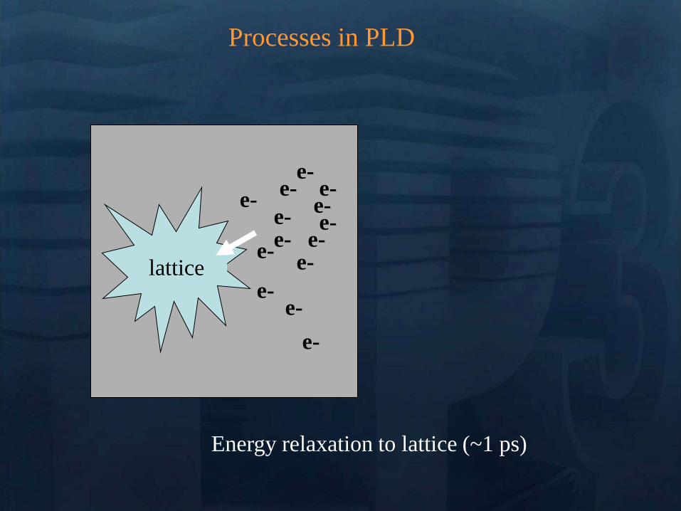
Processes in PLD
Heat diffusion (over microseconds)
lattice
Processes in PLD
Melting (tens of ns) Evaporation Plasma
Formation (microseconds) Resolidification
lattice
Processes in PLD
lattice
If laser pulse is long (ns) or
repetition rate is high laser may
continue interactions
Processes in Pulsed Laser Deposition
1 Absorption of laser pulse in material
Qab=(1-R)Ioe-aL
(metals absorption depths ~ 10 nm depends on )
2 Relaxation of energy (~ 1 ps) (electron-phonon interaction)
3 Heat transfer Melting and Evaporation
when electrons and lattice at thermal equilibrium (long pulses)
use heat conduction equation
(or heat diffusion model)
abp QTKt
TC
)(
At peak of laser pulse temperatures on target can reach
gt105 K (gt 40 eV)
Electric Fields gt 105 Vcm also high magnetic fields
Plasma Temperatures 3000-5000 K
Ablated Species with energies 1 ndash100 eV
Initially Incredibly Non-Equilibrium
Why biomaterials
- repair and reconstruction of parts of the musculo-skeletal system of vertebrates
- minimal biological requirement biocompatibility associated with the absence of
any adverse effect (non-toxic and non-allergic)
Other requests
- resistance to physiological fluids
- should not interfere with the bodyrsquos natural immunity system
- withstand mechanical stress during whole lifetime
- manufacturability in any appropriate shape
Possible classification
-a) biologically inactive (inert) alumina zirconia stainless steel CoCrNi
CoCrMo titanium titanium alloys carbon latex PE PMMA hellip
-b) porous Calcium Phosphates (CaPs) CaP-coated metal
-c) bioactive dense calcium phosphate ceramics bioactive glasses bioactive
glass-ceramics bioactive composites hellip
-d) Resorbable tricalcium phosphate calciumaliminate polylactic acid poly-L-
acetate
Our option biocompatible porous and bioactive CaP
Acronym
Chemical formula Compound name CaP
ratio
HA Ca10(PO4)6(OH)2 Hydroxylapatite 167
FA Ca10(PO4)6F2 Fluorapatite 167
CDHA Ca10x(HPO4)x(PO4)6x(OH)2x (0x2) Calcium-deficient Hydroxylapatite 133-167
BA Ca83(PO4)43 (CO3-HPO4)17(OH)03
BA=carbonated CDHA (x=17)
Biological apatite 138-193
Mn-CHA HA with (04-2) Mn2+ and (2-6) CO32- Mn2+ doped carbonated hydroxylapatite 151- 165
OHA Ca10(PO4)6(OH)22xOxٱx (0x1) Oxyhydroxylapatite 167
OA Ca10O(PO4)6 Oxyapatite 167
MCPM Ca(H2PO4)2middotH2O Monocalcium phosphate monohydrate 05
MCPA Ca(H2PO4)2 Monocalcium phosphate anhydrate 05
DCPD CaHPO4middot2H2O Dicalcium phosphate dihydrate (Brushite) 1
DCPA CaHPO4 Dicalcium phosphate anhydrate 1
OCP Ca8(HPO4)2(PO4)4middot5H2O Octacalcium phosphate 133
a-TCP Ca3(PO4)2 (monoclinic) Tricalcium phosphate (phase a) 15
-TCP Ca3(PO4)2 (rhombohedral) Tricalcium phosphate (phase Whitlockite) 15
TTCP Ca4O(PO4)2 Tetracalcium phosphate 2
a-DCP Ca2P2O7 (orthorhombic) Dicalcium phosphate (phase a) 1
-DCP Ca2P2O7 (tetragonal) Dicalcium phosphate (phase ) 1
ACP Cax(PO4)y nH2O (Amorphous Calcium pyrophosphate) 12-22
Bone
Resorbable CaP
CaP coating
Ti
Implant
Alternative solution Biomimetic coatings for metallic implants
Main deficiency of CaPs brittle in bulk
HA molecule Ca10(PO4)6OH2
Projection in the
(001) base plan of
the hydroxyapatite
unit cell (hexagonal
structure)
How difficult is to deposit CaPs (1)
- very complex molecules
How difficult is to deposit CaPs (2)
Crystal structure of OCP projected down the c-axis
Alternating apatite- and
hydrated- layers (100)
planes
The region with shaded
atoms the ldquoapatitic layerrdquo
is very similar to HA
The zone containing 10
water molecules is the
ldquohydrated layerrdquo
H atoms are omitted for
clarity
Octacalcium phosphate
(Ca8(HPO4)2(PO4)4middot5H2O)
Mesures par spectroscopie optique
drsquoemission sur le plasma issu de lrsquoirradiation
laser 248 nm drsquoune cible drsquoHA pure
Croissance impulsion par impulsion
Preacutesence des ions de
bull calcium (Ca I et Ca II)
bull oxygegravene (O I et O II) et
bull phosphore (P II)
Eacutemissions dues agrave des espegraveces moleacuteculaires preacutesentes dans le plasma
Couche mince drsquohydroxyapatite obtenue par
ablation laser
Goutelettes
CaP = 18 CaP = 21
CaP = 17 Trois reacutegions
typiques
i Reacutegion majoritaire lisse
ii Agglomeacuteration de gouttelettes sub-micromeacutetriques
iii Gouttelettes micromeacutetriques
Human primary osteoblasts (hOB) were cultured on OCP coated-Ti
Mn-CHA coatedndashTi HA coated-Ti Ti control (polystyrene)
hOB response SEM micrographs
- on bare Ti (a) after 7 days (b) after 21 days
Elongated rod-like morphology
Bioactivity tests
- OCP coatings (c) after 7 days (d) after 21 days
- In time cells spread and expand with flattened polyhedral-morphology
- Numerous cytoplasmatic extensions
hOB response SEM micrographs
- Mn-CHA coatings (e) after 7 days (f) after 21 days
- In time cells spread and expand with flattened polyhedral-morphology
- Less cytoplasmatic extensions
hOB response SEM micrographs
Biocompatible metallic thin films by PLD
Alloys NiTinol NiTi
NiTi characteristics ndash Super-Elastic Property
ndash Radiopaque
ndash Shape-Memory Effect
NiTi biomedical applications ndash Tweezers for removing foreign objects via small incisions
ndash Anchors for tendon fixation
ndash Stents for cardiovascular applications
ndash Dentistry - Orthodontic wires which no not need to be retightened and adjusted
Deposition chamber Multitarget system
PULSED LASER DEPOSITION (PLD)
SEM
Nitinol 30 sequences Nitinol 20 sequences
Cytotoxicity evaluation on microorganism culture
(a) E coli reference cell culture
(b) E coli cell culture on Ti surface
(c) E coli cell culture on NiTi surface
(d) S cerevisiae references cell culture
(e) S cerevisiae cell culture Ti surface
(f) S cerevisiae cell culture on NiTi surface
(g) SEM on E coli cell
culture on NiTi surface
(h) SEM on S cerevisiae cell culture on
NiTi surface E coli and S cerevisiae cell
cultures on the nitinol thin films
o Biocompatible
o Low adherence of microorganism on NiTi surface NiTi alloy does not stimulate the development at genetic level for specific genes which are involved in adhesion processes for microorganism cells
o Low release of Ni ions in solutions
CONCLUSIONS
MAPLE Matrix Assisted Pulsed Laser Evaporation
bull No need for UHV
bull Matrix prepared outside the
chamber
bull High flexibility
You can use everything
you can dissolve in a
solvent
MAPLE of polymers blends with Ag
nanoparticles
Controlled release of antibacterial agents
MAPLE deposition of PEGPLGA films with incorporated silver nanoparticles (AgNPs)
0 5 10 15 20 25 30000
005
010
015
020
025
Nu
mb
er o
f E
C
oli
X109
Time (hours)
Killing activity
0 5 10 15 20 25 30 3500
05
10
15
20
25
Bact
eria
l ce
ll n
um
ber
X109
Time (hours)
Delay of bacterial growth
Antibacterial efficiency against E Coli
Direct writing by femtosecond laser
pulses
Ormosil scaffolds for tissue engineering
50 lines ormosil sample
Distance between lines =
100 μm
30x30 lines ormosil
sample Distance between
lines = 50 μm
3 layers 30x30 lines ormosil
sample Distance between
lines = 100 μm
Scaffold functionalization
Protein deposition on the scaffolds by MAPLE
- Lysozyme antibacterial antitumor uses
- Fibrinogen biocompatible protein
Fibroblast cells grown on a polymeric grid with 100 microm distance between lines
Spectroscopie drsquoun panache plasma
laser femtoseconde
Laser
t =400 ns Spot 35x35 micromsup2 Cu
Z mm
Expeacuteriences reacutealiseacutees par (Grojo et al LP3Marseille)
nanoparticules
atoms
031 Jcm-2
Formation de nano agreacutegats
AFM
(Pereira et al )
TEM
(Kabashin et al)
Eacutetude des meacutecanismes de la formation drsquoagreacutegats
Couplage
- Direct Simulation Monte Carlo
- Dynamique Moleacuteculaire
Information deacutetailleacutee
-Continu du panache
-Distribution en taille des agreacutegats
K Gouriet thegravese (2008)
Simulation par Dynamique Moleacuteculaire (MD)
=gtdistribution drsquoagreacutegats
Cluster size N
No
rmaliz
ed
Yie
ld
100
101
102
103
104
105
10-9
10-8
10-7
10-6
10-5
10-4
10-3
10-2
10-1
100
N-426
N-122
t=1 ns
τ = 15 ps F ~ 2 Fth 4010 nm t =1 ns
Zhigilei et al (Virginia University)
Target applications of mobile nanomaterials
Bioimaging
Optical imaging deals with
visualization of biological objects
or tissues in order to detect
pathogens or follow the delivery
of drugs etc nanoparticles are
uses as contrast agents
In vitro
Cellular
imaging
In vivo Cancer
detection drug delivery
etc
Light-induced
therapies
Cancer therapy nanoparticles are used
as photosensitizers to produce local
targeted destruction of cancer cells
Photodynamic
therapy
Light-induced
hyperthermia
Diagnosis and Sensing A Diseases can be diagnosed through the (simultaneous) detection of a (set of) biomolecule(s) characteristic to a specific disease type and stage (biomarkers)
Huffman Nanomedicine and Nanobiotechnology Vol 1 1 2009
D
molecular signature of sick cell of infecting agent
(eg an antibody)
Cell membrane
Nanoparticle
Coating molecule specifically attracted to the molecular signature
C A nanoparticle can be functionalized in such a way that specifically targets a biomarker Thus the detection of the nanoparticle is linked to the detection of the biomarker and to the diagnosis of a disease
B Each cell type has unique molecular signatures that differentiate healthy and sick tissues Similarly an infection can be diagnosed by detecting the distinctive molecular signature of the infecting agent
Laser ablation of material from a solid target and production of
nanoparticlesnanostructures
Production of nanoclusters by laser
ablation and their subsequent cooling in
ambient medium Gaseous ambience
Nanoclusters can be
deposited on a
substrate forming a
nanostructured film
Aqueous ambience
Nanoclusters
crystallize and form
colloidal solution
NPs produced in gaseous ambience
0 20 40 60 80 100 120 140
0
2
4
6
8
Heig
ht
[nm
]
[nm]
01 1 102
3
4
5
6
7
Mean
part
icle
heig
ht
(nm
)
Helium pressure (Torr)
Silicon nanocrystal size AFM
Gas pressure decreases Nanocrystal size decreases
AFM of isolated laser-
ablated Si particles on
HOPG
Minimal particle size is 2-3 nm (from height measurements)
001Torr
1Torr
01Torr
10Torr
100Torr
NGC 2009
Photoluminescence of size selected nanoclusters
0 1 2 3 4 5 6
39 Jcm2
Num
ber
of
clust
ers
Size (nm)
30 Jcm2
14 Jcm2
0 1 2 3 4 5 6
He 4 Torr
10 Jcm2
15 20 25 30 35 40
0
500
1000
He 4 Torr
39 Jcmsup2
30 Jcmsup2
14 Jcmsup2
10 Jcmsup2
Inte
nsi
ty o
f P
L (
ua
)Photon energy (eV)
Silicon nanoclusters
Medical Imaging A Optical properties of nanoparticles depend greatly on its structure Particularly the color (wavelength) emitted by a quantum dot (a semiconductor nanoparticle) depends on its diameter
C The quantum dots (QD) can be injected to a
subject and then be detected by exciting them to emit light
Source Department of immunology University of Toronto
Solutions of CdSe QDrsquos of different diameter
CdSe nanoparticle (QD) structure
Source Laurence Livermore Laboratories
Imaging of QDrsquos targeted on cellular structures
Nano Letters 2008 Vol 8 pp3887-3892
B
In vivo cancer targeting and imaging with
semiconductor quantum dots
Gao et al Nat Biotech 22 969 - 976 (2004)
NPs produced in aqueous ambience
Advantages of laser ablation-based nanofabrication
compared to chemical methods
Disadvantage of chemical synthesis contaminationhelliphelliphellip
In particular chemical method for the fabrication of gold nanoparticles
Reduction of chloroauric acid (HAuCl4) with citrate in water
Control size by adding a stabilizing agent (thiol- (-SH) containing molecules)
Contamination impurities Cl- on surface non-biocompatible surfactantshellip
Problems in imaging
applications (especially
in vivo)
Problems in field-
enhanced applications (eg contaminants can
provide false Raman signals)
Focalized laser beam
Liquid medium
Target
Plasma
Laser ablation in liquids a way to avoid residual
contamination
- Laser energy can be transmitted through a liquid to ablate a solid
target the nanomaterial is released to the solution
- The process can be performed without any dirty chemical products in
clean liquid environment (deionized water PBS solution) no
contamination
Experiment
Ablation of gold final colloidal solutions
-Solution are ranging from deep wine red to pink and purple
- Color related to the size dependant surface plasmon resonance
Size distribution of Au nanoparticles prepared in
distilled water
0 50 100 150 200 250 300
0
200
400
600
800
1000
Rela
tive a
bundance (
arb
units)
Particle size (nm)
0 10 20 30 40 50 60 70 80 90 100
0
200
400
600
800
1000
Re
lative
Ab
un
da
nce
(a
rb u
nits)
Particle Size (nm) 0 2 4 6 8 10
0
200
400
600
800
1000
Re
lative
Ab
un
da
nce
(a
rb u
nits)
Particle Size (nm)
F = 20F0 Jcm2 F = 5F0 Jcm2 F = F0 Jcm2
-Size and dispersion of nanoparticles can be controlled by laser fluence
- Shape of craters on target at high F suggests the presence of cavitation phenomena
4 plusmn 15 nm
A V Kabashin M Meunier J Appl Phys 94 7941 (2003)
In water Very dispersed population of nanoparticles
Dextran (MW=6000gmol) Quenches the growth of the nanoparticles 2-4nm nanoparticles are produced (230-1850 atoms) Very fast dextran-gold interaction
Influence of the concentration
Dextran 6k
In pure water
300mJ
Ablation in biopolymers (dextran PEG)
Au OH
O
-
Dextran (C6H10O5)n
Nanoparticles react with OH
groups of biopolymers
Gold Nanoparticles vs Alzheimer
A Alzheimer and other degenerative diseases are caused my the clustering of amyloidal beta (Aβ) protein
D Gold nanoparticles can be functionalized to specifically attach to aggregates of this protein (amyloidosis)
Functionalized nanoparticle
Source wwwinternetchemistrycom
Chemical structure of Aβ-protein
Source wwwthefutureofthingscom
C
B
Alzheimerrsquos brain Healthy brain
Source Berkeley Lab
Gold Nanoparticles vs Alzheimer A The functionalized gold nanoparticles selectively attach to the aggregate of amyloidal protein The microwaves of certain frequency are irradiated on the sample Resonance with the gold nanoparticles increases the local temperature and destroy the aggregate
Nanoletters 2006 Vol 6 pp110-115
Before irradiation After irradiation
LIBS for biomedical applications in vivo analysis of tissue for real-time
analysis
real-time diagnosis of pathogen
presence in human fluids (bloodurine
CSF sputum)
71 35
ICCD
spectromegravetre
Time and space
Emission spectroscopie
LIBS (Laser Induced Breakdown Spectroscopy)
Principle of sample identification screening applications based on discriminant analysis here for warning when healthy tooth material is targeted during laser drilling
BMC Oral Health 2001 1 1 Ota Samek1 Helmut H Telle2 and David CS Beddows
Laser-induced breakdown spectroscopy a tool for real-time in vitro and in vivo identification of carious teeth
Sensitive Detection of Epithelial Ovarian Cancer Biomarkers
Using Tag-Laser Induced Breakdown Spectroscopy
Yuri Markushin and Noureddine Melikechi
Optical Science Center for Applied Research Delaware State University USA
X rays produced by laser plasmas
I = ~1016 agrave 1018 Wcm2
nc cm-3
1111021 mm 2
Thot Ilas2
1 3
Ilas2
3 4
Hot electrons
(characteristic lines)
X-ray
emission
laser
X-rays
Line emission Fast electrons knock
free core electrons
from the inner shell
Bremsstrahlung electrons deflected by nucleus
Laser-generated hot electrons produce x-rays by two dominant mechanisms
ne
critical surface (cf cathode)
conventional X-ray tube
Kmetec ldquoMeV X-ray generation with a femtosecond laserrdquo Phys Rev Lett 68 1527 (1992)
Rousse ldquoEfficient Ka X-ray source from fs-laser-produced plasmasrdquo Phys Rev E 50 2200 (1994)
(typical recombination lifetimes several fs)
Hou et al Appl Phys Lett 84 2259 (2004)
Tightly focused fs lasers create the worldrsquos smallest
hard x-ray source (~4 microm) for precision imaging
x-ray source size is ~ 4 larger than laser focus
because of lateral heat transport Bowes Opt Lett 31 116 (2006)
2004 premiegravere deacutemonstration drsquoimagerie avec contraste de phase avec une source X creacutee par laser
Source
Rayon non diffracteacute
Rayon diffracteacute
Lcoh 2R1
eacutechantillon
Absorption vs contraste de phase
dsource-object dobject-detector
X-ray
Source
Object
Detector
position
Dsource
Intensity
interface
dsource-object dobject-detector
X-ray
Source
Object
Detector
position
Fresnel Diffraction
Dsource
Intensity
interface
enhancement interface
R Toth et al
Rev Sci Instrum 76
083701 (2005)
Imagerie par contraste de phase
R Toth et al Rev Sci Instrum
76 083701 (2005)
copy INRS
bull Meilleure reacutesolution (5microm)
bull dose plus faible
bull meilleur contraste (phase)
bull lrsquoeacutenergie X peut ecirctre ajusteacutee
aux besoins de lrsquoimagerie
La reacutefeacuterence mondiale ALLS
(INRS- Montreacuteal) 200TW + Puissance moyenne 50W
47J 23fs 10Hz
in a 8microm spot
Sans miroir deacuteformable
microCT
Scan duration avec
50W10Hz (360 images)
2 min (mouse scan)
courtesy Prof Dr Oswald Willi U Duumlselldorf
MeV Protons produced by laser CR39 or RCF 1010 to 1013 H+
courtesy Prof Don Umstadter U Nebraska-Lincoln
Target Normal Sheath Acceleration hot electrons traversing
target electrostatically accelerate impurity hydrogen ions on the rear surface
virtual
cathode
I ge 1019 Wcm2
Protons de 10MeV (INRS )
~1022 Wcm2
Avantages des particules chargeacutees ou hadrons (protons ions) Rayons X de la Radio-Theacuterapie
Le pic de Bragg
La profondeur de peacuteneacutetration deacutepend de l rsquoeacutenergie incidente des protons
la tumeur la plus superficielle = l rsquoœil 70 MeV
la plus profonde = la prostate 200 - 250 MeV
Protontheacuterapie par laser (projet SAPHIR)
CPO
Exemple de dosimeacutetrie preacutevisionnelle
(photons vs protons)
Comparaison de lrsquoeacutepargne de dose dans les tissus sains en protons (image de droite)
par rapport aux photons (IMRT Intensity Modulated Radio-Therapy image de gauche)
Les irradiations colateacuterales en radiotheacuterapie et en protontheacuterapie
Avantage consideacuterable pour le traitement de tumeurs localiseacutees pregraves d rsquoorganes sensibles (œil cerveau)
currently treating patients 5
5 next 2 years
others awaiting approval
10000 m2 $125 M facility opened in 2006
12 m diameter 250 MeV synchrotron
ldquoThere are too few physicists in the world
and they are an incredibly important part of
doing this We have one of the largest
physics departments in the world with more than 50 medical physicistsrdquo
--- Dr James D Cox head of Radiation Oncology at MD Anderson Cancer Center Houston Texas
Proton Therapy enables precise exposure of small tumors
with minimal damage to surrounding healthy tissue
250 MeV
PROTON beam
10 20
Depth in Tissue [cm]
0 0
50
100
DO
SE
(
)
6 MV
PHOTON
beam
tunable
Bragg
peak
but requires large expensive facilities
eye tumors 65 MeV protons
deep tumors gt200 MeV
Laser proton therapy could be much smaller amp cheaper Fourkal Med Phys 29 2788 (2002)
Malka Med Phys 31 1587 (2004)
On-site production of short-lived isotopes for medical imaging
Laser-generated quasi-mono-energetic electrons efficiently photo-activate materials of interest
bull High Rep rate bull Low cost bull Compact
Limitations to the widespread use of PET arise from the high costs of cyclotrons needed to
produce the short-lived radionucleotides for PET scanning Few hospitals and universities
are capable of maintaining such systems - Wikipedia -
radiotracer activation reaction half-life medical use
15O 16O (n)15O 2 minutes neuro-imaging
11C 12C(n)11C 20 minutes neuro-receptor-specific brain imaging
18F 19F(n)18F 110 minutes clinical oncology
Positron Emission Tomography
on-site
production
essential
18F PET scan of tumor 15O PET scan of human brain
Reed ldquoEfficient initiation of photonuclear reactions using quasi-monoenergetic electron beams from LWFArdquo J Appl Phys 102 073103 (2007)
TEP in situ
+ in vivo
Modegravele animal
microCT +
contraste de phase
+ in vivo
modegraveles de cancer
Comprendre
le cancer
Preacuteparer le traitement
protons 50MeV
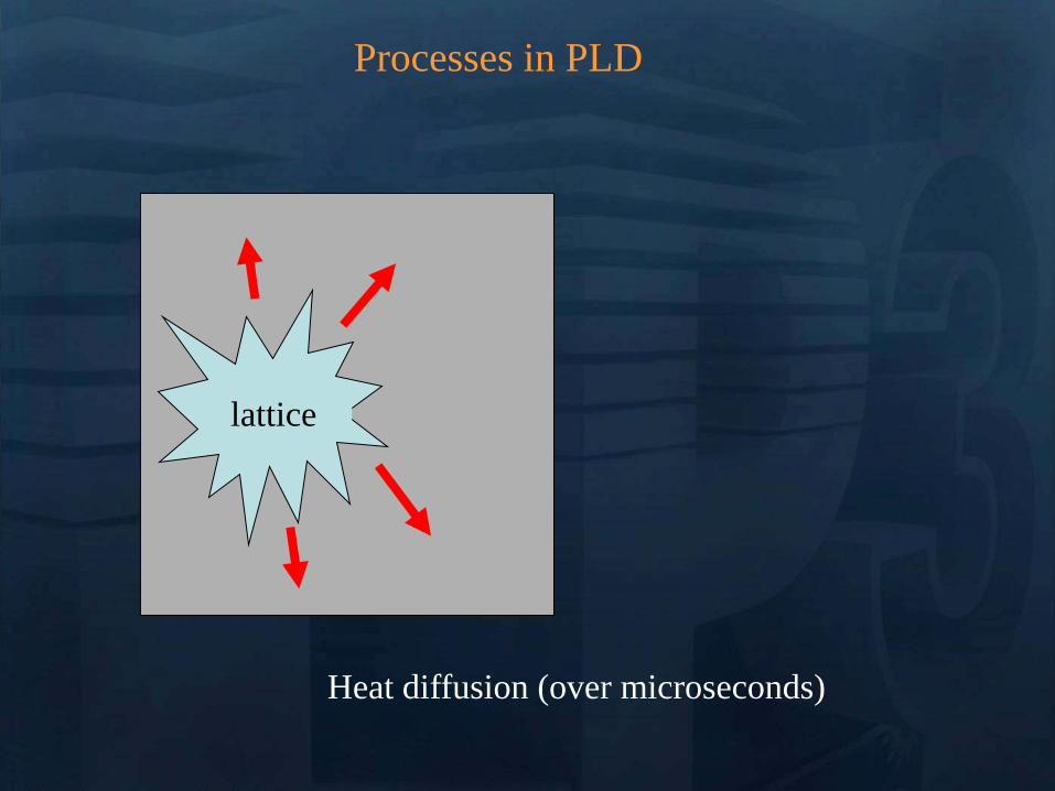
Processes in PLD
Melting (tens of ns) Evaporation Plasma
Formation (microseconds) Resolidification
lattice
Processes in PLD
lattice
If laser pulse is long (ns) or
repetition rate is high laser may
continue interactions
Processes in Pulsed Laser Deposition
1 Absorption of laser pulse in material
Qab=(1-R)Ioe-aL
(metals absorption depths ~ 10 nm depends on )
2 Relaxation of energy (~ 1 ps) (electron-phonon interaction)
3 Heat transfer Melting and Evaporation
when electrons and lattice at thermal equilibrium (long pulses)
use heat conduction equation
(or heat diffusion model)
abp QTKt
TC
)(
At peak of laser pulse temperatures on target can reach
gt105 K (gt 40 eV)
Electric Fields gt 105 Vcm also high magnetic fields
Plasma Temperatures 3000-5000 K
Ablated Species with energies 1 ndash100 eV
Initially Incredibly Non-Equilibrium
Why biomaterials
- repair and reconstruction of parts of the musculo-skeletal system of vertebrates
- minimal biological requirement biocompatibility associated with the absence of
any adverse effect (non-toxic and non-allergic)
Other requests
- resistance to physiological fluids
- should not interfere with the bodyrsquos natural immunity system
- withstand mechanical stress during whole lifetime
- manufacturability in any appropriate shape
Possible classification
-a) biologically inactive (inert) alumina zirconia stainless steel CoCrNi
CoCrMo titanium titanium alloys carbon latex PE PMMA hellip
-b) porous Calcium Phosphates (CaPs) CaP-coated metal
-c) bioactive dense calcium phosphate ceramics bioactive glasses bioactive
glass-ceramics bioactive composites hellip
-d) Resorbable tricalcium phosphate calciumaliminate polylactic acid poly-L-
acetate
Our option biocompatible porous and bioactive CaP
Acronym
Chemical formula Compound name CaP
ratio
HA Ca10(PO4)6(OH)2 Hydroxylapatite 167
FA Ca10(PO4)6F2 Fluorapatite 167
CDHA Ca10x(HPO4)x(PO4)6x(OH)2x (0x2) Calcium-deficient Hydroxylapatite 133-167
BA Ca83(PO4)43 (CO3-HPO4)17(OH)03
BA=carbonated CDHA (x=17)
Biological apatite 138-193
Mn-CHA HA with (04-2) Mn2+ and (2-6) CO32- Mn2+ doped carbonated hydroxylapatite 151- 165
OHA Ca10(PO4)6(OH)22xOxٱx (0x1) Oxyhydroxylapatite 167
OA Ca10O(PO4)6 Oxyapatite 167
MCPM Ca(H2PO4)2middotH2O Monocalcium phosphate monohydrate 05
MCPA Ca(H2PO4)2 Monocalcium phosphate anhydrate 05
DCPD CaHPO4middot2H2O Dicalcium phosphate dihydrate (Brushite) 1
DCPA CaHPO4 Dicalcium phosphate anhydrate 1
OCP Ca8(HPO4)2(PO4)4middot5H2O Octacalcium phosphate 133
a-TCP Ca3(PO4)2 (monoclinic) Tricalcium phosphate (phase a) 15
-TCP Ca3(PO4)2 (rhombohedral) Tricalcium phosphate (phase Whitlockite) 15
TTCP Ca4O(PO4)2 Tetracalcium phosphate 2
a-DCP Ca2P2O7 (orthorhombic) Dicalcium phosphate (phase a) 1
-DCP Ca2P2O7 (tetragonal) Dicalcium phosphate (phase ) 1
ACP Cax(PO4)y nH2O (Amorphous Calcium pyrophosphate) 12-22
Bone
Resorbable CaP
CaP coating
Ti
Implant
Alternative solution Biomimetic coatings for metallic implants
Main deficiency of CaPs brittle in bulk
HA molecule Ca10(PO4)6OH2
Projection in the
(001) base plan of
the hydroxyapatite
unit cell (hexagonal
structure)
How difficult is to deposit CaPs (1)
- very complex molecules
How difficult is to deposit CaPs (2)
Crystal structure of OCP projected down the c-axis
Alternating apatite- and
hydrated- layers (100)
planes
The region with shaded
atoms the ldquoapatitic layerrdquo
is very similar to HA
The zone containing 10
water molecules is the
ldquohydrated layerrdquo
H atoms are omitted for
clarity
Octacalcium phosphate
(Ca8(HPO4)2(PO4)4middot5H2O)
Mesures par spectroscopie optique
drsquoemission sur le plasma issu de lrsquoirradiation
laser 248 nm drsquoune cible drsquoHA pure
Croissance impulsion par impulsion
Preacutesence des ions de
bull calcium (Ca I et Ca II)
bull oxygegravene (O I et O II) et
bull phosphore (P II)
Eacutemissions dues agrave des espegraveces moleacuteculaires preacutesentes dans le plasma
Couche mince drsquohydroxyapatite obtenue par
ablation laser
Goutelettes
CaP = 18 CaP = 21
CaP = 17 Trois reacutegions
typiques
i Reacutegion majoritaire lisse
ii Agglomeacuteration de gouttelettes sub-micromeacutetriques
iii Gouttelettes micromeacutetriques
Human primary osteoblasts (hOB) were cultured on OCP coated-Ti
Mn-CHA coatedndashTi HA coated-Ti Ti control (polystyrene)
hOB response SEM micrographs
- on bare Ti (a) after 7 days (b) after 21 days
Elongated rod-like morphology
Bioactivity tests
- OCP coatings (c) after 7 days (d) after 21 days
- In time cells spread and expand with flattened polyhedral-morphology
- Numerous cytoplasmatic extensions
hOB response SEM micrographs
- Mn-CHA coatings (e) after 7 days (f) after 21 days
- In time cells spread and expand with flattened polyhedral-morphology
- Less cytoplasmatic extensions
hOB response SEM micrographs
Biocompatible metallic thin films by PLD
Alloys NiTinol NiTi
NiTi characteristics ndash Super-Elastic Property
ndash Radiopaque
ndash Shape-Memory Effect
NiTi biomedical applications ndash Tweezers for removing foreign objects via small incisions
ndash Anchors for tendon fixation
ndash Stents for cardiovascular applications
ndash Dentistry - Orthodontic wires which no not need to be retightened and adjusted
Deposition chamber Multitarget system
PULSED LASER DEPOSITION (PLD)
SEM
Nitinol 30 sequences Nitinol 20 sequences
Cytotoxicity evaluation on microorganism culture
(a) E coli reference cell culture
(b) E coli cell culture on Ti surface
(c) E coli cell culture on NiTi surface
(d) S cerevisiae references cell culture
(e) S cerevisiae cell culture Ti surface
(f) S cerevisiae cell culture on NiTi surface
(g) SEM on E coli cell
culture on NiTi surface
(h) SEM on S cerevisiae cell culture on
NiTi surface E coli and S cerevisiae cell
cultures on the nitinol thin films
o Biocompatible
o Low adherence of microorganism on NiTi surface NiTi alloy does not stimulate the development at genetic level for specific genes which are involved in adhesion processes for microorganism cells
o Low release of Ni ions in solutions
CONCLUSIONS
MAPLE Matrix Assisted Pulsed Laser Evaporation
bull No need for UHV
bull Matrix prepared outside the
chamber
bull High flexibility
You can use everything
you can dissolve in a
solvent
MAPLE of polymers blends with Ag
nanoparticles
Controlled release of antibacterial agents
MAPLE deposition of PEGPLGA films with incorporated silver nanoparticles (AgNPs)
0 5 10 15 20 25 30000
005
010
015
020
025
Nu
mb
er o
f E
C
oli
X109
Time (hours)
Killing activity
0 5 10 15 20 25 30 3500
05
10
15
20
25
Bact
eria
l ce
ll n
um
ber
X109
Time (hours)
Delay of bacterial growth
Antibacterial efficiency against E Coli
Direct writing by femtosecond laser
pulses
Ormosil scaffolds for tissue engineering
50 lines ormosil sample
Distance between lines =
100 μm
30x30 lines ormosil
sample Distance between
lines = 50 μm
3 layers 30x30 lines ormosil
sample Distance between
lines = 100 μm
Scaffold functionalization
Protein deposition on the scaffolds by MAPLE
- Lysozyme antibacterial antitumor uses
- Fibrinogen biocompatible protein
Fibroblast cells grown on a polymeric grid with 100 microm distance between lines
Spectroscopie drsquoun panache plasma
laser femtoseconde
Laser
t =400 ns Spot 35x35 micromsup2 Cu
Z mm
Expeacuteriences reacutealiseacutees par (Grojo et al LP3Marseille)
nanoparticules
atoms
031 Jcm-2
Formation de nano agreacutegats
AFM
(Pereira et al )
TEM
(Kabashin et al)
Eacutetude des meacutecanismes de la formation drsquoagreacutegats
Couplage
- Direct Simulation Monte Carlo
- Dynamique Moleacuteculaire
Information deacutetailleacutee
-Continu du panache
-Distribution en taille des agreacutegats
K Gouriet thegravese (2008)
Simulation par Dynamique Moleacuteculaire (MD)
=gtdistribution drsquoagreacutegats
Cluster size N
No
rmaliz
ed
Yie
ld
100
101
102
103
104
105
10-9
10-8
10-7
10-6
10-5
10-4
10-3
10-2
10-1
100
N-426
N-122
t=1 ns
τ = 15 ps F ~ 2 Fth 4010 nm t =1 ns
Zhigilei et al (Virginia University)
Target applications of mobile nanomaterials
Bioimaging
Optical imaging deals with
visualization of biological objects
or tissues in order to detect
pathogens or follow the delivery
of drugs etc nanoparticles are
uses as contrast agents
In vitro
Cellular
imaging
In vivo Cancer
detection drug delivery
etc
Light-induced
therapies
Cancer therapy nanoparticles are used
as photosensitizers to produce local
targeted destruction of cancer cells
Photodynamic
therapy
Light-induced
hyperthermia
Diagnosis and Sensing A Diseases can be diagnosed through the (simultaneous) detection of a (set of) biomolecule(s) characteristic to a specific disease type and stage (biomarkers)
Huffman Nanomedicine and Nanobiotechnology Vol 1 1 2009
D
molecular signature of sick cell of infecting agent
(eg an antibody)
Cell membrane
Nanoparticle
Coating molecule specifically attracted to the molecular signature
C A nanoparticle can be functionalized in such a way that specifically targets a biomarker Thus the detection of the nanoparticle is linked to the detection of the biomarker and to the diagnosis of a disease
B Each cell type has unique molecular signatures that differentiate healthy and sick tissues Similarly an infection can be diagnosed by detecting the distinctive molecular signature of the infecting agent
Laser ablation of material from a solid target and production of
nanoparticlesnanostructures
Production of nanoclusters by laser
ablation and their subsequent cooling in
ambient medium Gaseous ambience
Nanoclusters can be
deposited on a
substrate forming a
nanostructured film
Aqueous ambience
Nanoclusters
crystallize and form
colloidal solution
NPs produced in gaseous ambience
0 20 40 60 80 100 120 140
0
2
4
6
8
Heig
ht
[nm
]
[nm]
01 1 102
3
4
5
6
7
Mean
part
icle
heig
ht
(nm
)
Helium pressure (Torr)
Silicon nanocrystal size AFM
Gas pressure decreases Nanocrystal size decreases
AFM of isolated laser-
ablated Si particles on
HOPG
Minimal particle size is 2-3 nm (from height measurements)
001Torr
1Torr
01Torr
10Torr
100Torr
NGC 2009
Photoluminescence of size selected nanoclusters
0 1 2 3 4 5 6
39 Jcm2
Num
ber
of
clust
ers
Size (nm)
30 Jcm2
14 Jcm2
0 1 2 3 4 5 6
He 4 Torr
10 Jcm2
15 20 25 30 35 40
0
500
1000
He 4 Torr
39 Jcmsup2
30 Jcmsup2
14 Jcmsup2
10 Jcmsup2
Inte
nsi
ty o
f P
L (
ua
)Photon energy (eV)
Silicon nanoclusters
Medical Imaging A Optical properties of nanoparticles depend greatly on its structure Particularly the color (wavelength) emitted by a quantum dot (a semiconductor nanoparticle) depends on its diameter
C The quantum dots (QD) can be injected to a
subject and then be detected by exciting them to emit light
Source Department of immunology University of Toronto
Solutions of CdSe QDrsquos of different diameter
CdSe nanoparticle (QD) structure
Source Laurence Livermore Laboratories
Imaging of QDrsquos targeted on cellular structures
Nano Letters 2008 Vol 8 pp3887-3892
B
In vivo cancer targeting and imaging with
semiconductor quantum dots
Gao et al Nat Biotech 22 969 - 976 (2004)
NPs produced in aqueous ambience
Advantages of laser ablation-based nanofabrication
compared to chemical methods
Disadvantage of chemical synthesis contaminationhelliphelliphellip
In particular chemical method for the fabrication of gold nanoparticles
Reduction of chloroauric acid (HAuCl4) with citrate in water
Control size by adding a stabilizing agent (thiol- (-SH) containing molecules)
Contamination impurities Cl- on surface non-biocompatible surfactantshellip
Problems in imaging
applications (especially
in vivo)
Problems in field-
enhanced applications (eg contaminants can
provide false Raman signals)
Focalized laser beam
Liquid medium
Target
Plasma
Laser ablation in liquids a way to avoid residual
contamination
- Laser energy can be transmitted through a liquid to ablate a solid
target the nanomaterial is released to the solution
- The process can be performed without any dirty chemical products in
clean liquid environment (deionized water PBS solution) no
contamination
Experiment
Ablation of gold final colloidal solutions
-Solution are ranging from deep wine red to pink and purple
- Color related to the size dependant surface plasmon resonance
Size distribution of Au nanoparticles prepared in
distilled water
0 50 100 150 200 250 300
0
200
400
600
800
1000
Rela
tive a
bundance (
arb
units)
Particle size (nm)
0 10 20 30 40 50 60 70 80 90 100
0
200
400
600
800
1000
Re
lative
Ab
un
da
nce
(a
rb u
nits)
Particle Size (nm) 0 2 4 6 8 10
0
200
400
600
800
1000
Re
lative
Ab
un
da
nce
(a
rb u
nits)
Particle Size (nm)
F = 20F0 Jcm2 F = 5F0 Jcm2 F = F0 Jcm2
-Size and dispersion of nanoparticles can be controlled by laser fluence
- Shape of craters on target at high F suggests the presence of cavitation phenomena
4 plusmn 15 nm
A V Kabashin M Meunier J Appl Phys 94 7941 (2003)
In water Very dispersed population of nanoparticles
Dextran (MW=6000gmol) Quenches the growth of the nanoparticles 2-4nm nanoparticles are produced (230-1850 atoms) Very fast dextran-gold interaction
Influence of the concentration
Dextran 6k
In pure water
300mJ
Ablation in biopolymers (dextran PEG)
Au OH
O
-
Dextran (C6H10O5)n
Nanoparticles react with OH
groups of biopolymers
Gold Nanoparticles vs Alzheimer
A Alzheimer and other degenerative diseases are caused my the clustering of amyloidal beta (Aβ) protein
D Gold nanoparticles can be functionalized to specifically attach to aggregates of this protein (amyloidosis)
Functionalized nanoparticle
Source wwwinternetchemistrycom
Chemical structure of Aβ-protein
Source wwwthefutureofthingscom
C
B
Alzheimerrsquos brain Healthy brain
Source Berkeley Lab
Gold Nanoparticles vs Alzheimer A The functionalized gold nanoparticles selectively attach to the aggregate of amyloidal protein The microwaves of certain frequency are irradiated on the sample Resonance with the gold nanoparticles increases the local temperature and destroy the aggregate
Nanoletters 2006 Vol 6 pp110-115
Before irradiation After irradiation
LIBS for biomedical applications in vivo analysis of tissue for real-time
analysis
real-time diagnosis of pathogen
presence in human fluids (bloodurine
CSF sputum)
71 35
ICCD
spectromegravetre
Time and space
Emission spectroscopie
LIBS (Laser Induced Breakdown Spectroscopy)
Principle of sample identification screening applications based on discriminant analysis here for warning when healthy tooth material is targeted during laser drilling
BMC Oral Health 2001 1 1 Ota Samek1 Helmut H Telle2 and David CS Beddows
Laser-induced breakdown spectroscopy a tool for real-time in vitro and in vivo identification of carious teeth
Sensitive Detection of Epithelial Ovarian Cancer Biomarkers
Using Tag-Laser Induced Breakdown Spectroscopy
Yuri Markushin and Noureddine Melikechi
Optical Science Center for Applied Research Delaware State University USA
X rays produced by laser plasmas
I = ~1016 agrave 1018 Wcm2
nc cm-3
1111021 mm 2
Thot Ilas2
1 3
Ilas2
3 4
Hot electrons
(characteristic lines)
X-ray
emission
laser
X-rays
Line emission Fast electrons knock
free core electrons
from the inner shell
Bremsstrahlung electrons deflected by nucleus
Laser-generated hot electrons produce x-rays by two dominant mechanisms
ne
critical surface (cf cathode)
conventional X-ray tube
Kmetec ldquoMeV X-ray generation with a femtosecond laserrdquo Phys Rev Lett 68 1527 (1992)
Rousse ldquoEfficient Ka X-ray source from fs-laser-produced plasmasrdquo Phys Rev E 50 2200 (1994)
(typical recombination lifetimes several fs)
Hou et al Appl Phys Lett 84 2259 (2004)
Tightly focused fs lasers create the worldrsquos smallest
hard x-ray source (~4 microm) for precision imaging
x-ray source size is ~ 4 larger than laser focus
because of lateral heat transport Bowes Opt Lett 31 116 (2006)
2004 premiegravere deacutemonstration drsquoimagerie avec contraste de phase avec une source X creacutee par laser
Source
Rayon non diffracteacute
Rayon diffracteacute
Lcoh 2R1
eacutechantillon
Absorption vs contraste de phase
dsource-object dobject-detector
X-ray
Source
Object
Detector
position
Dsource
Intensity
interface
dsource-object dobject-detector
X-ray
Source
Object
Detector
position
Fresnel Diffraction
Dsource
Intensity
interface
enhancement interface
R Toth et al
Rev Sci Instrum 76
083701 (2005)
Imagerie par contraste de phase
R Toth et al Rev Sci Instrum
76 083701 (2005)
copy INRS
bull Meilleure reacutesolution (5microm)
bull dose plus faible
bull meilleur contraste (phase)
bull lrsquoeacutenergie X peut ecirctre ajusteacutee
aux besoins de lrsquoimagerie
La reacutefeacuterence mondiale ALLS
(INRS- Montreacuteal) 200TW + Puissance moyenne 50W
47J 23fs 10Hz
in a 8microm spot
Sans miroir deacuteformable
microCT
Scan duration avec
50W10Hz (360 images)
2 min (mouse scan)
courtesy Prof Dr Oswald Willi U Duumlselldorf
MeV Protons produced by laser CR39 or RCF 1010 to 1013 H+
courtesy Prof Don Umstadter U Nebraska-Lincoln
Target Normal Sheath Acceleration hot electrons traversing
target electrostatically accelerate impurity hydrogen ions on the rear surface
virtual
cathode
I ge 1019 Wcm2
Protons de 10MeV (INRS )
~1022 Wcm2
Avantages des particules chargeacutees ou hadrons (protons ions) Rayons X de la Radio-Theacuterapie
Le pic de Bragg
La profondeur de peacuteneacutetration deacutepend de l rsquoeacutenergie incidente des protons
la tumeur la plus superficielle = l rsquoœil 70 MeV
la plus profonde = la prostate 200 - 250 MeV
Protontheacuterapie par laser (projet SAPHIR)
CPO
Exemple de dosimeacutetrie preacutevisionnelle
(photons vs protons)
Comparaison de lrsquoeacutepargne de dose dans les tissus sains en protons (image de droite)
par rapport aux photons (IMRT Intensity Modulated Radio-Therapy image de gauche)
Les irradiations colateacuterales en radiotheacuterapie et en protontheacuterapie
Avantage consideacuterable pour le traitement de tumeurs localiseacutees pregraves d rsquoorganes sensibles (œil cerveau)
currently treating patients 5
5 next 2 years
others awaiting approval
10000 m2 $125 M facility opened in 2006
12 m diameter 250 MeV synchrotron
ldquoThere are too few physicists in the world
and they are an incredibly important part of
doing this We have one of the largest
physics departments in the world with more than 50 medical physicistsrdquo
--- Dr James D Cox head of Radiation Oncology at MD Anderson Cancer Center Houston Texas
Proton Therapy enables precise exposure of small tumors
with minimal damage to surrounding healthy tissue
250 MeV
PROTON beam
10 20
Depth in Tissue [cm]
0 0
50
100
DO
SE
(
)
6 MV
PHOTON
beam
tunable
Bragg
peak
but requires large expensive facilities
eye tumors 65 MeV protons
deep tumors gt200 MeV
Laser proton therapy could be much smaller amp cheaper Fourkal Med Phys 29 2788 (2002)
Malka Med Phys 31 1587 (2004)
On-site production of short-lived isotopes for medical imaging
Laser-generated quasi-mono-energetic electrons efficiently photo-activate materials of interest
bull High Rep rate bull Low cost bull Compact
Limitations to the widespread use of PET arise from the high costs of cyclotrons needed to
produce the short-lived radionucleotides for PET scanning Few hospitals and universities
are capable of maintaining such systems - Wikipedia -
radiotracer activation reaction half-life medical use
15O 16O (n)15O 2 minutes neuro-imaging
11C 12C(n)11C 20 minutes neuro-receptor-specific brain imaging
18F 19F(n)18F 110 minutes clinical oncology
Positron Emission Tomography
on-site
production
essential
18F PET scan of tumor 15O PET scan of human brain
Reed ldquoEfficient initiation of photonuclear reactions using quasi-monoenergetic electron beams from LWFArdquo J Appl Phys 102 073103 (2007)
TEP in situ
+ in vivo
Modegravele animal
microCT +
contraste de phase
+ in vivo
modegraveles de cancer
Comprendre
le cancer
Preacuteparer le traitement
protons 50MeV
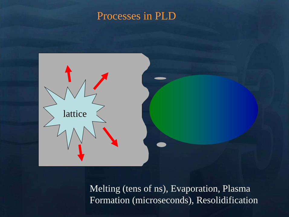
Processes in PLD
lattice
If laser pulse is long (ns) or
repetition rate is high laser may
continue interactions
Processes in Pulsed Laser Deposition
1 Absorption of laser pulse in material
Qab=(1-R)Ioe-aL
(metals absorption depths ~ 10 nm depends on )
2 Relaxation of energy (~ 1 ps) (electron-phonon interaction)
3 Heat transfer Melting and Evaporation
when electrons and lattice at thermal equilibrium (long pulses)
use heat conduction equation
(or heat diffusion model)
abp QTKt
TC
)(
At peak of laser pulse temperatures on target can reach
gt105 K (gt 40 eV)
Electric Fields gt 105 Vcm also high magnetic fields
Plasma Temperatures 3000-5000 K
Ablated Species with energies 1 ndash100 eV
Initially Incredibly Non-Equilibrium
Why biomaterials
- repair and reconstruction of parts of the musculo-skeletal system of vertebrates
- minimal biological requirement biocompatibility associated with the absence of
any adverse effect (non-toxic and non-allergic)
Other requests
- resistance to physiological fluids
- should not interfere with the bodyrsquos natural immunity system
- withstand mechanical stress during whole lifetime
- manufacturability in any appropriate shape
Possible classification
-a) biologically inactive (inert) alumina zirconia stainless steel CoCrNi
CoCrMo titanium titanium alloys carbon latex PE PMMA hellip
-b) porous Calcium Phosphates (CaPs) CaP-coated metal
-c) bioactive dense calcium phosphate ceramics bioactive glasses bioactive
glass-ceramics bioactive composites hellip
-d) Resorbable tricalcium phosphate calciumaliminate polylactic acid poly-L-
acetate
Our option biocompatible porous and bioactive CaP
Acronym
Chemical formula Compound name CaP
ratio
HA Ca10(PO4)6(OH)2 Hydroxylapatite 167
FA Ca10(PO4)6F2 Fluorapatite 167
CDHA Ca10x(HPO4)x(PO4)6x(OH)2x (0x2) Calcium-deficient Hydroxylapatite 133-167
BA Ca83(PO4)43 (CO3-HPO4)17(OH)03
BA=carbonated CDHA (x=17)
Biological apatite 138-193
Mn-CHA HA with (04-2) Mn2+ and (2-6) CO32- Mn2+ doped carbonated hydroxylapatite 151- 165
OHA Ca10(PO4)6(OH)22xOxٱx (0x1) Oxyhydroxylapatite 167
OA Ca10O(PO4)6 Oxyapatite 167
MCPM Ca(H2PO4)2middotH2O Monocalcium phosphate monohydrate 05
MCPA Ca(H2PO4)2 Monocalcium phosphate anhydrate 05
DCPD CaHPO4middot2H2O Dicalcium phosphate dihydrate (Brushite) 1
DCPA CaHPO4 Dicalcium phosphate anhydrate 1
OCP Ca8(HPO4)2(PO4)4middot5H2O Octacalcium phosphate 133
a-TCP Ca3(PO4)2 (monoclinic) Tricalcium phosphate (phase a) 15
-TCP Ca3(PO4)2 (rhombohedral) Tricalcium phosphate (phase Whitlockite) 15
TTCP Ca4O(PO4)2 Tetracalcium phosphate 2
a-DCP Ca2P2O7 (orthorhombic) Dicalcium phosphate (phase a) 1
-DCP Ca2P2O7 (tetragonal) Dicalcium phosphate (phase ) 1
ACP Cax(PO4)y nH2O (Amorphous Calcium pyrophosphate) 12-22
Bone
Resorbable CaP
CaP coating
Ti
Implant
Alternative solution Biomimetic coatings for metallic implants
Main deficiency of CaPs brittle in bulk
HA molecule Ca10(PO4)6OH2
Projection in the
(001) base plan of
the hydroxyapatite
unit cell (hexagonal
structure)
How difficult is to deposit CaPs (1)
- very complex molecules
How difficult is to deposit CaPs (2)
Crystal structure of OCP projected down the c-axis
Alternating apatite- and
hydrated- layers (100)
planes
The region with shaded
atoms the ldquoapatitic layerrdquo
is very similar to HA
The zone containing 10
water molecules is the
ldquohydrated layerrdquo
H atoms are omitted for
clarity
Octacalcium phosphate
(Ca8(HPO4)2(PO4)4middot5H2O)
Mesures par spectroscopie optique
drsquoemission sur le plasma issu de lrsquoirradiation
laser 248 nm drsquoune cible drsquoHA pure
Croissance impulsion par impulsion
Preacutesence des ions de
bull calcium (Ca I et Ca II)
bull oxygegravene (O I et O II) et
bull phosphore (P II)
Eacutemissions dues agrave des espegraveces moleacuteculaires preacutesentes dans le plasma
Couche mince drsquohydroxyapatite obtenue par
ablation laser
Goutelettes
CaP = 18 CaP = 21
CaP = 17 Trois reacutegions
typiques
i Reacutegion majoritaire lisse
ii Agglomeacuteration de gouttelettes sub-micromeacutetriques
iii Gouttelettes micromeacutetriques
Human primary osteoblasts (hOB) were cultured on OCP coated-Ti
Mn-CHA coatedndashTi HA coated-Ti Ti control (polystyrene)
hOB response SEM micrographs
- on bare Ti (a) after 7 days (b) after 21 days
Elongated rod-like morphology
Bioactivity tests
- OCP coatings (c) after 7 days (d) after 21 days
- In time cells spread and expand with flattened polyhedral-morphology
- Numerous cytoplasmatic extensions
hOB response SEM micrographs
- Mn-CHA coatings (e) after 7 days (f) after 21 days
- In time cells spread and expand with flattened polyhedral-morphology
- Less cytoplasmatic extensions
hOB response SEM micrographs
Biocompatible metallic thin films by PLD
Alloys NiTinol NiTi
NiTi characteristics ndash Super-Elastic Property
ndash Radiopaque
ndash Shape-Memory Effect
NiTi biomedical applications ndash Tweezers for removing foreign objects via small incisions
ndash Anchors for tendon fixation
ndash Stents for cardiovascular applications
ndash Dentistry - Orthodontic wires which no not need to be retightened and adjusted
Deposition chamber Multitarget system
PULSED LASER DEPOSITION (PLD)
SEM
Nitinol 30 sequences Nitinol 20 sequences
Cytotoxicity evaluation on microorganism culture
(a) E coli reference cell culture
(b) E coli cell culture on Ti surface
(c) E coli cell culture on NiTi surface
(d) S cerevisiae references cell culture
(e) S cerevisiae cell culture Ti surface
(f) S cerevisiae cell culture on NiTi surface
(g) SEM on E coli cell
culture on NiTi surface
(h) SEM on S cerevisiae cell culture on
NiTi surface E coli and S cerevisiae cell
cultures on the nitinol thin films
o Biocompatible
o Low adherence of microorganism on NiTi surface NiTi alloy does not stimulate the development at genetic level for specific genes which are involved in adhesion processes for microorganism cells
o Low release of Ni ions in solutions
CONCLUSIONS
MAPLE Matrix Assisted Pulsed Laser Evaporation
bull No need for UHV
bull Matrix prepared outside the
chamber
bull High flexibility
You can use everything
you can dissolve in a
solvent
MAPLE of polymers blends with Ag
nanoparticles
Controlled release of antibacterial agents
MAPLE deposition of PEGPLGA films with incorporated silver nanoparticles (AgNPs)
0 5 10 15 20 25 30000
005
010
015
020
025
Nu
mb
er o
f E
C
oli
X109
Time (hours)
Killing activity
0 5 10 15 20 25 30 3500
05
10
15
20
25
Bact
eria
l ce
ll n
um
ber
X109
Time (hours)
Delay of bacterial growth
Antibacterial efficiency against E Coli
Direct writing by femtosecond laser
pulses
Ormosil scaffolds for tissue engineering
50 lines ormosil sample
Distance between lines =
100 μm
30x30 lines ormosil
sample Distance between
lines = 50 μm
3 layers 30x30 lines ormosil
sample Distance between
lines = 100 μm
Scaffold functionalization
Protein deposition on the scaffolds by MAPLE
- Lysozyme antibacterial antitumor uses
- Fibrinogen biocompatible protein
Fibroblast cells grown on a polymeric grid with 100 microm distance between lines
Spectroscopie drsquoun panache plasma
laser femtoseconde
Laser
t =400 ns Spot 35x35 micromsup2 Cu
Z mm
Expeacuteriences reacutealiseacutees par (Grojo et al LP3Marseille)
nanoparticules
atoms
031 Jcm-2
Formation de nano agreacutegats
AFM
(Pereira et al )
TEM
(Kabashin et al)
Eacutetude des meacutecanismes de la formation drsquoagreacutegats
Couplage
- Direct Simulation Monte Carlo
- Dynamique Moleacuteculaire
Information deacutetailleacutee
-Continu du panache
-Distribution en taille des agreacutegats
K Gouriet thegravese (2008)
Simulation par Dynamique Moleacuteculaire (MD)
=gtdistribution drsquoagreacutegats
Cluster size N
No
rmaliz
ed
Yie
ld
100
101
102
103
104
105
10-9
10-8
10-7
10-6
10-5
10-4
10-3
10-2
10-1
100
N-426
N-122
t=1 ns
τ = 15 ps F ~ 2 Fth 4010 nm t =1 ns
Zhigilei et al (Virginia University)
Target applications of mobile nanomaterials
Bioimaging
Optical imaging deals with
visualization of biological objects
or tissues in order to detect
pathogens or follow the delivery
of drugs etc nanoparticles are
uses as contrast agents
In vitro
Cellular
imaging
In vivo Cancer
detection drug delivery
etc
Light-induced
therapies
Cancer therapy nanoparticles are used
as photosensitizers to produce local
targeted destruction of cancer cells
Photodynamic
therapy
Light-induced
hyperthermia
Diagnosis and Sensing A Diseases can be diagnosed through the (simultaneous) detection of a (set of) biomolecule(s) characteristic to a specific disease type and stage (biomarkers)
Huffman Nanomedicine and Nanobiotechnology Vol 1 1 2009
D
molecular signature of sick cell of infecting agent
(eg an antibody)
Cell membrane
Nanoparticle
Coating molecule specifically attracted to the molecular signature
C A nanoparticle can be functionalized in such a way that specifically targets a biomarker Thus the detection of the nanoparticle is linked to the detection of the biomarker and to the diagnosis of a disease
B Each cell type has unique molecular signatures that differentiate healthy and sick tissues Similarly an infection can be diagnosed by detecting the distinctive molecular signature of the infecting agent
Laser ablation of material from a solid target and production of
nanoparticlesnanostructures
Production of nanoclusters by laser
ablation and their subsequent cooling in
ambient medium Gaseous ambience
Nanoclusters can be
deposited on a
substrate forming a
nanostructured film
Aqueous ambience
Nanoclusters
crystallize and form
colloidal solution
NPs produced in gaseous ambience
0 20 40 60 80 100 120 140
0
2
4
6
8
Heig
ht
[nm
]
[nm]
01 1 102
3
4
5
6
7
Mean
part
icle
heig
ht
(nm
)
Helium pressure (Torr)
Silicon nanocrystal size AFM
Gas pressure decreases Nanocrystal size decreases
AFM of isolated laser-
ablated Si particles on
HOPG
Minimal particle size is 2-3 nm (from height measurements)
001Torr
1Torr
01Torr
10Torr
100Torr
NGC 2009
Photoluminescence of size selected nanoclusters
0 1 2 3 4 5 6
39 Jcm2
Num
ber
of
clust
ers
Size (nm)
30 Jcm2
14 Jcm2
0 1 2 3 4 5 6
He 4 Torr
10 Jcm2
15 20 25 30 35 40
0
500
1000
He 4 Torr
39 Jcmsup2
30 Jcmsup2
14 Jcmsup2
10 Jcmsup2
Inte
nsi
ty o
f P
L (
ua
)Photon energy (eV)
Silicon nanoclusters
Medical Imaging A Optical properties of nanoparticles depend greatly on its structure Particularly the color (wavelength) emitted by a quantum dot (a semiconductor nanoparticle) depends on its diameter
C The quantum dots (QD) can be injected to a
subject and then be detected by exciting them to emit light
Source Department of immunology University of Toronto
Solutions of CdSe QDrsquos of different diameter
CdSe nanoparticle (QD) structure
Source Laurence Livermore Laboratories
Imaging of QDrsquos targeted on cellular structures
Nano Letters 2008 Vol 8 pp3887-3892
B
In vivo cancer targeting and imaging with
semiconductor quantum dots
Gao et al Nat Biotech 22 969 - 976 (2004)
NPs produced in aqueous ambience
Advantages of laser ablation-based nanofabrication
compared to chemical methods
Disadvantage of chemical synthesis contaminationhelliphelliphellip
In particular chemical method for the fabrication of gold nanoparticles
Reduction of chloroauric acid (HAuCl4) with citrate in water
Control size by adding a stabilizing agent (thiol- (-SH) containing molecules)
Contamination impurities Cl- on surface non-biocompatible surfactantshellip
Problems in imaging
applications (especially
in vivo)
Problems in field-
enhanced applications (eg contaminants can
provide false Raman signals)
Focalized laser beam
Liquid medium
Target
Plasma
Laser ablation in liquids a way to avoid residual
contamination
- Laser energy can be transmitted through a liquid to ablate a solid
target the nanomaterial is released to the solution
- The process can be performed without any dirty chemical products in
clean liquid environment (deionized water PBS solution) no
contamination
Experiment
Ablation of gold final colloidal solutions
-Solution are ranging from deep wine red to pink and purple
- Color related to the size dependant surface plasmon resonance
Size distribution of Au nanoparticles prepared in
distilled water
0 50 100 150 200 250 300
0
200
400
600
800
1000
Rela
tive a
bundance (
arb
units)
Particle size (nm)
0 10 20 30 40 50 60 70 80 90 100
0
200
400
600
800
1000
Re
lative
Ab
un
da
nce
(a
rb u
nits)
Particle Size (nm) 0 2 4 6 8 10
0
200
400
600
800
1000
Re
lative
Ab
un
da
nce
(a
rb u
nits)
Particle Size (nm)
F = 20F0 Jcm2 F = 5F0 Jcm2 F = F0 Jcm2
-Size and dispersion of nanoparticles can be controlled by laser fluence
- Shape of craters on target at high F suggests the presence of cavitation phenomena
4 plusmn 15 nm
A V Kabashin M Meunier J Appl Phys 94 7941 (2003)
In water Very dispersed population of nanoparticles
Dextran (MW=6000gmol) Quenches the growth of the nanoparticles 2-4nm nanoparticles are produced (230-1850 atoms) Very fast dextran-gold interaction
Influence of the concentration
Dextran 6k
In pure water
300mJ
Ablation in biopolymers (dextran PEG)
Au OH
O
-
Dextran (C6H10O5)n
Nanoparticles react with OH
groups of biopolymers
Gold Nanoparticles vs Alzheimer
A Alzheimer and other degenerative diseases are caused my the clustering of amyloidal beta (Aβ) protein
D Gold nanoparticles can be functionalized to specifically attach to aggregates of this protein (amyloidosis)
Functionalized nanoparticle
Source wwwinternetchemistrycom
Chemical structure of Aβ-protein
Source wwwthefutureofthingscom
C
B
Alzheimerrsquos brain Healthy brain
Source Berkeley Lab
Gold Nanoparticles vs Alzheimer A The functionalized gold nanoparticles selectively attach to the aggregate of amyloidal protein The microwaves of certain frequency are irradiated on the sample Resonance with the gold nanoparticles increases the local temperature and destroy the aggregate
Nanoletters 2006 Vol 6 pp110-115
Before irradiation After irradiation
LIBS for biomedical applications in vivo analysis of tissue for real-time
analysis
real-time diagnosis of pathogen
presence in human fluids (bloodurine
CSF sputum)
71 35
ICCD
spectromegravetre
Time and space
Emission spectroscopie
LIBS (Laser Induced Breakdown Spectroscopy)
Principle of sample identification screening applications based on discriminant analysis here for warning when healthy tooth material is targeted during laser drilling
BMC Oral Health 2001 1 1 Ota Samek1 Helmut H Telle2 and David CS Beddows
Laser-induced breakdown spectroscopy a tool for real-time in vitro and in vivo identification of carious teeth
Sensitive Detection of Epithelial Ovarian Cancer Biomarkers
Using Tag-Laser Induced Breakdown Spectroscopy
Yuri Markushin and Noureddine Melikechi
Optical Science Center for Applied Research Delaware State University USA
X rays produced by laser plasmas
I = ~1016 agrave 1018 Wcm2
nc cm-3
1111021 mm 2
Thot Ilas2
1 3
Ilas2
3 4
Hot electrons
(characteristic lines)
X-ray
emission
laser
X-rays
Line emission Fast electrons knock
free core electrons
from the inner shell
Bremsstrahlung electrons deflected by nucleus
Laser-generated hot electrons produce x-rays by two dominant mechanisms
ne
critical surface (cf cathode)
conventional X-ray tube
Kmetec ldquoMeV X-ray generation with a femtosecond laserrdquo Phys Rev Lett 68 1527 (1992)
Rousse ldquoEfficient Ka X-ray source from fs-laser-produced plasmasrdquo Phys Rev E 50 2200 (1994)
(typical recombination lifetimes several fs)
Hou et al Appl Phys Lett 84 2259 (2004)
Tightly focused fs lasers create the worldrsquos smallest
hard x-ray source (~4 microm) for precision imaging
x-ray source size is ~ 4 larger than laser focus
because of lateral heat transport Bowes Opt Lett 31 116 (2006)
2004 premiegravere deacutemonstration drsquoimagerie avec contraste de phase avec une source X creacutee par laser
Source
Rayon non diffracteacute
Rayon diffracteacute
Lcoh 2R1
eacutechantillon
Absorption vs contraste de phase
dsource-object dobject-detector
X-ray
Source
Object
Detector
position
Dsource
Intensity
interface
dsource-object dobject-detector
X-ray
Source
Object
Detector
position
Fresnel Diffraction
Dsource
Intensity
interface
enhancement interface
R Toth et al
Rev Sci Instrum 76
083701 (2005)
Imagerie par contraste de phase
R Toth et al Rev Sci Instrum
76 083701 (2005)
copy INRS
bull Meilleure reacutesolution (5microm)
bull dose plus faible
bull meilleur contraste (phase)
bull lrsquoeacutenergie X peut ecirctre ajusteacutee
aux besoins de lrsquoimagerie
La reacutefeacuterence mondiale ALLS
(INRS- Montreacuteal) 200TW + Puissance moyenne 50W
47J 23fs 10Hz
in a 8microm spot
Sans miroir deacuteformable
microCT
Scan duration avec
50W10Hz (360 images)
2 min (mouse scan)
courtesy Prof Dr Oswald Willi U Duumlselldorf
MeV Protons produced by laser CR39 or RCF 1010 to 1013 H+
courtesy Prof Don Umstadter U Nebraska-Lincoln
Target Normal Sheath Acceleration hot electrons traversing
target electrostatically accelerate impurity hydrogen ions on the rear surface
virtual
cathode
I ge 1019 Wcm2
Protons de 10MeV (INRS )
~1022 Wcm2
Avantages des particules chargeacutees ou hadrons (protons ions) Rayons X de la Radio-Theacuterapie
Le pic de Bragg
La profondeur de peacuteneacutetration deacutepend de l rsquoeacutenergie incidente des protons
la tumeur la plus superficielle = l rsquoœil 70 MeV
la plus profonde = la prostate 200 - 250 MeV
Protontheacuterapie par laser (projet SAPHIR)
CPO
Exemple de dosimeacutetrie preacutevisionnelle
(photons vs protons)
Comparaison de lrsquoeacutepargne de dose dans les tissus sains en protons (image de droite)
par rapport aux photons (IMRT Intensity Modulated Radio-Therapy image de gauche)
Les irradiations colateacuterales en radiotheacuterapie et en protontheacuterapie
Avantage consideacuterable pour le traitement de tumeurs localiseacutees pregraves d rsquoorganes sensibles (œil cerveau)
currently treating patients 5
5 next 2 years
others awaiting approval
10000 m2 $125 M facility opened in 2006
12 m diameter 250 MeV synchrotron
ldquoThere are too few physicists in the world
and they are an incredibly important part of
doing this We have one of the largest
physics departments in the world with more than 50 medical physicistsrdquo
--- Dr James D Cox head of Radiation Oncology at MD Anderson Cancer Center Houston Texas
Proton Therapy enables precise exposure of small tumors
with minimal damage to surrounding healthy tissue
250 MeV
PROTON beam
10 20
Depth in Tissue [cm]
0 0
50
100
DO
SE
(
)
6 MV
PHOTON
beam
tunable
Bragg
peak
but requires large expensive facilities
eye tumors 65 MeV protons
deep tumors gt200 MeV
Laser proton therapy could be much smaller amp cheaper Fourkal Med Phys 29 2788 (2002)
Malka Med Phys 31 1587 (2004)
On-site production of short-lived isotopes for medical imaging
Laser-generated quasi-mono-energetic electrons efficiently photo-activate materials of interest
bull High Rep rate bull Low cost bull Compact
Limitations to the widespread use of PET arise from the high costs of cyclotrons needed to
produce the short-lived radionucleotides for PET scanning Few hospitals and universities
are capable of maintaining such systems - Wikipedia -
radiotracer activation reaction half-life medical use
15O 16O (n)15O 2 minutes neuro-imaging
11C 12C(n)11C 20 minutes neuro-receptor-specific brain imaging
18F 19F(n)18F 110 minutes clinical oncology
Positron Emission Tomography
on-site
production
essential
18F PET scan of tumor 15O PET scan of human brain
Reed ldquoEfficient initiation of photonuclear reactions using quasi-monoenergetic electron beams from LWFArdquo J Appl Phys 102 073103 (2007)
TEP in situ
+ in vivo
Modegravele animal
microCT +
contraste de phase
+ in vivo
modegraveles de cancer
Comprendre
le cancer
Preacuteparer le traitement
protons 50MeV
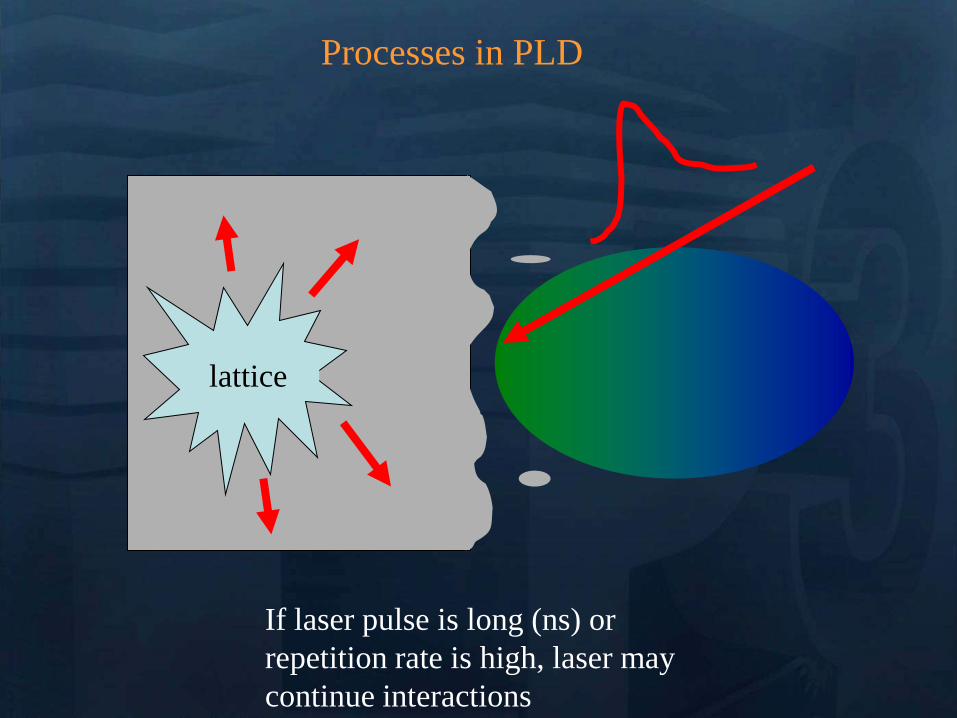
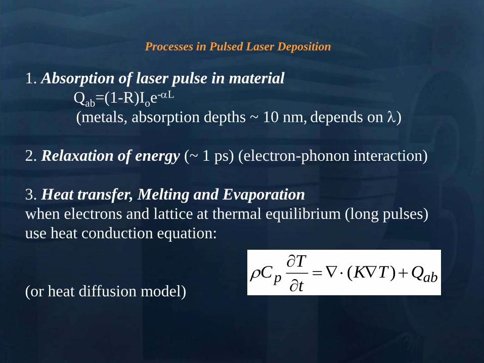
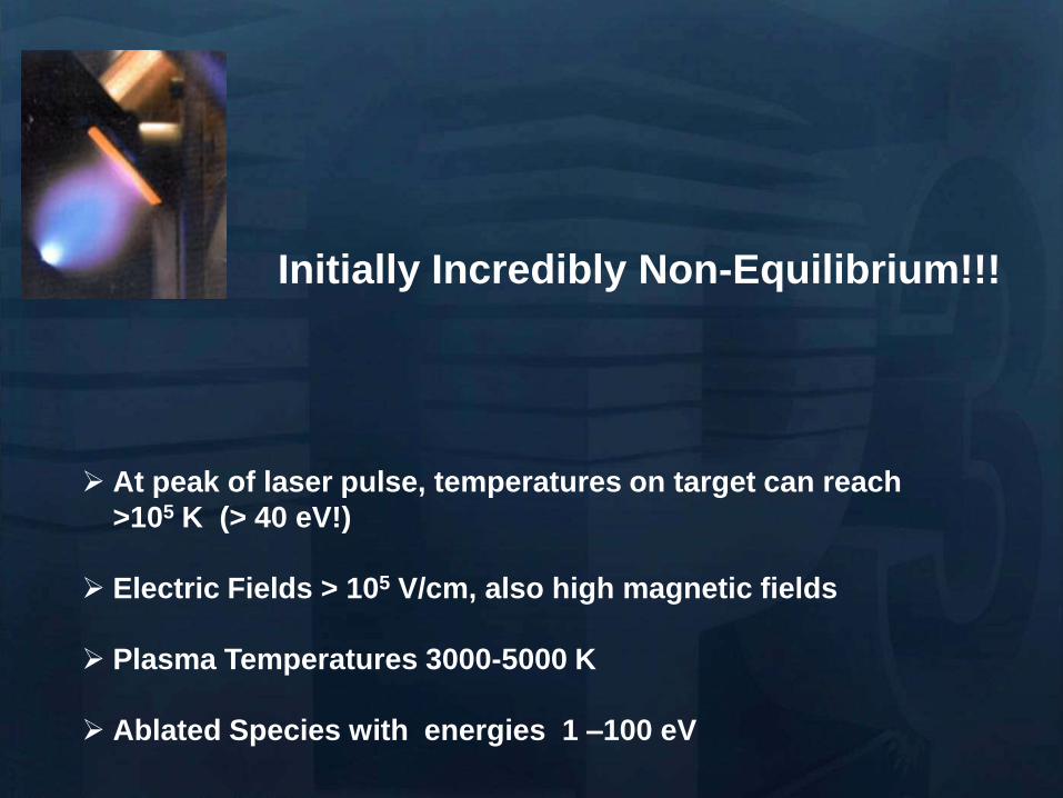
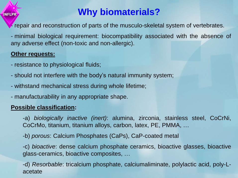
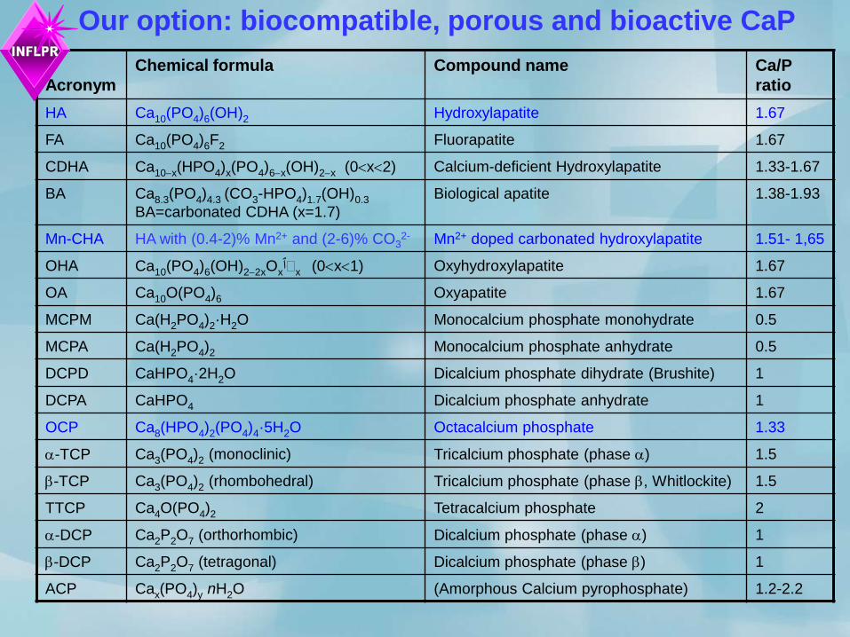
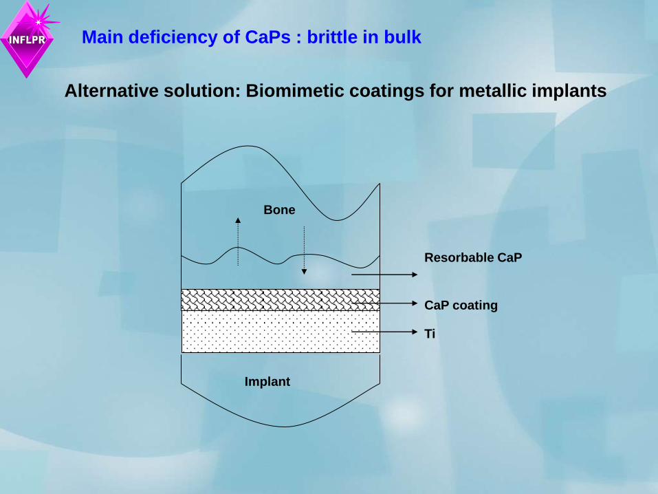
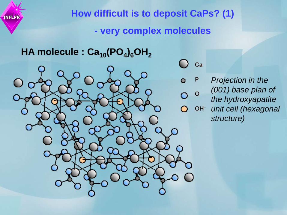
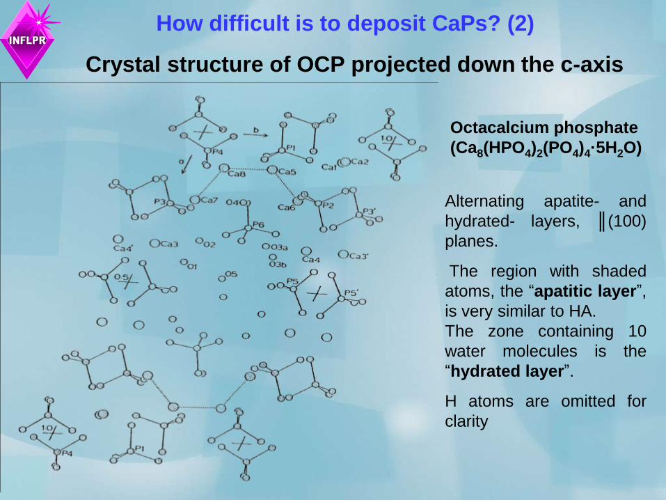
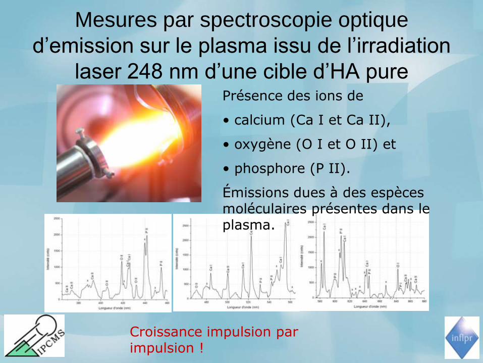
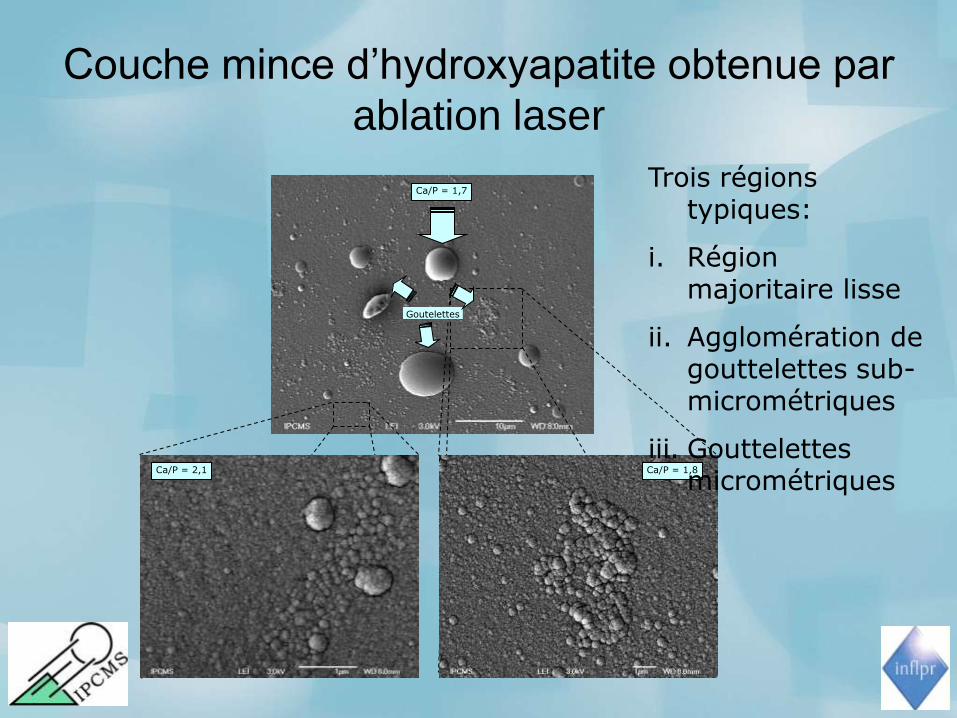
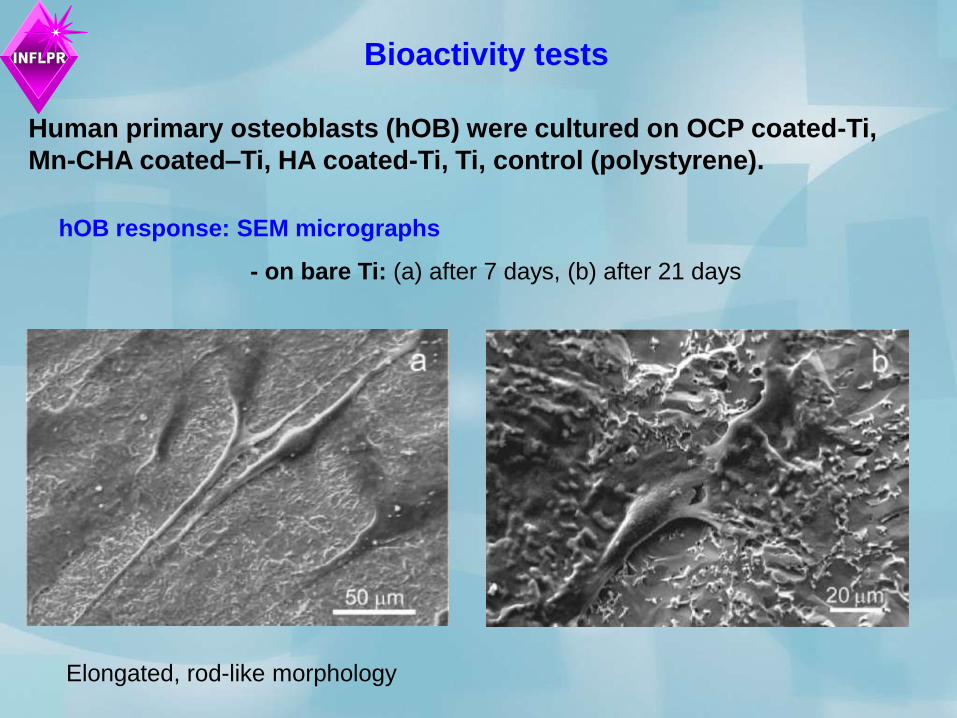
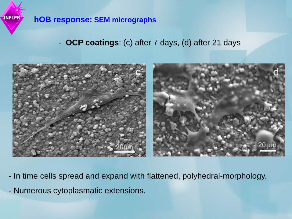
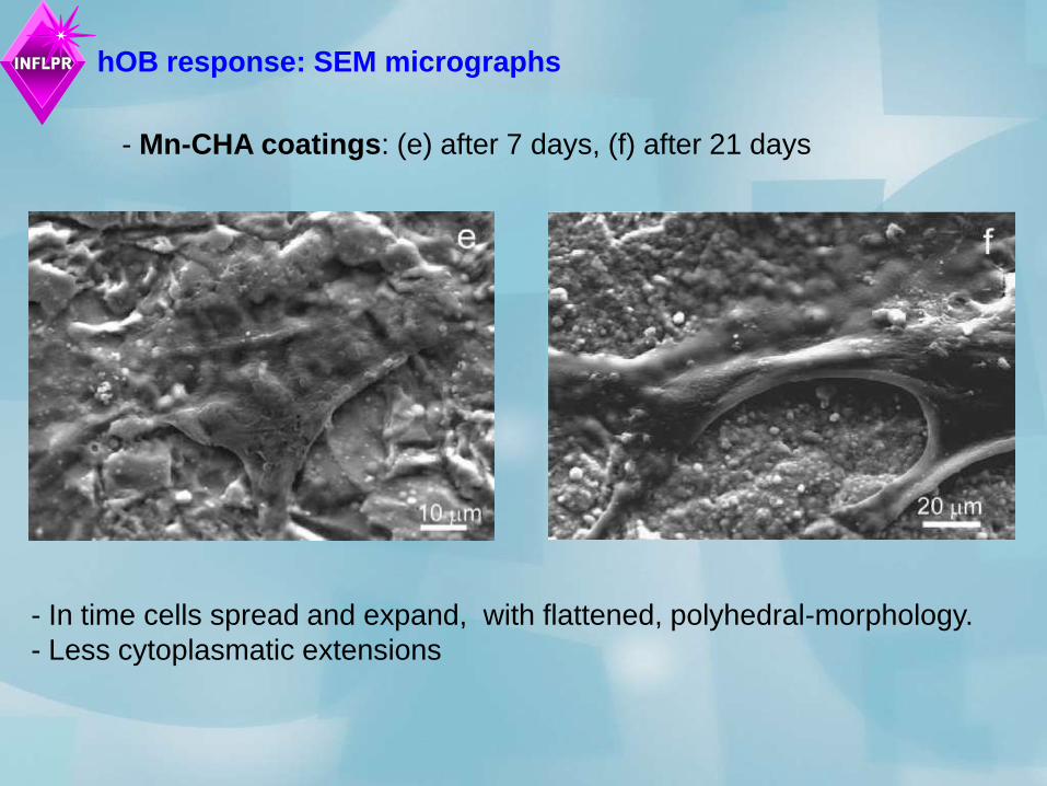
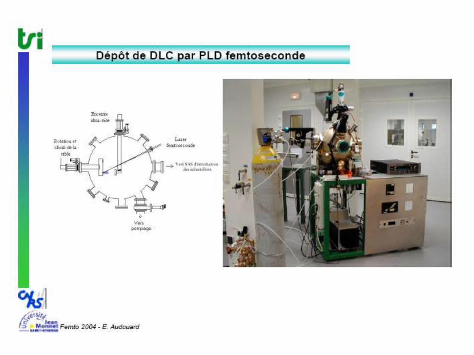
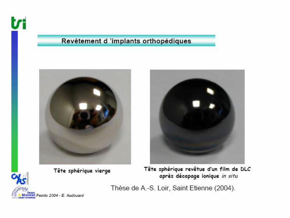
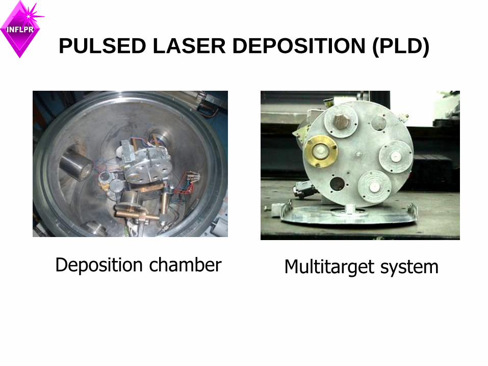
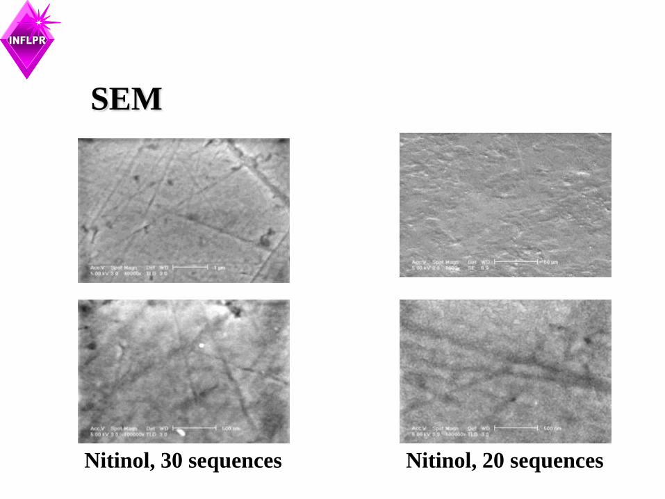
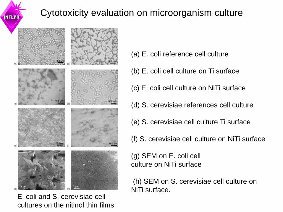
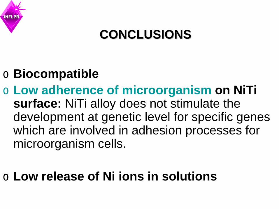
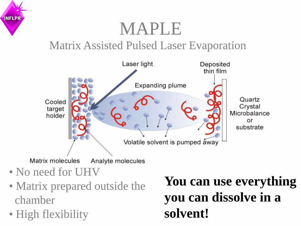
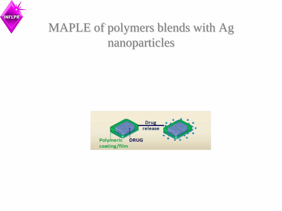
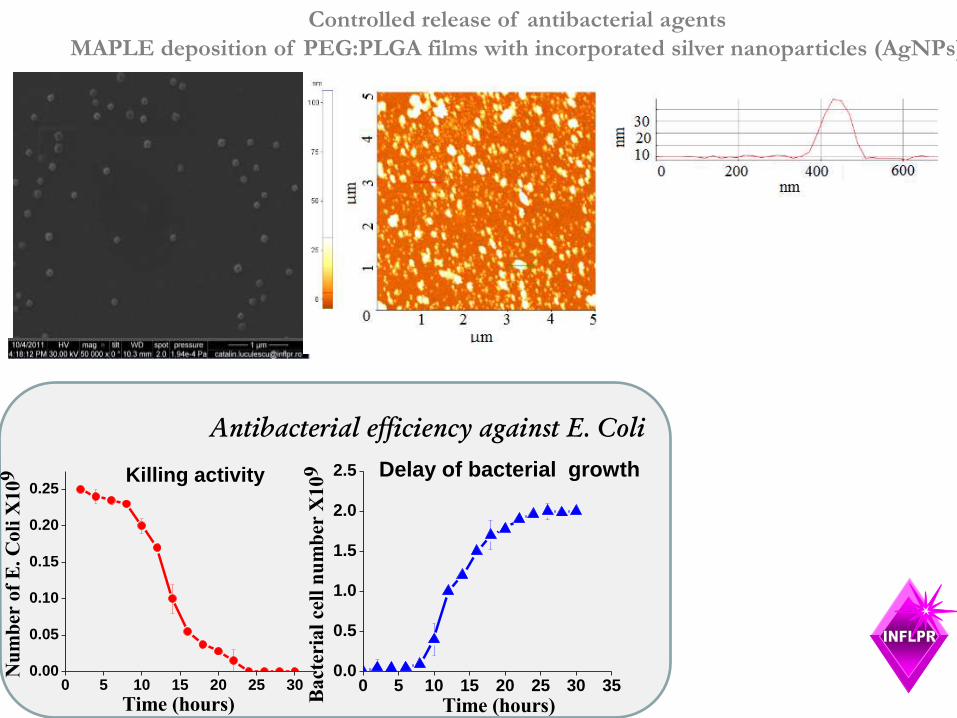
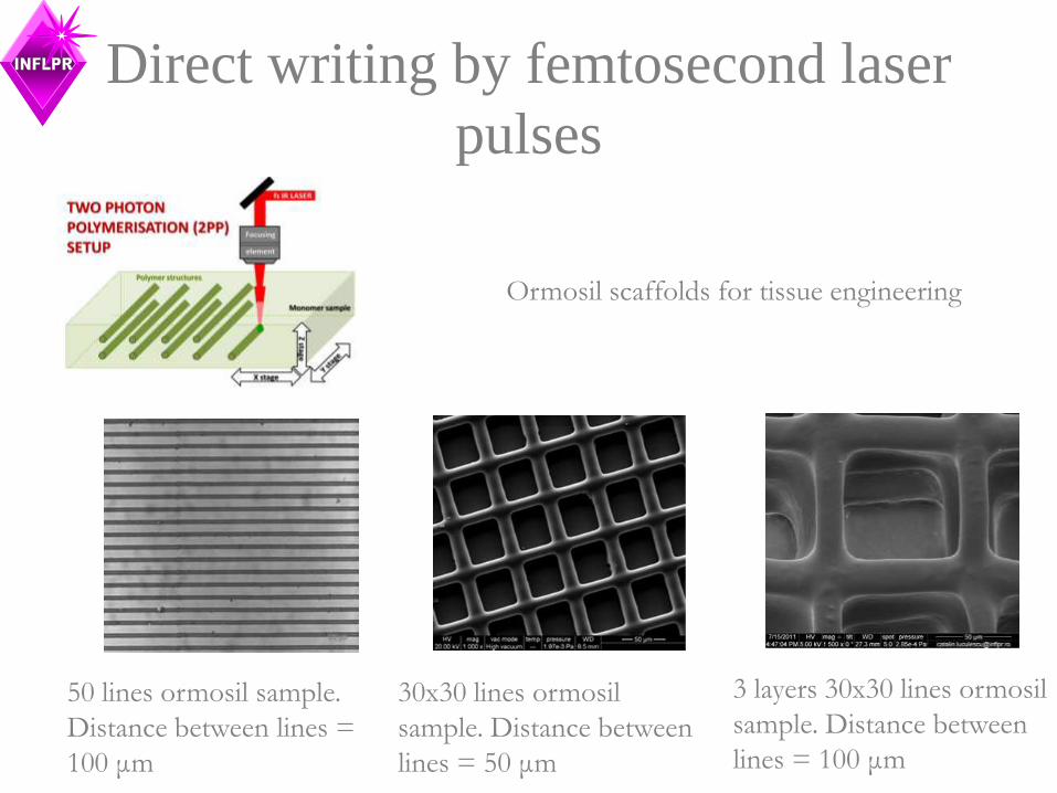
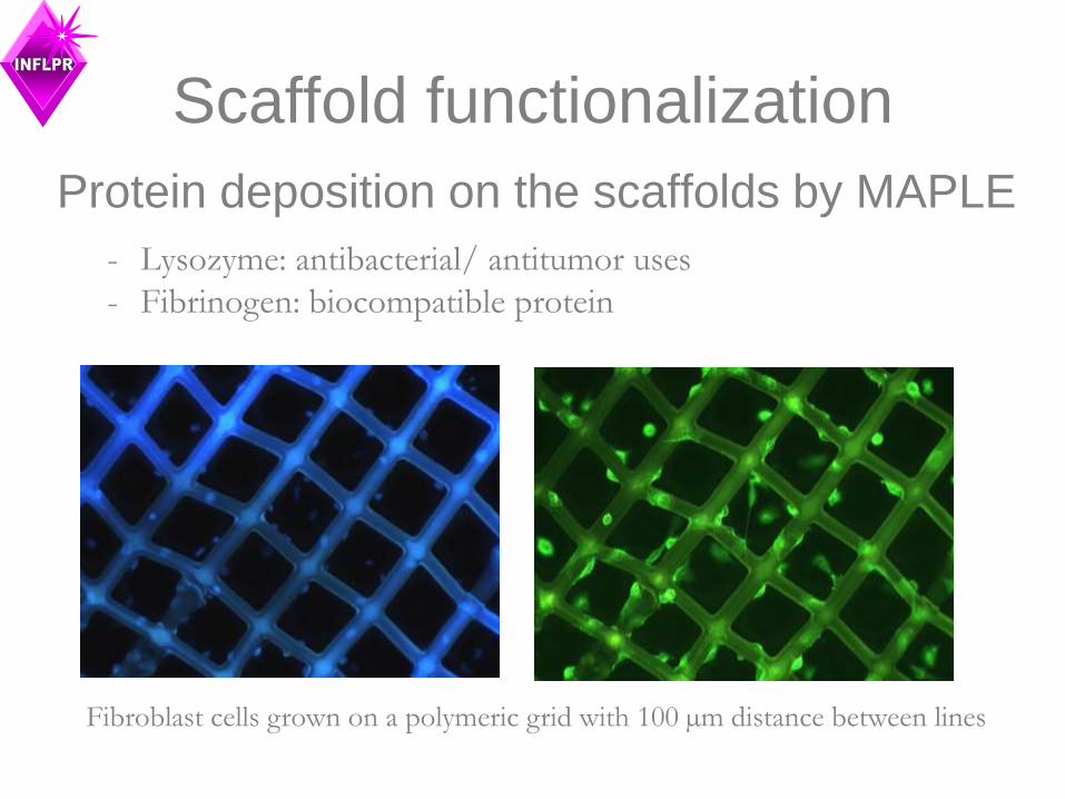
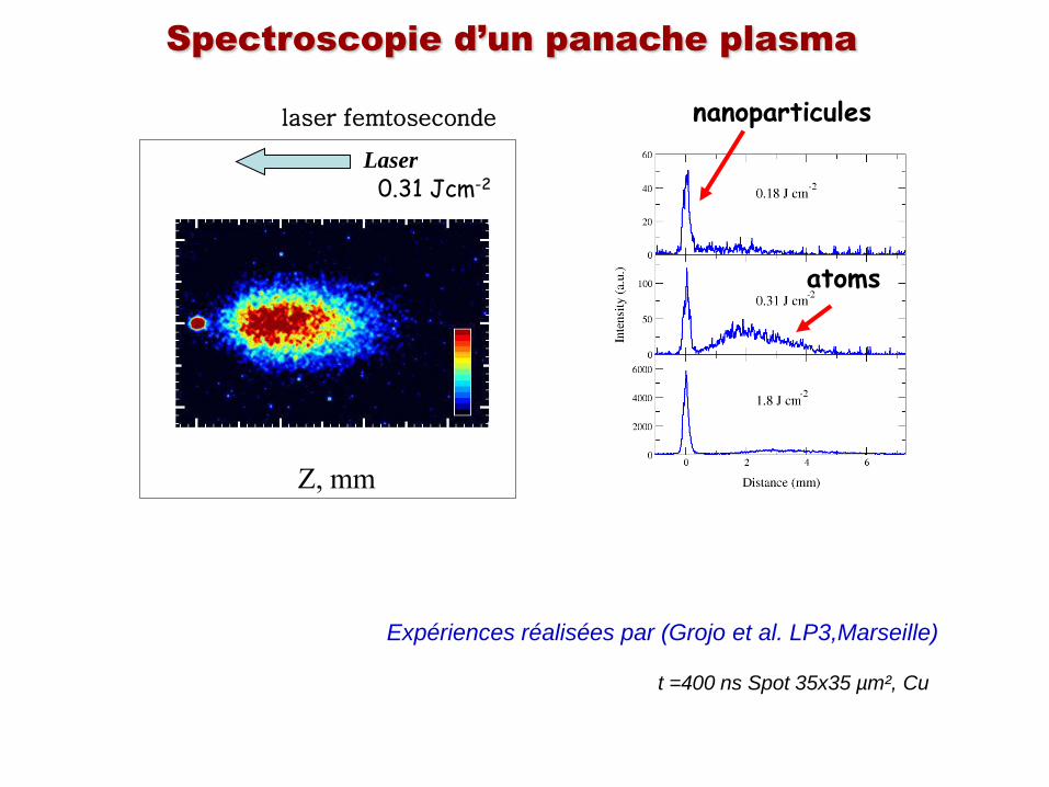
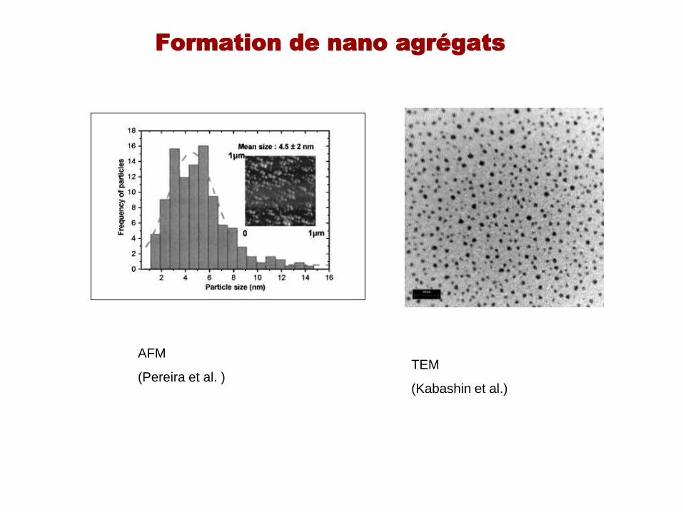
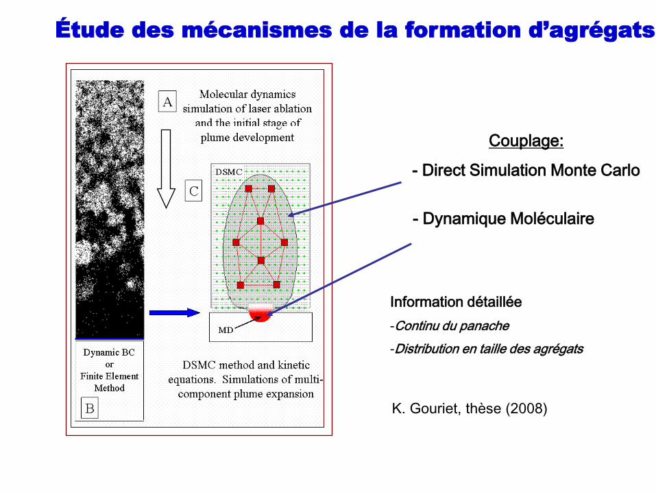
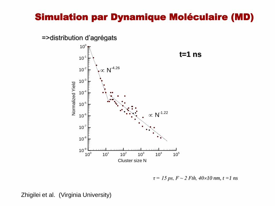
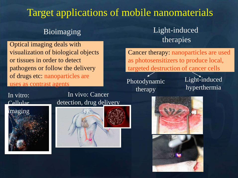
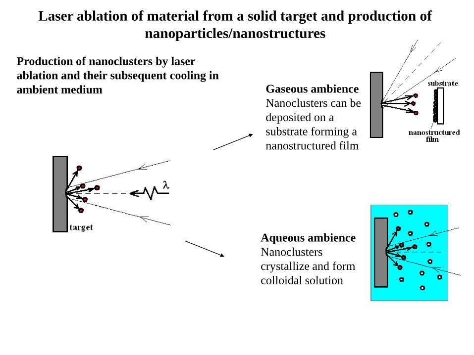
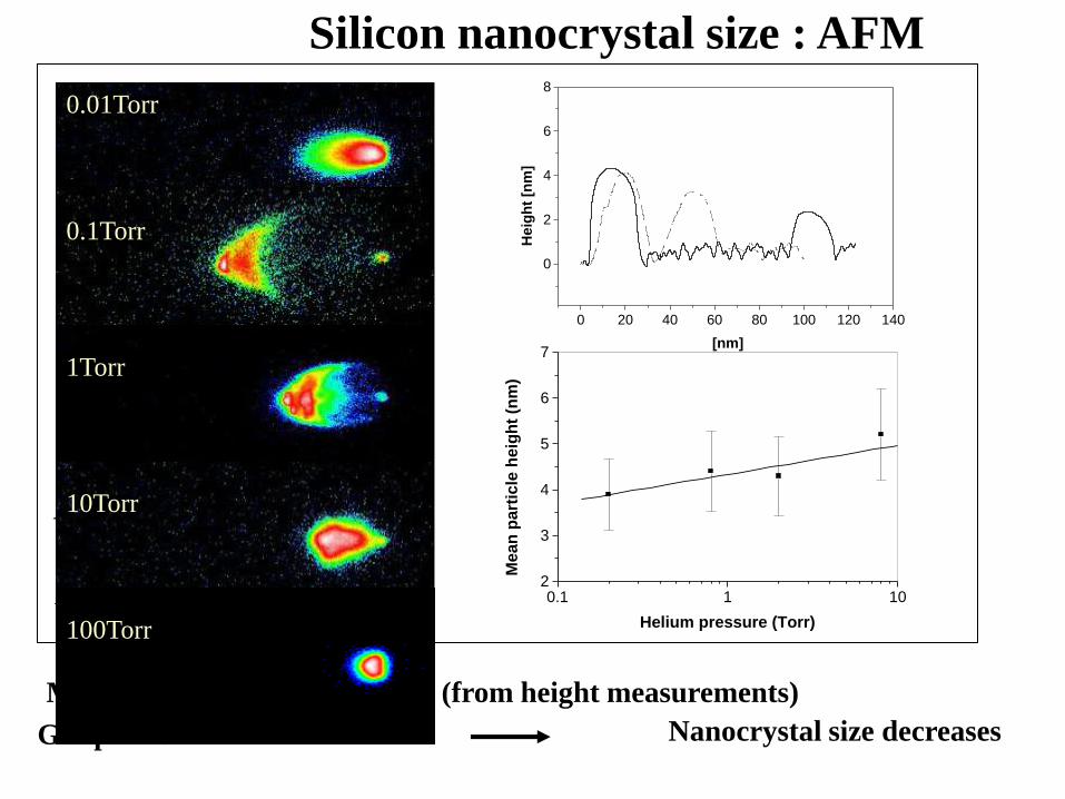
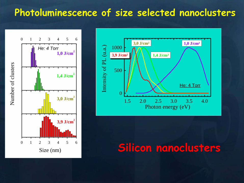
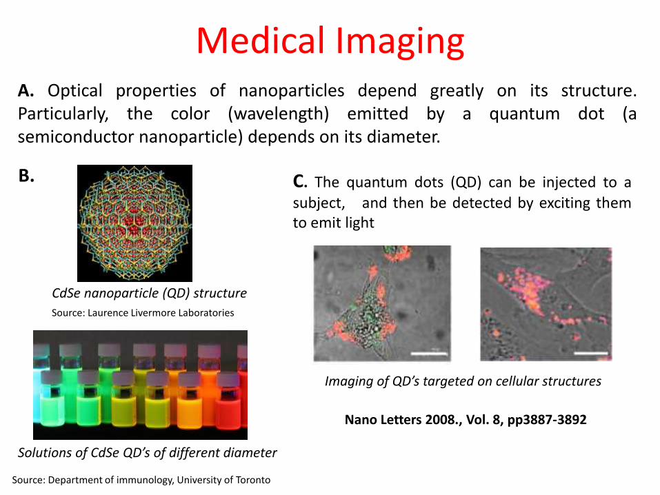
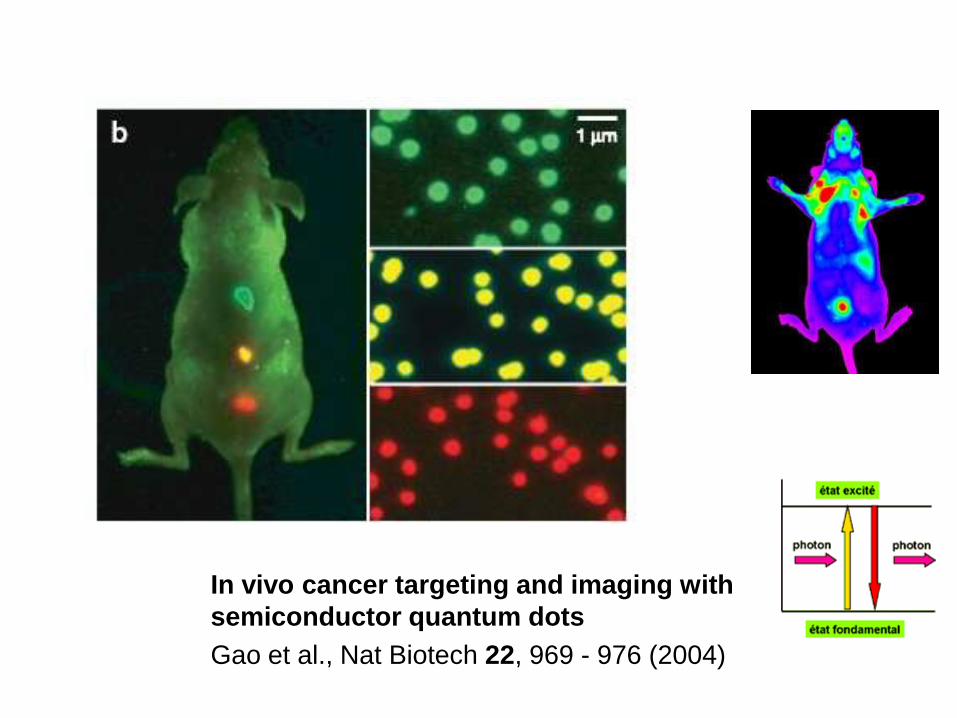
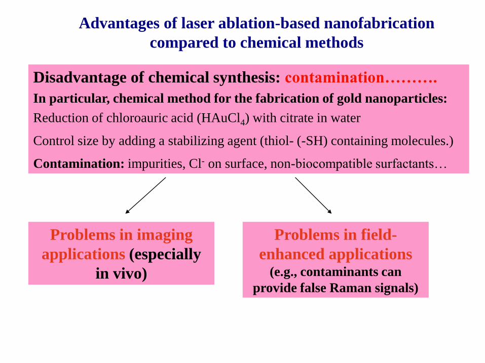
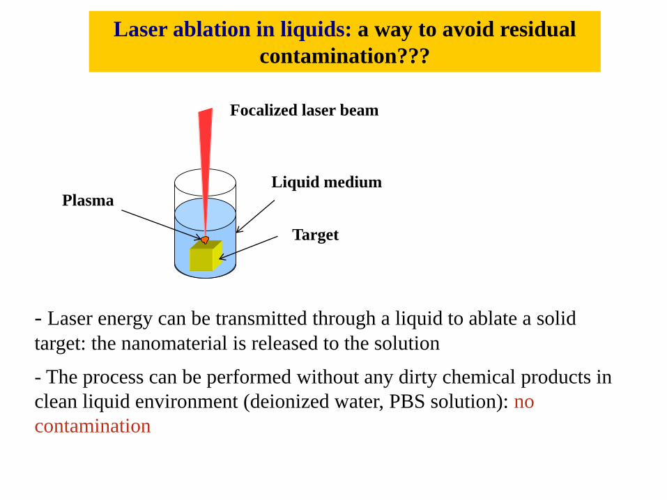
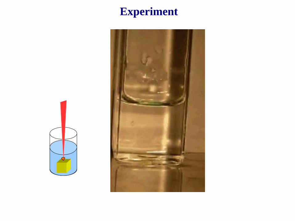
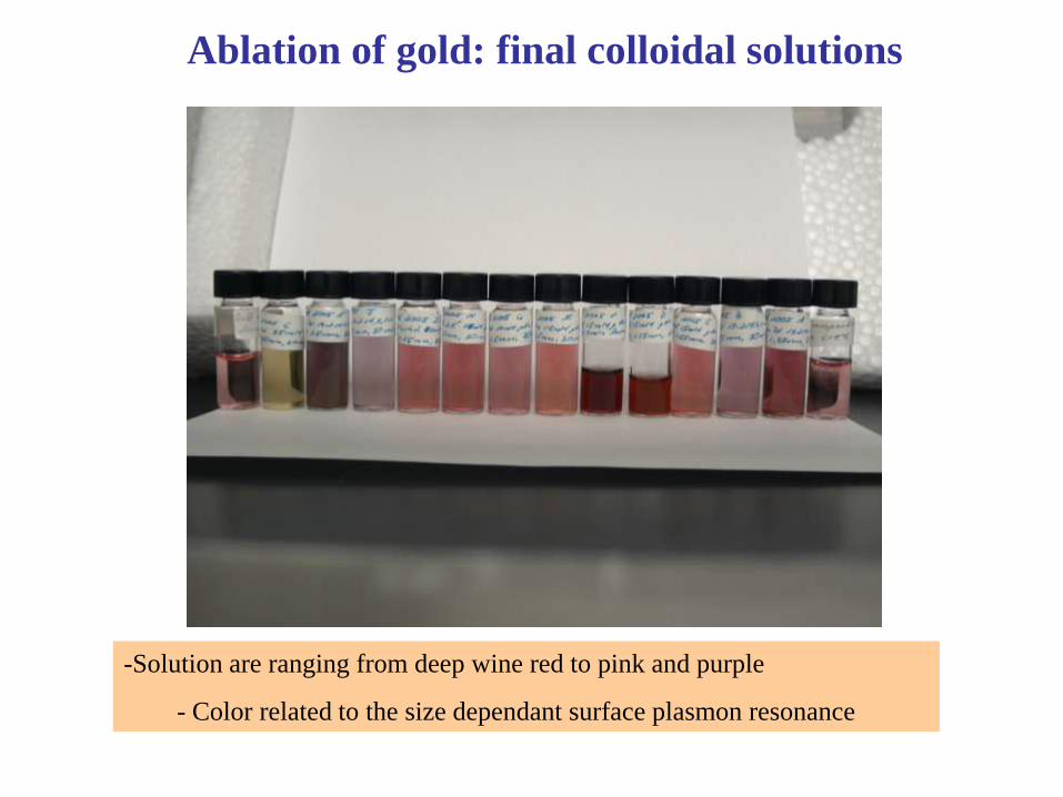
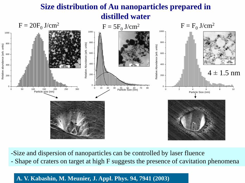
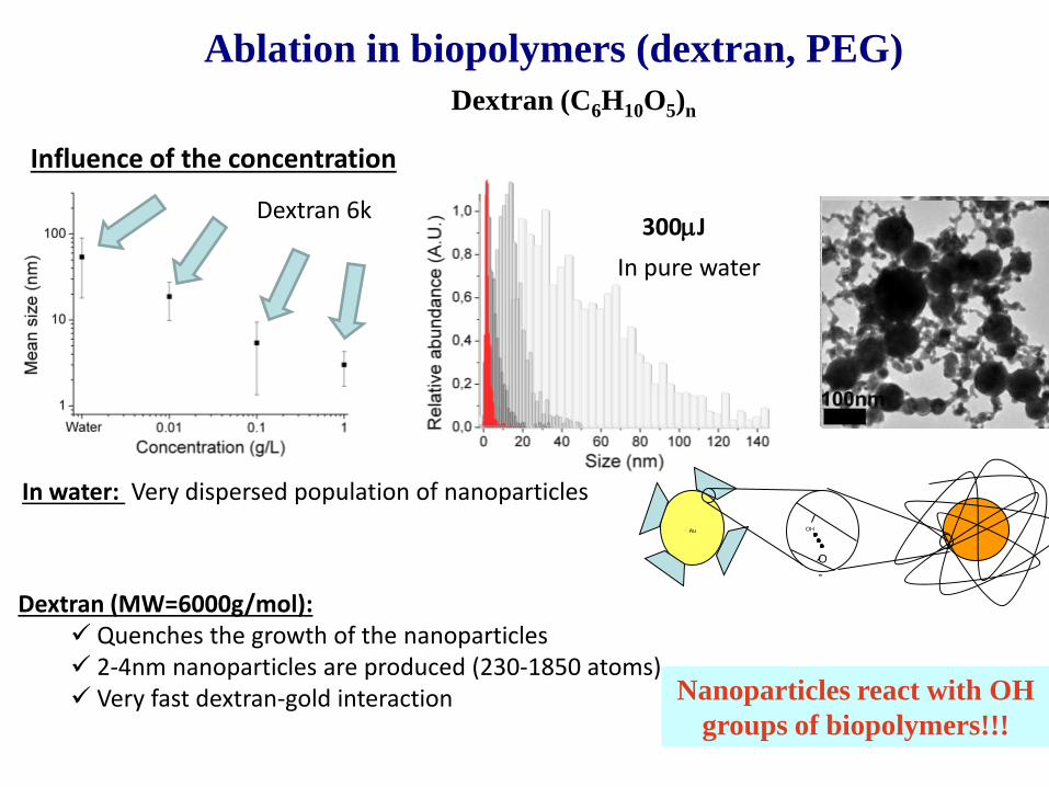
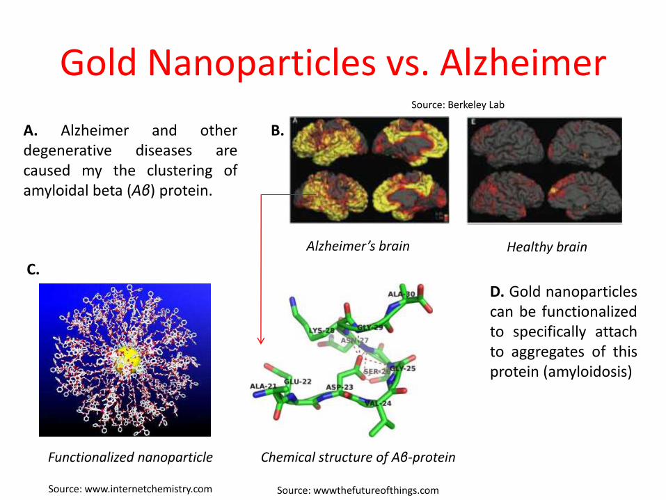
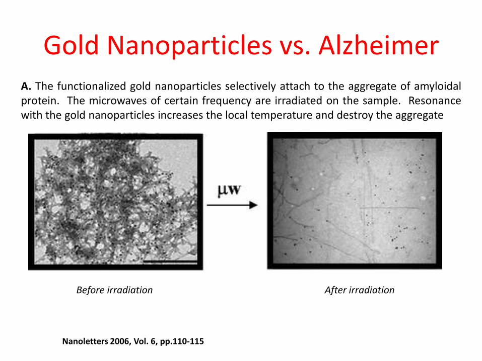
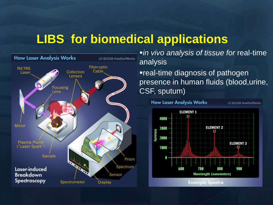
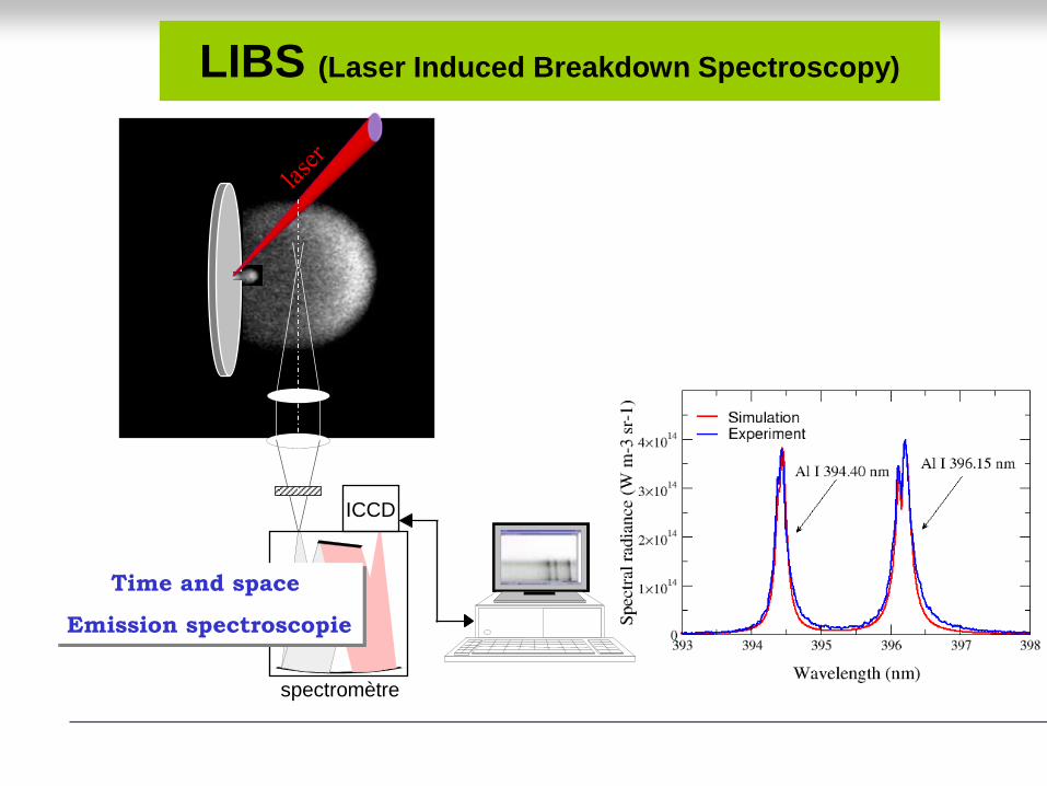
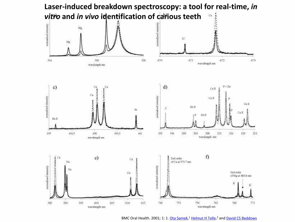
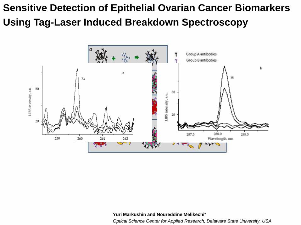
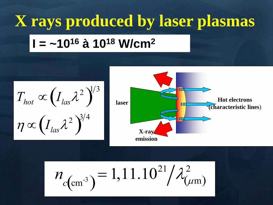
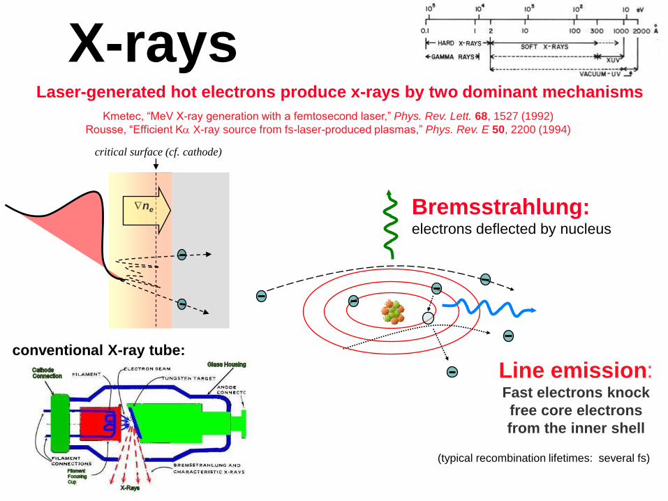
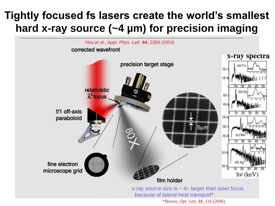
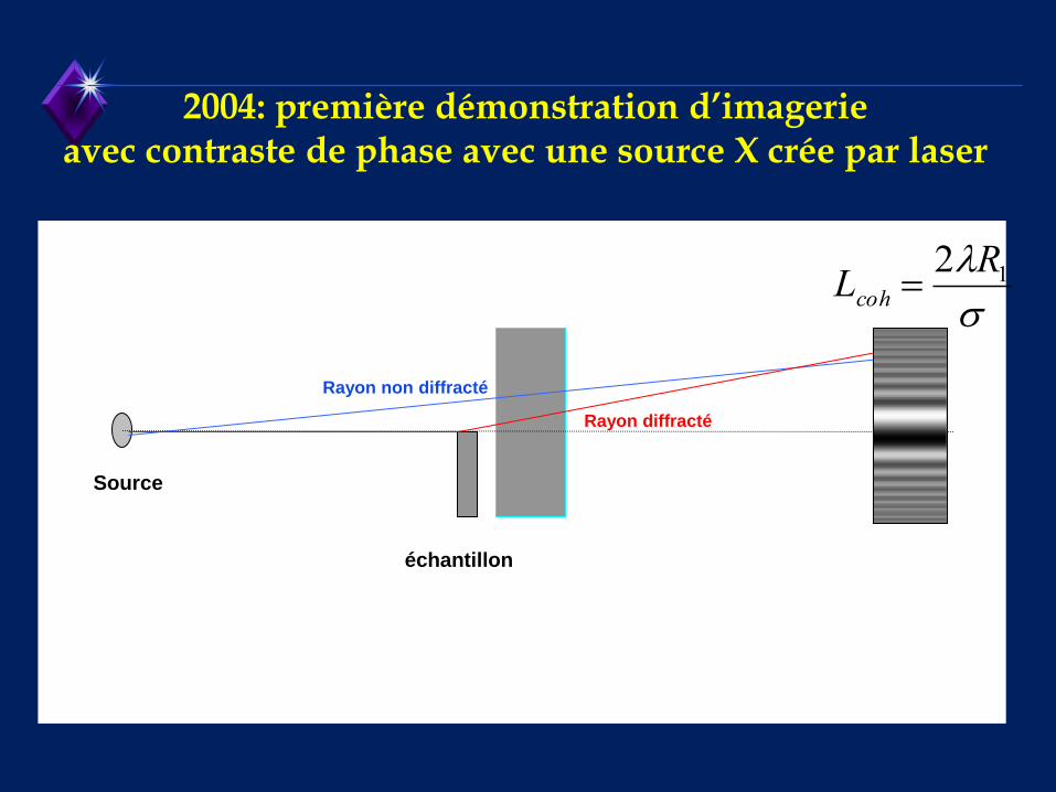
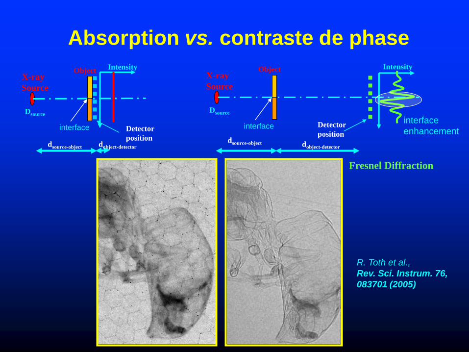
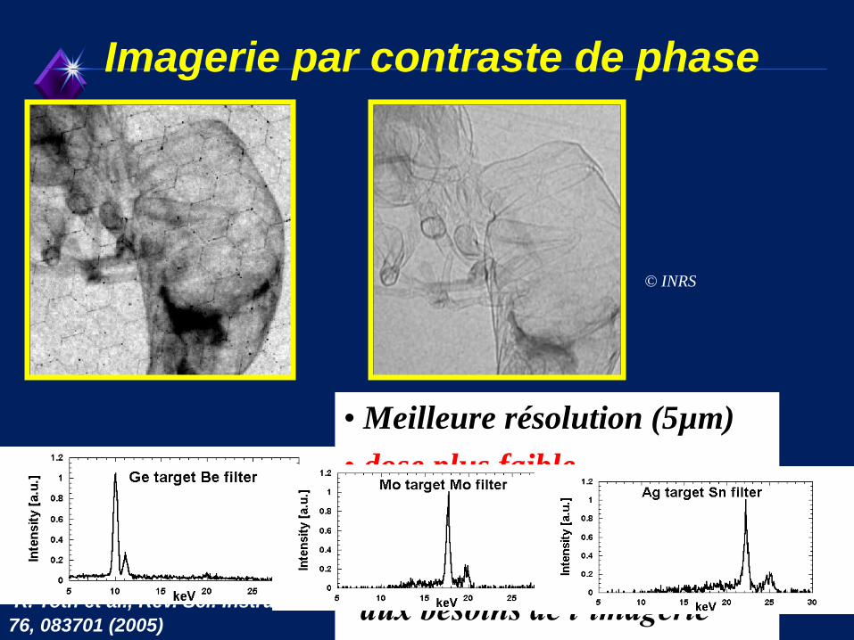
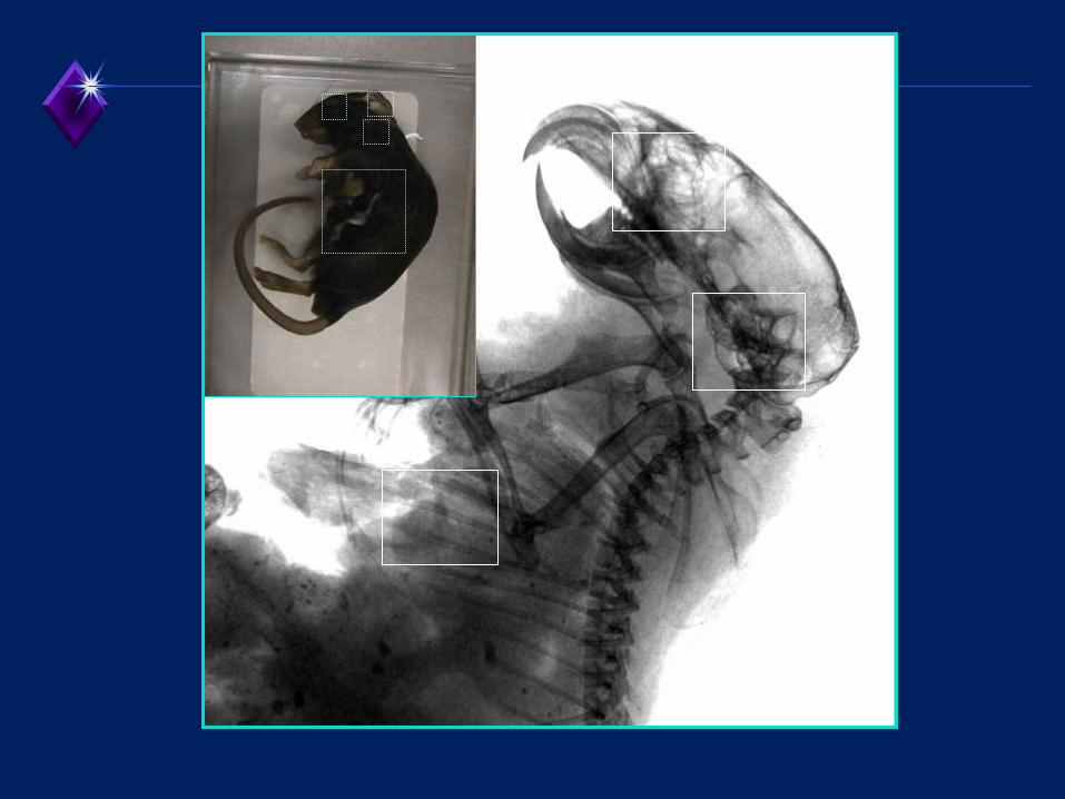
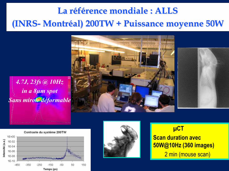
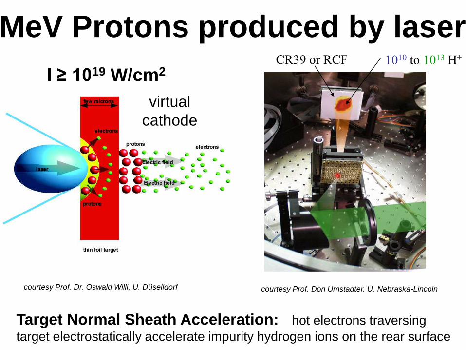
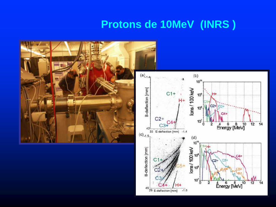
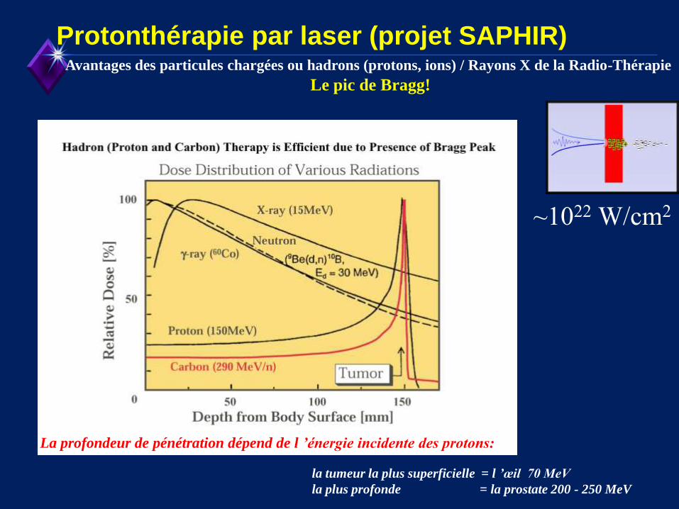
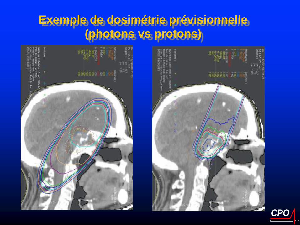
TEP in situ
+ in vivo
Modegravele animal
microCT +
contraste de phase
+ in vivo
modegraveles de cancer
Comprendre
le cancer
Preacuteparer le traitement
protons 50MeV