Embed Size (px)
Citation preview

1
Plastidial (p)ppGpp synthesis by the Ca2+-dependent RelA-SpoT homolog
regulates the adaptation of chloroplast gene expression to darkness in Arabidopsis
Sumire Ono1,4, Sae Suzuki1,4, Doshun Ito1 Shota Tagawa2, Takashi Shiina2, and Shinji
Masuda3,5
1School of Life Science & Technology, Tokyo Institute of Technology, Yokohama
226-8501, Japan
2Graduate School of Life and Environmental Sciences, Kyoto Prefectural University,
Sakyo-ku, Kyoto 606-8522, Japan�
3Center for Biological Resources & Informatics, Tokyo Institute of Technology,
Yokohama 226-8501, Japan
4These authors contributed equally to the study
5Corresponding author: E-mail, [email protected]
Abbreviations: CRSH, Ca2+-activated RSH; flg22, flagellin 22; (p)ppGpp,
5'-di(tri)phosphate 3'-diphosphate; PAMPs; pathogen-associated molecular patterns;
RSH, RelA-SpoT homolog; qRT-PCR, quantitative real-time PCR
author/funder. All rights reserved. No reuse allowed without permission. The copyright holder for this preprint (which was not peer-reviewed) is the. https://doi.org/10.1101/767004doi: bioRxiv preprint

2
Abstract
In bacteria, the hyper-phosphorylated nucleotides, guanosine 5'-diphosphate
3'-diphosphate (ppGpp) and guanosine 5'-triphosphate 3'-diphosphate (pppGpp),
function as secondary messengers in the regulation of various metabolic processes of
the cell, including transcription, translation, and enzymatic activities, especially under
nutrient deficiency. The activity carried out by these nucleotide messengers is known as
the stringent response. (p)ppGpp levels are controlled by two distinct enzymes, namely,
RelA and SpoT, in Escherichia coli. RelA-SpoT homologs (RSHs) are also conserved
in plants and algae where they function in the plastids. The model plant Arabidopsis
thaliana contains four RSHs: RSH1, RSH2, RSH3, and Ca2+-dependent RSH (CRSH).
Genetic characterizations of RSH1, RSH2, and RSH3 were undertaken, which showed
that the (p)ppGpp-dependent plastidial stringent response significantly influences plant
growth and stress acclimation. However, the physiological significance of
CRSH-dependent (p)ppGpp synthesis remains unclear, as no crsh-null mutant has been
available. Here to investigate the function of CRSH, a crsh-knockout mutant of
Arabidopsis was constructed using a site-specific gene-editing technique, and its
phenotype was characterized. A transient increase of ppGpp was observed for 30 min in
the wild type (WT) after light-to-dark transition, but this increase was not observed in
the crsh mutant. Similar analyzes were performed with the rsh2rsh3 double and
rsh1rsh2rsh3 triple mutants of Arabidopsis and showed that the transient increments of
ppGpp in the mutants were higher than those in the WT. The increase of (p)ppGpp in
the WT and rsh2rsh3 accompanied decrements in the mRNA levels of psbD transcribed
author/funder. All rights reserved. No reuse allowed without permission. The copyright holder for this preprint (which was not peer-reviewed) is the. https://doi.org/10.1101/767004doi: bioRxiv preprint
3
by the plastid-encoded plastid RNA polymerase. These results indicated that the
transient increase of intracellular ppGpp at night is due to CRSH-dependent ppGpp
synthesis and the (p)ppGpp level is maintained by the hydrolytic activities of RSH1,
RSH2, and RSH3 to accustom plastidial gene expression to darkness.
Key words: Arabidopsis, Chloroplast, ppGpp, Stringent response, Plastid gene
expression
author/funder. All rights reserved. No reuse allowed without permission. The copyright holder for this preprint (which was not peer-reviewed) is the. https://doi.org/10.1101/767004doi: bioRxiv preprint
4
Introduction
Organisms have various mechanisms by which they adapt their physiology to
environmental changes. Autophagy is a famous nutritional starvation response in
eukaryotes (Nakatogawa and Ohsumi 2014; Levine and Klionsky 2017), but this
process does not occur in bacteria. Instead, the well-known stringent response provides
the bacterial reaction to nutrient starvation (Potrykus and Cashel 2008; Dalebroux and
Swanson 2012). The stringent response was discovered almost a half-century ago as a
reaction that reduces the synthesis of rRNA and tRNA when Escherichia coli is
exposed to amino-acid starvation (Cashel 1969). This response is controlled by the
hyper-phosphorylated nucleotides guanosine 5'-diphosphate 3'-diphosphate (ppGpp)
and guanosine 5'-triphosphate 3'-diphosphate (pppGpp). Because these molecules are
similar in function, they are collectively called (p)ppGpp. Besides nutrient starvation,
the (p)ppGpp-dependent stringent response is important to combating multiple stresses,
including fatty-acid deficiency and high temperatures, and is conserved among almost
all bacteria (Potrykus and Cashel 2008).
The stringent response regulates various metabolic processes, including
transcription, translation, and enzymatic activities (Potrykus and Cashel 2008). In E.
coli, (p)ppGpp is synthesized by two enzymes, namely, RelA and SpoT, whereas
degradation is performed by SpoT. Thus, SpoT catalyzes (p)ppGpp synthesis and
hydrolysis, whereas RelA functions as a (p)ppGpp synthase (Fig. 1). The stringent
response has been studied extensively in bacteria, but in recent years, RelA-SpoT
homologs (RSHs) have been discovered in plants and algae (Tozawa and Nomura 2011;
author/funder. All rights reserved. No reuse allowed without permission. The copyright holder for this preprint (which was not peer-reviewed) is the. https://doi.org/10.1101/767004doi: bioRxiv preprint
5
Masuda 2012; Boniecka et al. 2017; Field 2018). RSHs were introduced into an
ancestral proto-plant cell through multiple horizontal gene transfers from two distinct
bacterial phyla, including Deinococcus–Thermus (Ito et al. 2017). Arabidopsis thaliana
contains four nuclear-encoded RSHs: RSH1, RSH2, RSH3, and Ca2+-dependent RSH
(CRSH) (Fig. 1). RSH1 may only have (p)ppGpp hydrolase activity because it lacks an
amino-acid necessary for (p)ppGpp synthase activity. RSH2 and RSH3 show high
similarity (~80% amino-acid identity) and may be bifunctional enzymes with (p)ppGpp
synthase and hydrolase activities. CRSH lacks a histidine-aspartate (HD) domain
necessary for (p)ppGpp hydrolase activity and functions only as a (p)ppGpp synthase
(Ito et al. 2017). CRSH has two Ca2+-binding EF hand motifs at the C-terminus, and its
(p)ppGpp synthase activity requires Ca2+ in vitro (Tozawa et al. 2007). Thus,
Arabidopsis RSHs have been classified into three distinct groups, namely, RSH1,
RSH2/3, and CRSH, all of which are localized in plastids. These classes of RSHs are
well conserved in many plant and algal species (Ito et al. 2017), suggesting that
(p)ppGpp levels in plant cells are maintained by coordinated regulation of their enzyme
activities.
The effects of (p)ppGpp on plant growth have been investigated using
RSH3-overexpression lines that over-accumulate ppGpp (Maekawa et al. 2015; Sugliani
et al. 2016; Honoki et al. 2018). Mutant-specific phenotypes include pale-green leaves,
dwarfization of chloroplasts, increased tolerance to nitrogen deficiency, and reduced
mRNA levels of chloroplast genes, which indicate that the plastidial stringent response
affects plant growth and adaptation to stresses, although the exact targets of (p)ppGpp
author/funder. All rights reserved. No reuse allowed without permission. The copyright holder for this preprint (which was not peer-reviewed) is the. https://doi.org/10.1101/767004doi: bioRxiv preprint
6
in chloroplasts are largely unclarified. The artificial accumulation of (p)ppGpp in the
cytosol induces a dwarf phenotype in Arabidopsis (Ihara and Masuda 2016), which
indicates that the (p)ppGpp-dependent stringent response may function in the cytosol;
however, this hypothesis requires confirmation.
In plants, Ca2+ functions as a major secondary messenger that is triggered by
various stresses, including wounding, darkness, and low temperatures (Knight et al.
1996; Sanders et al. 1999; Nguyen et al. 2018). A large fraction of Ca2+ is stored in the
vacuoles and endoplasmic reticulum of plant cells (White and Broadley 2003). In
addition, chloroplasts and mitochondria are used as Ca2+ storage sites and/or sinks
(Sanders et al. 1999). Plastidial Ca2+ concentrations alter in response to environmental
conditions (Hochmal et al. 2015). When plants are placed in the dark, the Ca2+
concentration in the chloroplast stroma transiently increases (Sai and Johnson 2002;
Loro et al. 2016), which is thought to occur due to the release of Ca2+ from the
thylakoid membranes and/or its uptake across the chloroplast envelope by
envelope-localized proteins that include BICAT2 (Hochmal et al. 2015; Frank et al.
2019). The significance of the increase in Ca2+ content upon the transition to darkness
has been long discussed. A recent prediction is that Ca2+ plays a role in shutting down
unnecessary photosynthetic reactions at night by suppressing enzymatic activities
involved in the Calvin–Benson cycle (Portis and Heldt 1976; Charles and Halliwell
1980; Hertig and Wolosiuk 1980; Rocha et al. 2014). The Arabidopsis bicat2 mutation,
which displays low Ca2+ uptake activity under transition to darkness, causes significant
growth retardation. The bicat2 phenotype can be restored in part by the addition of
author/funder. All rights reserved. No reuse allowed without permission. The copyright holder for this preprint (which was not peer-reviewed) is the. https://doi.org/10.1101/767004doi: bioRxiv preprint
7
sucrose (Frank et al. 2019), which may indicate that the negative impact of the bicat2
mutation on plant growth is due to an imbalance in the control of photosynthesis.
Therefore, Ca2+-dependent signaling in chloroplasts is important for the adaptation of
photosynthetic activity to darkness, although the mechanisms are not fully understood.
Plastidial Ca2+ signaling also triggers protective responses against pathogens.
When plants encounter infectious agents, such as pathogenic bacteria, the plastidial Ca2+
concentration increases, which further signals the nucleus to induce the transcription of
genes related to the defense response, including salicylic acid biosynthesis (Nomura et
al. 2012). Specifically, the mutational loss of the plastidial protein, namely, CAS,
results in the inability to increase Ca2+ concentrations in chloroplasts upon pathogen
attack and the incapacity to upregulate nuclear gene expression. However, the
mechanisms underlying the plastidial Ca2+-based retrograde signal to the nucleus
remain largely unknown.
A potential relationship between plastidial Ca2+ signaling and the
(p)ppGpp-dependent stringent response has been suggested, as the (p)ppGpp synthase
activity of CRSH requires Ca2+ in vitro (Tozawa et al. 2007). However, this hypothesis
remains to be tested, as no crsh-null mutant has been available. Here, to understand the
physiological significance of CRSH function, we constructed an Arabidopsis crsh-null
mutant using the CRISPR/Cas9-based gene-editing technique and characterized the
mutant phenotype. The results indicate that the darkness-induced accumulation of
(p)ppGpp relies on CRSH activity, which is necessary to downregulate some plastidial
gene expression and adapt the photosynthetic process to dark conditions.
author/funder. All rights reserved. No reuse allowed without permission. The copyright holder for this preprint (which was not peer-reviewed) is the. https://doi.org/10.1101/767004doi: bioRxiv preprint
8
Results
Isolation of the crsh-null mutant
Previously, a T-DNA insertional Arabidopsis crsh mutant crsh-1 (SAIL_1295_C04)
was reported to have a T-DNA insertion in the 3’-UTR of CRSH (Sugliani et al. 2016).
We obtained this mutant line from the stock center and the homozygous mutant was
isolated via PCR-based genotyping (Fig. 2B). Contrary to the previous report, our
sequencing analysis indicated that crsh-1 actually has the T-DNA insertion in the CRSH
coding region (Fig. 2A), suggesting that the T-DNA insertion may inactivate CRSH.
However, the T-DNA insertion creates a stop codon just after the T-DNA insertion site,
and the deduced molecular weight of the mutated CRSH is similar to (~0.7 kDa less
than) that of the wild type (WT). Reverse-transcription (RT)-PCR and western blot
analyzes clearly showed mRNA and protein accumulation, respectively, from the
mutated CRSH in crsh-1 (Figs. 2C, D), suggesting that the mutated CRSH is functional
in crsh-1. We therefore assumed that the isolation of the crsh-null mutant using other
strategies was required to further characterize CRSH function.
We employed a CRISPR/CAS9-based gene-editing method (Fauser et al.
2014) to obtain the crsh-null mutant, and designed a protospacer sequence in the first
exon of CRSH to induce a double-strand break at the correct position. Approximately 20
independent T1 plants were isolated, and one mutant showing a single base deletion at
the 99th nucleotide in the first exon of CRSH was found in the T3 generation (Fig. 2A).
author/funder. All rights reserved. No reuse allowed without permission. The copyright holder for this preprint (which was not peer-reviewed) is the. https://doi.org/10.1101/767004doi: bioRxiv preprint
9
This mutation induced a frameshift, which created a stop codon at the 72nd codon in the
first exon (the 38th codon from the frameshift site). Western blot analysis using an
anti-CRSH antibody confirmed the absence of CRSH in the mutant (Fig. 2E); we
designated the crsh-null mutant as crsh-2.
ppGpp accumulation in rsh mutants
Subsequently, we quantified ppGpp levels in crsh-2 to evaluate the contribution of
CRSH to ppGpp synthesis. No significant difference was observed in ppGpp levels
between the WT (172.9 ± 15.6 pmol g−1) and crsh-2 (186.8 ± 5.6 pmol g−1) under
continuous light conditions, indicating that CRSH is not required to synthesize basal
levels of ppGpp. Upon light-to-dark transition, the WT exhibited an increase in ppGpp
for 30 min, which decreased to basal levels after 60 min (Fig. 3A). The dark-induced
transient increment of ppGpp was also observed in crsh-2 after 10 min; however, the
increment rapidly decreased to low levels after 30 min (Fig. 3A). A complementing line
of crsh-2, harboring a 35S-promoter-driven CRSH, maintained high levels of ppGpp
beyond 30 min after the transition to darkness (Supplementary Fig. S2). This result
indicates that the attenuation of ppGpp levels upon dark treatment is due to the
upregulation of CRSH enzyme activity. Further support for this hypothesis was
established by measuring ppGpp levels following light-to-dark transition in the
rsh2rsh3 double mutant. Besides CRSH, the rsh2rsh3 mutant contains no ppGpp
synthase, as RSH1 has (p)ppGpp hydrolase activity but no (p)ppGpp synthase activity.
author/funder. All rights reserved. No reuse allowed without permission. The copyright holder for this preprint (which was not peer-reviewed) is the. https://doi.org/10.1101/767004doi: bioRxiv preprint
10
As reported previously (Maekawa et al. 2015), the amount of ppGpp in rsh2rsh3 before
the light-to-dark transition (162.5 ± 3.0 pmol g−1) was similar to that in the WT (as
indicated above). Upon light-to-dark transition, rsh2rsh3 exhibited a continuously
raised concentration of ppGpp for 60 min (Fig. 3B). Peak ppGpp levels in the WT and
rsh2rsh3 were 428 ± 60 pmol g−1 at 30 min and 818 ± 10 pmol g−1 at 60 min after the
transition to darkness, respectively. CRSH functions as the sole (p)ppGpp synthase in
the rsh2rsh3 mutant, indicating that increases in (p)ppGpp upon the shift from
light-to-dark are due to the upregulation of CRSH activity.
We also measured the ppGpp kinetics in the rsh1rsh2rsh3 triple mutant,
which lacks all (p)ppGpp-specific hydrolases. Before dark treatment, no significant
difference in ppGpp levels was observed between rsh1rsh2rsh3 (176.0 ± 18.5 pmol g−1)
and the WT (as indicated above). Following light-to-dark transition, ppGpp levels were
significantly raised for ~5 h in the rsh1rsh2rsh3 mutant (Fig. 3C). The ppGpp
concentration 5 h after the transition to darkness was 3,517 ± 807 pmol g−1, which was
~10- and ~5-fold higher than those found in the WT (after 30 min) and rsh2rsh3 mutant
(~2 h), respectively. These results indicated that CRSH is responsible for the
upregulation of ppGpp synthesis upon light-to-dark transition, and the hydrolase
activities of RSH1, RSH2, and RSH3 are required to maintain adequate levels of
(p)ppGpp during the night.
Western blot analysis using the anti-CRSH antibody was performed to
establish the CRSH levels in the rsh2rsh3 and rsh1rsh2rsh3 mutants. No differences in
CRSH levels were found in either mutant (Fig. 2E), indicating that the accumulation of
author/funder. All rights reserved. No reuse allowed without permission. The copyright holder for this preprint (which was not peer-reviewed) is the. https://doi.org/10.1101/767004doi: bioRxiv preprint

11
ppGpp upon transition to darkness in the mutants was not due to a change in CRSH
levels.
Dark-induced ppGpp accumulation regulates the transcription of chloroplast
genes
To investigate the effects of darkness-induced increases in ppGpp on chloroplast gene
expression, some chloroplast gene transcripts were measured by quantitative RT
(qRT)-PCR from the WT, crsh-2, and rsh2rsh3. We monitored the mRNA levels of
accD, clpP, atpB, rbcL, and psbD. The accD and clpP genes are transcribed by the
nuclear-encoded plastid RNA polymerase (NEP), rbcL and psbD are transcribed by the
plastid-encoded plastid RNA polymerase (PEP), and atpB is transcribed by the NEP and
PEP. The transcripts of psbD in the WT and rsh2rsh3 were reduced 1 h after
light-to-dark transition (Fig. 4). However, no significant reduction was observed in
crsh-2, suggesting that the reduction of psbD mRNA levels was due to
CRSH-dependent ppGpp synthesis. A significant reduction of atpB mRNA levels was
observed in rsh2rsh3 and the transcript of clpP in this mutant rose specifically at 0.5 h
from light-to-dark transition (Fig. 4), although the transient increment was not
observed in the WT or crsh-2. The accD and rbcL levels were insignificantly altered in
any of the tested lines.
Transcription profile following pathogen stimulation in the crsh mutant
author/funder. All rights reserved. No reuse allowed without permission. The copyright holder for this preprint (which was not peer-reviewed) is the. https://doi.org/10.1101/767004doi: bioRxiv preprint
12
Previous studies have indicated that after the application of pathogen-associated
molecular patterns (PAMPs), the Ca2+ concentration in the chloroplast stroma increases,
and the expression of certain nuclear genes is upregulated as part of the defense
response (Nomura and Shiina 2014; Sano et al. 2014). To test the hypothesis that
Ca2+-dependent (p)ppGpp synthesis is involved in the defense response, we analyzed
the mRNA levels of nuclear-encoded WRKY33 and WRKY46 upon the addition of a
PAMP, namely, flg22, in the WT and crsh-2 mutant. WRKY33 and WRKY46 were
previously shown to be upregulated at the transcriptional level by PAMPs (Sano et al.
2014). As shown in Fig. 5, no significant changes were observed in the transcription
profiles for these genes in the WT and crsh-2, suggesting that CRSH is not required for
the Ca2+-dependent signaling associated with exposure to PAMPs.
To obtain further insight into the role of the plastidial stringent response in
plant defense, we analyzed the transcript levels of WRKY33 upon transition to darkness
(Fig. 4). In fact, WRKY33 transcripts accrued significantly after the light-to-dark
transition; in the WT, ~20- and ~15-fold higher accumulations of mRNA were observed
at 0.5 and 1 h, respectively. A similar expression pattern was observed in crsh-2,
indicating that CRSH is not responsible for the darkness-induced accumulation of
WRKY33 transcripts. However, we found that rsh2rsh3 showed higher (1.5-fold) levels
of WRKY33 mRNA accumulation than the WT after 0.5 h of dark treatment, suggesting
that the ppGpp-dependent plastidial stringent response is involved in plant defense.
Growth phenotype of rsh mutants
author/funder. All rights reserved. No reuse allowed without permission. The copyright holder for this preprint (which was not peer-reviewed) is the. https://doi.org/10.1101/767004doi: bioRxiv preprint

13
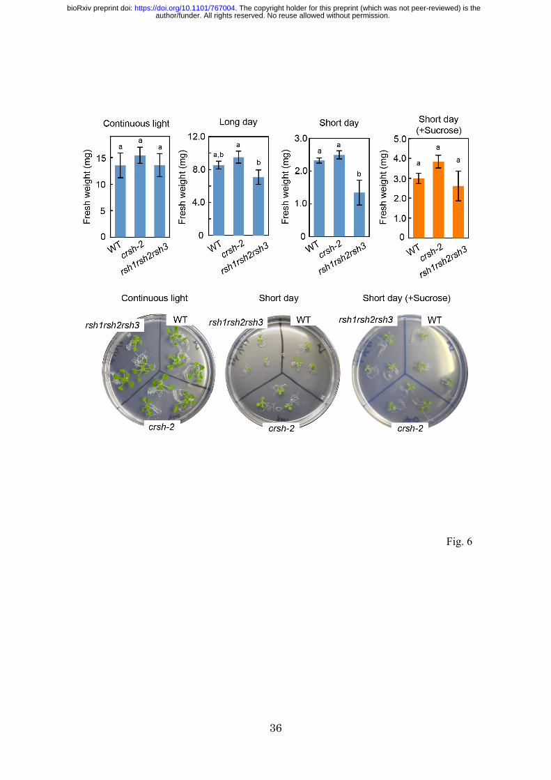
To observe the effects of nighttime ppGpp accumulation on plant growth, we grew WT,
crsh-2, and rsh1rsh2rsh3 mutants under continuous light, short-day (8 h light/16 h dark)
and long-day (16 h light/8 h dark) conditions for 14 days. The fresh weights of WT and
crsh-2 shoots were similar; however, a significant reduction in the fresh weight of
rsh1rsh2rsh3 compared with that of the WT was observed only under short-day
conditions (Fig. 6), suggesting that the hyper-accumulation of ppGpp in the triple
mutant under dark conditions (Fig. 3C) causes growth retardation. Under long-day
conditions, a significant reduction in the fresh weight of rsh1rsh2rsh3 compared with
crsh-2 was observed (Fig. 6), which supported the hypothesis that dark-induced
synthesis of ppGpp downregulates plant growth. Notably, the growth-retarding
phenotype of rsh1rsh2rsh3 was rescued by the exogenous addition of sucrose.
Previously, an Arabidopsis crsh co-suppression mutant (OX19) obtained from
35S-promoter-driven CRSH-overexpression lines showed abnormal flower development
(Masuda et al. 2008); however, this phenotype was not observed in crsh-2. A CRSH
knockdown mutant constructed using an artificial microRNA also did not show this
abnormal phenotype (Sugliani et al. 2016), suggesting that the OX19 phenotype was not
simply due to the less accumulation of CRSH. No further morphological differences
were observed for crsh-2 and rsh2rsh3 in comparison with the WT throughout the
entire growth period (Supplementary Fig. S3).
Discussion
author/funder. All rights reserved. No reuse allowed without permission. The copyright holder for this preprint (which was not peer-reviewed) is the. https://doi.org/10.1101/767004doi: bioRxiv preprint

14
In this study, we provide the first in vivo evidence demonstrating the
Ca2+-dependent ppGpp synthase activity of CRSH. We succeeded in isolating the
Arabidopsis crsh-null mutant, crsh-2, which did not attenuate a transient increase of
ppGpp upon shifting from light-to-dark conditions. Given that plastidial Ca2+
concentrations transiently increase upon light-to-dark transition (Sai and Johnson 2002),
CRSH most likely senses the increased Ca2+ in the chloroplast and upregulates its
(p)ppGpp synthase activity (Fig. 7). CRSH expression shows a diurnal rhythm with an
expression peak at dusk (Mizusawa et al. 2008), which may contribute to the rapid and
efficient accumulation of ppGpp during nighttime under natural conditions. The
accumulated ppGpp is rapidly degraded by RSH1, RSH2, and RSH3; this regulation
may be necessary to avoid the over-suppression of photosynthetic activity and
chloroplast metabolism. Once regulation was impaired, plant growth was significantly
influenced under photoautotrophic growth conditions. Notably, crsh-2 still showed a
small increase in ppGpp 10 min after the onset of dark treatment, indicating that the
increment is not due to the upregulation of CRSH activity; the elucidation of this
mechanism will be required in future studies.
ppGpp synthesis by CRSH under dark conditions accompanies the inhibition
of PEP-dependent psbD transcription in the WT and rsh2rsh3. ppGpp directly interacts
with PEP in vitro to suppress its transcriptional activity (Sato et al. 2009), suggesting
that ppGpp, which accumulates upon light-to-dark transition, binds to PEP and inhibits
its activity. The mRNA levels of atpB, which is transcribed by PEP and NEP, were
decreased upon the transition to darkness in rsh2rsh3, and super-accumulation of
author/funder. All rights reserved. No reuse allowed without permission. The copyright holder for this preprint (which was not peer-reviewed) is the. https://doi.org/10.1101/767004doi: bioRxiv preprint

15
ppGpp was seen, supporting the hypothesis that ppGpp negatively regulates PEP
activity. However, we found that transcripts of another PEP-dependent gene, rbcL, were
not downregulated upon the shift into darkness. The transcript levels of psbD and atpB
in the light are significantly higher than those in the dark; however, the transcript levels
of rbcL are constant, irrespective of the light conditions (Nakamura et al. 2003). These
observations suggest that the significance of the ppGpp-dependent inhibition of PEP
activity is variable in each promoter.
Our and another groups previously reported that RSH3-overexpression lines,
which constantly accumulate excess ppGpp, suppress the accumulation of transcripts of
PEP- and NEP-dependent genes (Maekawa et al. 2015; Sugliani et al. 2016), suggesting
that ppGpp also regulates the transcription of NEP-dependent genes by inhibiting
guanylate kinase activity and thus reducing the GTP pools necessary for mRNA
synthesis (Fig. 7). As direct transcriptional repression of ppGpp may be faster than
indirect transcriptional repression through the reduction of GTP pools, indirect
transcriptional repression of NEP-dependent genes could not be detected during the
timescale of this experiment. Notably, mRNA of clpP, which is transcribed by NEP, is
transiently increased in rsh2rsh3 upon the shift to dark conditions. Messenger RNA
levels of clpP are significantly (>fivefold) higher in the light than in the dark; however,
those of accD are constant, irrespective of the light conditions (Nakamura et al. 2003).
These findings suggest that RSH2 and/or RSH3 are involved in the control of NEP
activity and/or the post-transcriptional mRNA degradation of each chloroplast gene,
although the mechanism by which this case is carried out remains unclear.
author/funder. All rights reserved. No reuse allowed without permission. The copyright holder for this preprint (which was not peer-reviewed) is the. https://doi.org/10.1101/767004doi: bioRxiv preprint
16
Transient increments in ppGpp levels upon light-to-dark transition have also
been reported in the cyanobacterium Synechococcus elongatus (Hood et al. 2016;
Puszynska and O’Shea 2017), which indicates that the ppGpp-dependent stringent
response is universally required to adapt photosynthetic processes to dark conditions in
oxygenic phototrophs. Given that cyanobacteria do not contain CRSH homologs (Ito et
al. 2017), different strategies must be used to upregulate ppGpp synthesis in the dark in
a Ca2+-independent manner. Darkness-induced ppGpp accumulation in cyanobacteria
may be caused by the posing of the redox signal derived from photosynthetic electron
transfer (Hood et al. 2016). Notably, in cyanobacteria, accumulated (p)ppGpp can
regulate the expression of all photosynthesis-related genes at transcriptional and
translational levels because the genes are located on a single chromosome. However,
plants utilize the rise in plastidial Ca2+ concentrations and Ca2+-dependent ppGpp
synthesis to downregulate photosynthetic light and dark reactions in a coordinated
manner. Elevated levels of (p)ppGpp and Ca2+ negatively control the PEP-dependent
transcription of plastid genes, as determined in this study, and Calvin–Benson cycle
activities (Portis and Heldt 1976; Charles and Halliwell 1980; Hertig and Wolosiuk
1980; Rocha et al. 2014), respectively. Ca2+-dependent (p)ppGpp synthesis may have
been required after the endosymbiosis of the ancestral cyanobacterium, because most
enzymes that are required for CO2 fixation are encoded in the nucleus of plant cells and
must be regulated in chloroplasts at the post-translational level.
Although flg22 treatment increases plastidial Ca2+ concentrations (Nomura
and Shiina 2014), no significant differences were observed in the PAMP regulation of
author/funder. All rights reserved. No reuse allowed without permission. The copyright holder for this preprint (which was not peer-reviewed) is the. https://doi.org/10.1101/767004doi: bioRxiv preprint

17
nuclear gene expression in the WT and crsh-2, which suggests that the
CRSH-dependent stringent response does not interact with PAMP-induced signaling.
However, the accumulation of ppGpp has previously been found to affect the
susceptibility of Arabidopsis to Turnip mosaic virus, suggesting that (p)ppGpp
metabolism is of physiological importance in plant defense response (Abdelkefi et al.
2018). Furthermore, we found that WRKY33 transcripts increased upon the transition to
darkness and the increment in these transcripts in rsh2rsh3 0.5 h after the start of dark
incubation was ~2-fold higher than that in the WT, which supports the hypothesis that
the ppGpp-dependent plastidial stringent response is involved in plant–pathogen
interactions. Perhaps, however, the increase in Ca2+ following treatment with 1 µM
flg22 is insufficiently high to upregulate CRSH activity to increase plastidial (p)ppGpp
levels. The maximum concentration of Ca2+ is ~0.1 µM after 1 µM flg22 treatment,
which is lower than the value after light-to-dark transition (~0.2 µM) (Nomura et al.
2012), which supports this hypothesis. Future studies should test whether plastidial Ca2+
concentrations are increased by the application of other PAMPs; under such conditions,
CRSH activity should be upregulated.
In summary, CRSH synthesizes (p)ppGpp upon light-to-dark transition when
the Ca2+ concentration in chloroplasts increases to a certain level, which is important for
plants to adapt plastidial gene expression and metabolism to dark conditions. The
rsh2rsh3 and rsh1rsh2rsh3 mutants showed significantly higher ppGpp accumulation
and the raised levels were maintained longer than those in the WT, indicating that the
hydrolase activities of RSH1, RSH2, and RSH3 were required for the adequate
author/funder. All rights reserved. No reuse allowed without permission. The copyright holder for this preprint (which was not peer-reviewed) is the. https://doi.org/10.1101/767004doi: bioRxiv preprint

18
maintenance of ppGpp levels in the dark. Further work to determine other conditions in
which CRSH-dependent ppGpp synthesis is upregulated will be important in revealing
the physiological importance of CRSH function and the relationship between the
plastidial stringent response and intracellular Ca2+ signaling.
Materials and Methods
Plant materials and growth conditions
Plants (Columbia ecotype of A. thaliana) were grown on a half concentration of
Murashige and Skoog (1/2 MS) medium containing 0.8% agar at 23 °C. Sucrose was
added to 1% (w/v) when appropriate. Light conditions are indicated in each
experimental procedure. The rsh2-rsh3 double mutant was described previously
(Maekawa et al. 2015). The rsh1 (GABI_206D_08) and crsh-1 (SAIL_1295_C04)
mutants were obtained from the Arabidopsis Biological Resource Center. The
rsh1-rsh2-rsh3 triple mutant was obtained by crossing the rsh1 mutant with the
rsh2rsh3 mutant. The crsh-null mutant, namely, crsh-2, was constructed using the
CRISPR/Cas9-based method, as described previously (Fauser et al. 2014). We designed
a protospacer sequence in the first exon of CRSH (83−102 nucleotides from the
translation start site), and the protospacer oligonucleotides were annealed and cloned
into the plasmid pEn-Chimera based on the protocol (Fauser et al. 2014). After checking
the correct sequences of the inserted DNA, it was further cloned into pDe-CAS9 by
using Gateway LR Clonase (ThermoFisher). The resultant plasmid was introduced into
the Arabidopsis WT through the Agrobacterium-based standard flower-dip method to
author/funder. All rights reserved. No reuse allowed without permission. The copyright holder for this preprint (which was not peer-reviewed) is the. https://doi.org/10.1101/767004doi: bioRxiv preprint

19
induce the double-strand break at the protospacer position. Using gene-specific primers
(Table S1), homozygous mutations of rsh1, rsh2, rsh3, and/or crsh were confirmed by
PCR followed by sequencing. RNA was isolated using the SV Total RNA Isolation
System (Promega) to check for the presence of the CRSH transcript in crsh-1 (Fig. 2C).
The first-strand cDNA, synthesized using the PrimeScript RT regent Kit (TaKaRa), and
the genome DNA isolated from the WT were used as templates for RT-PCR with
gene-specific primers (Table S1).
The crsh-2 complementing line was constructed as follows. Previously, we
cloned the CRSH cDNA (without the stop codon) into the plasmid pDONR/Zeo
(Invitrogen) (Masuda et al. 2008). Using the plasmid as a template, five point (but
silent) mutations were introduced in the protospacer sequence used to construct crsh-2
to ensure that the introduced CRSH was not recognized by the guide RNA of the
CRISPR/Cas9 system. Forward and reverse primers were designed to contain the five
mutations (Table S1); the CRSH cDNA, together with the plasmid backbone, was
amplified by PCR. The In-Fusion cloning reagent (Clonetech) was used to circularize
the amplified fragment. After checking the correct sequence of the plasmid DNA, the
insert DNA was cloned into the pGWB2 vector (Nakagawa et al. 2007) by using
Gateway LR Clonase (ThermoFisher). Because the cloned CRSH did not contain the
stop codon, the resulting pGWB2-base plasmid expressed the CRSH fused with an ~1.5
kDa short peptide (YPAFLYKVVDNSA) at the C-terminus; the recombinant protein
was designated as CRSH*. The resulting plasmid was introduced into the crsh-2 mutant
by using the Agrobacterium-based standard flower-dip method. The expression of
author/funder. All rights reserved. No reuse allowed without permission. The copyright holder for this preprint (which was not peer-reviewed) is the. https://doi.org/10.1101/767004doi: bioRxiv preprint

20
CRSH* by the 35S promoter in the complementing line was confirmed by western blot
analysis using the anti-CRSH antibody (Supplementary Fig. S2A).
Quantitative mRNA analysis
To measure the mRNA levels of accD, clpP, atpB, rbcL, psbD, and WRKY33 upon
transition to darkness, plants were grown on agar-solidified 1/2 MS medium under
continuous light (40 µmol photons m−2 s−1) for 14 days. After harvesting shoots of the
plants (0 h), the remaining plants were transferred to the dark and shoots were harvested
after 0.5 and 1 h incubation. Total RNA was isolated using the SV Total RNA Isolation
System (Promega). The first-strand cDNA, synthesized using the PrimeScript RT regent
Kit (TaKaRa), was the subsequent template. qRT-PCR was performed using SYBR®
Premix EX TaqTM and Thermal Cycler Dice® (TaKaRa). Plastid genes, and
WRKY33-specific primers used for qRT-PCR were as described (Sano et al. 2014;
Maekawa et al. 2015).
For the quantification of mRNA levels of WRKY33 and WRKY46 after flg22
treatment, plants were grown on agar-solidified 1/2 MS medium under 16 h light (100
µmol photons m−2 s−1)/16 h dark cycles. Fourteen-day-old plants were treated with 1
µM flg22 (synthesized by Biologica, Nagoya) for 30 min, as described previously
(Sano et al. 2014). Total RNA isolation, cDNA synthesis, and gene-specific primers
used for qRT-PCR analysis were as described (Sano et al. 2014).
Quantification of ppGpp
author/funder. All rights reserved. No reuse allowed without permission. The copyright holder for this preprint (which was not peer-reviewed) is the. https://doi.org/10.1101/767004doi: bioRxiv preprint
21
Plants were grown on 0.8% agar-solidified 1/2 MS medium under continuous light (40
µmol photons m−2 s−1). Fourteen-day-old plants were transferred to the dark and ~0.1 g
shoots were harvested at each of the time points indicated (Fig. 3). Extraction and
quantification of ppGpp were as described previously (Ihara et al. 2015).
Western blot analysis
Protein content was measured using the Bradford assay Kit (Bio Lab). Total proteins (6
µg each) were applied to an SDS-PAGE gel and electroblotted onto a polyvinylidene
difluoride membrane (GE-Healthcare). The membrane was incubated with the
CRSH-specific antibody (Masuda et al. 2008). Immunoreactive proteins were detected
using the Alkaline Phosphatase Substrate Kit II (Vector Laboratories).
Acknowledgments
We thank Professor Holger Puchta (Karlsruhe Institute of Technology) for kindly
providing vectors for the CRISPR/Cas9-based genome editing and the Arabidopsis
Biological Resource Center for providing mutant seeds.
Funding
This work was supported by MEXT/JSPS KAKENHI (grant number 19H04719) to SM.
Disclosures
The authors have no conflicts of interest to declare.
author/funder. All rights reserved. No reuse allowed without permission. The copyright holder for this preprint (which was not peer-reviewed) is the. https://doi.org/10.1101/767004doi: bioRxiv preprint

23
References
Abdelkefi H, Sugliani M, Ke H, et al (2018) Guanosine tetraphosphate modulates
salicylic acid signalling and the resistance of Arabidopsis thaliana to Turnip
mosaic virus. Mol Plant Pathol 19:634–646.
Boniecka J, Prusińska J, Dąbrowska GB, Goc A (2017) Within and beyond the stringent
response-RSH and (p)ppGpp in plants. Planta 246:817–842.
Cashel M (1969) The control of ribonucleic acid synthesis in Escherichia coli IV.
Relevance of unusual phosphorylated compounds from amino acid-starved
stringent strains. J Biol Chem 244:3133–3141.
Charles SA, Halliwell B (1980) Action of calcium ions on spinach (Spinacia oleracea)
chloroplast fructose bisphosphatase and other enzymes of the Calvin cycle.
Biochem J 188:775–779.
Dalebroux ZD, Swanson MS (2012) ppGpp: magic beyond RNA polymerase. Nat Rev
Microbiol 10:203–212.
Fauser F, Schiml S, Puchta H (2014) Both CRISPR/Cas-based nucleases and nickases
can be used efficiently for genome engineering in Arabidopsis thaliana. Plant J
79:348–359.
Field B (2018) Green magic: regulation of the chloroplast stress response by (p)ppGpp
in plants and algae. J Exp Bot 69:2797–2807.
Frank J, Happeck R, Meier B, et al (2019) Chloroplast-localized BICAT proteins shape
stromal calcium signals and are required for efficient photosynthesis. New Phytol
221:866–880.
author/funder. All rights reserved. No reuse allowed without permission. The copyright holder for this preprint (which was not peer-reviewed) is the. https://doi.org/10.1101/767004doi: bioRxiv preprint

24
Hertig C, Wolosiuk RA (1980) A dual effect of Ca2+ on chloroplast
fructose-1,6-bisphosphatase. Biochem Biophys Res Commun 97:325–333.
Hochmal AK, Schulze S, Trompelt K, Hippler M (2015) Calcium-dependent regulation
of photosynthesis. Biochim Biophys Acta - Bioenerg 1847:993–1003.
Honoki R, Ono S, Oikawa A, et al (2018) Significance of accumulation of the alarmone
(p)ppGpp in chloroplasts for controlling photosynthesis and metabolite balance
during nitrogen starvation in Arabidopsis. Photosynth Res 135:299–308.
Hood RD, Higgins SA, Flamholz A, et al (2016) The stringent response regulates
adaptation to darkness in the cyanobacterium Synechococcus elongatus. Proc Natl
Acad Sci U S A 113:E4867-4876.
Ihara Y, Masuda S (2016) Cytosolic ppGpp accumulation induces retarded plant growth
and development. Plant Signal Behav. 11: e1132966.
Ihara Y, Ohta H, Masuda S (2015) A highly sensitive quantification method for the
accumulation of alarmone ppGpp in Arabidopsis thaliana using
UPLC-ESI-qMS/MS. J Plant Res 128:511–518.
Ito D, Ihara Y, Nishihara H, Masuda S (2017) Phylogenetic analysis of proteins
involved in the stringent response in plant cells. J Plant Res 130:625–634.
Knight H, Trewavas AJ, Knight MR (1996) Cold calcium signaling in Arabidopsis
involves two cellular pools and a change in calcium signature after ccclimation.
Plant Cell 8:489–503.
Levine B, Klionsky DJ (2017) Autophagy wins the 2016 Nobel Prize in Physiology or
Medicine: Breakthroughs in baker’s yeast fuel advances in biomedical research.
author/funder. All rights reserved. No reuse allowed without permission. The copyright holder for this preprint (which was not peer-reviewed) is the. https://doi.org/10.1101/767004doi: bioRxiv preprint

25
Proc Natl Acad Sci U S A 114:201–205.
Loro G, Wagner S, Doccula FG, et al (2016) Chloroplast-specific in vivo Ca2+ imaging
using Yellow Cameleon fluorescent protein sensors reveals organelle-autonomous
Ca2+ signatures in the stroma. Plant Physiol pp.00652.2016.
Maekawa M, Honoki R, Ihara Y, et al (2015) Impact of the plastidial stringent response
in plant growth and stress responses. Nat Plants 1:15167.
Masuda S (2012) The stringent response in phototrophs. In: Najafpour M (ed) Advances
in photosynthesis. In Tech., pp 487–500
Masuda S, Mizusawa K, Narisawa T, et al (2008) The bacterial stringent response,
conserved in chloroplasts, controls plant fertilization. Plant Cell Physiol 49:135–
41.
Mizusawa K, Masuda S, Ohta H (2008) Expression profiling of four RelA/SpoT-like
proteins, homologues of bacterial stringent factors, in Arabidopsis thaliana. Planta
228:553–562.
Nakagawa T, Kurose T, Hino T, et al (2007) Development of series of gateway binary
vectors, pGWBs, for realizing efficient construction of fusion genes for plant
transformation. J Biosci Bioeng 104:34–41.
Nakamura T, Furuhashi Y, Hasegawa K, et al (2003) Array-Based Analysis on Tobacco
Plastid Transcripts: Preparation of a Genomic Microarray Containing All Genes
and All Intergenic Regions. Plant Cell Physiol 44:861–867.
Nakatogawa H, Ohsumi Y (2014) Autophagy: close contact keeps out the uninvited.
Curr Biol 24:R560–R562.
author/funder. All rights reserved. No reuse allowed without permission. The copyright holder for this preprint (which was not peer-reviewed) is the. https://doi.org/10.1101/767004doi: bioRxiv preprint
26
Nguyen CT, Kurenda A, Stolz S, et al (2018) Identification of cell populations
necessary for leaf-to-leaf electrical signaling in a wounded plant. Proc Natl Acad
Sci U S A 115:10178–10183.
Nomura H, Komori T, Uemura S, et al (2012) Chloroplast-mediated activation of plant
immune signalling in Arabidopsis. Nat Commun 3:926.
Nomura H, Shiina T (2014) Calcium Signaling in Plant Endosymbiotic Organelles:
Mechanism and Role in Physiology. Mol Plant 7:1094–1104.
Portis AR, Heldt HW (1976) Light-dependent changes of the Mg2+ concentration in the
stroma in relation to the Mg2+ dependency of CO2 fixation in intact chloroplasts.
Biochim Biophys Acta - Bioenerg 449:434–446.
Potrykus K, Cashel M (2008) (p)ppGpp: Still Magical? . Annu Rev Microbiol 62:35–
51.
Puszynska AM, O’Shea EK (2017) ppGpp controls global gene expression in light and
in darkness in S. elongatus. Cell Rep 21:3155–3165.
Rocha AG, Mehlmer N, Stael S, et al (2014) Phosphorylation of Arabidopsis
transketolase at Ser 428 provides a potential paradigm for the metabolic control of
chloroplast carbon metabolism. Biochem J 458:313–322.
Sai J, Johnson CH (2002) Dark-stimulated calcium ion fluxes in the chloroplast stroma
and cytosol. Plant Cell 14:1279–1291.
Sanders J, Brownlee CH, Harper (1999) Communicating with calcium. Plant Cell
11:691–706.
Sano S, Aoyama M, Nakai K, et al (2014) Light-dependent expression of flg22-induced
author/funder. All rights reserved. No reuse allowed without permission. The copyright holder for this preprint (which was not peer-reviewed) is the. https://doi.org/10.1101/767004doi: bioRxiv preprint

27
defense genes in Arabidopsis. Front Plant Sci 5:531.
Sato M, Takahashi K, Ochiai Y, et al (2009) Bacterial alarmone, guanosine
5-diphosphate 3-diphosphate (ppGpp), predominantly binds the β subunit of
plastid-encoded plastid RNA polymerase in chloroplasts. ChemBioChem 10:1227–
1233.
Sugliani M, Abdelkefi H, Ke H, et al (2016) An ancient bacterial signaling pathway
regulates chloroplast function to influence growth and development in Arabidopsis.
Plant Cell 28:661–679.
Tozawa Y, Nomura Y (2011) Signalling by the global regulatory molecule ppGpp in
bacteria and chloroplasts of land plants. Plant Biol (Stuttg) 13:699–709.
Tozawa Y, Nozawa A, Kanno T, et al (2007) Calcium-activated (p)ppGpp synthetase in
chloroplasts of land plants. J Biol Chem 282:35536–45.
White PJ, Broadley MR (2003) Calcium in Plants. Ann Bot 92:487–511.
author/funder. All rights reserved. No reuse allowed without permission. The copyright holder for this preprint (which was not peer-reviewed) is the. https://doi.org/10.1101/767004doi: bioRxiv preprint
28
Figure legends
Fig. 1 Domain structures of RelA and SpoT from E. coli and RSHs in Arabidopsis. Hyd,
(p)ppGpp hydrolase domain; hyd, (p)ppGpp hydrolase domain lacking some critical
amino acids; Syn, (p)ppGpp synthase domain; syn, (p)ppGpp synthase domain lacking
the critical Gly residue; ACT, aspartate kinase, chorismate mutase, TyrA domain; EF
hand, Ca2+-binding helix E and F motif; TGS, threonyl-tRNA synthase, GTPase SpoT
domain; AA, amino acids; cTP, chloroplast transit peptide.
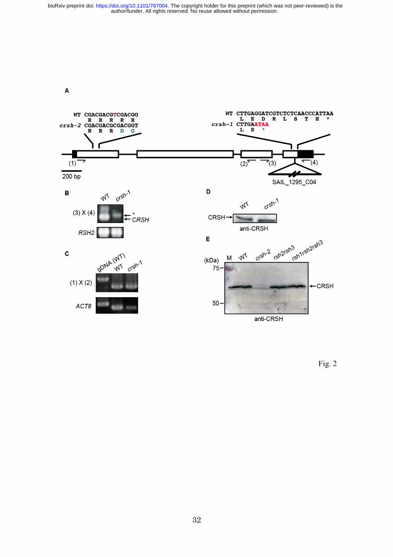
Fig. 2 Construction of crsh mutants. (A) Schematic of the exon–intron structure of
CRSH (At3g17470) with a single nucleotide deletion site in crsh-2 and a T-DNA
insertion site in crsh-1. A deleted nucleotide in crsh-2 and inserted T-DNA nucleotides
in crsh-1 are highlighted in red. Mutated codons are highlighted in blue. (B) PCR
analysis reveals homozygous T-DNA insertion in crsh-1. Positions of primers used (3
and 4) are indicated in (A). An asterisk indicates a nonspecific band. RSH2 was
amplified as a control for DNA isolation. (C) RT-PCR analysis reveals the presence of
CRSH mRNA in crsh-1. Positions of primers used (1 and 2) are indicated in (A).
Genome DNA isolated from WT was used as a control for genome DNA contamination,
and the actin8 (ACT8) gene was used as a control for mRNA isolation and reverse
transcription. (D) Western blot of proteins isolated from WT and crsh-1. (E) Western
blot of proteins isolated from WT, crsh-2, rsh2rsh3, and rsh1rsh2rsh3. Full images of
original photographs and western blot data are shown in Supplementary Fig. S1.
author/funder. All rights reserved. No reuse allowed without permission. The copyright holder for this preprint (which was not peer-reviewed) is the. https://doi.org/10.1101/767004doi: bioRxiv preprint

29
Fig. 3 ppGpp accumulation kinetics upon dark-to-light transition in the WT and crsh-2
(A), WT and rsh2rsh3 (B), and WT and rsh1rsh2rsh3 (C). Plants were grown on 0.8%
agar-solidified 1/2 MS medium under continuous light (40 µmol photons m−2 s−1).
Fourteen-day-old plants were transferred to the dark and shoots were harvested at the
time points for ppGpp quantification. Values are mean ± SD (n = 3). Data for the WT
are reproduced (dotted lines).
Fig. 4 Relative mRNA levels in the WT, rsh2rsh3, and crsh-2 plants before (0 h) and
after dark incubation for 0.5 and 1 h. Plants were grown on 0.8% agar-solidified 1/2 MS
medium under continuous light (40 µmol photons m−2 s−1) for 14 days and then
transferred to the dark. Messenger RNA quantification was carried out using RT-PCR
analysis. Values are mean ± SD (n = 3). The WT level at 0 h in the first experiment was
set to 1.0. P values based on Student’s t-test are indicated.
Fig. 5 Relative mRNA levels in the WT and crsh-2 before and after incubation with
flg22. Plants were grown on agar-solidified 1/2 MS medium under 16 h light (100 µmol
photons m−2 s−1)/16 h dark cycles. Fourteen-day-old plants were treated with 1 µM
flg22 and/or solvent (water) for 0.5 h. Values are means of two independent
experiments and the data are indicated by dots. The WT level before flg22 addition was
set to 1.0.
author/funder. All rights reserved. No reuse allowed without permission. The copyright holder for this preprint (which was not peer-reviewed) is the. https://doi.org/10.1101/767004doi: bioRxiv preprint

30
Fig. 6 Growth phenotype of rsh mutants. Plants were grown on 0.8% agar-solidified 1/2
MS medium with or without 1% sucrose under continuous light, long-day (16 h light/8
h dark) or short-day (8 h light/16 h dark) conditions for 14 days, and then fresh weights
of shoots were measured (n = 29–46). Different letters denote the significant difference
by using Tukey–Kramer’s multiple comparison test (P < 0.05).
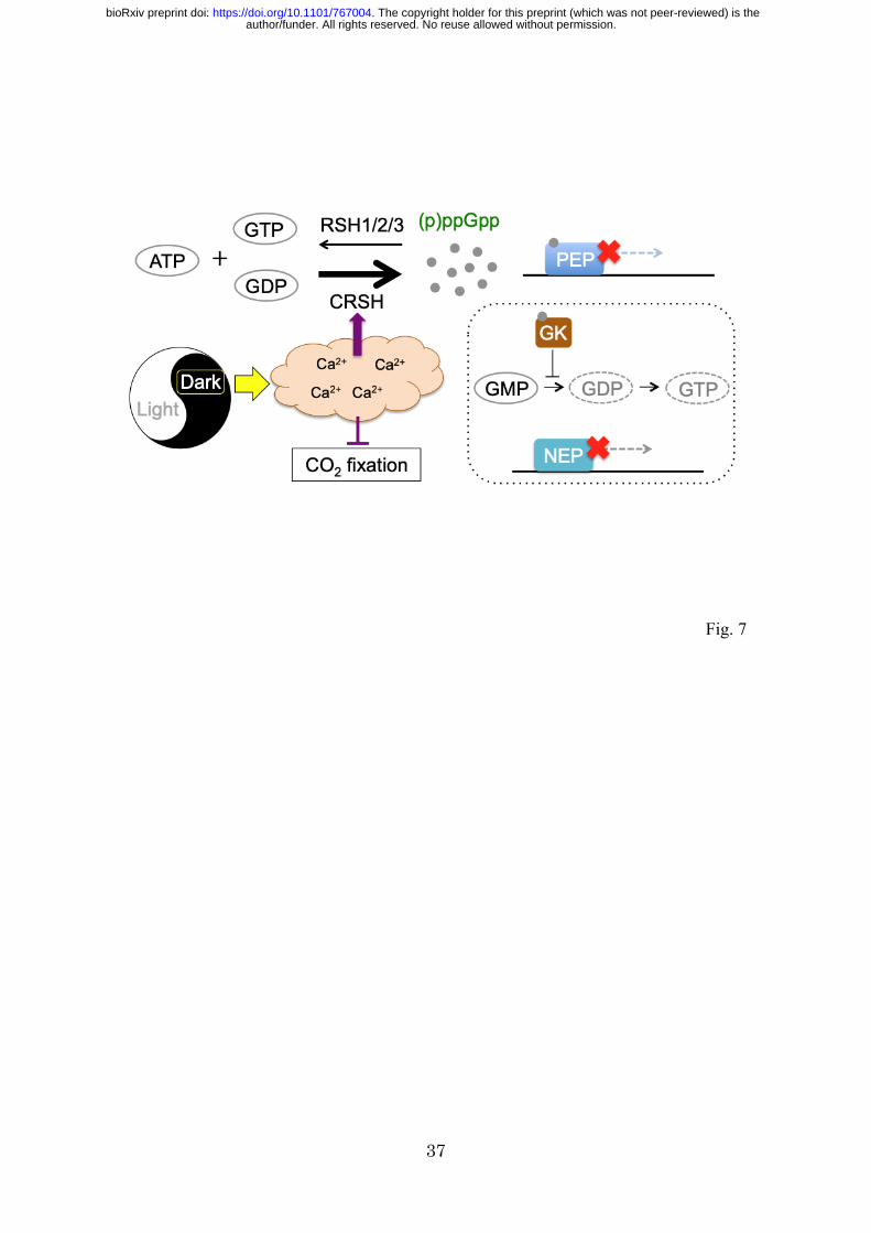
Fig. 7 Schematic of dark-induced ppGpp synthesis and regulation of photosynthesis.
Upon light-to-dark transition, the plastidial Ca2+ concentration is increased, which
results in the upregulation of CRSH-dependent (p)ppGpp synthesis and the
downregulation of CO2 fixation through the inhibition of Calvin–Benson cycle enzymes.
Synthesized (p)ppGpp directly inhibits the transcriptional activity of PEP. (p)ppGpp
potentially inhibits plastidial guanylate kinase (GK) activity, which results in indirect
regulation of transcription by NEP.
author/funder. All rights reserved. No reuse allowed without permission. The copyright holder for this preprint (which was not peer-reviewed) is the. https://doi.org/10.1101/767004doi: bioRxiv preprint

31
Fig. 1
author/funder. All rights reserved. No reuse allowed without permission. The copyright holder for this preprint (which was not peer-reviewed) is the. https://doi.org/10.1101/767004doi: bioRxiv preprint
32
Fig. 2
author/funder. All rights reserved. No reuse allowed without permission. The copyright holder for this preprint (which was not peer-reviewed) is the. https://doi.org/10.1101/767004doi: bioRxiv preprint

33
Fig. 3
author/funder. All rights reserved. No reuse allowed without permission. The copyright holder for this preprint (which was not peer-reviewed) is the. https://doi.org/10.1101/767004doi: bioRxiv preprint

34
Fig. 4
author/funder. All rights reserved. No reuse allowed without permission. The copyright holder for this preprint (which was not peer-reviewed) is the. https://doi.org/10.1101/767004doi: bioRxiv preprint

35
Fig. 5
author/funder. All rights reserved. No reuse allowed without permission. The copyright holder for this preprint (which was not peer-reviewed) is the. https://doi.org/10.1101/767004doi: bioRxiv preprint
36
Fig. 6
author/funder. All rights reserved. No reuse allowed without permission. The copyright holder for this preprint (which was not peer-reviewed) is the. https://doi.org/10.1101/767004doi: bioRxiv preprint
37
Fig. 7
author/funder. All rights reserved. No reuse allowed without permission. The copyright holder for this preprint (which was not peer-reviewed) is the. https://doi.org/10.1101/767004doi: bioRxiv preprint

38
Table S1. Primers used in this study Target gene(s) Sequence (5'->3') Usage T-DNA left border
CCCATTTGGACGTGAATGTAGACAC
rsh1 mutation genotyping
TAGCATCTGAATTTCATAACCAATCTCGATACAC rsh2 mutation genotyping
CCCATTTGGACGTGAATGTAGACAC
rsh3 mutation genotyping
ATTTCATAACCAATCTCGATACAC
crsh-1 mutation genotyping
RSH1 (At4g02260)
TCCAGATTATCTTGTGGTGG rsh1 mutation genotyping PCR AACCGGTCAGCTAGTTTGAC
RSH2 (At3g14050)
GAACTTTAGGATTATAAAGCAG rsh2 mutation genotyping PCR AAACAAGAATCTAAAACTAGTG
RSH3 (At1g54130)
GGTCGACATGGTGGTAGCAACGACC rsh3 mutation genotyping PCR CCAGCTACAACAACAGTCGAATTA
GGTCGACATGGTGGTAGCAACGACC rsh3 mutation genotyping RT-PCR GGTCGACATGGTGGTAGCAACGACA
CRSH (At3g17470)
ATGTCTGTGATTCGTCCTTC (Primer 1) TCGTTGTTGTCTTGAAGCTTTG (Primer 2) GAAGAGCTTGGAGCTCCAGG (Primer 3) AATAAGATGGGAGTAGTCTATAG (Primer 4)
crsh mutation genotyping PCR
ATCCAACGCCGTCGACGACGGTGGAATCCGAGGTC
For construction of the crsh-2 complementing line1 TCGACGGCGTTGGATTAATTGAATCGAATAAAGA
C 1Red characters are point mutations (silent mutations not to change the amino-acid sequence of CRSH) to avoid recognition by the guide RNA used for the CRISPR/Cas9-based genome editing. Underlines represent the complementary sequences used for In-Fusion cloning (see Methods section for more details).
author/funder. All rights reserved. No reuse allowed without permission. The copyright holder for this preprint (which was not peer-reviewed) is the. https://doi.org/10.1101/767004doi: bioRxiv preprint
39
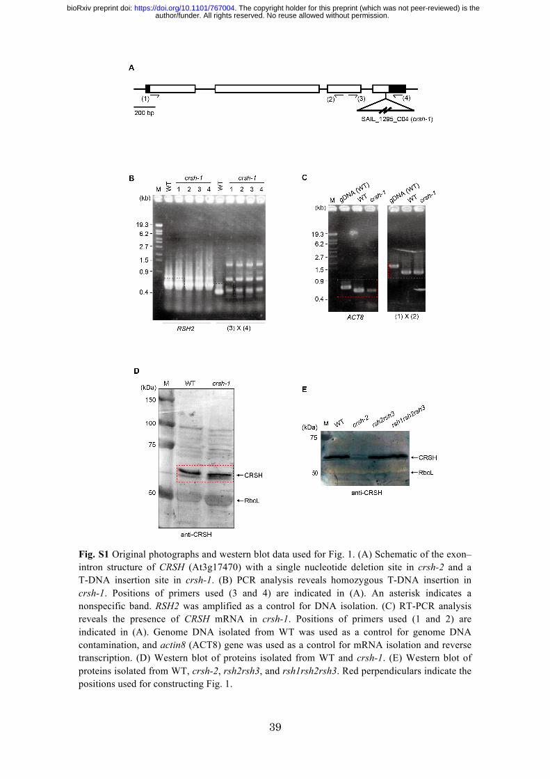
Fig. S1 Original photographs and western blot data used for Fig. 1. (A) Schematic of the exon–intron structure of CRSH (At3g17470) with a single nucleotide deletion site in crsh-2 and a T-DNA insertion site in crsh-1. (B) PCR analysis reveals homozygous T-DNA insertion in crsh-1. Positions of primers used (3 and 4) are indicated in (A). An asterisk indicates a nonspecific band. RSH2 was amplified as a control for DNA isolation. (C) RT-PCR analysis reveals the presence of CRSH mRNA in crsh-1. Positions of primers used (1 and 2) are indicated in (A). Genome DNA isolated from WT was used as a control for genome DNA contamination, and actin8 (ACT8) gene was used as a control for mRNA isolation and reverse transcription. (D) Western blot of proteins isolated from WT and crsh-1. (E) Western blot of proteins isolated from WT, crsh-2, rsh2rsh3, and rsh1rsh2rsh3. Red perpendiculars indicate the positions used for constructing Fig. 1.
author/funder. All rights reserved. No reuse allowed without permission. The copyright holder for this preprint (which was not peer-reviewed) is the. https://doi.org/10.1101/767004doi: bioRxiv preprint
40
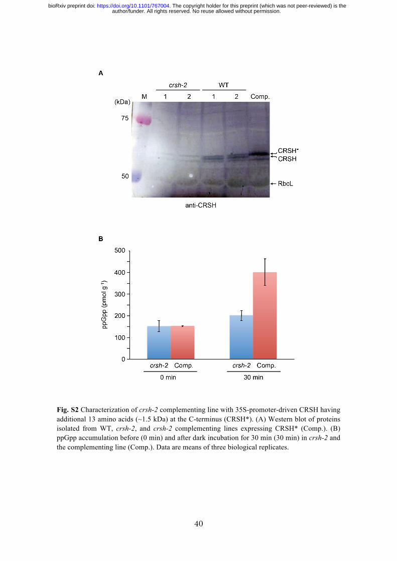
Fig. S2 Characterization of crsh-2 complementing line with 35S-promoter-driven CRSH having additional 13 amino acids (~1.5 kDa) at the C-terminus (CRSH*). (A) Western blot of proteins isolated from WT, crsh-2, and crsh-2 complementing lines expressing CRSH* (Comp.). (B) ppGpp accumulation before (0 min) and after dark incubation for 30 min (30 min) in crsh-2 and the complementing line (Comp.). Data are means of three biological replicates.
author/funder. All rights reserved. No reuse allowed without permission. The copyright holder for this preprint (which was not peer-reviewed) is the. https://doi.org/10.1101/767004doi: bioRxiv preprint

41
Fig. S3 No significant visible phenotype was observed for crsh-2 and rsh2rsh3 mutants. Plants were germinated on 1/2 MS plates and grown for 2 weeks. Plants were then transferred to soil and grown for 1 month under continuous-light conditions.
author/funder. All rights reserved. No reuse allowed without permission. The copyright holder for this preprint (which was not peer-reviewed) is the. https://doi.org/10.1101/767004doi: bioRxiv preprint


![The Plastidial Glyceraldehyde-3-Phosphate Dehydrogenase Is … · The Plastidial Glyceraldehyde-3-Phosphate Dehydrogenase Is Critical for Viable Pollen Development in Arabidopsis1[W]](https://img.pdfslide.net/doc/110x75/600aa8912522092462533f3e/the-plastidial-glyceraldehyde-3-phosphate-dehydrogenase-is-the-plastidial-glyceraldehyde-3-phosphate.jpg)


![Annual Review of Microbiology Volume 62 Issue 1 2008 [Doi 10.1146%2Fannurev.micro.62.081307.162903] Potrykus, Katarzyna; Cashel, Michael -- (p)PpGpp- Still Magical](https://img.pdfslide.net/doc/110x75/55cf905f550346703ba55ba6/annual-review-of-microbiology-volume-62-issue-1-2008-doi-1011462fannurevmicro62081307162903.jpg)