Embed Size (px)
Citation preview
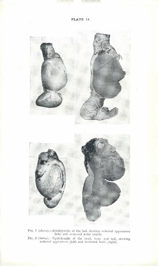
PLATE 11
Fig. 1 (above).—Epididymitis of the tail, showing external appearance (left) and sectioned testis (right).
Fig. 2 (below).—Epididymitis of the head, body and tail, showing externa] appearance (left) and sectioned testis (right).


![Medieval Sheep and Wool Types · Mouflon* 0.70 short tail Soay* 0.96 short tail Orkney]" -- short tail Shetlandt o.69 short tail St Kilda (Hebridean) *(4) Black short tail Manx Loghtan](https://img.pdfslide.net/doc/110x75/5fc6398b3821403e177e8284/medieval-sheep-and-wool-types-mouflon-070-short-tail-soay-096-short-tail-orkney.jpg)


![4thy lecture [وضع التوافق] - kau.edu.sakau.edu.sa/Files/140/Files/29546_4thy lecture.pdf · epididymal cyst Tumor Epididymitis Hydrocele Hematocele Torsion Epididymitis](https://img.pdfslide.net/doc/110x75/5c852ee009d3f2ea4b8c2a98/4thy-lecture-kauedusakauedusafiles140files295464thy.jpg)