Embed Size (px)
Citation preview

Brit. J. industr. Med., 1963, 20, 226.
PNEUMOCONIOSIS IN CHINA-CLAY WORKERSBY
S. WARRAKI and Y. HERANT
From Ain-Shams University Hospital and the Ministry of Public Health, U.A.R.
(RECEIVED FOR PUBLICATION OCTOBER 2, 1962)
A 70 mm. radiographic survey in 914 workmen exposed to china-clay dust was made in 1959in an industrial plant in which kaolin deposits are processed for the earthenware industry inAyyat, United Arab Republic. All were ex-agricultural workers and thus not exposed to anyindustrial dust hazards.The purpose of the survey was to study the prevalence of pneumoconiosis and active tuberculosis.
Fifty subjects were considered to require further investigation and large films were thereforetaken which showed the following results: five cases of pneumoconiosis; one case of pneumo-coniosis and tuberculosis; and 13 cases of active tuberculosis. Of the five pneumoconiosis cases,two were classified as progressive massive fibrosis (P.M.F.), two as category 3 and one as category 1simple pneumoconiosis. All had been heavily exposed for 15 years or more.Case records of the pneumoconiosis group are given with follow-up observations for two and
a half years. There were no important clinical or radiographic changes except that one of thetwo patients with P.M.F. died of cor pulmonale. Correlation of the clinical features and radio-graphic abnormalities was poor. The low prevalence of pneumoconiosis, in spite of the heavyand prolonged exposure, is emphasized. A sample of the airborne dust contained about 2% of Si02.
China-clay or kaolin has long been suspected ofcausing pneumoconiosis. Middleton (1936) suggest-ed that there might be a dust hazard in the china-clayindustry. McNally and Trostler (1941) reportedcases with densely mottled areas in the upper lobesof the lungs among workers in fuller's earth inIllinois. Similar cases have been reported by Basten-ier (1950), and by Cavigneaux, Charles, Fuchs, andTara (1950). Thomas (1952) described the findings oftwo necropsies; one on a worker in the ball clayindustry, and one on a man who had been engaged ingrinding china stone, a granite containing china-clay.Kaolinosis is the term given by Lynch and McIver(1954), who described the pathological studies in twocases of massive pulmonary fibrosis in kaolin work-ers in South Carolina. Six cases of pneumoconiosisin kaolin workers were described by Hale, Gough,King, and Nagelschmidt (1956) in Cornwall; two ofthese died and post-mortem examination showedlarge amounts of kaolin dust in the lungs.
This report describes six cases of pneumoconiosisdiscovered during the mass miniature radiographicsurvey of workers processing kaolin deposits for theearthenware industry in Ayyat, U.A.R. This groupof workers was ideal for such a survey since the
majority were ex-agricultural workers from thenearby land, and thus not engaged in dusty occupa-tions before joining the plant.The factory was built in 1880 by a private com-
pany. During the second world war the factory wasextended with a considerable increase in the numberof workers. No methods of dust suppression wereused. Since 1961 the factory has become the propertyof the state, and new measures for dust control havebeen introduced. It is the only factory of its kind inthe country for manufacturing chinaware and vari-ous refractory materials. Recently, ceramics havealso been produced.The china-clay is obtained by digging and is then
brought to the factory where it is ground and sieved.The processes are all extremely dusty. The finalground dust is mixed with water to form the mould-ing clay. The method of washing away the wall of thepit with a high pressure stream of water, as in Corn-wall, has not been adopted.
Methods
A mass miniature radiographic survey was carriedout in September, 1959, on 914 men working in an
226
on January 13, 2020 by guest. Protected by copyright.
http://oem.bm
j.com/
Br J Ind M
ed: first published as 10.1136/oem.20.3.226 on 1 July 1963. D
ownloaded from

PNEUMOCONIOSIS IN CHINA-CLA Y WORKERS
industrial plant for processing china-clay deposits.A 70 mm. "Odelca" mirror camera attachment witha four-valve mobile unit was used. The object of thesurvey was to ascertain the prevalence of pneumo-coniosis and active tuberculosis.The total population surveyed was 914. There
were no refusals. The periods of exposure at the timeof the survey were as follows: less than 10 years, 264workers; 10 to 15 years, 133; 15 to 20 years, 326 (4with pneumoconiosis); and more than 20 years, 191workers (two with pneumoconiosis).The films were read by three observers independ-
ently; differences were resolved by discussion andagreement between the readers in conference.
Large films (12 x 15 in. (30-48 x 38-10 cm.)) weretaken when at least two of the three observers agreedthat the cases were suspect. The 50 cases in whichlarge films were made are categorized (I.L.O.Classification, 1950) in the Table.
TABLE
Results from Large FilmsClassified from M.M.R. No.
Pneumoconiosis ActiveTuberculosis
Simple: Category 1 14 1 2Category 2 11 1 2Category 3 17 1 4
Complicated 8 3 5
The 13 cases of active tuberculosis discovered didnot include case 4 (below), which was regarded as acase of pneumoconiosis. In the pneumoconiosiscases, routine medical and industrial histories weretaken, a clinical examination was carried out, andsputa were examined for tubercle bacilli. The E.S.R.was measured by the Westergren method. All caseswere followed up until April, 1962, except onepatient who died in 1960.
Case Reports
Case 1.-M.M.A., aged 57, had worked in grinding forthe last 17 years. He had previously been a farmer. Hehad no symptoms. On clinical examination only a fewrhonchi were heard at both bases. The chest radiographrevealed increased bronchovascular markings with dis-persed mottling, especially in the right mid-zone peri-pherally (category 1). Slight mottling was also noticedbelow the left clavicle. Sputum examination was negativefor tubercle bacilli by direct methods and culture. TheE.S.R. was 18 mm. in the first hour. He continued withthe same job and the last examination in 1962 revealed nomarked change.
Case 2.-M.A.H., aged 50, worked in grinding andsieving for 15 years, before which he had worked on theland. He complained of mild dyspnoea on exertion andslight mucoid sputum for the last 11 months. Clinical
FIG. l.-Case 2. Right upper zone showing simple pneumoconiosis(category 3, mixed).
examination of the chest and cardiovascular and othersystems was negative. A radiograph showed fine lightmottling all over both lung fields (category 3, mixed) (Fig.1). Sputum examination was twice negative for tuberclebacilli by direct smear only. The E.S.R. was 40 mm. inone hour. He worked in the same processes and when lastexamined in 1962 no important clinical or radiographicchange was reported.
Case 3.-S.A.B., aged 46, worked in loading of china-clay, grinding, and sieving for 18 years. He had notpreviously been engaged in any dusty occupation. He hadno complaints and clinical examination was withinnormal range. A chest radiograph (Fig. 2) showedbilateral diffuse nodular shadows with no accentuation ofthe bronchovascular markings (category 2, nodular).Sputum was negative for tubercle bacilli by repeateddirect smears. The E.S.R. was 30 mm. in the first hour.He continued to work in grinding until the last examina-tion in 1962 when no change was reported.
Case 4.-A.E.A., aged 47, had worked at the mill forthe last 16 years. Previously he had been an agriculturalworker. During the last six months he had noticed thathe had anorexia and was easily fatigued with mild dysp-noea on exertion. He also noticed that he had a cough
227
on January 13, 2020 by guest. Protected by copyright.
http://oem.bm
j.com/
Br J Ind M
ed: first published as 10.1136/oem.20.3.226 on 1 July 1963. D
ownloaded from
BRITISH JOURNAL OF INDUSTRIAL MEDICINE
FIG. 2.-Case 3. Right upper zone showing simple pneumoconiosis(category 2, nodular).
with sputum which he did not have before. Frank bloodwas coughed up with sputum once. On examination hisgeneral condition was less than moderate and a fewcrepitations were heard over the interscapular region onthe left side. A chest radiograph showed a rounded 5 cm.cavity below the left clavicle; both the lung fields showedprofuse mottling throughout, particularly in the mid-zones (category 2A, cavity). Sputum examination waspositive for tubercle bacilli both by direct methods andon culture. The E.S.R. was 90 mm. in the first hour.Streptomycin and isoniazid were given in hospital fornine months with great improvement in his general con-dition and radiographic appearance. The mild dyspnoeawas however unaltered. He continued working in theplant as a door-keeper and the last examination in 1962showed the same findings. The absence of any markedchange in the nodulation in the film of the right lung aftertreatment for tuberculosis is the main reason for regard-ing this as a case of pneumoconiosis.
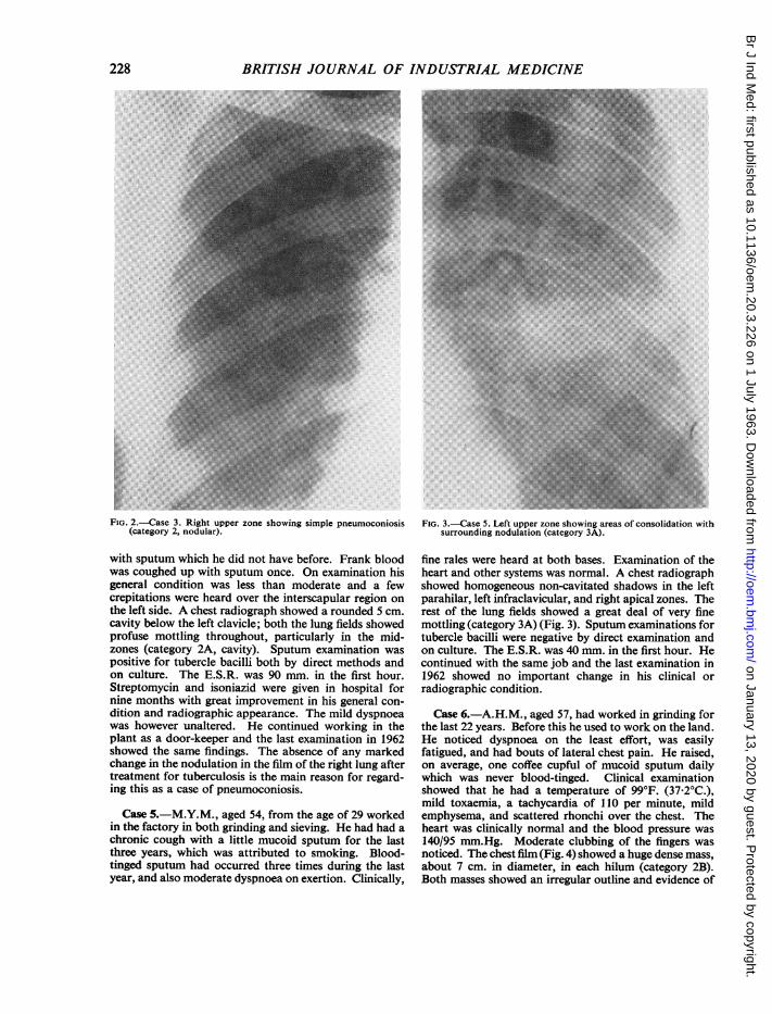
Case 5.-M.Y.M., aged 54, from the age of 29 workedin the factory in both grinding and sieving. He had had achronic cough with a little mucoid sputum for the lastthree years, which was attributed to smoking. Blood-tinged sputum had occurred three times during the lastyear, and also moderate dyspnoea on exertion. Clinically,
FIG. 3.-Case 5. Left upper zone showing areas of consolidation withsurrounding nodulation (category 3A).
fine rales were heard at both bases. Examination of theheart and other systems was normal. A chest radiographshowed homogeneous non-cavitated shadows in the leftparahilar, left infraclavicular, and right apical zones. Therest of the lung fields showed a great deal of very finemottling (category 3A) (Fig. 3). Sputum examinations fortubercle bacilli were negative by direct examination andon culture. The E.S.R. was 40 mm. in the first hour. Hecontinued with the same job and the last examination in1962 showed no important change in his clinical orradiographic condition.
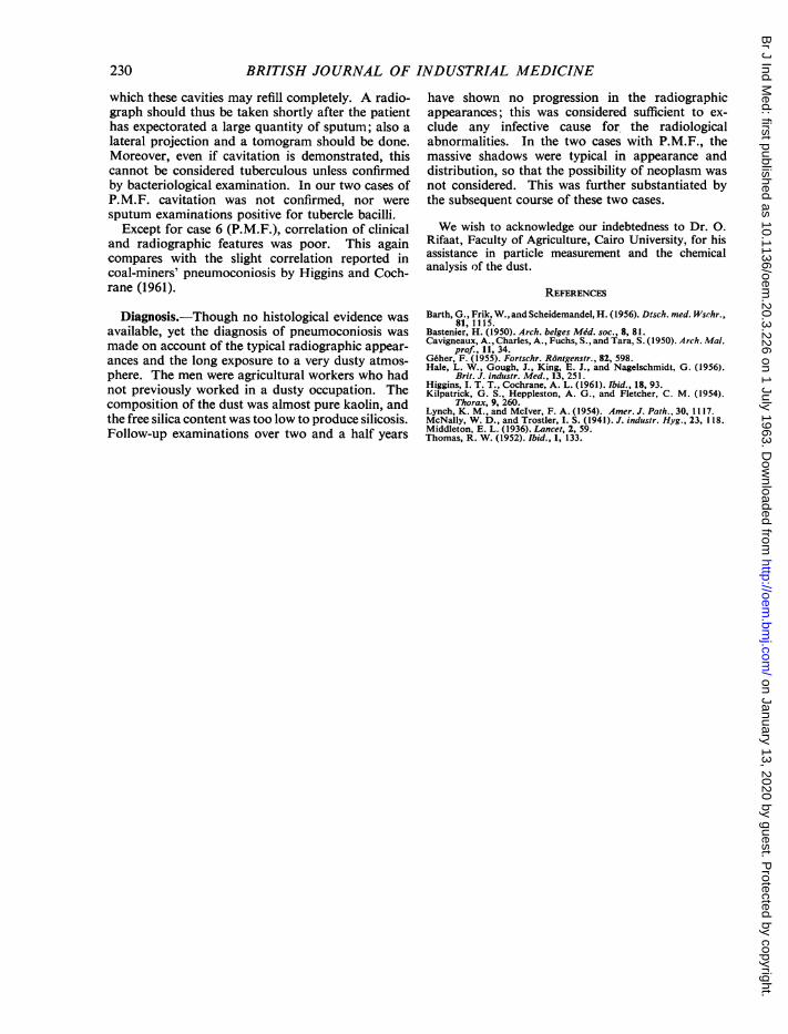
Case 6.-A.H.M., aged 57, had worked in grinding forthe last 22 years. Before this he used to work on the land.He noticed dyspnoea on the least effort, was easilyfatigued, and had bouts of lateral chest pain. He raised,on average, one coffee cupful of mucoid sputum dailywhich was never blood-tinged. Clinical examinationshowed that he had a temperature of 99°F. (37 2°C.),mild toxaemia, a tachycardia of 110 per minute, mildemphysema, and scattered rhonchi over the chest. Theheart was clinically normal and the blood pressure was140/95 mm.Hg. Moderate clubbing of the fingers wasnoticed. The chest film (Fig. 4) showed a huge dense mass,about 7 cm. in diameter, in each hilum (category 2B).Both masses showed an irregular outline and evidence of
228
on January 13, 2020 by guest. Protected by copyright.
http://oem.bm
j.com/
Br J Ind M
ed: first published as 10.1136/oem.20.3.226 on 1 July 1963. D
ownloaded from

PNEUMOCONIOSIS IN CHINA-CLA Y WORKERS
FIG. 4.-Case 6. Right mid and upper zone showing large area ofconsolidation (category 2B).
breakdown was noticed. The rest of the lung fields was
irregularly and lightly mottled, with areas of focalemphysema. Sputum examination was repeatedly nega-tive for tubercle bacilli by direct methods and on culture.The E.S.R. ranged between 70 and 110 mm. in the firsthour. He continued working in the same process for twomonths in spite of his disabling dyspnoea. From January,1960, he took to bed with marked weakness and wastingtogether with episodes of "bronchitis". He died on June30, 1960, and a diagnosis of cor pulmonale was made.Permission for necropsy was not obtained.
Discussion
Industrial Hazards.-China-clay is prepared forlarge-scale use in the pottery industry as an import-ant constituent of chinaware and also in the manu-
facture of refractory materials. Several processes are
carried out in its production but the most dusty ones
are grinding and sieving which were done in a closedchamber. Masks were available but were not used bythe workers. Chemical analysis of dust samplescollected at the mill showed that its chief constituent(83 to 86 %) was potassium aluminium silicate5
(K4 A12 Si2 09); there was only 1 to 20% free silica.Microscopical examination of the same dust showedall particles were below 3 4 microns. The exposuretime of the workers diagnosed as having pneumo-coniosis varied from 15 to 25 years of 8 hours workdaily. No quantitative samples of airborne dust wereobtained. The factory was visibly very dusty.
Clinical Features.-While no complaint was ob-tained in two cases, the remaining four men hadsymptoms. These were mainly dyspnoea on exertion,cough, and slight mucoid sputum of no character-istic colour. In case 6, dyspnoea was marked, andone coffee-cupful of sputum was raised per day.This is a case of progressive massive fibrosis (P.M.F.)with probable cavitation. Repeated blood-tingedsputum occurred in case 5 (P.M.F.), while frankblood-spitting occurred once in case 4 (pneumo-coniosis with tuberculosis). Clubbing of the fingerswas only noticed in case 6 (P.M.F.).
Clinical examination was negative in cases 2, 3,and 5, a few rhonchi on both sides were heard in case1, and crepitations were present in case 4 who had atuberculous cavity. The raised E.S.R. in three casescan be partly explained by various degrees ofanaemia due to endemic infections of the intestinaland urinary tracts by parasites (schistosomiasis,ancylostomiasis, and amoebiasis) since all the menwere ex-landworkers. Sputum examinations werenegative for tubercle bacilli in all cases except case 4.
Radiographic Examination.-Hale et al. (1956),reporting on china-clay pneumoconiosis in Cornwall,mentioned that lesions were in the form of nodularmottling comparable in appearance to those ofsilicosis, or P.M.F. as seen in coal-miners' pneumo-coniosis.The radiographic patterns observed in the present
series conform mostly with those reported by Hale.The mottling was not particularly dense, as describedby McNally and Trostler (1941) in workmen exposedto fuller's earth; neither was there any elevation ofhilar shadows or strand-like opacities, as describedby Geher (1955) and by Barth, Frik, and Scheide-mandel (1956) in pneumoconiosis due to aluminiumdust. Case 4, whose condition was complicated bytuberculosis, had a nodular type of lesion. Theappearance and location of the confluent opacities incases 5 and 6 were comparable to those of P.M.F. incoal-miners. In these two cases cavitation was notconfirmed, though in case 6 there was a suspicion ofcavitation on account of the breakdown shown bythe postero-anterior radiograph as well as thequantity of sputum. Kilpatrick, Heppleston andFletcher (1954) reported that cavitation may not bedetected in P.M.F. because of the remarkable way in
229
.n A
.. ... ... ..........
on January 13, 2020 by guest. Protected by copyright.
http://oem.bm
j.com/
Br J Ind M
ed: first published as 10.1136/oem.20.3.226 on 1 July 1963. D
ownloaded from
BRITISH JOURNAL OF INDUSTRIAL MEDICINE
which these cavities may refill completely. A radio-graph should thus be taken shortly after the patienthas expectorated a large quantity of sputum; also alateral projection and a tomogram should be done.Moreover, even if cavitation is demonstrated, thiscannot be considered tuberculous unless confirmedby bacteriological examination. In our two cases ofP.M.F. cavitation was not confirmed, nor weresputum examinations positive for tubercle bacilli.
Except for case 6 (P.M.F.), correlation of clinicaland radiographic features was poor. This againcompares with the slight correlation reported incoal-miners' pneumoconiosis by Higgins and Coch-rane (1961).
Diagnosis.-Though no histological evidence wasavailable, yet the diagnosis of pneumoconiosis wasmade on account of the typical radiographic appear-ances and the long exposure to a very dusty atmos-phere. The men were agricultural workers who hadnot previously worked in a dusty occupation. Thecomposition of the dust was almost pure kaolin, andthe free silica content was too low to produce silicosis.Follow-up examinations over two and a half years
have shown no progression in the radiographicappearances; this was considered sufficient to ex-clude any infective cause for the radiologicalabnormalities. In the two cases with P.M.F., themassive shadows were typical in appearance anddistribution, so that the possibility of neoplasm wasnot considered. This was further substantiated bythe subsequent course of these two cases.
We wish to acknowledge our indebtedness to Dr. 0.Rifaat, Faculty of Agriculture, Cairo University, for hisassistance in particle measurement and the chemicalanalysis of the dust.
REFERENCES
Barth, G., Frik, W., and Scheidemandel, H. (1956). Dtsch. med. Wsrhr.,81, 1115.
Bastenier, H. (1950). Arch. belges Mdd. soc., 8, 81.Cavigneaux, A., Charles, A., Fuchs, S., and Tara, S. (1950). Arch. Mal.
prof., 11, 34.Geher, F. (1955). Fortschr. Rontgenstr., 82, 598.Hale, L. W., Gough, J., King, E. J., and Nagelschmidt, G. (1956).
Brit. J. industr. Med., 13, 251.Higgins, I. T. T., Cochrane, A. L. (1961). Ibid., 18, 93.Kilpatrick, G. S., Heppleston, A. G., and Fletcher, C. M. (1954).
Thorax, 9, 260.Lynch, K. M., and Mclver, F. A. (1954). Amer. J. Path., 30, 1117.McNally, W. D., and Trostler, I. S. (1941). J. industr. Hyg., 23, 118.Middleton, E. L. (1936). Lancet, 2, 59.Thomas, R. W. (1952). Ibid., 1, 133.
230
on January 13, 2020 by guest. Protected by copyright.
http://oem.bm
j.com/
Br J Ind M
ed: first published as 10.1136/oem.20.3.226 on 1 July 1963. D
ownloaded from