Embed Size (px)
Citation preview

PNEUMONIA Diagnosis Definition & Explanation
of Diagnosis Etiology & Risk Factors Incidences Among
Gender, Age, & Ethnicity Prognosis Signs & Symptoms
By:Marjorie Johnson

definition
Pneumonia: inflammation of the respiratory bronchioles and alveoli
Either infectious or non-infectious
Infectious: bacteria, viruses, fungi, protozoa and other microbes
Non-infectious: aspiration of gastric contents and inhalation of toxic or irritating gases

DIAGNOSIS
Pneumonia is often diagnosed with difficult breathing because the airway is filled with mucus and fluid
A person with pneumonia often produces delicate crackling noises or wheezing in the area of the lung affected by pneumonia
In a lung with pneumonia, the sound can be dull or muffled because the air sacs are filled with fluid instead of air
A doctor may take a chest x-ray to confirm a diagnosis of pneumonia

Other risk factors
People with problems with heart, liver, or kidneys.
Patient with HIV/AIDS whose immune system are very weak.
Patient who taking medication that weaken the immune system such as cancer patients.
Patients that smoke, abuse alcohol.
Exposure to certain chemicals, past surgery
Patients hospitalized in an intensive care of certain disease (ex: coma)

prognosis
More than a million people are hospitalized each year for pneumonia
1 in 4 people who had pneumonia died
Community-acquired pneumonia is responsible for 350,000 to 620,000 hospitalizations
Older adults have lower survival rates than younger people
Pneumonia kills between 40,000 to 70,000 people each year

Signs & Symptoms
Most people who develop pneumonia initially have symptoms of a cold which are followed by:
High feverShakingChillsSharp chest painTachycardiaTachypneaCough with sputum
production

PNEUMONIA Diagnostic tests (include
rationale & nursing implications
Medical Treatment and Nursing Implications
Surgical Treatment and Nursing Implications
Prevention Complications Discharge/Client
teaching
By:Stacy Moyston-Duckie

DIAGNOSTIC TESTS
• Diagnostic tests are usually done by a medical doctor who would examine the client by auscultations of the lungs for evidence of delicate crackling noises or wheezing in the area that is infected. In a lung with pneumonia, the sound can be dull or muffled because the air sacs are filled with fluid instead of air.
• Based on these findings the doctor will order certain test to be done to identify the organism that is responsible for this illness.
DIAGNOSTIC TESTS
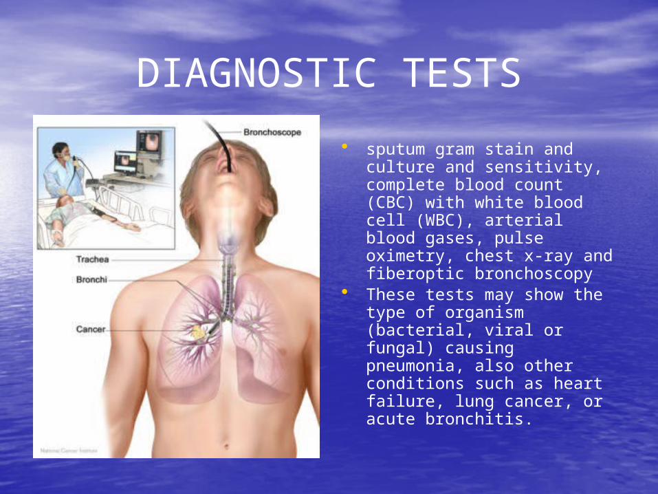
• sputum gram stain and culture and sensitivity, complete blood count (CBC) with white blood cell (WBC), arterial blood gases, pulse oximetry, chest x-ray and fiberoptic bronchoscopy
• These tests may show the type of organism (bacterial, viral or fungal) causing pneumonia, also other conditions such as heart failure, lung cancer, or acute bronchitis.

Medical treatmentWith medical treatment the patient with pneumonia is
generally treated with: Immunization: Pneumococcal vaccine is recommended for people
over age 65, for immunocompromised people, and for those with chronic cardiac or respiratory conditions, diabetes mellitus, alcoholism, or other chronic disease.
Medications: Medications used to treat pneumonia may include antibiotics to eradicate causative organisms and bronchodilators to reduce bronchospam and improve ventilation.
Oxygen therapy: Oxygen may be ordered when pneumonia interferes with gas exchange. The nasal cannula delivers 24% to 45% oxygen (room air is 21% oxygen) with flow rates of 2 to 6 liters per minute.
Other therapies: Increasing fluid intake to 2,500 to 3,000 mL/day helps liquefy secretions, making them easier to cough up and expectorate.
Another effective method is percussion, which is done by rhythmically striking or clapping the chest wall with cupped hands. Cupping traps air between the palm and the skin, causing vibrations that loosen respiratory secretions. Postural drainage uses gravity to help remove secretions from a particular lung segment.

SURGICAL TREATMENT
• Thoracotomy is the standard surgery for pneumonia.
• It requires general anesthesia and an incision to open the chest and view the lungs. The surgeon most times has to remove dead or damaged lung tissue. In extreme cases the entire lobe of the lung is removed.

Treatment and nursing implications
• Clients being treated for pneumonia need therapeutic care in order to successfully recover. The priority of nursing care for clients with pneumonia is its effects on the client’s ability to maintain open airways and on the exchange of gases in the alveoli.
• The nurse must always wash his/her hands and wear gloves, if indicated. Hand washing and gloves help to prevent the spread of infection.
• If the person has difficulty breathing, he or she is given oxygen, usually by mask or cannula. Adjust the client’s position.
• An orthopneic position may be necessary. Proper positioning helps the person to be more comfortable and to breathe more easily. Place a pillow lengthwise under the back. This action encourages fuller chest expansion.
• Assess the client’s vital signs at least every 4 hours. Frequent monitoring is necessary to allow for prompt detection and early intervention if problems arise.

PREVENTION• There are a number of steps a
person can take to help prevent getting pneumonia.
• Stop Smoking!!! Smoking puts a smoker at high risk in getting pneumonia.
• One must avoid contact with people who suffer from respiratory tract infections, such as colds and influenza (flu), measles and chickenpox.
• Pneumonia may develop or put the client at risk for developing these types of infections. Vaccination is given mostly to adults over 65 years of age who are at greater risk of developing pneumonia as well as for younger people who are chronically ill.

PREVENTION
• Also daily diets should include foods such as fresh, dark-colored fruits and vegetables, which are rich in antioxidants and other important food chemicals that help, boost the immune system.
• Children are given vaccination against certain diseases such as measles, and chickenpox. Mineral zinc may also reduce the risk of pneumonia in children. Zinc can be found in foods that we eat such as, seafood, lean red meats, beans, and cereals.

complications
• Clients with pneumonia must be treated immediately to avoid serious complications such as by way of entering in the bloodstream or by cross- contamination to other parts of the body.
• Without proper treatment, pneumonia can lead to such life-threatening complications as septic shock, hypoxemia, and respiratory failure. The infection can also spread within the patient’s lungs, causing empyema or lung abscess.

Discharge/client teaching• It is very important that the
nurse educate the client about the disease and how to do self-care.
• The nurse must explain all procedures (especially intubation and suctioning) to the patient and his family. Emphasize the importance of adequate rest to promote full recovery and prevent a relapse.
• Stress the need to take the entire course of medication, even if he feels better, to prevent a relapse.
• Teach the patient procedures to clear lung secretions, such as deep-breathing and coughing exercises, as well as home oxygen therapy. Explain deep breathing and pursue-lip breathing.

Congestive heart failure Yvonne Prempeh
Medical Surgical Nursing 1
July 23, 2007

Objectives
Definition and explanation of congestive heart failure
Epidemiology: incidences among gender, age and ethnicity
Etiology and risk factors Diagnosis of CHF

Definition and explanation of CHF structural or functional cardiac
disorder impairing the filling and pumping ability of the heart
highly specialized muscular organnormal heart pump 50% of total bloodfailing heart pumps </= 40% of total blood

Epidemiology
Younger men are more likely than women to develop myocardial infarction
People >/= 65 years have higher risks for CHF
48,500 heart attacks are associated with CHF 23,500 heart attacks occur in women

Epidemiology cont’d
Asians and Latinos tend to suffer least cases of cardiovascular diseases
African Americans are at a higher risk for developing severe CHF
recurrent hospitalization rate increased mortality rate due to severe CHF
( www.american heart.org; National Institutes of Health)

Etiology and risk factors
Causes of CHF: myocardial ischemia hypertension various types of arrhythmias anemia hyperthyroidism hypothyroidism diabetes mellitus endocarditis myocarditis congenital heart defects artherosclerosis cardiomyopathy

Example of cardiomyopathy

Etiology & Risk Factors

Etiology and risk factor cont’d
increased cholesterol intake increased intake of fats increased sodium intake lack of exercise morbid obesity diabetes mellitus hypertension

Diagnosis of CHF
Targeted history and physical exam Tests:
BNP ( B-type Natriuretic Peptide)
normally secreted in ventricles during filling and stretching
BNP < 100 pg/ml : No CHF BNP between 100-300 pg/ml: suggestive CHF BNP > 600 pg/ml: moderate CHF BNP > 900pg/ml: severe CHF
(www.clevelandclinic.org/heart center)

Diagnosis cont’d
Tests cont’d: Serial EKG’s and cardiac iso-enzymes q6h x 3
Troponin I CK (creatine kinase) CKMB (specific from heart muscle fraction) CKMB Index (calculated by lab)
CBC Chest x-ray: to detect pulmonary edemas a result of edema Two dimensional echocardiogram: to check the pumping
ability of the heart and the function of the heart Stress test to evaluate for myocardial ischemia

Signs and SymptomsMain manifestations to watch out for:
Fatigue and dyspnea ( difficulty breathing) = Due to excess fluid in the body and fluid in the lungs that causes congestion.
= Mostly observable when a person is walking halfway and gets tired easily eventhough he just walk halfway.
Edema or swelling of the ankle and feet) = Due to fluid overload in the tissue which causes congestion.
= mainly observe during the day and every time the feet is used.
Other symptoms include:
Nausea, abdominal pain and decreased appetite.

Diagnostic TestsTests to confirmed the presence of congestive heart failure:
1. EKG or EGC ( Electrocardiogram)
2. X-ray examination
3. Echocardiography
4. Cardiac catherization
Purpose: Evaluate heart function and valve dysfunction.
NURSING IMPLICATION:
Discuss situation with the client the procedure.
Distinguish the possible effects of the procedure to the well-being of the patient.
Rationale:
Discussing will help the client to be more familiar with the procedure hence, it will decrease anxiety.
Client will be able to deal with her fear by the information gathered from the nurse.

Medical TreatmentDigoxin
= Cardioglycoside
= slows heart rate.
= stimulate heart muscle which increases the force of systole.
=Decreased the workload of the heart.
NURSING IMPLICATION:
= Monitor apical pulse before administering the medication.
= If less than 60 bpm, withhold the medication, then notify MD.
Diuretics
=Help fluid from building up
=Decrease fluid retention
= make the fluids run through the kidneys
NURSING IMPLICATION:
Monitor BP, I&O, daily weight.
Assess feet, legs, and sacral area for edema DAILY>

PrognosisCongestive heart failure has a very poor
prognosis.
Only 50% of CHF patients lives within 5 years.
On the other hand, only 20% survive within 8 to 12 years prior to diagnose.
HALF OF THE PATIENT WILL DIE IN 5 years.
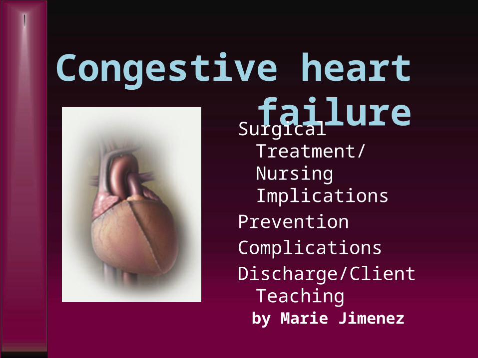
Congestive heart failure
Surgical Treatment/ Nursing Implications
PreventionComplicationsDischarge/Client
Teaching by Marie Jimenez

Surgical Treatment
Types of surgical treatment for CHF:
1. Cardiac transplantation2. Dynamic cardiomyoplasty

Cardiac Transplantation
Primary treatment for end-stage heart failure
Transplanted organs are from young accident victims with no evidence of cardiac trauma
Client or (recipient) heart is removed, leaving posterior atria intact
Donor heart is sutured to the remaining atrial walls

Cardiac Transplant: NURSING
IMPLICATIONS
Risks for surgical treatment of CHF:
Infection Rejection of donor’s
heart
Nursing Implication: Monitor for infection Administering
immunosuppresive drugs to prevent the rejection of the transplanted organ
Route: IV, PO, IM

Dynamic cardiomyoplasty
Dynamic cardiomyoplasty involves using the client’s own skeletal muscle (particularly the latissimus dorsi) to enhance the function of the heart and improve circulation.
The muscle is positioned around the heart or aorta, and a cardiomyostimulator and leads are implanted to stimulate muscle contractions.
This type of surgical procedure may be implemented as an alternative to cardiac transplantation for clients who refuse organ transplantation.

PREVENTION
The prevention of CHF includes modifiable lifestyle changes relating to diet, smoking, alcohol, and exercise
Limit sodium intake Alcoholics Anonymous & Smoking cessation programs Providing awareness of the negative effects of
smoking and alcohol and recommending “regular, moderate exercise to improve their overall fitness” will greatly reduce in the prevention of congestive heart failure. (Merck Manual, 1997)

complications
Hepatomegaly (enlargment of the liver)
Spenomegaly (enlargement of the spleen)
Thrombus & emboli Cardiogenic shock which
“occurs when the left ventricle is unable to supply the tissues with enough oxygen and nutrients to meet their needs. [It] is a life-threatening condition that requires immediate treatment (Williams, 2003, p. 332)
Picture on right: IABP (intra-aortic balloon pump) to increase coronary perfusion

Discharge & client teaching
Nursing interventions for discharge:
Explain heart failure and its effects on the client’s life – this helps the client understand the reasons for ordered treatments
Stress the importance of medications in managing heart failure and provide verbal and written information regarding each specific medication to encourage compliance
Teach the client and family about the prescribed diet
Instruct to keep regular follow-up appointments to monitor disease progression and effects of therapy

Chronic Obstructive Pulmonary Disease (COPD)
Student Name: Na Pang

Nursing Diagnosis:
• Ineffective Airway Clearance
• Imbalanced nutrition: less than body requirements,
• compromised family copying
• Decisional conflict: smoking.

Definition and Explanation of Diagnosis:
• Chronic obstructive pulmonary disease (COPD) is a term referring to two lung diseases, chronic bronchitis and emphysema, that are characterized by obstruction to airflow that interferes with normal breathing. Both of these conditions frequently co-exist, hence physicians prefer the term COPD.

Etiology and Risk Factors:
• Etiology: In COPD, there is decreased elastic recoil of the lungs (mainly emphysema) and increased airway resistance (mainly bronchitis).
• Risk factors: Smoking is the primary risk factor for COPD. Approximately 80 to 90 percent of COPD deaths are caused by smoking.

Incidence Among Gender, Age and Ethnicity:• 1. Chronic bronchitis affects people of all ages, but is higher
in those over 45 years old. • 2. Females are more than twice as likely to be diagnosed
with chronic bronchitis as males. In 2004, 2.8 million males had a diagnosis of chronic bronchitis compared to 6.3 million females.
• 3. Chronic bronchitis prevalence for Hispanics (27.7 per 1,000) was significantly lower than for both Whites (45.7 per 1,000) and Blacks

COPD PROGNOSIS
• None of the existing medications for COPD has been shown to modify the long-term decline in lung function that is the hallmark of this disease. Therefore, the goal of pharmacotherapy for COPD is to provide relief of symptoms and prevent complications and/or progression of the disease with a minimum of side effects.

Signs and symptoms:
• 1. Cough • 2. Sputum (mucus) production • 3. Shortness of breath,
especially with exercise • 4. Wheezing (a whistling or
squeaky sound when you breathe)
• 5. Chest tightness

Chronic Obstructive Pulmonary Disease
Student Practical Nurse, Anaïse Ikama
Spring II 2007

Diagnostic TestsThe following test can be used to diagnose COPD
• Pulmonary function testinga spirometry is used to identifies how much air your lung can hold and how fast you can blow the air out of your lung
• Chest X-ray helps in the identification
of emphysema (causes large abnormal air spaces in the lungs by destroying alveolar walls)

Diagnostic Test
• Serum alpha1-antitrypsin levelsto screen the enzyme deficiency that can cause lung tissue destruction
• Arterial Blood Gasmeasures deficiency of oxygen
• Other test such as a computed Tomography can determine if the client will benefit from surgery

Medical Treatment
• Bronchodilators as prescribed to relax the muscles that wrap around the airwaysallow tubes to become larger and easier to breath through
• Inhaled steroids/corticosteroidsreduces swelling in the breathing tube

Surgical Treatment
• Lung Volume ReductionThe removable of the inflated air sacs that causes healthy air sacs to work poorly
• Lung TransplantReplacement of one or both lungs

Nursing Interventions
Administer inhaler and oxygen as ordered before activity
Assist the pt. with the use of a spirometer, percussion and postural drainage
Reassure and provide emotional support (r/t fear of death)

Prevention
• “Avoiding – never starting or stopping – smoking is the only way to prevent COPD and to its progression” (Burke, Lemone & Mohn-Brown, pp 557).

Complication
• Due to impaired defense mechanisms *Respiratory infection such as influenza
and pneumonia can develop *Increase BP in pulmonary veins *Lung cancer *Feeling of depression (from progressive & incurable disease)

Client Teaching
• Nurses should:a- Teach effective coughing and
breathing mechanism b- Advice client to maintain fluid intake of 2 to 2 ½ quarts daily
c- Discuss aerobic physical exercise (e.g.:
20 mn walkd- Stress the importance of eating
small frequent meal e- Reinforce teaching about prescribed
medication

REMEMBER
TO TEACH YOUR CLIENT TO NEVER SMOKE

Thoracotomy
By Janell TrotmanPetra Ramnarine

- surgery done by an incision into the thorax or chest cavity to perform surgery on the lungs
- incision is made between the ribs and all or part of the infected lung is removed depending on the location, size and type of lung cancer that is present
Eitology
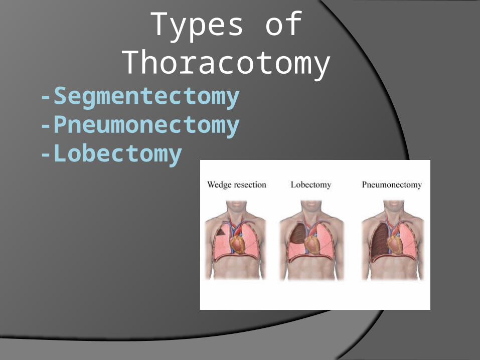
-Segmentectomy -Pneumonectomy -Lobectomy
Types of Thoracotomy
- Ineffective breathing pattern- Ineffective airway clearance- Ineffective gas exchange- Pain: Chest
Nursing Diagnosis

Nursing Implication Assess client for s&s of impaired res.
function. (rapid, shallow or slow respiration)
Perform action to reduce pain Perform actions to maintain patency and
integrity of chest drainage system if present
Maintain oxygen therapy as ordered Administer bronchodilators as ordered

Risk Factors Smoking