Embed Size (px)
Citation preview
FACTA UNIVERSITATIS Series: Medicine and Biology Vol.14, No 2, 2007, pp. 98 - 100 UC 616.12-053.31
PNEUMOPERICARDIUM: A POSSIBLE RARE CAUSE OF NEONATAL DEATH
Radovan Karadžić, Aleksandra Antović, Goran Ilić, Lidija Kostić-Banović
Institute of Forensic Medicine, Faculty of Medicine, Niš, Serbia E-mail: [email protected]
Summary. The aim of this paper is to point to the dangers of pneumopericardium (PPC), which is defined as a collection of air or gas in the pericardial cavity. PPC belongs to the most dangerous kinds of extra-alveolar air and only physicians' awareness of that condition and immediate air evacuation (pericardiocentesis) prevents the deleterious results. Neonatal pneumopericardium is a rare clinical condition. We report a case of neonatal pneumopericardium with the fatal outcome, occurring in the presence of significant lung pathology (in utero meconial aspiration, pulmonary hypertension) and a history of neonatal resuscitation. The patophysiological mechanism is the obstruction of large and small airways with the aspirated material and consecutive air trapping distal to obstruction, over distension and rupture of alveolar walls, allowing gas to escape and dissect along perivascular and peribronchial sheaths towards pericardial sac within which this air accumulates. Mechanically ventilated patients are particularly at risk for tension pneumopericardium (TPPC) secondary to barotrauma. Acute cardiac shock, pulsus paradoxus and decreased systolic blood pressure (the heart is ineffectual pump), muffled heart sounds and a metallic sound of high frequency refer to the presence of a pneumopericardium. Tension pneumopericardium is a life threatening condition, so early recognition and intervention (pericardiocentesis) is very important.
Key words: Pneumopericardium, neonatal, meconial aspiration, death
Introduction
Pneumopericardium is defined as a collection of air or gas in the pericardial cavity and was first described by Bricheteau in 1844. Many cases have since been re-ported, mostly due to blunt or penetrating chest injuries in adults and due to respiratory distress syndrome com-bined with mechanical ventilation in infants (1, 2).
Neonatal pneumopericardium is a rare clinical condi-tion that usually occurs in association with other air leak syndromes (pneumomediastinum, pneumothorax, pneu-moperitoneum, subcutaneous and interstitial emphysema) especially when there is severe lung pathology, post vig-orous resuscitation, or in the presence of assisted ventila-tion (3, 4, 5). We report a case of neonatal pneumoperi-cardium occurring in the presence of significant lung pathology (in utero meconial aspiration, pulmonary hy-pertension) and a history of neonatal resuscitation.
The autopsy was performed at the Court's request, in order to determine the cause of death of the newborn but also the presence of a doctor's criminal responsibility elements. Also, the autopsy was requested by the doctor, since the death of the newborn occurred soon after the delivery and the cause of death remained unclarified.
Patients and Methods In order to present the case, we use the available
medical files collected during hospital treatment of a female newborn, as well as complete autopsy findings
taken during the examination. The autopsy of the body was done 12 hours after the death. Routine histology of the sample was carried out after fixation in 10% buff-ered formalin. After that, the tissues were embedded in paraffin, and the paraffin samples were stained with haematoxylin-eosin (HE).
Results According to the medical data, the woman deliver-
ing a child, multipara (who, by the anamnesis, has one healthy child and one spontaneous abortion), was ad-mitted to hospital 7 to 10 days prior to the expected de-livery term. Not long after her being admitted, the pro-fusion bleeding occurred from the birth canals due to premature detachment of the placenta. Tocographic finding showed a slow and arrhythmic heart rate of the offspring. The delivery was conducted by caesarean section. On its birth the child was 3150 g heavy, 53 cm long, with the signs of pale asphyxia (asphyxia pallida), and according to APGAR score it was marked by 1. The aspiration, intubations, assisted mechanical ventilation, medicament therapy and exterior cardiac massage were immediately performed. Although all the measures of the intensive CPR (cardio pulmonary resuscitation) were conducted, the lethal outcome occurred 35 minutes after the birth.
By the autopsy it was found that the newborn was mature female in X lunar month of the maternal life. The skin was explicitly pale, and the umbilical cord
PNEUMOPERICARDIUM: A POSSIBLE RARE CAUSE OF NEONATAL DEATH 99
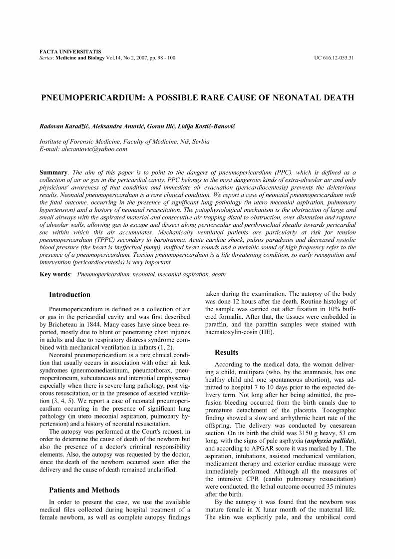
tied. The pericardial sac was filled with air, tense and it resembled fish swim bladder. The findings on the other organs were regular. Pathohistological finding on the lungs shows signs of meconial aspiration and ruptured alveolar walls (Figure 1) and persistent lung hyperten-sion (Figure 2). Apart from stress-involution of thymus and hydropic-vacuolar degeneration of the liver tissue, other pathological conditions in the internal organs have not been found. Injuries to the body have not been found either, except for injections needle wounds. On the basis on the clinical and post-mortem findings, the death in this case was attributed to cardiac tamponade present in the air of pericardial sac.
Fig. 1. Histopathology in fatal neonatal
pneumopericardium: presence of aspirated contents (meconium, vernix caseosa, cellular debridement) into small airways and alveoli; ruptured alveolar walls (HE staining, original magnification 100x).
Fig. 2. Histopathology in fatal neonatal
pneumopericardium: thick muscle layer of small lung arterioles-pulmonary hypertension (HE staining, original magnification 100x).
Discussion Meconium in the amniotic fluid occurs in approxi-
mately 12% of term-birth neonates. As a consequence, meconium aspiration is considered to be a relatively common event. Other substances such as blood or am-niotic fluid can also be aspirated. The aspiration syn-drome of meconium is fatal in 40% of the cases. Pul-monary hypertension usually develops when meconium aspiration occurs in conjunction with varying degrees of in-utero asphyxia. Damaged lungs are incapable of dis-carding inhaled meconium due to the damaged cilia of epithelial cells of respiratory airways so that circulus vitiosus occurs within which vasoconstriction aggra-vates the hypoxia and vasospasm (6).
The patophysiological mechanism is the obstruction of large and small airways with the aspirated material (meconium, blood, amniotic fluid contents) and con-secutive air trapping distal to obstruction. Following, an increase in intra-alveolar pressure with alveolar over distension results in rupture of marginal alveolar walls, allowing gas escape into the perivascular space (6, 7). The escaping air travels through pulmonary interstitium dissecting the peribronchial and perivascular sheaths, with resulting presence of air into the pleural space (pneumotorax) or in the hilum, where air dissects be-tween the fascial sheath and moves into the mediasti-num (pneumomediastinum) and/or pericardium (pneu-mopericardium). Pericardial connective tissue is dis-continuous at the reflection of parietal onto visceral pericardium near the ostia of the pulmonary veins so that there is a site of potential weakness where a micro-scopic dissection of air into the pericardial sac is possi-ble (4, 8, 9, 10).
A complication of ventilatory management of neo-natal respiratory distress also may result in cardiac tam-ponade. Mechanically ventilated patients are particu-larly at risk for pneumopericardium secondary to baro-trauma (alveoli rupture when interstitial pressures ex-ceed airway parenchyma pressures, and air dissects along perivascular and peribronchial sheaths towards pericardial sac) (11, 12).
Early recognition and intervention for tension pneumopericardium are very important. Clinical find-ings include acute haemodynamic deterioration with acute cardiac shock, dyspnoea, cyanosis, bradycardia or tachycardia, pulsus paradoxus and decreased systolic blood pressure (the heart is ineffectual pump); muffled heart sounds and a metallic sound of high frequency refer to the presence of a pneumopericardium (12, 13).
Roentgen graphically, posteroanterior and lateral ro-entgenograms demonstrate that, with pneumopericar-dium, air is confined to the space immediately around the heart. Treatment includes preventing further air en-try into the pericardium and decompressing pericardial space by using pericardiocentesis or tube drainage for all neonates because of the high risk of re-accumulation of air (3, 9, 14).
100 R. Karadžić, A. Antović, G. Ilić, L. Kostić-Banović
Conclusion This paper shows that the pneumopericardium be-
longs to the most dangerous kinds of extra-alveolar air
and only physicians' awareness of that condition and immediate air evacuation (pericardiocentesis) prevents the deleterious results.
References1. Cummings RG, Wesly RL, Adams DH, Lowe JE. Pneumopericar-
dium resulting in cardiac tamponade. Ann Thorac Surg 1984; 37: 511-18.
2. Westermann GW, Suwelack B. Spontaneous pneumopericardium due to exertion. Southern Medical Journal 2003; 96(1): 50-52.
3. Fellous L, Tourneux P, Brule-Pepin R, Goissen C, Krim G. Pneu-mopericardium: a rare complication of meconium aspiration syn-drome. Archives de Pediatrie 2005; 12(1): 83.
4. Rucker J. Pneumopericardium in hyaline membrane syndrome in premature infants. Pediatr. Padol. 1987; 22(1): 51-58.
5. Kumar A, Bhatnagar V. Respiratory distress in neonates. Indian J Pediatr 2005; 72(5): 425-28.
6. Mihailović D. Opšta pedijatrijska patologija. In: Mihailović D, Stojanović D (eds), Osnovi pedijatrijske patologije. 1st ed. Prosveta, Niš, 2002: 13-16.
7. Varano LA, Maisels MJ. Pneumopericardium in the newborn: diagnosis and pathogenesis. Pediatrics 1974; 53(6): 941-45
8. Mirzayan R, Cepkinian V, Asensio JA. Subcutaneous emphysema, pneumomediastinum, pneumothorax, pneumopericardium and
pneumoperitoneum from rectal barotrauma. The Journal of Trauma-Injury, Infection and Critical Care 1996; 41(6): 1073-75.
9. Fisher DH, Walker WM. Tension pneumopericardium in an infant. Chest 1992; 102: 1888-91.
10. Toledo TM, Moore WL, Nash DA, North RL. Spontaneous pneu-mopericardium in acute asthma: case report and review of the lit-erature. Chest 1972; 62: 118-120.
11. Emery RW, Foker J, Thompson TR. Neonatal pneumopericardium: a surgical emergency. Annals of Thoracic Surgery 1984; 37(2): 128-32.
12. Auden SM, Lapin SL, Joseph-Reynolds AM. Cardiac arrest from tension pneumopericardium in a premature infant. Anesth. Analg. 2002; 95(5): 1245-47.
13. Reppert SM, Ment LR. The treatment of pneumopericardium in the newborn infant. J Pediatr. 1977; 90(1): 115-7.
14. Galenski JA, Hali RT. Neonatal pneumopericardium: analysis of ventilatory variables. Crti Care Med, 1984; 12: 439-42.
PNEUMOPERIKARD: MOGUĆI REDAK UZROK SMRTI NOVORODJENČETA
Radovan Karadžić, Aleksandra Antović, Goran Ilić, Lidija Kostić-Banović
Zavod za sudsku medicinu, Medicinski fakultet, Niš E-mail: [email protected]
Kratak sadržaj: Cilj ovog rada je da ukaže na opasnost od pneumoperikarda (PPK), koji se definiše kao kolekcija vazduha ili gasa u srčanoj kesi. PPK predstavlja najopasniji vid ekstra-alveolarnog prodora vazduha i samo oprez i svest lekara o ovom stanju, kao i urgentna evakuacija vazduha (perikardiocenteza) prevenira štetan ishod. Pneumoperikard kod novorođenčadi predstavlja relativno retko kliničko stanje. U ovom radu opisali smo slučaj pneumoperikarda novorođenčeta sa fatalnim ishodom, koji je nastao na terenu izražene plućne patologije (mekonijalna aspiracija in utero, plućna hipertenzija) i izvršene kardio-pulmonalne reanimacije novorođenčeta. Patofiziološki mehanizam nastanka PPK ogleda se u opstrukciji velikih i malih disajnih puteva aspiriranim materijalom sa konsekutivnim zarobljavanjem vazduha distalno od opstrukcije, prekomernom distenzijom i rupturom alveolarnih zidova, izlaskom vazduha i raslojavanjem perivaskularnih i peribronhijalnih omotača sve do srčane kese i akumulacijom vazduha u njoj. Mehanički ventiliranim pacijentima preti povećani rizik od nastanka tenzionog pneumoperikarda (TPPK), a kao posledica barotraume. Akutni kardiogeni šok, paradoksni puls i smanjen sistolni krvni pritisak (srce je neefikasna pumpa), potmuli srčani tonovi i metalni zvuk visoke frekvencije odgovaraju pneumoperikardu. Tenzioni pneumoperikard predstavlja po život opasno stanje, pa su rano prepoznavanje i intervencija (perikardiocenteza) od presudnog značaja u terapijskom smislu.
Ključne reči: Pneumoperikard, novorođenče, aspiracija mekonijuma, smrt.