Embed Size (px)
Citation preview
1
Polycythemia in Neonates
Polycythemia or an increased hematocrit is associated with hyperviscosity of blood. As the blood viscosity increases, there is impairment of tissue oxygenation and perfusion and tendency to form microthrombi. Significant damage may occur if these events occur in the cerebral cortex, kidneys and adrenal glands. Hence this condition requires urgent diagnosis and prompt management. The viscosity of blood is directly proportional to the hematocrit and plasma viscosity and inversely proportional to the deformability of red blood cells. Symptoms of hypoperfusion correlate better with viscosity as compared to hematocrit. Viscosity is, however, difficult to measure at the bedside. Hyperviscosity is therefore suspected in the presence of an abnormally high hematocrit with or without suggestive symptoms. Relationship between viscosity and hematocrit is almost linear up to a hematocrit of 65% and exponential thereafter.1,2 The polycythemia-hyperviscocity syndrome is thus usually confined to infants with hematocrits of more than 65%; it is very rare with hematocrits of <60%. Definition A diagnosis of polycythemia is made in the presence of a venous hematocrit more than 65% or a venous hemoglobin concentration in excess of 22 gm/dL. Hyperviscosity is defined as a viscosity greater than 14.6 centipoise at a shear rate of 11.5 per second.3 Incidence The incidence of polycythemia is 1.5% to 4% of all live births.4,5 The incidence is higher among both small for gestational age (SGA) and large for gestational age (LGA) infants. The incidence of polycythemia is 15% among term SGA infants as compared to 2% in term AGA infants.6 Neonates born at high altitudes also have a higher incidence of polycythemia.1 Maternal smoking is an important risk factor for polycythemia.7 Term neonates born to mothers engaged in smoking during pregnancy are 2.5 times more likely to require a partial exchange transfusion for polycythemia than the counterparts of non-smoker mothers.7 Infants born by cesarean section have a lower hematocrit values than those delivered vaginally.8 Infants subjected to delayed cord clamping carry nearly four times greater risk of asymptomatic polycythemia.9 In the last 3 years the incidence of polycythemia ranged from 0.95% to 1.5% in our centre. Physiological changes in postnatal life Significant changes take place in the hematocrit from birth through the first 24 to 48 hr of life. The hematocrit peaks at 2 hr of age and values up to 71% may be normal at this age.10-11 It gradually declines to 68% by 6 hr and usually stabilizes by 12 to 24 hr. The initial rise in hematocrit is related to a transudation of fluid out of the intravascular space.
2
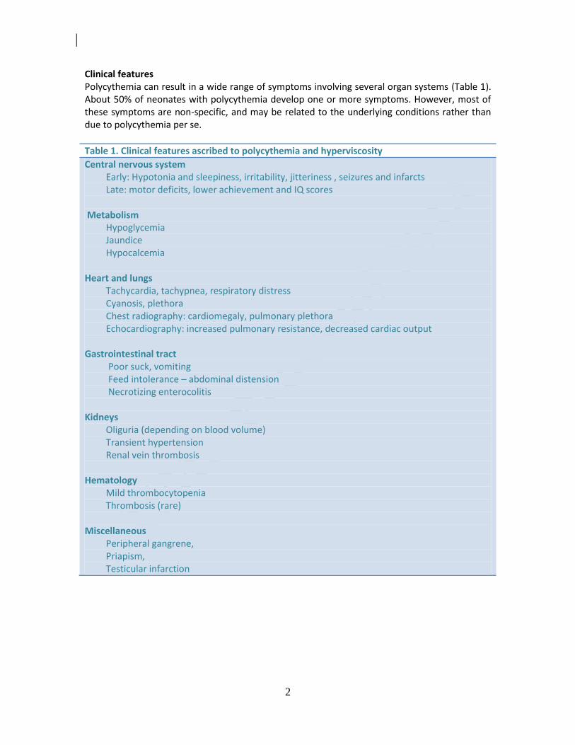
Clinical features Polycythemia can result in a wide range of symptoms involving several organ systems (Table 1). About 50% of neonates with polycythemia develop one or more symptoms. However, most of these symptoms are non-specific, and may be related to the underlying conditions rather than due to polycythemia per se.
Table 1. Clinical features ascribed to polycythemia and hyperviscosity
Central nervous system Early: Hypotonia and sleepiness, irritability, jitteriness , seizures and infarcts Late: motor deficits, lower achievement and IQ scores
Metabolism
Hypoglycemia Jaundice Hypocalcemia
Heart and lungs Tachycardia, tachypnea, respiratory distress Cyanosis, plethora Chest radiography: cardiomegaly, pulmonary plethora Echocardiography: increased pulmonary resistance, decreased cardiac output
Gastrointestinal tract Poor suck, vomiting Feed intolerance – abdominal distension Necrotizing enterocolitis
Kidneys Oliguria (depending on blood volume) Transient hypertension Renal vein thrombosis
Hematology Mild thrombocytopenia Thrombosis (rare)
Miscellaneous Peripheral gangrene, Priapism, Testicular infarction
3
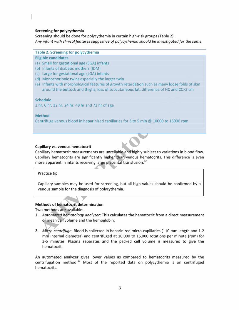
Screening for polycythemia Screening should be done for polycythemia in certain high-risk groups (Table 2). Any infant with clinical features suggestive of polycythemia should be investigated for the same.
Table 2. Screening for polycythemia
Eligible candidates (a) Small for gestational age (SGA) infants (b) Infants of diabetic mothers (IDM) (c) Large for gestational age (LGA) infants (d) Monochorionic twins especially the larger twin (e) Infants with morphological features of growth retardation such as many loose folds of skin
around the buttock and thighs, loss of subcutaneous fat, difference of HC and CC>3 cm Schedule 2 hr, 6 hr, 12 hr, 24 hr, 48 hr and 72 hr of age
Method Centrifuge venous blood in heparinized capillaries for 3 to 5 min @ 10000 to 15000 rpm
Capillary vs. venous hematocrit Capillary hematocrit measurements are unreliable and highly subject to variations in blood flow. Capillary hematocrits are significantly higher than venous hematocrits. This difference is even more apparent in infants receiving large placental transfusion.12 Methods of hematocrit determination Two methods are available: 1. Automated hematology analyzer: This calculates the hematocrit from a direct measurement
of mean cell volume and the hemoglobin.
2. Micro-centrifuge: Blood is collected in heparinized micro-capillaries (110 mm length and 1-2 mm internal diameter) and centrifuged at 10,000 to 15,000 rotations per minute (rpm) for 3-5 minutes. Plasma separates and the packed cell volume is measured to give the hematocrit.
An automated analyzer gives lower values as compared to hematocrits measured by the centrifugation method.13 Most of the reported data on polycythemia is on centrifuged hematocrits.
Practice tip Capillary samples may be used for screening, but all high values should be confirmed by a venous sample for the diagnosis of polycythemia.
4
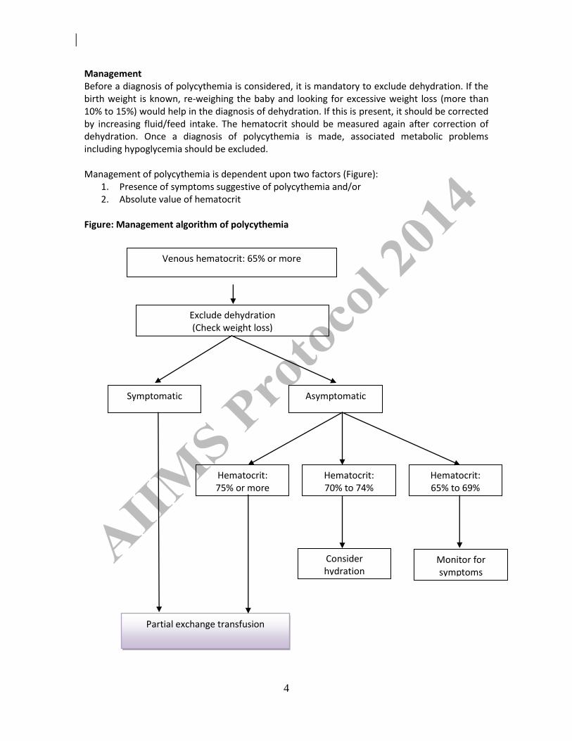
Management Before a diagnosis of polycythemia is considered, it is mandatory to exclude dehydration. If the birth weight is known, re-weighing the baby and looking for excessive weight loss (more than 10% to 15%) would help in the diagnosis of dehydration. If this is present, it should be corrected by increasing fluid/feed intake. The hematocrit should be measured again after correction of dehydration. Once a diagnosis of polycythemia is made, associated metabolic problems including hypoglycemia should be excluded. Management of polycythemia is dependent upon two factors (Figure):
1. Presence of symptoms suggestive of polycythemia and/or 2. Absolute value of hematocrit
Figure: Management algorithm of polycythemia
Venous hematocrit: 65% or more
Symptomatic
Exclude dehydration (Check weight loss)
Asymptomatic
Partial exchange transfusion
Hematocrit: 75% or more
Hematocrit: 70% to 74%
Hematocrit: 65% to 69%
Consider hydration
Monitor for symptoms
5
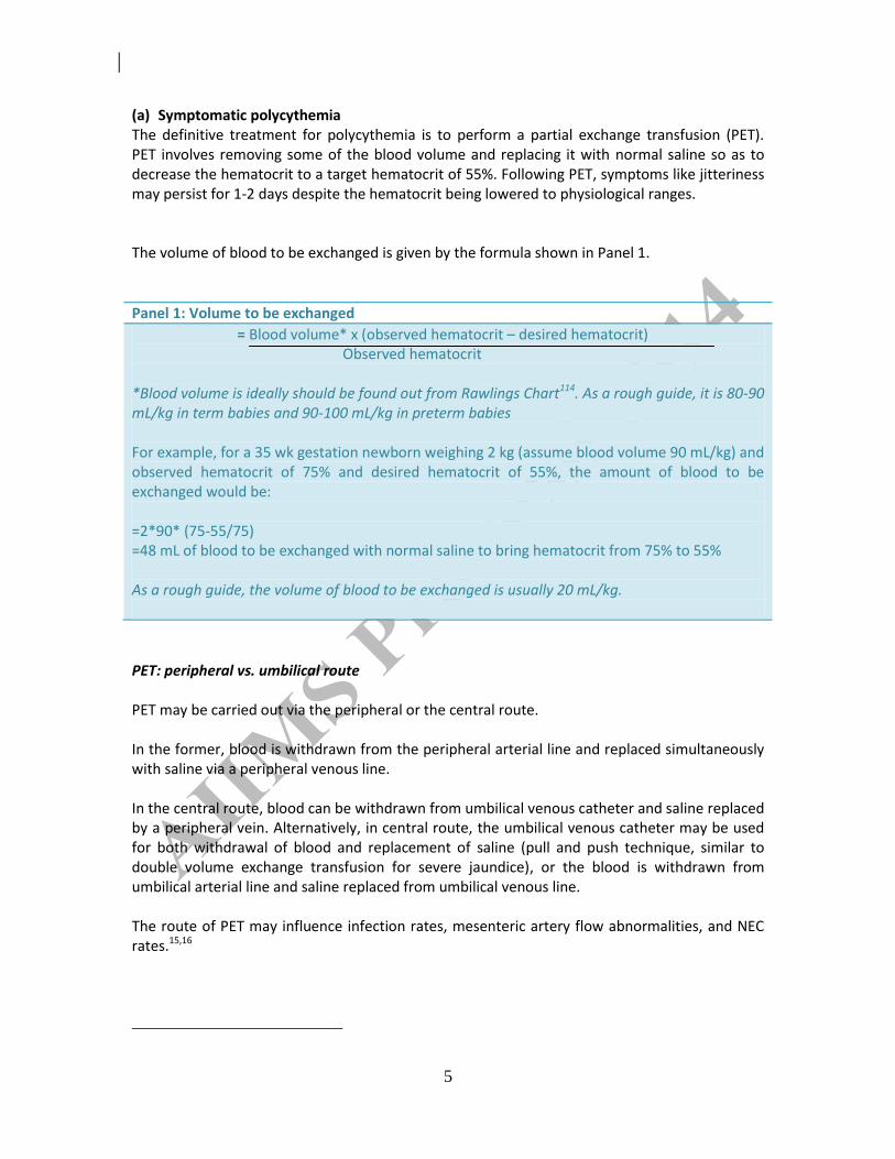
(a) Symptomatic polycythemia The definitive treatment for polycythemia is to perform a partial exchange transfusion (PET). PET involves removing some of the blood volume and replacing it with normal saline so as to decrease the hematocrit to a target hematocrit of 55%. Following PET, symptoms like jitteriness may persist for 1-2 days despite the hematocrit being lowered to physiological ranges. The volume of blood to be exchanged is given by the formula shown in Panel 1.
Panel 1: Volume to be exchanged
= Blood volume* x (observed hematocrit – desired hematocrit) Observed hematocrit
*Blood volume is ideally should be found out from Rawlings Chart114. As a rough guide, it is 80-90 mL/kg in term babies and 90-100 mL/kg in preterm babies For example, for a 35 wk gestation newborn weighing 2 kg (assume blood volume 90 mL/kg) and observed hematocrit of 75% and desired hematocrit of 55%, the amount of blood to be exchanged would be: =2*90* (75-55/75) =48 mL of blood to be exchanged with normal saline to bring hematocrit from 75% to 55% As a rough guide, the volume of blood to be exchanged is usually 20 mL/kg.
PET: peripheral vs. umbilical route PET may be carried out via the peripheral or the central route. In the former, blood is withdrawn from the peripheral arterial line and replaced simultaneously with saline via a peripheral venous line. In the central route, blood can be withdrawn from umbilical venous catheter and saline replaced by a peripheral vein. Alternatively, in central route, the umbilical venous catheter may be used for both withdrawal of blood and replacement of saline (pull and push technique, similar to double volume exchange transfusion for severe jaundice), or the blood is withdrawn from umbilical arterial line and saline replaced from umbilical venous line. The route of PET may influence infection rates, mesenteric artery flow abnormalities, and NEC rates.15,16
6
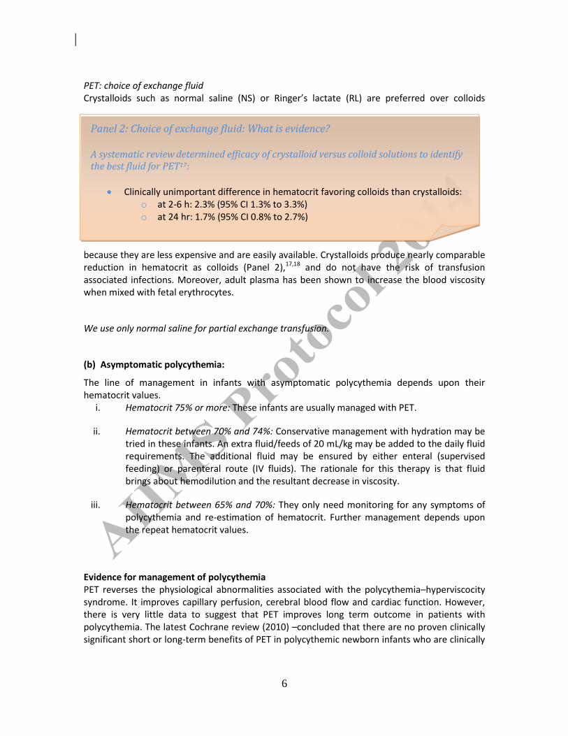
PET: choice of exchange fluid Crystalloids such as normal saline (NS) or Ringer’s lactate (RL) are preferred over colloids
because they are less expensive and are easily available. Crystalloids produce nearly comparable reduction in hematocrit as colloids (Panel 2),17,18 and do not have the risk of transfusion associated infections. Moreover, adult plasma has been shown to increase the blood viscosity when mixed with fetal erythrocytes.
We use only normal saline for partial exchange transfusion.
(b) Asymptomatic polycythemia:
The line of management in infants with asymptomatic polycythemia depends upon their hematocrit values.
i. Hematocrit 75% or more: These infants are usually managed with PET.
ii. Hematocrit between 70% and 74%: Conservative management with hydration may be tried in these infants. An extra fluid/feeds of 20 mL/kg may be added to the daily fluid requirements. The additional fluid may be ensured by either enteral (supervised feeding) or parenteral route (IV fluids). The rationale for this therapy is that fluid brings about hemodilution and the resultant decrease in viscosity.
iii. Hematocrit between 65% and 70%: They only need monitoring for any symptoms of polycythemia and re-estimation of hematocrit. Further management depends upon the repeat hematocrit values.
Evidence for management of polycythemia PET reverses the physiological abnormalities associated with the polycythemia–hyperviscocity syndrome. It improves capillary perfusion, cerebral blood flow and cardiac function. However, there is very little data to suggest that PET improves long term outcome in patients with polycythemia. The latest Cochrane review (2010) –concluded that there are no proven clinically significant short or long-term benefits of PET in polycythemic newborn infants who are clinically
Panel 2: Choice of exchange fluid: What is evidence? A systematic review determined efficacy of crystalloid versus colloid solutions to identify the best fluid for PET17:
Clinically unimportant difference in hematocrit favoring colloids than crystalloids:
o at 2-6 h: 2.3% (95% CI 1.3% to 3.3%) o at 24 hr: 1.7% (95% CI 0.8% to 2.7%)
7
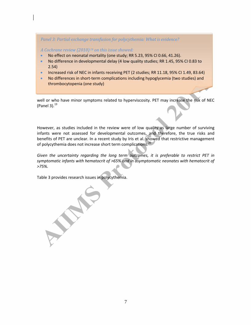
well or who have minor symptoms related to hyperviscosity. PET may increase the risk of NEC (Panel 3).19 However, as studies included in the review were of low quality as large number of surviving infants were not assessed for developmental outcomes, and therefore, the true risks and benefits of PET are unclear. In a recent study by Iris et al. showed that restrictive management of polycythemia does not increase short term complications.20
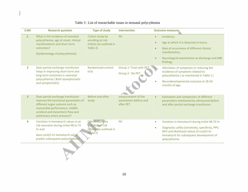
Given the uncertainty regarding the long term outcomes, it is preferable to restrict PET in symptomatic infants with hematocrit of >65% and in asymptomatic neonates with hematocrit of >75%. Table 3 provides research issues in polycythemia.
Panel 3: Partial exchange transfusion for polycythemia: What is evidence? A Cochrane review (2010) 19 on this issue showed: No effect on neonatal mortality (one study; RR 5.23, 95% CI 0.66, 41.26).
No difference in developmental delay (4 low quality studies; RR 1.45, 95% CI 0.83 to 2.54)
Increased risk of NEC in infants receiving PET (2 studies; RR 11.18, 95% CI 1.49, 83.64)
No differences in short-term complications including hypoglycemia (two studies) and thrombocytopenia (one study)
8
References 1. Mackintosh TF, Walkar CH. Blood viscosity in the newborn. Arch Dis Child 1973; 48: 547-53.
2. Phibbs RH. Neonatal Polycythemia. In: Rudolph AB(ed): Pediatrics, 16th
ed. New York: Appleton Century Crofts, 1997, pp 179.
3. Ramamurthy RS, Brans WY. Neonatal Polycythemia I. Criteria for diagnosis and treatment. Pediatrics 1981; 68: 168-74.
4. Wirth FH, Goldberg KE, Lubchenco LO: Neonatal hyperviscocity I. Incidence. Pediatrics 1979; 63: 833-6.
5. Stevens K, Wirth FH. Incidence of neonatal hyperviscosity at sea level. Pediatrics 1980; 97: 118
6. Bada HS, Korones SB, Pourcyrous M, et al. Asymptomatic syndrome of polycythemic hyperviscocity: effect of partial exchange transfusion. J Pediatr 1992; 120: 579-85.
7. Awonusonu FO, Pauly TH, Hutchison AA. Maternal smoking and partial exchange transfusion for neonatal polycythemia. Am J Perinatol 2002; 19: 349-54.
8. Lubetzky R, Ben-Shachar S, Mimouni FB, et al. Mode of delivery and neonatal hematocrit. Am J Perinatol 2000; 17: 163-5.
9. Hutton EK, Hassan ES. Late vs early clamping of the umbilical cord in full-term neonates: systematic review and meta-analysis of controlled trials. JAMA 2007; 297: 1241-52.
10. Shohat M, Merlob P, Reisner SH: Neonatal Polycythemia. I. Early diagnosis and incidence relating to time of sampling. Pediatrics 1984; 73: 7-10.
11. Shohat M, Reisner SH, Mimouni F, et al. Neonatal polycythemia II. Definition related to time of sampling. Pediatrics 1984; 73:11-3.
12. Oh W. Neonatal polycythemia and hyperviscosity. Pediatr Clin North Am 1986; 33: 523-32.
13. Goldberg K, Wirth FH, Hathaway WE, et al. Neonatal hyperviscocity II. Effect of partial exchange transfusion. Pediatrics 1982; 69: 419-25.
14. Rawlings JS, Pettett G, Wiswell TE, et al. Estimated blood volumes in polycythemic neonates as a function of birth weight. J Pediatr 1982; 101: 594-9.
15. Rodriguez-Balderrama I, Rodriguez-Juarez DA, Cisneros-Garcia N, et al. Comparison of 2 methods of partial exchange transfusion in newborns with polycythemia: peripheral-peripheral and central-peripheral]. Bol Med Hosp Infant Mex 1993; 50: 633-8
16. Hein HA, Lathrop SS. Partial exchange transfusion in term, polycythemic neonates: absence of association with severe gastrointestinal injury. Pediatrics 1987; 80: 75-8.
17. de Waal KA, Baerts W, Offringa M. Systematic review of the optimal fluid for dilutional exchange transfusion in neonatal polycythaemia. Arch Dis Child Fetal Neonatal Ed 2006; 91: F7-10.
18. Deorari AK, Paul VK, Shreshta L, Singh M. Symptomatic neonatal polycythemia: Comparison of partial exchange transfusion with saline versus plasma. Indian Pediatr 1995; 32: 1167-71.
9
19. Ozek E, Soll R, Schimmel MS. Partial exchange transfusion to prevent neurodevelopmental disability in infants with polycythemia. Cochrane Database Syst Rev 2010 Jan 20;(1):CD005089.
20. Morag I, Strauss T, Lubin D, Schushan-Eisen I, Kenet G, Kuint J. Restrictive management of neonatal
Polycythemia. Am J Perinatol 2011; 28: 677-682.
10
S.NO Research question Type of study Intervention Outcome measures
1. What is the incidence of neonatal polycythemia, age at onset, clinical manifestations and short term outcomes?
(Epidemiology of polycythemia)
Cohort study by enrolling at risk infants (as outlined in Table 2)
Nil Incidence,
Age at which it is detected in hours,
Rate of occurrence of different clinical manifestation,
Neurological examination at discharge and MRI findings,
2 Does partial exchange transfusion helps in improving short term and long term outcomes in neonatal polycythemia ( Both asymptomatic and symptomatic)
Randomized control trial
Group 1: Treat with PET
Group 2 : No PET
Alleviation of symptoms or reducing the incidence of symptoms related to polycythemia ( as mentioned in Table 1 )
Neurodevelopmental outcome at 18-24 months of age.
3 Does partial exchange transfusion improve the functional parameters of different organ systems such as myocardial performance, middle cerebral and mesenteric flow and pulmonary artery pressure?
Before-and-after study
measurement of the parameters before and after PET
Estimation and comparison of different parameters mentioned by ultrasound before and after partial exchange transfusion.
4 Variation in hematocrit values in at risk neonates during initial 48 to 72 hr and
does cord/2-hr hematocrit value predict subsequent polycythemia?
Cohort study by enrolling at risk infants (as outlined in Table 2)
Nil Variation in hematocrit during initial 48-72 hr
Diagnostic utility (sensitivity, specificity, PPV, NPV and likelihood ratios) of cord/2-hr hematocrit for subsequent development of polycythemia
Table 3 : List of researchable issues in neonatal polycythemia