Embed Size (px)
Citation preview
Polysaccharide from Lentinus edodes Inhibits theImmunosuppressive Function of Myeloid-DerivedSuppressor CellsHao Wu1., Ning Tao2., Xiaoman Liu2, Xiao Li2, Jian Tang3, Chungwah Ma3, Xiaofei Xu3, Haitao Shao4,
Baidong Hou4*, Hui Wang1*, Zhihai Qin2*
1 Xinxiang Medical University, Xinxiang, China, 2 Protein and Peptide Pharmaceutical Laboratory, Institute of Biophysics, Chinese Academy of Sciences, Beijing, China,
3 Infinitus (China) Company Ltd., Guangzhou, China, 4 Key Laboratory of Infection and Immunity, Institute of Biophysics, Chinese Academy of Sciences, Beijing, China
Abstract
Reversing the function of immune suppressor cells may improve the efficacy of cancer therapy. Here, we have isolated anovel polysaccharide MPSSS (577.2 Kd) from Lentinus edodes and examined its effects on differentiation and function ofmyeloid-derived suppressor cells (MDSCs). MPSSS is composed of glucose (75.0%), galactose (11.7%), mannose (7.8%), andxylose (0.4%). In vivo, it inhibits the growth of McgR32 tumor cells, which is correlated with a reduced percentage of MDSCsin peripheral blood. In vitro, it induces both morphological and biophysical changes in MDSCs. Importantly, MPSSS up-regulates MHC II and F4/80 expression on MDSCs, and reverses their inhibition effect on CD4+ T cells in a dose-dependentmanner. The mechanism study shows that MPSSS may stimulate MDSCs through a MyD88 dependent NF-kB signalingpathway. Together, we demonstrated for the first time that MPSSS stimulates the differentiation of MDSCs and reverses itsimmunosuppressive functions, shedding new light on developing novel anti-cancer strategies by targeting MDSCs.
Citation: Wu H, Tao N, Liu X, Li X, Tang J, et al. (2012) Polysaccharide from Lentinus edodes Inhibits the Immunosuppressive Function of Myeloid-DerivedSuppressor Cells. PLoS ONE 7(12): e51751. doi:10.1371/journal.pone.0051751
Editor: Paulo Lee Ho, Instituto Butantan, Brazil
Received June 5, 2012; Accepted November 5, 2012; Published December 18, 2012
Copyright: � 2012 Wu et al. This is an open-access article distributed under the terms of the Creative Commons Attribution License, which permits unrestricteduse, distribution, and reproduction in any medium, provided the original author and source are credited.
Funding: This project was supported by Plan for Scientific Innovation Talent of Henan Province (104200510001), and the National Natural Science Foundation ofChina (31071261). The funders had no role in study design, data collection and analysis, decision to publish, or preparation of the manuscript.
Competing Interests: Some authors are employed by a commercial company ‘‘Infinitus (China) Company Ltd’’. There are no other declarations relating toemployment, consultancy, patents, products in development or marketed products. This does not alter the authors’ adherence to all the PLOS ONE policies onsharing data and materials.
* E-mail: [email protected] (BH); [email protected] (HW); [email protected] (ZQ)
. These authors contributed equally to this work.
Introduction
Myeloid-derived suppressor cells (MDSCs) are a group of
Gr1+CD11b+ cells and were first identified in tumor bearing mice.
Increasing evidence suggests that they are important immune
regulatory cells. In human, the major function of MDSCs during
tumor progression is to inhibit T cell activity and promote tumor
growth [1–6]. Preventing the generation of MDSCs or inhibiting
their function may promote anti-tumor immunity, and could be a
new strategy for cancer treatment.
Polysaccharides from L. edodes exhibit strong anti-cancer
activities [7,8], which are host-mediated and associated with the
activation of immune cells such as T cells, natural killer cells and
macrophages [9–12]. However, whether polysaccharides from L.
edodes are able to reduce immunological suppression in tumor,
especially by MDSCs, is still unknown.
In this study, we isolated a novel polysaccharide component
(MPSSS) from L. edodes and investigated its effect on MDSCs. We
found that MPSSS induced MDSC differentiation and prevented
their immunosuppressive function. This reveals a novel mecha-
nism underlying the anti-tumor activity of polysaccharides and
indicates that MPSSS is a potential candidate for anti-tumor
therapy.
Results
Purification of MPSSSL. edodes was extracted with hot water to obtain a total water
soluble polysaccharide extract, and it was precipitated sequentially
with 30%, 50% and 70% ethanol to obtain three pure fractions:
MPPP, MPSSP2 and MPSSS. Given that MPSSS had the
strongest stimulating effect on splenocytes, this fraction was used
in the study. The yield of MPSSS was low, at only 1% of the total
dry weight. The homogeneity and molecular weight of MPSSS
were determined by size-exclusion chromatography and it gave a
single symmetrical narrow peak on a Shodex SB column, with a
molecular weight of 5.7726105 Daltons (Da). Analysis of its
monosaccharide composition showed that this polysaccharide
fraction was mainly composed of glucose (75.0%), galactose
(11.7%) and mannose (7.8%) (Fig. 1B, C). Its polymer fraction
mainly consists of a b-1, 6-linked glucan branched at C-4 with side
chains which are also b-1, 6-linked-glucans (Fig. 1 D, E; Table S1
in Supplemental material).
MPSSS Inhibited Tumor Growth in vivoTo test whether MPSSS has anti-tumor activity, we first
examined its effect on the growth of McgR32 tumor cells in mice.
As shown in Fig. 2A, tumors from the MPSSS-treated group grew
PLOS ONE | www.plosone.org 1 December 2012 | Volume 7 | Issue 12 | e51751
slower and were significantly smaller (200 mm3) than those in the
control group (580 mm3) at day 16. Respectively, the percentage
of MDSCs in the peripheral blood was also significantly lower in
the MPSSS group (19%) (Fig. 2B, C) compared to the control
group (38%) at the same time. This result demonstrated that
MPSSS inhibited tumor growth, which was accompanied by a
decrease of MDSCs in mice.
Figure 1. Composition and structure analysis of MPSSS. (A) MPSSS appeared as a single peak in Sepharose CL-6B chromatography, indicatingits high degree of purity. (B) The monosaccharides derived from MPSSS were detected by HPLC. (C) Analytical results of the composition of MPSSS. (D)Results of 13C NMR detection of MPSSS. (E) Analytical results of the MPSSS polysaccharide basic structure unit.doi:10.1371/journal.pone.0051751.g001
Polysaccharide on Myeloid-derived Suppressor Cells
PLOS ONE | www.plosone.org 2 December 2012 | Volume 7 | Issue 12 | e51751
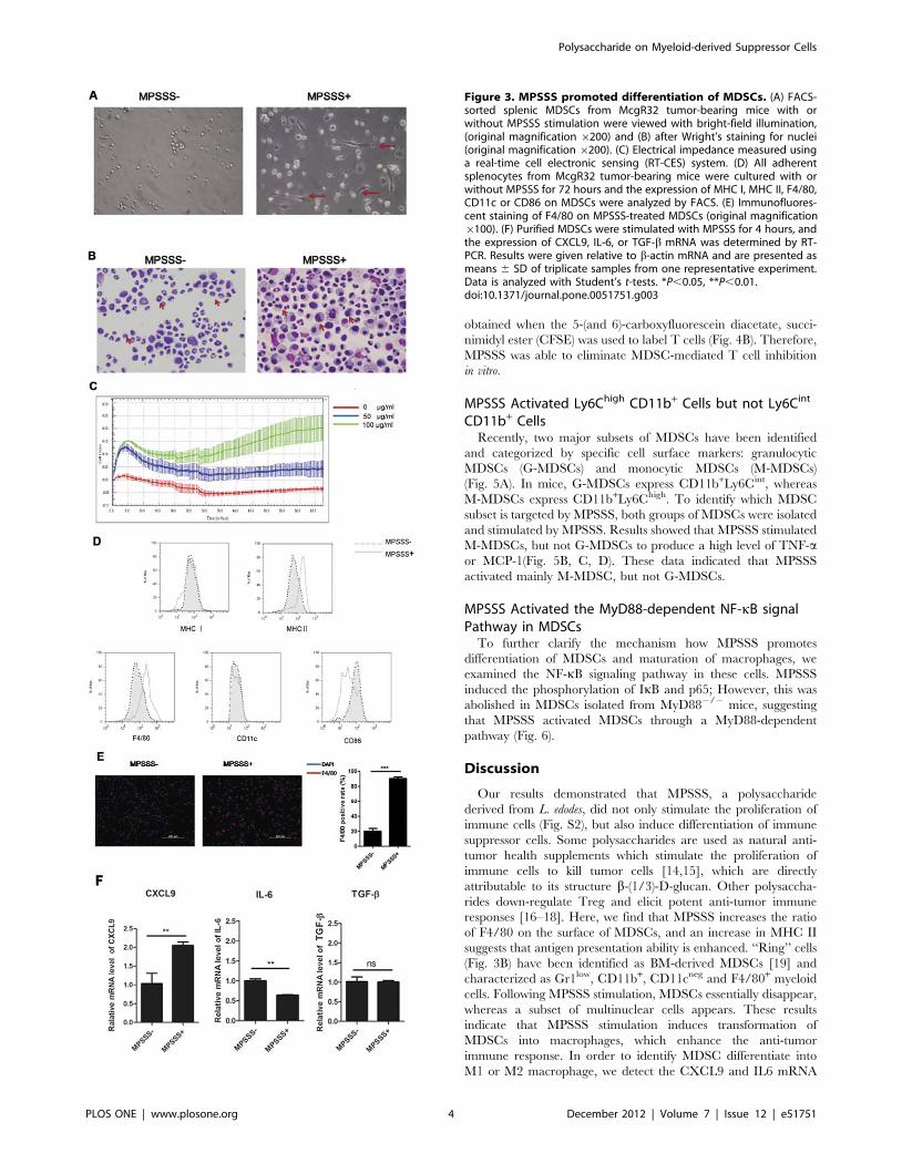
MPSSS Promoted Differentiation of MDSCsTo contribute the effect of MPSSS against tumor progression to
its influence on MDSC differentiation, we investigated its effect on
purified MDSCs in vitro. The Ly6C+CD11b+ cells were sorted
from the spleen of McgR32 tumor-bearing mice by FACS, and
stimulated with MPSSS or PBS for 3 days. Interestingly, the
morphology of MDSCs altered significantly after MPSSS treat-
ment. Untreated cells were mainly round, whereas MPSSS-treated
cells exhibited an elongated and irregular form, with long
pseudopodia, suggesting that MPSSS induces MDSC differentia-
tion (Fig. 3A). Moreover, the nuclear morphology of MDSCs was
also changed as shown in Fig. 3B. In the control group, a large
percentage of MDSCs exhibited a ‘‘ring-like’’ nucleus, while in the
MPSSS-treated group the ring-like structure disappeared, and
instead there were more cells containing multi-nuclei.
A real-time cell electronic sensing system (RT-CES) was used to
measure the biophysical characters of MDSCs [13], and the results
showed that MPSSS induced a dose-dependent increase in the
electrical impedance of MDSCs (Fig. 3C).
Furthermore, the expression of MHC II and F4/80 was up-
regulated, CD86 down-regulated though the expression of CD11c
and MHC I showed no significant changes after MPSSS treatment
(Fig. 3D, E). Interestingly, the expression of CXCL9 was
increased, while the expression of IL-6 was reduced after MPSSS
stimulation (Fig. 3F). Together, the results indicated that MPSSS
promoted the differentiation of MDSCs into more mature
macrophages.
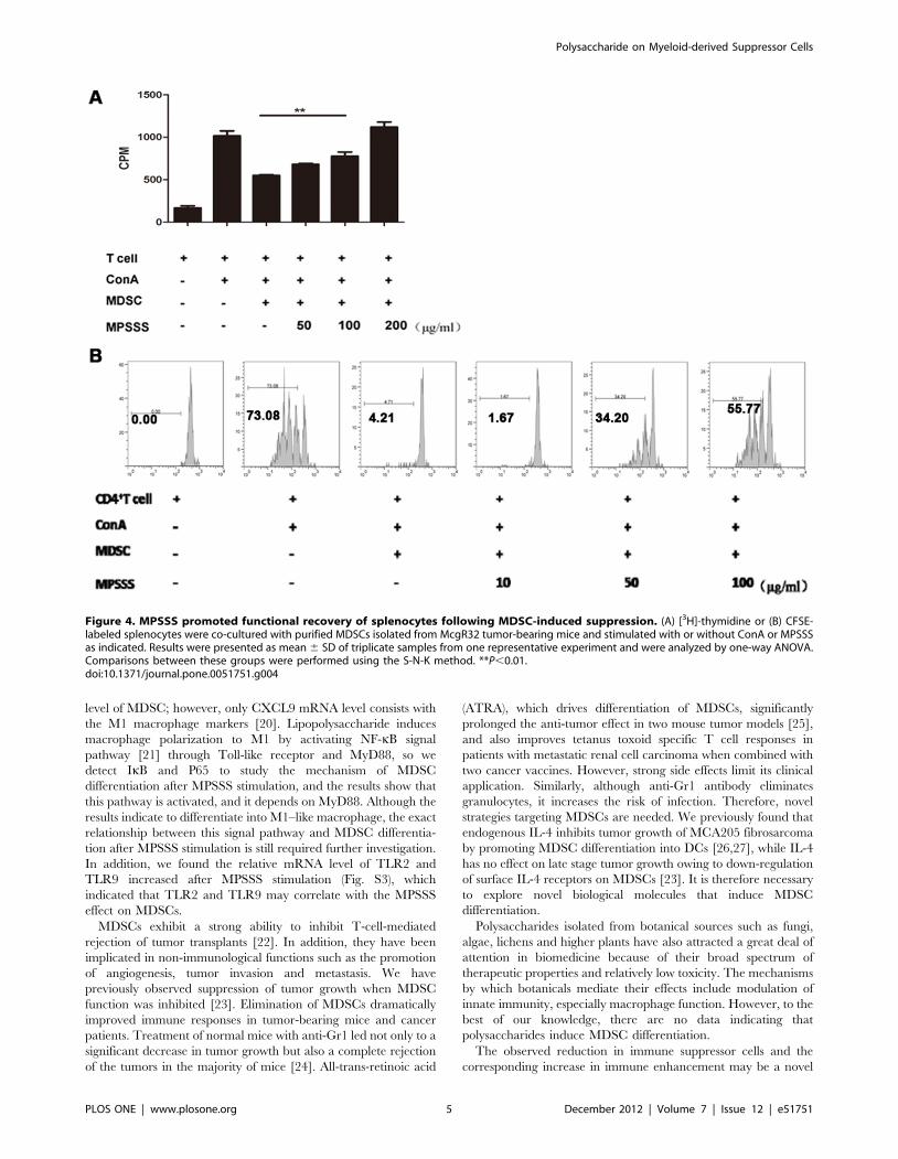
MPSSS Eliminated the MDSC-mediated T cell InhibitionAlthough the morphology of MDSCs changed markedly after
MPSSS stimulation (Fig. 3A, B), it is still unclear whether their
functions were influenced. MDSCs isolated from tumor-bearing
mice were co-cultured with a suspension of splenocytes, labeled
with [3H]-thymidine, and stimulated with ConA in the presence or
absence of MPSSS. As shown in Fig. 4A, the T cells proliferation
upon ConA stimulation was drastically inhibited by MDSCs;
however, when MPSSS was added into the culture system, it
recovered in a dose-dependent manner. Similar results were
Figure 2. MPSSS inhibited tumor growth and reduced MDSC numbers. (A) C57BL/6 mice were injected subcutaneously with 16106 McgR32cellsand 8 day later, with MPSSS or PBS intraperitoneally once every two days. Tumor volume was recorded as the mean 6 SD and each groupcontained 3–4 mice and was analyzed by one-way ANOVA for repeated measures data. (B) At the indicated days after tumor cell injection, peripheralblood was taken for MDSC detection by flow cytometry, Quantitative data are expressed as mean 6 SD and analyzed by Student’s t tests. *P,0.05,**P,0.01. (C) A representative flow cytometry analysis was shown for the staining of CD11b and Gr1.doi:10.1371/journal.pone.0051751.g002
Polysaccharide on Myeloid-derived Suppressor Cells
PLOS ONE | www.plosone.org 3 December 2012 | Volume 7 | Issue 12 | e51751
obtained when the 5-(and 6)-carboxyfluorescein diacetate, succi-
nimidyl ester (CFSE) was used to label T cells (Fig. 4B). Therefore,
MPSSS was able to eliminate MDSC-mediated T cell inhibition
in vitro.
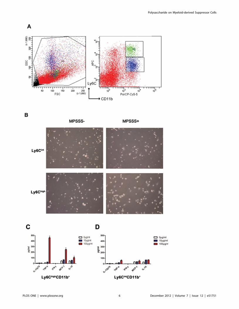
MPSSS Activated Ly6Chigh CD11b+ Cells but not Ly6Cint
CD11b+ CellsRecently, two major subsets of MDSCs have been identified
and categorized by specific cell surface markers: granulocytic
MDSCs (G-MDSCs) and monocytic MDSCs (M-MDSCs)
(Fig. 5A). In mice, G-MDSCs express CD11b+Ly6Cint, whereas
M-MDSCs express CD11b+Ly6Chigh. To identify which MDSC
subset is targeted by MPSSS, both groups of MDSCs were isolated
and stimulated by MPSSS. Results showed that MPSSS stimulated
M-MDSCs, but not G-MDSCs to produce a high level of TNF-aor MCP-1(Fig. 5B, C, D). These data indicated that MPSSS
activated mainly M-MDSC, but not G-MDSCs.
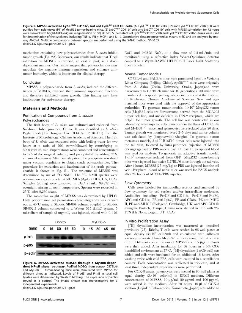
MPSSS Activated the MyD88-dependent NF-kB signalPathway in MDSCs
To further clarify the mechanism how MPSSS promotes
differentiation of MDSCs and maturation of macrophages, we
examined the NF-kB signaling pathway in these cells. MPSSS
induced the phosphorylation of IkB and p65; However, this was
abolished in MDSCs isolated from MyD882/2 mice, suggesting
that MPSSS activated MDSCs through a MyD88-dependent
pathway (Fig. 6).
Discussion
Our results demonstrated that MPSSS, a polysaccharide
derived from L. edodes, did not only stimulate the proliferation of
immune cells (Fig. S2), but also induce differentiation of immune
suppressor cells. Some polysaccharides are used as natural anti-
tumor health supplements which stimulate the proliferation of
immune cells to kill tumor cells [14,15], which are directly
attributable to its structure b-(1/3)-D-glucan. Other polysaccha-
rides down-regulate Treg and elicit potent anti-tumor immune
responses [16–18]. Here, we find that MPSSS increases the ratio
of F4/80 on the surface of MDSCs, and an increase in MHC II
suggests that antigen presentation ability is enhanced. ‘‘Ring’’ cells
(Fig. 3B) have been identified as BM-derived MDSCs [19] and
characterized as Gr1low, CD11b+, CD11cneg and F4/80+ myeloid
cells. Following MPSSS stimulation, MDSCs essentially disappear,
whereas a subset of multinuclear cells appears. These results
indicate that MPSSS stimulation induces transformation of
MDSCs into macrophages, which enhance the anti-tumor
immune response. In order to identify MDSC differentiate into
M1 or M2 macrophage, we detect the CXCL9 and IL6 mRNA
Figure 3. MPSSS promoted differentiation of MDSCs. (A) FACS-sorted splenic MDSCs from McgR32 tumor-bearing mice with orwithout MPSSS stimulation were viewed with bright-field illumination,(original magnification 6200) and (B) after Wright’s staining for nuclei(original magnification 6200). (C) Electrical impedance measured usinga real-time cell electronic sensing (RT-CES) system. (D) All adherentsplenocytes from McgR32 tumor-bearing mice were cultured with orwithout MPSSS for 72 hours and the expression of MHC I, MHC II, F4/80,CD11c or CD86 on MDSCs were analyzed by FACS. (E) Immunofluores-cent staining of F4/80 on MPSSS-treated MDSCs (original magnification6100). (F) Purified MDSCs were stimulated with MPSSS for 4 hours, andthe expression of CXCL9, IL-6, or TGF-b mRNA was determined by RT-PCR. Results were given relative to b-actin mRNA and are presented asmeans 6 SD of triplicate samples from one representative experiment.Data is analyzed with Student’s t-tests. *P,0.05, **P,0.01.doi:10.1371/journal.pone.0051751.g003
Polysaccharide on Myeloid-derived Suppressor Cells
PLOS ONE | www.plosone.org 4 December 2012 | Volume 7 | Issue 12 | e51751
level of MDSC; however, only CXCL9 mRNA level consists with
the M1 macrophage markers [20]. Lipopolysaccharide induces
macrophage polarization to M1 by activating NF-kB signal
pathway [21] through Toll-like receptor and MyD88, so we
detect IkB and P65 to study the mechanism of MDSC
differentiation after MPSSS stimulation, and the results show that
this pathway is activated, and it depends on MyD88. Although the
results indicate to differentiate into M1–like macrophage, the exact
relationship between this signal pathway and MDSC differentia-
tion after MPSSS stimulation is still required further investigation.
In addition, we found the relative mRNA level of TLR2 and
TLR9 increased after MPSSS stimulation (Fig. S3), which
indicated that TLR2 and TLR9 may correlate with the MPSSS
effect on MDSCs.
MDSCs exhibit a strong ability to inhibit T-cell-mediated
rejection of tumor transplants [22]. In addition, they have been
implicated in non-immunological functions such as the promotion
of angiogenesis, tumor invasion and metastasis. We have
previously observed suppression of tumor growth when MDSC
function was inhibited [23]. Elimination of MDSCs dramatically
improved immune responses in tumor-bearing mice and cancer
patients. Treatment of normal mice with anti-Gr1 led not only to a
significant decrease in tumor growth but also a complete rejection
of the tumors in the majority of mice [24]. All-trans-retinoic acid
(ATRA), which drives differentiation of MDSCs, significantly
prolonged the anti-tumor effect in two mouse tumor models [25],
and also improves tetanus toxoid specific T cell responses in
patients with metastatic renal cell carcinoma when combined with
two cancer vaccines. However, strong side effects limit its clinical
application. Similarly, although anti-Gr1 antibody eliminates
granulocytes, it increases the risk of infection. Therefore, novel
strategies targeting MDSCs are needed. We previously found that
endogenous IL-4 inhibits tumor growth of MCA205 fibrosarcoma
by promoting MDSC differentiation into DCs [26,27], while IL-4
has no effect on late stage tumor growth owing to down-regulation
of surface IL-4 receptors on MDSCs [23]. It is therefore necessary
to explore novel biological molecules that induce MDSC
differentiation.
Polysaccharides isolated from botanical sources such as fungi,
algae, lichens and higher plants have also attracted a great deal of
attention in biomedicine because of their broad spectrum of
therapeutic properties and relatively low toxicity. The mechanisms
by which botanicals mediate their effects include modulation of
innate immunity, especially macrophage function. However, to the
best of our knowledge, there are no data indicating that
polysaccharides induce MDSC differentiation.
The observed reduction in immune suppressor cells and the
corresponding increase in immune enhancement may be a novel
Figure 4. MPSSS promoted functional recovery of splenocytes following MDSC-induced suppression. (A) [3H]-thymidine or (B) CFSE-labeled splenocytes were co-cultured with purified MDSCs isolated from McgR32 tumor-bearing mice and stimulated with or without ConA or MPSSSas indicated. Results were presented as mean 6 SD of triplicate samples from one representative experiment and were analyzed by one-way ANOVA.Comparisons between these groups were performed using the S-N-K method. **P,0.01.doi:10.1371/journal.pone.0051751.g004
Polysaccharide on Myeloid-derived Suppressor Cells
PLOS ONE | www.plosone.org 5 December 2012 | Volume 7 | Issue 12 | e51751
Polysaccharide on Myeloid-derived Suppressor Cells
PLOS ONE | www.plosone.org 6 December 2012 | Volume 7 | Issue 12 | e51751
mechanism explaining how polysaccharides from L. edodes inhibit
tumor growth (Fig. 2A). Moreover, our results indicate that T cell
inhibition by MDSCs is reversed, at least in part, in a dose-
dependent manner. Our results suggest that polysaccharides may
modulate the negative immune regulation, and enhance anti-
tumor immunity, which is important for clinical therapy.
ConclusionMPSSS, a polysaccharide from L. edodes, induced the differen-
tiation of MDSCs, reversed their immune suppressor functions
and therefore inhibited tumor growth. This finding may have
implications for anti-cancer therapy.
Materials and Methods
Purification of Compounds from L. edodesPolysaccharides
The fruit body of L. edodes was cultured and collected from
Suizhou, Hubei province, China. It was identified as L. edodes
Pegler (Berk.) by Hongwei Liu (COA No: 2010–131) from the
Institute of Microbiology, Chinese Academy of Sciences. The fruit
body of L. edodes was extracted twice with boiling water for two
hours at a ratio of 20:1 (w/w)followed by centrifuging at
5000 rpm65 min. Supernatants were combined and concentrated
to 1/5 of the original volume, and precipitated by adding 95%
ethanol (4 volumes). After centrifugation, the precipitate was dried
under vacuum conditions to obtain crude polysaccharides. The
procedure for extraction and fractionation of the crude polysac-
charide is shown in Fig. S1. The structure of MPSSS was
determined by use of 13C NMR. The 13C NMR spectra were
obtained on a spectrometer at 500 MHz (Agilent DD2, CA, USA).
Samples (20 mg) were dissolved in D2O (1 mL, 99.8%) with
overnight stirring at room temperature. Spectra were recorded at
25uC after 9,280 scans.
The molecular weight of MPSSS was determined by HPLC.
High performance gel permeation chromatography was carried
out at 45uC using a Shodex SB-804 column coupled to Shodex
SB-802.5 column connected to a Waters 515 HPLC system. 5
microliters of sample (5 mg/mL) was injected, eluted with 0.1 M
NaCl and 0.02 M NaN3 at a flow rate of 0.5 mL/min and
monitored using a refractive index Wyatt-Optilabrex detector
coupled to a Wyatt-DAWN HELEOS-II Laser Light Scattering
Spectrometer.
Mouse Tumor ModelsC57BL/6 and BALB/c mice were purchased from the Weitong
Lihua Company (Beijing, China). myd882/2 mice were originally
from S. Akira (Osaka University, Osaka, Japan)and were
backcrossed to C57BL/6 mice for 10 generations. All mice were
maintained in a specific pathogen-free environment at the Institute
of Biophysics, Chinese Academy of Sciences. Sex- and age-
matched mice were used with the approval of the appropriate
authorities. To generate tumor models, 16106 McgR32 tumor
cells (McgR32 cells are fibrosarcoma derived from the MCA205
tumor cell line, and are deficient in IFN-c receptors, which are
helpful for tumor growth. The cell line was constructed in our
laboratory) were injected subcutaneously in the flank of C57BL/6
and MyD882/2 mice, and splenocytes were isolated after 20 days.
Tumor growth was monitored every 2–5 days and tumor volume
was calculated by (length6width6height). To generate tumor
metastasis models, 36105 B16F10 tumor cells were injected into
the tail vein, followed by intra-peritoneal injection of MPSSS
(25 mg/kg/day) or PBS once a day. On day 15, peripheral blood
was used for analysis. To generate an adoptive transfer model,
16107 splenocytes isolated from GFP+ McgR32 tumor-bearing
mice were injected into naive C57BL/6 mice through the tail vein.
After 4 hours, MPSSS (25 mg/kg) or PBS was injected into the tail
vein. Peripheral blood of naive mice was used for FACS analysis
after 24 hours of MPSSS/PBS injection.
Flow CytometryCells were labeled for immunofluorescence and analyzed by
flow cytometry for cell surface and/or intracellular molecules.
Antibodies including PerCP-anti-CD11b, PerCP-anti-F4/80,
APC-anti-CD11c, PE-anti-Ly6C, PE-anti-CD86, PE-anti-MHC
II, PE-anti-MHC I (Biolegend, Cambridge, UK) and APC-CD11b
(Sungene Biotech, Tianjin, China) were diluted in PBS with 2%
FCS (HyClone, Logan, UT, USA).
In vitro Proliferation Assay[3H] thymidine incorporation was measured as described
previously [25]. Briefly, T cells were seeded in 96-well plates at
equal density (36105 cells/ml) and co-cultured with adhesion
splenocytes isolated from McgR32 tumor-bearing mice at a ratio
of 5:1. Different concentrations of MPSSS and 0.5 mg/ml ConA
were then added. After incubation for 56 hours in a 5% CO2
humidified environment at 37uC, [3H]-thymidine (1 mCi/well) was
added and cells were incubated for an additional 16 hours. After
washing twice with cold PBS, cells were counted in a scintillation
counter. Each concentration was replicated in triplicate, and at
least three independent experiments were performed.
For CCK-8 assays, splenocytes were seeded in 96-well plates at
equal density (36105 cells/ml) in RPMI medium. Different
concentration of MPSSS, 10 mg/ml, 50 mg/ml and 100 mg/ml,
were added in the medium. After 20 hours, 10 ml of CCK-8
solution (DojinDo Laboratories, Kumamoto, Japan) was added to
Figure 5. MPSSS activated Ly6Chigh CD11b+, but not Ly6Cint CD11b+ cells. (A) Ly6Chigh CD11b+ cells (P2) and Ly6Cint CD11b+ cells (P3) werepurified from splenocyte (P1) of McgR32 tumor-bearing mice. (B) Ly6Chigh CD11b+ cells and Ly6Cint CD11b+ cells with MPSSS stimulation for 72 hourswere viewed with bright-field (original magnification6100). (C & D) Supernatants of Ly6Chigh CD11b+ cells and Ly6Cint CD11b+ cell cultures were usedfor determination of the cytokines, including TNF-a, IFN-c, MCP-1 and IL-10. Quantitative data are presented as means 6 SD and are analyzed by one-way ANOVA. Multiple comparisons between groups are performed using the S-N-K method. *P,0.05.doi:10.1371/journal.pone.0051751.g005
Figure 6. MPSSS activated MDSCs through a MyD88-depen-dent NF-kB signal pathway. Purified MDSCs from control C57BL/6and MyD882/2 tumor-bearing mice were stimulated with MPSSS fordifferent times as indicated. Levels of P-p65, and P-IkB in total celllysates were determined by Western blotting. The expression of b-actinserved as a control. The image shown was representative for 3independent experiments.doi:10.1371/journal.pone.0051751.g006
Polysaccharide on Myeloid-derived Suppressor Cells
PLOS ONE | www.plosone.org 7 December 2012 | Volume 7 | Issue 12 | e51751
fresh medium, and cells were incubated further for 4 hours.
Absorbance at 450 nm was determined using a microplate reader
(BIO-RAD Laboratories, Philadelphia, PA, USA). Results are
expressed as a percentage of the absorbance of control cultures. All
experiments were performed in triplicate.
For CFSE labeling and proliferation assays, splenocytes were
isolated from naive mice and incubated in RPMI 1640 for 4 hours.
Suspension cells were then collected and stained with 0.5–1 mM
carboxyfluorescein diacetate and CFSE. CFSE-labeled splenocytes
were co-cultured at a 5:1 ratio with MDSCs, stimulated with
0.5 mg/ml ConA, and sorted by FACS Aria III (BD Biosciences,
San Diego, CA, USA). After 3 days, cells were stained with CD4
(Biolegend, Cambridge, UK), and the CFSE signal of gated
lymphocytes was analyzed.
Cell and Nuclear MorphologyMDSCs were sorted from McgR32 tumor-bearing mice by
FACS with markers for Ly6C and CD11b and stimulated with
10 ng/ml IL-4 and 10 ng/ml GM-CSF with or without MPSSS
(100 mg/ml) for 3 days. Adherent cells were digested in 5 mM
EDTA, re-suspended to 26106 cells/ml in 50 ml PBS and
centrifuged in a cytospin (Thermo Scientific, Waltham, MA,
USA). Cells were then stained with Wright’s stain for 15 minutes
and washed with water prior to analysis.
Electrical Impedance of MDSCsThe RT-CES system (ACEA Biosciences Inc., San Diego, CA,
USA) is comprised of three components: an electronic sensor
analyzer, a device station and a 96-well strip. The cell culture
conditions on the 966 sensor device were the same as those used
for tissue culture. Growth media (100 ml) was gently dispensed into
a 96-well strip for background readings by the RT-CES system
prior to adding to 200 ml of cells at 16106 cells/ml.
Devices containing cells were kept at room temperature in a
tissue culture hood for 15 minutes prior to insertion into the RT-
CES device. Cell-sensor impedance was displayed as an arbitrary
unit called the cell index.
The cell index at each time point is defined according to the
formula:
CI~ maxi~1,2,:::,N
(Rcell(fi)
R0(fi){1)
where N is the number of frequency points at which the
impedance is measured (i.e., N = 3 for 10 kHz, 25 kHz and
50 kHz), and Rcell(fi) and R0(fi) are the frequency-dependent
electrode resistances with or without cells present in the wells,
respectively [13].
Before the start of the experiment, the media in each well was
changed to RPMI 1640 and equilibrated for 2 hours. Cells were
then incubated in the presence or absence of different concentra-
tions of MPSSS.
Immune Fluorescence AssayMDSCs (Ly6C+ CD11b+ cells) were sorted from spleens of
McgR32 tumor-bearing mice and maintained on glass slides with
or without 100 mg/ml MPSSS stimulation for 3 days. The mAb
used for staining was anti-F4/80 monoclonal antibody (BD
Biosciences). Alex Fluor 555-conjugated goat anti-rat IgG (H+L)
(Invitrogen, Carlsbad, CA, USA) was used as a secondary
antibody. Cell nuclei were counterstained with 49, 6-diamidino-
2-phenylindole. Photos of cells were taken using a digital
microscope (OLYMPUS, Tokyo, Japan).
Real Time RT-PCRTotal RNA was extracted from 56106 isolated CD11b+Gr1+
cells stimulated by MPSSS with TRIzol (Invitrogen, Carlsbad,
CA, USA) and quantified on a ND-1000 spectrophotometer
(NanoDrop Technologies, Wilmington, DE, USA). cDNA was
synthesized using 2 mg RNA, 9 nt random primer (TaKaRa,
Ohtu, Japan) and M-MLV reverse transcriptase (Promega,
Heidelberg, Germany). The amounts of TLR2, TLR4, TLR9,
CXCL9, IL-6 and TGF-b mRNA were determined using iQTM
SYBR Green Supermix on a MyiQTM system (Bio-Rad
Laboratories). b-actin mRNA was used as internal control. The
specific primers were:
TLR2:5’-CTCTTCAGCAAACGCTGTTCT-3’; 5’-
GGCGTCTCCCTCTATTGTATTG-3’;
TLR4:5’-CCTGATGACATTCCTTCT-3’; 5’-AGCCACCA-
GATTCTCTAA-3’;
TLR9:5’-ATGGTTCTCCGTCGAAGGACT-3’; 5’-
GAGGCTTCAGCTCACAGGG-3’;
IL-6:5’-ATGGATGCTACCAAACTGGAT-3’; 5’-TGAAG-
GACTCTGGCTTTGTCT-3’;
CXCL9:5’-TGCTACACTGAAGAACGGAGAT-3’; 5’-
TCCTTGAACGACGACGACTT-3’;
TGF-b: 5’-CAACAATTCCTGGCGTTACCT-3’; 5’-
TGTATTCCGTCTCCTTGGTTCA-3’;
actin: 5’-GAAGTGTGACGTTGACATCCGTA-3’; 5’-
CTCAGGAGGAGCAATGATCTTGA-3’.
Primers for IL-6, CXCL9 and TGF-b were purchased from
Sangon Biotech Shanghai Co., Ltd.
Western BlottingCells were lysed by RIPA solution [50 mM Tris-HCl (pH 7.5),
150 mM NaCl, 1.0% Nonidet P-40, 0.5% (w/v) sodium deoxy-
cholate, 0.1% (w/v) SDS, 1 mM EDTA] supplemented with
100 mM phenylmethanesulfonyl fluoride, 25 mg/ml aprotinin,
1 mM sodium orthovanadate and 50 mM NaF. Aliquots of cell
extracts were resolved on 10% SDS-PAGE gel and then
transferred to a nitrocellulose membrane (GE Healthcare,
Milwaukee, WI, USA) using a semi-dry transfer apparatus (Bio-
Rad Laboratories). The primary antibodies used were: Phospho
(P)-NF-kB p65 (Ser536; 93H1), and P-IkB (S32; 14D4; Cell
Signaling Technology), all of which were diluted by 1:1000, and b-
actin (1:8000, Sigma-Aldrich, St. Louis, Missouri, USA). HRP-
conjugated goat anti-mouse or goat anti-rabbit IgG (Thermo) were
used as secondary antibodies. After washing with PBST, the
membrane was incubated with chemiluminescent substrate
(Thermo) for 5 minutes. Specific bands were visualized by
exposing the membrane to X-ray film (Kodak, Rochester, NY,
USA) in a dark room.
Cytokine DetectionLy6Chigh CD11b+ and Ly6Cint CD11b+ cells were sorted from
spleens of McgR32 tumor-bearing mice and cultured with or
without 100 mg/ml MPSSS stimulation for 3 days. Supernatant
were analyzed by cytometric bead arrays (CBA, BD Biosciences) in
accordance with the manufacturer’s instructions. Briefly, five bead
populations with distinct fluorescence intensities were coated with
capture antibodies specific for IL-10, MCP-1, IFN-c, TNF-a and
IL-12p70 proteins. The five bead populations were mixed together
to form the CBA, which was resolved in the FL3 channel. The
capture beads, PE-conjugated detection antibodies and recombi-
nant standards or test samples were incubated together to form
sandwich complexes. Following data acquisition, flow cytometry
results were generated using CBA Analysis Software (BD
Biosciences).
Polysaccharide on Myeloid-derived Suppressor Cells
PLOS ONE | www.plosone.org 8 December 2012 | Volume 7 | Issue 12 | e51751
Statistical AnalysisData are analyzed using Student’s t tests, or one-way ANOVA,
and are presented as means 6 SD. Differences are considered to
be statistically significant when P,0.05.
Supporting Information
Figure S1 Purification of polysaccharides from L.edodes. We obtained three pure compositions: MPPP, MPSSP2
and MPSSS.
(TIF)
Figure S2 MPSSS promotes splenocyte proliferation.Splenocytes isolated from naive C57BL/6 mice (A) or McgR32
tumor-bearing mice (B) were seeded on 96-well plates and
stimulated with different concentrations of MPSSS. The product
of CCK-8 assay formazan was measured with optical density
450 nm. Results presented are mean 6 SD of triplicate samples
from one representative experiment. *P,0.05, **P,0.01.
(TIF)
Figure S3 MPSSS increases the mRNA level of TLR2 andTLR9. MDSCs were isolated by FACS and stimulated with
MPSSS for 0, 3, 6 hours and then the expression of TLR2/4/9
were measured by real-time PCR. Results presented are mean 6
SD of triplicates from one representative experiment. *P,0.05,
***P,0.001.
(TIF)
Table S1 The relationship between 13C NMR chemicalshift and the corresponding C.
(DOCX)
Acknowledgments
The authors are grateful to Li Yu for analysis of MPSSS structure, to
Shanshan Zang for technical support, to Xuehui Liu for MPSSS NMR
detection, and to Joy Fleming and Shuna Cui for critical reading of the
manuscript.
Author Contributions
Conceived and designed the experiments: ZQ HW BH. Performed the
experiments: HW NT X. Liu X. Li. Analyzed the data: HW NT.
Contributed reagents/materials/analysis tools: JT CM HS. Wrote the
paper: HW NT ZQ BH XX.
References
1. Almand B, Clark JI, Nikitina E, van Beynen J, English NR, et al. (2001)
Increased production of immature myeloid cells in cancer patients: a mechanism
of immunosuppression in cancer. J Immunol 166: 678–689.
2. Gabrilovich D (2004) Mechanisms and functional significance of tumour-
induced dendritic-cell defects. Nat Rev Immunol 4: 941–952.
3. Gabrilovich DI, Bronte V, Chen SH, Colombo MP, Ochoa A, et al. (2007) The
terminology issue for myeloid-derived suppressor cells. Cancer Res 67: 425;
author reply 426.
4. Gallina G, Dolcetti L, Serafini P, De Santo C, Marigo I, et al. (2006) Tumors
induce a subset of inflammatory monocytes with immunosuppressive activity on
CD8+ T cells. J Clin Invest 116: 2777–2790.
5. Melani C, Chiodoni C, Forni G, Colombo MP (2003) Myeloid cell expansion
elicited by the progression of spontaneous mammary carcinomas in c-erbB-2
transgenic BALB/c mice suppresses immune reactivity. Blood 102: 2138–2145.
6. Yang L, DeBusk LM, Fukuda K, Fingleton B, Green-Jarvis B, et al. (2004)
Expansion of myeloid immune suppressor Gr+CD11b+ cells in tumor-bearing
host directly promotes tumor angiogenesis. Cancer Cell 6: 409–421.
7. Chihara G, Maeda Y, Hamuro J, Sasaki T, Fukuoka F (1969) Inhibition of
mouse sarcoma 180 by polysaccharides from Lentinus edodes (Berk.) sing.
Nature 222: 687–688.
8. Chihara G, Hamuro J, Maeda Y, Arai Y, Fukuoka F (1970) Fractionation and
purification of the polysaccharides with marked antitumor activity, especially
lentinan, from Lentinus edodes (Berk.) Sing. (an edible mushroom). Cancer Res
30: 2776–2781.
9. Hamuro J, Rollinghoff M, Wagner H (1980) Induction of cytotoxic peritoneal
exudate cells by T-cell immune adjuvants of the beta(1 leads to 3) glucan-type
lentinan and its analogues. Immunology 39: 551–559.
10. Ladanyi A, Timar J, Lapis K (1993) Effect of lentinan on macrophage
cytotoxicity against metastatic tumor cells. Cancer Immunol Immunother 36:
123–126.
11. Maeda YY, Chihara G (1973) The effects of neonatal thymectomy on the
antitumour activity of lentinan, carboxymethylpachymaran and zymosan, and
their effects on various immune responses. Int J Cancer 11: 153–161.
12. Peter G, Karoly V, Imre B, Janos F, Kaneko Y (1988) Effects of lentinan on
cytotoxic functions of human lymphocytes. Immunopharmacol Immunotoxicol
10: 157–163.
13. Solly K, Wang X, Xu X, Strulovici B, Zheng W (2004) Application of real-time
cell electronic sensing (RT-CES) technology to cell-based assays. Assay Drug
Dev Technol 2: 363–372.
14. Zhang Y, Li S, Wang X, Zhang L, Cheung PCK (2011) Advances in lentinan:
Isolation, structure, chain conformation and bioactivities. Food hydrocolloids 25:
196–206.15. Jung HY, Bae IY, Lee S, Lee HG (2011) Effect of the degree of sulfation on the
physicochemical and biological properties of, i.Pleurotus eryngii,/i.poly-saccharides. Food hydrocolloids 25: 1291–1295.
16. Qi C, Cai Y, Gunn L, Ding C, Li B, et al. (2011) Differential pathways
regulating innate and adaptive antitumor immune responses by particulate andsoluble yeast-derived beta-glucans. Blood 117: 6825–6836.
17. Ochoa-Reparaz J, Mielcarz DW, Wang Y, Begum-Haque S, Dasgupta S, et al.(2010) A polysaccharide from the human commensal Bacteroides fragilis protects
against CNS demyelinating disease. Mucosal Immunol 3: 487–495.18. Li XB, He XJ, Liu B, Xu L, Ju DH, et al. (2010) [Immunoregulatory function of
Radix Glycyrrhizae polysaccharide in tumor-bearing mice]. Zhong Xi Yi Jie He
Xue Bao 8: 363–367.19. Greifenberg V, Ribechini E, Rossner S, Lutz MB (2009) Myeloid-derived
suppressor cell activation by combined LPS and IFN-gamma treatment impairsDC development. Eur J Immunol 39: 2865–2876.
20. Martinez FO, Gordon S, Locati M, Mantovani A (2006) Transcriptional
profiling of the human monocyte-to-macrophage differentiation and polariza-tion: New molecules and patterns of gene expression. Journal of Immunology
177: 7303–7311.21. Lawrence T, Natoli G (2011) Transcriptional regulation of macrophage
polarization: enabling diversity with identity. Nat Rev Immunol 11: 750–761.22. Wang Z, Jiang J, Li Z, Zhang J, Wang H, et al. (2010) A myeloid cell population
induced by Freund adjuvant suppresses T-cell-mediated antitumor immunity.
J Immunother 33: 167–177.23. Jiang J, Wang Z, Li Z, Zhang J, Wang C, et al. (2010) Early exposure of high-
dose interleukin-4 to tumor stroma reverses myeloid cell-mediated T-cellsuppression. Gene Ther 17: 991–999.
24. Kusmartsev S, Cheng F, Yu B, Nefedova Y, Sotomayor E, et al. (2003) All-trans-
retinoic acid eliminates immature myeloid cells from tumor-bearing mice andimproves the effect of vaccination. Cancer Res 63: 4441–4449.
25. Mirza N, Fishman M, Fricke I, Dunn M, Neuger AM, et al. (2006) All-trans-retinoic acid improves differentiation of myeloid cells and immune response in
cancer patients. Cancer Res 66: 9299.
26. Li Z, Jiang J, Wang Z, Zhang J, Xiao M, et al. (2008) Endogenous interleukin-4promotes tumor development by increasing tumor cell resistance to apoptosis.
Cancer Res 68: 8687–8694.27. Li Z, Chen L, Qin Z (2009) Paradoxical roles of IL-4 in tumor immunity. Cell
Mol Immunol 6: 415–422.
Polysaccharide on Myeloid-derived Suppressor Cells
PLOS ONE | www.plosone.org 9 December 2012 | Volume 7 | Issue 12 | e51751