Embed Size (px)
Citation preview
1
POSSIBLE CAUSES FOR THE COLONY COLLAPSE DISORDER (CCD)
Zoran Stanimirović*, Nevenka Aleksić* and Jevrosima Stevanović*
*The University of Belgrade, Faculty of Veterinary Medicine, Belgrade, Boulevard
oslobodjenja 18, Serbia
Abstract. The possible reasons for the diminishing of bees i.e. for the phenomenon
known as the Colony Collapse Disorder (CCD) are given, as well as the biology,
clinical findings, diagnosis and prevention/protection of honeybee colonies from
American foulbrood, nosemosis, varoosis and certain viral infections. The importance
of good condition of honeybee colonies, adequate diet supply, together with the
reinforcing of hygienic and grooming behaviour which influence the higher resistance
to aforementioned diseases is underlined. Further on, the biological means to combat
the diseases of bees and bee colonies, which contribute to the development of ecological
honeybee keeping and production of bee products devoid of residua of pharmaceuticals
used in conventional bee keeping.
Key words: CCD, American foulbrood, nosemosis, varroosis, honeybee viruses,
biological means of combat, hygienic and grooming behaviour
COLONY COLLAPSE DISORDER (CCD)
Massive death of honeybees was described worldwide long ago; for example it
happened in Ireland in 950, 992 and 1443. At the beginning of the 20th century, in
spring of 1906, on the White Island (Great Britain) the majority of beekeepers lost their
bee colonies. Furthermore, American beekeepers occasionally suffered from heavy
losses. In the Cache Valley (Utah, USA) in 1903 two-thousand bee colonies died from
mysterious phenomenon of disappearing throughout cold winter and spring. More
recently, in 1995, beekeepers in Pennsylvania lost 53% of their bee colonies (Oldroyd,
2007).
2
Colony Collapse Disorder (CCD) is a phenomenon which was not known until recently
but has become the most serious disease and cause of sudden death of honeybee
colonies characterised by the disappearance of adult bees in and in front of beehives.
Both honey and ’bee bread’ are usually present in abandoned beehives as well as the
indications of recent brood rearing. Sometimes the queen and few bees can be found in
the nest. Characteristically, in the hives without bees honey robbery appears late and the
hives are invaded by usual pests (wax moth Galleria mellonella and small hive beetle
Aethina tumida) more slowly than expected or are not at all.
CCD is a multifactorial disease of honeybee colonies, i.e. it cannot be claimed to be
caused by one agent only. The fact leads to the difficulty of recommendation of a single
remedy which can prove the most efficacious (Stanimirović et al., 2008a,b,c).
The most frequent causes of CCD are:
- The deficiency in high-quality diet (bee bread and honey)
- Bacterial infections (American foulbrood)
- Fungal diseases (nosemosis and ascospherosis)
- Parasitic infections of honeybee colonies, most often with Varroa destructor
and Acarapis woodi
- Mixed viral infections of honeybee colonies
- Management in the apiary
THE DEFICIENCY IN HIGH-QUALITY DIET (BEE BREAD AND HONEY)
Global climatic changes, pollution and chemisation in all human activities, particularly
in agricultural production, lead to disturbances of ecosystems, diminishing or
problematic herbal production and, consequently, to reduced production of sufficient
high-quality food for honeybee societies. Intensive (frequent and widespread)
application of pesticides results in reduced production of high-quality pollen, which is
the basis for high-quality bee bread preparation, an indispensable source of proteins. On
3
the other hand, the reduced livestock, especially the reduction in the number of sheep
and goats, which means diminishing manure production, result in significant fall in
high-quality nectar and pollen production since many herbal species have diminished or
completely disappeared from the fields and pastures (Stanimirović et al., 2008a,b,c). For
example, in the regions of Homolje and Peshter (Serbia) solely the number of domestic
animals dramatically decreased being one tenth of the number in 1989, which also
means similar decline in manure production. Having in mind that a sheep produces
approximately 500 g of manure on average, the consequences are more easily
understandable. On the other hand, the number of honeybee colonies rose meanwhile
due to the idea that households can increase their budget by beekeeping. In attempt to
earn as much as possible honeybee keepers do not mind the biological needs of their
bees, i.e. they do not only deprive them of the surpluses in honey but also take away the
one from the brood chambers. This honey is to belong to the bees only and it is not to be
removed, since it is not nectar but represents a source of energy, essential amino acids,
micro- and macroelements, vitamins and other active substances. It is a biologically
active material manufactured from nectar with which it is mixed as well as with the
secretion of bees’ glands. Besides, the honey from brood chambers comprises
considerable amounts of pollen which under the influence of acids as well as of the
enzymes of the bees’ exocrine glands bursts at some time and its content is mixed with
the honey itself. That is why this honey is an extremely high-quality energetic protein-
rich diet of the bees (Stanimirović et al., 2008a,b,c). It is the most important prerequisite
for the wintering and rapid development of the colonies in spring. When depriving a
colony of the honey from brood chambers beekeepers do severe damage to the bees as
well as to themselves. Taking away this honey also means taking the residua of various
preparations (amitraz, coumaphos, cymiazole hydrochloride, flumethrin, fluvalinate,
dicyclohexylamine) used for the treatment of the bees in brood chambers (Stanimirović
et al., 2003a,c, 2005a, 2006, 2007a,b; Pejin et al., 2006; Stevanović et al., 2006, 2008).
Thus, the residua got into the honey either directly or from wax which had been
contaminated previously. Such honey is not to be used by humans. In addition, apart
from being deprived of honey bees are deprived of the highest-quality diet which
strongly influences the ability of development and survival of the colonies and the
immunological potential of each individual bee as well as the colony as a whole
4
(Stevanović, 2007; Stanimirović et al., 2008a,b,c). Due to the deprivation of honey the
potential for development declines, the age polyethism is altered and by the addition of
sugar as a substitute to the honey (by no means can sugar substitute honey) bees are
exhausted additionally and the colony will not be ready for the main honey harvest. The
destruction of the proportion of bees of different age in the colony leads to the decline in
the number of cleaning bees (aged from 15 to 17 days, Arathi et al., 2000) which results
in the diminished defensive potential to the infective agents always present in the hive
(Paenibacillus larvae, Nosema apis, Nosema ceranae, Ascosphera apis, Varroa
destructor and various viruses) (Stanimirović et al., 2005b, 2008a,b,c).
Being capable of regulating humidity the honey from brood chambers influences the
microclimate, temperature and the exchange of gases in hives. The excessive moisture,
which can be detrimental to the thermoregulation in a hive, is absorbed by the honey,
which helps the bees with the regulation of moisture and temperature in the brood.
Honeybees efficaciously maintain the temperature in brood chambers at approximately
34.5oC regardless of the environmental conditions. If the temperature is higher or lower,
the bees will develop into seemingly normal adults, but with damaged reception of
information and memory. Worker bees reared on temperatures lower than optimal tend
to get lost in the field and are incapable of performing waggle dances efficaciously. If
bee colonies fail to maintain the temperature and moisture in the brood continuously,
symptoms similar to those of the CCD will develop (Oldroyd, 2007).
If the humidity declines (in summer at high temperatures) the water from the honey in
the brood chambers is released, which helps the maintenance of optimal moisture and
temperature in the hives. Thus, the need for fanning bees and water carriers declines and
worker bees can devote themselves to honey harvest and contribute to the honey yield
(Stanimirović et al., 2008a,b,c). It is well-known that excessive moisture is a
prerequisite for the appearance of fungi (the causative agents of chalk brood,
Ascosphera apis, and nosemosis: Nosema apis and Nosema ceranae). In spring the
temperature increases both outside and in the hives, the air becomes drier and if the bee
colony is weak and there is no honey substantial decline in humidity is unavoidable. As
a result, the brood dries, suffers from lack of moisture and dies thus providing
conditions for bacterial infections. By all means Varroa destructor contributes to the
5
damage being not only an ectoparasite of the brood and adult bees but also a vector of
mixed viral infections. In addition, apart from propolis, the honey from brood chambers
has an antimicotic and bacteriostatic effect (Stanimirović et al., 2008a,b,c).
Besides propolis and honey, pollen also contributes to the immunological properties and
potential of honeybee colonies but in conditions described afore either there is a
shortage of pollen or it is of weak quality. Pollen does not maintain its quality during
the pasture season. The best is the one from early polliniferous plants (ephemeral
flowering plants), the pollen of hazelnut, willow, from fruits, meadow grasses, corn,
sophora etc. Global climatic changes have influenced the dynamics of flowering as well
as the use of pollen of certain polliniferous plants. For example, these years there was
little pollen of ephemeral flowering plants, hazelnut and plum and it was difficult to use
due to bad weather conditions (Stanimirović et al., 2008a,b,c).
Various pesticides can also influence the quality of pollen and nectar. Apart from
numerous other pesticides used on vegetable, fruit and crop farming the use of
imidacloprid- and fipronil-based pesticides has recently increased. These are poisonous
for honeybees, acting by contact and after ingestion. Imidacloprid and fipronil are
neonicotinoids. They are absorbed by plants through their roots and distributed into
higher organs: flowers, fruits, leaves and seeds, where they remain for a long time and
accumulate in the nectar and pollen (Šovljanski, 2008a,b). Neonicotinoids, such as
imidacloprid, acts on acetylcholine receptors while fipronil influences the chlorine
channels thus enhancing the permeability of neurons; in other words, fipronil is capable
of blocking the passage of chloride ions through the GABA receptor and glutamate-
gated chloride channels, components of the central nervous system of insects. While
seeking for food and collecting nectar bees remember the scent of flowers and make
some kind of ‘maps’ which they use in the future. Unfortunately, the aforementioned
insecticides do damage to the brain centres responsible for memory and orientation,
which is why the bees become disoriented and incapable of returning to their hives;
consequently they wander and eventually die of starvation (Oldroyd, 2007). In addition,
the poisoned bees are agitated, their movements are uncoordinated; for example, they
can hang from the sunflowers; at first they are very active but soon become apathetic,
suffer cramps, droop and die in the end (Stanimirović et al. 2008a,b,c).
6
AMERICAN FOULBROOD
Infective diseases of bees pose serious problems which influence the development of
beekeeping in Serbia, as well as worldwide. Among them American foul brood (AFB),
varroosis, nosemosis, viral and fungal diseases are of utmost importance. From a health
protection and financial viewpoint American foulbrood is a hindrance claimed that had
reached panzootic proportions a long time ago (Đuričić and Radojičić, 2000). It has
been spread in various numbers of beehives in Serbia for several decades and is
believed to have been present in all regions of the country (Đuričić et al., 2001;
Laušević et al., 2001).
American foulbrood is a highly contagious disease of brood, enzootic in the beginning,
but can reach panzootic dimensions due to its assertiveness, capability of maintenance
and slow spread in an apiary and the surroundings (Đuričić et al., 2001). Two forms of
the bacterium which causes the disease can be distinguished: the mobile vegetative
bacillary form and the spore incapable of any movements. Spores of the Paenibacillus
larvae are extremely resistant to environmental factors and chemicals. The spores can
survive in old hives as long as 35 years and still remain infective. At 110oC (autoclave)
they remain viable 30 minutes, in boiling wax at 125oC 20 minutes and in dry soil they
maintain their infectivity 228 days. The bacterium is in connection with the brood of
honeybees (larvae) only. The infection occurs by spores of P. larvae that were brought
into the brood by nursing bees. Vegetative forms develop from spores after the brood
cells are closed. The infection of the diseased brood is extremely severe as the number
of P. larvae per larva can exceed one billion, which is of utmost importance from the
epizootic and healthcare viewpoint (OIE Manual of standards Diagnostic Tests and
Vaccines, 2000). The diseased and dead bees, scales, honey, pollen and the interior of
the hive of a diseases colony are the primary source of infection. Furthermore, the
spores of P. larvae can be easily mechanically transmitted by Varroa destructor and
adult wax moths. The honey from the honey chamber of infected hives is the secondary
source of infection and is the cause of recrudescences. Young nursing bees disperse the
spores inside the hives, and the infection is spread to other colonies by beekeepers when
handling hives, moving weak and infected colonies to pastures, swarm trading, honey
7
robbery, lending/borrowing tools, preparing comb foundations from unsterile wax etc.
The route of infection is oral, with spores only rather than with vegetative forms.
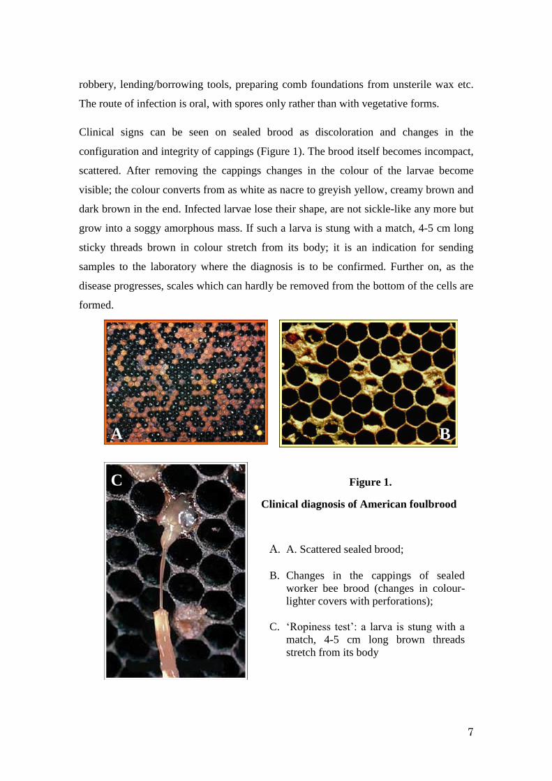
Clinical signs can be seen on sealed brood as discoloration and changes in the
configuration and integrity of cappings (Figure 1). The brood itself becomes incompact,
scattered. After removing the cappings changes in the colour of the larvae become
visible; the colour converts from as white as nacre to greyish yellow, creamy brown and
dark brown in the end. Infected larvae lose their shape, are not sickle-like any more but
grow into a soggy amorphous mass. If such a larva is stung with a match, 4-5 cm long
sticky threads brown in colour stretch from its body; it is an indication for sending
samples to the laboratory where the diagnosis is to be confirmed. Further on, as the
disease progresses, scales which can hardly be removed from the bottom of the cells are
formed.
B A
C
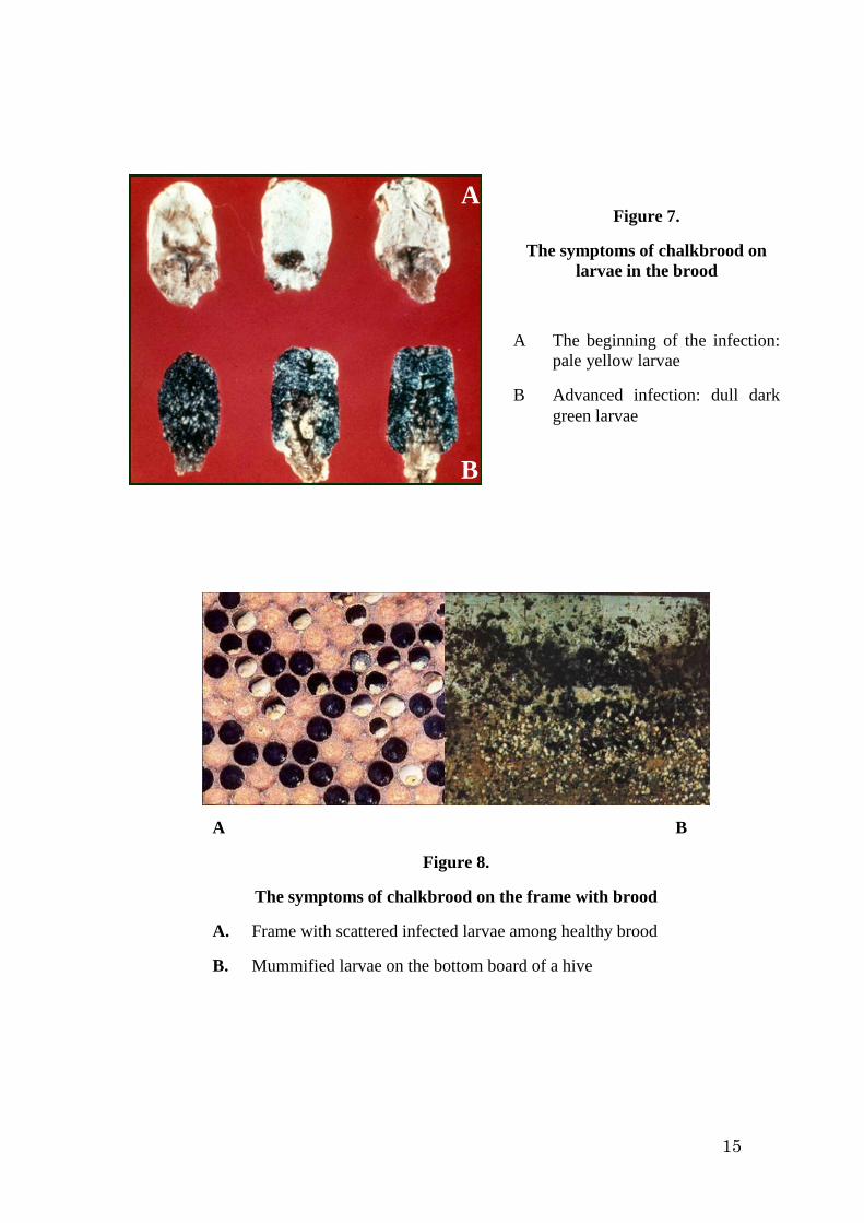
Figure 1.
Clinical diagnosis of American foulbrood
A. A. Scattered sealed brood;
B. Changes in the cappings of sealed
worker bee brood (changes in colour-
lighter covers with perforations);
C. ‘Ropiness test’: a larva is stung with a
match, 4-5 cm long brown threads
stretch from its body
A B
C
8
The process of hydrolysis of diseased larvae and their disintegration into scales last
approximately two months. The colony gets weaker and finally becomes the victim of
numerous wax moths and honey robbery. The diagnosis is confirmed in the laboratory
after the isolation of the pathogenic agent. The material for diagnosis is a frame of a
diseased colony which is to be wrapped in paper. Sacbrood, European foulbrood and
varroosis should be taken into consideration as a differential diagnosis. Unfortunately,
recently beekeepers as well as some experts tend to take various ’prophilactic’ measures
against American foulbrood such as administration of grease patties containing
antibiotics which are sold over the counter (Đuričić et al., 2001). The preventive
application of antibiotics is proposed not only by individuals, but also by
pharmaceutical industry. In addition, private manufacturers of medicines for honeybees
recommend the application of oxytetracycline in patties on the basis of the results of
Wilson et al. and Kulinčević et al., which proved that sugary-oily patties with
oxytetracycline can be used successfully to control American and European foulbrood
when used at the beginning of the diseases (Mlađan and Živanov, 1996). Loss resulting
from American foulbrood which has still been present proves that this is wrongly
believed. In addition, the use of tetracyclines is prohibited. A team of Swedish and
Serbian authors confirmed that there is no reason for the use of antibiotics to control
American foulbrood. They found that there are four strains of Paenibacillus larvae
(ERIC I, ERIC II, ERIC III and ERIC IV) which differ not only in resistance to high
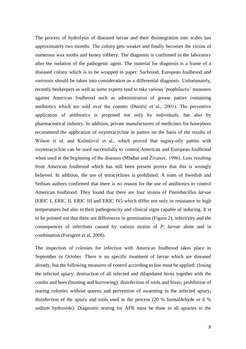
temperatures but also in their pathogenicity and clinical signs capable of inducing. It is
to be pointed out that there are differences in germination (Figure 2), infectivity and the
consequences of infections caused by various strains of P. larvae alone and in
combination (Forsgren et al, 2008).
The inspection of colonies for infection with American foulbrood takes place in
September or October. There is no specific treatment of larvae which are diseased
already, but the following measures of control according to law must be applied: closing
the infected apiary; destruction of all infected and dilapidated hives together with the
combs and bees (burning and burrowing); disinfection of tools and hives; prohibition of
rearing colonies without queens and prevention of swarming in the infected apiary;
disinfection of the apiary and tools used in the process (20 % formaldehyde or 6 %
sodium hydroxide). Diagnostic testing for AFB must be done in all apiaries in the
9
vicinity of the infected (up to 3 km far). After two months the testing is repeated in the
infected apiary and if the results are negative the infection is considered not to be
present any more.
The best means to control AFB is rearing strong colonies of autochthonous
ecogenotypes with pronounced hygienic and grooming behaviour as well as taking care
of biological needs of honeybees as live creatures (Stanimirović et al., 2002, 2003c). In
addition, biological means of combat (replacement of old queens and old combs) and
maintaining hygienic conditions of beekeeping contribute to the production of bee
products free from residua but with all autochthonous biological features.
Having in mind the aforementioned, it is clear that AFB is not connected directly with
CCD (Oldroyd, 2007), but if present latently, which occurs frequently, leads to the
disturbances in the immunity of bee colonies, altered age polyethism and thus the
shortage in high-quality diet and weak hygienic conditions in the colony, which
contributes to CCD (Stanimirović et al., 2008a,b,c).
Figure 2.
Differences in germination of the
four genotypes of Paenibacillus
larvae at various temperatures and
in the presence of antibiotics
(Forsgren et al., 2008).
10
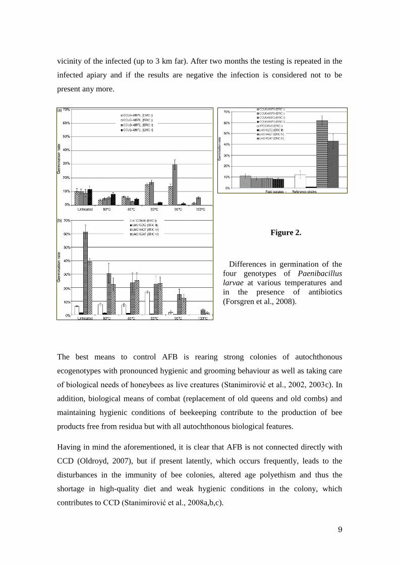
NOSEMOSIS
Nosemosis is a protozoal disease caused by the microsporidium Nosema apis (Figure 3)
which is thought to be in cohabitation with the honeybee for over 60 million years.
There are several successive stages in the lifecycle of Nosema apis: spora, planont,
meront, sporont and sporoblast. The development of spores and sporulation depends on
the temperature, the optimum being between 30 and 34oC. Nosema apis develops in the
cells of the adult bee’s midgut (Mlađan et al., 2000). The infection is not only specific
regarding the tissue, but also the type of the cells which are parasitised.
Spores of Nosema apis remain infective in bees’ cadavers after being kept at 4oC and
90-100% of relative humidity at least 81 days. Spores retain their infectivity in water or
when dried even after 93 days.
Collecting samples for laboratory analysis is of utmost importance since by clinical
means putative diagnosis is obtained only. The best samples to collect are the bees from
the flight entrance and from the top bars of the frames. The best time to identify the
spores is after the winter, at the beginning of the main season. It is difficult to prove the
presence of the spores in summer. There is a slight increase in the number of infected
bees in autumn. Spreading of the disease occurs in the hive, among the colonies and
among the apiaries. In each occasion, forage bees are the primary vectors of nosemosis.
The infected bees’ lifespan is shorter when compared to the uninfected; it might even be
25 to 58 % shorter than expected (Mlađan et al., 1990). It leads to the replacement of
Figure 3. Spores of Nosema apis
11
infected queens (clustering). In addition to this, orientation flights of young worker bees
are delayed, the intake of nectar and pollen decreases or is even omitted, the bees are
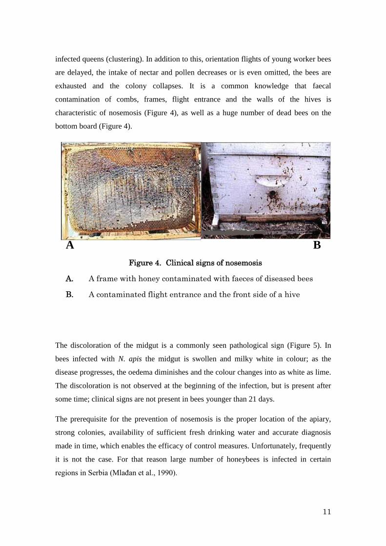
exhausted and the colony collapses. It is a common knowledge that faecal
contamination of combs, frames, flight entrance and the walls of the hives is
characteristic of nosemosis (Figure 4), as well as a huge number of dead bees on the
bottom board (Figure 4).
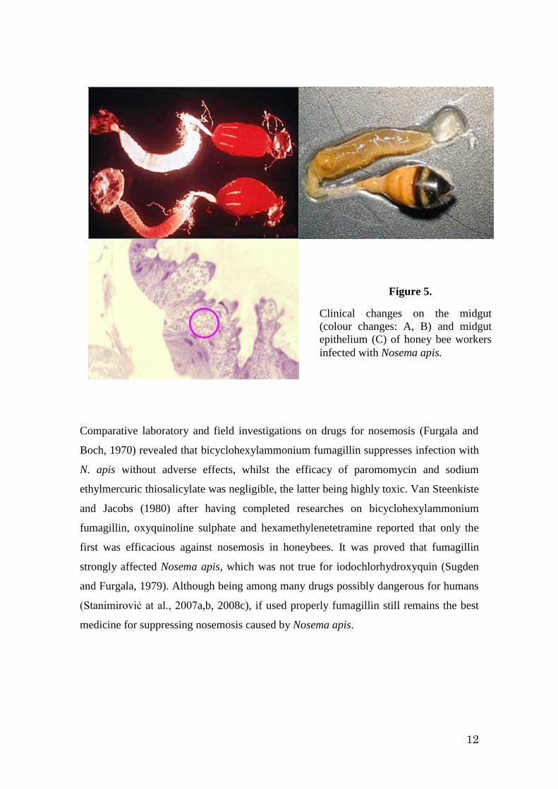
The discoloration of the midgut is a commonly seen pathological sign (Figure 5). In
bees infected with N. apis the midgut is swollen and milky white in colour; as the
disease progresses, the oedema diminishes and the colour changes into as white as lime.
The discoloration is not observed at the beginning of the infection, but is present after
some time; clinical signs are not present in bees younger than 21 days.
The prerequisite for the prevention of nosemosis is the proper location of the apiary,
strong colonies, availability of sufficient fresh drinking water and accurate diagnosis
made in time, which enables the efficacy of control measures. Unfortunately, frequently
it is not the case. For that reason large number of honeybees is infected in certain
regions in Serbia (Mlađan et al., 1990).
A B
A B
Figure 4. Clinical signs of nosemosis
A. A frame with honey contaminated with faeces of diseased bees
B. A contaminated flight entrance and the front side of a hive
12
Comparative laboratory and field investigations on drugs for nosemosis (Furgala and
Boch, 1970) revealed that bicyclohexylammonium fumagillin suppresses infection with
N. apis without adverse effects, whilst the efficacy of paromomycin and sodium
ethylmercuric thiosalicylate was negligible, the latter being highly toxic. Van Steenkiste
and Jacobs (1980) after having completed researches on bicyclohexylammonium
fumagillin, oxyquinoline sulphate and hexamethylenetetramine reported that only the
first was efficacious against nosemosis in honeybees. It was proved that fumagillin
strongly affected Nosema apis, which was not true for iodochlorhydroxyquin (Sugden
and Furgala, 1979). Although being among many drugs possibly dangerous for humans
(Stanimirović at al., 2007a,b, 2008c), if used properly fumagillin still remains the best
medicine for suppressing nosemosis caused by Nosema apis.
A B
C
Figure 5.
Clinical changes on the midgut
(colour changes: A, B) and midgut
epithelium (C) of honey bee workers
infected with Nosema apis.
13
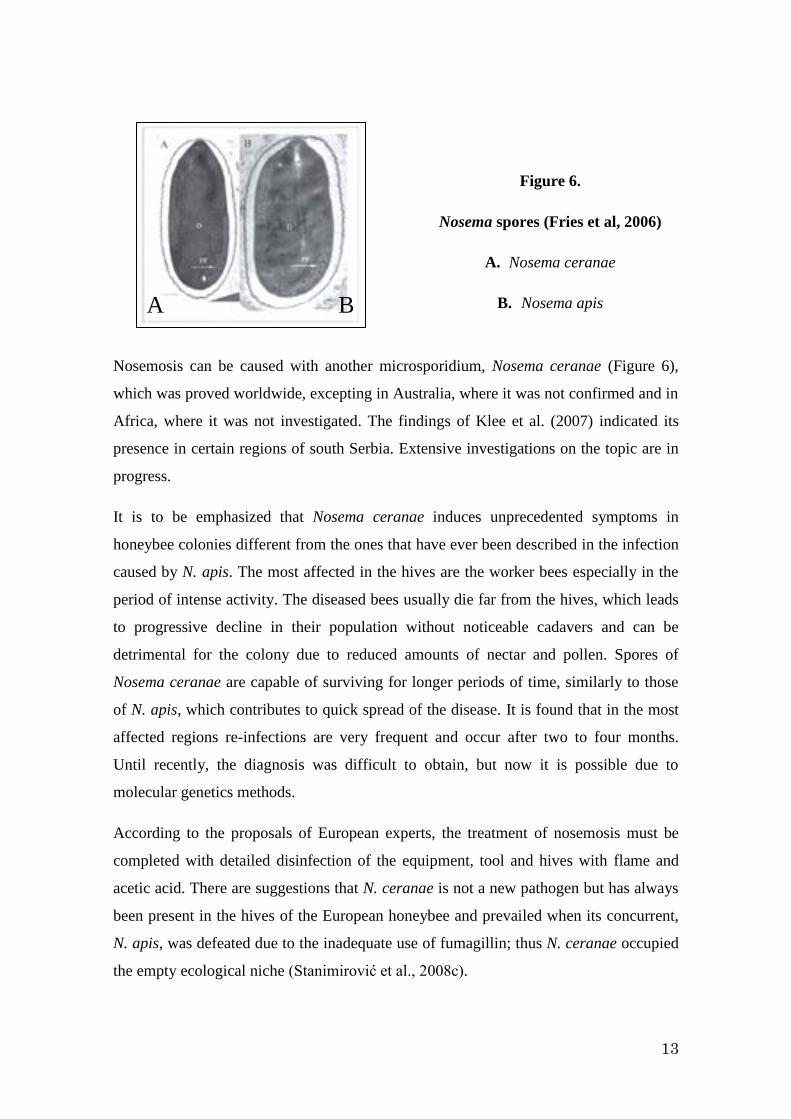
Nosemosis can be caused with another microsporidium, Nosema ceranae (Figure 6),
which was proved worldwide, excepting in Australia, where it was not confirmed and in
Africa, where it was not investigated. The findings of Klee et al. (2007) indicated its
presence in certain regions of south Serbia. Extensive investigations on the topic are in
progress.
It is to be emphasized that Nosema ceranae induces unprecedented symptoms in
honeybee colonies different from the ones that have ever been described in the infection
caused by N. apis. The most affected in the hives are the worker bees especially in the
period of intense activity. The diseased bees usually die far from the hives, which leads
to progressive decline in their population without noticeable cadavers and can be
detrimental for the colony due to reduced amounts of nectar and pollen. Spores of
Nosema ceranae are capable of surviving for longer periods of time, similarly to those
of N. apis, which contributes to quick spread of the disease. It is found that in the most
affected regions re-infections are very frequent and occur after two to four months.
Until recently, the diagnosis was difficult to obtain, but now it is possible due to
molecular genetics methods.
According to the proposals of European experts, the treatment of nosemosis must be
completed with detailed disinfection of the equipment, tool and hives with flame and
acetic acid. There are suggestions that N. ceranae is not a new pathogen but has always
been present in the hives of the European honeybee and prevailed when its concurrent,
N. apis, was defeated due to the inadequate use of fumagillin; thus N. ceranae occupied
the empty ecological niche (Stanimirović et al., 2008c).
A
A
A
B
Figure 6.
Nosema spores (Fries et al, 2006)
A. Nosema ceranae
B. Nosema apis
14
Nosema ceranae attack the guts of adult honeybees and when present in large bumbers,
lead to their disorientation, which can be related to CCD (Oldroyd, 2007). In addition,
this endoparasite provokes and maintains a high-level energetic stress, which results in
high food consumption, continuous hunger and heavy agitation of bees (Mayack and
Naug, 2009). They leave the hives even in bad weather conditions which contributes to
a decrease in their number, alter the age polyethism and contributes to CCD
(Stanimirović et al., 2008a,b,c).
CHALKBROOD
Chalkbrood is a disease of the honeybee which is caused by the fungus Ascosphaera
apis.
a) Symptoms on the larvae in the brood (Figure 7)
At the beginning of the infection the larvae are pale yellowish in colour, soft, smooth,
their shape varies and as the disease progresses they grow light yellow, become rough,
their consistence is skinny and can be fragile. They are smothered in white mycelium
wrappings which thicken rapidly and in a short time fill the whole space in the cells.
The mycelium adheres to hind part of the larva whilst the head remains free, dry and
resembles a button. Further on, the size of old mummified larvae decline due to
dehydration and they seem to be transformed into chunks of chalk; hence, the name of
the disease. The larvae in the advanced disease are dark dull green.
b) Symptoms on the frames with the brood (Figure 8)
Infected young larvae, which still seem healthy, are usually scattered among healthy
brood, whilst the older ones, already mummified, are in sealed cells or sometimes in
cells that the workers already opened. Generally, the cappings appear normal, but can be
mottled or slightly concave. When the combs with sealed cells are cut lengthways, the
mummified larvae (Figure 9) easily fall out. When the mycelium penetrates the
cappings and covers them from outside, sealed broods seem as if it was sprinkled with
flour, lime or greyish dust.
A B
15
A B
Figure 8.
The symptoms of chalkbrood on the frame with brood
A. Frame with scattered infected larvae among healthy brood
B. Mummified larvae on the bottom board of a hive
A B
Figure 7.
The symptoms of chalkbrood on
larvae in the brood
A The beginning of the infection:
pale yellow larvae
B Advanced infection: dull dark
green larvae
A
B
16
The tentative diagnosis is based on the clinical signs, time of the appearance and the
absence of the disease in adult worker bees. The accurate diagnosis can be obtained in
the laboratory after microscopic assessment of mummified larvae (Figure 9) or by
isolation of Ascosphaera apis in vitro. The samples for diagnosis are frames with
mummified drone or worker brood or parts of brood sized 10 x 10-14 cm. Each sample
must be packed separately in a paper box.
There is no registered chemical for the treatment of chalkbrood. If the disease is benign,
it is sufficient to remove, destroy (burn) and replace frames with diseased brood, move
the hives to dry sunny places, and maintain well ventilated colonies to keep the interior
of hives dry and without fungus. In badly infected colonies re-queening is necessary and
thorough cleaning and disinfection of the hives with flame. Combs with infected brood
are to be burnt or remelted. In order to encourage hygienic behaviour supplemental
feeding or sprinkling with sugar is recommended.
Having in mind the aforementioned, it can be seen that chalkbrood is not connected
directly with CCD (Oldroyd, 2007), but if present, contributes to disturbances in the
immunity of the colonies, affects the age polyethism and results in the shortage in high-
quality diet and weak hygienic conditions in the colony, which lead to CCD
(Stanimirović et al., 2008a,b,c).
Figure 9.
Mummified larvae suitable for the
laboratory diagnosis of chalkbrood
17
VARROOSIS
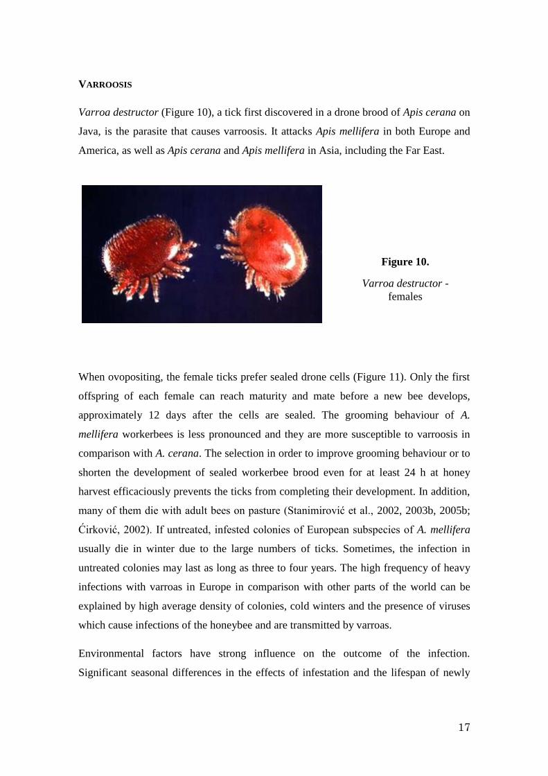
Varroa destructor (Figure 10), a tick first discovered in a drone brood of Apis cerana on
Java, is the parasite that causes varroosis. It attacks Apis mellifera in both Europe and
America, as well as Apis cerana and Apis mellifera in Asia, including the Far East.
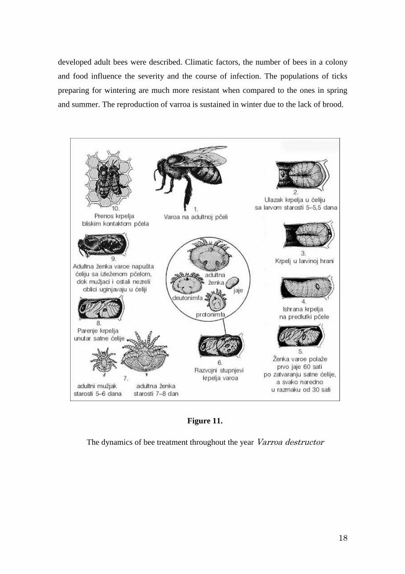
When ovopositing, the female ticks prefer sealed drone cells (Figure 11). Only the first
offspring of each female can reach maturity and mate before a new bee develops,
approximately 12 days after the cells are sealed. The grooming behaviour of A.
mellifera workerbees is less pronounced and they are more susceptible to varroosis in
comparison with A. cerana. The selection in order to improve grooming behaviour or to
shorten the development of sealed workerbee brood even for at least 24 h at honey
harvest efficaciously prevents the ticks from completing their development. In addition,
many of them die with adult bees on pasture (Stanimirović et al., 2002, 2003b, 2005b;
Ćirković, 2002). If untreated, infested colonies of European subspecies of A. mellifera
usually die in winter due to the large numbers of ticks. Sometimes, the infection in
untreated colonies may last as long as three to four years. The high frequency of heavy
infections with varroas in Europe in comparison with other parts of the world can be
explained by high average density of colonies, cold winters and the presence of viruses
which cause infections of the honeybee and are transmitted by varroas.
Environmental factors have strong influence on the outcome of the infection.
Significant seasonal differences in the effects of infestation and the lifespan of newly
Figure 10.
Varroa destructor -
females
18
developed adult bees were described. Climatic factors, the number of bees in a colony
and food influence the severity and the course of infection. The populations of ticks
preparing for wintering are much more resistant when compared to the ones in spring
and summer. The reproduction of varroa is sustained in winter due to the lack of brood.
Figure 11.
The dynamics of bee treatment throughout the year Varroa destructor
19
Varroosis is the disease of both the brood and adult bees. Sources of infection are
infected colonies, package bees, contact with the diseased bees, natural swarms, queens
and brood. In summer, varoosis can spread as far as 11 km and more within three
months. In heavy infections (more than 20 ticks per 100 bees in a hive) in autumn and
summer death of the brood can be observed, as well as the discharge of dead larvae
(drone and workerbee), young bees and drones. In autumn and winter, the bees in
infected colonies are unsettled and frequently die at the beginning of winter.
At first, the disease develops slowly, is unnoticed and does not influence the
productivity of the colony. Clinical symptoms appear after 2-3 years. The ticks reduce
the quantity of dry substance, total nitrogen, fatty acids and fat body in infected larvae,
and increase the energy waste during respiration. There is a decrease in resistance to
diseases and the strength of the colony. The symptoms appear if more than 20% of bees
are infected. In winter unsettlement, buzzing, leaving the hives, diarrhoea and death
occur. In spring and summer pupae die and the colonies’ strength lessens as a
consequence of incapability of offspring for surviving. The fertility of queens
diminishes significantly, they do not mate and the brood is scattered. Workerbees are
inactive during honey harvest, which leads to reduced honey production and the
incapability of bees to accommodate enough food for themselves.
The spread of infection in the brood changes throughout the year. In spring and autumn,
in the absence of drone brood, the workerbee brood is infected and vice versa. The
majority of ticks is on the worker brood, which causes the emergence of high number of
damaged bees incapable of flying. In summer female ticks reproduce in drone brood,
where there is plenty of high-quality protein diet and the temperature is much lower
than in the worker brood. The damage caused by varroas depends not only on the
number of ticks in the attacked colony, but is also connected with secondary viral
infections. The virus of acute bee paralysis is the most deleterious, at least in Europe. It
leads to latent infections, not causing visible body damage. The ticks activate the
viruses when infesting bees, transport them onto open and sealed broods, which show
unspecific signs, especially in heavily infested colonies. Adult bees, in which the
viruses are active, can infect young larvae when feeding them with the secretion of
mandibular and thoracic glands. Having ingested sufficient quantities of viruses, larvae
20
die before their cells are sealed; those who survive continue development into latently
infected adult bees. The virus of acute paralysis can sometimes be found even in the
pollen collected by seemingly uninfected bees as well as in their thoracic salivary
glands (Ponten and Ritter, 1992; Pohl and Ritter, 1997; Békési et al., 1999).
Varroosis is nowadays the biggest problem in beekeeping in Serbia and, similarly, in the
majority of the world. Each and every year it is necessary to treat bees in order to
control the infection, in other words, to keep less than 3 % of the bees infected.
It is of utmost importance that the number of ticks on bees is as little as possible at the
beginning of winter. In Europe, several acaricides against varroa are approved, but all
exert adverse effects on bees (Stanimirović et al., 2003a,c, 2005a). The problem is that
acaricides cannot reach the ticks in the sealed brood. Systemic acaricides given to bees
in food which can reach larvae in sealed brood via food are considered ideal (e.g.
cymiazol hydrochloride). Short- or long-term application of the evaporated formic acid
is deleterious to the majority of varroas including the ones in sealed cells. Geraniol, the
component of Nasanov’s glands of forager bees, repels the migrating mites, which was
proved in the numerous experiments. Chemicals should be applied with great care due
to their residua in bee products and possible toxic effects (Stanimirović et al., 2003a,c,
2005a, 2006, 2007a,b; Stevanović et al., 2008). In addition, the application of acaricides
can result in resistance, as it is proven for fluvalinates.
Manipulative treatment against varroosis includes diminishing drone broods in infested
colonies (building frame, bait of drone and workerbee comb, TNT frames etc) in order
to prevent the migration of female Varroa destructor into sealed cells, where they avoid
chemical treatment. The drawbacks of this method are the destruction of monthly brood
production, it is tedious and favours the effects of nosemosis and acarosis.
Recently, a biophysical method, heating of the brood, is more frequently being applied
in combat with varoosis. Huang (2001) applied temperature treatment of the hives
themselves. The difficulty which arises from this treatment is the melting of wax, but it
can be solved by replacing wax comb foundation with the one made of heatproof
plastic.
21
Certain biological means of combat against varroas are recommended: the use of their
natural enemies, pathogen fungi Hirsutella thompsonii and Metarrhizium anisopliae,
which are quite efficacious against the ticks and are harmless to bees (Shaw et al., 2002;
Kanga et al., 2002, 2003; Peng et al., 2002). It has been proved experimentally that
treatment with precisely defined numbers of dry spores of these fungi do not influence
the number of eggs laid by queens, and do no harm to the brood, larvae, pupae and adult
bees. In laboratory conditions, ticks were infected with fungi while allowed to walk on
the culture of H. thompsonii for five minutes. It was discovered by SEM that the
membranous ambulacrae on the ticks’ legs were the places where the conidia of the
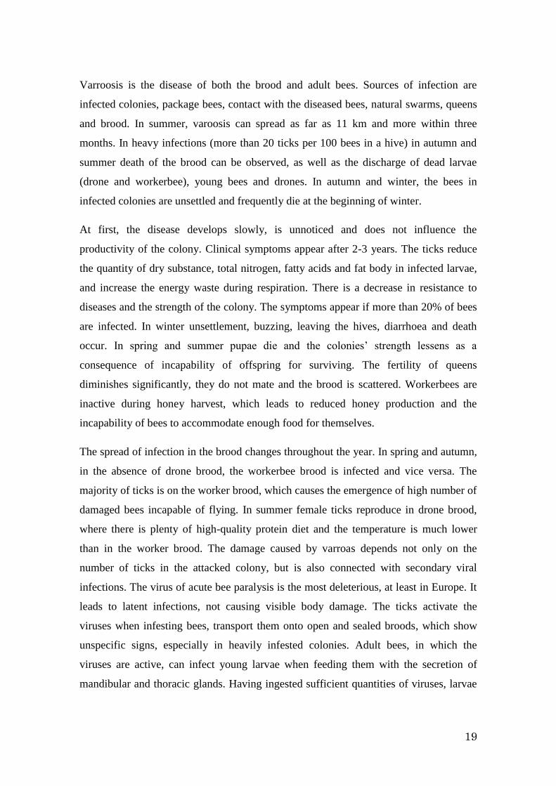
fungi adhere and germinate (Figure 12). The infected ticks died from mycosis within
52.7 to 96.7 hours (LT50), which depended on the isolate of fungi.
VIRAL INFECTIONS
Viruses are obligate intracellular parasites which can virtually be found in any living
organism. They are incapable of any metabolic activity on their own and, thus, can live
and reproduce only in live cells. Once in host cells viruses use their metabolism,
machinery and components to produce their own offspring, the virions. This process
Figure 12.
Distal parts of legs of Varroa destructor before and after the treatment with
Hirsutella thompsonii spores
22
does harm to the host, leading to diseases or even death. Due to their powerful effects
viruses are possibly the most serious challenge to the health of living organisms.
In general, there are two means of transmission of viruses: horizontal and vertical.
Horizontal transmission occurs among the organisms of the same generation and can be
direct or indirect. Direct transmission is completed by contact, or via food, water and
air. On the other hand, indirect transmission depends on vectors, among which varroas
and nosemas are the most important. Vertical transmission occurs from mothers to the
offspring via eggs (transovum). It is supposed that these various means of transmission
influence the virulence of pathogens. Typically, horizontal transmission favours the
onset of diseases and enhances the prevalence of infections under certain circumstances,
for example in high-density populations (as are in beekeeping) and in cases where there
is a high replication of pathogens. In contrast, vertical transmission is a mechanism that
enables the long-lasting persistence and survival of viruses and favours the evolution of
benign infections. The result of a viral infection can depend on the balance of these two
means of its transmission.
Similarly to other organisms, honeybees are exposed to various pathogens including
viruses, which pose major threat to their health. Until now, at least eighteen viruses are
described which attack bees worldwide and can dramatically influence their health
under certain conditions (Martin, 2001). Due to dense populations and frequent contacts
among the members of the society (feeding, chemical communication), honeybee
colonies are especially prone to the transmission of diseases. Although there are gaps in
the knowledge of the most important processes underlying in the dynamics of the
transmission of viruses, casting light on the means of transmission of viruses among
bees is being developed quickly, thus our knowledge of transmission and epidemiology
of viral infection in bees has considerably improved during the last decade.
There is no possible direct and efficacious treatment of the viral infection of honeybees.
Some viral infections (acute paralysis) can be solved by re-queening with queens from
different parts of the world, which, on the other hand, poses a high risk of importing
exotic pathogens. Having considered that many viruses are connected with the varroa
and that there is no known adequate medicine to combat them, it is plausible that only
having defeated varroosis we can defeat viral infections.
23
Heavy infections with ticks result in the weakness of colonies and it can be claimed that
varroas virtually lead to the decrease in the immunity of honeybee colonies. Untreated
colonies usually die in 3-4 years. Very frequently viruses transmitted by varroas
contribute to the collapse of a colony (Martin, 2001). However, it is asserted that in
Europe there were many viruses detected in honeybee colonies even before the presence
of the varroa, but clinical manifestations were observed only sporadically. Thus the
presence of viruses did not influence the losses in beekeeping and was disregarded
(Allen and Ball, 1996). The situation changed dramatically with the appearance of
varroa ticks in Europe. Having in mind the direct correlation between the intensity of
infection with varroas and the appearance of viral diseases, it is supposed that the
presence of ticks plays the main role in the onset of clinical signs of viral diseases
(Nordstrom et al., 1999). The ticks exhaust bees and, in addition, have negative
consequences as biological and/or mechanical vectors and/or activators of other
pathogens, especially viruses (Yue and Genersch, 2005; Shen et al., 2005a,b; Berényi et
al., 2006) which lead to the collapse of bee colonies (CCD). The presence of viruses in
ticks and their transmission by Varroa destructor have recently been proved by
molecular methods, especially concerning the virus which causes the deformation of
wings (DWV) (Genersch, 2005; Chen et al., 2005), the virus of acute bee paralysis
(ABPV) (Bakonyi et al., 2002; Tencheva et al., 2004), the virus of sacbrood (SBV)
(Chen et al., 2004; Shen et al., 2005a,b) and the virus of black queen cells (BQCV)
(Chantawannakul et al., 2006). It was also proved that one single V. destructor can be
infected with all aforementioned viruses. The co-egsistance of numeral viruses clearly
proves their role in the transmission of viruses and development of viral diseases in bee
colonies (Chantawannakul et al., 2006). Recently, the replication of Kashmir bee virus
(KBV), SBV and DWV in varroas has experimentally been proved, as well as their
presence in the ticks’ saliva, which undoubtedly confirms the role of these ectoparasites
as biological vectors of the viruses that attack honeybees (Ongus et al., 2004; Shen et
al., 2005a,b).
The connection between viral infections and varroa-infestation in bee colonies is the
most complicated aspect of parasitic relationship between bees and V. destructor.
Nowadays, much attention is being given to this problem in order to cast light on the
means of the transmission of viruses (Berényi et al., 2006; Chen et al., 2006). In Europe,
24
viruses the most often transmitted by varroas are DWV and acute paralysis virus (APV)
(Tentcheva et al., 2006; Berényi et al., 2006). The most harmful is APV, which infects
the bees latently not causing any visible damage to their bodies. The ticks activate the
viruses when infesting bees and transmit them to both open and sealed brood which
exerts non-specific signs. The lifespan of bees that were infected with APV in the pupal
stage is shorter, thus they can only work as nursing bees for a short time (Békési et al.,
1999). Adult bees with active viruses can infect young larvae most probably via
mandibular and thoracic secretions when feeding the offspring. Larvae which ingested
large quantities of viruses die before the brood is sealed; those who survive continue
their development into latently infected adults. APV can sometimes be found even in
pollen collected by seemingly healthy bees, as well as in their thoracic salivary glands.
DWV replicates slowly and if present in large amounts leads to malformation of the
bee’s wings in praepupal stage (even before the pigmentation of eyes). However, the
presence of this virus was also proved in bees with normal wings, but in numbers
approximately ten times smaller than in the ones with deformities (Chen et al., 2005;
Tentcheva et al., 2006).
For unknown reasons, in some years, bee viruses appear scarcely ever. In the absence of
viral infections connected with the ticks, honeybee colonies easily tolerate several-
thousand populations of varroas. However, when the bees are attacked with both ticks
and viruses, much lower number of varroas can result in CCD because the viruses
enhance the effects of varroosis (Denholm, 1999).
CCD and the Israeli virus of acute paralysis (IAPV). Recent findings of Cox-Foster et
al. (2007) indicated the connection between CCD and a new virus, the Israeli virus of
acute paralysis. This virus was found in all the colonies suffering from CCD, and, by
contrast, was not identified in healthy ones. IAPV was first recognised in Israel and
later in bees imported from Australia and royal jelly from China. The precise
geographic origin of this virus is yet to be known.
Is it proved that the IAPV is the reason for CCD? No, it is not for certain. It is claimed
that IAPV is possibly connected to CCD, but further investigations into this problem are
necessary to confirm or discard this assumption. It can be concluded that IAPV is a
25
marker for CCD but that, most possibly, other stressogens contribute to the onset of
CCD, such as Varroa, other viruses, Nosema, fungi, pesticides, inadequate diet and
management in the apiary.
To conclude, multi-task care of honeybee colonies is necessary since it is the only way
how to prevent the presence of complicated parasitic-viral infections. It should include:
adequate hygienic-sanitary measures, application of drugs which varroas are not
resistent to, the selection of honeybees in order to favour colonies with highly expressed
hygienic and grooming behaviour, and selection of queens which posses the SMR gene
responsible for the synthesis of proteins that influence the reproduction of adult female
varroas (Harbo and Harris, 1999).
MANAGEMENT IN THE APIARY
Management in the apiary, from the choice of the place where to put it, the type of
hives, the quality and timely replacement of the wax in the hives, to the choice and
administration of medicines etc. is very important for the functioning of bee colonies. If
any of these is omitted or is done wrongly, CCD is very likely to occur. Hereby, the
importance of timely replacement of wax in the hives is emphasized.
If the beekeepers had replaced only one third of old combs in a year, the losses caused
by infestation with varroas would be reduced. In old combs (in cocoons of several
generations hatched workerbees) there is a chemical originated from the cocoons of
fifth-stage larvae, which stimulates the oviposition in female Varroa (Garrido and
Rosenkranz, 2004) . Clean wax from newly made combs is free of the substance since
there were no brood in their cells. This was proved by low numbers of activated
terminal oocytes in varroas tested in the presence of larvae in newly built combs. The
only difference between new wax and the one in which several generations of bees were
hatched is in the presence of exuvia (sheets discarded by moulting larvae) in the latter.
The higher activation in the presence of larvae from combs that was used for several
cycles is possibly the consequence of additive effects of cuticular substances of larvae
and their exuvia in the cells (Stevanović, 2007).
26
What is to be done when CCD occurs?
In the remaining hives:
► Control the infection with varroas
► Treatment against Nosema, if it is present
► Do not use anything from the abandoned hives
What else can be done?
Since not all the factors that contribute to CCD are known and there is no treatment
against bee viruses, the best option is to maintain and support the health and strength of
bee colonies, provide young and healthy queens and enough high-quality food, avoid
import of reproductive material and swarms from regions where CCD was registered
and regular control of reproductive and other bee material.
REFERENCES:
1. Allen MF, Ball BV (1996) The incidence and world distribution of honey bee
viruses. Bee World, 77, 141-162.
2. Arathi HS, Burns I, Spivak M (2000) Ethology of hygienic behaviour in the the
honey bee, Apis mellifera (Hymenoptera: Apidae): Behavioural repertoire of
hygienic bees. Ethology, 106 (4) 365-379.
3. Bakonyi T, Farkas R, Szendroi A, Dobos-Kovacs M, Rusvai M (2002) Detection
of acute bee paralysis virus by RT-PCR in honey bee and Varroa destructor
field samples: rapid screening of representative Hungarian apiaries. Apidologie,
33, 63–74.
4. Békési L, Brenda VD, Dobos-Kovàcs M, Bakonyi K, Rusvai M (1999)
Occurence of acute paralysis virus of the honey bee (Apis mellifera) in a
Hungarian apiary infested with the parasitic mite Varroa jacobsoni. Acta
Veterinaria Hungarica, 47, 319-329.
5. Berényi O, Bakony T, Derakhshifar I, Köglberger H, Nowotny N (2006)
Occurrence of Six Honeybee Viruses in Diseased Austrian Apiaries. Applied &
Environmental Microbiology, 72, 2414-2420.
6. Chantawannakul P, Ward L, Boonham N, Brown M (2006) A scientific note on
the detection of honeybee viruses using real-time PCR (TaqMan) in Varroa
27
mites collected from a Thai honeybee (Apis mellifera) apiary. Journal of
Invertebrate Pathology, 91, 69–73
7. Chen YP, Higgins JA, Feldlaufer MF (2005) Quantitative Real-Time Reverse
Transcription-PCR Analysis of Deformed Wing Virus Infection in the Honeybee
(Apis mellifera L.). Applied & Environmental Microbiology, 71, 436-441.
8. Chen Y, Evans J, Feldlaufer M (2006) Horizontal and vertical transmission of
viruses in the honey bee, Apis mellifera. Journal of Invertebrate Pathology, 92,
152–159.
9. Cox-Foster DL, Conlan S, Holmes EC, Palacios G, Evans JD, Moran NA, Quan
PL, Briese T, Hornig M, Geiser DM, Martinson V, vanEngelsdorp D, Kalkstein
AL, Drysdale A, Hui J, Zhai J, Cui L, Hutchison SK, Simons JF, Egholm M,
Pettis JS, Lipkin WI. (2007) A metagenomic survey of microbes in honey bee
colony collapse disorder. Science, 318 (5848) 283-287.
10. Ćirković D, 2002, Reproduktivno - produktivna i higijensko - negovateljska
karakterizacija sjeničko-pešterskog ekotipa medonosne pčele. Magistarski rad,
Fakultet veterinarske medicine, Univerzitet u Beogradu.
11. Đuričić Bosiljka, Radojičić Sonja, 2000, Uloga veterinarske struke u očuvanju
zdravlja pčela i ljudi i razvoju pčelarstva. Zbornik radova 7. Savetovanja
veterinara Republike Srpske sa mećunarodnim učešćem, 6-10. jun, Teslić, Banja
Vrućica.
12. Đuričić Bosiljka, Bošnjak Mirjana, Plavša Nada, 2001, Epizootiološka slika
američke kuge pčelinjeg legla sa posebnim osvrtom na moguće greške u terapiji.
Zbornik plenarnih radova. I Savetovanje o biologiji i zdravstvenoj zaštiti pčela,
Dec 22, Beograd, 1-7.
13. Forsgren E, Stevanovic J, Fries I, Variability in germination and in temperature
and storage resistance among Paenibacillus larvae genotypes, Veterinary
Microbiology, 2008, 129 (3-4) 342-349
14. Fries I, Martín R, Meana A, García-Palencia P, Higes M (2006) Natural
infections of Nosema ceranae in European honey bees. Journal of Apicultural
Research, 45 (4), 230–233.
15. Furgala B, Boch R (1970) The Effect of Fumidil-B, Nosemack and Humatin on
Nosema apis. Journal of Apicultural Research, 9 (2) 79-85.
16. Garrido C, Rosenkranz P (2004) Volatiles of the honey bee larva initiate
oogenesis in the parasitic mite Varroa destructor. Chemoecology, 14, 193-197.
17. Genersch E (2005) Development of a rapid and sensitive RT-PCR method for
the detection of deformed wing virus, a pathogen of the honeybee (Apis
mellifera). Veterinary Journal, 169, 121–123.
18. Harbo JR, Harris JW, 1999, Selecting honey bees for resistance to Varroa
jacobsoni. Apidologie 30, 183-196.
28
19. Huang Z (2001) Mite zapper – a new and effective method for Varroa mite
control. American Bee Journal, 141 (10) 730–732.
20. Kanga LHB, James RR, Boucias DG (2002) Hirsutella thompsonii and
Metarhizium anisopliae as potential microbial control agents of Varroa
destructor, a honey bee parasiite. Journal of Invertebrate Pathology, 81, 175-
184.
21. Kanga LHB, Jones WA, James RR (2003) Field Trials Using the Fungal
Pathogen, Metarhizium anisopliae (Deuteromycetes: Hyphomycetes) to Control
the Ectoparasitic Mite, Varroa destructor (Acari: Varroidae) in Honey Bee, Apis
mellifera (Hymenoptera: Apidae) Colonies. Journal of Economic Entomology,
96 (4) 1091-1099.
22. Klee J, Besana AB, Genersch E, Gisder S, Nanetti A, Tam DQ, Chinh TX,
Puerta F, Ruz JM, Kryger P, Message D, Hatjina F, Korpela S, Fries I, Paxton
RJ (2007) Widespread dispersal of the microsporidian Nosema ceranae, an
emergent pathogen of the western honey bee, Apis mellifera. Journal of
Invertebrate Pathology, 96 (1) 1-10
23. Laušević D, Milovanović R, Pejović N, 2001, Epizootiologija američke kuge
legla u Crnoj gori. Zbornik plenarnih radova. I Savetovanje o biologiji i
zdravstvenoj zaštiti pčela, Dec 22, Beograd, 54-60.
24. Martin SJ (2001) The role of Varroa and viral pathogens in the collapse of
honeybee colonies: a modelling approach. Journal of Applied Ecology, 38,
1082–1093.
25. Mayack C, Naug D (2009) Energetic stress in the honeybee Apis mellifera from
Nosema ceranae infection. Journal of Invertebrate Pathology, 100, 185-188.
26. Mlađan V, Lolin Miroslava, Cokić J, 1990, Epizootiološka slika bolesti pčela u
Zaječarskom regionu. Veterinarski glasnik, 44, 845-850.
27. Mlađan V, Živanov D, 1996, Upotreba antibiotika u pčelarstvu, Zbornik radova
2. Savetovanja o lekovima za upotrebu u veterini, Igalo.
28. Mlađan V, Todorović D, Lolin M, 2000, Preventive action of Fumagillin on the
degree of infection with Nosema apis in the digestive tract of bees. Acta
Veterinaria, 50 (4) 241-252.
29. Nordström S, Fries Aarhus A, Hansen H, Korpela S (1999) Virus infection in
Nordic honey bee colonies with no, low or severe Varroa jacobsoni infestations.
Apidologie 30, 475–484.
30. OIE Manual of standards Diagnostic Tests and Vaccines, 2000, Chapter 2.9.2.
American foulbrood, http://www.oie.int/Eng/Normes/Mmanual/A_00118.htm
31. Oldroyd BP (2007) What's killing American honey bees? PLoS Biology, 5(6)
1195-1199.
29
32. Ongus JR, Peters D, Bonmatin JM, Bengsch E, Vlak JM, Oers MMV (2004)
Complete sequence of a picorna-like virus of the genus Iflavirus replicating in
the mite Varroa destructor. Journal of General Virology, 85, 3747–3755.
33. Pejin II, Stanimirović Z, Stevanović J, Kulišić Z (2006) Evaluacija
genotoksičnosti potencijala amitraza citogenetičkim testom in vivo, Veterinarski
glasnik, 60 (3-4) 163-173.
34. Peng CYS, Zhou X, Kaya HK (2002) Virulence and site of infection of the
fungus, Hirsutella thompsonii, to the honey bee ectoparasitic mite, Varroa
destructor. Journal of Invertebrate Pathology, 81, 185-195.
35. Pohl F, Ritter W (1997) Neue ergebnisse zu zwei virosen (APV, SBV) der
honigbiene. Apidologie, 28, 174-176.
36. Ponten A, Ritter W (1992) Influence of acute paralysis virus attacks on brood
care in honeybees. Apidologie, 23, 363-365.
37. Shaw KE, Davidson G, Clark SJ, Ball BV, Pell JK, Chandler D, Sunderland KD
(2002) Laboratory bioassays to assess the pathogenicity of mitosporic fungi to
Varroa destructor (Acari: Mesostigmata), an ectoparasitic mite of the honeybee,
Apis mellifera. Biological Control, 24, 266-276.
38. Shen MQ, Cui LW, Ostiguy N, Cox-Foster D (2005a) Intricate transmission
routes and interactions between picorna-like viruses (Kashmir bee virus and
sacbrood virus) with the honeybee host and the parasitic varroa mite. Journal of
General Virology, 86, 2281–2289.
39. Shen MQ, Yang XL, Cox-Foster D, Cui LW (2005b) The role of varroa mites in
infections of Kashmir bee virus (KBV) and deformed wing virus (DWV) in
honey bees. Virology, 342, 141–149.
40. Stanimirović Z, Pejović D, Stevanović J, Vučinić M, Mirilović M (2002)
Investigations of hygienic behaviour and disease resistance in organic
beekeeping of two honeybee ecogeographic varieties from Serbia. Acta
Veterinaria, 52 (2-3) 169-180.
41. Stanimirović Z, Todorović D, Stevanović J, Mladenović M, Janković L,
Djordjević M (2003a) Influence of cymiazole hydrochloride on mitotic and
proliferative activities of cultured human lymphocytes, Acta Veterinaria 53 (1)
47-55.
42. Stanimirovic Z, Stevanovic J, Cirkovic D (2003b) Investigations of
reproductive, productive, hygienic and grooming features of Syenichko-
Peshterski honey bee ecotype. Apidologie 34 (5) 487-488.
43. Stanimirović Z, Fišter Svetlana and Stevanović Jevrosima (2003c) Analysis of
sister chromatid exchanges in cultured human lymphocytes treated with
cymiazole hydrochloride. Acta Veterinaria 53 (5-6) 419-425
30
44. Stanimirović Z, Stevanović J, Mladenović M, Nedić N (2003d) Ekološka
kontrola i strategija borbe protiv varoze. II Savetovanje o biologiji i
zdravstvenoj zaštiti pčela, Nov 22, pp 45-66. Beograd, YU.
45. Stanimirović Z, Stevanović J, Jovanović S, Andjelković M (2005a) Evaluation
of genotoxic effects of Apitol® (cymiazole hydrochloride) in vitro by
measurement of sister chromatid exchange, Mutation Research, 588 (2), 152-
157.
46. Stanimirovic Z, Stevanovic J, Cirkovic D (2005b) Behavioural defenses of the
honey bee ecotype from Sjenica – Pester against Varroa destructor, Acta
Veterinaria, 55 (1) 69-82.
47. Stanimirović Z, Stevanović J, Kulić M, Stojić V (2006) Frequency of
chromosomal aberrations in the evaluation of genotoxic potential of
dicyclohexylamine (fumagillin) in vivo, Acta Veterinaria, 56 (4), 353-366.
48. Stanimirović Z, Stevanović J, Bajić V, Radović
I (2007a) Evaluation of
genotoxic effects of fumagillin (dicyclohexylamine) by citogenetic tests in vivo,
Mutation Research, 628 (1), 1-10.
49. Stanimirović Z, Pejin II, Kulišić Z, Djiporović M (2007b) Evaluation of
genotoxic effects of fumagillin by sister chromatide exchange and chromosomal
aberration tests in human cell cultures. Acta Veterinaria, 57 (2-3), 257-273.
50. Stanimirović Z, Stevanović J, Ćirković D (2008a) Possible causes of the collony
collapse disorder (CCD) I deo. Pčelarski žurnal 1, 2-7.
51. Stanimirović Z, Stevanović J, Ćirković D (2008b) Possible causes of the collony
collapse disorder (CCD) I deo. Pčelarski žurnal 2, 10-15.
52. Stanimirović Z, Stevanović J, Ćirković D (2008c) Possible causes of the collony
collapse disorder (CCD) III deo. Pčelarski žurnal 3, 21-25.
53. Stevanovic J, Stanimirovic Z, Pejin II, Lazarevic M (2006) Monitoring of
mitotic index and frequency of micronuclei in evaluation of genotoxic potential
of fumagillin (dicyclohexylamine) in vivo, Acta Veterinaria, 2006, 56 (5-6) 437-
448.
54. Stevanovic J, Stanimirovic Z, Radakovic M, Stojic V (2008) In vitro evaluation
of the clastogenicity of fumagillin, Environmental and Molecular Mutagenesis,
49 (8) 594-601.
55. Stevanović J (2007) Ekološko-etološki odbrambeni mehanizmi Apis mellifera
carnica prema ektoparazitu Varroa destructor na području Srbije. PhD thesis.
Biološki fakultet, Univerzitet u Beogradu, pp 1-202.
56. Sugden MA, Furgala B (1979) Enteroseptol ineffective against Nosema apis.
American Bee Journal, 119 (8) 594-596.
57. Šovljanski R (2008a) Pesticidi i pčele. Zbornik radova XXVI Savetovanja
pčelara, Novi Sad.
31
58. Šovljanski R (2008b) Trovanje pčela insekticidima. Zbornik radova XXVI
Savetovanja pčelara, Novi Sad.
59. Tentcheva D, Gauthier L, Bagny L, Fievet J, Dainat B, Cousserans F, Colin ME,
Bergoin M (2006) Comparative analysis of deformed wing virus (DWV) RNA
in Apis mellifera and Varroa destructor. Apidologie 37, 41–50.
60. Van Steenkiste D, Jacobs FJ (1980) A Comparison on the Activity of Fumidil-B,
Chinosol and Urotropine Against Nosema apis Zander. XXVIIth International
Congress of Apiculture. Athens, Apim Pub House 380-382.
61. Yue C, Genersch E (2005) RT-PCR analysis of Deformed wing virus (DWV) in
honey bees (Apis mellifera) and mites (Varroa destructor). Journal of General
Virology, 86, 3419–3424.