Embed Size (px)
Citation preview

children
Article
Bidirectional Ductal Shunting and Preductal toPostductal Oxygenation Gradient in PersistentPulmonary Hypertension of the Newborn
Amy Lesneski, Morgan Hardie, William Ferrier, Satyan Lakshminrusimha and Payam Vali *
Davis School of Medicine, University of California, Sacramento, CA 95817, USA; [email protected] (A.L.);[email protected] (M.H.); [email protected] (W.F.); [email protected] (S.L.)* Correspondence: [email protected]
Received: 2 August 2020; Accepted: 14 September 2020; Published: 15 September 2020�����������������
Abstract: Background: The aim was to evaluate the relationship between the direction of the patentductus arteriosus (PDA) shunt and the pre- and postductal gradient for arterial blood gas (ABG)parameters in a lamb model of meconium aspiration syndrome (MAS) with persistent pulmonaryhypertension of the newborn (PPHN). Methods: PPHN was induced by intermittent umbilicalcord occlusion and the aspiration of meconium through the tracheal tube. After delivery, 13 lambswere ventilated and simultaneous 129 pairs of pre- and postductal ABG were drawn (right carotidand umbilical artery, respectively) while recording the PDA and the carotid and pulmonary bloodflow. Results: Meconium aspiration resulted in hypoxemia. The bidirectional ductal shunt had alower postductal partial arterial oxygen tension ([PaO2] with lower PaO2/FiO2 ratio—97 ± 36 vs.130 ± 65 mmHg) and left pulmonary flow (81 ± 52 vs. 133 ± 82 mL/kg/min). However, 56% ofthe samples with a bidirectional shunt had a pre- and postductal saturation gradient of < 3%.Conclusions: The presence of a bidirectional ductal shunt is associated with hypoxemia and lowpulmonary blood flow. The absence of a pre- and postductal saturation difference is frequentlyobserved with bidirectional right-to-left shunting through the PDA, and does not exclude a diagnosisof PPHN in this model.
Keywords: oxygenation saturation; patent ductus arteriosus; pulmonary hypertension
1. Introduction
Persistent pulmonary hypertension of the newborn (PPHN) is a disorder of unsuccessful circulatorytransition at birth [1]. It is characterized by the extrapulmonary shunting of blood at the level ofthe patent ductus arteriosus (PDA) and patent foramen ovale (PFO), resulting in hypoxemia. Labilehypoxemia and a preductal to postductal oxygen saturation by the pulse oximetry (SpO2) gradient areclassic features of PPHN. The pre- and postductal gradient is thought to be secondary to right-to-left orbidirectional shunting across the PDA [2]. This gradient can be measured using simultaneous arterialblood gases (ABG) or by pulse oximetry.
As part of the screening for critical congenital heart disease (CCHD), an oxygen saturation by pulseoximetry (SpO2) gradient of > 3% [3] is taken as an indication for further testing [4]. This screeningmethod often detects conditions such as PPHN in addition to CCHD [5–7]. However, there areno studies directly measuring the relation of ductal shunts to simultaneous pre- and postductalblood gases.
We evaluated the relationship between the direction of ductal shunt and blood gas parametersin simultaneous preductal (right carotid) and postductal (umbilical artery) samples in a lamb modelof meconium aspiration syndrome (MAS) with PPHN. We hypothesized that the presence of a
Children 2020, 7, 137; doi:10.3390/children7090137 www.mdpi.com/journal/children

Children 2020, 7, 137 2 of 9
bidirectional or right-to-left shunt at the PDA will be associated with a clinically significant gradientbetween the pre- and postductal partial arterial oxygen tension (PaO2) and saturation of arterial oxygen(SaO2) concentrations.
2. Materials and Methods
The study protocol was approved by the Institutional Animal Care and Use Committee (IACUC,protocol #20267) at the University of California Davis (UCD) and has been described in detailpreviously [8–10]. This institution is accredited by the Association for Assessment and Accreditation ofLaboratory Animal Care, International (AAALAC). UCD has an Animal Welfare Assurance on file withthe Office of Laboratory Animal Welfare (OLAW). The Assurance Number is D16-00272 (A3433-01).The IACUC is constituted in accordance with the U.S. Public Health Service (PHS) Animal WelfarePolicy and includes a member of the public and a non-scientist.
2.1. Animal Preparation
Time-dated near-term (138–141 day gestation; term ~145 days) pregnant ewes were bred byVan Laningham Farm, Arbuckle, CA, USA. Following an overnight fast, the ewe was sedated withintravenous propofol or diazepam and ketamine. The ewe was then intubated with a 9.5-mm cuffedendotracheal tube (ETT), provided general anesthesia with 2–4% inhaled isoflurane, and continuouslymonitored with a pulse oximeter and an end-tidal carbon dioxide (ETCO2) monitor. Following alaparotomy, the fetal lamb was partially exteriorized and intubated with a 4.5-mm cuffed endotrachealtube (ETT). The fetal lung fluid in the ETT was passively drained by lowering the head and, thereafter,the ETT was occluded to prevent gas exchange during gasping, following asphyxiation by cordocclusion. Under maternal anesthesia, and after infiltrating the site with subcutaneous bupivacaine,an incision was made to place a catheter in the lamb’s right carotid artery for the measurement of bloodpressures and the collection of blood samples. The right jugular vein was catheterized for fluid andmedication administration. A 3-mm flow probe (Transonic, Ithaca, NY, USA) was placed around theleft carotid to measure the blood flow. A left thoracotomy was performed for placement of flow probesto measure blood flow in the left pulmonary artery (QP; 4-mm probe) and the ductus arteriosus (QDA;6-mm probe). Finally, both the thoracotomy and neck incisions were surgically closed. The baselinehemodynamic measurements and arterial blood gases were recorded.
2.2. Experimental Protocol
After instrumentation and baseline measurements, a 30 mL syringe was attached to the ETTand a 20% solution of meconium in amniotic fluid (approximately 5 mL/kg) was instilled into theETT. Intravenous analgesic support was started prior to cord clamping. Acute prenatal asphyxiationwas induced by occluding the umbilical cord for five minutes or until the heart rate decreased below40 beats per minute. During this time period, the meconium solution was “spontaneously” aspiratedduring gasping and distributed into the lungs. The umbilical cord compression was relieved for twominutes to allow for hemodynamic recovery, followed by a second five-minute cord occlusion interval.Following meconium aspiration, the umbilical cord was tied and cut, and the lambs were delivered.
The lambs were transferred to a radiant warmer and mechanically ventilated. The peak inspiratorypressure (PIP) was adjusted based on the exhaled tidal volume, ETCO2, and partial arterial carbondioxide (PaCO2). A catheter was then placed in the umbilical artery to collect postductal bloodsamples. A pulse oximeter was placed on the right forelimb for continuous saturation monitoring(SpO2). A second pulse oximeter was placed around a hind limb for postductal SpO2 measurements.The inspired oxygen concentration was adjusted to achieve a preductal SpO2 as per the NeonatalResuscitation Program guidelines for the first 15 min. Subsequent management was based on theconsensus statement from the European Pediatric Pulmonary Vascular Disease Network (EPPVDN) [11].We targeted a preductal SpO2 between 91% and 95%, a PaO2 between 50 and 70 mmHg, and a PaCO2
between 45 and 60 mmHg. Lambs were monitored for up to six hours, whereupon the blood gases

Children 2020, 7, 137 3 of 9
and hemodynamic data were analyzed at 15 min intervals. At the completion of the study period,the lambs were euthanized.
2.3. Statistical Analysis
Blood gases were analyzed using a blood gas analyzer (Radiometer ABL90 FLEX, Denmark),and hemodynamic variables were continuously recorded using computer acquisition and analysissoftware (BIOPAC Systems, Goleta, CA, USA). The ductal flow (systolic maximum, diastolic minimum,and mean) was recorded simultaneously at the time of the pre- and postductal blood gas sampling.Blood gas variables, specifically SaO2, and flows are expressed as means with standard deviations(SDs). By convention, the right-to-left ductal flow was labeled negative and the left-to-right flow waslabeled positive on the acquisition device. The ductal flow was recorded as the minimum (diastolicflow), mean, and maximum (systolic flow). Throughout the analysis, the right-to-left or bidirectionalductal shunting was defined by values with negative (right-to-left) flow either during the diastole,systole, or both phases of the cardiac cycle. The left-to-right ductal shunting was defined by positiveflow throughout the cardiac cycle. We also evaluated the hemodynamic and gas exchange implicationsof a pre- and postductal SaO2 gradient of ≥ 3% and < 3%. The pre- and postductal comparisons wereanalyzed by a two-tailed, paired Student’s t-test. Comparisons between the right-to-left/bidirectionaland left-to-right ductal shunt groups were analyzed using a two-tailed, Student’s t-test with unequalvariances. Statistical significance was defined as p < 0.01.
3. Results
A total of 129 hemodynamic and blood gas time-points were compiled and sorted intoright-to-left/bidirectional and left-to-right ductal shunt groups based on the ductal blood flow direction(Table 1).
Table 1. Blood gas parameters comparing lambs with a right-to-left or bidirectional ductal shunt vs.exclusively left-to-right ductal shunt.
ShuntingRight-to-Left or Bidirectional
Ductal ShuntingLeft-to-Right
Ductal Shunting
(n = 80) (n = 49)
SpO2 (%), preductal 92 ± 6 92 ± 9SaO2 (%), preductal 91 ± 7 ♦ 93 ± 6SaO2 (%), postductal 90 ± 8 93 ± 6
SaO2 (%), postductal ** 88 ± 10 88 ± 7PaO2 (mmHg), preductal 61 ± 26 ♦ 74 ± 33 ♦
PaO2 (mmHg), postductal 57 ± 24 * 70 ± 31PaCO2 (mmHg), preductal 56 ± 12 * 48 ± 12 ♦
PaCO2 (mmHg), postductal 56 ± 12 * 50 ± 12Mean QCA (mL/kg/min) 12 ± 4 * 15 ± 6
Mean systemic blood pressure (mmHg) 54 ± 10 * 65 ± 11Mean QDA (mL/kg/min) 32 ± 7 * 84 ± 57Max QDA (mL/kg/min) 137 ± 112 110 ± 78Min QDA (mL/kg/min) −85 ± 71 * 48 ± 41
Mean Left QP (mL/kg/min) 81 ± 52 * 133 ± 83CaO2 (mLO2/dL), preductal 16 ± 2 16 ± 3CaO2 (mLO2/dL), postductal 16 ± 2 16 ± 3
Lactate (mmol/L) 3.2 ± 1.2 3.7 ± 1.5pH 7.2 ± 0.1 7.2 ± 0.1
Hb (g/dL) 13.1 ± 1.2 12.8 ± 1.8FiO2 0.54 ± 0.24 0.59 ± 0.23

Children 2020, 7, 137 4 of 9
Table 1. Cont.
ShuntingRight-to-Left or Bidirectional
Ductal ShuntingLeft-to-Right
Ductal Shunting
(n = 80) (n = 49)
Mean Airway Pressure (cm H2O) 10 ± 1.8 10 ± 1.5Oxygenation Index (OI), preductal 11 ± 2.5 10 ± 6.1Oxygenation Index (OI), postductal 11 ± 2.9 10 ± 6.2
PaO2/FiO2 ratio, preductal 126 ± 60 ♦ 137 ± 72 ♦
PaO2/FiO2 ratio, postductal 119 ± 32 132 ± 69A-a DO2 (preductal) 269 ± 162 286 ± 179A-a DO2 (postductal) 273 ± 163 287 ± 179
Brain O2 delivery (mL/kg/min) 1.9 ± 0.4 * 2.4 ± 0.9Brain oxygen consumption (mL/kg/min) 0.4 ± 0.15 * 0.5 ± 0.16
Data shown as mean ± SD. A-a DO2 = Alveolar-arterial oxygen gradient; CaO2 = content of arterial oxygen;FiO2 = fraction of inspired oxygen; PaCO2 = partial tension of arterial carbon dioxide; PaO2 = partial tensionof arterial oxygen; QCA = carotid blood flow; QDA = ductal blood flow; QP = pulmonary arterial blood flow;SpO2 = saturation of pulsed oxyhemoglobin; SaO2 = saturation of arterial oxygenation * p < 0.01 cf. left-to-rightgroup and ♦ p < 0.01 cf. postductal. ** Postductal SpO2 values were available for only 13 samples in the bidirectionalshunt group and 8 samples in the left-to-right group.
The blood gas and hemodynamic parameters were also classified based on the pre- to postductalSaO2 gradient (Table 2). The hemoglobin, preductal SpO2, pH, blood lactate, fractional inspired oxygen(FiO2), and mean airway pressures were not significantly different between these two groups.
Table 2. Blood gas parameters comparing greater than or equal to 3% differences and less than 3%differences between the pre- and postductal oxygen saturations.
Oxygen Saturation Gradient∆SaO2 ≥ 3% ∆SaO2 < 3%
(n = 18) (n = 111)
SaO2 (%), preductal 86 ± 10 *,♦ 93 ± 5 ♦
SaO2 (%), postductal 81 ± 10 92 ± 5PaO2 (mmHg), preductal 51 ± 17 *,♦ 68 ± 30 ♦
PaO2 (mmHg), postductal 42 ± 12 65 ± 28FiO2 0.61 ± 0.25 0.56 ± 0.24
Mean Airway Pressure (cm H2O) 11 ± 2.3 10 ± 1.6Oxygenation Index (OI), preductal 14 ± 6.0 ♦ 10 ± 5.4 ♦
Oxygenation Index (OI), postductal 16 ± 6.4 * 10 ± 5.6PaCO2 (mmHg), preductal 59 ± 10 * 52 ± 12PaCO2 (mmHg), postductal 62 ± 9.2 * 52 ± 12.3
Mean QCA (mL/kg/min) 10 ± 2.5 * 13 ± 5.0Mean QDA (mL/kg/min) 60 ± 69 50 ± 55Max QDA (mL/kg/min) 233 ± 95 * 109 ± 91Min QDA (mL/kg/min) −96 ± 99 * −25 ± 84
Mean Left QP (mL/kg/min) 71 ± 66 106 ± 70Samples with bidirectional ductal or
right-to-left shunt 16 (89%) 62 (56%)
Data shown as mean ± SD. QCA = carotid blood flow; QDA = ductal blood flow; QP = pulmonary arterial blood flow.* p < 0.01 cf. left-to-right group (∆SaO2 < 3%) and ♦ p < 0.01 cf. postductal.
3.1. Shunting and Ductal Blood Flow
Blood gas samples drawn during the presence of right-to-left (or bidirectional) ductal shunting(as defined in the methodology) had significantly lower postductal PaO2 values and significantlyhigher preductal and postductal PaCO2 values compared to the samples drawn in the presence ofleft-to-right shunting (Table 1). Only two instances of exclusive right-to-left shunting throughout thecardiac cycle were observed in the right-to-left (or bidirectional) ductal shunting, and hence this group

Children 2020, 7, 137 5 of 9
is henceforth referred as the bidirectional shunt group. The left-to-right ductal flow is an importantcontributor to pulmonary blood flow, and samples with a bidirectional shunt had a lower QP whenplotted against the preductal PaO2 (Figure 1).
Children 2020, 7, x FOR PEER REVIEW 5 of 9
3.1. Shunting and Ductal Blood Flow
Blood gas samples drawn during the presence of right-to-left (or bidirectional) ductal shunting (as defined in the methodology) had significantly lower postductal PaO2 values and significantly higher preductal and postductal PaCO2 values compared to the samples drawn in the presence of left-to-right shunting (Table 1). Only two instances of exclusive right-to-left shunting throughout the cardiac cycle were observed in the right-to-left (or bidirectional) ductal shunting, and hence this group is henceforth referred as the bidirectional shunt group. The left-to-right ductal flow is an important contributor to pulmonary blood flow, and samples with a bidirectional shunt had a lower Qp when plotted against the preductal PaO2 (Figure 1).
Figure 1. Left pulmonary blood flow (Qp) plotted against the preductal partial arterial oxygen tension (PaO2). Left Qp is significantly lower when there is bidirectional shunt through the ductus arteriosus compared to when the shunting is from left to right (aorta towards pulmonary artery).
Mean systemic blood pressure was significantly lower in the bidirectional group compared to the left-to-right ductal shunt group (53 ± 12 vs. 68 ± 10 mm Hg, p < 0.01). The left QCA was significantly lower in the bidirectional shunt group (12.1 ± 3.9 vs. 14.6 ± 5.7 mL/kg/min, p < 0.01).
Analysis within groups revealed that the postductal arterial oxygen content (CaO2) and SaO2 were significantly lower than their preductal counterparts during bidirectional shunting (Table 1). There was no significant difference between the pre- and postductal CaO2 and SaO2 values in the presence of left-to-right shunting (Table 1). When evaluating for the oxygenation index (OI), the preductal samples were significantly lower than the postductal samples regardless of the group/shunting directionality, with no difference in OI between the bidirectional group compared to the left-to-right ductal shunt group (Table 1). Oxygen delivery to the brain was calculated by multiplying the carotid blood flow with the preductal arterial oxygen content (QCA × CaO2/100). Oxygen consumption by the brain was calculated using the following equation QCA × (CaO2 − CvO2)/100, where CaO2 is the carotid arterial oxygen content and CvO2 is the jugular venous oxygen content. The brain oxygen delivery and oxygen consumption by the brain were lower in the bidirectional shunt group (Table 1). A representative BIOPAC image of bidirectional shunt and left-to-right shunt are shown in Figure 2.
Figure 1. Left pulmonary blood flow (QP) plotted against the preductal partial arterial oxygen tension(PaO2). Left QP is significantly lower when there is bidirectional shunt through the ductus arteriosuscompared to when the shunting is from left to right (aorta towards pulmonary artery).
Mean systemic blood pressure was significantly lower in the bidirectional group compared to theleft-to-right ductal shunt group (53 ± 12 vs. 68 ± 10 mm Hg, p < 0.01). The left QCA was significantlylower in the bidirectional shunt group (12.1 ± 3.9 vs. 14.6 ± 5.7 mL/kg/min, p < 0.01).
Analysis within groups revealed that the postductal arterial oxygen content (CaO2) and SaO2 weresignificantly lower than their preductal counterparts during bidirectional shunting (Table 1). There wasno significant difference between the pre- and postductal CaO2 and SaO2 values in the presence ofleft-to-right shunting (Table 1). When evaluating for the oxygenation index (OI), the preductal sampleswere significantly lower than the postductal samples regardless of the group/shunting directionality,with no difference in OI between the bidirectional group compared to the left-to-right ductal shuntgroup (Table 1). Oxygen delivery to the brain was calculated by multiplying the carotid blood flowwith the preductal arterial oxygen content (QCA × CaO2/100). Oxygen consumption by the brain wascalculated using the following equation QCA × (CaO2 − CvO2)/100, where CaO2 is the carotid arterialoxygen content and CvO2 is the jugular venous oxygen content. The brain oxygen delivery and oxygenconsumption by the brain were lower in the bidirectional shunt group (Table 1). A representativeBIOPAC image of bidirectional shunt and left-to-right shunt are shown in Figure 2.

Children 2020, 7, 137 6 of 9
Children 2020, 7, x FOR PEER REVIEW 6 of 9
Figure 2. Screenshot of the left pulmonary (QP) blood flow, left carotid blood flow (QCA), and ductal arteriosus (DA) blood flow showing a transition from left-to-right shunting (top, (A)) and bidirectional shunting (bottom, (B)) through the DA. The left QP is lower when the shunt is bidirectional.
3.2. Shunting and Pre- and Postductal Difference in SaO2
Samples with a pre- and postductal oxygen saturation gradient of ≥3% had a lower pre- and postductal SaO2, lower preductal PaO2, and higher PaCO2 compared to the <3% group (Table 2). The mean airway pressure and FiO2 were similar between the two groups. When analyzing the pre- and postductal arterial saturation differences, 56% of samples with a bidirectional or right-to-left shunt had a pre- and postductal saturation difference of <3% (Table 2). However, only 11% of the samples containing a ≥3% difference were associated with exclusive left-to-right shunts (Table 2).
A graphic summary of the results is presented in Figure 3.
Figure 2. Screenshot of the left pulmonary (QP) blood flow, left carotid blood flow (QCA), and ductalarteriosus (DA) blood flow showing a transition from left-to-right shunting (top, (A)) and bidirectionalshunting (bottom, (B)) through the DA. The left QP is lower when the shunt is bidirectional.
3.2. Shunting and Pre- and Postductal Difference in SaO2
Samples with a pre- and postductal oxygen saturation gradient of ≥ 3% had a lower pre- andpostductal SaO2, lower preductal PaO2, and higher PaCO2 compared to the < 3% group (Table 2).The mean airway pressure and FiO2 were similar between the two groups. When analyzing the pre-and postductal arterial saturation differences, 56% of samples with a bidirectional or right-to-left shunthad a pre- and postductal saturation difference of < 3% (Table 2). However, only 11% of the samplescontaining a ≥ 3% difference were associated with exclusive left-to-right shunts (Table 2).
A graphic summary of the results is presented in Figure 3.

Children 2020, 7, 137 7 of 9Children 2020, 7, x FOR PEER REVIEW 7 of 9
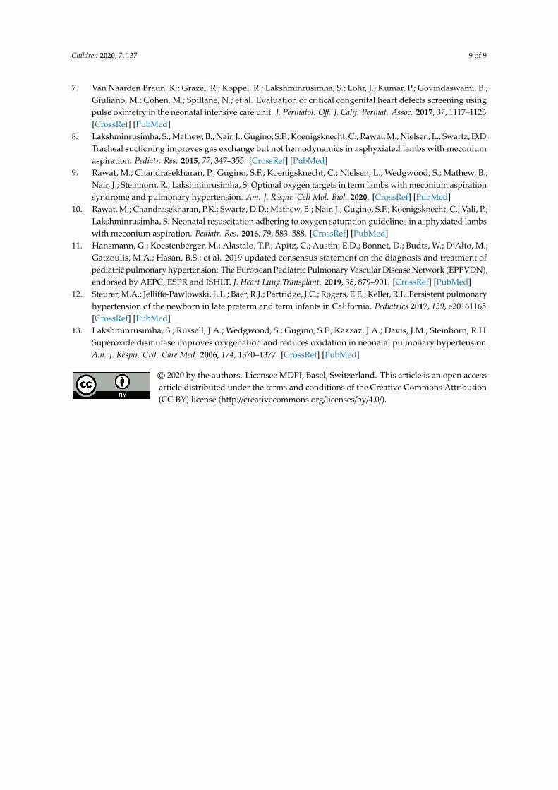
Figure 3. Graphical abstract illustrating hemodynamic and oxygen saturation differences comparing the bidirectional to left-to-right ductus arteriosus shunting in a lamb model of meconium aspiration syndrome and pulmonary hypertension. Lambs that experience a bidirectional ductal shunt have a significantly lower left pulmonary arterial blood flow (QP), left carotid blood flow (QCA), mean systolic blood pressure (Sys BP), cerebral oxygen delivery (C-DO2), and oxygen consumption (VO2) compared to left-to-right ductal shunting. FiO2 = fraction of inspired oxygen; PaO2/PaCO2 = partial arterial oxygen/carbon dioxide tension; SaO2 = saturation of arterial oxygen. Copyright Satyan Lakshminrusimha.
4. Discussion
Extrapulmonary shunt at the PDA and PFO level leading to hypoxemia is characteristic of PPHN. Birth asphyxia with meconium aspiration is a common cause of PPHN in term infants [12]. The presence of right-to-left ductal shunting is inferred by the presence of an oxygenation gradient between the pre- and postductal regions. In this study, using a model of birth asphyxia, meconium aspiration syndrome, and secondary PPHN, we demonstrate that bidirectional ductal shunting with hypoxemia can occur without a significant pre- to postductal oxygenation gradient.
There are several limitations to this study. The degree of hypoxemic respiratory failure was mild to moderate. We did not evaluate shunting at the PFO level. Significant right-to-left shunting at the PFO may potentially reduce the pre- and postductal oxygen gradient. We did not consistently measure the postductal SpO2 in all lambs due to the lack of an adequate number of pulse oximeters during multiple, simultaneous studies. The degree of right-to-left shunting may potentially be more significant in the presence of severe hypoxemia and PPHN. The ovine model of severe PPHN can be induced by antenatal ductal ligation [13]. However, it is not possible to assess ductal shunting in this model. We did not measure the pulmonary arterial pressure in these studies and did not assess the severity of PPHN. We did document hypoxemia, low pulmonary blood flow, and bidirectional or right-to-left shunting (2 samples) in this study, and these findings are suggestive of PPHN. Finally, we did not statistically correct for multiple values obtained from the same lamb. There was widespread fluctuation in the direction of shunting, pulmonary blood flow, and blood gases in the same lamb with time.
The novel aspect of the study is the simultaneous measurement of ductal, pulmonary, and carotid flows along with pre- and postductal blood gases. A right-to-left or bidirectional flow at the ductus is associated with a significant reduction in the pulmonary blood flow, leading to significantly lower PaO2/FiO2 ratios in these lambs. The presence of a bidirectional shunt was also associated with
FiO2 0.54 ± 0.24
Preductal PaO2 61 ± 26 mmHg
Mean Sys BP – 54 ± 10* mmHg
Qca – 12 ± 4*
ml/kg/min
C-DO2 – 1.9 ± 0.4* VO2 – 0.4 ± 0.15* ml O2/kg/min
A. Bidirectional /Right-to-left Ductal shunt
PDA
PA
PV Qp 81 ± 52* ml/
kg/min Pulmonary blood flow
Preductal SpO2 92 ± 6%
Preductal SaO2 91 ± 7%
Postductal SpO2 88 ± 10%
Postductal SaO2 90 ± 8%
Postductal PaO2 57 ± 24*
mmHg
Descending aorta
Bidirectional ductal shunt –+137 ± 112* to -85 ± 71* ml/kg/min
FiO2 0.59 ± 0.23
Preductal PaO2 74 ± 33 mmHg
Mean Sys BP – 65 ± 11 mmHg
Qca – 15 ± 6 ml/kg/min
C-DO2 – 2.4 ± 0.9 VO2 – 0.5 ± 0.16 ml
O2/kg/min
B. Left-to-right Ductal shunt
PDA
PA
PV Qp 133 ± 83 ml/
kg/min Pulmonary blood flow
Preductal SpO2 92 ± 9%
Preductal SaO2 93 ± 6%
Postductal SpO2 88 ± 7% Postductal
SaO2 93 ± 6%
Postductal PaO2 70 ± 31*
mmHg
Descending aorta
Left-to-right ductal shunt –+110 ± 78 to +48 ± 41 ml/
kg/min
Preductal PaCO2 56 ± 12* mmHg
Preductal PaCO2 48 ± 12 mmHg
* P < 0.01 cf. left-to-right shunt in figure B
Figure 3. Graphical abstract illustrating hemodynamic and oxygen saturation differences comparingthe bidirectional to left-to-right ductus arteriosus shunting in a lamb model of meconium aspirationsyndrome and pulmonary hypertension. Lambs that experience a bidirectional ductal shunthave a significantly lower left pulmonary arterial blood flow (QP), left carotid blood flow (QCA),mean systolic blood pressure (Sys BP), cerebral oxygen delivery (C-DO2), and oxygen consumption(VO2) compared to left-to-right ductal shunting. FiO2 = fraction of inspired oxygen; PaO2/PaCO2
= partial arterial oxygen/carbon dioxide tension; SaO2 = saturation of arterial oxygen. CopyrightSatyan Lakshminrusimha.
4. Discussion
Extrapulmonary shunt at the PDA and PFO level leading to hypoxemia is characteristic of PPHN.Birth asphyxia with meconium aspiration is a common cause of PPHN in term infants [12]. The presenceof right-to-left ductal shunting is inferred by the presence of an oxygenation gradient between the pre-and postductal regions. In this study, using a model of birth asphyxia, meconium aspiration syndrome,and secondary PPHN, we demonstrate that bidirectional ductal shunting with hypoxemia can occurwithout a significant pre- to postductal oxygenation gradient.
There are several limitations to this study. The degree of hypoxemic respiratory failure was mildto moderate. We did not evaluate shunting at the PFO level. Significant right-to-left shunting atthe PFO may potentially reduce the pre- and postductal oxygen gradient. We did not consistentlymeasure the postductal SpO2 in all lambs due to the lack of an adequate number of pulse oximetersduring multiple, simultaneous studies. The degree of right-to-left shunting may potentially be moresignificant in the presence of severe hypoxemia and PPHN. The ovine model of severe PPHN canbe induced by antenatal ductal ligation [13]. However, it is not possible to assess ductal shunting inthis model. We did not measure the pulmonary arterial pressure in these studies and did not assessthe severity of PPHN. We did document hypoxemia, low pulmonary blood flow, and bidirectional orright-to-left shunting (2 samples) in this study, and these findings are suggestive of PPHN. Finally,we did not statistically correct for multiple values obtained from the same lamb. There was widespreadfluctuation in the direction of shunting, pulmonary blood flow, and blood gases in the same lambwith time.
The novel aspect of the study is the simultaneous measurement of ductal, pulmonary, and carotidflows along with pre- and postductal blood gases. A right-to-left or bidirectional flow at the ductus isassociated with a significant reduction in the pulmonary blood flow, leading to significantly lowerPaO2/FiO2 ratios in these lambs. The presence of a bidirectional shunt was also associated with lower

Children 2020, 7, 137 8 of 9
systemic blood pressures and a higher PaCO2. Despite high PaCO2 concentrations, the carotid bloodflow, oxygen delivery, and oxygen consumption by the brain were significantly lower in the presence ofa bidirectional shunt. This suggests that oxygen delivery to the brain is compromised in the presenceof bidirectional shunt in this model.
In neonates with PPHN the absence of a pre- and postductal oxygenation gradient is thoughtto be due to the closure of the PDA or an exclusively left-to-right shunt at the PDA [1]. The currentstudy shows that even in the presence of a low pulmonary blood flow and bidirectional ductal shunt,there may not be a significant saturation or PaO2 gradient between the preductal and postductalregions. The oxygen saturation in the lower limb is dependent on the admixture of pulmonary arterialblood (with a mixed venous oxygen saturation of 70% ± 9% in our study) and preductal arterial blood(with SaO2 of 90.9% ± 6.5%). Using the shunt equation, the right-to-left effective ductal shunt onlycontributes to 6.6% of the descending aortic flow in lambs with a bidirectional ductal flow. The presenceof a saturation difference between the preductal and postductal samples is a reliable indicator ofPPHN and a bidirectional or right-to-left shunt (89% of instances—Table 2). However, the absence of asaturation gradient between the preductal and postductal samples does not rule out PPHN despite thepresence of hypoxemia with compromised oxygen delivery to the brain.
5. Conclusions
In lambs with parenchymal lung disease and secondary PPHN, the presence of a pre- andpostductal oxygenation gradient was indicative of a right-to-left or bidirectional shunt. However,the lack of a pre- and postductal oxygenation gradient does not rule out bidirectional shunt. In clinicalsituations with parenchymal lung disease, the clinical absence of a pre- and postductal oxygenationgradient should not be considered to suggest the absence of PPHN, and higher emphasis should beplaced on obtaining echocardiography to confirm the diagnosis.
Author Contributions: A.L.: data extraction and analysis, conducting experiments, and writing the manuscript;M.H.: data extraction and analysis, conducting experiments, and critiquing and writing the manuscript;W.F.: conducting experiments and critiquing manuscript; S.L.: concept, analysis of data, and critiquing and writingthe manuscript; P.V.: concept, conducting studies, analysis of data, and critiquing and writing the manuscript.All authors have read and agreed to the published version of the manuscript.
Funding: This work has been supported by NIH grants HD096299 (PV) and HD072929 (SL).
Conflicts of Interest: The authors declare no conflict of interest.
References
1. Lakshminrusimha, S.; Keszler, M. Persistent pulmonary hypertension of the newborn. Neoreviews 2015, 16,e680–e692. [CrossRef] [PubMed]
2. Nair, J.; Lakshminrusimha, S. Update on PPHN: Mechanisms and treatment. Semin. Perinatol. 2014, 38, 78–91.[CrossRef] [PubMed]
3. Kemper, A.R.; Mahle, W.T.; Martin, G.R.; Cooley, W.C.; Kumar, P.; Morrow, W.R.; Kelm, K.; Pearson, G.D.;Glidewell, J.; Grosse, S.D.; et al. Strategies for implementing screening for critical congenital heart disease.Pediatrics 2011, 128, e1259–e1267. [CrossRef] [PubMed]
4. Mahle, W.; Koppel, R. Screening with pulse oximetry for congenital heart disease. Lancet 2011, 378, 749–750.[CrossRef]
5. Fernandes, N.; Lakshminrusimha, S. The limitations of pulse oximetry for critical congenital heart diseasescreening in the neonatal intensive care units. Acta Paediatr. 2017, 106, 1007. [CrossRef] [PubMed]
6. Manja, V.; Mathew, B.; Carrion, V.; Lakshminrusimha, S. Critical congenital heart disease screening by pulseoximetry in a neonatal intensive care unit. J. Perinatol. Off. J. Calif. Perinat. Assoc. 2015, 35, 67–71. [CrossRef][PubMed]
Children 2020, 7, 137 9 of 9
7. Van Naarden Braun, K.; Grazel, R.; Koppel, R.; Lakshminrusimha, S.; Lohr, J.; Kumar, P.; Govindaswami, B.;Giuliano, M.; Cohen, M.; Spillane, N.; et al. Evaluation of critical congenital heart defects screening usingpulse oximetry in the neonatal intensive care unit. J. Perinatol. Off. J. Calif. Perinat. Assoc. 2017, 37, 1117–1123.[CrossRef] [PubMed]
8. Lakshminrusimha, S.; Mathew, B.; Nair, J.; Gugino, S.F.; Koenigsknecht, C.; Rawat, M.; Nielsen, L.; Swartz, D.D.Tracheal suctioning improves gas exchange but not hemodynamics in asphyxiated lambs with meconiumaspiration. Pediatr. Res. 2015, 77, 347–355. [CrossRef] [PubMed]
9. Rawat, M.; Chandrasekharan, P.; Gugino, S.F.; Koenigsknecht, C.; Nielsen, L.; Wedgwood, S.; Mathew, B.;Nair, J.; Steinhorn, R.; Lakshminrusimha, S. Optimal oxygen targets in term lambs with meconium aspirationsyndrome and pulmonary hypertension. Am. J. Respir. Cell Mol. Biol. 2020. [CrossRef] [PubMed]
10. Rawat, M.; Chandrasekharan, P.K.; Swartz, D.D.; Mathew, B.; Nair, J.; Gugino, S.F.; Koenigsknecht, C.; Vali, P.;Lakshminrusimha, S. Neonatal resuscitation adhering to oxygen saturation guidelines in asphyxiated lambswith meconium aspiration. Pediatr. Res. 2016, 79, 583–588. [CrossRef] [PubMed]
11. Hansmann, G.; Koestenberger, M.; Alastalo, T.P.; Apitz, C.; Austin, E.D.; Bonnet, D.; Budts, W.; D’Alto, M.;Gatzoulis, M.A.; Hasan, B.S.; et al. 2019 updated consensus statement on the diagnosis and treatment ofpediatric pulmonary hypertension: The European Pediatric Pulmonary Vascular Disease Network (EPPVDN),endorsed by AEPC, ESPR and ISHLT. J. Heart Lung Transplant. 2019, 38, 879–901. [CrossRef] [PubMed]
12. Steurer, M.A.; Jelliffe-Pawlowski, L.L.; Baer, R.J.; Partridge, J.C.; Rogers, E.E.; Keller, R.L. Persistent pulmonaryhypertension of the newborn in late preterm and term infants in California. Pediatrics 2017, 139, e20161165.[CrossRef] [PubMed]
13. Lakshminrusimha, S.; Russell, J.A.; Wedgwood, S.; Gugino, S.F.; Kazzaz, J.A.; Davis, J.M.; Steinhorn, R.H.Superoxide dismutase improves oxygenation and reduces oxidation in neonatal pulmonary hypertension.Am. J. Respir. Crit. Care Med. 2006, 174, 1370–1377. [CrossRef] [PubMed]
© 2020 by the authors. Licensee MDPI, Basel, Switzerland. This article is an open accessarticle distributed under the terms and conditions of the Creative Commons Attribution(CC BY) license (http://creativecommons.org/licenses/by/4.0/).