Embed Size (px)
Citation preview

3. International Emergency Symposium. 27-29 March 2015. Almaty/Kazakhstan
POSTER PRESENTATION BOOK
The Symposium was Adopted Just the Poster Papers

3. International Emergency Symposium. 27-29 March 2015. Almaty/Kazakhstan
Symposium Boards
Honorary Chairman of the Symposium
Prof.Dr. Akanov Aikan Akanovich
Chairmans of the Symposium
Prof.Dr. Başar Cander
Prof. Dr. Darmenov Oralbay Kenzhebaevich
Prof.Dr. Polat Durukan
Symposium Secretaries
Prof. Dr. Nurmanbetova Farida Nusupzhanovna
Doç. Dr. Cemil Kavalcı
Uzm. Dr. Ömer Salt
Dr. Murat Muratoğlu
Symposium Organizing Committee
Prof.Dr. Mehmet Gül
Prof.Dr. Batırhanov Shayhsalam Kilibaevich
Prof.Dr. Egemberdiev Tolegen Zholimbetovich
Doç.Dr. İbraeva Gulmira Alpısbaevna
Doç.Dr. Eralina Svetlana Nurushevna
Doç.Dr.Salim Satar
Doç. Dr. Selahattin Kıyan
Doç. Dr. Sadık Girişgin
Doç. Dr. Yunsur Çevik
Doç. Dr. Murat Orak
Doç. Dr. Mustafa Burak Sayhan

3. International Emergency Symposium. 27-29 March 2015. Almaty/Kazakhstan
Symposium Report Evaluation Board
Prof. Dr. Darmenov Oralbay Kenzhebaevich
Prof.Dr. Polat Durukan
Doç.Dr. Cemil Kavalcı
Doç.Dr. Mustafa Burak Sayhan
Dr. Murat Muratoğlu
3. International Emergency Symposium. 27-29 March 2015. Almaty/Kazakhstan
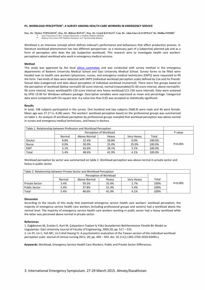
P1. WORKLOAD PERCEPTİON”: A SURVEY AMONG HEALTH CARE WORKERS IN EMERGENCY SERVICE
Doç. Dr. Türker TOPALHAN1, Doç. Dr. Bülent BAYAT1, Doç. Dr. Cemil KAVALCI2, Uzm. Dr. Afşin Emre KAYIPMAZ2
, Dr. Meliha FINDIK2
1) Gazi Üniversitesi İ.İ.B.F. Çalışma Ekonomisi ve Endüstri İlişkileri Bölümü 2) Başkent Üniversitesi TIP Fakültesi Hastanesi Acil Tıp Hekimliği Bölümü
Workload is an intensive concept which defines indivual’s performance and behaviours that affect production process. In literature workload phenomenon has two different perspectives: as a necessary part of a (objective) planned job and as a form of perception who does the job (subjective workload). This research aims to investigate health care workers’ perceptions about workload who work in emergency medical services. Method This study was approved by the local ethics committee and was conducted with survey method in the emergency departments of Baskent University Medical School and Gazi University Medical School. Survey forms to be filled were handed over to health care workers (physicians, nurses, and emergency medical technicians (EMT)) were requested to fill the form. Two kinds of data were detected with IWPS (individual workload perception scale) defined by Cox and his friends: factual data (categorical) and data about perception of individual workload (numerical). There were five groups based on the perception of workload (below normal/0-30 score interval, normal (reasonable)/31-60 score interval, above normal/61-90 score interval, heavy workload/91-120 score interval very heavy workload/121-150 score interval). Data were analysed by SPSS 13.00 for Windows software package. Descriptive variables were expressed as mean and percentage. Categorical data were compared with Chi-square test. A p value less than 0.05 was accepted as statistically significant. Results In total, 148 subjects participated in the survey. One hundred and two subjects (%68.9) were male and 46 were female. Mean age was 27.17 (+,-6.06) years. The workers’ workload perception based on the professional groups was summarized on table 1. An analysis of workload perception by professional groups revealed that workload perception was above normal in nurses and emergency medical technicians, and heavy in doctors.
Table 1. Relationship between Profession and Workload Perception
Perception of Workload P value
Normal Above Normal Heavy Very Heavy Total
P<0.001 Doctor 8.8% 32.4% 58.8% 0.0% 100.0%
Nurse 0.0% 50.0% 25.0% 25.0% 100.0%
EMT 3.1% 65.6% 28.1% 3.1% 100.0%
Total 5.4% 48.6% 41.9% 4.1% 100.0%
Workload perception by sector was summarized on table 2. Workload perception was above normal in private sector and heavy in public sector.
Table 2: Relationship between Private Sector and Workload Perception
Perception of Workload Total
P<0.005 Normal Above Normal Heavy Very Heavy
Private Sector 5.4% 59.5% 32.4% 2.7% 100%
Public Sector 5.4% 37.8% 51.4% 5.4% 100%
Total 5.4% 48.6% 41.9% 4.1% 100%
Discussion According to the results of this study that examined emergency service health care workers’ workload perception, the majority of emergency service health care workers (including professional groups and sectors) had a workload above the normal level. The majority of emergency service health care workers working in public sector had a heavy workload while the latter was perceived above normal in private sector. References 1. Dağdeviren M, Eraslan E, Kurt M. Çalışanların Toplam İş Yükü Seviyelerinin Belirlenmesine Yönelik Bir Model ve Uygulaması: Gazi University Journal of Faculty of Engineering, 2005;20; pp. 517 – 525. 2. Lin SY, Lin C, Yeh MC, Lin S And Hwang YJ. A psychometric evaluation of the Taiwan version of the individual workload perception scale. Journal of clinical nursing 2011; 20; pp. 494 – 503. doi: 10.111/j.1365-2702-2010-03495.x Keywords: Workload, Emergency Service Health Care Workers, Public and Private Sector Differences.

3. International Emergency Symposium. 27-29 March 2015. Almaty/Kazakhstan
P2. A STUDY OF THE PRESENCE AND LEVEL OF ESTRANGEMENT AMONG EMERGENCY SERVİCE HEALTH CARE WORKERS
Doç. Dr. Bülent BAYAT 1,
Doç. Dr. Türker TOPALHAN 1, Doç. Dr. Cemil KAVALCI2, Uzm. Dr. Afşin Emre KAYIPMAZ2, Dr. Meliha FINDIK2
1) Gazi Üniversitesi İ.İ.B.F. Çalışma Ekonomisi ve Endüstri İlişkileri Bölümü 2) Başkent Üniversitesi TIP Fakültesi Hastanesi Acil Tıp Hekimliği Bölümü
INTRODUCTION
Estrangement is emotional and social condition that is highly dependent on working life/working conditions. If the person who constructs himself by working is forced to hard working conditions, he draws himself away himself and society. In literature most researchers linked estrangement to working conditions. Estrangement is determined by organization in structural level and working conditions in functional level when it is dealed with organizational level.
The aim of this study was to explore the presence and level of estrangement in emergency service health care workers.
METHODS
This study was conducted in the emergency departments of Baskent University Medical School and Gazi University Medical School via questionnaire method after being approved by the local Ethics Committee. The questionnaire forms were filled by healthcare staff (physicians, nurses, and emergency medical technicians). The estrangement scale used in the research was developed by Bayat and based on the estrangement theory of Johnson and Seeman. The scale consists of 36 proposals. The participants can get 36-180points. The categories by points were as follows: “0-36 points: no estrangement, 37-72 points: low estrangement, 73-109 points: medium estrangement, 110-156 points: high estrangement, 157-180 points: very high estrangement”. The data were analyzed with SPSS 13.00 for Windows software package. The descriptive data were presented as “n, % and mean”. Chi-square test was used for comparison of categorical data. A p value less than 0.05 was accepted as statistically significant.
RESULTS
A total of 148 participants were enrolled. They consisted of 102 males (%68,9) and 46 females (%31,1). The mean age was 27.7+6.06 years. The estrangement level of workers by the professional groups was summarized on Table 1. The analysis of the estrangement level of workers by professional groups revealed a low and medium estrangement among nurses, high estrangement among emergency medical technicians, and medium estrangement among doctors.
Table 1: The Relationship Between Professional Groups and Estrangement Level
Profession
Estrangement Level
Total
P value
Low Medium High Very High
p<0.001
Physician 20,6% 64,7% 11,8% 2,9% 100,0%
Nurse 50,0% 50,0% 0,0% 0,0% 100,0%
EMT 31,2% 28,1% 34,4% 6,2% 100,0%
Total 28,4% 47,3% 20,3% 4,1% 100,0%
The estrangement level of workers by sector was summarized on Table 2. The analysis of the estrangement level of workers by sector revealed a maximum medium estrangement level in public and private sector.
Table 2: The Relationship Between Sector and Estrangement Level
Estrangement Level Total P value
Low Medium High Very High
p<0.001
Private Sector 21,6% 45,9% 29,7% 2,7% 100,0%
Public Sector 35,1% 48,6% 10,8% 5,4% 100,0%
Total 28,4% 47,3% 20,3% 4,1% 100,0%
DISCUSSION
According to the results of this study investigating the presence and level of estrangement among emergency service healthcare workers, doctors had a more prevalent and severe estrangement than the other professional groups.
REFERENCES
1.Taştan S İşci E, Arslan B. Örgütsel destek algısının işe yabancılaşma ve örgutsel bağlılığa etkisinin incelenmesi: İstanbul özel hastanelerinde bir çalışma. Journal of Pamukkale University Social Sciences, 2014; 19: pp: 121-138.
2.Usul H ve Atan A, Sağlık sektöründe yabancılasma düzeyi”, Journal of KMÜ Social And Economic Researchs, 2014; 16 (26): pp: 1-10
Keywords: Estrangement Level, Public Sector, Private Sector. Emergency Health Care Workers.

3. International Emergency Symposium. 27-29 March 2015. Almaty/Kazakhstan
P3. SURGICAL TREATMENT OF ACUTE CALCULOUS CHOLECYSTITIS
Aimagambetov MJ, Bulegenov TA,Asylbekov EM, Elchibaev BM, Akparov TL, Serikova GS
Department of internship of surgerySemey State Medical University
Introduction
Minimally invasive interventions for gallstone disease and its complications are gradually gaining recognition due to
minor trauma, sufficient efficiency, and with experience are everyday manipulation [1-6]. Conversion and postoperative
mortality of minimally invasive interventions is 2 - 23.6%, 0.5 - 2.5% [2, 4, 5, 6-10].
Material and methods Analyzed the results of treatment of 225 patients with different forms of acute calculous
cholecystitis. The age of patients ranged from 20 to 86 years. Women was 167 (74.2%), men - 58 (25.8%). Up to 24 hours
from the onset of the disease were hospitalized 32 (14.2%) in the period 24 - 72 hours - 76 (33.8%), in terms of more than 72
hours - 117 (52.0%) patients. Of these, 33 (14.7%) patients had previous surgery on the organs of the lower floor of the
abdominal cavity, 7 (3.1%) - on the upper abdominal cavity. The majority of patients had comorbidities: 135 (60, 0%)
patients suffering from various disorders of the cardiovascular system, chronic lung disease (chronic bronchitis, pulmonary
fibrosis, emphysema, bronchiectasis, bronchial asthma), diseases of the digestive system, urinary, metabolism, pathology of
bone - joint system and others.
All patients on admission to hospital was conducted a comprehensive physical examination. The presence of lesions of
the gallbladder and bile ducts were diagnosed based on clinical, laboratory data, ultrasound, endoscopic methods of
investigation and verification during surgery, and histological findings. In 11 (4.9%) patients had catarrhal calculous
cholecystitis in 165 (73.3%) - an abscess in 49 (21.8%) - gangrenous calculous cholecystitis. The incidence of destructive
cholecystitis observed in 106 (47.1%) patients (paravesical infiltrate and abscess, empyema, jaundice, choledocholithiasis,
cholangitis, peritonitis, internal biliary fistula)..
Results and discussion In 5 (5.8%) of the 86 patients Making laparotomy and completion operations in the traditional
way done for laparoscopically operated In one case was damage to the structure of the duodenum. The transition to a mini -
access to the right upper quadrant with a set of tools mini - assistant. When multiple cholangiography revealed
choledocholithiasis, and the incision is extended downwards. The operation is completed suturing defect duodenum,
cholecystectomy, holedoholitotomiey, choledochoduodenostomy and drainage of common bile duct through the cystic duct
stump. In three observations are also the transition to the mini-access right upper quadrant with a set of tools mini-assistant in
the presence of a dense infiltrate and due to technical difficulties incision is extended downwards, cholecystectomy
performed by Pribram. In one case also revealed a dense infiltrate and sclerotic gall bladder. Made upper midline laparotomy
and cholecystectomy, given the availability of a wide cystic duct, common bile duct and the expansion of the presence of
small stones made cholangiography. In this case, revealed multiple stones in the common bile duct. The transaction is
completed holedoholitotomiey, choledochoduodenostomy and drainage through the common bile duct stump of the cystic
duct. In 5 (5.8%) patients with no deaths were postoperative complications. One of them, the left femoral vein thrombosis. In
two patients was lower lobe pneumonia. In two patients flowed bile, which stopped on their own. Reoperation was not. All
patients were discharged from the recovery.
Patients operated on the traditional way of deaths was not. Re-operated on two patients. One of them for 6 day
performed an appendectomy, a second resection of the greater omentum over the purulent omentita. Postoperative
complications occurred in 4 (11.1%) patients. In 2 of them were lower lobe pneumonia, in 2 - liver failure. Wound
complications were not observed. All patients were discharged from the recovery.
Conclusion Thus, the complementarity of laparoscopic cholecystectomy cholecystectomy from mini-approach in acute
cholecystitis and its complications lead to a decrease in the conversion of postoperative complications.

3. International Emergency Symposium. 27-29 March 2015. Almaty/Kazakhstan
P4.FALL AND INJURIES AMONG ELDERLY (LITERATURE REVIEW)
A.S. Tlemissov, TA Bulegenov, Semey State Medical University
Abstract
In this paper we studied the literature data problems falling and injuries among elderly
According to United Nations estimates that by 2025 the number of older people will double from the
current 600 million. To 1.2 billion. Although the proportion of older people in the total population is higher in
developed countries, the percentage increase in the elderly population is much greater in developing countries
(UN Population Division, 2004). The number of people aged 60 years and older as a proportion of the world's
population will double from 11% in 2006 to 22% in 2050 [1]. The median age of the European Region is the
highest in the world. With the increase in life expectancy, largely increasing proportion of older people. By
2050, more than 27% of the population will be elderly [2]. Elderly patients at increased risk of injury - for a
more active lifestyle and a violation of cognitive functions. For older people enough much less effort to injury
[3].
According to experts, having sharp differences in injury rates between countries indicate a significant
potential for improvement. Thousands of deaths could be prevented if all over Europe deaths from injury would
be reduced to the level of the most prosperous countries in this regard. To implement preventive measures
requires political will and consistently implemented a comprehensive program. It is necessary to creating a
culture of safety, where unintentional injuries are not regarded as inevitable [19].
Keywords: elderly, fall, injuries.

3. International Emergency Symposium. 27-29 March 2015. Almaty/Kazakhstan
P5.Modern methods of treatment of perforated duodenal ulcer
Aimagambetov M.J, OmarovN.B., Bulegenov T.A, Elchibaev B.M, Serikova G.S.
Department of internship in surgery, State Medical University of Semey, Kazakhstan
A key issue in the treatment of perforated ulcers is the problem of choosing the optimal surgical approach,
both to reduce the risk of death and to achieve the most favorable functional outcome [2,5,8,11,13].
This approach should take into account not only the general principles of abdominal surgery, but the
specific mechanisms that are typical for the development of peptic ulcer disease [1,3,9,10]. Thus, in a number of
experimental and clinical work revealed the important role of vascular disorders of the stomach and in the wall
of the duodenum in the pathogenesis of duodenal ulcers [4,5,6,7]. The development of these disorders, and
pathological increase in acid production associated with functional disorders of the autonomic regulation,
hyperactivity segmental level of the parasympathetic nervous system [Gisbert JP, Calvet X., 2009; Lundell L.,
2011].
Material and methods:
The study is based on a study of the results of surgical treatment of 112 patients with perforated duodenal
ulcer.
All patients were operated on an emergency basis.
Of the 112 operated the vast majority were men - 100 people (89.2%), and was only 12 (10.8%) women.
Among our patients 109 (97.3%) were of working age. The age of patients ranged from 16 to 73 years,
mean age 38,9 ± 1,0 years
The combination of perforation duodenal stenosis degree I-II was detected in 32 (28.6%), with the presence
of the second ulcer - 15 (13.4%) and its penetration into the head of the pancreas in 3 patients (2.7%). Covered
with perforation was detected in 5 (4.5%) cases (Table 1).
Table - 1 Characteristics of intraoperative data
Intraoperative find
Total patients
the number of patients М±m
Isolation of the front wall of perforated
ulcer duodenum 69 61,6±4,6
In conjunction with duodenum
compensated stenosis 22 19,6±3,8
In conjunction with duodenum
subcompensated stenosis 8 7,1±2,4
In combination with the rear wall of
duodenum ulcer 15 13,4±3,2
Total: 112 100,0
The choice of surgical approach was aimed at simultaneously solving the following tasks - the elimination
of the source of peritonitis, radical treatment of peptic ulcer disease, a small trauma.
All patients were divided into two groups. The first accounted for 55 (49.1%) patients who made
videolaparoscopy, sanitation, stitching holes perforated duodenum (the comparison group). The second group
consisted of 57 (50.9%) patients who underwent videolaparoscopy, brushing with excision of duodenal ulcer
with the implementation of small duodenoplasty access (study group).
Conclusions:
1. Evaluation long-term clinical outcomes after laparoscopic excision of perforated duodenal ulcer,
duodenoplasty of mini access followed in 54bolnyh.Otlichny and good results were obtained in 49 (93.3%),
satisfactory-5 (6.6%). All patients had positive dynamics of body weight, ability to work restored in 21-28 days
after surgery.
2. Posleoperatsionnye complications occurred in 8 (18%) patients: festering wounds -1 (2.2%), dysphagia, 2
(4.4%), gatrostaz-2 (4.4%), acute pancreatitis -3 (7 , 2%). Endoscopic and radiological significant pathological
changes were found.
3. Uchityvaya results videolaparoscopy, excision of perforated duodenal ulcer with duodenoplasty of mini access
suggests that this operation is a valid method of treatment of perforated duodenal ulcer.

3. International Emergency Symposium. 27-29 March 2015. Almaty/Kazakhstan
P6. Acute and emergency medicine on the level of primary care for children and adults
М.А. Kamaliev1, А.B. Almukhanova2
1
Doctor of medicine, professor, chief of department of public health, Kazakh National Medical University named after S. D. Asfendiyarov, Almaty, Kazachstan 2
Student of phD in specialization of Public health, Kazakh National Medical University named after S. D. Asfendiyarov, Almaty, Kazachstan
Abstract
Reduction of circulation diseases mortality of population is proved as a priority target of Health Care
Service and its integrated solution includes improvement of cardiac care on the stages of treatment. This article shows the most relevant organizational aspects in provision of emergency treatment for patients with circulation diseases in conditions of high–tech hospital.
Key words: Circulation diseases, cardiac and cardiac surgery treatment.
Picture – Indicators of newly diagnosed morbidity and mortality from CVD in the Republic of Kazakhstan in 2000-2012 years. (per 100 000 population)
-------- morbidity -------- mortality
1288,7 1396,4
1984,4
1754,3 1845,1
1749,1
1911,4 1906,6
2170,5 2273,1
2087,7
2277,1
2454
502,3 494,6 511,1 539 517,7 535,5 533,1 528,3 489,66 416,4 403,7
308,8 251,88
2000 г. 2001 г. 2002 г. 2003 г. 2004 г. 2005 г. 2006 г. 2007 г. 2008 г. 2009 г. 2010г. 2011г. 2012г.

3. International Emergency Symposium. 27-29 March 2015. Almaty/Kazakhstan
P7. Financial integration of ambulance and emergency services in primary
health care
Imangazinov S. B., Kabulov K. S., Tanbaeva K. D.
Pavlodar branch of MSU, Semey
It is interesting and important from a health-economic perspective that the rate of referral for emergency
medical assistance decreased. There is a significant reduction in uptake by some medical practitioners to 30%
and more with the simple implementation of per capita financing at the level of family group practices (FGPs).
Patients regularly observed by family physicians, rarely require emergency care. Up until the doctors don't
organize correct treatment of simple clinical disease, in urgent cases, their populations will ask for help on the
ambulance. In the structure of appeals for the ambulance emergency care is 80%. More than 60% in the structure
of the emergency takes care to patients with hypertension, bronchial asthma, various hyperthermie.
The purpose of the message - submission of the results of a retrospective comparative analysis of emergency
services, Pavlodar, worked in various conditions of financial cooperation with the network of primary
medicosanitary care (PHC).
Materials and methods. The analysis is based on archival data of the health system Pavlodar during the
integration of PHC services in the organization of emergency and urgent care, Pavlodar in 2000 and after project
completion. The ambulance, Pavlodar in 2000 served the population with a population of 302 thousand, 2004 -
309,8 thousand. For the period comparative study of the number of employees did not undergo significant
deviations (table 1).
As can be seen from table 3, 2004 emergency visits ambulance on observation chronic pathologies in
cases that are subject to constant observation and treatment by specialists in primary health care, carried out in
the 64080 cases against 36890 in 2000, which is 1.7 times more than in 2000, and among children this indicator
has increased in 2 times.
As for other urgent conditions requiring emergency medical care in case of exacerbation or deterioration
in patients with chronic pulmonary and cardiovascular systems are subject to constant observation and treatment
by specialists of primary health care has been a rise of 1.5 and 1.4 times respectively. Provides information prove
the influence of the mechanism of co-financing at the emergency level on the nature of incoming calls at the
service of "03" and on the quality of medical assistance in emergency cases and exceptions unreasonable visits
ambulance crews. The increase in the number of emergency calls was the basis of the increase in the number of
emergency teams from 21 in 2000 to 25 in 2004 and the cost of providing the needs of the service "03" (table 4).
Thus, one of the mechanisms for the effective use of the health budget is financing services of emergency and
urgent care at the expense of primary care.
Literature
1 . T. K. Rahypbekov, Financial management in health care. -Astana, 2002.-p. 288 .

3. International Emergency Symposium. 27-29 March 2015. Almaty/Kazakhstan
P8. COMPARISION OF TWO CORONARY ARTERY BYPASS SURGERY TECHNIQUE WITH
RESPECT TO ACUTE KIDNEY INJURY
Deniz Sarp Beyazpinar1, Bahadir Gultekin
1, Cagri Kayipmaz
1, Atilla Sezgin
1, Afsin Emre Kayipmaz
2,
Tufan Akin Giray 2
1Başkent University, Faculty of Medicine, Department of Cardiovascular Surgery, Ankara, Turkey
2Başkent University, Faculty of Medicine, Emergency Department, Ankara, Turkey
INTRODUCTION
Percutaneous coronary intervention and coronary bypass surgery together form the interventional therapy for
atherosclerotic heart disease. In the conventional coronary artery bypass grafting operation cardiac arrest is
established by cardioplegia, aorta is clamped, and tissue perfusion is maintained by extracorporeal circulation. In
the on-pump beating heart bypass grafting technique, on the other hand, cardioplegia and aortic clamping are not
utilized, and extracorporeal circulation is used whenever needed for maintaining mean arterial pressure at a
certain level.
The aim of our study was to compare the conventional technique and on-pump beating heart technique with
respect to acute renal injury and attendant dialysis requirement.
METHOD
The study was performed retrospectively after ethics committee approval. Seventy-seven patients who
underwent isolated coronary bypass surgery for coronary artery disease between 2012 and 2013 formed Group 1
and 76 patients who were applied on-pump beating heart technique at the same time window formed Group 2.
RESULTS
The two groups were not significantly different with respect to preoperative renal function tests. There was,
however, a significant difference between them with regard to cardiopulmonary bypass time and duration of
intensive care unit stay (p<0.05). Seven (9.21%) of 76 cases in Group 2 and 11 (14.28%) of 77 cases in Group 2
developed acute renal injury. Both groups were similar with respect to rates of acute renal injury,
however (p>0.05). One patient in Group 2 and 4 in Group 1 had dialysis requirement later.
CONCLUSION
Despite being statistically non-significant, the results of the present study suggested that on-pump beating heart
coronary bypass surgery was superior to the conventional technique in terms of acute renal injury and, more
importantly, development of acute renal failure in patients with a serum creatinine of 1-1.3 mg/dl.

3. International Emergency Symposium. 27-29 March 2015. Almaty/Kazakhstan
P9. APPLICATION OF MODERN EDUCATIONAL TECHNOLOGY IN THE POST-
GRADUATE TRAINING OF ANESTHESIOLOGISTS - RESUSCITATORS
Kuluspaev E.S., Alpischeva S.V., Karibaeva A.Ye., Terekhov D.V.
State Medical University, Semey city, Kazakhstan
The social order of local health authorities for anesthesiology and resuscitation course is to
prepare anesthesiologists for central district hospitals. Post-graduate training in our
department is carried out in the form of 20-week primary specialization in anesthesiology and
intensive care medicine. The regional need in anesthesiologists – resuscitators specialists is so
great that in recent years, the number of wishing to study is 15-20 people.
Important role in preparing trainees is to development practical skills of intensive care on
mannequins. The course has 8 mannequins of «Laerdal Medical» company allows fulfilling
almost all the existing intensive care methods. Mannequins have important additional
functions that can be applied to pediatric patients. «ALS Baby Trainer» mannequin simulates
3 months baby with 5 kg and allows intraosseous access for drug therapy. «Uetimate Hurt»
mannequin simulates various injuries and allows to master skills of trachea intubation at
traumatic head injuries, treatment of wounds, release and transport of casualties. These
mannequins can join VitalSim the system, which is a simulator of the different parameters of
human life:
all kinds of abnormal heart rhythm and response to defibrillation and outer
cardiostimulation, imitation of heart sounds and noises, normal and abnormal lungs
wheezing, bowel sounds and voice - moaning, crying, vomiting, laryngospasm. «VitalSim»
mannequin allows setting different levels of systolic and diastolic blood pressure from 2 to
300 mm Hg v., to vary the degree of pulse filling. This makes possible to create scenarios of
emergency clinical situations: acute myocardial infarction, ventricular fibrillation,
atrioventricular block, bilateral pneumothorax etc., and develop an algorithm of resuscitation
event. VitalSim memory can store 25 scenarios simultaneously.
A significant part of free time anesthesiologists - resuscitators trainee devote to develop
practical skills on mannequins. Final exam consists of two stages: computer test control and
solution of situational problems in all fields of specialty. Positive mark is 65% of correct
answers; diagnosis of emergency conditions and provision of resuscitation events on
mannequins.
Conclusion.
Anesthesiology and Intensive Care course at Semey State Medical University has unique
experience in a wide application and implementation of the innovative technologies to the
professional postgraduate education system, including elements of the virtual approach to
learn practical skills.

3. International Emergency Symposium. 27-29 March 2015. Almaty/Kazakhstan
P10. Diagnostic value of troponin T in patients with multiple organ failure syndrome
Kuluspaev E.S.,Terechov D.V ., Karibaeva A.E., Alpischeva S.V., Buhanchenko A.G.
State medical university of Semey city, Semey c., Kazakhstan
Abstract: The study included 122 patients [75 men, 47 women aged 42 to 76 years, mean 59±10.3
years] hospitalized in ITD. Cardio specific tropin T in blood of patients indicates myocardial involvement in the
pathological process.
Keywords: research, Tropin T, acute myocardial infarction, inflammatory process.

3. International Emergency Symposium. 27-29 March 2015. Almaty/Kazakhstan
P11.Modern experience of surgical care to patients with maxillofacial area
trauma.
Dauletkhozhaev N.A.; Urazalin Zh.B.; Sabdanalyev A.M.; Seytkulov A.B.; Chairoyev M.M.
KazNMU named after S.D.Asfendiyarov, №5 City Clinical Hospital
Modern experience of surgical care to patients with maxillofacial area trauma.
In 2013 in Almaty city – about 5,045 people applied to the hospital with face trauma. The characteristics
of new types of injuries are given: gunshot wounds by complex “Osa”, facial wounds by angle grinder. The
main principles of providing emergency care to patients with facial trauma in modern conditions were
formulated: precision-guided diagnostics of injuries; single-stage and operative intervention; minimally invasive
surgery; convenience during the postoperative and rehabilitation periods for patients. There is given own
experience of organization of the diagnostics and treatment of facial trauma using CТ и 3D X-ray work,
endoscopic methods, osteosynthesis miniplates. Two clinical examples of using methods mentioned above are
given.
Key words: Maxillofacial trauma, gunshot wound of face, incised wound of face, computer
tomography of skull.

3. International Emergency Symposium. 27-29 March 2015. Almaty/Kazakhstan
P12. PROTECTING EYES AGAINST FOREIGN BODIES DUE TO THE EXPLOSIONS
AKSOY Yakup1, SEVINC Mehmet Koray
2, COLAKOGLU Kadir
3, EYİ Yusuf Emrah
4
1 Girne Military Hospital, Dept. of Ophthalmology, Girne, TRNC
2 Gülhane Military Medical Academy, Dept. of Ophthalmology, Ankara, TURKEY
3 Kasımpasa Military Hospital, Dept. of Ophthalmology, İstanbul, TURKEY
4 Gülhane Military Medical Academy, Dept. of Emergency Medicine, Ankara, TURKEY
OBJECTIVE: Explosions due to the mines, handmade explosives, grenades etc. may cause
severe eye injuries due to the foreign bodies. These injuries may be only superficial or
sometimes deep which may cause permanent visual loss. We present here a case of bilateral
multiple conjunctival and corneal injury after mine explosion.
CASE REPORT: The case was a 28 years old male patient who was presented our emergency
policlinic with complaint of bilateral pain in eyes, lacrimation and decrease in vision. He had
encountered a mine explosion about 3 meters away from him. On examination vital signs
were in normal limits. There were many foreign bodies buried in skin of hands, neck and face.
Ophthalmological examination showed many foreign bodies in conjunctiva and cornea. Much
of them were superficial but a few had penetrated to the deep stroma of cornea. A deeply
penetrated one had located in the center of the left cornea. Visual acuity was 20/25 on right
and 20/40 on left. All foreign bodies were removed from conjunctiva and cornea after topical
anesthesia in operating room. Final visual acuity was 20/20 in right eye but 20/25 in left eye 4
months later.
CONCLUSION: Explosions may cause mortal injuries. Besides this eye injuries are not
commonly mortal but they are sight threatening. For an active military unit which is on a
mission it’s always possible to encounter an explosion which may cause eye injury. To
decrease the damage of explosion protecting goggles always should be worn during missions.

3. International Emergency Symposium. 27-29 March 2015. Almaty/Kazakhstan
P13. Mianserin Cardiotoxicty: A Case Report and Short Review of Literature
Şeref Kerem Çorbacıoğlu, Murat Güvendi, Yunsur Çevik
Keciören Traning and Research Hospital, Emergency Department, Ankara, Turkey
Abstract
Introduction: Mianserin, which has a tetracyclic structure, is frequently used in depression, sleep disorders, and
anxiety treatment in clinical practice. Although mianserin is considered to be more safely than other anti-
depressant agents in terms of fewer side effects and cardiotoxicity, results of the studies and case reports on
effects of mianserin in the literature are controversial.
Case Report:In this paper, we reported that case of a woman who was admitted for self-poising with mianserin
and detected first-degree AV block on electrocardiogram without clinical findings such as hypotension,
bradycardia, and arrhythmias.
Conclusion: Although mianserin is known as a safe drug terms of cardiotoxicity, physicians should be aware
that overdosing of these drugs can cause changing from nonlife-threatening situations to life-threatening
situations.
Keywords: Mianserin, Poisoning, Cardiotoxicity, First-degree AV block, PR interval
3. International Emergency Symposium. 27-29 March 2015. Almaty/Kazakhstan
P14.What is the cause of seizure: isoniazid poising or epidural hematoma? A case report.
Tuba Şafak, Ali Ekber Karabulut, Şeref Kerem Çorbacıoğlu, Yunsur Çevik
Keciören Training and Research Hospital, Emergency Department, Ankara, Turkey
Abstract
Introduction: Isoniazid is the primary drug that widely used in the treatment of tuberculosis. Due to the
decrease of the incidence of tuberculosis usage of isoniazid and related intoxications less seen day by day. In this
case we present a patient who was admitted for self-poising with isoniazid and falling down the stairs.
Case report: A 26-year-old woman was brought to emergency department for self-poisoning with a large
unknown dosage of isoniazid. On arrival at the ED she had a generalized tonic-clonic seizure. Arterial blood gas
analysis revealed that lactic acidosis and hyperglycemia. At the same time she had a head trauma and brain
computed tomography demonstrated epidural hematoma, thus 18mg/kg phenytoin was started for seizure
prophylaxis. Intravenous pyridoxine treatment was planned for intoxication of isoniazid but treatment could not
be available due to absence of this drug on our local region. On the follow-up of patient metabolic acidosis,
bicarbonate and glucose levels were reduced and the patient had no seizures again during follow period, thus she
was discharged.
Conclusion: When physicians encounter the patient with lactic acidosis, hyperglycemia, and seizure, they
should consider that the patient might use large dosage of INAH and the seizure attack may not respond to
classical anticonvulsant agents.
3. International Emergency Symposium. 27-29 March 2015. Almaty/Kazakhstan
P15. A Cause of Back Pain in Emergency Department: Superior Vena Cava
Thrombosis
1 Afsin Emre Kayipmaz,
1 Cemil Kavalci,
2 Cagri Kayipmaz
1 Baskent University Faculty of Medicine, Emergency Department
2 Baskent University Faculty of Medicine, Department of Cardiovascular Surgery
Introduction
Back pain and shortness of breath are the two of the most common 10 reasons of emergency admission
(1). Etiology of these two symptoms should be studied carefully in the emergency department. Because, back
pain may arise from a simple disorder as musculoskeletal problems on the one hand, it can also have fatal causes
like aortic diseases (2). In this report, we aimed to present a case admitted to our emergency department with the
complaints of back pain and dyspnea and diagnosed with superior vena cava thrombosis.
Case Report
A 65-year-old woman was admitted to our emergency department with the complaints of back pain and
shortness of breath for 2 days. She had chronic renal failure and she was on hemodialysis three days per week.
Her arterial blood pressure was 90/60 mmHg. She had a sinus tachycardia at a rate of 120 beats per minute on
ECG. She had no notable abnormality on physical examination. Her blood results showed BUN: 40 mg/dL,
Creatinine: 6.27 mg/dL, CRP: 71.1 mg/dL, and White blood count: 16520/µL. Thoracoabdominal computed
tomography and computed tomographic angiography were performed with the working diagnoses of aortic
dissection and pulmonary embolism. The results of these tomographic scans revealed a thrombosis in the
superior vena cava extending to the internal jugular vein and right atrium. The cardiovascular surgery
department hospitalized the patient upon these findings. On the second day of hospitalization a transesophageal
echocardiography was performed, showing a mobile thrombus with a diameter of 6x8 mm in the thoracic aorta.
The patient was discharged with medical therapy and an elective surgery was scheduled.
Conclusion
It has been reported that superior vena cava thrombus could occur in 30% of the patients who had
central venous catheters. Likewise, our patient had a history of hemodialysis therapy via a central venous
catheter. Hence we suggest that the central catheter could be a predisposing factor for thrombus formation.
Superior vena cava thrombosis should be kept in mind in patients presenting to emergency department with back
pain and dyspnea, particularly when they also had a history of endovascular intervention.
References
1. Pines JM, Mullins PM, Cooper JK, Feng LB, Roth KE. National trends in emergency department use,
care patterns, and quality of care of older adults in the United States. J Am Geriatr Soc. 2013; 61: 12–7.
2. Edlow J a. Managing Nontraumatic Acute Back Pain. Ann Emerg Med. American College of Emergency
Physicians; 2015; 1–6.

3. International Emergency Symposium. 27-29 March 2015. Almaty/Kazakhstan
P16. A Case of Eels Fish Bite in Emergency Department
AKSOY Yakup1, EYİ Yusuf Emrah
2
1 Girne Military Hospital, Dept. of Ophthalmology, Girne, TRNC
2 Gülhane Military Medical Academy, Dept. of Emergency Medicine, Ankara, TURKEY
OBJECTIVE: Eels fishes are carnivorous which lives both in shallow and deep water in sea.
They live in small caves and hunters different sea creatures such as fishes and lobsters. They
don’t attack people if they don’t feel dangerous. We report here a case whose finger was
bitten by an Eels fish.
CASE REPORT: A 28 years old male patient was presented our emergency policlinic with
complaint of bleeding of hand finger after bite of an eels fish. He informed that he had just
finished fishing and started to clean and prepare the hunted fishes on rocks of the coast before
about two hours. He was cleaning the fishes in sea water and suddenly an Eels fish bite his
left second hand finger. He reflexively took his hand out of the water and saw the fish biting
his finger. Immediately hold the fish and throw in the sea. On examination vital signs were in
normal limits. There were small cuts in shape of parallel lines on each side of second finger of
left hand (Figure 1). There was not hyperemia, increase of heat or edema on hand. We cleaned
the cuts with povidon iodine, applied antibacterial pomade and closed with gauze. We
prescribed a broad-spectrum antibiotic and controlled the injury every day. On about 21th day
the finger was normal except small scars.
CONCLUSION: Possibly the eels fish thought the cleaning fish as an easy injured hunt and
attacked it. But it also it saw the finger a part of the hunt and bite it. Although many kinds of
poisonous creatures in sea, eels fishes are not. But they have really sharp and powerful teeth
which may break bones of fingers. Our case was chancy. Also they had different kinds of
bacteria in mouth and this may cause secondary infection. The doctors especially working in a
place close the sea should be ready for this kind of injuries and intervene quickly.
Figure 1; a:Sample Eels Fish, b: Left second finger, c: Left second finger

3. International Emergency Symposium. 27-29 March 2015. Almaty/Kazakhstan
P17. A Case of Eye Injury Due to Airbag Who Didn’t Use Seat Belt
AKSOY Yakup1, EYİ Yusuf Emrah
2Kaya Abdullah
3, Colakoğlu Kadir
4
1Girne Military Hospital, Dept. of Ophthalmology, Girne, TRNC
2Gülhane Military Medical Academy, Dept. of Emergency Medicine, Ankara, TURKEY
3GATA Haydarpasa Training Hospital, Dept. of Ophthalmology, İstanbul, TURKEY
4 Kasımpasa Military Hospital, Dept. of Ophthalmology, İstanbul, TURKEY
OBJECTIVE: The seat belts and airbag in cars are created to save life and decrease the risk of
injury in crashes. But in our country using seat belt while driving is not applied by many
drivers and other passengers in cars. Here we present a case of ocular injury due to airbag in a
patient who wasn’t using seat belt at the time of accident
CASE REPORT: A 33 years old male patient was admitted to our policlinic with complaint of
redness and edema on eyelids and periocular area on left eye. He informed that he was in the
front seat of a car which was being used by his friend and suddenly they crashed to wall 2
days before. Then airbag suddenly stared and hit to his left cheek and eye. On examination
vital signs of patient were in normal limits. There were edema and echimosis on his left cheek
and lids of the left eye. Biomicroscopy showed subconjonctival hemorrhage at infero-lateral
location of the left eye (Figure 1). On fundus examination we detected retinal edema at
position which was fixing to location of subconjunctival hemorrhage. But visual acuity was
20/20 in both eyes. He controlled at seventh day and subconjunctival hemorrhage and retinal
edema was disappeared.
CONCLUSION: The seat belt is really a vital instrument in cars. But the importance of it is
felled in an accident much more than a normal driving. Another very important instrument of
a car, the airbag may injury the driver or passenger instead of protecting them if seat belt is
not used in an accident. This case showed us that the importance of seatbelt is not understood
by society. Therefore education about it should be given by authorities and controls should be
done by officers.
Figure 1; a:Edema and echimosis on his left cheek and lids of the left eye, b:Biomicroscopy
showed subconjonctival hemorrhage at infero-lateral location of the left eye

3. International Emergency Symposium. 27-29 March 2015. Almaty/Kazakhstan
P18. A Nightmare of Ocular Trauma After a Beautiful Day in Playground
AKSOY Yakup1, SEVINC Mehmet Koray
2, COLAKOGLU Kadir
3, EYI Yusuf Emrah
4
1 Girne Military Hospital, Dept. of Ophthalmology, Girne, TRNC
2 Gülhane Military Medical Academy, Dept. of Ophthalmology, Ankara, TURKEY
3 Kasımpasa Military Hospital, Dept. of Ophthalmology, İstanbul, TURKEY
4 Gülhane Military Medical Academy, Dept. of Emergency Medicine, Ankara, TURKEY
OBJECTIVE: The children who are in period of growing, developing and learning are full of
energy. Because of this energy children many times they shows uncontrolled movements and
behaviors which may cause severe body injuries. This risk increases in neglected playgrounds
which are common destinations of children. Here we present a case of eye injury emerged due
to the broken ladder.
CASE REPORT: A 8 years old male patient was admitted to the emergency policlinic with
complaint of incised upper eye lid and red, painfull eye by his parents. They reported that
child was playing in playground and suddenly he stumbled because of broken ladder and fell
on ground. They noticed that he had also hit his right eye to the stick that was in his hand. On
examination vital signs were in normal limits. There were periorbital edema, echimosis and 2
vertically located superficial skin incisions on upper eyelid. On biomicroscopy
subconjunctival hemorrhage in lateral conjunctiva and corneal epithelial loss in lateral cornea
was detected. Visual acuity was 20/20 in left eye and 20/50 in right. Intraocular Pressure was
15 mm hg in both eyes. Antibiotic pomade treatment was started and eye was closed with eye
pad. On examination at third day corneal epithelium was totally healed and visual acuity was
20/20 in both eyes.
CONCLUSION: Playgrounds are places were children spent most of their time. Therefore
they should be made from safe materials for children. Also regular controls and maintenance
should be done by qualified persons. Otherwise AAU may emerge commonly due to the
ankylosing spondylitis in young males. But we couldn’t find a sign of this in our case. AAU
causes pain, lacrimation and redness in eye and decreased visual acuity in different degrees.
In emergency department AAU should be remembered as a cause of red eye and the patient
should be referred to the ophthalmologist to start early treatment and investigate the etiology
of uveitis.
Figure 1: a; Periorbital edema, echimosis and skin incisions on upper eyelid, b: Corneal
epithelial loss

3. International Emergency Symposium. 27-29 March 2015. Almaty/Kazakhstan
P19. Biomicroscopy is Important in Eye İnjuries at Emergencies
AKSOY Yakup1, Kar Taner
2, Sevinc Mehmet Koray
3, EYİ Yusuf Emrah
4
1 Girne Military Hospital, Dept. of Ophthalmology, Girne, TRNC
2 GATA Haydarpasa Training Hospital, Dept. of Ophthalmology, İstanbul, TURKEY
3 Gülhane Military Medical Academy, Dept. of Ophthalmology, İstanbul, TURKEY
4 Gülhane Military Medical Academy, Dept. of Emergency Medicine, Ankara, TURKEY
OBJECTIVE: Biomicroscopy is an important part of eye examination. The eye structures are
small to see and evaluate without an apparatus is very difficult. Especially on examination of
an injured eye to see and confirm the integrity of the eye structures may be vital for eye
health. Here we present a case of overlooked corneal penetration
CASE REPORT: A 21 years old male patient was presented our policlinic with complaint of
decreased visual acuity and pain in left eye. He reported that he had hit his left eye to the
branch of a tree. He was examined by an emergency doctor in an health center. On phisical
examination vital signs were in normal limits. On ophthalmological examination visual acuity
was 20/20 on right eye and 20/40 in left eye. Intraocular Pressure (IOP) was 15 mm hg in
right and 10 mm hg in left one. On biomicroscopic examination we saw a triangle shaped
(1,5x 1x5x1 mm) epithelial- anterior stromal flep at 2 oclock position on parasantral cornea.
A stromal channel (0.3mm diameter) was starting under the flep and was continued toward
anterior chamber. Also there was an opening on endothelia at the finish point of this stromal
channel. After painting with florescein we saw that seidel test was positive. This means that
anterior chamber liquid was leaking from anterior chamber to corneal surface. Also anterior camber
deepth was shallower than right eye. The patient was hospitalized, bandage contact lens was fixed on
injured cornea to decrease leaking and prophilactic antibiotic and anti-fungal treatment was started.
The leaking ended two days later and IOP was 16 mm hg in right and 15 mm hg in left. The vision
was 20/20 in both eyes at seventh day of hospitalization.
CONCLUSION: Corneal penetration is an important cause of blindness. The blindness is
essentially caused by primary tissue damage. Besides this secondary infection and hypotonia
may also cause blindness. To detect and not to skip penetration during examination in injured
eyes biomicroscopy should be taught and applied in emergencies.
Figure 1; Biomicroscopy a: Triangle shaped epithelial- anterior stromal flep b; Anterior
chamber liquid was leaking from anterior chamber to corneal surface

3. International Emergency Symposium. 27-29 March 2015. Almaty/Kazakhstan
P20. Emergency Doctor's Attention Led to The Correct Diagnosis.
AKSOY Yakup1, BEDIR Yavuz Bahadır
2. KAYA Abdullah
3, COLAKOGLU Kadir
4, EYİ
Yusuf Emrah5
1 Girne Military Hospital, Dept. of Ophthalmology, Girne, TRNC
2 Girne Military Hospital, Girne, TRNC
3 GATA Haydarpasa Training Hospital, Dept. of Ophthalmology, İstanbul, TURKEY
4 Kasımpasa Military Hospital, Dept. of Ophthalmology, İstanbul, TURKEY
5Gülhane Military Medical Academy, Dept. of Emergency Medicine, Ankara, TURKEY
OBJECTIVE: Emergency doctors see many eye traumas in emergency patient. But most of
these are simple injuries such as corneal foreign bodies and photokeratitis. Orbital wall
fracture is an important pathology after eye traumas and skipping this pathology may cause
severe ophthalmological damage. We present here a case of orbital fracture diagnosed with
attention of emergency doctor.
CASE REPORT: The case was a 24 years old male patient who was presented emergency
policlinic with complaint of eye pain and eye lid swelling and skin tear on left eye. He
informed that a part of a machine at work had hit about 1 cm inferior of the inferior orbital
rim. On examination vital signs were in normal limits. There was skin incision on inferior
periorbital region and 360 degree edema and echymosis on left side.Visual acuity was 20/20
in both eyes. There was conjunctival chemosis, subconjunctival hemorrhage and minimal
corneal epithelial defect in left eye. The doctor in emergency completed examination and
detected eye movement restriction while patient looking superior and lateral positions. Then
he suspected if there could be orbital wall fracture. On orbital computed tomography.we saw
that there was fracture which was lining from inferior rim to medial wall of the orbita. Also
intra orbital edema at inferior and medial location which was restricting movement of eye was
detected. The incision was sutured and patient was referred to department of ophthalmology
of an university for operation.
CONCLUSION: Orbital fractures may cause really important complications such as orbital
cellulitis, orbital muscle restriction, orbital fat prolapsus and blindness. Therefore early
detection of this pathology is important. Especially restriction of eye movement is an
important clue for orbital fracture.
Figure 1: The skin incision on
inferior periorbital region and 360
degree edema and echimosis on
left side. Conjunctival chemosis
and subconjunctival hemorrhage

3. International Emergency Symposium. 27-29 March 2015. Almaty/Kazakhstan
P21.Examination of Fornixes and Palpebral Conjunctiva in Patients with Foreign Body
in Eye
AKSOY Yakup1, KAYA Abdullah
2, COLAKOGLU Kadir
3, EYİ Yusuf Emrah
4
1 Girne Military Hospital, Dept. of Ophthalmology, Girne, TRNC
2 GATA Haydarpasa Training Hospital, Dept. of Ophthalmology, İstanbul, TURKEY
3 Kasımpasa Military Hospital, Dept. of Ophthalmology, İstanbul, TURKEY
4 Gülhane Military Medical Academy, Dept. of Emergency Medicine, Ankara, TURKEY
OBJECTIVE: Ophthalmological injuries due to the different kind of foreign bodies are
common problems in emergency departments. These injuries are generally not mortal and
may be only superficial. But sometimes they may cause permanent blindness. Therefore
correct and full eye examination with early treatment is important in emergencies. Here we
present a case of total corneal epithelial defect after soil exposure to eye.
CASE REPORT: The case was a 24 years old male patient who was presented our policlinic
with complaint of bilateral pain in eyes, lacrimation and swelling in eye lids. He reported that
his friend had thrown soil in his eyes by mistake. He also informed that he had been examined
by a doctor in emergency department and both eyes were washed with physiological saline by
doctor. On examination vital signs were in normal limits. There was edema in upper and
lower eyelids of left eye. He couldn’t open his eyelids due to this edema. We opened the
eyelids by forcing with fingers and continued the examination. Firstly we instilled alcaine
drop to decrease the pain and relieve the patient. On biomicroscopy we saw that there was
severe chemosis and upper and lower fornixes were full of seared soil parts in left eye. We
cleared this soil parts using antibiotic ointment plowed buds and penset. Also we saw that
there was total corneal epithelial loss in left cornea. The examination of the right eye was
normal. The vision was 20/20 in right bt 20/100 in left eye. The patient was hospitalized and
started treatment. Final visual acuity was 20/20 in both eyes on control 1 month later.
CONCLUSION: Washing the eye after foreign body exposure is an important part of the
treatment in emergencies. But washing is not enough especially in chemical materials and soil
exposures. Because in these injuries these materials may remain hidden under the eyelids and
fornix and continues to damage to the cornea and conjunctiva. So it is important to turn the lid
and clean the inner side and fornix.
Figure 1;Total
corneal epithelial
loss in left cornea, a:
Without Florescein,
b: With Florescein

3. International Emergency Symposium. 27-29 March 2015. Almaty/Kazakhstan
P22. Three Cases of Allergic Urticaria and Allergic Conjunctivitis Due to the Contact
with Caterpillar Powder
AKSOY Yakup1, COLAKOGLU Kadir
2, EYI Yusuf Emrah
3, SEVINC Mehmet Koray
4
1 Girne Military Hospital, Dept. of Ophthalmology, Girne, TRNC
2 Kasımpasa Military Hospital, Dept. of Ophthalmology, İstanbul, TURKEY
3 Gülhane Military Medical Academy, Dept. of Emergency Medicine, Ankara, TURKEY
4Gülhane Military Medical Academy, Dept. of Ophthalmology, Ankara, TURKEY
OBJECTIVE: Allergic diseases increase in spring months. Because, many kinds of allergens
such as pollens, fruits and insects emerge in these months. Powder of caterpillars is another
powerfull allergen that also emerges in spring and summer. Here we present three cases of
allergic urticaria and allergic conjunctivitis due to the contact with caterpillar powder
CASE REPORT: Te cases were 21, 21, and 22 years old males. All of them admitted to the
our emergency policlinic with complaint of itching and redness on different parts of the skin
and itching, swelling and redness in eyes. They informed that they were cleaning backyard of
their friends’ and one of them inadvertently touched to the nest of caterpillars and suddenly a
kind of dust spread around. Then they started to cough and different parts of their body
containing eye were started to itch. On examination vital signs were in normal limits in all
cases. The cases had red and fluffy-looking urticarial lesions in different parts such as arm,
neck face chest etc. Also there was edema on upper eyelid, conjunctival chemosis, and ciliary
injection on left eye of a case. All case was treated with intramuscular dexamethasone and
Feniramine Maleat injections in emergency department. About 30 minutes later the lesions
were regressed and itching was decreased in all cases.
CONCLUSION: The urticaria due to the caterpillars powder is frequently encountered condition
in North Cprus this powder is very powerfull allergen which causes severe itching, redness on
skin, itching in eyes and breathlessness in some cases. So the people living near trees should
be careful about caterpillars and powder of it. Also health professionals should also know
about them to diagnose and treat these cases in a short time.
Figure 1; a: Urticaria on chest, b:
Urticaria on neck, c: Periorbital
urticaria, d: Urticaria on arm

3. International Emergency Symposium. 27-29 March 2015. Almaty/Kazakhstan
P23. Neglected Control and Post Traumatic Visual Loss
AKSOY Yakup1, SEVİNC Mehmet Koray, EYİ Yusuf Emrah
3
1 Girne Military Hospital, Dept. of Ophthalmology, Girne, TRNC
2 Gülhane Military Medical Academy, Dept. of Ophthalmology, Ankara, TURKEY
3 Gülhane Military Medical Academy, Dept. of Emergency Medicine, Ankara, TURKEY
OBJECTIVE: Eye traumas are commonly seen in emergencies. But usually these are simple
injuries which may be treated easily and don’t cause visual loss. But especially after strong
eye traumas second and third control is important to detect secondary developed ocular
pathologies. Here we present a case who had ocular trauma on right eye but neglected to come
for control and come with visual loss 3 weeks later.
CASE REPORT: A 21 years old male patient was presented our emergency policlinic with
complaint of ocular trauma at right side. At emergency examination vital signs were normal
and the patient was referred to ophthalmologist. On ophthalmologic examination we saw that
visual acuity was 20/25 at right side. Biomicroscopy showed epithelial defect on central
cornea. Fundus was normal by ophthalmoscopy. The patient was told to come for control at
third and seventh day but the patient came about 21 days later with complaint of visual loss at
right eye. On examination at third week we detected that visial acuity was at level of ‘’see
hand movements’’. Corneal surface was normal but on fundus examination we saw that
foveal region had swollen. Optical Coherence Tomography was applied at another hospital
and subretinal hemorrhage was detected in foveal region
CONCLUSION: Controls after ocular trauma is important. Retinal detachment intravitreal
hemorrhage, intraocular pressure changes, choroidal detachment and intra and subretinal
hemorrhage may emerge in following days of trauma. So the patient should be advised to
come for control at third and seventh day of trauma.

3. International Emergency Symposium. 27-29 March 2015. Almaty/Kazakhstan
P24. A cause of Red Eye in Emergency: Acute Anterior Uveitis
AKSOY Yakup1, SEVINC Mehmet Koray
2, COLAKOGLU Kadir
3, EYI Yusuf Emrah
4
1 Girne Military Hospital, Dept. of Ophthalmology, Girne, TRNC
2 Gülhane Military Medical Academy, Dept. of Ophthalmology, Ankara, TURKEY
3 Kasımpasa Military Hospital, Dept. of Ophthalmology, İstanbul, TURKEY
4 Gülhane Military Medical Academy, Dept. of Emergency Medicine, Ankara, TURKEY
OBJECTIVE: There are many causes of red eye in patients admitted to the emergency
policlinic. subconjunctival hemorrhage, acute conjunctivitis, photo keratitis, chemical
exposure and others. Acute uveitis may also cause severe red and painful eye. We presented
here a case of acute anterior uveitis who admitted to the emergency policlinic with red eye.
CASE REPORT: A 22 years old male patient was admitted to the emergency policlinic with
complaint of red and painfull eye and decreased vision. He had noticed this when he woke up
in the morning. This was first time that he had such pain in his eye. On phisical examination
vital signs were in normal limits. On ophthalmological examination visual acuity was 20/20 in
left eye and 20/200 in right. Biomicroscopic examination showed conjunctival hyperemia,
keratic presipitats on inner surface of the cornea. There was widespread cell in anterior
chamber which had created 1 mm level at the bottom. Also we saw pupillary iris adhesions on
lens. Intraocular Pressure was 1O mm hg in right and 15 mm hg in left one. We hospitalized
the patient with diagnosis of acute anterior uveitis (AAU) and prednisolone acetate and
siklopentolat drop treatment was started. The uveitis responded to the treatment and on
seventh day of treatment the uveitis was regressed and visual acuity was 20/20 in both eyes.
CONCLUSION: AAU may emerge commonly due to the ankylosing spondylitis in young
males. But we couldn’t find a sign of this in our case. AAU causes pain, lacrimation and
redness in eye and decreased visual acuity in different degrees. In emergency department
AAU should be remembered as a cause of red eye and the patient should be referred to the
ophthalmologist to start early treatment and investigate the etiology of uveitis.

3. International Emergency Symposium. 27-29 March 2015. Almaty/Kazakhstan
P25. Long-Term Use of Cervical Collar and Trauma Board
Ümit Kaldırım1, Salim Kemal Tuncer
2, Yusuf Emrah Eyi
3, Ibrahim Arzıman
2, Adem Parlak
4,
Yakup Aksoy5
1Department of Emergency Medicine, Siirt Military Hospital, Siirt, Turkey
2Department of Emergency Medicine, Gulhane Military Medical Academy, Ankara, Turkey
3Department of Emergency Medicine, President Guard Regimen, Ankara, Turkey
4 Department of Family Medicine, President Guard Regimen, Ankara, Turkey
5 Department of Ophthalmology, Girne Military Hospital, Girne, Cyprus
INTRODUCTION: Fixing the vertebral column is one of the most important aspects in the
management of first aid and emergency management of posttraumatic injured patient. After
each trauma, performing these transactions on a regular basis have some undesired effects
outside the known benefits such as obstructing the medical intervention and diagnosing of
injured as well as affecting the quality of transport of the injured. This paper aimed to
evaluate an unnecessary application created by the use of trauma board.
CASE: A 34-year-old male attached cervical collar suffering from loin pain and abrasion due
to motor vehicle accident was admitted to emergency department on trauma board. On
admission consciousness was clear and oriented to cooperate, moreover his vital signs were
within normal range. He stated that he got out of the car without help and having loin pain due
to crash. Although he had no waist and back pain he was put to trauma board by health care
providers for 30 minutes. No other pathologic results of the primary survey were obtained.
Secondary survey revealed tenderness on thoracic spine 6th to 8th and lumbar spine 2nd to
4th. He declared that his pain arose after he was put on the trauma board. No other pathologic
results of the X-Ray examination were obtained. Vertebral tomography was planned due to
ongoing tenderness of thoracic and lumbar spine. Tomography confirmed that no other
pathologic results were present. Vertebral sensitivity was thought to be due to long stay on
trauma board. Analgesic was administered. Patient were discharged after a period of
observation.
CONCLUSION: Use of cervical collar and trauma board remains importance in trauma
patients during transport in the hospital or from scene to hospital. It is necessary to make an
effective assessment about trauma board and cervical collar use on the scene for healthcare
providers. Therefore, if trauma board and servikal collar attached on the scene is not required,
must be removed earlier especially after effective evaluation in emergency departments.

3. International Emergency Symposium. 27-29 March 2015. Almaty/Kazakhstan
P26. Echocardiography and Emergency Department
Ümit Kaldırım1, Adem Parlak
2, Yusuf Emrah Eyi
3, Ibrahim Arzıman
4, Salim Kemal Tuncer
4,
Ali Osman Yıldırım4
1Department of Emergency Medicine, Siirt Military Hospital, Siirt, Turkey
2Department of Family Medicine, President Guard Regimen, Ankara, Turkey
3Department of Emergency Medicine, President Guard Regimen, Ankara, Turkey
4Department of Emergency Medicine, Gulhane Military Medical Academy, Ankara, Turkey
INTRODUCTION: A patient, who applies to emergency with dyspnea, should be evaluated
for cardiac and respiratory causes. For differential diagnosis, using echocardiography (ECHO)
and ultrasonography in emergency service is vital. In emergency departments, ECHO may be
life saving for some patients. In this study, we aimed to express a case apllied to emergency
service with a respiratory distress got worse last two days and had no medical problem
previously and used ECHO for diagnosis.
CASE: A 38 year old man, was applied to emergency service with dyspnea and fever, has
been used antibiotherapy for 2 days. His blood pressure was: 70/55 mmHg, fever: 38 °C,
pulse; 124 beat / min, oxygen saturation;92%, respiratory rate; 26. In patient’s physical
examination; respiratory sound was decreased on the basal region of the right lung and had
infiltrative lesion at the same regio on computed tomography (Figure 1). Patient had no
medical problem and his dyspnea got worse last 2 days. He had a normal electrocardiogram
and dilated right heart chambers and 2 to 3 degree tricuspid valves failure in ECHO.
Pulmonary artery pressure was 40 – 45 mmHg (Figure 2). Patient was suspected as unstable
pulmonary embolism. Patient was admitted to intensive care unit and thrombolytic therapy
was started. One day later, patient's situation had become stable and contrast enhanced
thoracic spiral computerized tomography was administered and diagnosis of pulmonary
embolism was confirmed. Existing treatment was continued.
DİSCUSSİON / CONCLUSION: Massive pulmonary embolism is one of the life threatening
condition. Early diagnosis of pulmonary embolism and starting thrombolytic therapy are life-
saving procedures. Detecting the presence of cardiac symptoms and using ECHO for
differential diagnosis make an important contribution to diagnose as pulmonary embolism.
Emergency medicine specialist should be evaluate suspected patient with pulmonary
embolism with carrefully and use ECHO for differential diagnosis.

3. International Emergency Symposium. 27-29 March 2015. Almaty/Kazakhstan
P27. Early Ischemia Finding in Tomography
Ümit Kaldırım1, Ibrahim Arzıman
2, Yusuf Emrah Eyi
3, Salim Kemal Tuncer
2, Ali Osman
Yıldırım2, Murat Durusu
2
1Department of Emergency Medicine, Siirt Military Hospital, Siirt, Turkey
2Department of Emergency Medicine, Gulhane Military Medical Academy, Ankara, Turkey
3Department of Emergency Medicine, President Guard Regimen, Ankara, Turkey
OBJECTIVE: Cerebral ischemia is an often clinical situation, and d one of the frequent
admission reasons in elderly. In early period (first 6 hours) there cannot be observed any
finding in the brain computed tomography (BCT) for ischemia. In this presentation, we
wanted to share early ischemia findings in the BCT of a patient having speech disorder and
comprehension disorder for two hours.
CASE: A 62 year old male was brought to ED by his relatives with the complaints of speech
disorder, comprehension disorder and imperception for 2-3 hours. There were no history of
any medication and illness. Vital signs were in normal ranges. He was trying to talk with
meaningless sentences. BCT was planned with aphasia pre-diagnosis. Hypo echoic region was
observed in left frontal lobe. Compatible pathological lesion with BCT was observed at
Diffusion MRI. The patient hospitalized to the neurology clinic with aphasia due to ischemia
diagnosis.
CONCLUSIONS: Early findings (first 6 hours) in BCT cannot be observed in ischemic
cerebrovascular event. In these cases, Diffusion MRI is the first choice as imaging method. In
BCT, although it not obvious, ischemia finding that can explain the clinic presentation can be
observed. And it will be beneficial for the services that they do not have the opportunity for
Diffusion MRI. In these conditions, BCT should be investigated carefully and correlated with
the clinical presentations.

3. International Emergency Symposium. 27-29 March 2015. Almaty/Kazakhstan
P28.Clavicle Fracture and The Importance of Figure-of-eight Wrap
Ümit Kaldırım1, Yusuf Emrah Eyi
2, Ibrahim Arzıman
3, Salim Kemal Tuncer
3, Şükrü Ardıç
4,
Murat Durusu3
1Department of Emergency Medicine, Siirt Military Hospital, Siirt, Turkey
2 Department of Emergency Medicine, President Guard Regimen, Ankara, Turkey
3 Department of Emergency Medicine, Gulhane Military Medical Academy, Ankara, Turkey
4Department of Emergency Medicine, Elazığ Military Hospital, Elazığ, Turkey
OBJECTIVE: Clavicle fractures are one of the most seen orthopedic emergencies. Although
the percentage of fracture admissions to ED in adults is 8-10%, it is 15-20% in children.
Usually figure-of-eight wrap is enough for recovery except extreme displaced or open
fractures. We wanted to share a patient whose clavicle fracture got worst after removing
figure-of-eight wrap.
CASE: A 52 year old male patient admitted to ED with complaint of pain at right shoulder
that occurred after falling during sport. His right hand and arm functions were in normal
ranges. Right shoulder moves were limited and painful. In X-ray examination, non-displaced
fracture was observed in the midline of right clavicle. Figure-of-eight wrap was used and he
was discharged with analgesic. After a week, the patient admitted to ED again with the same
complaints. In his history, he has removed the bandage himself with his decision. He felt
sudden acute pain while he was moving his shoulder without bandage. Separation of clavicle
fracture and being displaced fracture from non-displaced fracture were observed in the x-ray
graphics. Figure-of-eight wrap was used again and referred to orthopedics clinic.
CONCLUSION: Sometimes the patients remove the figure-of-eight wrap before the treatment
period (approximately 4-6 weeks).This behavior may cause separation of clavicle fracture and
prolong the recovery period as in our case. The patients shall be warned by the emergency
doctors about the complications of clavicle fractures and possible situations which might
happen removing the bandage without informing the doctor.

3. International Emergency Symposium. 27-29 March 2015. Almaty/Kazakhstan
P29. Acute Pancreatitis and Metronidazole
Ümit Kaldırım1, Yusuf Emrah Eyi
2, Ibrahim Arzıman
3, Ali Osman Yıldırım
3, Şükrü Ardıç
4,
Salim Kemal Tuncer3
1Department of Emergency Medicine, Siirt Military Hospital, Siirt, Turkey
2Department of Emergency Medicine, Gulhane Military Medical Academy, Ankara, Turkey
3Department of Emergency Medicine, President Guard Regimen, Ankara, Turkey
4Department of Emergency Medicine, Elazığ Military Hospital, Elazığ, Turkey
Object: Acute pancreatitis is an inflammatory process that begins at the pancreas tissue and
can affect the other organ systems. It can vary from mild inflammation to progressive
pancreas necrosis and can cause multi organ failures that have 20-30% mortality. 80% of
acute pancreatitis cases are because of bile stones and alcohol consumption. Rare cause of
acute pancreatitis is drugs. In this case we wanted to study metronidazole induced acute
pancreatitis patient.
CASE: 65 year old male patient admitted ED with complaints of abdominal pain and nausea.
Abdominal pain started 2 hours ago and was intense at the umbilical part. He had
hypertension for 12 years and had diverticulitis diagnosis for 5 years. He had three times
diverticulitis attacks. One week before this admission, he had acute abdominal pain and began
to use ciprofloxacin 500 mg 2x1, metronidazole 500 mg 2x1 with diagnosis of diverticulitis.
He did not have any complaints till his admission day. In his physical examination, abdomen
was distended, rebound tenderness was positive. Radiologic imaging was in normal ranges.
Biochemical values were observed as WBC: 17.000, serum amylase 2715, HsCRP: 186. He
was diagnosed as metronidazole induced acute pancreatitis. Oral intake was stopped and
500cc/h IV saline was started. 150 mcg fentanyl was used for pain control. He was
hospitalized to the gastroenterology clinic.
CONCLUSION: Acute pancreatitis is a sudden onset inflammation of pancreas with upper
abdominal pain radiating to the back. Although most common causes are gallstones and
alcohol, numbers of acute pancreatitis cases due to multi drug use are increasing. Drug
induced acute pancreatitis takes 1.4-2% of all acute pancreatitis patients, and it is known that
metronidazole is very rare cause. Emergency physicians must be aware of this often
prescribed and used drug in the EDs – metronidazole can cause acute sudden abdominal pain
and acute pancreatitis.

3. International Emergency Symposium. 27-29 March 2015. Almaty/Kazakhstan
P30. ALTERNATIVE METHOD FOR DIAGNOSIS OF TRAUMATIC LENS
DISLOCATION: EMERGENCY ULTRASONOGRAPHY
Arda Kocataş, Mustafa Burak Sayhan
Trakya Universty Medical School, Department of Emergency Medicine, Edirne
Ectopia lentis is different positions (dislocation or malposition) of the lens of the eye. The most common cause
of a lens dislocation is blunt ocular trauma. Ectopia lentis can be diagnosed by ultrasonography (US), computed
tomography (CT) scan, or magnetic resonance imaging (MRI).
A 50-year-old man presented to the Emergency Department (ED) with loss of vision, periorbital
ecchymosis and edema after blunt trauma to his left eye. His vital signs were body temperature 36.7°C; blood
pressure 130/80 mmHg; and heart rate 92 beats per minute. His physical examination revealed periorbital
ecchymosis, painful eye movement and edema (Figure 1). The patient did not open the left eyelids. Because the
patient can not open the eyelids, US performed by an Emergency Physician in ED, revealed a lens dislocation in
the left eye globe, indicating a freely floating lens inside the eye globe. The patient was diagnosed with ectopia
lentis (Figure 2). The patient transfered to Ophthalmology Department.
Although traumatic lens dislocations are recommended to be diagnosed with MRI and CT, US is a
suitable alternative method in the ED because it is a fast and cost effective method and patient does not expose to
ionized radiation.
Figure 1: The patient’s physical examination revealed periorbital ecchymosis and edema
Figure 2: Image of the left eye ultrasonography, showing the lens dislocated in the left eye
globe.

3. International Emergency Symposium. 27-29 March 2015. Almaty/Kazakhstan
P31. Ligation of the internal iliac arteries in the emergency cases for obstetric
hemorrhages
Musuraliev M.S., Makenjan uulu A.
Kyrgyz State Medical Academy by I.K.Akhunbaev,
Department of Obstetrics and Gynecology №1, Bishkek, Kyrgyz Republic.
The aim of research: Substantiation of ligation of internal iliac artery to stop severe obstetric
hemorrhages and means/opportunities of preserving an organ in emergency situations.
Methods: In the research done, there were analyzed 284 cases of ligation of iliac artery to stop
severe obstetric hemorrhages during rendering emergency obstetric aid for the period 2004-2013 in the
Kyrgyz Republic
Results: In conditions of provision urgent medical care, for pregnant and parturient women in
critical state, on timely ligation of iliac arteries made it possible to save uterus: the placental abruption
– 69,6% (32 parturient women); postpartum hemorrhage (hypotonic hemorrhage) – 58,3% (35
parturient women); traumatic injuries of the uterus – 55,2% (16 parturient women). As a result 48,6%
of cases there were managed to avoid hysterectomy.
Conclusion: Ligation of the internal iliac arteries is one of the most effective methods of
stopping severe obstetric hemorrhages. This method makes possible to reduce an intraoperative
hemorrhage which makes possible to save the uterus.
Key words: ligation of internal iliac arteries, severe obstetric hemorrhages, hysterectomy.

3. International Emergency Symposium. 27-29 March 2015. Almaty/Kazakhstan
P32. Pulmonary Embolism with Abdominal Pain: A Case Report
Murat Muratoglu 1 Tufan Akın Giray
1 Elif Celikel
1 Cemil Kavalcı
1 Serdar Arslan
2 Polat Durukan
3
1 Baskent University Department of Emergency Medicine, Ankara, Turkey , 2 Baskent University Department of Radiology,
Ankara, Turkey
3 Erciyes University Department of Emergency Medicine, Kayseri, Turkey
Introduction
One in every 500 to 1000 ED patients who present to the ED has a PE, and atypical presentations are common .
No matter how aggressively one pursues the diagnosis and work-up, it is believed that about 1-2% of patients
with PE will be missed.
Case Report:
A 29-year-old female presented to the emergency department with a 5 days history of right upper quadrant
abdominal pain. The patient was a smoker and over 3 years use contraceptives. She not had any comorbid
disease. Her pain aggravated with deeply respiration. She not had a shortness of breath. The patient report cough
and minimally hemoptysis with sputum. The vital signs were as follows: Blood pressure 125/52 mmHg, pulse 90
per minute, respiratory rate 16 per minute, oxygen saturation 98% on room air and body temperature at 36.6 °C.
Her abdominal examination was remarkable for severe right upper quadrant pain to palpation without defense or
rebound. There was no costovertebral angle tenderness. Her lung sounds were clear bilaterally. The patient
remarked that the abdominal pain was made worse with deep inspiration. Her cardiac exam was normal. ECG
was a normal sinus rithm with rate of 90 per minute. Arterial blood gas analysis revealed PH at 7.45, Pco2 at
32.1, HCO3 at 25.4, PaO2 at 58.3 mmHg and oxygen saturation 95%. Laboratory analysis revealed a normal
complete blood count, complete metabolic profile, urinalysis, and amylase - lipase. A right upper quadrant
ultrasound examination was normal. A chest radiograph was significant for a right lower minimally lobe
infiltrate. A d-dimer test was added and was positive ( 4,3 mg/mL ) at which point a computed tomography
angiogram (CTA) of the chest was performed. On CT we found a PE in segmental pulmoner artery. Fig (1) The
right lower lung lobe contained a peripheral based wedge-shaped opacity consistent with pulmonary infarct.
Fig(2) Anticoagulation (Enoxaparin) was started in the emergency department.
Discussion:
The consequences of missing a PE can be catastrophic. Rapid diagnosis and treatment are essential in order to
decrease mortality. Up to 70% of PEs are misdiagnosed by practitioners to whom the patient presents and are
discovered postmortem. In patients presenting with a PE in the main or lobar pulmonary arteries the “classic”
symptoms of dyspnea or tachypnea occur in 92%. However, when smaller subsegmental PEs are taken into
account, only 73% of patients presented with dyspnea, 70% with tachypnea and 66% had pleuritic chest pain.[4]
Our patient had no classic symptoms. Abdominal pain may be seen in 6.7% of cases of PE.[5] The mechanism of
abdominal pain is unknown, but may be due to hepatic congestion (secondary to right heart strain), distension of
Glisson's capsule, or diaphragmatic pleurisy resulting from pulmonary infarction.
Without a high index of suspicion, PE is an easily missed diagnosis with dire consequences. As a result, it should
be considered in the differential for a variety of symptoms, including abdominal pain. A PE can masquerade as a
variety of other entities and physicians should be aware of the various signs, symptoms, and radiographic
findings that can lead to this life-saving diagnosis.

3. International Emergency Symposium. 27-29 March 2015. Almaty/Kazakhstan
P33. Diagnosis and treatment of acute appendicitis in pregnant women
Ibadildin A.S., Scharoun G.I., Ayapbergenov E.B.
Kazakh National Medical University
them. S.D. Asfendiyarov
Department of Surgical Diseases №2
Treated 152 patients with acute appendicitis in pregnant women. Volume of operations: appendectomy.
Delivery in 5 patients with acute appendicitis (3.3%). Agreed tactics with obstetrician-gynecologists,
continuity in the treatment of acute appendicitis in pregnant women allowed to reduce preterm birth in
acute appendicitis to 3.3%, to prevent maternal mortality.
Key words: acute appendicitis, pregnancy, treatment.

3. International Emergency Symposium. 27-29 March 2015. Almaty/Kazakhstan
P34. Analysis of Geriatric Vertebra Fractures: Review of Four Years
Tufan Akın Giray1, Erkin Sönmez
2, Murat Haberal
3, Cem Yılmaz
2, Cemil Kavalcı
1
1Baskent University Faculty of Medicine, Emergency department, Ankara, Turkey
2Baskent University Faculty of Medicine, Neursurgery department, Ankara, Turkey
3Baskent University Faculty of Medicine, Radiology department, Ankara, Turkey
Introduction:Currently, traumatic injuries constitute a major public health problem and are characterized
by high morbidity and mortality rates. Traumatic injury may reflect the injury severity and patient life is
more deeply affected by spinal fractures than other types of injuries.The aim of the present study was to
examine any non-spinal tissue injuries accompanying spinal fractures in an attempt to determine if spinal
fractures are an indicator of the presence, severity, and prognosis of concomitant organ/tissue injuries in a
Level 1 reference trauma center in Turkey.
Methods:This cross-sectional, retrospective study had a design that colloborated the information of the patients
admitting to the emergency services of level 1 trauma centers in the Central Anatolian Region of Turkey between
January 2011 and January 2015.The patients were grouped into major, minor, and complex spine fracture groups.
The major spinal fracture group included compression and burst fractures, the minor group involved process
fractures, and the complex group contained combined fractures of more than 1 pattern or dislocation. Descriptive
variables were presented as n, proportions and mean ±SD. Categorical variables were compared between the
groups using the Chi-square test.
Results Among 111 spinal fracture patients, 28 (25.2%) had accompanying injuries in other regions.
Forty-one (64,1%) of them were female and 23 (35,9%) were male, with a female- to-male ratio of
approximately 1.78. The age range of the patients with accompanying injuries was 65 to 100 years. Patients
aged 75 to 84 years (n=26, 40,6%) were most likely to have accompanying injuries in other tissues/systems.
Males were significantly more prone to accompanying injuries in other tissues/systems than females
(p=0.021).
The most common t h r a u m a m e c h a n i s m w a s a falls (n=71, 64%). Other thrauma
mechanism was presented on table 1.
Table 1.Trauma mechanism
Mechanism n %
mVC 7 10,9
Pedestrian 6 9,4
Falls 51 79,7
Anatomical localization of vertebral fractures are summarized in Table 2.
Table 2. Localization of fracture
Localization n %
Cervical 10 15,6
Thoracal 24 37,5
Lumbar 30 46,9
There was a 14 (21,9%) Accompanying injuries of a patients. Accompanying injuries to spine fractures were
examined, thorax (n=7, 10.9%) and extremity injuries (n=7, 10.9%) were the most common accompanying
injuries. There was no significant difference between the anatomical levels of the spinal fractures with respect to
the accompanying injuries (Table 1, 2).
Conclusion:
Geriatric spinal trauma presented the most common fracture as a lumbar vertebra and in the 75-84 age group.
Keywords: Geriatric trauma, vertebra fracture, emergency

3. International Emergency Symposium. 27-29 March 2015. Almaty/Kazakhstan
P35. Cervical dislocation secondary to epileptic seizure: CASE REPORT
Tufan Akın Giray1, Erkin Sönmez2, Murat Haberal3, Cem Yılmaz2, Cemil Kavalcı1 1Baskent University Faculty of Medicine, Emergency department, Ankara, Turkey 2Baskent University Faculty of Medicine, Neursurgery department, Ankara, Turkey 3Baskent University Faculty of Medicine, Radiology department, Ankara, Turkey Introduction: Cervical dislocation secondary to epileptic seizure is a rare condition. This case report
aims to present a case of cervical dislocation after epileptic seizure.
Case report: A 66-year-old man presented to emergency department with neck pain after epileptic
seizure. His vital signs were as follows: blood pressure 130/40 mmHg, pulse rate 86 bpm, SpO2 98%,
and respiratory rate 18/minute. He had a history of epilepsy, with two epileptic attacks on average
annually. His seizures were of the generalized tonic clonic variety. He was on oxcarbazepine therapy
for his disorder. After his last epileptic seizure that happened on the day of emergency department
admission he remembered nothing but had neck pain. On physcial examination he had tenderneas in
cervical vertebrae and left monoparesis. A cervical collar was placed and the patient was sent to
cervical computed tomography that revealed a dislocation of c4 and c5 vertebrae (Figure 1). The
patient was consulted with the neurosurgery department and treated by surgerically (Figure 2).
Figure 1 Figure 2 Conclusion: While epileptic seizure-induced trauma has frequently been reported, cervical trauma is
very rare (1,2,3,4). Physicians should be vigilant for potential trauma after epileptic seizures.
Keywords: epileptic seizure, cervical dislocation, emergency

3. International Emergency Symposium. 27-29 March 2015. Almaty/Kazakhstan
P36. Cervical fracture in aquapark: CASE REPORT
Tufan Akın Giray1, Erkin Sönmez2, Murat Haberal3, Cem Yılmaz2, Cemil Kavalcı1 1Baskent University Faculty of Medicine, Emergency department, Ankara, Turkey 2Baskent University Faculty of Medicine, Neursurgery department, Ankara, Turkey 3Baskent University Faculty of Medicine, Radiology department, Ankara, Turkey Introduction: Cervical fractures have very high mortality and morbidity rates.
Case report: A 44-year-old man presented to emergency department with neck pain. He stated that
the day before the emergency department admission he went to an aquapark where he slided down a
water slide head down to jump into a pool with an unknown water depth. On physical examination he
had cervical tenderness; he was put on a cervical collar and sent for a cervical CT, which showed C5-
C6 dislocation-fracture. A neurosurgery consultation was obtained and the patient was admitted for
surgery.
Conclusion: Aquaparks water slides have a tube-like slide on which water flows down into a pool that
is usually not so deep. Unfortunately, they can lead to untoward consequences such as cervical fracture
(1). Diving into a relatively shallow water with high speed is reportedly one of the main reasons of
spinal injuries (2,3,4).
This case report highlights that diving injuries during aquapark activities may have significant health
consequences. Thus, rules and necessary measures should be strictly applied for aquapark activities.
Figure 1 Figure 2
Keyword:Cervical fracture, emergency, aquapark

3. International Emergency Symposium. 27-29 March 2015. Almaty/Kazakhstan
P37. OPTIMIZATION OF SURGICAL CARE FOR PATIENTS WITH
CONCOMITANT INJURY IN A MULTIDISCIPLINARY HOSPITAL
А.N. BAIMAKHANOV1, G.А. AKHMEDZHANOVA
1, ZH.М. MOLDAKULOV
2, KH.А.
DASSIBEKOV2,
Т.К. KOZHAKHMETOV2
Kazakh National Medical University named after S.D.Asphendiyarov1
Municipal Clinical Hospital No.4 of Almaty2
In the overall structure of injuries the share of concomitant injuries ranges from 12 to
36% (S.F. Bagnenko) and has no downward trend in the city that is due to the increase in the
number of road accidents, the number of injuries received at construction sites,
manufacturing, and in everyday life. According to world statistics, a concomitant injury as a
cause of death among patients younger than 50 years ranks first. Mortality in severe cases
ranges from 23.3 to 85% (V.A. Sokolov), and disablement and disability level is 10 times
higher than in the case with isolated injuries (M.M. Abakumov, N.N. Lebedev, V.I.
Malyarchuk). According to information from the database of traffic accidents and persons
affected by them 23359 accidents were registered in Kazakhstan within 12 months in 2013,
compared to 14168 - in 2012, more than 3000 persons died, more than 29000 had injuries of
varying severity. Increase in the number of patients with concomitant injuries, major mortality
of the population of working age require creation of a clear algorithm for diagnosis and
treatment for this group of patients, especially in modern multi-disciplinary clinic, which has
not only high-tech diagnostic and treatment facilities operating around the clock, but is also
provided with subject-matter specialists.
The aim of our study was to investigate the etiology and dynamics of injuries in the
megalopolis, as well as summarize the experience of introducing modern diagnostic algorithm
and treatment of patients with concomitant injury of the abdominal cavity organs through
creation of a multidisciplinary "active traumatology group" and the most efficient use of the
"golden hour" after injury.
3. International Emergency Symposium. 27-29 March 2015. Almaty/Kazakhstan
P38. Optimization of First-Aid Training of Interns in General Practice
R.S. Begimbetova, S.S. Salimova, Zh.M. Alimanova
Asfendiyarov Kazakh National Medical University, Department of Primary Health Care, Almaty
According to the level of medical care used during training of interns – the general practitioners
– one of the interactive training methods is the role play.
We have developed the hypertension role play which is a common disease in physician practice
at the level of primary health care.
The stages of the role play with interns of 6 year in GP on delivery of health care in case of
hypertensive crisis were described. The conclusions were drawn on the effectiveness of the role play
as an interactive training method.
Key words: role play, interactive methods, first-aid, general practitioner (GP).

3. International Emergency Symposium. 27-29 March 2015. Almaty/Kazakhstan
P39.Expansion of Innovative Training Methods Applied in General Medical Practice of
Emergency Care
D.O. Karibayeva, R.S. Begimbetova, K.K. Akanova
Asfendiyarov Kazakh National Medical University,
Department of Primary Health Care, Almaty
Usage of emergency situation scenarios involving a standardized patient in combination with
simulation computer-controlled manikins/models for the purpose of development of specific skills
allows increasing professional experience in emergency medical aid of a general practitioner.
Key words: innovative methods, medical education, general practitioner, emergency care,
clinical situation scenarios, standardized patient

3. International Emergency Symposium. 27-29 March 2015. Almaty/Kazakhstan
P40. Risk factors and modern principles of diagnostics of pelvic inflammatory diseases by
women of reproductive age
D.M. Nuradilova *, L.K. Kaliyeva ᵇ, S.B. Kalimoldayeva , Kh.A. Dassibekov
ᵇKazakh National Medical University named after S.D. Asfendiyarov, Department of obstetrics and
gynecology No.2, Almaty city, the Republic of Kazakhstan
State-owned public enterprise with right of economic jurisdiction “Regional diagnostics center”,
Clinical Diagnostics Laboratory, Almaty city, the Republic of Kazakhstan
City clinical hospital No.4, Almaty city, the Republic of Kazakhstan
Pelvic inflammatory diseases are a significant medical and social problem, since they not only
lower quality of life, but also influence reproductive health of modern women. The problem is
worsened by low effectiveness of traditional methods of treatment, when the character of disease-
causing microflora is not taken into account. Due to this development of modern strategy of
diagnostics of pelvic inflammatory diseases of women of reproductive age is currently important.
Detecting risk factors of this pathology will ensure development of complex of measures of preventive
character.

3. International Emergency Symposium. 27-29 March 2015. Almaty/Kazakhstan
P41.TRAUMATIC DISSECTION OF AORTA: A CASE REPORT
Sule Yakar1, Necmi Baykan
1, Nesij Dogan Kaymaz
1, Ömer Salt
2, Cemil Kavalcı
3, Polat
Durukan1
1Erciyes University Faculty of Medicine, Department of Emergency Medicine, Kayseri, Turkey
2Yozgat State Hospital, Emergency Department, Yozgat, Turkey
3Baskent University Faculty of Medicine, Department of Emergency Medicine, Ankara, Turkey
Introduction
Traumatic dissection of aorta (TDA); is a rare but life-threatening condition. Approximately 90% of acute
traumatic lesions are caused by non-penetrating car accidents. Two different anatomic classification systems
have been used to describe aortic dissections; the De Bakey and the Stanford systems. In this study, we aimed to
present radiological and clinical characteristics of a patient who was with TDA.
Case Report
A 60 year old, male patient was admitted to our emergency department. He was referred to our hospital for
evaluation of chest pain after a car accident. On arrival, his consciousness level was E4V4M6 on the Glasgow
Coma Scale (GCS). Cranial nerves were intact with no focal neurological deficits, and pupils were isochoric
with normal light reflexes on both sides. Heart rate was 88 beats/min, and blood pressure was 170/90 mmHg.
Respiratory rate was 20 times/min, with decreased breath sounds in both lungs. In thorax examination; there was
marked tenderness with palpation in the left hemithorax and the patient’s abdominal examination revealed
common defense on all quadrants. Levels of serum electrolytes, glucose, blood urea, creatinine, and complete
blood counts were normal. Electrocardiogram of the patient was in sinus rhythm and revealed no ischemic
changes. After the stabilization of the patient, radiography and complete body CT were performed Thorax CT
revealed bilateral pneumothorax, bilateral multiple rib fractures and type 1 traumatic aortic dissection (Fig.1-2-
3). There was mandible fracture detected on cranial CT. No traumatic changes were observed in other areas of
the body or extremities. Bilateral chest tubes were inserted and the patient was intubated. The patient was
consulted to cardiovascular surgery and admitted to the surgical ward.
Conclusion
Major vascular injuries; may occur after high-energy trauma and deaths are seen in the first minutes due to the
injury. However; TDA can be easily diagnosed by computed tomography. In conclusion; TDA should be
considered in the differential diagnosis in patients who present with chest pain after trauma.
Figure 1 Figure 2

3. International Emergency Symposium. 27-29 March 2015. Almaty/Kazakhstan
P42.AN INTERESTING CAUSE OF ABDOMINAL PAIN: SURGICAL
INSTRUMENT
Sule Yakar1, Polat Durukan
1, Bahadır Taslıdere
1, Necmi Baykan
1, Omer Salt
2, Nesij Dogan
Kaymaz1
1Erciyes University Faculty of Medicine, Department of Emergency Medicine, Kayseri, Turkey
2Yozgat State Hospital, Emergency Department, Yozgat, Turkey
Introduction
Retained surgical foreign bodies are rare conditions and computed tomography is a valuable tool in diagnosis.
Patients may present with abdominal pain, nausea and/or vomiting. Abscess or fistula formation may be
observed.
We present a case of an intra-abdominal abscess associated with retained foreign body after surgical treatment.
Retained surgical foreign bodies should be considered in patients who have abdominal pain and intra-abdominal
abscess symptoms particularly in patients had abdominal operation previously.
A fifty-seven-years-old woman referred to our emergency department suffering post operational abdominal pain
and fever . She has a history of appendectomy 21 days before. On arrival, blood pressure was 150/80 mm Hg,
heart rate 83 beats/min, respiratory rate 20 breaths/min, and temperature 36,6 °C. Pain was constant and
localized beneath the incision line. On physical examination, the abdomen was distended and there was no
tenderness, guarding or rebound. Her hemoglobin level was 11,5 g/dL, white cell count 9400/mm3, and platelet
count 327000/mm3, and her biochemical parameters revealed normal. 12-lead electrocardiogram and abdominal
x-ray were revealed normal. There was a suspected abcess formation on abdominal ultrasound (US) imaging, in
this reason a dynamic spiral abdominopelvic computed tomography ( CT) scan with radio-opaque contrast media
was performed to determine the source of intra-abdominal pain. CT scan revealed a collection area measuring
9,5*4,5 cm in sizes which contained inflammatory changes and an object resembling a foreign body (Fig.1-2).

3. International Emergency Symposium. 27-29 March 2015. Almaty/Kazakhstan
P43.FRACTURE OF THE STYLOID PROCESS: A CASE REPORT
Polat Durukan1, Sule Yakar1, Necmi Baykan1, Omer Salt2, Dogan Nesij Kaymaz1
1Erciyes University Faculty of Medicine, Department of Emergency Medicine, Kayseri, Turkey
2Yozgat State Hospital, Emergency Department, Yozgat, Turkey
Introduction
Styloid process (SP) fracture of the temporal bone is rare. Eagle syndrome refers to
symptomatic elongation of the styloid process or calcified stylohyoid ligament. Styloid
process fractures could be seen in such kind of patients. In this study, we aimed to present
radiological and clinical characteristics of a patient who has SP fracture without mandibular
fracture or Eagle syndrome.
Case Report
A 62-year-old male patient was admitted to emergency department after motor vehicle
accident. On arrival, his consciousness level was E4V5M6 on the Glasgow Coma Scale
(GCS). Cranial nerves were intact with no focal neurological deficits, and pupils were
isochoric with normal light reflexes on both sides. Heart rate was 78 beats/min, respiratory
rate was 20 times/min and blood pressure was 106/70 mmHg. Physical examination revealed
no abnormality except tenderness on the right maxillary sinus anterior wall. Levels of serum
electrolytes, glucose, blood urea and creatinine, and complete blood counts were normal.
Head CT revealed traumatic subarachnoid hemorrhage in the right temporoparietal region
(Fig.1). Bone window of the cranial CT revealed left styloid process fracture and right
maxillary sinus anterior wall fracture (Fig.2-3). No traumatic changes were observed in other
areas of the body or extremities. The patient was consulted to neurosurgery, plastic surgery
and otorhinolaryngology. He was admitted to the neurosurgery department and treated with
conservative non-surgical therapy. His condition improved clinically within 2 days after
admission. He was discharged after 5 days. No problem was observed during the follow-up
period.
Conclusion
Diagnosis of SP fractures may be made easily and reliably with high-resolution bone window
imaging. Emergency physicians should keep in mind that; SP fractures can be seen in isolated
form without mandibular fractures.

3. International Emergency Symposium. 27-29 March 2015. Almaty/Kazakhstan
P44. AVULSION FRACTURE OF SPINA ILIACA ANTERIOR INFERIOR: CASE
REPORT
Necmi Baykan
1, Polat Durukan
1, Şule Yakar
1, Nesij Doğan Kaymaz
1,Ömer Salt
2
1Erciyes University Faculty of Medicine, Department of Emergency Medicine, Kayseri, Turkey
2Yozgat State Hospital, Emergency Department, Yozgat, Turkey
Introduction
Avulsion fractures of pelvic apophysis are not quite common but well frequently tended to be seen through the
adolescent period, before growth plates’ closure.Most common and typical injury locations are Spina Iliaca
Anterior Superior (SIAS), Spina Iliaca Anterior Inferior (SIAI) and tuberositas ischii. Sudden, forceful and
uncontrolled muscle contraction during sport, constitutes the typical mechanism of injury. Avulsion fracture of
SIAI occurs after strong contraction of rectus femoris muscle. Shooting a ball, running and jumping are the most
commonly reported injury mechanisms.Avulsion fractures of SIAS and SIAI are reported in literature.Majority
of these patients are young and have sufferred sport related injuries.
Key words: Spina iliaca anterior superior, avulsion fracture, emergency medicine
Case Report
16 years old male patient referred to our E.R. with right inguinal pain, suddenly started after shooting a ball
approximately 6-8 hours ago, before arrival.Passive motion of right hip was painful, just as stepping on.There
was a remarkable relief in pain while resting.There was no obvious inspection finding, but the patient had a
serious pain on right inguinal zone and right lower quadrant, with palpation. Active flexion and passive
hyperextension of right hip joint was aggravating the pain. An antero-posterior (AP) radiograph of pelvis showed
us a fragmented bone fracture 1 cm inferolaterally to SIAI (Figure 1). A pelvic Computerized Tomography (CT)
was obtained in order to assign the exact position of fragmanted bone. After consultation to orthopedic surgeons,
patient was discharged with order of absolute bed resting and avoiding from stepping on.

3. International Emergency Symposium. 27-29 March 2015. Almaty/Kazakhstan
P45.PENETRATING NECK TRAUMA IN A TODDLER: A CASE REPORT
Dogan Nesij Kaymaz1, Polat Durukan1, Necmi Baykan1, Omer Salt2, Sule Yakar1
1Erciyes University Faculty of Medicine, Department of Emergency Medicine, Kayseri, Turkey
2Yozgat State Hospital, Emergency Department, Yozgat, Turkey
Introduction
In our clinic, penetrating neck injuries are accepted relatively rare but hard to handle events, similar with world
wide literature. Oropharyngeal foreign body traumas may cause intracranial, neurovascular, respiratory or upper
gastrointestinal injuries which can forge ahead life threatening conditions eventually. Management of such
injuries have tended to be less invasive with widespread clinical use of CTA after 1980's.
Case Report
2 years old male toddler suffering a foreign body (pen) in his mouth which due to a home accident, was admitted
to our emergency room. In the examination during the admission; he was agitated, with "AVPU score A". His
vital signs were normal. .First physical assessment showed no tachypnea, stridor, subcutaneous emphysema or
active bleeding.On the detailed examination, there was no neuromotor deficitis or any finding suggests a carotid
sheat injury; such as murmur, unisocoria or absent pupillary light reflex.
Patient was hemodinamically stable and immediately transferred to Computerized Tomography (CT) unit.
demonstrated the foreign body (pen) stuck into C2 transvers foramen neighboring through the posterior
pharyngeal wall (Figure 1 a-b). Patient was admitted to neurosurgery intensive care unit ICU and operated by
head and neck surgeons . Under general anesthesia the foreign body was cautiously pulled back. 1*1 cm sized
tissue gap was sutured subsequent to control of bleeding. After clinical follow up patient was discharged without
any complications
Conclusion
Although neck trauma due to foreign body is rare, it can be a life threatening condition. In this
reason parents need to be very vigilant in this regard. And emergency physicians must keep in
mind the potential complications of such traumas.
Key words: Neck trauma, penetration, emergency department

3. International Emergency Symposium. 27-29 March 2015. Almaty/Kazakhstan
P46.PATIENT WITH POST TRAUMATIC INTERHEMISPHERIC SUBDURAL
HEMATOMA: A CASE REPORT
Necmi Baykan1, Polat Durukan
1, Ömer Salt
2, Şule Yakar
1, Nesij Doğan Kaymaz
1, Cemil
Kavalci3
1Erciyes University Faculty of Medicine, Department of Emergency Medicine, Kayseri, Turkey
2Yozgat State Hospital, Emergency Department, Yozgat, Turkey
3Baskent University Faculty of Medicine, Department of Emergency Medicine, Ankara, Turkey
Introduction
Traumatic interhemispheric subdural hematoma (ISH) is a rare but substantial clinical condition.
Interhemispheric subdural hematoma constitutes 6% of all traumatic subdural hematom cases. It usually occurs
unilaterallly, but can also be bilaterally in some cases. Interhemispheric subdural hematoma mostly presents with
contralateral monoparesis of lower extemity or hemiparesis when lower extremity suffers more than upper
extremity; which is known as falx syndrome. Falx syndrome constitutes 30% of all IHS cases. Trauma,
coagulopathy, Shaken Baby Syndrome, ruptured aneursym anticoagulant use are the main etiologic factors.
Among all ISH cases 87-91% patient suffer trauma. ISH is more common in 6. and 7. decades and male
patients.Patients can be treated conservatively in most cases but surgical management may also be neccessary
occasionally. We present a post traumatic ISH which doesn't show any neuropathologic finding.
Case Report
48 year-old male patient was brought to the Emergency Department after a motor vehicle accident. In first
clinical assessment; patient was concious, cooperated and oriented with 15 Glasgow Coma Scale score (GCS).
There was no lateralized motor deficit or any neuropathological finding. We only noted 2-3 cm laceration
laterally to right eyebrow and abrasion on right frontal area. There is no evidence of intracranial hypertension on
examination. Laceration was sutured. Direct graphies of the patient were obtained. Because of high energy head
trauma we were concerned about brain parenchyma, so we performed a cranial computerized tomography (CT)
which demonstrated a hyperdense area through falx cerebri, reached 13 mm in the largest part (Figure 1a).
Patient was consulted to brain surgeons. A conservative care plan was established, thus patient was hospitalized
for observation. 48. hour head CT showed resolution of hematoma (Figure 1b) .After 7 days of observation
patient was discharged.
Conclusion
Interhemispheric subdural hematoma is a rare post traumatic complication. Falx
syndrome findings might be presented. And falx syndrome might be seen in %30 of all ISH
cases. It might be related with <1 cm hematoma size, why majority of patients doesn't show
falx syndrome clinic. Management of ISH has been controversial. Surgical or conservative
approaches may be chosen.

3. International Emergency Symposium. 27-29 March 2015. Almaty/Kazakhstan
P47. MODERN APPROACHES TO TACTICS OF MAINTAINING THE
COMPLICATED ACUTE CORONARY SYNDROME
Bedelbayeva G.G., Nurmakhanova Zh.M., Mukhambetyarova S. A.,
Sarmalayev A.S., Aminov A.
Kazakh National Medical University named after S.D. Asfendiyarov, KAZAKHSTAN
Abstract
Objectives: To examine whether the treatments and outcomes of acute coronary
syndrome (ACS)-associated acute heart failure (AHF) are different from non-ACS-associated
AHF.
Methods: We examined outcomes of AHF patients with and without ACS as its
principal cause. Results: Of the 102 patients in our national heart failure survey, 2336 (56.9%) had AHF,
of whom 923 (39.5%) had ACS-associated AHF. These patients were more likely to receive
intravenous inotropes and vasodilators and to undergo coronary angiography and
revascularization, but less likely to receive intravenous diuretics. The unadjusted in-hospital,
30-day, one-year, and four-year mortality rates for AHF patients with or without ACS were
6.5% versus 5.0% (P =0.13), 10.3% versus 7.5% (P = 0.02), 26.6% versus 31.0% (P 0.02),
and 55.3% versus 63.3% (P= 0.0001), respectively. In the multivariate analysis, the adjusted
mortality risk for patients with ACS at the respective time points were 1.46 (0.99 – 2.10), 1.67
(1.22 – 2.30), 1.02 (0.86 – 1.20), and 0.93 (0.82 – 1.04).
Conclusions: Patients with ACS-associated AHF seem to have a unique clinical course
and perhaps should be distinguished from other AHF patients.
Keywords: Acute heart failure, acute coronary syndrome, outcomes.

3. International Emergency Symposium. 27-29 March 2015. Almaty/Kazakhstan
P48. ARRHYTHMIAS IN PATIENTS WITH ACUTE CORONARY SYNDROME
Bedelbayeva G.G., Nurmakhanova Zh.M., Mukhambetyarova S. A.,
Sarmalayev A.S., Aminov A.
Kazakh National Medical University named after S.D. Asfendiyarov, KAZAKHSTAN
Abstract
Objectives: In patients with acute coronary syndrome (ACS) sought describe
arrhythmias during hospitalization, explore the association between arrhythmias and patient
outcomes, and explore predictors of the occurrence of arrhythmias.
Methods: In a prospective sub-study of the immediate aim study, we analyzed
electrocardiographic (ECG) data from 78 patients with ACS.
Results: Approximately 22% of patients had more than 50 premature ventricular
contractions (PVCs) per hour. Non-sustained ventricular tachycardia (VT) occurred in 15% of
patients. Very few patients (≤1%) had a malignant arrhythmia (sustained VT, asystole, torsade
de pointes, or ventricular fibrillation). Only more than 50 PVCs/hour independently predicted
an increased length of stay (p<.0001). No arrhythmias predicted mortality. Age greater than
65 years and a final diagnosis of acute myocardial infarction independently predicted more
than 50 PVCs per hour (p=0.0004).
Conclusions: Patients with ACS seem to have fewer serious arrhythmias today, which
may have implications for the appropriate use of continuous ECG monitoring.
Keywords: Acute myocardial infarction, Arrhythmia, Acute coronary syndrome,
Outcomes

3. International Emergency Symposium. 27-29 March 2015. Almaty/Kazakhstan
P49. Doctor's autonomy in case of patient's refusal of emergency surgery
Almaty municipal clinical hospital No. 4, the Republic of Kazakhstan.Higher school of public
health care in Almaty, the Republic of Kazakhstan.
Smagulov A.M., Baimakhanov A.N., Kalmatayeva Zh.A., Mustafina A.M.
Situation with complex and ambiguous vision has become a problem of relations
between a doctor and a patient in the Republic of Kazakhstan (hereinafter the RoK). On the
one hand, adoption of legislation in accordance with the principles of international law and
bioethics had to correctly highlight the key points, and on the other hand, the actual situation
in the health care sector brought to light vacuum and inconsistency of legal and ethical
standards. Kazakhstan's medicine after being for a long time a part of the Soviet health care
system, now is implementing a sweeping painful reform, which includes rethinking on
principles of delivery of health care on the basis of generally accepted international standards.
Transition to the informed consent doctrine adopted in the RoK and embodied in the
Code "On People's Health and Healthcare System" (hereinafter the Code) adopted on
September 18, 2009 No.193-IV in the articles 91 p. 3 and 139 p. 1 drew strong criticism from
both health professionals and mass media. These articles express the legal norm regarding the
voluntary patient informed consent to diagnostics and treatment. The basis of them is the
absolute norm enshrined in the Constitution of the RoK and ensuring the right of the
population to the health care. This right is the essential element of principles of personal legal
status of a human and a citizen.

3. International Emergency Symposium. 27-29 March 2015. Almaty/Kazakhstan