Embed Size (px)
Citation preview

10.1128/CMR.13.1.122-143.2000.
2000, 13(1):122. DOI:Clin. Microbiol. Rev. Mahmoud A. Ghannoum Virulence and Fungal PathogenesisPotential Role of Phospholipases in
http://cmr.asm.org/content/13/1/122Updated information and services can be found at:
These include:
REFERENCEShttp://cmr.asm.org/content/13/1/122#ref-list-1at:
This article cites 179 articles, 95 of which can be accessed free
CONTENT ALERTS more»articles cite this article),
Receive: RSS Feeds, eTOCs, free email alerts (when new
http://journals.asm.org/site/misc/reprints.xhtmlInformation about commercial reprint orders: http://journals.asm.org/site/subscriptions/To subscribe to to another ASM Journal go to:
on February 23, 2013 by P
EN
N S
TA
TE
UN
IVhttp://cm
r.asm.org/
Dow
nloaded from

CLINICAL MICROBIOLOGY REVIEWS,0893-8512/00/$04.0010
Jan. 2000, p. 122–143 Vol. 13, No. 1
Copyright © 2000, American Society for Microbiology. All Rights Reserved.
Potential Role of Phospholipases in Virulence and Fungal PathogenesisMAHMOUD A. GHANNOUM*
Center for Medical Mycology, Mycology Reference Laboratory, University Hospitals of Cleveland, andDepartment of Dermatology, Case Western Reserve University, Cleveland, Ohio 44106-5028
INTRODUCTION .......................................................................................................................................................122DEFINITION OF PHOSPHOLIPASES AND RATIONALE IN CONSIDERING THEM FOR
A ROLE IN VIRULENCE .................................................................................................................................123PHOSPHOLIPASES IN ORGANISMS OTHER THAN FUNGI.........................................................................123
Bacterial Phospholipases .......................................................................................................................................124Clostridium perfringens ........................................................................................................................................124Listeria monocytogenes .........................................................................................................................................124Pseudomonas aeruginosa......................................................................................................................................125Bacillus cereus ......................................................................................................................................................125Rickettsia ...............................................................................................................................................................125Corynebacterium pseudotuberculosis....................................................................................................................125
Protozoan Phospholipases .....................................................................................................................................126PHOSPHOLIPASES IN PATHOGENIC FUNGI ..................................................................................................126
Phospholipases of Candida albicans .....................................................................................................................126Early work on candidal phospholipases ..........................................................................................................126
(i) Candidal phospholipase ...........................................................................................................................126(ii) Production of phospholipase by various candidal species .................................................................127(iii) Types of candidal phospholipases ........................................................................................................127
Evidence correlating phospholipases and virulence ......................................................................................128Cloning of the gene(s) encoding candidal phospholipases ...........................................................................128
(i) Candidal phospholipases B......................................................................................................................128(ii) Candidal phospholipase D......................................................................................................................131
Disruption of phospholipase B1........................................................................................................................132Phenotypic characterization of phospholipase B1-deficient mutants ..........................................................132Testing of phospholipase B1-deficient mutants in murine models of candidiasis.....................................132
(i) Hematogenous-dissemination model ......................................................................................................132(ii) Oral-intragastric infant-mouse model...................................................................................................133
Expression of phospholipase B1 during host tissue invasion.......................................................................133Phospholipases of Candida glabrata......................................................................................................................134Phospholipases of Cryptococcus neoformans.........................................................................................................135
Correlation of phospholipase and virulence ...................................................................................................135Cloning of the gene encoding phospholipase B..............................................................................................135
Phospholipases of Aspergillus Species and Their Relation to Virulence .........................................................135MECHANISMS BY WHICH PHOSPHOLIPASE AUGMENTS CANDIDAL VIRULENCE...........................136
Influence of Phospholipase B1 on Candidal Adherence to Host Cells............................................................136Involvement of Phospholipase B1 in Host Cell Injury and Penetration .........................................................136Do Fungal Extracellular Phospholipases Have Other Functions That Facilitate Virulence in
Addition to Cell Lysis and Damage? ...............................................................................................................137Role of Fungal Phospholipases in the Antifungal Activity of Amphotericin B Lipid Complex...................138
FUNGAL PHOSPHOLIPASES AS A THERAPEUTIC AND DIAGNOSTIC TARGET...................................138CONCLUSION AND OUTLOOK ............................................................................................................................139ACKNOWLEDGMENTS ...........................................................................................................................................139REFERENCES ............................................................................................................................................................139
INTRODUCTION
To aid in the invasion of host tissues, microbial cells possessconstitutive and inducible hydrolytic enzymes that destroy orderange constituents of host cell membranes, leading to mem-brane dysfunction and/or physical disruption (167). Since
membranes are made up of lipids and proteins, these bio-chemicals constitute the target of enzyme attack. Pathogenicfungi (e.g., Candida albicans) secrete enzymes which are con-sidered to be integral to their pathogenesis; these are catego-rized into two main types, proteinases (72, 73), which hydrolyzepeptide bonds, and phospholipases (75), which hydrolyze phos-pholipids. In this review, we focus on fungal phospholipases. Itshould be emphasized that this field is evolving rapidly and thatmost of the data reviewed in this article were published re-cently. New insights into the biology and contribution of theseenzymes to fungal virulence will be forthcoming in the nearfuture.
* Mailing address: Center for Medical Mycology, Mycology Refer-ence Laboratory, University Hospitals of Cleveland and Departmentof Dermatology, Case Western Reserve University, Cleveland, OH44106-5028. Phone: (216) 844 8580. Fax: (216) 844 1076. E-mail: [email protected].
122
on February 23, 2013 by P
EN
N S
TA
TE
UN
IVhttp://cm
r.asm.org/
Dow
nloaded from
DEFINITION OF PHOSPHOLIPASES AND RATIONALEIN CONSIDERING THEM FOR A ROLE IN VIRULENCE
The term “phospholipases” refers to a heterogeneous groupof enzymes that share the ability to hydrolyze one or moreester linkage in glycerophospholipids. Although all phospho-lipases target phospholipids as substrates, each enzyme has theability to cleave a specific ester bond (Fig. 1a). Thus, qualifyingletters, such as A, B, C, and D, are used to differentiate amongphospholipases and to indicate the specific bond targeted inthe phospholipid molecule (4). For example, phospholipase A1(PLA1) hydrolyzes the fatty acyl ester bond at the sn-1 positionof the glycerol moiety, while phospholipase A2 (PLA2) re-moves the fatty acid at the sn-2 position of this molecule. Theaction of PLA1 (EC 3.1.1.32) and PLA2 (EC 3.1.1.4) results inthe accumulation of free fatty acids and 2-acyl lysophospho-lipid or 1-acyl lysophospholipid, respectively. The fatty acidstill linked to the lysophospholipid is, in turn, cleaved by otherenzymes termed lysophospholipases (Lyso-PL) (EC 3.1.1.5)(Fig. 1b). Phospholipase C (PLC) (EC 3.1.4.3) hydrolyzes thephosphodiester bond in the phospholipid backbone to yield1,2-diacylglycerol and, depending on the specific phospholipidspecies involved, phosphatidylcholine, phosphatidylethano-lamine, etc. The second phosphodiester bond is cleaved byphospholipase D (PLD) (EC 3.1.4.4) to yield phosphatidic acidand choline or ethanolamine, again depending on the phos-pholipid class involved (4).
Although phospholipase B (PLB) (synonyms: lysophospho-lipase, lysophospholipase-transacylase) refers to an enzymethat can remove both sn-1 and sn-2 fatty acids, its nomencla-ture is confusing (4). This confusion arises because PLB hasboth hydrolase (fatty acid release) and lysophospholipase-transacylase (LPTA) activities. The hydrolase activity allowsthe enzyme to cleave fatty acids from both phospholipids (PLBactivity) and lysophospholipids (lysophospholipase [Lyso-PL]activity), while the transacylase activity allows the enzyme toproduce phospholipid by transferring a free fatty acid to alysophospholipid (Fig. 1b). The finding that PLB has bothhydrolase and acyltransferase activities has been reported forenzymes purified from C. albicans, Penicillium notatum, and
Saccharomyces cerevisiae (100, 122, 164, 224, 225). This com-plex pattern of activity led to difficulties in the nomenclature ofthe enzyme, with some authors naming it PLB (as in the caseof S. cerevisiae and P. notatum) and others naming it LPTA (10,122, 193). Recent cloning and disruption of genes encodingPLB provided further evidence that this enzyme has both PLBand Lyso-PL activities. Deletion of caPLB1, the gene encodingPLB from C. albicans, showed that both PLB and Lyso-PLactivities were reduced in the PLB1-deficient mutant relativeto the wild type, confirming that caPLB1 encodes an enzymeresponsible for the PLB and Lyso-PL activities (75, 101). Sim-ilar findings were reported for PLB deletion in S. cerevisiae(100).
Invasion of host cells by microbes entails penetration anddamage of the outer cell envelope. This transmigration processis mediated, most probably, by either physical or enzymaticmeans or a combination of the two. Phospholipids and proteinsrepresent the major chemical constituents of the host cell en-velope. Therefore, enzymes capable of hydrolyzing thesechemical classes, such as phospholipases and proteinases, arelikely to be involved in the membrane disruption processes thatoccur during host cell invasion. By cleaving phospholipids,phospholipases destabilize the membrane and cell lysis results(166). Evidence implicating phospholipases in host cell pene-tration, injury, and lysis by microorganisms has been reportedfor Rickettsia rickettsii (207), Toxoplasma gondii (160, 161),Entamoeba histolytica (154), and C. albicans (101). Conse-quently, phospholipases have been included among the viru-lence factors that damage host cells (166).
PHOSPHOLIPASES IN ORGANISMSOTHER THAN FUNGI
Besides fungi, which are the subject of this review, extracel-lular phospholipases have been implicated as pathogenicityfactors for bacteria (Table 1). Since bacterial phospholipaseshave been the subject of at least two recent reviews (183, 194),to avoid repetition, only a brief description of these enzymesand their role in virulence is covered in this article. Further-more, including some information about phospholipases from
FIG. 1. Sites of action of various phospholipases. (a) A1 and A2, PLA1 and PLA2, respectively; B, PLB; C, PLC; D, PLD. (b) Lyso-PL and Lyso-PL transacylase.
VOL. 13, 2000 PHOSPHOLIPASES IN VIRULENCE AND FUNGAL PATHOGENESIS 123
on February 23, 2013 by P
EN
N S
TA
TE
UN
IVhttp://cm
r.asm.org/
Dow
nloaded from
organisms other than fungi will show a global perspective ofmicrobial phospholipases.
Bacterial Phospholipases
Clostridium perfringens. PLC (synonyms: alpha-toxin, lecithi-nase), which is one of the most active bacterial phospholipases,is 1 of at least 12 different soluble antigens referred to astoxins, produced by Clostridium perfringens, that may be in-volved in pathogenesis. The findings that clostridial PLC was apotent toxin (109, 110, 123) with hemolytic (162, 195), lethal(196), dermonecrotic (115), vascular permeabilization (186),and platelet-aggregating (134, 185) properties attracted a largenumber of investigators to study this secreted protein, makingit the most extensively studied bacterial toxin. Thus, it is notsurprising that extensive reviews have been written about thisenzyme (67, 79, 194), its application to the study of cell mem-branes (3, 5, 48, 126, 159, 229), and its role in virulence (183).
Over the past 60 years, extensive data have accumulatedconcerning both the physiology of toxin production by C. per-fringens (179) and some of the physical and biological proper-ties of the enzyme (115). However, many of the earlier datahave been difficult to interpret because of the lack of purity ofthe toxin preparations used (127). In these earlier studies,many authors used preparations of the enzyme from commer-cial or other sources with insufficient or no regard for the factthat these preparations, unless produced under rigorous puri-fication standards, may contain as many as 11 contaminatingsubstances (115). Furthermore, purification of this enzyme isdifficult. Fortunately, recent molecular biology techniques al-lowed the cloning and sequencing of the gene encoding clos-tridial PLC (195, 227) and subsequent synthesis of purified
recombinant enzyme (227). Availability of the gene and thepurified protein made studies aimed at defining the role ofPLC in the virulence of C. perfringens feasible.
To study the pathogenesis of C. perfringens-mediated gasgangrene, Awad et al. (6) used reverse genetics to construct asuicide plasmid in which the plc gene, encoding PLC, wasinactive. Using this plasmid, they isolated mutants that had losttheir ability to produce detectable PLC. Comparing the mu-tants and their parent in a mouse virulence model showed thatthe mutants were markedly less pathogenic than the parentstrain (6). Unlike animals infected with the phospholipase-deficient mutants, which showed minimal swelling, muscle de-struction, inflammation, or necrosis of the infected tissues andremained active with an otherwise healthy appearance, animalschallenged with the parent strain had extensive swelling of theinfected foot, leg, and hip, as well as a severely necrotic in-fected foot, demonstrable signs of hematuria, and extensivemuscle destruction. This study provided definitive genetic ev-idence of the essential role of PLC in gas gangrene or clostrid-ial myonecrosis. For a more in-depth review of clostridial PLCwith regard to its substrate specificity, molecular architecture,interaction with phospholipids and membranes, and role indisease, the reader is referred to excellent reviews by Titball(194) and Songer (183).
Listeria monocytogenes. Similar to C. perfringens, L. monocy-togenes secretes a number of extracellular enzymes includinglisteriolysin (encoded by hly) and two phospholipases: i) PI-PLC or PLC-A, encoded by plcA, which is an inositol-specificPLC (21), and (ii) PC-PLC or PLC-B, encoded by plcB, whichis a broadly active PLC with the ability to hydrolyze mostcellular phospholipids (203). Although each phospholipasemay contribute to the virulence of L. monocytogenes by itself
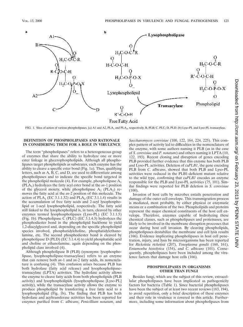
TABLE 1. Phospholipases from microorganisms other than fungi
Microbial class Enzymea Gene cloned Substrate specificitya Reference(s)
BacteriaClostridium perfringens Alpha-toxin Yes PC, SM, PS, LPC 103, 136, 162, 195, 197Clostridium novyi Gamma-toxin No PC, SM, LPC, PE, PI, PG 191
PI-PLC NoClostridium bifermentans PLC Yes Unknown 197Listeria monocytogenes PLC-A Yes PI 102, 119, 203
PLC-B Yes PC, PE, PS, SMPseudomonas aeruginosa PLC-H Yes PC, LPC, SM 13, 29, 137
PLC-NPseudomonas cepacia PLC Yes 202Staphylococcus aureus Beta-toxin Yes SM, LPC 38, 76, 148, 157, 216, 217
PI-PLC No PI, LPIBacillus cereus PC-PLC Yes PC, PE 77, 85, 96, 104
SMase Yes SMPI-PLC Yes PI, LPI
Bacillus thuringiensis PI-PLC Yes PI, LPI 68, 78, 192Mycobacterium tuberculosis MPC-Ac SM, PC 83
MPC-Bc SM, PCPLD PCPLA 177
Rickettsia prowazekii PLAcorynebacterium pseudotuberculosis PLD Yes 118Arcanobacterium haemolyticum PLD Yes 118Legionella pneumophila PLC No PC 8Ureaplasma urealyticum PLC No 15, 36
ProtozoaToxoplasma gondii PLA No 160, 161Entamoeba histolytica PLA No 106, 153
a Abbreviations: PI, phosphatidylinositol; LPI, lysophosphatidylinositol; PG, phosphatidylglycerol; SM, sphingomyelin; LPC, lysophosphatidylcholine; LPG, lyso-phosphatidylglycerol; PLC-N, nonhemolytic PLC; PLC-H, hemolytic PLC.
124 GHANNOUM CLIN. MICROBIOL. REV.
on February 23, 2013 by P
EN
N S
TA
TE
UN
IVhttp://cm
r.asm.org/
Dow
nloaded from

(21, 119, 203), elegant work by Portnoy and colleagues showedthat the two phospholipases play overlapping roles in thepathogenesis of L. monocytogenes (178). In their study, mu-tants harboring a deletion in each PLC, as well as a doublemutant lacking both enzymes, were characterized with regardto virulence. Abolishing PI-PLC and PC-PLC resulted instrains that were 2- and 20-fold less virulent than the parentstrain, respectively. In addition, these strains were defective inescape from the vacuole and in cell-to-cell spread, dependingon which enzyme was deleted. Interestingly, the mutant lackingboth PLCs was 500-fold less virulent in mice and was severelydiminished in its ability to escape from the vacuole and tospread from cell to cell (178). These findings are consistentwith the two enzymes having overlapping functions throughoutthe course of infection. Importantly, deleting a single generesulted in only a modest reduction in virulence, while simul-taneous deletion of the two genes in the same strain led to ahighly significant decrease in the virulence, suggesting that thetwo enzymes have a synergistic effect on the ability of theorganism to invade host tissues. For more details on entry of L.monocytogenes into phagocytic cells and the role of secretedenzymes, including phospholipases, in the ability of the organ-ism to escape from the vacuole and transmigrate cell-to-cell,the reader is referred to references 144, 183, and 194.
Pseudomonas aeruginosa. Two distinct PLCs are produced byPseudomonas aeruginosa: PLC-N (nonhemolytic) and PLC-H(hemolytic) (183). The genes (plcN and plcS) encoding theseenzymes have been cloned (137). Although the expression andsecretion of both enzymes are phosphate regulated, each en-zyme has a distinct substrate specificity, with PLC-H hydrolyz-ing sphingomyelin in addition to phosphatidylcholine (PC),while PLC-N is active in phosphatidylserine and PC degrada-tion (137). This difference in substrate specificity has a bearingon the efficiency with which these enzymes degrade eukaryoticmembranes. Ostroff et al. (138) proposed that the two enzymescould work sequentially and synergistically to lyse host cells.PLC-H initiates membrane lysis by hydrolyzing PC and sphin-gomyelin (the major constituents of the membrane outer leaf-let), and this is followed by the action of PLC-N which cleavesphosphatidylserine (the major constituent of the membraneinner leaflet). Furthermore, substrate specificity studies haveshown that PLC-H preferentially cleaves phospholipids con-taining quaternary ammonium groups, such as phosphatidyl-choline, which are found primarily in eukaryotic membranesand lung surfactants, but that it has minimal activity towardphospholipids such as phosphatidylethanolamine, which arefound in the prokaryotic membrane (14). This selective abilitymay explain why the invading organism can lyse the host cellswithout damaging its own membrane.
Correlation between PLC-H and P. aeruginosa pathogenicitycan be derived from studies showing that the enzyme is se-creted in vivo and in experiments comparing the wild type withmutants in which this gene is deleted in animal models. Threelines of evidence suggest that PLC-H is secreted in vivo: (i)clinical isolates from the lungs produce PLC (13), (ii) PLC isproduced when the bacterium is grown in bronchial washings(105), and (iii) high titers of antibody against PLC-H are de-tected in patients with chronic P. aeruginosa infection (62).
Mutants disrupted in the plcS gene were generated and usedto determine the role of PLC-H in pseudomonal virulence(138). In this study, the virulence of wild-type strains andplcS-disrupted mutants was compared in a mouse burn modelof infection. A reduction was observed in the ability of thedisruptant grown under phosphate-limiting conditions to killmice (200-fold increase in the 50% lethal dose compared withthe wild-type strain). Since virulence in these studies was phos-
phate dependent, mutants disrupted in plcR (a phosphate-regulatory gene) were generated to investigate its effect aloneand combined with plcS. These studies showed that when thestrains were grown under phosphate-limiting conditions, thevirulence of the DplcS DplcR strain was 200- to 10,000-foldlower than that of the parent strain (138). Interestingly, mu-tants with only plcR deleted were also attenuated in virulenceeven though they produced greater amounts of PLC and he-molysin (138). In spite of this apparent enigma, which suggestsa role for plcR in pseudomonal pathogenicity, the significantlygreater reduction in the pathogenicity of the double disruptantsuggests that PLC-H is associated with virulence (194).
Bacillus cereus. Several PLCs, including phosphatidylinosi-tol-specific and PC-preferring enzymes, as well as sphingomy-elinase, are produced by Bacillus cereus (80, 95, 198, 199). Thegenes encoding the three different proteins have been clonedand sequenced (84, 85, 96). PC-preferring and sphingomyeli-nase-encoding genes form a gene cluster (tandemly located)(59), which is not positioned in close proximity to the geneencoding phosphatidylinositol-specific PLC (59, 96). The PC-preferring enzyme has structural homology to the C. perfrin-gens alpha-toxin (103), while the phosphatidylinositol-specificenzyme has stretches of sequence homology to other eukary-otic phospholipases (97).
The contribution of phospholipases to the virulence of B.cereus has not been investigated in animals. Although PLC ofthis bacterium is considered to be nontoxic (126), data sugges-tive of its involvement in host cell lysis have been reported.Gilmore et al. (59) suggested that by creating a duplex hemo-lysin named cereolysin AB, the PC-preferring phospholipaseand the sphingomyelinase act in concert to cause hemolysis.Others (212) showed that B. cereus strains producing PLCcause degranulation of human neutrophils with a dose-depen-dent release of lysosomal enzymes, which may mediate tissuedamage. Finally, it has been suggested that B. cereus protectsitself against phagocytosis by releasing phospholipases (152).
Rickettsia. Two species of the genus Rickettsia (Rickettsiarickettsii and Rickettsia prowazekii) were shown to possess PLAthat was suggested to mediate host cell lysis (63, 177, 220, 222).Previously, Winkler and Miller (221) reported that rickettsiaeenter cells through a mechanism involving a PLA. Their hy-pothesis was based on the demonstration that host cells ex-posed to large numbers of R. prowazekii release considerableamounts of lysophospholipids and free fatty acids into theculture medium. Involvement of phospholipases in the pene-tration and damage of host cells by R. rickettsii was first sug-gested by Walker et al. (207). More recently, Silverman et al.(177) confirmed these findings and provided suggestive evi-dence that phospholipase activity associated with internaliza-tion of this intracellular parasite lies directly with the infectingorganism rather than with the host cell. However, as stated bythe authors, definitive evidence that the phospholipase origi-nates from these organisms could be provided only by isolationand cloning of specific rickettsial gene(s) involved in the inter-nalization process (177).
Corynebacterium pseudotuberculosis. Corynebacterium pseu-dotuberculosis, the agent of caseous lymphadenitis in smallruminants and ulcerative lymphangitis in horses, produces ex-tracellular PLD, which possibly plays a role in the pathogenic-ity of this and similar bacteria (Corynebacterium ulcerans andArcanobacterium haemolyticum) (1, 34, 118, 184). The genesencoding the PLD in C. pseudotuberculosis and A. haemolyti-cum have been cloned and sequenced (118). Targeted mu-tagenesis of PLD in C. pseudotuberculosis reduced the ability ofthis bacterium to establish a primary infection or cause chronicabscess formation in regional lymph nodes (117). These results
VOL. 13, 2000 PHOSPHOLIPASES IN VIRULENCE AND FUNGAL PATHOGENESIS 125
on February 23, 2013 by P
EN
N S
TA
TE
UN
IVhttp://cm
r.asm.org/
Dow
nloaded from

indicate that PLD is a virulence determinant of C. pseudotu-berculosis, increasing the persistence and spread of the bacteriawithin the host (117).
Protozoan Phospholipases
Phospholipase A facilitates host cell penetration by the twoprotozoan species Toxoplasma gondii and Entamoeba histo-lytica.
Indirect evidence implicating a calcium dependent-PLA2 inhost cell invasion by T. gondii was obtained by Saffer et al.(160), who showed that incorporation of exogenous PLA2 fromsnake venom increases host cell penetration by T. gondii. Fur-thermore, penetration of host fibroblasts by the parasite wasinhibited following preincubation with PLA2 inhibitors, such asp-bromophenacyl bromide and nordihydroguaiaretic acid, orantisera to this enzyme (160). In another study, the same groupextended these findings and demonstrated that treating fibro-blasts with fractions of disrupted T. gondii led to accumulationof degradation products of the phospholipids (fatty acids andlysophospholipids). The data suggest that PLA may be associ-ated with T. gondii cells (161). Moreover, fractions of T. gondiithat had PLA enzymatic activity also increased host cell pen-etration (161). Although the findings of Saffer and Schwartz-man (161) implicate phospholipases in the process of penetra-tion of host cells by T. gondii, these workers used a crudeenzyme preparation, which makes interpretation of the resultsquite difficult. To unequivocally establish a correlation be-tween PLA and T. gondii virulence and to elucide how theenzyme may influence the penetration process of host cells bythis parasite, gene cloning and enzyme purification are re-quired.
E. histolytica, similar to T. gondii, possesses PLA (106, 153).Ravdin and coworkers (106) reported that this ameba has twoPLA enzymes: a calcium-dependent protein which is associ-ated with the plasma membrane and is most active at an alka-line pH, and a calcium-independent enzyme that is localizedpredominantly to soluble subcellular fractions of E. histolyticaand is optimally active at an acidic pH. Amebic cytolytic activ-ity, and thereby virulence, is associated with the calcium-de-pendent PLA. However, as in the case with T. gondii, definitivedemonstration of the role of amebic PLA enzymes in thecytolytic event awaits their purification, gene cloning, and dis-ruption.
PHOSPHOLIPASES IN PATHOGENIC FUNGI
Phospholipases of Candida albicans
The overall incidence of Candida bloodstream infections hasincreased significantly in the last two decades (9, 140, 143),ranging from a 75% increase in small hospitals to an over400% increase in some large tertiary-care centers (140). Thisincrease led to a tremendous interest in the study of candidalpathogenesis and strategies for control and prevention of thisclinically important fungus. Candidal virulence factors havealso attracted interest as a possible means for developing noveltherapeutic interventions against candidiasis (33, 52, 139).Such virulence factors include adherence (20, 55, 90), germi-nation (180), extracellular proteinases (71, 72) and phospho-lipases (75), and phenotypic switching (181, 182).
Early work on candidal phospholipases. (i) Candidal phos-pholipase. The secretion of extracellular phospholipases by C.albicans was first reported in the 1960s by Costa et al. (A.Costa, A. Misefari, and A. Amaro, Abstr. Atti XIV Congr.Naz. Microbiol. Messina, abstr. P35 and P36 1967) and Werner
(213) by growing the yeast on solid media containing egg yolkor lecithin and analyzing the lipid breakdown products. Later,phospholipase activity was found in many pathogenic C. albi-cans strains by using media containing blood serum and sheeperythrocytes (30). The observation that C. albicans secretesphospholipase prompted Pugh and Cawson (150) to develop alecithin-based cytochemical method to detect this enzyme. In asubsequent study, these authors used this method (149) inconjunction with a chicken chorioallantoic membrane model toevaluate ultrastructural details of candidal invasion and to de-termine the site of phospholipase production. Invasion wasinitiated by placing stationary-phase blastospores of C. albicanson the membrane, which stimulated cellular changes in theblastospores. Many of the blastospores developed hyphae withphospholipase activity concentrated at the growing tip. Theactivity was highest where the hyphae were in direct contactwith the membrane (149). In general, only hyphae invaded themembrane successfully. Based on these results, the investiga-tors proposed that extracellular phospholipases were impor-tant in the invasion of tissue by C. albicans.
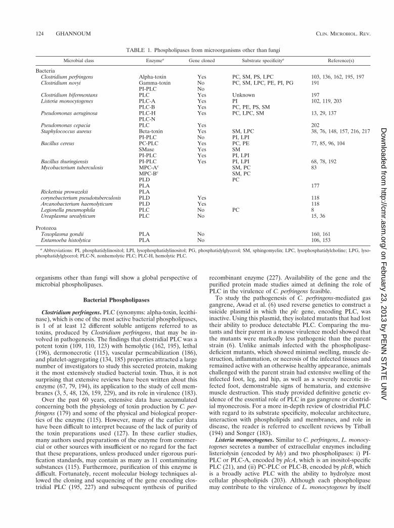
Subsequent studies by other groups were directed at devel-oping simple methods to detect candidal phospholipases. Oddsand Abbott (132) described a biochemical assay to measureintracellular PLA and Lyso-PL activity in C. albicans. By usingthis assay, it was found that PC was hydrolyzed to give Lyso-PC. A small amount of this degradation product was alwaysdetected (132). One disadvantage of this assay is that it istime-consuming and therefore unsuitable for testing a largenumber of isolates. Price et al. (147) described a plate methodfor the detection of phospholipase activity in C. albicans whichcircumvents this disadvantage. Since egg yolk contains largeamounts of phospholipids, predominantly PC and phosphati-dylethanolamine, it was incorporated into a Sabouraud dex-trose agar-based medium. When grown on this medium,phospholipase-positive candidal isolates produce a distinct,well-defined, dense white zone of precipitation around thecolony (Fig. 2). This white zone is probably due to the forma-tion of calcium complex with the fatty acids released by theaction of phospholipase on the phospholipids present in theegg yolk (110). In this assay, phospholipase activity (expressedas a Pz value) is defined as the ratio of colony diameter to thediameter of the dense white zone of precipitation around phos-pholipase positive colonies. This easy plate method became thetraditional screening method for phospholipase activity forCandida species (66, 99, 168, 215) and other fungi such as
FIG. 2. Production of phospholipase by three different clinical C. albicansisolates. Note the difference in the precipitation zones around the three colonies.
126 GHANNOUM CLIN. MICROBIOL. REV.
on February 23, 2013 by P
EN
N S
TA
TE
UN
IVhttp://cm
r.asm.org/
Dow
nloaded from

Cryptococcus neoformans (23). However, because egg yolk con-tains substrates for both phospholipases (phospholipids) andlipases (triglycerides), the egg yolk-based assay is not specific,and therefore its use should be limited to initial screens only(49). Furthermore, the assay is not suitable for the screening offungal isolates that produce low levels of phospholipase. Con-firmation of phospholipase activity necessitates the use of aspecific radiometric (10) or colorimetric (122, 176) assay andthe use of concentrated culture filtrate, particularly in poorlyphospholipase-producing strains. Awareness of this caveat isespecially relevant for researchers attempting to determine thephospholipase activity of genetically manipulated mutants,e.g., phospholipase-deficient clones generated by site-specificmutagenesis (49, 101).
(ii) Production of phospholipase by various candidal spe-cies. Early studies in which the egg yolk-based assay was usedto evaluate the ability of different Candida species to producephospholipase showed that only C. albicans and not otherspecies of Candida produce extracellular phospholipase. Sa-maranayake et al. (168) screened 41 Candida isolates for phos-pholipase activity by using a plate assay method and found that79% of the C. albicans strains tested produced extracellularphospholipases whereas no strains of C. tropicalis, C. glabrata,and C. parapsilosis produced the enzymes. The quantity ofphospholipase produced by C. albicans varied with the specificisolate and correlated with the site of infection. Blood isolatesgenerally produce much higher levels than do isolates fromwounds or urine (147). In contrast, Clancy et al. (C. J. Clancy,M. A. Ghannoun, and M. H. Nguyen, Programs Abstr. 36thAnnu. Meet. Infect. Dis. Soc. Am., abstr. 317, 1998) recentlyshowed that non-albicans Candida species produce extracellu-lar phospholipases as determined by both egg yolk-based andcolorimetric assays. In this study, 41% of C. glabrata, 50% of C.parapsilosis, 70% of C. tropicalis, 80% of C. lusitaniae, and100% of C. krusei strains produced detectable phospholipaseactivity. However, it is important to emphasize that relative toC. albicans, the non-albicans species secreted significantlysmaller amounts of phospholipase (for example, C. krusei hasapproximately 10 times less phospholipase activity [P. Mukher-jee and M. Ghannoum, unpublished data]). Although Clancyet al. demonstrated that non-albicans species can secrete phos-pholipase similar to C. albicans, it is important to point out thatthe number of candidal isolates tested was limited (n 5 51).Therefore, the percentages reported in this study may havebeen significantly different if a larger number of isolates hadbeen examined. Furthermore, the discrepancy observed by dif-ferent workers in the phospholipase activity for the non-albi-cans species may be attributed to strain-to-strain variation ordifferences in the preparation of the egg yolk agar plates usedto detect phospholipase secretion in these species.
Lane and Garcia (99), using an egg yolk-based assay, testedthe ability of C. albicans switching variants (wild type, star,stipple, and ring) to produce phospholipase (181). Star andring variants produced a similar amount of phospholipase tothe wild type. In contrast, the stipple variant produced between27 and 34% more phospholipase than did the other threestrains tested. Although candidal switching is considered to bea virulence factor (181), the relevance of the findings of Laneand Garcia to candidal pathogenicity remains to be shownthrough in vivo experimentation.
(iii) Types of candidal phospholipases. The literature con-tains contradicting reports on the number and specific types ofphospholipase enzymes secreted by C. albicans. Costa et al.(Abstr. Atti XIV Cong. Naz. Microbiol. Messina, abstr. P35and P36, 1967) reported the secretion of both PLA and PLC bythis clinically important yeast. Their results were based on the
isolation of palmitic acid and phosphatidylcholine from theproximity of candidal colonies cultured on Sabouraud agarsupplemented with serum and sheep erythrocytes. Since thismedium is not chemically defined, the sources of the hydrolysisproducts are uncertain. Banno et al. (10) performed a crudefractionation of the proteins in culture filtrates of C. albicansby using DEAE-Sephadex and then assayed the fractions forphospholipase activity. Their data suggested that C. albicanssecreted three types of phospholipases: Lyso-PL, LPTA, andPLB. Takahashi et al. (193) purified two distinct forms ofLPTA from culture filtrates of C. albicans. These candidalenzymes differed from mammalian enzymes in amino acidcomposition; however, the substrate specificities were not de-termined (193).
To determine the type and substrate specificity of phospho-lipases secreted by C. albicans, a high-phospholipase-produc-ing strain was grown to late log phase and the supernatant wasconcentrated and assayed for phospholipase activity by usingtwo complementary assays: a specific-substrate radial diffusionassay capable of differentiating between PLA, PLB, and PLCactivities (64) and a colorimetric acyl coenzyme A-oxidase sys-tem (101). Only PLB activity was observed in the diffusion-based assay (128). In the colorimetric assay, the concentratedsupernatant was incubated with a substrate that is specific forPLB (PC) or Lyso-PL (Lyso-PC) activities. Both PLB andLyso-PL activities were detected in the supernatant, suggestingthat there are two enzymes with individual activity, or oneenzyme with two activities (see “Definition of phospholipasesand rationale in considering them for a role in virulence”above). To resolve this dilemma, protein purification and genecloning were undertaken.
In collaboration with Yoshinori Nozawa (Gifu University,Gifu, Japan), we purified to homogeneity the protein respon-sible for candidal extracellular phospholipase activities. Thisenzyme is a glycoprotein with a molecular mass of 84 kDa onsodium dodecyl sulfate-polyacrylamide gel electrophoresis.The specific activities of the enzyme were 117 mmol/min/mg ofprotein for fatty acid release (hydrolase), and 459 mmol/min/mg of protein for PC formation (LPTA activity). Theapparent Km of the hydrolase activity of the enzyme for1-palmitoyl-sn-glycero-3-phosphocholine was 60.6 mM. PLBactivity was optimal at pH 6.0. The activity of the purifiedenzyme was not dependent on divalent cations (Ca21 andMg21) and was not inhibited by the addition of EDTA orEGTA (122).
To characterize the functional activity of the purified en-zyme, Mirbod et al. (122) used both the radiometric and col-orimetric methods described above. In these assays the enzymewas incubated with a substrate that is specific for PLB activityor Lyso-PL activities. In addition, to determine the LPTAactivity, Lyso-PC was incubated with the purified enzyme andthe rate of production of PC was monitored. These studiesrevealed that the purified C. albicans enzyme has both hydro-lase (PLB and Lyso-PL) and LPTA activities. The finding thatone enzyme has two hydrolase activities is not unique to C.albicans. A similar observation has been reported for PLBsecreted by Penicillium notatum. Saito et al. (164) reported thatP. notatum PLB is a glycoprotein with intrinsic Lyso-PL andPLB activities. Similarly, Lee et al. (100) showed that theSaccharomyces cerevisiae PLB1 gene encodes a protein re-quired for Lyso-PL and PLB activities. These data clearly dem-onstrate that the purified enzyme is a phospholipase with dualactivities. Similar to the candidal enzyme, PLB from S. cerevi-siae is reported to have an acyltransferase activity (224, 225).This complex pattern of activity led to difficulties in the no-menclature of the fungal enzyme (see above). Further evi-
VOL. 13, 2000 PHOSPHOLIPASES IN VIRULENCE AND FUNGAL PATHOGENESIS 127
on February 23, 2013 by P
EN
N S
TA
TE
UN
IVhttp://cm
r.asm.org/
Dow
nloaded from
dence that candidal PLB has both PLB and Lyso-PL activitieswas obtained by assaying phospholipase activities in superna-tants from the parent and PLB1-deleted mutants (see belowfor construction and characterization of mutants). Unlike theparent, deletion of caPLB1 led to loss of both PLB andLyso-PL activities.
The phospholipase activities described so far are due tosecreted candidal enzymes. Recently, McLain and Dolan (116)reported on a membrane-associated enzyme in C. albicans.This enzyme has a PLD activity and is capable of performing atransphosphatidylation reaction in the presence of primaryalcohols (see below for cloning of candidal PLD).
Evidence correlating phospholipases and virulence. Al-though the contribution of phospholipases to the pathogenesisof bacteria and protozoa has been known for some time, in-vestigations into whether these enzymes are associated withfungal virulence have only recently been undertaken. Barrett-Bee et al. (11) was the first to evaluate the role of extracellularcandidal phospholipases in virulence by using a murine modelof candidiasis. When phospholipase activity was measured insix yeasts (four strains of C. albicans and a single strain each ofC. parapsilosis and S. cerevisiae), a correlation was found be-tween phospholipase activity and two potential parameters ofpathogenicity. The C. albicans isolates which adhered moststrongly to buccal epithelial cells and were most pathogenic inmice had the highest phospholipase activities. Less pathogenicisolates of C. albicans, C. parapsilosis, and S. cerevisiae wereless adherent to epithelial cells and less lethal to mice and hadlower phospholipase activities (11). Although these findingssuggest a correlation between phospholipase activity and can-didal virulence, the strains used were not genetically related,and therefore the differences observed in the virulence of thetested strains could be attributed to factors other than phos-pholipase secretion.
Two strategies were followed by Ibrahim et al. (75) to de-termine the role of phospholipase in candidal virulence: (i) theability of blood isolates of C. albicans from patients and oralisolates from healthy volunteers to produce phospholipasewere compared, and (ii) the pathogenicity of clinical isolateswith different levels of phospholipase secretion was comparedin a murine model of disseminated candidiasis (75). In the firstcomparative study, 22 isolates of C. albicans were tested (11were blood isolates obtained from patients with disseminatedcandidiasis, and 11 were commensal isolates recovered fromthe oral cavities of healthy volunteers). Marked differencesamong strains from the two sources were observed. Signifi-cantly higher levels of phospholipase production were found inthe blood isolates than in the commensals (P 5 0.0081). Inaddition, the blood isolates had significantly higher rates ofgermination (P 5 0.03), and their germ tubes were longer thanthe germ tubes formed by the commensal strains (P 5 0.016).These findings suggest that blood isolates, unlike commensals,may have enhanced the expression of several virulence factors,including both germination and phospholipase production, toenable them to invade host tissues (75).
In the same study, Ibrahim et al. (75) prospectively exam-ined nine blood isolates for expression of virulence factors,including phospholipase and proteinase production, adher-ence, germination, growth rate, and ability to damage endo-thelial cells. Additionally, the mortality of mice infected witheach of these isolates was determined and the predictive valueof each virulence factor for mortality was determined by Coxproportional-hazards analysis. Of the virulence factors studied,only extracellular phospholipase activity was predictive of mor-tality (75).
To obtain further evidence of the contribution of phospho-
lipases to candidal pathogenicity, the virulence of two C. albi-cans isolates, CA30 and CA87, was compared in an infant-mouse model (28). Strain CA87 failed to cross the bowel wallof the infant mouse after oral-intragastric challenge and didnot cause disseminated infection, while strain CA30 was ableto cross the bowel wall and to disseminate hematogenously. Avariety of putative virulence factors of these two strains weremeasured in vitro to determine the relationship of these factorsto virulence (75). The only apparent difference in expression ofvirulence factors between the two strains was in the superiorability of CA30 to produce phospholipase (75). Since thisstrain was distinguished from CA87 by its ability to invade thebowel wall and undergo subsequent hematogenous dissemina-tion and tissue invasion, these data further suggest that phos-pholipase is involved in the invasion process of C. albicans.
Although these studies provide evidence that implicatesphospholipases in the pathogenesis of C. albicans, they do notprove this association. As in the study of Barrett-Bee et al.(11), the candidal isolates used were not genetically related.The use of such strains does not rule out the possibility thatdifferences observed in the virulence of these strains could beattributed to factors other than phospholipases. Thus, the cor-relation found between phospholipase and candidal virulenceshould be confirmed by the use of an isogenic strain pair thatdiffers only in phospholipase production. Molecular cloning ofthe candidal gene(s) encoding the extracellular phospholipasesis the essential first step in the molecular genetic dissection ofthe role of these enzymes in pathogenesis.
Cloning of the gene(s) encoding candidal phospholipases.Efforts to clone genes encoding candidal phospholipases haveresulted in the cloning of two genes encoding PLB (caPLB1and caPLB2) and one gene (caPLD) coding for PLD.
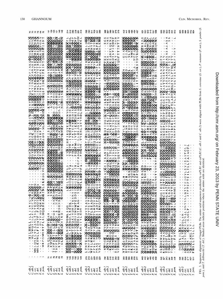
(i) Candidal phospholipases B. (a) caPLB1. The C. albicansPLB1 gene was cloned by using a PCR-based approach relyingon degenerate oligonucleotide primers designed on the basis ofthe amino acid sequences of two peptide fragments obtainedfrom a purified candidal enzyme displaying phospholipase ac-tivity (122). Sequence analysis of a 6.7-kb EcoRI-ClaI genomicclone revealed a single open reading frame of 1,818 bp thatpredicts a preprotein of 605 residues. The size of the candidalPLB1 protein is similar to those reported for other fungalPLBs, which ranged between 612 and 664 amino acids (Table2). The genomic DNA sequence encodes 17 amino acid resi-dues that are absent from the NH2 terminus of the matureprotein. This stretch of residues represents a possible signalsequence (111, 169). The predicted protein contains sevenAsn-X-Ser/Thr motifs (residues 199, 261, 399, 451, 465, 492,and 573) that could potentially be N glycosylated. One possibletyrosine phosphorylation site, Lys-Ser-Asn-Ile-Asp-Val-Ser-Ala-Tyr (residues 369 to 377), was also identified (101). Hy-dropathy analysis of the predicted protein sequence (98) re-vealed the presence of a single stretch of hydrophobic aminoacids present at the amino terminus (residues 1 to 18). Thissegment of amino acids most probably functions as a singlepeptide which targets the protein to the endoplasmic reticulumfor subsequent processing and, ultimately, secretion. Compar-ison of the putative candidal phospholipase with other proteinsin the redundant database (BLASTP program) revealed sig-nificant homology to known fungal PLBs from S. cerevisiae(45%), P. notatum (42%), Torulaspora delbrueckii (48%), andSchizosaccharomyces pombe (38%) (Fig. 3). This gene, desig-nated caPLB1, was mapped to chromosome 6 (101). Consis-tent with our findings, Hoover et al. (69a) used degenerateoligonucleotide primers derived from conserved regions ofPLB1 genes from S. cerevisiae and other fungi to clone the
128 GHANNOUM CLIN. MICROBIOL. REV.
on February 23, 2013 by P
EN
N S
TA
TE
UN
IVhttp://cm
r.asm.org/
Dow
nloaded from
TA
BL
E2.
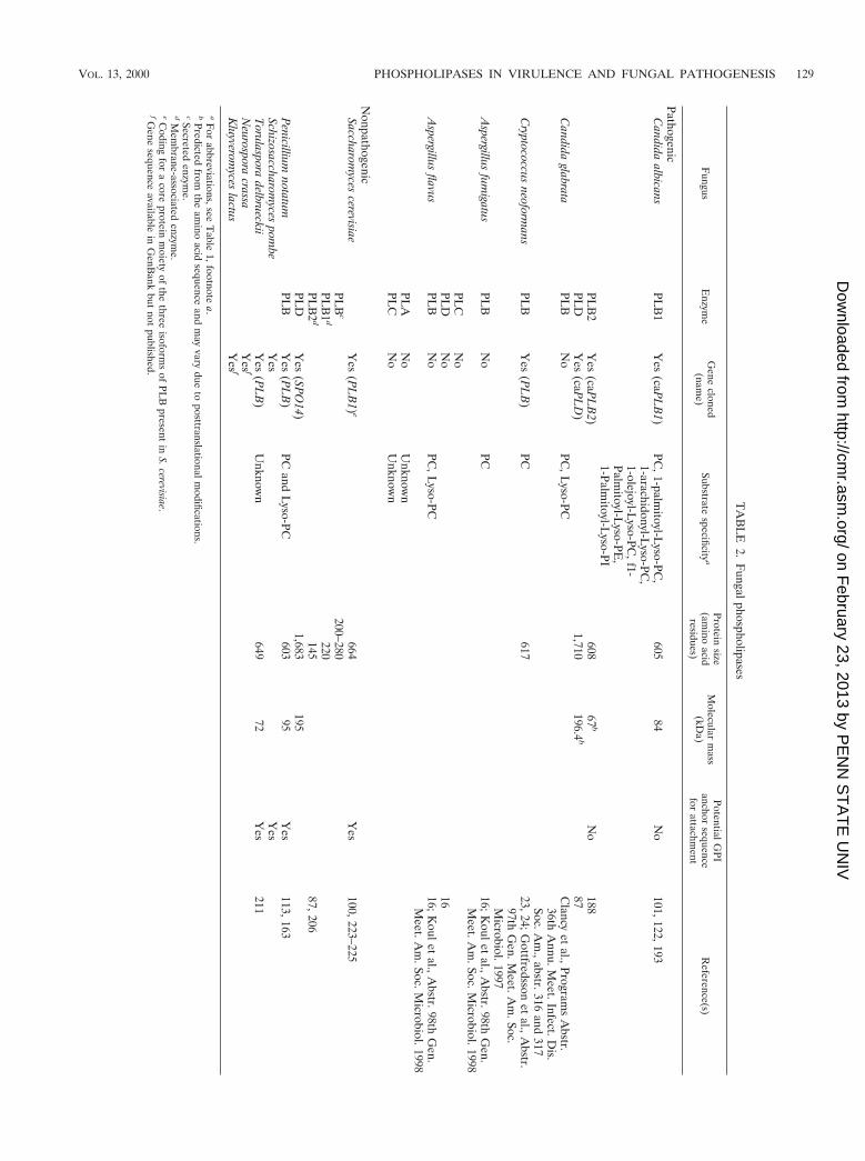
Fungalphospholipases
Fungus
Enzym
eG
enecloned
(name)
Substratespecificity
aProtein
size(am
inoacid
residues)
Molecular
mass
(kDa)
PotentialGPI
anchorsequence
forattachm
entR
eference(s)
PathogenicC
andidaalbicans
PLB
1Y
es(caP
LB
1)PC
,1-palmitoyl-L
yso-PC,
1-arachidonyl-Lyso-PC
,1-olejoyl-L
yso-PC,f1-
Palmitoyl-L
yso-PE,
1-Palmitoyl-L
yso-PI
60584
No
101,122,193
PLB
2Y
es(caP
LB
2)608
67b
No
188PL
DY
es(caP
LD
)1,710
196.4b
87C
andidaglabrata
PLB
No
PC,L
yso-PCC
lancyet
al.,Programs
Abstr.
36thA
nnu.Meet.Infect.D
is.Soc.A
m.,abstr.316
and317
Cryptococcus
neoformans
PLB
Yes
(PL
B)
PC617
23,24;Gottfredsson
etal.,A
bstr.97th
Gen.M
eet.Am
.Soc.M
icrobiol.1997A
spergillusfum
igatusPL
BN
oPC
16;Koulet
al.,Abstr.98th
Gen.
Meet.A
m.Soc.M
icrobiol.1998PL
CN
oPL
DN
o16
Aspergillus
flavusPL
BN
oPC
,Lyso-PC
16;Koulet
al.,Abstr.98th
Gen.
Meet.A
m.Soc.M
icrobiol.1998PL
AN
oU
nknown
PLC
No
Unknow
n
NonpathogenicSaccharom
ycescerevisiae
Yes
(PL
B1)
e664
Yes
100,223–225PL
Bc
200–280PL
B1
d220
PLB
2d
14587,206
PLD
Yes
(SPO
14)1,683
195P
enicilliumnotatum
PLB
Yes
(PL
B)
PCand
Lyso-PC
60395
Yes
113,163Schizosaccharom
ycespom
beY
esY
esT
orulasporadelbrueckii
Yes
(PL
B)
Unknow
n649
72Y
es211
Neurospora
crassaY
esf
Kluyverom
yceslactus
Yes
f
aF
orabbreviations,see
Table
1,footnotea.
bPredicted
fromthe
amino
acidsequence
andm
ayvary
dueto
posttranslationalmodifications.
cSecretedenzym
e.d
Mem
brane-associatedenzym
e.eC
odingfor
acore
proteinm
oietyof
thethree
isoforms
ofPL
Bpresent
inS.cerevisiae.
fGene
sequenceavailable
inG
enBank
butnot
published.
VOL. 13, 2000 PHOSPHOLIPASES IN VIRULENCE AND FUNGAL PATHOGENESIS 129
on February 23, 2013 by P
EN
N S
TA
TE
UN
IVhttp://cm
r.asm.org/
Dow
nloaded from
FIG
.3.
Sequ
ence
alig
nmen
tof
fung
alPL
Bs.
The
amin
oac
idse
quen
ces
pred
icte
dby
caP
LB
1an
dca
PL
B2
(C.a
lb.1
and
C.a
lb.2
)w
ere
alig
ned
with
PLB
sfr
omS.
cere
visi
ae(S
.cer
.),P
.not
atum
(P.n
ot.)
,S.p
ombe
(S.
pom
.),a
ndT
.del
brue
ckii
(T.d
el.)
.Sha
ded
area
sin
dica
tepo
sitio
nsw
here
the
amin
oac
ids
are
iden
tical
.
130 GHANNOUM CLIN. MICROBIOL. REV.
on February 23, 2013 by P
EN
N S
TA
TE
UN
IVhttp://cm
r.asm.org/
Dow
nloaded from

corresponding candidal homolog. Their results confirm ourfindings and the identity of caPLB1.
A deeper analysis of the PLB1 protein sequence revealed afeature that distinguishes the candidal enzyme from the otherknown fungal PLBs. Unlike the PLBs of the nonpathogenicfungi, S. cerevisiae, S. pombe, T. delbrueckii, and P. notatum,candidal PLB1 is characterized by the absence of a hydropho-bic COOH terminus (Table 2). Such a hydrophobic COOH-terminal region may ultimately be replaced by a glycosylphati-dylinositol (GPI) anchor. Indeed, potential GPI anchor sites,immediate to the hydrophobic COOH-terminus, were identi-fied in fungal PLBs from S. cerevisiae, S. pombe, T. delbrueckii,and P. notatum (22, 53). Proteins modified with a GPI anchormay be transiently tethered to the plasma membrane or ulti-mately cross-linked to the insoluble glucan component of thecell wall (43, 107, 108). Release of proteins associated with theplasma membrane would require the action of a GPI-specificphospholipase. In this view, a GPI anchor may serve to regu-late the release of the enzyme to the surroundings. UnlikePLBs from nonpathogenic fungi, the candidal PLB, escapingGPI anchoring, would probably be directly secreted. Such acharacteristic may enhance the virulence of C. albicans. Fur-ther characterization of PLBs from each fungal species will benecessary to clarify whether any are GPI anchored, and whateffect this modification may have on the function and subcel-lular localization of PLBs, as well as on the pathogenicity ofthese fungi.
(b) caPLB2. Attempts to clone caPLB2 were prompted byevidence suggesting that C. albicans may possess more thanone gene encoding PLB. This evidence is summarized as fol-lows: (i) two proteins which share phospholipase activity werepurified by Takahashi et al. (193); (ii) similarly, three enzymeswith PLB activity have been purified and characterized from S.cerevisiae (224, 225); (iii) the completion of the sequencing ofthe S. cerevisiae genome revealed that this organism has at leastthree genes encoding PLB that are highly homologous (Gen-Bank accession no. L23089, S53035, and S66693); (iv) the S.pombe genome has two sequences which potentially encodePLB (D89183 and D89204); and (v) deletion of caPLB1 didnot lead to 100% loss of phospholipase activity, suggesting thatthe residual activity (about 1% and 10% PLB and Lyso-PLactivities, respectively) may be due to a second gene.
To clone caPLB2, Sugiyama et al. (188) used a PCR-basedapproach similar to the one used to clone caPLB1. A numberof similarities are observed between caPLB1 and caPLB2 insize, availability of N-glycosylation sites, the presence of asingle stretch of hydrophobic amino acids at the amino termi-nus, and the absence of GPI attachment site (Table 2). Thenucleotide sequence of caPLB2 contained a single open read-ing frame encoding a putative 608-amino-acid protein with anestimated molecular mass of about 67 kDa. The predictedamino acid sequence contains six potential N-glycosylationsites [Asn-X-(Ser/Thr) motifs] at residues 259, 365, 450, 464,491, and 572. The deduced amino acid sequence of caPLB2was homologous to that of caPLB1 (65% identity). caPLB2 wasalso similar to PLBs from S. cerevisiae, T. delbrueckii, and P.notatum (42, 46, and 42% identity, respectively) (Fig. 3). Hy-dropathy analysis of the predicted protein (98) revealed thepresence of a cluster of hydrophobic amino acids at the Nterminus. Similar to S. cerevisiae and T. delbrueckii PLBs (100,211), caPLB2 possesses a potential signal sequence at the N-terminal region of caPLB2, where two polar amino acids (Gln-Ser) are followed by a cluster of six hydrophobic amino acids(Ile-Leu-Leu-Phe-Val-Val). Such sequence may guide proteinsto the secretory pathway. Like caPLB1, caPLB2 lacks the GPIattachment site (a cluster of hydrophobic amino acids at the
carboxy terminal) found in the PLBs from the nonpathogenicfungi such as S. cerevisiae, P. notatum, and T. delbrueckii PLBs(100, 113, 211) (see above). Therefore, caPLB2, as withcaPLB1, is probably not GPI anchored. Since fungal PLBs andmammalian PLA2 have lysophospholipase activities, it is likelythat these enzymes also share common conserved amino acidregions. In this regard, three amino acid residues essential forthe catalytic function have been identified in PLA2 (200Arg,228Ser, and 549Asp) (142). Among them, the serine residueparticipates in the catalytic function. Interestingly, three re-gions surrounding these amino acids are also conserved inPLBs from fungi. The deduced amino acid sequence ofcaPLB2 contains the motifs SGGGX97RA(M/L), GL133SG(G/S), and 381D(S/G)G(E/L)XXXN, which may have a catalyticfunction (188).
It is clear that fungal PLBs have certain features that arecommon and others in which they differ. To determine thephylogenetic relationship among various fungal PLBs, Sug-iyama et al. (188) constructed a phylogenetic tree of PLBs byusing the neighbor-joining method (165). Sequence data fromS. cerevisiae PLB (GenBank accession no. L23089), S. cerevi-siae SP01 (P53541), S. cerevisiae YLM006c (S53035), S. cerevi-siae YOLO11w (S66693), T. delbrueckii PLB (D32134), P. no-tatum PLB (P39457), S. pombe (Z99258), Neurospora crassa(AF045575), and C. albicans caPLB1 and caPLB2 were used inthe tree construction. Figure 4 shows that PLBs and potentialPLB analogues are contained in a large cluster of PLB familymembers. caPLB1 and caPLB2 are closely related to eachother, and caPLB genes are more closely related to PLB genesfrom S. pombe and P. notatum than to PLBs genes from S.cerevisiae and T. delbrueckii.
(ii) Candidal phospholipase D. PLD catalyzes the hydrolysisof PC to produce phosphatidic acid and choline (Fig. 1aabove). Both mammalian and fungal (S. cerevisiae) genes en-coding PLD have been cloned and characterized (130, 206).Mammalian PLD has emerged as one of the key enzymes inintracellular signaling (44), while PLD from S. cerevisiae (en-coded by SPO14) is essential for meiosis (41, 158, 206). Theabsence of meiosis in C. albicans, which, unlike S. cerevisiae,exists as a diploid, indicates that PLD in this clinically impor-tant yeast may play other roles than meiosis. McLain andDolan (116) have recently shown that PLD may be an impor-
FIG. 4. Phylogenetic tree analysis of fungal PLBs. S. cerevisiae PLB (Gen-Bank accession no. L 23089), S. cerevisiae SPO1 (P 53541), S. cerevisiae YLM006c(S53035), S. cerevisiae YOL011w (S66693), T. delbrueckii PLB (D 32134), P.notatum PLB (P 39457), S. pombe PLB (Z 99258), N. crassa PLB (AF045575),and C. albicans caPLB1 and caPLB2.
VOL. 13, 2000 PHOSPHOLIPASES IN VIRULENCE AND FUNGAL PATHOGENESIS 131
on February 23, 2013 by P
EN
N S
TA
TE
UN
IVhttp://cm
r.asm.org/
Dow
nloaded from
tant regulator of the dimorphic transition. Since this yeast-hypha transformation is thought to be an important virulencedeterminant in C. albicans (33, 54, 133), cloning and disruptionof candidal PLD may contribute to our understanding of thebiological role of this phospholipase.
For cloning candidal PLD, Kanoh et al. (87) designed twooligonucleotide primers based on the conserved amino acidsequences in human PLD1a and SPO14. These primers wereused in a PCR-based approach to clone the full-length gene.The cloned PLD sequence had a potential open reading frameencoding a protein of 1,710 amino acids with a calculatedmolecular mass of 196.4 kDa. The putative ATG codon wassurrounded by the consensus Kozak sequence for translationinitiation (94), and the in-frame stop codon and the putativeTATA box sequence (TATATAA) were found 6 and 172 basesupstream from the start codon, respectively. The deducedamino acid sequence contained the four conserved regions (I,II, III, and IV) defined by the primary structures of plant,yeast, and human PLDs (125). Furthermore, the HKD motif(HxKxxxxD) in regions I and IV and a serine residue in theGSRS motif in region IV, which are critical for PLD biochem-ical activity (189), were also completely conserved.
The number of amino acids and the calculated molecularmass of C. albicans PLD closely resembled these of SPO14protein (1,683 amino acids; 195 kDa). Comparison of the pri-mary structure with those of other PLDs showed that it had thehighest homology to the SPO14 protein. The overall homologybetween the amino acid sequences of C. albicans PLD and theSPO14 protein was 42%. At the four conserved regions, thehomology between C. albicans PLD and the SPO14 proteinranged from 65 to 71%, while that between C. albicans PLDand rat PLDs was 42 to 55%. In addition to the four conservedregions found in both fungal and mammalian PLDs, candidalPLD has seven regions (A to G) of 10 to 91 amino acids thatare highly homologous to SPO14 protein. Of the seven regions,only two, F and G, are conserved in mammalian PLDs. There-fore, Kanoh et al. (87) speculated that these five regions (A toE) may compose some functional domains specific for fungi.
A phylogenetic tree for PLDs of various species showedthree major clusters (87). The first cluster is composed ofmammalian PLD1 and plant (151, 209), nematode, and Strep-tomyces PLDs. The second cluster was composed of mamma-lian PLD2, and the third cluster was composed of fungal PLDs,including C. albicans PLD and the SPO14 protein. Becausefungal PLDS are in a separate grouping from mammalian andother PLDs, it is tempting to speculate that this enzyme mayserve a fungus-specific function(s). Unfortunately, elucidationof the biological function of candidal PLD awaits the disrup-tion of the gene encoding it.
Disruption of phospholipase B1. Gene disruption (deletion)is frequently used to determine the functionality of a specificgene. This is particularly relevant in assessing the contributionof a given gene to microbial virulence (45). Several workershave disrupted a number of genes to evaluate their contribu-tion to the virulence of C. albicans (37, 61, 74, 170, 228). Mostrecently, C. albicans strains with deletions of the INT1 gene,which encodes a cell surface protein with similarity to mam-malian integrins, were constructed (50). Adhesion, hyphal for-mation, and virulence were subsequently found to be corre-lated with the expression of this gene.
The successful cloning of caPLB1 allowed us to constructPLB-deficient mutants by targeted gene disruption by using theura-blaster technique (47) (see reference 101 for details of themethod used to disrupt caPLB1). Initial disruption of caPLB1was complicated by the finding that three rounds of transfor-mation were required to delete the gene, suggesting that strain
CAI-4 (a candidal strain derived from C. albicans SC5314 bydeletion of the ura3 gene) was triploid for this locus. Thisfinding seemed plausible since disruption of the C. albicanschitin synthase (CHS2) and catalase (CAT1) genes also re-vealed that they are triploid (61, 226). However, chromosome-blotting experiments suggested that the triallelic state of thecaPLB1 genetic locus probably originated from a gene dupli-cation/translocation event rather than an inherent triploidstate in the parental strain.
Given the above finding, we proceeded to disrupt thecaPLB1 gene a second time, starting with the original CAI-4strain. Chromosomal analysis was performed at every stage ofthis second disruption as a means of screening newly con-structed strains for genetic translocation or incorrect targetingof transforming DNA. Southern hybridization, chromosomalanalysis, and measurement of functional activity confirmed thesuccessful disruption of caPLB1 and demonstrated that Can-dida is diploid for this locus (101). Although we cannot dis-count the fact that some candidal genes may be aneuploid (61,226), our study with caPLB1 highlights the importance of fullycharacterizing the nature of a suspected triploidy even if mu-tant strains are constructed by the generally reliable techniqueof targeted gene disruption. Similar experience has been en-countered by a number of Candida researchers (personal com-munications); therefore, intensive molecular genetic analysisof the constructed mutants should be undertaken so as todisclose potential mutations that otherwise go undetected, es-pecially if disruption of the gene in question produces noobvious phenotypic defect.
Phenotypic characterization of phospholipase B1-deficientmutants. Phenotypic characterization of the parent and dele-tion mutants is necessary prior to evaluating the virulence ofthe isogenic strain pair. Such characterization may providesome measure of assurance that deleting the caPLB1 generesults in only loss of PLB production and not other unrelatedphenotypic properties. Comparison of the growth rates andgermination capabilities of PLB-deficient mutant with that ofthe parental strain revealed that disruption of caPLB1 did notaffect growth and germination of C. albicans (101), indicatingthat caPLB1 is not essential for these processes.
Disruption of caPLB1 reduced the ability of C. albicans tosecrete the enzyme. When examined by Western blot analysis,PLB was found only in culture filtrates produced by parentaland plb1-D1 strains but not in the supernatant produced bystrain plb1-D2 (null mutant). Furthermore, assay of superna-tants collected from the parent and the PLB-deficient mutantsfor PLB and Lyso-PL activities by using specific substratesrevealed that the two activities were reduced in the PLB-deficient mutant by approximately 99 and 80% respectively,relative to the wild type. The residual phospholipase activitiessecreted by the caPLB1-deficient strain may be the result of anadditional candidal phospholipase-encoding gene. In this re-gard, a second gene, caPLB2, which has significant homologyto caPLB1, was recently cloned by our group (see “Candidalphospholipases B” above). Experiments to delete this secondgene are under way. Characterization of mutants with caPLB1and caPLB2 deleted may clarify the source of the residualphospholipase activity observed in caPLB1-deficient mutant.
Testing of phospholipase B1-deficient mutants in murinemodels of candidiasis. Two different models of candidiasis,representing different clinical settings, were used to determinethe role of caPLB1 in the virulence of C. albicans: (i) a hemat-ogenous-dissemination murine model (58) and (ii) an oral-intragastric infant mouse model (28).
(i) Hematogenous-dissemination model. Leidich et al. (101)challenged BALB/c mice with 5 3 105 yeast cells of either the
132 GHANNOUM CLIN. MICROBIOL. REV.
on February 23, 2013 by P
EN
N S
TA
TE
UN
IVhttp://cm
r.asm.org/
Dow
nloaded from

parent or the PLB-deficient strains intravenously through thelateral tail vein. Both survival and tissue fungal burden wereused to assess the pathogenicity of the infecting strains. Theirdata showed that all mice infected with parental strain SC5314succumbed to candidal infection within 9 days. In contrast, 50and 60% of mice challenged with either the PLB-deficientstrain plb1-D1 or plb1-D2, respectively, were alive at day 15.The mean survival time 6 standard deviation for mice infectedwith parent was 4.4 6 2.1 days, compared to 12.7 6 2.7 and13.3 6 2.6 days for mice infected with plb1-D1 or plb1-D2,respectively. Statistical analyses revealed that mice infectedwith either strain plb1-D1 or plb1-D2 survived significantlylonger than did mice infected with strain SC5314 (P , 0.0001for both comparisons).
Gross inspection of the kidneys showed that numerous vis-ible candidal foci covered the renal cortex of mice infectedwith the parental strain. In contrast, no candidal foci werevisible on renal surfaces of mice infected with either of thePLB-deficient strains. Tissue fungal burden experiments werecarried out to assess the severity of infection caused by thewild-type and mutant strains (101). Consistent with the resultsof the survival experiment, candidal strains plb1-D1 andplb1-D2 were cleared significantly faster from the kidneys andbrain than was parental strain SC5314. For example, the meanfungal burden in the kidneys 6 standard deviation was 6.13 60.05 and 4.33 6 0.35 CFU/g of tissue for the parent andplb1-D2 mutant, respectively (P , 0.004). The relative rates ofclearance were as follows: plb1-D2 . plb1-D1 . SC5314.
Taken together, these findings demonstrate that caPLB1 isassociated with candidal virulence. Survival and tissue fungalburden data showed that mortality of and tissue invasion inmice infected with strain plb1-D1, which harbors only a singledeleted caPLB1 allele, was also significantly reduced comparedto those for the wild type, suggesting that there may be adose-dependent relationship between PLB and virulence.Thus, a threshold level of PLB may be required to effectivelyenhance candidal virulence. In this regard, analysis of culturefiltrates for PLB activity revealed that the levels of free fattyacid activity, relative to the parent, released from dipalmi-toyl-PC following incubation with culture filtrates obtainedfrom CAI-4, plb1-D1, and plb-D2 were 100, 54, and 1%, respec-tively. Moreover, the fact that deletion of caPLB1 did notrender C. albicans strains completely avirulent underscores thenotion that candidal pathogenicity is multifactorial and is reg-ulated by more than one determinant (33).
(ii) Oral-intragastric infant-mouse model. Seshan et al.(174) used an oral-intragastric infant-mouse model to examinethe effect of caPLB1 on the ability of C. albicans to traverse thegastrointestinal barrier and colonize systemic target organs,such as the kidneys and liver. This model simulates candidalmigration across the gastrointestinal tract, one of the majorroutes for contracting disseminated candidiasis (131). In theseexperiments, inbred infant mice [crl:CFW (SW) BR] were in-oculated intragastrically with 2 3 108 blastospores of either thewild-type or caPLB1-deficient strain. Transmigration of can-didal cells across the gastrointestinal tract was monitored mi-croscopically as well as by determining tissue fungal burden oftarget organs.
The histopathologic appearance of the gastric mucosa ofmice infected with the parental strain differed markedly fromthat of mice infected with the PLB-deficient mutant. Light andtransmission electron microscopic examination revealed yeastand hyphal elements in the gastric mucosa 14 days postchal-lenge. Fungal elements were observed after challenge withboth the parental and PLB-deficient strains. However, mucosalinvasion in mice challenged with the PLB-deficient strain was
confined to the stomach lumen and inner layers of the gastricmucosa, and a minimal neutrophil-dependent inflammatoryresponse was elicited. Furthermore, several areas of the stom-ach revealed no demonstrable infection. In contrast, the pa-rental strain invaded the submucosal tissue of the stomach,elicited a neutrophil-dependent inflammatory response, andproduced systemic candidiasis to a far greater extent. Addi-tionally, the parental strain was able to transverse the vascu-lature, since numerous hyphal elements were observed withinblood vessel lumens (see below) (174).
The number of mice that developed systemic candidal infec-tions differed markedly following challenge with either theparental or PLB-deficient strain. Of the mice challenged withthe parental strain, 90 and 70%, respectively, exhibited liverand kidney colonization. In contrast, only 45 and 27% of miceinfected with the PLB-deficient mutant exhibited liver andkidney involvement, respectively (174). This difference was re-flected in the number of candidal cells (CFU) recovered fromthese organs. The relative candidal cells recovered from thekidneys and liver of mice challenged with the parental orcaPLB1-deficient strain were 2.05 6 0.4 and 0.77 6 0.4 CFU/gfor the kidney and 2.87 6 0.4 and 1.33 6 0.4 CFU/g for theliver, respectively. These differences were statistically signifi-cant (P values for kidneys and liver were 0.042 and 0.014,respectively).
The findings of Seshan et al. (174) that the parental strainwas more efficient at crossing the gastrointestinal tract andinvading internal organs than was its PLB-deficient counter-part suggest a role for PLB in candidal transmigration acrossthe gastrointestinal tract and subsequent dissemination to tar-get organs. Furthermore, their study presents further evidencein support of PLB as a virulence determinant for C. albicans.Finally, the results of the studies of Leidich et al. (101) andSeshan et al. (174) indicate that candidal PLB may be criticalfor dissemination of C. albicans by the gastrointestinal andhematogenous routes.
Expression of phospholipase B1 during host tissue invasion.One of the criteria to prove that a particular gene or its prod-uct plays an important role in the disease process is to showthat it is expressed by the microorganism during the infectiousprocess (45, 167). Both the hematogenous-dissemination (101)and oral-intragastric mouse (174) models for candidiasis wereused to determine whether PLB is secreted in vivo.
In the hematogenous-dissemination model, Leidich et al.(101) challenged mice with either the parental or the caPLB1-deleted mutant. Kidneys were harvested, and sections wereprocessed for immunoelectron microscopy by being incubatedwith either PLB antiserum or goat serum, which served as anegative control. The data revealed that while PLB was se-creted from the parental strain during the infectious process,as evidenced by the formation of immunogold complexes fol-lowing incubation of the tissue sections with PLB antiserum, itwas not secreted from the PLB-deficient mutant. In otherexperiments, C. albicans cells were recovered from the kidneysof mice infected with either the parental or the PLB-deficientstrain and prepared for immunogold electron microscopy. Sec-tions examination showed that immunogold labeling was ob-served only with cells recovered from mice infected with thewild type strain and not with cells recovered from animalsinfected with the PLB-deficient mutant (J. Vitullo, S. D.Leidich, C. J. Jessup, and M. A. Ghannoum, Program Abstr.98th Gen. Meet. Am. Soc. Med., abstr. F-39, 1998). These datademonstrate that C. albicans secretes PLB during the invasionof target organs. Moreover, deletion of PLB abrogate the fun-gal cell ability to secrete this lytic enzyme (see “Phenotypic and
VOL. 13, 2000 PHOSPHOLIPASES IN VIRULENCE AND FUNGAL PATHOGENESIS 133
on February 23, 2013 by P
EN
N S
TA
TE
UN
IVhttp://cm
r.asm.org/
Dow
nloaded from
genotypic characterization of phospholipase B1-deficient mu-tants” above).
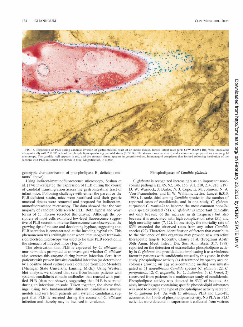
Using indirect-immunofluorescence microscopy, Seshan etal. (174) investigated the expression of PLB during the courseof candidal transmigration across the gastrointestinal tract ofinfant mice. Following challenge with either the parent or thePLB-deficient strain, mice were sacrificed and their gastricmucosal tissues were removed and prepared for indirect-im-munofluorescence microscopy. The data showed that the vastmajority of candidal cells secrete PLB. Both hyphal and yeastforms of C. albicans secreted the enzyme. Although the pe-riphery of most cells exhibited low-level fluorescence sugges-tive of PLB secretion, intense fluorescence was observed at thegrowing tips of mature and developing hyphae, suggesting thatPLB secretion is concentrated at the invading hyphal tip. Thisphenomenon was strikingly clear when immunogold transmis-sion electron microscopy was used to localize PLB secretion inthe stomach of infected mice (Fig. 5).
The observation that PLB is expressed by C. albicans inmurine models prompted us to investigate whether C. albicansalso secretes this enzyme during human infection. Sera frompatients with proven invasive candidal infection (as determinedby a positive blood culture) were obtained from Leo Mendoza(Michigan State University, Lansing, Mich.). Using Westernblot analysis, we showed that sera from human patients withsystemic candidiasis contain antibodies that reacted with puri-fied PLB (data not shown), suggesting that PLB is secretedduring an infectious episode. Taken together, the above find-ings, using two fundamentally different candidiasis murinemodels and sera from patients with systemic candidiasis, sug-gest that PLB is secreted during the course of C. albicansinfection and thereby may be involved in virulence.
Phospholipases of Candida glabrata
C. glabrata is recognized increasingly as an important noso-comial pathogen (2, 89, 92, 146, 156, 201, 210, 214, 218, 219);D. W. Warnock, J. Burke, N. J. Cope, E. M. Johnson, N. A.Von Frauenhofer, and E. W. Williams, Letter, Lancet ii:310,1988). It ranks third among Candida species in the number ofreported cases of candidemia, and in one study, C. glabratasurpassed C. tropicalis to become the most common nonalbi-cans species isolated (51). C. glabrata is important clinically,not only because of the increase in its frequency but alsobecause it is associated with high complication rates (51) andhigh mortality rates (7, 12). In one study, the mortality rate of83% exceeded the observed rates from any other Candidaspecies (92). Therefore, identification of factors that contributeto the virulence of this organism may provide new attractivetherapeutic targets. Recently, Clancy et al. (Programs Abstr.36th Annu. Meet. Infect. Dis. Soc. Am., abstr. 317, 1998)reported on the detection of extracellular phospholipase activ-ity in C. glabrata and provided data implicating it as a virulencefactor in patients with candidemia caused by this yeast. In theirstudy, phospholipase activity (as determined by opacity aroundcolonies growing on egg yolk-containing media) was investi-gated in 51 non-albicans Candida species (C. glabrata, 22; C.parapsilosis, 12; C. tropicalis, 10; C. lusitaniae, 5; C. krusei, 2)recovered from patients in a multicenter study of candidemia.Phospholipase activity was detected in 53% of isolates. Anassay involving agar containing specific phospholipid substrateswas used to identify the type of phospholipase activity secretedby C. glabrata (64). As with C. albicans, PLB and Lyso-PLaccounted for 100% of phospholipase activity. No PLA or PLCactivities were detected in supernatants collected from various
FIG. 5. Expression of PLB during candidal invasion of gastrointestinal tract of an infant mouse. Inbred infant mice [cr1: CFW (CSW) BR] were inoculatedintragastrically with 2 3 108 cells of the phospholipase-producing parental strain (SC5314). The stomach was harvested, and sections were prepared for immunogoldmicroscopy. The candidal cell appears in red, and the stomach tissue appears in greenish-yellow. Immunogold complexes that formed following incubation of thesections with PLB antiserum are shown in blue. Magnification, 310,000.
134 GHANNOUM CLIN. MICROBIOL. REV.
on February 23, 2013 by P
EN
N S
TA
TE
UN
IVhttp://cm
r.asm.org/
Dow
nloaded from
C. glabrata isolates tested. To determine whether a correlationexists between phospholipase activity and clinical virulence, theauthors classified the C. glabrata isolates into two groups: per-sistent isolates were strains that remained in the blood despitetherapy with antifungal agents effective in vitro and despiteremoval of all intravenous catheters, and nonpersistent isolateswere strains that were cleared. Among non-albicans Candidaspecies tested, the association between phospholipase activityand persistent candidemia was strongest for C. glabrata (P 50.004). Moreover, the median PLB and Lyso-PL activities werehigher for persistent isolates (9.5 and 24 pmol/mg, respec-tively) than for nonpersistent isolates (0 and 5.8 pmol/mg) (P ,0.0001 and P 5 0.02, respectively).
To establish the role of PLB in the pathogenesis of C. gla-brata, an isogenic strain pair of this yeast which differed only inPLB activity was created (C. J. Clancy, A. Leuin, M. A. Gha-nnoum, and M. H. Nguyen, Programs Abstr. 36th Annu. Meet.Infect. Dis. Soc. Am., abstr. 316, 1998). Conserved sequencesof scPLB1, the gene responsible for PLB activity in S. cerevi-siae, were used to design guessmer primers for PCR with DNAfrom a clinical C. glabrata isolate. A 1,030-bp PCR product thatencoded part of a putative protein with 78% homology toscPLB1 protein was generated. To create an isogenic mutantwith disruption of the PLB gene, Clancy et al. first created aura32 auxotroph of the parent C. glabrata strain. S. cerevisiaeURA3, which is .80% identical to C. glabrata URA3, wasamplified by PCR, BamHI restriction sites were added to ei-ther end, and the resulting product was inserted in the uniqueBamHI site of the 1,030-bp PCR fragment. The disruptedproduct was liberated from the plasmid by EcoRI digestionand was then used to transform C. glabrata ura32 auxotrophs.The resulting mutants were selected by growth on Ura2 media.Targeted disruption was confirmed by Southern blot analysis.Colorimetric assay of phospholipase activity revealed .90%elimination of both PLB and Lyso-PL activities in the mutantisolate (URA31 plb2) compared to the parent C. glabrata(URA31 plb1), confirming that the cloned gene was responsi-ble for the vast majority of PLB and Lyso-PL activities. Com-parative in vivo pathogenicity studies are currently being per-formed to determine the role of the cloned gene in C. glabratavirulence.
Phospholipases of Cryptococcus neoformans
Cryptococcus neoformans is the cause of the most commonlife-threatening fungal infection in patients with AIDS. De-pending on the study, estimates of the frequency of cryptococ-cosis among AIDS patients range from 5% to slightly greaterthan 10% (25, 42, 172). Although the occurrence has decreasedin the last couple of years in the developed world due to theintroduction of triple HIV therapy (B. Dupont, Abstr. 4thCongr. Eur. Confed. Med. Mycol., abstr. S1, 1998), this inci-dence is still high, particularly in developing countries such asUganda. Given the high rate of relapse after initial antifungaltherapy, the current management of C. neoformans infectionsincludes lifelong suppressive therapy with antifungals.
Correlation of phospholipase and virulence. The ability ofC. neoformans to secrete phospholipase was first reported byVidotto et al. (204). Their data showed that 22 of 23 crypto-coccal isolates tested produced phospholipase. As with Can-dida, a wide variation was observed in the ability of variousstrains to secrete phospholipase (Pz values ranged between0.271 and 0.49). Although these authors found a correlationbetween phospholipase production and the size of the capsulein the strains isolated from AIDS patients, the number of
isolates analyzed for these factors was small. Therefore, moreisolates should be examined to confirm this notion. In anotherstudy, Sorrell and coworkers (23) examined 50 C. neoformansisolates for extracellular phospholipase activity by using an eggyolk-based agar assay. Forty-nine of these isolates produced apericolonial precipitate indicative of phospholipase activity.No difference in phospholipase production was observed be-tween environmental and clinical isolates of C. neoformans var.gattii. Quantitation of cryptococci in the lungs and brains ofBALB/c mice inoculated intravenously with four strains ex-pressing high, intermediate, or low phospholipase activity re-vealed a correlation between phospholipase activity and viru-lence (23). Based on this finding, the authors proposed thatphospholipases secreted by C. neoformans may be associatedwith cryptococcal virulence.
In a second study, Chen et al. (24), using 1H and 31P nuclearmagnetic resonance spectroscopy combined with thin-layerchromatography analyses, extended their work to define thenature of the phospholipase activity produced by C. neofor-mans. Nuclear magnetic resonance spectroscopy revealed thatthe sole phospholipid degradation product of the reaction be-tween the substrate PC and cryptococcal culture supernatantswas glycerophosphocholine, indicating the presence of PLB.No products indicative of PLA, PLC, PLD, or other lipaseactivity were detected (24). Thin-layer chromatography analy-sis confirmed that PLB and Lyso-PL activities were detected inC. neoformans supernatants. Additionally, LPTA activity wasidentified, by the formation of radioactive PC from Lyso-PC, inthe supernatants. The Lyso-PL activity was 10- to 20-foldgreater than the PLB activity in these supernatants, with meanspecific activities 6 standard deviation of 34.9 6 7.9 and 3.18 60.2 mmol of substrate hydrolyzed/min/mg of protein, respec-tively. Enzyme activities were stable at acidic pHs (pH 3.8),with pH optima of 3.5 to 4.5. Activities were unchanged in thepresence of exogenous serine protease inhibitors, divalent cat-ions, and EDTA. Thus, C. neoformans secretes phospholipaseswith similar activity (PLB, Lyso-PL, and LPTA) to these ob-served in C. albicans.
Cloning of the gene encoding phospholipase B. A PCR-based approach similar to the one used to clone other fungalphospholipases was used to clone cryptococcal PLB (M. Gott-fredsson, G. M. Cox, M. Ghannoum, and J. R. Perfect, Abstr.98th Gen. Meet. Am. Soc. Microbiol. 1998, abstr. F-71, 1998).Multiple degenerate PCR primers were designed to conservedregions in known fungal PLB genes. From one of the primercombinations, a 1.2-kb amplicon was obtained and cloned intoplasmid SK. This fragment was sequenced to confirm its iden-tity and used to screen an EMBL3 C. neoformans genomiclibrary to isolate the entire gene. The data showed that thecryptococcal PLB gene exists in a single copy of approximately2.4 kb and the putative protein is made up of 617 amino acids.Comparison of the cloned C. neoformans gene to other fungalPLB genes revealed 37, 36, and 36% homology to S. cerevisiae,P. notatum, and C. albicans PLB, respectively, at the aminoacid level.
The importance of PLB production for virulence propertiesin C. neoformans is still unknown and awaits the constructionof an isogenic strain pair with specific deletion of PLB. Con-struction of this pair is currently being accomplished.
Phospholipases of Aspergillus Species andTheir Relation to Virulence
Aspergillus fumigatus is the most pathogenic member of itsgenus, being responsible for 90% of Aspergillus infections. Inspite of tremendous efforts, no specific virulence factors have
VOL. 13, 2000 PHOSPHOLIPASES IN VIRULENCE AND FUNGAL PATHOGENESIS 135
on February 23, 2013 by P
EN
N S
TA
TE
UN
IVhttp://cm
r.asm.org/
Dow
nloaded from
been identified in this genus. Only two studies attempted todetermine whether Aspergillus strains secrete phospholipase.Birch et al. (16) investigated the ability of A. fumigatus toproduce extracellular phospholipases. The use of fast atombombardment (which compares lipid-containing media, beforeand at intervals during A. fumigatus growth, for the presence ofanions corresponding to major classes of phospholipids andfatty acids) enabled the detection of several anions corre-sponding to phospholipid breakdown products. Based on spe-cific degradation products Birch et al. (16) suggested that A.fumigatus secretes multiple extracellular phospholipases, in-cluding PLA, PLB, PLC, and PLD. Further characterization ofPLC revealed that this activity was initially observed after 30 hof growth and accumulated in broth cultures up to 50 h. Max-imal PLC activity coincided with fungal cultures entering thestationary phase, with greater activity occurring at 37°C than atlower temperatures.
Koul et al. studied eight strains of A. fumigatus and A. flavus(A. fumigatus, n 5 5; A. flavus, n 5 3) for their ability toproduce phospholipase (A. Koul, C. J. Jessup, D. J. Deluca,C. J. Elnicky, M. Nunez, R. G. Washburn, and M. A. Ghan-noum, Abstr. 987. Gen. Meet. Am. Soc. Microbiol. 1998, abstr.F-78, 1998). Following growth for 3 days at 30 or 37°C inSabouraud dextrose broth supplemented with 4% glucose, su-pernatants were concentrated and assayed for phospholipaseactivity by using a specific substrate radial-diffusion assay ca-pable of differentiating between PLA, PLB, and PLC. Phos-pholipase activity was detected in all strains regardless of tem-perature. For both species, PLB was the predominant enzyme.Two of three A. flavus strains also secreted PLA and PLC. ForA. fumigatus grown at 30 and 37°C, quantitative PLB activitywas 76 to 98 U/100 ml (mean 6 standard deviation 5 84 6 9)and 67 to 90 U/100 ml (mean 79 6 11), respectively. Similarly,for A. flavus, activities were 50 to 81 U/100 ml (mean 62 6 17)and 33 to 68 U/100 ml (mean, 45 6 20), respectively. UnlikeBirch et al. (16), Koul et al. did not detect enzymatic activityindicative of PLD. Furthermore, the latter group detected onlyPLB activity in A. fumigatus. This discrepancy could be due tostrain-to strain variation. Alternatively, the discrepancy in thedetected activities could be because Birch et al. deduced thetype of phospholipase secreted by A. fumigatus based on phos-pholipid degradation products and not assays incorporatingspecific substrates as utilized by Koul et al.
Although the role of phospholipase in Aspergillus virulencemust await cloning and disruption of the gene(s) encodingthese enzymes, Koul et al. (Abstr. 98th Gen. Meet. Am. Soc.Microbiol. 1998) showed that reagent-grade PLB producedextensive cytolysis of cultured pneumocytes, offering the pos-sibility that this enzyme enhances the virulence of Aspergillusby causing damage to host cells.
MECHANISMS BY WHICH PHOSPHOLIPASEAUGMENTS CANDIDAL VIRULENCE
The mechanisms through which the secreted PLB contrib-utes to candidal virulence are currently unknown. Intuitively, itis reasonable to assume that this enzyme is likely to be involvedin the early steps of host invasion (adherence, penetration, anddamage).
Influence of Phospholipase B1 on CandidalAdherence to Host Cells
A number of investigators provided evidence implicatingphospholipases in microbial adherence to host cells. Silvermanet al. (177) suggested that phospholipases mediate adhesion to
and lysis of host cell membranes during infections by Rickettsia.Prakobphol et al. (145) presented evidence suggesting thatenzymes which use lysophospholipids as substrates, e.g., PLBand Lyso-PL, mediate adhesive interactions between host cellsand C. tropicalis. Additionally, Barrett-Bee et al. (11) found acorrelation between the ability of yeasts, including C. albicans,to adhere to buccal epithelial cells and phospholipase activity.In contrast, comparison of two C. albicans isolates that differedin their ability to produce phospholipase showed that the lowphospholipase producer adhered more avidly to the gastroin-testinal tract of infant mice than did the high phospholipaseproducer (28). These contradictory findings could be becausethe above studies used genetically unrelated strains. Alterna-tively, phospholipases may facilitate adherence in some organ-isms and not in others or in certain specific settings. To deter-mine whether caPLB1 influences candidal adherence, theabilities of PLB-deficient mutant and parental strain to adhereto epithelial and endothelial cells were compared by using astandard in vitro assay (57). Our data showed that the percentadherence for the PLB-deficient mutant and parent strain toepithelial cells (HT-29) were 36% 6 3.9% and 35% 6 2.2%,respectively. Similarly, no significant difference was noted inthe ability of the isogenic strain pairs to adhere to humanumbilical vein endothelial cells (HUVEC) (101). These datasuggest that PLB does not appear to directly affect the abilityof C. albicans to adhere to host cells. Susan Fisher (Universityof California, San Francisco), in collaboration with our group,is currently comparing the adherence capabilities of the parentand mutant strains by using more sensitive adherence assaysdeveloped by her group. These investigations should deter-mine if subtle differences exist in the adherence of thesestrains.
Involvement of Phospholipase B1 in HostCell Injury and Penetration
Since phospholipase targets membrane phospholipids anddigests these components, leading to cell lysis (166), directhost cell damage and lysis has been proposed as a majormechanism contributing to microbial virulence. Such host cellinjury would be expected to facilitate the penetration of theinfecting agent. In this context, Klotz et al. (91), using an invitro model depicting the earliest events of metastatic Candidainfection, showed that Candida organisms first adhere toand then penetrate the endothelium. During transmigration,endothelial-cell continuity was disrupted by the yeasts. As de-struction of the endothelium progressed, the fungus pene-trated deeper into the substance of the vascular tissue. Theseauthors attributed the dissolution of a portion of the endothe-lial cells to phospholipase activity (91). Similar suggestiveevidence for enzymatic activity by C. albicans in the penetra-tion of the glossal epithelium in both rats (70) and humans(124) has been described, and studies that document thismechanism in murine cervical tissue and skin have been re-ported (171).
More recently, evidence implicating phospholipase in hostcell penetration and injury has been derived from in vitro andin vivo studies. The in vitro studies compared the ability of theparent and PLB-deficient strains to penetrate epithelial andendothelial cell monolayers (101). Also, the abilities of super-natants from cultures of these strains to cause damage toepithelial cells were compared (P. K. Mukhergee, S. D.Leidich, J. Vitulla and M. A. Ghannoum, Program Abstr. 38thIntersci. Conf. Antimicrob. Agents Chemother., abstr. J-81,1998). Leidich et al. (101), using scanning electron microscopy,
136 GHANNOUM CLIN. MICROBIOL. REV.
on February 23, 2013 by P
EN
N S
TA
TE
UN
IVhttp://cm
r.asm.org/
Dow
nloaded from
compared the abilities of the parent and caPLB1-deficient mu-tant to penetrate both HUVEC and HT-29 epithelial cells.Their data showed that both the parent and caPLB1-deficientmutant formed germ tubes which penetrated HUVEC andHT-29 cells. However, the capacity of the caPLB1-deficientstrain to penetrate HUVEC and HT-29 host cells was signifi-cantly lower than that of the parent. The percentage of pene-trating parental hyphae was 66.7% 6 1.7% for the endothelialcell line and 57.8% 6 2.5% for the epithelial cell line. Incontrast, the percentages of penetrating PLB-deficient hyphaewere 37.3% 6 1.3% and 29.0% 6 2.9% for the HUVEC andHT-29 cell lines, respectively (P , 0.0002 for HUVEC; P ,0.0003 for HT-29) (101).
The ability of supernatant from cultures of the parental orPLB-deficient strains to cause damage to epithelial cells wascompared by using a thiazolium blue-based assay (Mukhergeeet al., 38th ICAAC). Supernatant from the parental culturewas about twofold more efficient in causing cell damage thanthat from the caPLB1-deficient culture (supernatant from theparental strain caused 62.33% 6 1.15% damage, while super-natant from the caPLB1-deficient strain caused 34.67% 60.58% damage [P , 0.0001). In the same experiments, damagecaused by supernatants obtained from C. albicans and thenonpathogenic yeast S. cerevisiae was compared. In contrast tothe significant damage caused by C. albicans, only minor injurywas caused by S. cerevisiae (3.95% 6 0.48%). The above datasuggest that PLB may enhance candidal virulence by directlydamaging host cell membranes.
Seshan et al. (174) provided in vivo data demonstrating thatcandidal PLB facilitates candidal penetration of host tissues.Using an oral-intragastric infant-mouse model of candidiasis,these authors showed that the phospholipase-producing paren-tal strain penetrate deep into the gastric mucosal and submu-cosal tissues following intragastric challenge. In contrast, thecaPLB1-deficient mutant was not as invasive and was generallysequestered to the stomach lumen. The invasiveness of theparental strain is likely to have increased its access to thegastric vasculature, allowing the organism to hematogenouslydisseminate more efficiently than the PLB-deficient mutant.Consistent with this notion, more hyphal elements were ob-served in blood vessel lumens following challenge with theparental strain. These differences in penetration and dissemi-nation were reflected by the number of candidal cells recov-ered from the livers and kidneys of mice infected with eitherthe parental or caPLB1-deficient strains. Organs (kidneys andliver) from mice infected with the parental strain had signifi-cantly higher candidal CFU than did organs from mice infectedwith the PLB-deficient mutant (174) (see above).
Therefore, the decrease in the capability of PLB1-deficientstrains to cross the gastrointestinal tract and cause systemicinfection, combined with their decreased capability to pene-trate host cell monolayers, and the ability of the enzymaticpreparation to lyse the host cell indicate that PLB may play arole in candidal virulence by causing direct damage to host cellmembranes. Such injury would allow fungal hyphal elements tomore effectively traverse the vascular endothelium, ultimatelyincreasing the rapidity of dissemination to and invasion oftarget organs.
Do Fungal Extracellular Phospholipases Have OtherFunctions That Facilitate Virulence in Addition
to Cell Lysis and Damage?
Evidence accumulated so far suggests that fungal phospho-lipases enhance virulence by damaging host cell membranes.However, based on findings derived from studies of phospho-
lipases from bacteria, it is likely that these hydrolytic enzymesmay have other functions that facilitate virulence, in additionto causing direct tissue damage (183, 194). Clues to the func-tions and mechanisms of phospholipases during fungal infec-tion will be provided by the availability of purified fungal phos-pholipases and isogenic strains from different pathogenicfungi. While C. albicans strains differing in phospholipase pro-duction became available only recently and were tested in vivofor their virulence (101, 174), sets from other pathogenic fungisuch as C. neoformans are still being constructed, and in somecases (e.g., A. fumigatus) the genes encoding phospholipasesawait cloning. Thus, mechanistic studies represent importantareas of future inquiry.
A number of potential investigational areas relevant to thephospholipase mechanism(s) could be proposed, including(but not limited to) the contributions by phospholipases tosignal transduction, stimulation of host cells to release cyto-kines, and the host inflammatory response.
Many lipids and lipid-derived products generated by phos-pholipases acting on phospholipids present in host cell mem-branes are implicated as mediators and second messengers insignal transduction (35, 173). Given the substrate specificity ofthe enzyme and the lipid degradation products, candidal PLBcould also be involved in an as yet unidentified signal trans-duction pathway potentiated at the lysophospholipid levels(187). In this regard, by-products of PLB degradation, such aslysophospholipids, are reported to induce the activation ofprotein kinase C (135). Activation of protein kinase C couldplay a role in the deregulation of cell signaling through localeffects or as a consequence of global impacts on host cellphysiology (178, 183).
Stimulation of host cells to produce cytokines in response tosoluble factors of microbial origin is well documented. Forexample, PLC from Clostridium perfringens induces the expres-sion of interleukin-8 (IL-8) synthesis by endothelial cells (18,19). Entamoeba histolytica, which utilizes phospholipase to in-jure host cells, also stimulates epithelial cells to secrete IL-6and IL-8 (40). Stimulation of cytokine production in responseto lytic enzymes of microbial origin has also been demon-strated in vivo (114). Since phospholipase production is closelyassociated with candidal host cell injury (see above and refer-ence (101), it is conceivable that this enzyme may directly orindirectly stimulate host cells to produce specific cytokines. Apossible mechanism responsible for the expression of genesencoding IL-6 and IL-8 by epithelial cells following microbialinjury was proposed by Eckemann et al. (40). These authorsshowed that E. histolytica injures epithelial cells and causesthem to release IL-1a into the medium. This cytokine stim-ulates adjacent epithelial cells to produce IL-8 and IL-6.Similarly, Kaplanski et al. (88) reported that injury of en-dothelial cells following exposure to Rickettsia conorii led tothe synthesis of IL-6 and IL-8 via a cell-associated IL-1a-dependent pathway. Thus, it is feasible that fungal phospho-lipases may mediate cytokine production via a similar mech-anism.
Microbial phospholipases have been demonstrated to bepotent inflammatory agents, inducing the accumulation of in-flammatory cells and plasma proteins and the release of vari-ous inflammatory mediators in vivo (120). Moreover, microbialphospholipases are able to mobilize arachidonic acid and sub-sequent prostaglandin synthesis (208). The availability of pu-rified fungal phospholipases and isogenic strain pairs that differin phospholipase production only will make it feasible to probethe above exciting areas of investigation.
VOL. 13, 2000 PHOSPHOLIPASES IN VIRULENCE AND FUNGAL PATHOGENESIS 137
on February 23, 2013 by P
EN
N S
TA
TE
UN
IVhttp://cm
r.asm.org/
Dow
nloaded from

Role of Fungal Phospholipases in the Antifungal Activityof Amphotericin B Lipid Complex
New lipid formulations of amphotericin B with improvedsafety and efficacy profiles were approved in the United Statesin the mid-1990s (175). It has been hypothesized that theenhanced therapeutic index of amphotericin B lipid complex(ABELCET) relative to conventional amphotericin B is due, inpart, to selective release of active amphotericin B at sites offungal infection (81). Moreover, this release may occurthrough the action of phospholipases that are released by thefungus itself or by activated host cells (81, 200). To test thishypothesis, Minassian et al. generated phospholipase-deficientC. albicans mutants by nitrous acid treatment and evaluatedthem for extracellular phospholipase activity and killing kinet-ics in the presence of both amphotericin B and amphotericin Blipid complex (ABLC) (B. Minassian, R. K. Flamm, R. R.Whitney, L. Kurselman, L. Bender, R. Summerill, and D. P.Bonner, Program Abstr. 30th Intersci. Conf. Antimicrob.Agents Chemother, abstr. 847, 1990). Compared to the parentthe mutants were phospholipase-deficient with increasedABLC MICs and slightly elevated amphotericin B values(121). When tested in vivo, ABLC and conventional ampho-tericin B were equally efficacious (80 to 90% animals survivedfollowing treatment with either agent). Based on these find-ings, the authors concluded that in vitro release of amphoter-icin B from ABLC in the case of C. albicans may be due to thebreakdown of phospholipids by fungal phospholipases. Fur-thermore, reduced phospholipase may account for the elevatedMICs of ABLC compared to amphotericin B. Moreover, the invitro activity of amphotericin B but not of ABLC correlateswith in vivo efficacy of ABLC against certain phospholipase-deficient Candida. Finally, it was suggested that standard invitro susceptibility tests with ABLC itself (rather than conven-tional amphotericin B) may not accurately predict in vivo ac-tivity. More recently, Swenson et al. performed an in-depthanalysis of the role of candidal and host phospholipases in theantifungal activity of ABLC (190). To evaluate the contribu-tion of fungal phospholipases, these authors used the samecandidal mutants created by Minassian et al. (30th ICAAC)and reached similar conclusions. One drawback to these ele-gant studies is that they were performed with genetically un-related strains that were created by chemical mutagenesis andwere performed prior to the availability of isogenic candidalstrains that differ only in phospholipase activity. It is recog-nized that chemical mutagenesis may have resulted in otherchanges besides abrogation of phospholipase and thereby mayhave complicated the data interpretation.
Recently, our group cloned and disrupted the gene caPLB1,which encodes PLB, the dominant phospholipase in C. albi-cans, and constructed an isogenic strain pair which differ onlyin phospholipase secretion (101). Next, we used the isogenicstrain pair to determine whether a difference exists in thesusceptibility of ABLC and conventional amphotericin B invitro. Our data showed that both conventional amphotericin Band ABLC had similar MICs (0.125-0.25 mg/ml) for the phos-pholipase-producing wild-type strain (SC5314) and the phos-pholipase-deficient mutant (plb-1D2). This finding was consis-tent even when different media (RPMI 1640, Sabouraud’sdextrose broth, and yeast nitrogen base) were used to assay thein vitro activities of these antifungals (C. Jessup and M. A.Gharroum, unpublished data). Similarly, no difference was ob-served in the in vitro susceptibility of parent and phospho-lipase-deficient C. neoformans strains (constructed by Got-tfredsson, et al., Abstr. 98th Gen. Meet. Am. Soc. Microbiol.1998) against amphotericin B and ABLC (Jessup and Ghan-
noum, unpublished). Therefore, data derived from our groupand that of Perfect clearly demonstrate that deletion of fungalphospholipases does not influence antifungal susceptibility ofABLC. Furthermore, our data support the conclusion of Mi-nassian et al. (30th ICAAC), who suggested that in vivo efficacyof ABLC may be more closely correlated with host phospho-lipase activity and not with yeast or fungal phospholipase ac-tivity. It is conceivable that the effects seen in the studies ofMinassian et al. (30th ICAAC) and Swenson et al. (190) couldbe due to a second phospholipase beside PLB. However, this isunlikely since PLB is the dominant phospholipase and is re-sponsible for about 90% of the extracellular activity of thecandidal phospholipase.
FUNGAL PHOSPHOLIPASES AS A THERAPEUTICAND DIAGNOSTIC TARGET
The validation of PLB as a virulence factor in C. albicans byusing animal models of hematogenously disseminated (101)and gastrointestinal (174) candidiasis and the detection of thishydrolytic enzyme in other pathogenic fungi including C. neo-formans, A. fumigatus, and A. flavus makes it a potential ther-apeutic target. The finding that phospholipases are secreted bydifferent clinically important fungal genera and evidence thatthese fungi share the same class of phospholipase, namely,PLB, suggest that these hydrolytic enzymes may represent acommon theme utilized by pathogenic fungi as a universalvirulence factor (56). Moreover, demonstration that phospho-lipase secretion is not limited to one fungal genus increases thepotential of fungal phospholipases as a therapeutic target.
A number of drug development approaches could be pur-sued which may lead to the discovery of novel agents targetingfungal phospholipases. These approaches include the develop-ment of vaccines, identification of agents that inhibit theproduction or release of fungal phospholipases (medicinalchemistry synthetic programs, screening of chemical or com-binatorial libraries), and rational inhibitor design (based on thetertiary structure of the protein).
The use of microbial phospholipases as vaccines in protec-tion against diseases has been investigated. Clostridial alpha-toxin (PLC) was reported by Kameyama et al. (86) to induceprotection against C. perfringens-mediated gas gangrene. Wil-liamson and Titball (214a) were able to protect sheep againstexperimentally induced Clostridium novyi and C. perfringens gasgangrene by using a toxoid mixture from C. perfringens-C. sep-ticum-C. novyi as a vaccine. Instead of using PLD to develop avaccine against C. pseudotuberculosis, Hodgson et al. (69) con-structed a PLD-negative strain (called Toxminus), which wasincapable of inducing caseous lymphadenitis, and used it tovaccinate sheep. Sheep vaccination and challenge trials withthis mutant showed that it elicited strong humoral and cell-mediated immune response and protected the animals fromwild-type challenge (69).
Preliminary attempts to identify inhibitors to candidal phos-pholipases have been previously undertaken by Hanel et al.(65), who screened for synthetic phospholipase inhibitory sub-stances by using in vitro and in vivo models. Their effortsresulted in the identification of lead structures capable of in-hibiting phospholipase activity in vitro (65). Additionally, pro-longation of animal survival was observed when these com-pounds were used in combination with fluconazole (65). Theabove studies provide some support to the concept of targetingfungal phospholipases for drug discovery and warrant a sys-temic approach for drug discovery based on these lytic en-zymes.
Another potential use of fungal extracellular phospho-
138 GHANNOUM CLIN. MICROBIOL. REV.
on February 23, 2013 by P
EN
N S
TA
TE
UN
IVhttp://cm
r.asm.org/
Dow
nloaded from
lipases, particularly candidal PLB, is as a diagnostic tool. Sinceit has been demonstrated that candidal PLB is released duringthe progression of candidal infection in murine models ofcandidiasis (101, 174) and that sera from patients with systemiccandidiasis contain antibodies which react with purified can-didal PLB (see above), it is conceivable that this particularantigen could be applied in an antibody detection assay. PLBpossesses a number of advantages which make it attractive fordevelopment as a diagnostic tool: (i) it is a naturally secretedprotein and not a cell wall or cytoplasmic component as is thecase with all fungal antigens, with the exception of aspartylproteinases (46), under development (for reviews, see refer-ences 112 and 155); (ii) it is a purified well-defined antigenrather than a crude extract containing cell wall polysaccharide(112); and (iii) blood isolates generally secrete much higherlevels of enzyme than do isolates from the oral cavity of healthyindividuals (75) or isolates from wounds or urine (147).
Secretion of PLB by a number of pathogenic fungi, includingCandida, Cryptococcus neoformans, and Aspergillus, may pose achallenge for the development of a highly specific and sensitivediagnostic test. However, this challenge could be met as moreinformation on genes encoding PLB from specific pathogensbecome available and these proteins are purified. As stated byMartinez et al. (112), “with their amino acid sequences readilyavailable and the application of new specific epitope-mappingtechnique, it is now easier to identify specific epitopes withinthese antigens and to adapt this knowledge to development ofimproved serologic tests for the diagnosis of candidiasis”.
CONCLUSION AND OUTLOOK
The aim of this review was to evaluate our current knowl-edge of fungal phospholipases, with special emphasis on theirpossible role in virulence. It is clear that we are at an earlystage in our investigations and that exciting areas for futurestudies lie ahead. Among the pathogenic fungi, cloning, dis-ruption, and virulence evaluation of genes encoding extracel-lular phospholipases have been completed only for C. albicans.It is also clear that efforts to clone the genes encoding theseenzymes in other pathogenic species are being pursued inearnest. Investigations aimed at determining the role of phos-pholipases in the virulence of different pathogenic fungi willreveal whether these enzymes are universal virulence factor inpathogenic fungi. Elucidation of the mechanism(s) by whichphospholipases contribute to virulence is another area thatwarrants attention. In addition to gene cloning, enzyme puri-fication should be pursued. Purification of fungal phospho-lipases will affect our mechanistic studies and, in the long term,may help in rational drug design of novel antifungal targets. Aword of caution: purification of these enzymes is not a trivialmatter. They are produced in small quantities, and thereforelarge volumes of medium are required to produce reagentamounts of protein. Moreover, the high lytic potency of theseenzymes may interfere with recombinant approaches to purifythe gene products. Finally, purification procedures could becomplicated further by organism-related factors. For example,in C. neoformans, the presence of a large capsule may make itdifficult to purify the enzyme. There is a lack of knowledge ofphospholipase gene regulation in vivo, as well as whether thisclass of enzymes acts in concert with other secreted proteins,such as aspartic proteinases, to augment fungal virulence. Re-search into these areas will further our understanding of thepathobiology of fungal infection. Once the importance of phos-pholipases in fungal virulence is proven, the next challenge willbe to use these enzymes in drug discovery efforts to identifyand design inhibitors and/or develop a vaccine. The utility of
phospholipases as diagnostic markers of fungal infections is yetanother area of study that may prove to be fruitful.
ACKNOWLEDGMENTS
I thank Steven Leidich for useful discussions and Pranab Mukherjeefor his assistance in figure preparation and putting the manuscripttogether. Also, I thank Kathleen Smith for her secretarial help and K.Seshan, Medical College of Ohio, for providing Fig. 5.
The work reviewed in this article which originated from the mylaboratory was partly supported by grants from the National Institutesof Health (AI35097), and Pfizer Pharmaceutical Group, New York,N.Y.
REFERENCES1. Abrehem, K., and I. Zamiri. 1980. Purification of a protein toxin from
Corynebacterium ulcerans. J. Med. Microbiol. 13:587–592.2. Aisner, J., S. C. Schimpff, J. C. Sutherland, V. M. Young, and P. H.
Wiernik. 1976. Torulopsis glabrata infections in patients with cancer: in-creasing incidence and relationship to colonization. Am. J. Med. 61:23–28.
3. Alouf, J. E. 1977. Cell membranes and cytolytic bacterial toxins, p. 219–270.In P. Cuatrecasas (ed.), The specificity and action of animal, bacterial andplant toxins. Receptors and recognition, series B, vol. 1. John Wiley & Sons,Inc., New York, N.Y.
4. Ansell, G. B., and J. N. Hawthorne. 1964. Catabolism, p. 152–174. In G. B.Ansell and J. N. Hawthorne (ed.), Phospholipids. Elsevier Publishing Co.,Amsterdam, The Netherlands.
5. Avigad, G. 1976. Microbial phospholipases, p. 99–167. In A. W. Bernheimer(ed.), Mechanisms in bacterial toxinology. John Wiley & Sons, Inc., NewYork, N.Y.
6. Awad, M. M., A. E. Bryant, D. L. Stevens, and J. I. Rood. 1995. Virulencestudies on chromosomal a-toxin and u-toxin mutants constructed by allelicexchange provide genetic evidence for the essential role of a-toxin inClostridium perfringens-mediated gas gangrene. Mol. Microbiol. 15:191–202.
7. Bailey, J. E., R. M. Kliegman, W. L. Annable, B. B. Dahms, and A. A.Fanaroff. 1984. Torulopsis glabrata sepsis appearing as necrotizing entero-colitis and endophthalmitis. Am. J. Dis. Child. 183:965–966.
8. Baine, W. B. 1988. A phospholipase C from the Dallas 1E strain of Legio-nella pneumophila serogroup 5: purification and characterization of condi-tions for optimal activity with an artificial substrate. J. Gen. Microbiol.134:489–498.
9. Banerjee, S. N., T. G. Emori, D. H. Culver, R. P. Gaynes, W. R. Jarvis, T.Horan, J. E. Edwards, J. Tolson, T. Henderson, W. J. Martone and Na-tional Nosocomial Infections Surveillance System. 1995. Secular trends innosocomial primary bloodstream infections in the United States, 1980–1989, p. 3–8. In J. Rex and M. D. Anderson (ed.), Serious Candida infec-tions: risk factors, treatment, and prevention. Selected readings: focus onfluconazole. Medical Information Press, Pfizer, Inc., New York, N.Y.
10. Banno, Y., T. Yamada, and Y. Nozawa. 1985. Secreted phospholipases ofthe dimorphic fungus, Candida albicans; separation of three enzymes andsome biological properties. Sabouraudia 23:47–54.
11. Barrett-Bee, K., Y. Hayes, R. G. Wilson, and J. F. Ryley. 1985. A compar-ison of phospholipase activity, cellular adherence and pathogenicity ofyeasts. J. Gen. Microbiol. 131:1217–1221.
12. Beck-Sague, C. M., W. R. Jarvis, and National Nosocomial InfectionsSurveillance System. 1993. Secular trends in the epidemiology of nosoco-mial fungal infections in the United States, 1980–1990. J. Infect. Dis. 167:1247–1251.
13. Berka, R., G. Gray, and M. Vasil. 1981. Studies of phospholipase C (heat-labile haemolysin) in Pseudomonas aeruginosa. Infect. Immun. 34:1071–1074.
14. Berka, R., and M. Vasil. 1982. Phospholipase C (heat-labile hemolysin) ofPseudomonas aeruginosa: purification and preliminary characterization. J.Bacteriol. 152:239–245.
15. Bernheimer, A. W., and R. F. Bey. 1986. Copurification of Leptospira inter-rogans serovar pomona hemolysin and sphingomyelinase C. Infect. Immun.54:262–264.
16. Birch, M., G. Robson, D. L. Law, and D. W. Denning. 1996. Evidence ofmultiple extracellular phospholipase activities of Aspergillus fumigatus. In-fect. Immun. 64:751–755.
17. Bowman, M. H., A. C. Ottolenghi, and C. E. Mengel. 1972. The effects ofphospholipase C on human erythrocytes. J. Med. Microbiol. 5:467–472.
18. Bryant, A. E., and D. L. Stevens. 1996. Phospholipase C and perfringolysinO from Clostridium perfringens upregulate endothelial cell-leukocyte adher-ence molecule 1 and intercellular leukocyte adherence molecule 1 expres-sion and induce interleukin-8 synthesis in cultured human umbilical veinendothelial cells. Infect. Immun. 64:358–362.
19. Bunting, M., D. E. Lorant, A. E. Bryant, G. A. Zimmerman, T. M. McIntyre,D. L. Stevens, and S. M. Prescott. 1997. Alpha toxin from Clostridiumperfringens induces proinflammatory changes in endothelial cells. J. Clin.Investig. 100:565–574.
VOL. 13, 2000 PHOSPHOLIPASES IN VIRULENCE AND FUNGAL PATHOGENESIS 139
on February 23, 2013 by P
EN
N S
TA
TE
UN
IVhttp://cm
r.asm.org/
Dow
nloaded from
20. Calderone, R., and P. Braun. 1991. Adherence and receptor relationshipsof Candida albicans. Microbiol. Rev. 55:1–20.
21. Camilli, A., H. Goldfine, and D. A. Portnoy. 1991. Listeria monocytogenesmutants lacking phosphatidylinositol-specific phospholipase C are aviru-lent. J. Exp. Med. 173:751–754.
22. Caro, L. H. P., H. Tettelin, J. H. Vossen, A. F. Ram, H. van den Ende, andF. M. Klis. 1997. In silicio identification of glycosyl-phosphatidylinositol-anchored plasma-membrane and cell wall proteins of Saccharomyces cer-evisiae. Yeast 13:1477–1489.
23. Chen, S., M. Muller, Z. Zhou, L. Wright, and T. Sorrell. 1997. Phospho-lipase activity in Cryptococcus neoformans: a new virulence factor? J. Infect.Dis. 175:414–420.
24. Chen, S., L. C. Wright, R. T. Santangelo, M. Muller, V. R. Moran, P. W.Kuchel, and T. C. Sorrell. 1997. Identification of extracellular phospho-lipase B, lysophospholipase, and acyltransferase produced by Cryptococcusneoformans. Infect. Immun. 65:405–411.
25. Chuck, S. L., and M. A. Sande. 1989. Infections with Cryptococcus neofor-mans in the acquired immunodeficiency syndrome. N. Engl. J. Med. 321:794–799.
26. Reference deleted.27. Reference deleted.28. Cole, G. T., K. T. Lynn, and K. R. Seshan. 1990. An animal model for
oropharyngeal, esophageal and gastric candidosis. Mycoses 33:7–19.29. Coleman, K., G. Dougan, and J. P. Arbuthnott. 1983. Cloning and expres-
sion in Escherichia coli K12, of the chromosomal hemolysin (phospholipaseC) determinant of Pseudomonas aeruginosa. J. Bacteriol. 153:909–915.
30. Costa, A., C. Costa, A. Misefari, and A. Amato. 1968. On the enzymaticactivity of certain fungi. VII. Phosphatidase activity on media containingsheep’s blood of pathogenic strains of Candida albicans. Atti Soc. Sci. Fis.Mat. Nat. XIV:93–101.
31. Reference deleted.32. Reference deleted.33. Cutler, J. E. 1991. Putative virulence factors of Candida albicans. Annu.
Rev. Microbiol. 45:187–218.34. Cutter, D., and A. Kreger. 1990. Cloning and expression of the damselysin
gene from Vibrio damsela. Infect. Immun. 58:266–268.35. Dennis, E. A., S. G. Rhee, M. M. Billah, and Y. A. Hannun. 1991. Role of
phospholipases in generating lipid second messengers in signal transduc-tion. FASEB J. 5:2068–2077.
36. De Silva, N. S., and P. A. Quinn. 1987. Rapid screening for phospholipaseC activity in mycoplasmas. J. Clin. Microbiol. 25:729–731.
37. Diez-Orejas, R., G. Molero, F. Navarro-Garcia, J. Pla, C. Nombela, and M.Sanchez-Perez. 1997. Reduced virulence of Candida albicans MKC1 mu-tants: a role for mitogen-activated protein kinase in pathogenesis. Infect.Immun. 65:833–837.
38. Doery, H. M., B. J. Magnusson, J. Gulasekharam, and J. E. Pearson. 1965.The properties of phospholipase enzymes in staphylococcal toxins. J. Gen.Microbiol. 40:283–296.
39. Reference deleted.40. Eckmann, L., S. L. Reed, J. R. Smith, and M. F. Kagnoff. 1995. Entamoeba
histolytica trophozoites induce an inflammatory cytokine response by cul-tured human cells through the paracrine action of cytolytically releasedinterleukin-1a. J. Clin. Investig. 96:1269–1279.
41. Ella, K. M., J. W. Dolan, C. Qi, and K. E. Meier. 1996. Characterization ofSaccharomyces cerevisiae deficient in expression of phospholipase D. Bio-chem. J. 314:15–19.
42. Eng, R. H. K., E. Bishburg, and S. M. Smith. 1986. Cryptococcal infectionsin patients with acquired immune deficiency syndrome. Am. J. Med. 81:19–23.
43. Englund, P. T. 1993. The structure and biosynthesis of glycosyl phosphati-dylinositol protein anchors. Annu. Rev. Biochem. 62:121–138.
44. Exton, J. H. 1997. New developments in phospholipase D. J. Biol. Chem.272:15579–15582.
45. Falkow, S. 1988. Molecular Koch’s postulates applied to microbial patho-genicity. Rev. Infect. Dis. 10:S274–S276.
46. Flahaut, M. L., D. Sanglard, M. Monod, J. Bille, and M. Rossier. 1998.Rapid detection of Candida albicans in clinical sample by DNA amplifica-tion of common regions from C. albicans-secreted aspartic proteinasegenes. J. Clin. Microbiol. 36:395–401.
47. Fonzi, F., and M. Y. Irwin. 1998. Isogenic strain construction and genemapping in Candida albicans. Genetics 134:717–728.
48. Freer, J. H., and J. P. Arbuthnott. 1976. Biochemical and morphologicalterations of membranes by bacterial toxins. p. 169–193. In A. W. Bern-heimer (ed.), Mechanisms of bacterial toxicology. John Wiley & Sons, Inc.,New York, N.Y.
49. Fu, Y., A. Ibrahim, F. Fonzi, X. Zhou, C. Ramos, and M. Ghannoum. 1997.Cloning and characterization of a gene (LIP1) which encodes a lipase fromthe pathogenic yeast Candida albicans. Microbiology 143:340.
50. Gale, C. A., C. M. Bendel, M. McClellan, M. Hauser, J. M. Becker, J.Berman, and M. K. Hostetter. 1998. Linkage of adhesion, filamentousgrowth, and virulence in Candida albicans to a single gene, INT1. Science279:1355–1358.
51. Gassama-Diagne, A., P. Rogalle, J. Fauvel, M. Willson, A. Klaebe, and H.Chaps. 1992. Substrate specificity of phospholipase B from guinea pigintestine. J. Biol. Chem. 267:13418–13424.
52. Georgopapadakou, N. H., and T. J. Walsh. 1996. Antifungal Agents: che-motherapeutic targets and immunologic strategies. Antimicrob. AgentsChemother. 40:279–291.
53. Gerber, L. D., K. Kodukula, and S. Udenfriend. 1992. Phosphatidylinositolglycan (PI-G) anchored membrane proteins. Amino acid requirements ad-jacent to the site of cleavage and PI-G attachment in the COOH-terminalsignal peptide. J. Biol. Chem. 267:12168–12173.
54. Ghannoum, M., and K. H. Abu-Elteen. 1990. Pathogenicity determinants inCandida: a review. J. Mycol. Med. 33:265–282.
55. Ghannoum, M., and S. Radwan. 1990. Candida adherence to epithelialcells. CRC Press, Inc., Boca Raton, Fla.
56. Ghannoum, M. A. 1998. Extracellular phospholipases as universal virulencefactor in pathogenic fungi. Jpn. J. Med. Mycol. 39:55–59.
57. Ghannoum, M. A., S. G. Filler, A. S. Ibrahim, Y. Fu, and J. E. Edwards, Jr.1992. Modulation of interactions of Candida albicans and endothelial cellsby fluconazole and amphotericin B. Antimicrob. Agents Chemother. 36:2239–2244.
58. Ghannoum, M. A., B. J. Spellberg, S. M. Saporito-Irwin, and W. Fonzi.1995. Reduced Virulence of Candida albicans PHR1 mutants. Infect. Im-mun. 63:4528–4530.
59. Gilmore, M. S., A. L. Cruz-Rodgz, M. Leimeister-Wachter, J. Kreft, and W.Goebel. 1989. A Bacillus cereus cytolytic determinant, cereolysin AB, whichcomprises the phospholipase C and sphingomyelinase genes: nucleotidesequence and genetic linkage. J. Bacteriol. 171:744–753.
60. Reference deleted.61. Gow, N. A. R., P. W. Robbins, J. Lester, A. Brown, F. Fonzi, T. Chapman,
and O. Kinsman. 1994. A hyphal-specific chitin synthase gene (CHS2) is notessential for growth, dimorphism, or virulence of Candida albicans. Proc.Natl. Acad. Sci. USA 91:6216–6220.
62. Granstrom, M., A. Erickson, B. Strandvik, B. Wretlind, O. Pavlovskis, R.Berka, and M. Vasil. 1984. Relationship between antibody response toPseudomonas aeruginosa exoproteins and colonization/infection in patientswith cystic fibrosis. Acta Paediatr. Scand. 73:772–777.
63. Graybill, J. R., E. Montalbo, W. R. Kirkpatrick, M. F. Luther, S. G.Revankar, and T. F. Patterson. 1998. Fluconazole versus Candida albicans:a complex relationship. Antimicrob. Agents Chemother. 42:2938–2942.
64. Habermann, E., and K. L. Hardt. 1972. A sensitive and specific plate testfor the quantitation of phospholipases. Anal. Biochem. 50:163–173.
65. Hanel, H., R. Kirsch, H.-L. Schmidts, and H. Kottmann. 1995. New sys-tematically active antimycotics from the beta-blocker category. Mycoses38:251–264.
66. Hanel, H., I. Menzel, and H. Holzmann. 1988. High phospholipase Aactivity of Candida albicans isolated from the intestine of psoriatic patients.Mycoses 31:451–453. (In German.)
67. Hauschild, A. H. W., M. J. Walcroft, and W. Campbell. 1971. Emesis anddiarrhea induced by enterotoxin of Clostridium perfringens type A in mon-keys. Can. J. Microbiol. 17:1141–1143.
68. Henner, D. J., M. Yang, E. Chen, R. Hellmiss, H. Rodriguez, and M. G.Low. 1988. Sequence of the Bacillus thuringiensis phosphatidylinositol-spe-cific phospholipase C. Nucleic Acids Res. 16:10383.
69. Hodgson, A. L. M., J. Krywult, L. A. Corner, J. S. Rothel, and A. J. Radford.1992. Rational Attenuation of Corynebacterium pseudotuberculosis: poten-tial cheesy gland vaccine and live delivery vehicle. Infect. Immun. 60:2900–2905.
69a.Hoover, C. I., M. J. Jantapour, G. Newport, N. Agabian, and S. J. Fischer.1998. Cloning and regulated expression of the Candida albicans phospho-lipase B (PLB1) gene. FEMS Microbiol. Lett. 167:163–169.
70. Howlett, J. A., and C. A. Squier. 1980. Candida albicans ultrastructure:colonization and invasion of oral epithelium. Infect. Immun. 29:252–260.
71. Hube, B. 1996. Candida albicans secreted aspartyl proteinases. Curr. Top.Med. Mycol. 7:55–69.
72. Hube, B. 1998. Possible role of secreted proteinases in Candida albicansinfections. Rev. Iberoam. Micol. 15:65–68.
73. Hube, B., M. Monod, M. Monod, D. Sanglard and F. Odds. 1998. Func-tional aspects of secreted Candida proteinases, p. 339–344. In M. N. G.James (ed.), Aspartic proteinases. Plenum Press, New York, N.Y.
74. Hube, B., D. Sanglard, F. Odds, D. Hess, M. Monod, W. Schafer, A. J. P.Brown, and N. A. R. Gow. 1997. Disruption of each of the secreted aspartylproteinase genes SAP1, SAP2, and SAP3 of Candida albicans attenuatesvirulence. Infect. Immun. 65:3529–3538.
75. Ibrahim, A. S., F. Mirbod, S. G. Filler, Y. Banno, G. T. Cole, Y. Kitajima,J. E. Edwards, Jr., Y. Nozawa, and M. A. Ghannoum. 1995. Evidenceimplicating phospholipase as a virulence factor of Candida albicans. Infect.Immun. 63:1993–1998.
76. Ikezawa, H. 1986. The physiological action of bacterial phosphatidylinosi-tol-specific phospholipase C. The release of ectoenzymes and other effects.J. Toxicol. Toxin Rev. 5:1–24.
77. Ikezawa, H., M. Matsushita, M. Tomita, and R. Taguchi. 1986. Effects of
140 GHANNOUM CLIN. MICROBIOL. REV.
on February 23, 2013 by P
EN
N S
TA
TE
UN
IVhttp://cm
r.asm.org/
Dow
nloaded from
metal ions on sphingomyelinase activity of Bacillus cereus. Arch. Biochem.Biophys. 249:588–595.
78. Ikezawa, H., T. Nakabayashi, K. Suzuki, M. Nakajima, T. Taguchi, and R.Taguchi. 1983. Complete purification of a phosphatidylinositol-specificphospholipase C from a strain of Bacillus thuringiensis. J. Biochem. 93:1717–1719.
79. Ispolatovskaya, M. V. 1971. Type A Clostridium perfringens toxin, p. 109–158. In S. Kadis, T. C. Montie, and S. J. Ajl (ed.), Microbial toxins, vol. IIA.Bacterial protein toxins. Academic Press, Inc., New York, N.Y.
80. Jackson, S. G. 1989. Development of a fluorescent immunodot assay forBacillus cereus enterotoxin. J. Immunol. Methods 120:215–220.
81. Janoff, A. S., W. R. Perkins, S. L. Saletan, and C. E. Swenson. 1993.Amphotericin B lipid complex (ABLC®): a molecular rationale for theattenuation of amphotericin B related toxicities. J. Liposome Res. 3:451–471.
82. Reference deleted.83. Johansen, K. A., R. E. Gill, and M. L. Vasin. 1996. Biochemical and
molecular analysis of phospholipase C and phospholipase D activity inmycobacteria. Infect. Immun. 64:3259–3266.
84. Johansen, T., H. Haugli, H. Ikezawa, and C. Little. 1988. Bacillus cereusstrain SE-1: nucleotide sequence of the sphingomyelinase C gene. NucleicAcids Res. 16:10370.
85. Johansen, T., T. Holm, P. H. Guddal, K. Sletten, F. B. Haugli, and C. Little.1988. Cloning and sequencing of the gene encoding the phosphatidylcho-line-preferring phospholipase C of Bacillus cereus. Gene 65:293–304.
86. Kameyama, S., H. Sato, and R. Murata. 1975. The role of a-toxin ofClostridium perfringens in experimental gas gangrene in guinea pigs. Jpn.J. Med. Sci. Biol. 25:200.
87. Kanoh, H., S. Nakashima, Y. Zhao, Y. Sugiyama, Y. Kitajima, and Y.Nozawa. 1998. Molecular cloning of a gene encoding phospholipase D fromthe pathogenic and dimorphic fungus, Candida albicans. Biochim. Biophys.Acta 1398:359–364.
88. Kaplanski, G., N. Teysseire, C. Farnarier, S. Kaplanski, J. Lissitzky, J.Durand, J. Soubeyrand, C. A. Dinarello, and P. Bongard. 1995. IL-6 andIL-8 production from cultured human endothelial cells stimulated by in-fection with Rickettsia conorii via a cell-associated IL-1 alpha-dependentpathway. J. Clin. Investig. 96:2839–2844.
89. Kaufmann, C. S., and W. G. Merz. 1989. Electrophoretic karyotypes ofTorulopsis glabrata. J. Clin. Microbiol. 27:2165–2168.
90. Klotz, S. 1992. Fungal adherence to the vascular compartment: a criticalstep in the pathogenesis of disseminated candidiasis. Clin. Infect. Dis.14:340–347.
91. Klotz, S. A., D. J. Drutz, J. L. Harrison, and M. Huppert. 1983. Adherenceand penetration of vascular endothelium by Candida yeasts. Infect. Immun.42:374–384.
92. Komshian, S. V., A. K. Uwaydah, J. D. Sobel, and L. R. Crane. 1989.Fungemia caused by Candida species and Torulopsis glabrata in the hospi-talized patient: frequency, characteristics, and evaluation of factors influ-encing outcome. Rev. Infect. Dis. 11:379–390.
93. Reference deleted.94. Kozak, M. 1987. An analysis of 59-encoding sequences from 699 vertebrate
messenger RNAs. Nucleic Acids Res. 15:8125–8148.95. Kramer, J. M., and G. Gilbert. 1989. Bacillus cereus and other Bacillus
species, p. 21–70. In M. P. Doyle (ed.), Foodborne bacterial pathogens.Marcel Dekker, Inc., New York, N.Y.
96. Kuppe, A., L. M. Evans, D. A. McMillen, and O. H. Griffith. 1989. Phos-phatidylinositol-specific phospholipase C of Bacillus cereus: cloning, se-quencing, and relationship to other phospholipases. J. Bacteriol. 171:6077–6083.
97. Kuppe, A., K. K. Hedberg, J. J. Volwerk, and O. H. Griffith. 1990. Inhibitionof the phosphatidylinositol-specific phospholipase C from Bacillus cereus bya monoclonal antibody binding to a region with sequence similarity toeukaryotic phospholipases. Biochim. Biophys. Acta 1047:41–48.
98. Kyte, J., and R. F. Doolittle. 1982. A simple method for displaying thehydropathic character of a protein. J. Mol. Biol. 157:105–132.
99. Lane, T., and J. R. Garcia. 1991. Phospholipase production in morpholog-ical variants of Candida albicans. Mycoses 34:217–220.
100. Lee, K. S., J. L. Patton, M. Fido, L. K. Hines, S. D. Kohlwein, F. Paltauf,S. A. Henry, and D. E. Levin. 1994. The Saccharomyces cerevisiae PLB1gene encodes a protein required for lysophospholipase and phospholipaseB activity. J. Biol. Chem. 269:19725–19730.
101. Leidich, S. D., A. S. Ibrahim, Y. Fu, A. Koul, C. Jessup, J. Vitullo, W. Fonzi,F. Mirbod, S. Nakashima, Y. Nozawa, and M. A. Ghannoum. 1998. Cloningand disruption of caPLB1, a phospholipase B gene involved in the patho-genicity of Candida albicans. J. Biol. Chem. 273:26078–26086.
102. Leimeister-Wachter, M., E. Domann, and T. Chakraborty. 1991. Detectionof a gene encoding a phosphatidylinositol-specific phospholipase C that isco-ordinately expressed with listeriolysin in Listeria monocytogenes. Mol.Microbiol. 5:361–366.
103. Leslie, D., N. Fairweather, D. Pickard, G. Dougan, and M. Kehoe. 1989.Phospholipase C and haemolytic activities of Clostridium perfringens alpha-toxin cloned in Escherichia coli: sequence and homology with a Bacillus
cereus phospholipase C. Mol. Microbiol. 3:383–392.104. Little, C., B. Aurebekk, and A.-B. Otnaess. 1975. Purification by affinity
chromatography of phospholipase C from Bacillus cereus. FEBS Lett. 52:175–179.
105. Liu, P. 1979. Toxins of Pseudomonas aeruginosa, p. 63–68. In R. G. Dogget.(ed.), Pseudomonas aeruginosa: clinical manifestations of infection and cur-rent therapy. Academic Press, Inc., New York, N.Y.
106. Long-Krug, S., K. Fischer, R. Hysmith, and J. Ravdin. 1985. PhospholipaseA enzymes of Entamoeba histolytica: description and subcellular localiza-tion. J. Infect. Dis. 152:536–541.
107. Lu, C. F., J. Kurjan, and P. N. Lipke. 1994. A pathway for cell wallanchorage of Saccharomyces cerevisiae alpha-agglutinin. Mol. Cell. Biol.14:4825–4833.
108. Lu, C. F., R. C. Montijn, J. L. Brown, F. Klis, J. Kurjan, H. Bussey, andP. N. Lipke. 1995. Glycosyl phosphatidylinositol-dependent cross-linking ofa-agglutinin and b-1,6-glucan in the Saccharomyces cerevisiae cell wall.J. Cell Biol. 128:333–340.
109. MacFarlane, M. G. 1955. On the biochemical mechanisms of action ofgas-gangrene toxins, p. 57–77. In J. W. Howie and A. J. Ollea (ed.), Mech-anisms of microbial pathogenicity. Cambridge University Press, Cambridge,United Kingdom.
110. MacFarlane, M. G., and B. C. J. G. Knight. 1941. The biochemistry ofbacterial toxins. I. Lecithinase activity of Cl. welchii toxins. Biochem. J.35:884–902.
111. Marck, C. 1988. ‘DNA Strider’: a ‘C’ program for the fast analysis of DNAand protein sequences on the Apple Macintosh family of computers. Nu-cleic Acids Res. 16:1829–1836.
112. Martinez, J. P., M. L. Gil, J. L. Lopez-Ribot, and W. L. Chaffin. 1998.Serologic response to cell wall mannoproteins and proteins of Candidaalbicans. Clin. Microbiol. Rev. 11:121–141.
113. Masuda, N., N. Kitamura, and K. Saito. 1991. Primary structure of proteinmoiety of Penicillium notatum phospholipase B deduced from the cDNA.Eur. J. Biochem. 202:783–787.
114. May, A. K., R. G. Sawyer, T. Gleason, A. Whitworth, and T. L. Pruett. 1996.In vivo cytokine response to Escherichia coli alpha-hemolysin determinedwith genetically engineered hemolytic and nonhemolytic E. coli variants.Infect. Immun. 64:2167–2171.
115. McDonel, J. L. 1986. Toxins of Clostridium perfringens types A, B, C, D, andE, p. 477–517. In F. Dorner and J. Drews (ed.), Pharmacology of bacterialtoxins. Pergamon Press, Oxford, United Kingdom.
116. McLain, N., and J. W. Dolan. 1997. Phospholipase D activity is required fordimorphic transition in Candida albicans. Microbiology 143:3521–3526.
117. McNamara, P. J., G. A. Bradley, and J. G. Songer. 1994. Targeted mu-tagenesis of the phospholipase D gene results in decreased virulence ofCorynebacterium pseudotuberculosis. Mol. Microbiol. 12:921–930.
118. McNamara, P. J., W. A. Cuevas, and J. G. Songer. 1995. Toxic phospho-lipases D of Corynebacterium pseudotuberculosis, C. ulcerans and Ar-canobacterium haemolyticum: cloning and sequence homology. Gene 156:113–118.
119. Mengaud, J., C. Barun-Breton, and P. Cossart. 1991. Identification of aphosphatidylinositol-specific phospholipase C in Listeria monocytogenes: anovel type of virulence factor? Mol. Microbiol. 5:367–372.
120. Meyers, D. J., and R. S. Berk. 1990. Characterization of phospholipase Cfrom Pseudomonas aeruginosa as a potent inflammatory agent. Infect. Im-mun. 58:659–666.
121. Reference deleted.122. Mirbod, F., Y. Banno, M. A. Ghannoum, A. S. Ibrahim, S. Nakashima, Y.
Kitajima, G. T. Cole, and Y. Nozawa. 1995. Purification and characteriza-tion of lysophospholipase-transacylase (h-LPTA) from a highly virulentstrain of Candida albicans. Biochim. Biophys. Acta 1257:181–188.
123. Mitsui, K., N. Mitsui, and J. Hase. 1973. Clostridium perfringens exotoxins.I. Purification and properties of the a-toxin. Jpn. J. Exp. 43:65–80.
124. Montes, L. F., and W. H. Wilborn. 1981. Ultrastructural features of host-parasite relationship in oral candidiasis. J. Bacteriol. 96:1349–1356.
125. Morris, A. J., J. Engebrecht, and M. A. Frohman. 1996. Structure andregulation of phospholipase D. Trends Pharmacol. Sci. 17:182–185.
126. Mollby, R. 1978. Bacterial phospholipases, p. 367–424. In J. Jeljaszewiczand T. Wadstrom, (ed.), Bacterial toxins and cell membranes. AcademicPress, Inc., New York, N.Y.
127. Mollby, R., C. E. Nord, and T. Wadstrom. 1973. Biological activities con-taminating preparations of phospholipase C (a-toxin) from Clostridiumperfringens. Toxicon 11:139–147.
128. Reference deleted.129. Reference deleted.130. Nakashima, S., Y. Matsuda, Y. Akao, S. Yoshimura, H. Sakai, K. Hay-
akawa, M. Andoh, and Y. Nozawa. 1997. Molecular cloning and chromo-some mapping of rat phospholipase D genes, Pld1a, Pld1b and Pld2. Cy-togenet. Cell Genet. 79:109–113.
131. Odds, F. 1988. Candidosis of the gastrointestinal tract, p. 156–163. InCandida and candidosis. Bailliere Tindall, London, United Kingdom.
132. Odds, F., and A. Abbott. 1980. A simple system for presumptive identifi-
VOL. 13, 2000 PHOSPHOLIPASES IN VIRULENCE AND FUNGAL PATHOGENESIS 141
on February 23, 2013 by P
EN
N S
TA
TE
UN
IVhttp://cm
r.asm.org/
Dow
nloaded from
cation of Candida albicans and differentiation of strains within the species.Sabouraudia 18:301–307.
133. Odds, F. C. 1994. Candida species and virulence. ASM News 60:313–318.134. Ohsaka, A., M. Tsuchiya, C. Oshio, M. Miyari, K. Suzuki, and Y. Ya-
makawa. 1978. Aggregation of platelets in the mesenteric microcirculationof the rat induced by a-toxin (phospholipase C) of Clostridium perfringens.Toxicon 16:333–341.
135. Oishi, K., R. L. Raynor, P. A. Charp, and J. F. Kuo. 1988. Regulation ofprotein kinase C by lysophospholipids. Potential role in signal transduction.J. Biol. Chem. 263:6865–6871.
136. Okabe, A., T. Shimizu, and H. Hayashi. 1989. Cloning and sequencing of aphospholipase C gene of Clostridium perfringens. Biochem. Biophys. Res.Commun. 160:33–39.
137. Ostroff, R. M., A. I. Vasil, and M. L. Vasil. 1990. Molecular comparison ofa nonhemolytic and a hemolytic phospholipase C from Pseudomonas aerugi-nosa. J. Bacteriol. 172:5915–5923.
138. Ostroff, R. M., B. Wretlind, and M. L. Vasil. 1989. Mutations in the hemo-lytic-phospholipase C operon result in decreased virulence of Pseudomonasaeruginosa PAO1 grown under phosphate-limiting conditions. Infect. Im-mun. 57:1369–1373.
139. Perfect, J. R. 1996. Fungal virulence genes as targets for antifungal chemo-therapy. Antimicrob. Agents Chemother. 40:1577–1583.
140. Pfaller, M., and R. Wenzel. 1992. Impact of the changing epidemiology offungal infections in the 90’s. Eur. J. Clin. Microbiol. Infect. Dis. 11:287–291.
141. Pfaller, M. A., J. Zhang, S. A. Messer, M. E. Brandt, R. A. Hajjeh, and C. J.Jessup. 1998. In vitro activities on voriconazole, fluconazole, and itracon-azole against 566 clinical isolates of Cryptococcus neoformans from theUnited States and Africa. Antimicrob. Agents Chemother. 43:169–171.
142. Pickard, R. T., X. G. Chiou, B. A. Strifler, M. R. DeFelippis, P. A. Hyslop,A. L. Tebbe, Y. K. Yee, L. J. Reynolds, E. A. Dennis, R. M. Kramer, andJ. D. Sharp. 1996. Identification of essential residues for the catalyticfunction of 85-kDa cytosolic phospholipase A2. J. Biol. Chem. 271:19225–19231.
143. Pittet, D., D. Tarrara, and R. P. Wenzel. 1985. Nosocomial bloodstreaminfection in critically ill patients: excess length of stay, extra costs, andattributable mortality, p. 17–24. In J. Rex and M. D. Anderson (ed.),Serious Candida Infections: risk factors, treatment, and prevention. Se-lected readings: focus on fluconazole. Medical Information Press, Pfizer,Inc., New York, N.Y.
144. Portnoy, D. A. 1994. Cellular biology of Listeria monocytogenes infection, p.279–293. In V. L. Miller, J. B. Kaper, D. A. Portnoy, and R. R. Isberg (ed.),Molecular genetics of bacterial pathogenesis. American Society for Micro-biology, Washington, D.C.
145. Prakobphol, A., H. Leffler, and S. J. Fisher. 1998. Specific adherence ofCandida tropicalis to lysophospholipids. Biochemistry 33:9496–9503.
146. Price, M. F., M. T. LaRocco, and L. O. Gentry. 1994. Fluconazole suscep-tibilities of Candida species and distribution of species recovered fromblood cultures over a 5-year period. Antimicrob. Agents Chemother. 38:1422–1424.
147. Price, M. F., I. D. Wilkinson, and L. O. Gentry. 1982. Plate method fordetection of phospholipase activity in Candida albicans. Sabouraudia 20:7–14.
148. Projan, S. J., J. Kornblum, B. Kreiswirth, S. L. Moghazeh, W. Eiser, andR. P. Novick. 1989. Nucleotide sequence of the b-hemolysin gene of Staph-ylococcus aureus. Nucleic Acids Res. 17:3305.
149. Pugh, D., and R. A. Cawson. 1977. The cytochemical localization of phos-pholipase in Candida albicans infecting the chick chorio-allantoic mem-brane. Sabouraudia 15:29–35.
150. Pugh, D., and R. A. Cawson. 1975. The cytochemical localization of phos-pholipase A and lysophospholipase in Candida albicans. Sabouraudia 13:110–115.
151. Qin, W., K. Pappan, and X. Wang. 1997. Molecular heterogeneity of phos-pholipase D (PLD): cloning of PLDg and regulation of plant PLDg, -b, and-a by polyphosphoinositides and calcium. J. Biol. Chem. 272:28267–28273.
152. Rahmet-Alla, M., and A. F. Rowley. 1990. Studies on the cellular defensereactions of the madeira cockroach, Leucophaea maderae: in vitro phago-cytosis of different strains of Bacillus cereus and their effect on hemocyteviability. J. Invertebr. Pathol. 55:350–356.
153. Ravdin, J., C. Murphy, R. Guerrant, and S. Long-Krug. 1985. Effect ofantagonists of calcium and phospholipase A on the cytopathogenicity of E.histolytica. J. Infect. Dis. 152:542–549.
154. Ravdin, J. I., B. Y. Croft, and R. L. Guerrant. 1998. Cytopathogenic mech-anisms of Entamoeba histolytica. J. Exp. Med. 152:377.
155. Reiss, E., and C. J. Morrison. 1993. Nonculture methods for diagnosis ofdisseminated candidiasis. Clin. Microbiol. Rev. 6:311–323.
156. Rex, J. H., J. E. Bennett, A. M. Sugar, P. G. Pappas, C. M. van der Horst,J. E. Edwards, et al. 1994. A randomized trial comparing fluconazole withamphotericin B for the treatment of candidemia in patients without neu-tropenia. N. Engl. J. Med. 331:1325–1330.
157. Rogolsky, M. 1979. Nonenteric toxins of Staphylococcus aureus. Microbiol.Rev. 43:320–360.
158. Rose, K., S. A. Rudge, M. A. Frohman, A. J. Morris, and J. Engebrecht.
1995. Phospholipase D signaling is essential for meiosis. Proc. Natl. Acad.Sci. USA 92:12151–12155.
159. Rosenberg, P. 1976. Bacterial and snake venom phospholipases: enzymaticprobes in the study of structure and function in bioelectrically excitabletissues, p. 229–262. In A. Ohsaka, K. Hayashi, and Y. Sawai (ed.), Animal,plant, and microbial toxins, vol. 2. Plenum Press, New York, N.Y.
160. Saffer, L. D., S. A. L. Krug, and J. D. Schwartzman. 1989. The role ofphospholipase in host cell penetration by Toxoplasma gondii. Am. J. Trop.Med. Hyg. 40:145–149.
161. Saffer, L. D., and J. D. Schwartzman. 1991. A soluble phospholipase ofToxoplasma gondii associated with host cell penetration. J. Protozool. 38:454–460.
162. Saint-Joanis, B., T. Garnier, and S. Cole. 1989. Gene cloning shows thealpha-toxin of Clostridium perfringens to contain both sphingomyelinase andlecithinase activities. Mol. Gen. Genet. 219:453–460.
163. Saito, K., and M. Kates. 1974. Substrate specificity of a highly purifiedphospholipase B from Penicillium notatum. Biochim. Biophys. Acta 369:245–253.
164. Saito, K., J. Sugatani, and T. Okumura. 1991. Phospholipase B fromPenicillium notatum. Methods Enzymol. 197:446–456.
165. Saitou, N., and M. Nei. 1987. The neighbor-joining method: a new methodfor reconstructing phylogenetic trees. Mol. Biol. Evol. 4:406–425.
166. Salyers, A., and D. Witt. 1994. Virulence factors that damage the host, p.47–62. In A. Salyers and D. Witt (ed.), Bacterial pathogenesis: a molecularapproach. ASM Press, Washington, D.C.
167. Salyers, A., and D. Witt. 1994. Virulence factors that promote colonization,p. 30–46. In A. Salyers and D. Witt (ed.), Bacterial pathogenesis: a molec-ular approach. ASM Press, Washington, D.C.
168. Samaranayake, L. P., J. M. Raeside, and T. W. MacFarlane. 1984. Factorsaffecting the phospholipase activity of Candida species in vitro. Sabouraudia22:201–207.
169. Sanger, F., S. Nicklen, and A. R. Coulson. 1977. DNA sequencing withchain-terminating inhibitors. Proc. Natl. Acad. Sci. USA 74:5463–5467.
170. Sanglard, D., B. Hube, M. Monod, F. Odds, and N. A. R. Gow. 1997. Atriple deletion of the secreted aspartyl proteinase genes SAP4, SAP5, andSAP6 of Candida albicans causes attenuated virulence. Infect. Immun.65:3539–3546.
171. Scherwitz, C. 1982. Ultrastructure of human cutaneous candidosis. J. In-vestig. Dermatol. 78:200–205.
172. Selik, R. M., E. T. Starcher, and J. W. Curran. 1987. Opportunistic diseasesreported in AIDS patients: frequencies, association, and trends. AIDS1:175–182.
173. Serhan, C. N., J. Z. Haeggstrom, and C. C. Leslie. 1996. Lipid mediatornetworks in cell signaling: update and impact of cytokines. FASEB J.10:1147–1158.
174. Seshan, K. R., J. C. Vitullo, S. D. Leidich, C. Jessup, G. T. Cole, and M. A.Ghannoum. A genetically defined phospholipase B-deficient Candida albi-cans mutant is less virulent in the oral-intragastric infant mouse model.Submitted for publication.
175. Sheehan, D. J., C. A. Hitchcock, and C. M. Sibley. 1999. Current andemerging azole antifungal agents. Clin. Microbiol. Rev. 12:40–79.
176. Shimizu, S., Y. Tani, H. Yamada, M. Tabata, and T. Murachi. 1980. En-zymatic determination of serum-free fatty acids: a colorimetric method.Anal. Biochem. 107:193–198.
177. Silverman, D. J., L. A. Santucci, N. Meyers, and Z. Sekeyova. 1992. Pene-tration of host cells by Rickettsia rickettsii appears to be mediated by aphospholipase of rickettsial origin. Infect. Immun. 60:2733–2740.
178. Smith, G. A., H. Marquis, S. Jones, N. C. Johnston, D. A. Portnoy, and H.Goldfine. 1995. The two distinct phospholipase C of Listeria monocytogeneshave overlapping roles in escape from a vacuole and cell-to-cell spread.Infect. Immun. 63:4231–4237.
179. Smith, L. D. S. 1979. Virulence factors of Clostridium perfringens. Rev.Infect. Dis. 1:254–262.
180. Soll, D. 1985. Candida albicans, p. 167–195. In P. Szaniszlo (ed.), Fungaldimorphism with emphasis on fungi pathogenic to humans. Plenum Press,New York, N.Y.
181. Soll, D. 1992. High-frequency switching in Candida albicans. Clin. Micro-biol. Rev. 5:183–203.
182. Soll, D. R. 1997. Gene regulation during high frequency switching in Can-dida albicans. Microbiology 143:279–288.
183. Songer, J. G. 1997. Bacterial phospholipases and their role in virulence.Trends Microbiol. 5:156–161.
184. Soucek, A., C. Michalec, and A. Souckova. 1971. Identification and charac-terization of a new enzyme of the group ‘phospholipase D’ isolated fromCorynebacterium ovis. Biochim. Biophys. Acta 227:116–128.
185. Sugahara, T., T. Takahashi, S. Yamaya, and A. Ohsaka. 1976. In vitroaggregation of platelets induced by a-toxin (phospholipase C) of Clostrid-ium perfringens. Jpn. J. Med. Sci. Biol. 29:255–263.
186. Sugahara, T., T. Takahashi, S. Yamaya, and A. Ohsaka. 1977. Vascularpermeability increase by a-toxin (phospholipase C) of Clostridium perfrin-gens. Toxicon 15:81–87.
187. Sugimoto, H., and S. Yamashita. 1994. Purification, characterization, and
142 GHANNOUM CLIN. MICROBIOL. REV.
on February 23, 2013 by P
EN
N S
TA
TE
UN
IVhttp://cm
r.asm.org/
Dow
nloaded from

inhibition by phosphatidic acid of lysophospholipase transacylase from ratliver. J. Biol. Chem. 269:6252–6258.
188. Sugiyama, Y., S. Nakashima, F. Mirbod, H. Kanoh, Y. Kitajima, M. A.Ghannoum, and Y. Nozawa. 1999. Molecular cloning of a second phospho-lipase B gene, caPLB2 from Candida albicans. Med. Mycol. 37:61–67.
189. Sung, T.-C., R. L. Roper, Y. Zhang, S. A. Rudge, R. Temel, S. M. Hammond,A. J. Morris, B. Moss, J. Engebrecht, and M. A. Frohman. 1997. Mutagen-esis of phospholipase D defines a super-family including a trans-Golgi viralprotein required for pox-virus pathogenicity. EMBO J. 16:4519–4530.
190. Swenson, C. E., W. R. Perkins, P. Roberts, I. Ahmad, R. Stevens, D. A.Stevens, and A. S. Janoff. 1998. In vitro and in vivo antifungal activity ofamphotericin B lipid complex: are phospholipases important? Antimicrob.Agents Chemother. 42:767–771.
191. Taguchi, R., and H. Ikezawa. 1975. Phospholipase C from Clostridium novyitype A.I. Biochim. Biophys. Acta 409:75–85.
192. Taguchi, R., and H. Ikezawa. 1978. Phosphatidylinositol-specific phospho-lipase C from Clostridium novyi type A. Arch. Biochem. Biophys. 186:196–201.
193. Takahashi, M., Y. Banno, and Y. Nozawa. 1991. Secreted Candida albicansphospholipases: purification and characterization of two forms of lysophos-pholipase-transacylase. J. Med. Vet. Mycol. 29:193–204.
194. Titball, R. W. 1993. Bacterial phospholipases C. Microbiol. Rev. 57:347–366.
195. Titball, R. W., S. E. C. Hunter, K. L. Martin, B. C. Morris, A. D. Shuttle-worth, T. Rubidge, D. W. Anderson, and D. C. Kelly. 1989. Molecularcloning and nucleotide sequence of the alpha-toxin (phospholipase C) ofClostridium perfringens. Infect. Immun. 57:367–376.
196. Titball, R. W., D. L. Leslie, S. Harvey, and D. Kelly. 1991. Haemolytic andsphingomyelinase activities of Clostridium perfringens alpha-toxin are de-pendent on a domain homologous to that of an enzyme from the humanarachidonic acid pathway. Infect. Immun. 59:1872–1874.
197. Tso, J. Y., and C. Siebel. 1989. Cloning and expression of the phospholipaseC gene from Clostridium perfringens and Clostridium bifermentans. Infect.Immun. 57:468–476.
198. Turnbull, P. C., J. Kramer, and J. Melling. 1990. Bacillus, p. 188–210. InW. W. C. Topley and G. S. Wilson (ed.), Topley and Wilson’s principles ofbacteriology, virology and immunity, vol. 2, 8th ed. Edward Arnold, Lon-don, United Kingdom.
199. Turnbull, P. C. B. 1986. Bacillus cereus toxins, p. 397–448. In F. Dorner andJ. Drews (ed.), Pharmacology of bacterial protein toxins. Permagon Press,New York, N.Y.
200. Vadas, P., J. Browning, J. Edelson, and W. Pruzanski. 1993. Extracellularphospholipase A2 expression and inflammation: the relationship with asso-ciated disease states. J. Lipid Mediators 8:1–30.
201. Vanden Bossche, H., H. P. Marichal, F. Odds, L. Le Jeune, and M.-C.Coene. 1992. Characterization of an azole-resistant Candida glabrata iso-late. Antimicrob. Agents Chemother. 36:2602–2610.
202. Vasil, M. L., D. P. Krieg, J. S. Kuhns, J. W. Ogle, V. D. Shortridge, R. M.Ostroff, and A. I. Vasil. 1990. Molecular analysis of haemolytic and phos-pholipase C activities of Pseudomonas cepacia. Infect. Immun. 58:4020–4029.
203. Vazquez-Boland, J. A., C. Kocks, S. Dramsi, H. Ohayon, C. Geoffroy, J.Mengaud, and P. Cossart. 1992. Nucleotide sequence of the lecithinaseoperon of Listeria monocytogenes and possible role of lecithinase in cell-to-cell spread. Infect. Immun. 60:219–230.
204. Vidotto, V., A. Sinicco, D. Di Fraia, S. Cardaropoli, S. Aoki, and S. Ito-Kuwa. 1997. Phospholipase activity in Cryptococcus neoformans. Myco-pathologia 136:119–123.
205. Reference deleted.206. Waksman, M., Y. Eli, M. Liscovitch, and J. E. Gerst. 1998. Identification
and characterization of a gene encoding phospholipase D activity in yeast.J. Biol. Chem. 271:2361–2364.
207. Walker, D. H., W. T. Firth, J. G. Ballard, and B. C. Hegarty. 1983. Role ofphospholipase-associated penetration mechanism in cell injury by Rickettsiarickettsii. Infect. Immun. 40:840–842.
208. Walker, T. S., J. S. Brown, C. S. Hoover, and D. A. Morgan. 1990. Endo-
thelial prostaglandin secretion: effects of typhus rickettsiae. J. Infect. Dis.162:1136–1144.
209. Wang, X., L. Xu, and L. Xheng. 1994. Cloning and expression of phosphati-dylcholine-hydrolyzing phospholipase D from Ricinus communis L. J. Biol.Chem. 269:20312–20317.
210. Reference deleted.211. Watanabe, Y., Y. Yashiki, G. N.-N. Sultana, M. Maruyama, K. Kangawa,
and Y. Tamai. 1994. Cloning and sequencing of phospholipase B gene fromthe yeast Torulaspora delbrueckii. FEMS Microbiol. Lett. 124:29–34.
212. Wazny, T. K., N. Mummaw, and B. Styrt. 1990. Degranulation of humanneutrophils after exposure to bacterial phospholipase C. Eur. J. Clin. Mi-crobiol. Infect. Dis. 9:830–832.
213. Werner, H. 1966. Untersuchungen uber die Lipase-Aktivitat bei Hefen undHefeahnlichen Pilzen. Zentbl. Bakteriol. Mikrobiol. Hyg. 1 Abt Orig. A200:113.
214. Wickerham, L. J. 1957. Apparent increase in frequency of infections in-volving Torulopsis glabrata: procedure for its identification. JAMA 165:47–48.
214a.Williamson, E. D., and R. W. Titball. 1993. A genetically engineered vac-cine against a-toxin of Clostridium perfringens protects mice against exper-imental gas gangrene. Vaccine 11:1253–1258.
215. Williamson, M. I., L. P. Samaranayake, and T. W. MacFarlane. 1986.Phospholipase activity as a criterion for biotyping Candida albicans. J. Med.Vet. Mycol. 24:415–417.
216. Willis, T. A. 1969. Clostridia of wound infection. Butterworths, London,United Kingdom.
217. Willis, T. A. 1977. Anaerobic bacteriology: clinical and laboratory practice,3rd ed. Butterworths, London, United Kingdom.
218. Wingard, J. R. 1995. Importance of Candida species other than C. albicansas pathogens in oncology patients. Clin. Infect. Dis. 20:115–125.
219. Wingard, J. R., W. G. Merz, M. G. Rinaldi, C. B. Miller, J. E. Karp, and R.Saral. 1993. Association of Torulopsis glabrata infections with fluconazoleprophylaxis in neutropenic bone marrow transplant patients. Antimicrob.Agents Chemother. 37:1847–1849.
220. Winkler, H., and E. Miller. 1980. Phospholipase A activity in the hemolysisof sheep and human erythrocytes by Rickettsia prowazekii. Infect. Immun.29:316–321.
221. Winkler, H., and E. Miller. 1981. Immediate cytotoxicity and phospholipaseA: the role of phospholipase in the internalization of Rickettsia prowazekiiand L-cells, p. 327–333. In W. Burgdorfer and R. Anacker (ed.), Proceed-ings of the Rocky Mountain Laboratory Conference on Rickettsiae andRickettsial Diseases. Academic Press, Inc., New York, N.Y.
222. Winkler, H. H. 1986. Early events in the interaction of the obligate intra-cytoplasmic parasite, Rickettsia prowazekii, with eucaryotic cells: entry andlysis. Ann. Inst. Pasteur Microbiol. 137A:333–336.
223. Witt, W., H.-J. Bruller, G. Falker, and G. F. Fuhrmann. 1982. Purificationand properties of a phospholipid acyl hydrolase from plasma membranes ofSaccharomyces cerevisiae. Biochim. Biophys. Acta 711:403–410.
224. Witt, W., A. Mertsching, and E. Konig. 1984. Secretion of phospholipase Bfrom Saccharomyces cerevisiae. Biochim. Biophys. Acta 795:117–124.
225. Witt, W., M. E. Schweingruber, and A. Mertsching. 1984. Phospholipase Bfrom the plasma membrane of Saccharomyces cerevisiae. Separation of twoforms with different carbohydrate content. Biochim. Biophys. Acta 795:108–116.
226. Wysong, D. R., L. Christin, A. M. Sugar, P. W. Robbins, and R. D. Dia-mond. 1998. Cloning and sequencing of a Candida albicans catalase geneand effects of disruption of this gene. Infect. Immun. 66:1953–1961.
227. Yun Tso, J., and C. Siebel. 1989. Cloning and expression of the phospho-lipase C gene from Clostridium perfringens and Clostridium bifermentans.Infect. Immun. 57:468–476.
228. Zhao, X. J., G. E. McElhaney-Feser, M. J. Sheridan, and S. E. Broedel, Jr.1997. Avirulence of Candida albicans FAS2 mutants in a mouse model ofsystemic candidiasis. Infect. Immun. 65:829–832.
229. Zwaal, R. F. A., B. Roelofsen, and C. M. Colley. 1973. Localization of redcell membrane constituents. Biochim. Biophys. Acta 300:159–182.
VOL. 13, 2000 PHOSPHOLIPASES IN VIRULENCE AND FUNGAL PATHOGENESIS 143
on February 23, 2013 by P
EN
N S
TA
TE
UN
IVhttp://cm
r.asm.org/
Dow
nloaded from


![The Chloroplast-Localized Phospholipases D 4and5 Regulate ...The Chloroplast-Localized Phospholipases D a4and5a Regulate Herbivore-Induced Direct and Indirect Defenses in Rice1[C][W]](https://img.pdfslide.net/doc/110x75/5f0867d77e708231d421d990/the-chloroplast-localized-phospholipases-d-4and5-regulate-the-chloroplast-localized.jpg)