Embed Size (px)
Citation preview
Loughborough UniversityInstitutional Repository
Studies of potentiallyinflammatory phospholipases
A2 and the effects ofchloroquine-like agents
This item was submitted to Loughborough University's Institutional Repositoryby the/an author.
Additional Information:
• A Doctoral Thesis. Submitted in partial fulfilment of the requirementsfor the award of Doctor of Philosophy at Loughborough University.
Metadata Record: https://dspace.lboro.ac.uk/2134/27008
Publisher: c© Kalwant S. Authi
Rights: This work is made available according to the conditions of the CreativeCommons Attribution-NonCommercial-NoDerivatives 2.5 Generic (CC BY-NC-ND 2.5) licence. Full details of this licence are available at: http://creativecommons.org/licenses/by-nc-nd/2.5/
Please cite the published version.

This item was submitted to Loughborough University as a PhD thesis by the author and is made available in the Institutional Repository
(https://dspace.lboro.ac.uk/) under the following Creative Commons Licence conditions.
For the full text of this licence, please go to: http://creativecommons.org/licenses/by-nc-nd/2.5/
..
LOUGHBOROUGH UNIVERSITY OF TECHNOLOGY
LIBRARY
AUTHOR/FILING TiTlE
i __________ .A~"_Tl:ll1---K-~-------------------
----------------------------------- ----- - ------- ....... ACCESSION/COPY NO.
! --VOL~NO~-----J~~L~~M~R}----·-~----~------
1·7 DEC 1988 L.oA,..!· Cof'j

STUDIES OF POTENTIALLY INFLAMMATORY
PHOSPHOLIPASES A2 AND THE EFFECTS
OF CHLOROQUINE-LIKE AGENTS
by
KALWAN T SINGH AUTH I
(B.Se., M.Se.)
Supervisor;
Or. J. R. Traynol'.
A Doctoral Thesis
. Submitted in partial fulfilment of the requirements
for the.awaFdof Doctor of Philosophy of the
Loughborough University of Technology.
October 1981.
<E) Kalwant. S. Authi. 1981.

, (;.,ghboro~.ul\lvIJfO!ty .f I.e<:tW·~!o,v Li~r • ..,
,~.. \I\~-h --Class
Acc. I \:)~b73/0'Z.-"'0. I

To my fami Iy
• 'r ..
, '."" . ': .... :-.:. ..;
A C K NOW LEO GEM ENT S
I would like to thank Dr. J.R. Traynor for his help
and guidance throughout this project.
I would also like to thank all those members of the
academic staff and my researchcollsgueein the Department of Chemistry
who offered helpful comments and discussions and mads my stay at
Loughborough a pleasant one.
I am grateful to the technical staff especially Mrs.
M. Hardy, Mr. ·M.J. Brennan,Miss. J. Stocker, and Mr. E. Miller for their
help and assistance.
My sincere thanke go to my sister Tiruth K. Authi
who typed and help prepare this thesis. My thanks also go to Mr. Mukesh
Patel who kindly allo~ed use of his typewriter.
ii
CONTENTS
•
Acknowledgements •••••••••••••••••••••••••••••••••••••••••••
Contents................................................... ii Fi . ~ii gures •••••••••••••• ! •••••••••••••••••••••••••••••••••••••
Tables..................................................... X Structures of drugs used in the work....................... xii
CHAPTER ONE INTRODUCTION
PART A INFLAMMATION
1.1 Inflammation........................................... 1 1.2 Mediators of inflammation.............................. 2 1.3 Proetaglandine ee mediators of inflemmation •••••••••• ~. 2
.:. .-....,
(1) Dis·covery......................... ....•.........•.•. 4 (ii) Releasa during inflammatory reections............. 4
(1il).Induction of inflammation •••••••••••••••••••••••• 8
(iv) Effecte of anti - inflammatory drugs.............. 9
(v) Prostaglandins as modulators of inflammation....... 10
1.4 Involvement of polymorphonuclear leucocytes in inflamm-atlon...................................................... 11
(1) Function and morphology............................ 11 (ii) Release of lysosomal enzymae from PMN leucocytes
during stimulation..................................... 12 (iii) The involvement of PMN leucocytes in the generat-
ion of prostaglandin - like materials.................. 15 (a) Prostaglandlns..................................... 15
(b) Production of thrombaxanes by PMN leucocytes....... 17 (c) Production of hydroxy acids by PMN leucocytes...... 18
1.5 Importance of phospholipase A2 in the release of prost-aglandins ••••••••••••••••••••••••• ~................... ••••• 19
1.6 Phospholipases of PMN leucocytes....................... 23
PART B THE ANTI - RHEUMATIC EFFECTS OF CHLOROQUINE
1.7 Introduction........................................... 26 1.8 Tissue distribution and metabolism..................... 26
1.9 The snti - rheumatic mode of action of chloroquine..... 27
\
iii
1.10 Lysosomotrophic activity and effects on lysosoms funct-
10n........................................................ 27 (1) Uptake into cells.................................. 27 (11) Effects on lysosomal .enzymes...................... 30 (iii) Actions on lysosomal membranes................... 30 (iv) Effects on lysosomal lipolytic processes.......... 32
1.11 Effects of chloroquine and related drugs on lipolytic
enzymes.................................................... 32
1.12 Aims of the project................................... 34
CHAPTER 00 ~TERIALS & METHODS
SECTION I MATERIALS
1.1 Chemicals and reagents ••••••••••••••••••••••••••••••••• 35
1.2 Media and buffers........................................ 37
SECTION 11 GENERAL PROCEDURES
PART A ASSAY METHODS
(1) Protein estimation................................. 39 (ii) Lipid extractions................................. 39
(iii) Phospholipid determinationa...................... 40 (iv) Estimation of endogenous calcium.................. 43
(v) Partition coefficient a of antimalarial drugs....... 43
PART B COLLECTION Or BIOLOGICAL SAMPLES
(i) Collection of PMN leucocytes from rabbits.......... 44 (ii) Collection of rabbit inflammatory peritoneal fluid 45
SECTION III EXPERIMENTAL METHODS
PART A PHOSPHOLIPASE SUBSTRATE
1.0 Preparation of labelled substrate for determination of phospholipase A2 activity................................... 46 1.1 Determination of the position of the (1 - 14C) oleic
acid label in E.coli phospholipids......................... 47
iv
PART B STUDY Of" IrIf"LAMMATORY PHOSPHOLIPASES
2.1 Release and properties of a phospholipase A2 from rabbit PMN leucocytes (neutrophils) ••••••••••• ~·............ 47
2.2 Properties of a phospholipese A present in the rabbit
inflammatory peritoneal fluid • 0, •
~ .. :., ••• ·,...~.;.·ti:t ••••••••••••••••
PART C Ef"f"ECTS Of" CHLOROQUINE - LIKE AGENTS ON INf"LAMMATORY
AND OTHER PHOSPHOLIPASES
51
3.1 Effects of chloroquine - like agents on phospholipase A2
from CrotalUs adamenteus venom and pig pancreas............ 52
3.2 Effects of chloroquine - like agents on the activity of peritoneal fluid phospholipase A........................... 54
3.3 Effects of chloroquine - like agents on the phospholipase activity of PMN leucocytes................................. 54
3.4 Interections of hsperin with antimalerial druge.~...... 55 3.5 Membrane actions of anti malarial drugs on guinea pig
red - blood cells., ••••••••••••• e".............. ............. 57
PART D STUDIES Of" AN ENDOGENOUS INHIBITOR Of" PHOSPHOLIPASE A2
IN THE 8,200g SUPERNAT~NT Of" NEUTROPHIL LEUCOCYTES
4.1 Preparation and determination of inhibitory activity in
the 8,200g supernatant of neutrophi1s...................... 58
4.2 Effects of the PMN 8,200g supernatant on the phospho1i-.
pases from pig pancreas, Crotalus adamenteus venom and Naja naJa venom................................................. 59
CHAPTER THREE RESULTS
PART A PHOSPHOLIPASE A2 SUBSTRATE 14 1.0 Radiolabel1ing of E.co1i with (1 - C) oleic acid...... 60
1.1 Distribution of label among lipids of E.co1i........... 63
1.2 Position of (1 _14c) oleic acid in E.coli phospholipids 63
PART B STUDY Of" INf"LAMMATORY PHOSPHOLIPASES
2.0 Release and properties of a phospholipase A2 from rabbit PMN laucocytes.............................................. 68 2.1 Relsase' of phospholipase A2 during phagocytosis of zy-
mosan - complement par~icles.' ••• e"........... ........... .... 68
v
2.2 Release of phospholipase by calcium ions ••••••••••••••• ~ 75
2.3 Properties of the zymosan - complement released phosp
holipase A2 of neutrophil leucocytas....................... 75
2.4 Properties of a phospholipase present in the rabbit inflammatory peritoneal exudate............................ 85
PART C EFFECTS OF CHLOROQUINE - LIKE AGENTS ON INFLAMMATORY AND OTHER PHOSPHOLIPASES
3.1 Studies of effects of chloroquine - like agents on phos
pholipases A2
from Crotalus sdsmentaus venom and porcine ps~
ncress... .•••.•••••••••••••••••••••••••• ••• •.••• .••.•••. •••• 93
3.2 Effects of chloroquine - like agsnts on the psritoneal eXUdate phospholipase A2 activity.......................... 103
3.3 Effects of chloroquine - like agents on the phospholipase
activity of PMN leucQcytes................................. 114 3.4 Interactiona of heparin with inflammstory phoapholipases
end with antimalarial drugs................................ 122 3.5 Membrene.' action. of antimalarial drugs on guinea pig erythrocytes............................................... 129
PART D STUDIES OF AN ENDOGENOUS INHIBITOR OF PHOSPHOLIPASE A2 IN THE 8,200g SUPERNATRNT OF NEUTROPHIL LEUCOCYTES
I
4.1 Effects of the neutrophil 8,200g supernatant on the peri-toneal fluid PLA2 activity.................................. 131 4.2 Effect of the PMN 8,200g supernat~t on the phospholipases
from pig pancreas and erotal,s adamenteue and Naja nsl'a vanoms 135
CHAPTER FOUR DISCUSSION
PART A E.COLl SU85TRATE.................................... 139
PART B'STUDY OF INFLAMMATORY PHOSPHOLIPASES
2.1 Release of phospholipase A from stimulated neutrophils. 141
2.2 Properties of the released phospholipase A............. 143
2.3 Subcellular localisation of phosphOlipase activities... 145 2.4 The role of lysosomal phospholipase A2 in tha inflammatory
response................................................... 146
2.5 Phospholipase A activity of the cell·- free peritoneal
inflammatory exudate •••••••••••••••. __ •••••• _............... 148
\
vi
PART C EffECTS Of CHLOROQUINE - LIKE AGENTS ON INfLAMMATORY AND OTHER PHOSPHOLIPASES
3.1 Effect of chloroquine on phospholipase A2 of Crotalas edamenteus venom and of pig pancreas........................ 150
3.2 Effect of chloroquine - like agents on the inflammatory
peritoneal exudate PLA2 activity........................... 152 3.3 Kinetics of inhibition of the peritoneal exudate PLA2 towards E.coli phospholipids by mepacrine.................. 156 3.4 Important consequences of inhibition of phospholipase A2
activities by chloroquine.................................. 156 3.5 Low - dose stimulation of phospholipase activity by
chloroquine and mepacrine.................................. 160 3.6 Effects of other agents on the peritoneal eXUdate PLA2• 161
3.7 Effects of chloroquine - like agsnts on the phospholipase
A2 activity of PMN leucDcYtes ••••••• ,........................ 161
3.8 Consequences of a stimulatory effect of PLA2 of PMN leucocytes at acid pH................................................. 165
PART D STUDIES Of AN ENDOGENOUS INHIBITOR Of PHOSPHOLIPASE A2 . IN RABBIT PERITONEAL PMN LEUCOCYTES
4.1 Cellular localisation + properties..................... 168 4.2 Endogenous inhibitors of PLA2 activity and the mode of .
action of steroidal anti - inflammatory drugs.............. 171
CONCLUSIONS................................................ 174
BIBLIOGRAPHY............................................... 177
1.1
1.2
vii
.IGURES
Pathways of arachidonic acid oxygenation ••
Time course of mediators in the carrageenan
'induced oedema of rat paw .. .. 1.3 Mechanisms of lysosomal enzyme release From
1.4
1.5
2.1
PMN leucocyte •• .. ,Action of phospholipase A2 ••
Uptake of cfuQoroquine into cells
Calibration curve For protein estimation
..
.. •• ..
2.2 Phosphorus determination by the method o'F
Raheja et al, 1973 •• .. .. 2.3 'low chart For release experiments .. 2.4 Partial puriFication of rabbit neutrophil
phospholipase A .. •• • • 3.1
, 14 Uptake of radioactivity, (1 - C) oleic acid,
into E.coli W during thQ labelling period ••
3.2 Release of enzyme activities From neutrophils
by zymosan - complement particles .. 3.3 Time dependant release of neutrop~l enzymes
3.4
3.5
by zymosan - complement particles .. Release
of Ca 2+
of neutrophil enzymes in the presence
' .. .. .. .. Time dependancy of neutrophil enzyme release
wit Ca2+ .. .. ' ... • • 3.6 Hydr:al.ysis of E.coli phospholipids with the
zymosan - complemenb released PLA of neutrophils
3.7 The effect of pH 'on tlhle activity of lC - released
J.B
3.9
phospholipase A2 from neutrophil leucocyte ..
EfFect of Ca2+ on the neubrophil released
pfuospholipase A2 at pH 6.0 •• ••
Effect of Ca2+ on the neutrophil released
phospholipase A2 at pH 9.0 .. .. 3.10 Effect of protein concentration on enzyme
activity of tt.e neutrophil released PLA 2 ••
3.11 Effect of substrate variation on the neutrophil
released PLA 2 activity .. ..
..
..
..
..
..
..
..
..
..
..
..
..
..
..
..
..
• •
..
..
PAGES
5
7
14
21
29
41
42
50
56
62
69
71
73
74
78
80
81
81
82
.. 83
3.12
3.13
3.15
3.17(a)
+ (b)
viii
Double-reciprocal plot of substrate variation •• -- Variation of time on the peritoneal exudate PLA2
activity _ •• •• •• •• • • Effect of protein concentration on the peritoneal
exudate PLA2 •• •• •• ••
Hydrolysis of E.coli phospholipids by the
peritoneal exudate PLA2 Effect of pH variation on
PLA2 •• •• 2+ The effect ·-01' Ca ,on the
•• •• ••
•• •• the peritoneal exudate
•• •• peri toned exudate. PLA
2 •• • •
Hydrolysis of egg yolk phospholipids by Crotalus
••
••
• •
. , .. ·yadamenteus venom. Progress of reaction in the
3.21
3.22
3.24
3.25a
3.25b
3.26a
+b
3.27 -
presence of varying amounts of Ca2+ ••
Effect of Ca2+ variation on the hydrolysis of egg
yolk phospholipids by Crotalus adamenteus veno~
Variation of deoxycholate on the hydrolysis of
egg yolk phospholipids by Crotalus adamenteus
venom •• •• •• •• 2+ Variation of Ca ~ith enzyme activity of pig
pancreas PLA2
•• •• ••
Effect of chloroquine diphosphate on PLA2
activity
of pig pancreas and Crotalus adamenteus venom
Double reciprocel plot of substrate variation in the presance and absence of 0.75mM chloroquine
2+ Double reciprocal plot of Ca variation in the
presence and absence of
using pancreatic PLA2
Effect of antimalarial
chloroquine and dibucaine
•• •• drugs on the peritoneal
•• •• • • fluid PLA2 ••
Correlation bet~ean inhibitory potency of anti-
malarial drugs end lipid solubilities ••
Varietion of Ca2+ and inhibition of peritoneal
exudate PLA2
by mepacrine •• •• Lo~ dose effects of mepacrine and chloroquine on
~ peritoneal exudate enzyme preparation
••
••
••
••
••
••
••
••
••
••
••
PAGE -83
86
86
87
89
90
94
95
95
96
97
- 101
102
104
105
108
109
ix
Effects of antimalarial drugs on the PLA2
ectivity of sonicated neutrophils .. 3.29 Partial purification of neutrophil. leucocyte
PLA2
by sulphuric acid extraction • • 3.30 Effects of antimalarial drug~. on the acid
3.32
3.33
purified preparation of neutrophils • • Interaction of hlparin with antimalarial drugs
Effect of hepa~in on the peritoneal exudate PLA 2 Effects of antimalarial drug~ on the hyptonic
haemolysis of guinea - pig erythrocytes ••
3.34 Effect of neutrophil 8,200g supernatent prepara -
3.35
tion on the peritoneal exudate PLA2
activity at
pH 9.0.. •• •• ••
Effect of pH on the enzyme activity of peritoneal
exudate PLA 2 in the presence and absence of
neutrophil 8,2009 .supernatant preparation
4.1 Structural differences between proton movements
between chioroquine- and mepacrinesni::J primaquine
4.2 Site of inhibition by anti - inflammatory drugs
along the generation of· prostaglandin - like.
metabolites •• • • • •
• •
• •
• • • •
• •
• •
• •
••
PAGES
115
120b
121
123
125
130
132
133
• • ---- -154--
•• 158
x
TABLES
'.:./ PAGES
Mediators of Inflammation
Oistribubion of·(1 _14e)
of E.coli .. .. .. ••
oleate among,lipids
.. .. ·3.2 Hydrolysis of oleate labelled phosp olipids
of E.coli by a specific phospholipase A2 from
the snake venom of erotalas adamenteus .. 3.3 Incorporation of radiolabelled fatty acids
in E.coli phospholil'ids .. .. Release of phospholipase A2 and ph·agocytosis
of zymosan - complement particles (ze) ••
3.5 Time dependant release of phospholipase A and
3.6
3.7
phagocytosis of zymosan - complement particles
(ze) .. ..
Hydrolisis of (1 _ 14e )
.. .. palmitate labelled
E.coli phospholipids by PMN sonicates at p 6.0
Stablity of phospholipase A2 of sonicated • neutrophil leucocytes. Storage at 4 e ••
..
..
..
..
..
..
..
.. Stmrage of peritoneal exudate PLA
2 activity ••
Effect of indomethacin, salicylic acid, dibucaine
and cinchonine sUlphate on the phospholipase
from pig· pancreas towards egg.,yolk .emulsions
Inhibition of peritoneal exudate PLA 2 by
quine and the variation of pH ••
chloro
.. .. ..
3.11 Inhibition of peritoneal exudate PLA2
.by mepacrine
.. .. (at pH 6.0 and lcngterm
Effects of mepacrine (~t low doses) on the
peritoneal exudate PLA2
using
(.coli phoSPholipids ••
isolated, sonicated
.. ..
..
.. 3.13 Screening of anti - rheumatic· and anti - inflammatory
3.15+
3.16
agents on
Effect of
suspended
the peritoneal fluid PLA2
acting at pH
0.5mM mepacrine PLA2 from neutrophils 7 in water (5 ~ 10 cells/ml) ..
Stimulation of neutrophil·granule (8,200g pellet)
phospholipase A2 by mepacrine •• ..
6.0
..
..
..
3
64
65
66
70
72
79
84
91
99
107
107
110
113
116 117
117
xi
3.17 Stimulation of ZC released PLA2 from neutrophil
leucocytes, by mepacrine at pH 6.0 • • 3.18 Effect of chloroquine sulphate (1mM) on the
zymosan - complement released PLA 2 activity
of PMN neutrophils •• •• ••
Action of heparir on
and variation of pH
the peritoneal exudate PLA2
•• • • 3.20 Effect of heparin. on the acid purified PLA
2 from neutrophil leucocytes •• • •
3.21 Effect of mepacrine and varying amounts of
heparin together on the acid purified PLA2
3.22
.{ 3.23
3.24
from neutrophil leucocytes •• • • Effect of 8,200g supernatent of the neutrophil
granule phospholipase activity at p 6.5
Effect of 8,200g neutrophil leucocyte supern
atent, and bovine serum albumin (8SA) on the
hydrolitic activity of soluble phospholipases A2
towards (1 _14C) oleate labelled Escherichia
coli phosphollpids •• •• ••
Effect of EOTA on phospholipases. Calcium
dependancy .. .. .. 3.25 Inhibitory potency of 8,200g supernatent against
4.1
rabbit peritoneal PLA2
and storage at 4 C
Summary of kinetic studies of inhibition of
phospholipases by local anaesthetics and anti -
melarial drugs .. .. .. 4.2 Comparison of properties of the rabbit released
phospholipase A2 and the rabbit peritoneal
exudate PLA2 .. .. ..
PAGES
.. 118
.. 119
.. 126
.. 127
.. 128
.. 134
.. 136
.. 137
.. 138
.. 157
.. 175
xii
STRUCTURES OF DRUGS USED IN TIlE WOllK
Chloroquine . I
Cl ~
Mepacrine
~H3 CH CH N H CH [HC H CH N" 2 3
. 2 2 2 'c H CH 2 3
-..-:: 0 C H3
Cl ~
Pri maquine
g CH CH . C NHC Ii CH N.... 2 3
2 2 .... CH CH 2 3
Dibucaine
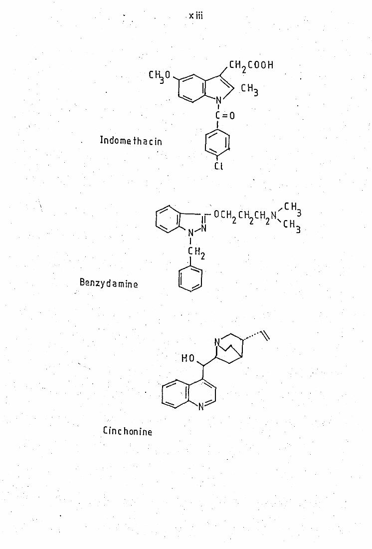
Indomethacin
Benzydamine
Cinc honine
· x Hi
(1-I 2(00H
~ CH3
~ N I
(=0
o Cl
.... ~ .- "

Chapter One
INT RODUCTION
1
PART A INFLAMMATION
1.1 I nrlammation
Inflammation is an essentially normal and protective
response to any noxious stimulus that may threaten the well-being of a host.
The injurious stimUlus may be a chemical agent (croton oil, carrageenan,
glycogen), a physical agent (burns, ultra - violet light), or a biological
agent· (bacteria, fungi, viruses). In addition an endogenous factor may
give rise to inflammation as is the case in auto immune diseases.
Inflammation can vary fram an acute, transient, and highly
localised response such as a pin prick, to a complex sustained chronic
. response involving.the whole organism as occurs with rheumatoid diseases.
When tissue injury is caused by a single event or a
single exposure to a non - replicating agent, the inflammatory process
progresses smoothly from injury to healing" and is termed acute inflamm - .
ation. If howeyer, the injurious·agent is self - replicating (parasites,
bacteria and viruses), or attempts to remove the agent fail, then the
inflammatory response becomes much'more complex and will continue for as
long as the agent itself persists. This gives rise to a'chronic inflamm -
atory process with much tissue damage due to the secretion of destructive
lysosomal enzymes and may result in loss of function.
Inflammation is manifested by five cardinal signs of in -
flammation. These are erythema, oedema, pain, heat and eventually loss of
function. During an inflammatory response there is an initial brief cons -
triction of the arterioles followed by prolonged dilation of the blood
vessels. This produces an erythema which :is then followed by leakage of
.2
plasma protein from the blood causing an oedema. During .. the oedema phase
the migration of leucocytes into the inflammed area is a'characteristic
feature of the inflammatory response. The liberation of mediators through·-
out the response leads to the production of pain and heat.
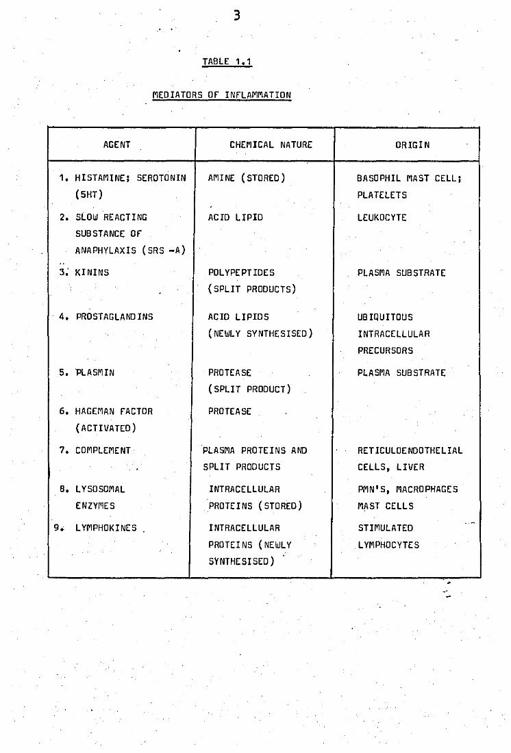
1.2 Mediators of Inflammation
The inflammatory process involves the li -
beration of many potent substances which can themselves duplicate the
inflammatory response and lead to pain, heat and to the chronic condition,
loss of function. Table 1.1 summarises the chemical nature and origin of
these substances.
Each substance, as its involvement was proposed or dem -
onstrated has been studied intensively. and attempts have been made to
link its actions to that of other mediators. Mediators generally have
three properties: (1) they can induce some or all of the signs of in
flammation, (2) they can be released during an inflammatory reaction, and
(3) their release or action can be affected by anti - inflammatory drugs.
The importance of the mediators in the various types of
'inflammation depends upon the sensitivities of the tissues in which they
are released a'nd on the sequence of mediator release. F"or example, in
anaphylactic shock there is an explosive and virtually simultaneous -"
release of many mediators, for example histamine and SRS - A, whereas-~n
the carrageenan - induced inflammatory response there is a sequential
release of mediators.
1.3 Prostaglandins as mediators of inflammation
3
TABLE 1.1
MEDIATORS Of INfLAMMATION
AGENT CHEMICAL NATURE ORIGIN
1. HISTAMINE; SEROTONIN AMINE (STORED ) BASOPHIL MAST CELL;
(5HT) PLATELETS .
2. SLOW REACTING ACID LIPID LEUKOCYTE
SUBSTANCE Of
ANAPHYLAXIS (SRS -A) .. 3; KININS POLYPEPTIDES PLASMA SUBSTRATE
(SPLIT PRODUCTS)
. 4. PROSTAGLAND INS ACID LIPIDS UB IQUITOUS
(NEWL Y SYNTHESISED) INTRACELLULAR
PRECURSORS
5. 'PLASMIN PROTEASE PLASMA SUBSTRATE
(SPLI T PRODUCT)
6. HAGEMAN FACTOR PROTEASE
(ACTIVATED)
7. COMPLEMENT PLASMA PROTEINS AND RETICULOENDOTHELIAL
, SPLIT PRODUCTS CELLS, LIVER
B. LYSOSOMAL INTRACELLULAR PMN'S, MACRO PHAGE S
ENZYMES PROTEINS (STORED) MAST CELLS
.-9. L YMPHOKINES INTRACELLULAR STIMULATED
PROTEI NS (NEWLY L YMPHOCYTE S
SYNTHESISED) .
4
(1) Discovery
In 1930 Kurzrock and Lieb observed that humen myometrium
tissue showed rhythmic contractions and relaxation when incubated with
fresh:.human semen. This observation was confirmed by GOLDBLATT (1933)
and Von Euler (1936) and the latter author identified the active component
as an acidic lipid and named it "prostaglandin" think in!; ,that it was
produced in the prostate gland. In 1959, Eliasson showed that prostag
landins in human semen were' derived from 'seminal veslclesand'Bergstrom
and SJovall (1960) showed that the active sUbstances were several closely
related compounds. Prostaglandins are now known to be ubiquitous having
been identified in almost every tissue.
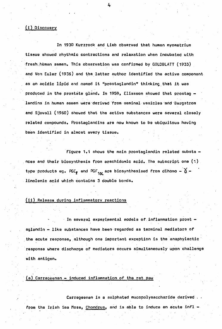
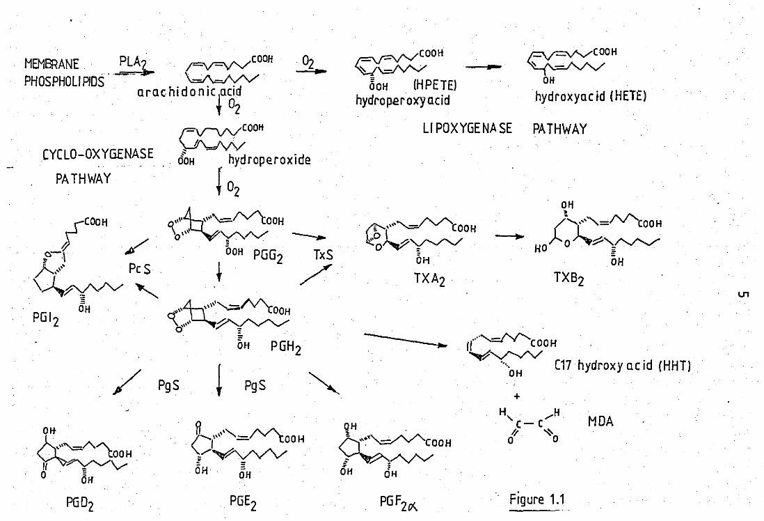
figure 1.1 shows the main prostaglandin related substa -
, nces end their biosynthesis from arachidonic acid. The subscript one (1)
type products eg. PGE1 and PG'1C)(. are biosynthesised from dihomo -'0'linolenic acid which contains 3 double bonds.
(ii) Release during inflammatory reections
In several experimental models of, inflammation prost
aglandin - like substances have ,been regarded as terminal mediators of
, the acute response, although one important exception is the anaphylactic'
'response where discharge of mediators occurs simultaneously upon challenge
1011 th antigen.
(a) Carrageenan - induced inflammation of tho rat paw
Carragoenan is a sulphated mucopolysaccharide derived , .
from the Irish Saa Moss, Chondrus, and is able to induce an acute inrl

•
\
•
LEGE NO TO FIGURE 1.1
Arachidonic acid can be.oxygenated via 2 pathways:
1) CYCLOOXYGENASE Known to be present in all cell. types.
except erythrocytes.
First PGG2
is formed. This can be converted to
PGH2, which can break down enzymically or non-enzymically to a
variety of other products (see fig~re ). Inhibited by NSAIOS.
Enzymes PLA 2 = phospholipase A2
TXS = thromboxane synthetase
PgS = prostaglandin synthetase
PcS = prostacyclili synthetase
3) LIPOXYGENASE Identified so far in lungs, platelets r
and leucocytes. An unstable hydroperoxide is formed (HPETE) which can
then breakdown to the stable hydroxyacids or can be further tran -
sformed to other products such as leukotrienes. Upoxygenase is
inhibited by SW 755C.
In lung!! .1;poxidase acting at C11 + C12
In platelets djpoxi~ase acting at C12
In leukocytes:lrpoxidase acting at CS.
_~OOIi ~~20 11 Pf
ARACHIDONIC ACID
MEMBRANE PLAl,... ;=v=\I'V"COOH
PHOSPHOLlPIDS ._- ~ . orae hi don ic la cid
'f °2 Fv-vvCOOH
\...ffVVV CYCLO-OXYGENASE OOH hydroperoxide
! °2 PATHWAY
'\\\\\~\\'\"=IV"'tOOH O\'\\~'Y'/V' ____
DOH PGG2 TxS
t / '\\\\, ........ _AA.
. -' ''(OOH ~.('v'/'~
bH PGH2
/ PgS PgS
r-:v==vvCOOH
~\.d'v"V' 60H (HPETE)
hydroperoxyacid
- ~COOH
~ OH
hydroxyQcid (HETE)
LI POXYGENASE PATHWAY
OH
TXB2
.. 1t····~COOH y~
·····OH C17 hydroxy ac id (HHT) .
-
+
qH . ~COOH ; ,A,/'v' OH OH
H, ..... H C- (
1/ ~ o 0
MDA
Figure 1.1
U1
o
ammatory response when injected into the paw of a rat. In this model which
is used widely in the search for new anti - inflammatory agents, there
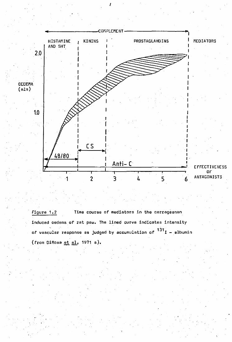
appears to be distinct phases of mediator releasa (DiRosa ~ aI, 1971a)
(fi9ure 1.2). It appears that during the first 11hrs histamine and 5HT
are the main mediators released. This was shown by the use of the ante -
gonist compound 4B / BD which depletes stores of histamine and 5HT. 4B / BD
Given over 4 days before carrag~enan injection showed a marked reduction
in paw oedema during the first 11hrs after carrageenan injection; The
second phase (11 - 21 hrs) of this reacti~n is mediated by Kinins. Simil
arly the use of cellulose sulphate, which lowers the plasma Kininogenen
level by 50%, reduced the formation of the oedema duringc,this phase. The
use of 4B / BD end cellulose sulphate together led to a supression of
the oedema up to 21hrs.
The appearance of prostaglandins in the inflammed paws
was demonstrated at 21 - 6hrs after carrageenan injection by Willis (1969), ,
suggesting that prostaglandins may mediate this phase of the response.
Compound 4B / BD and cellulose sulphate had no effect on the oedema during
this phase and neither did they affect associated migration of polymor -
phonuclear (PM») leucocytes into the area. However the depletion of com -
p'fement by the uue of antisera towards leucocytes or antigen - antibody
complexes, redueed the oedema up to 6hrs. This demonstrated that comple
ment was required for the'release of all the medietors and it was suggested
that activation of complement, either by the irritant itself or fixation by,
altered tissue proteins was the event that led to the release of mediators
and the progression of the inflammatory response.
(b) The carrageenan air - bleb
This is an 'experimental model in which an ordered release
I
..... I-------_-'l·corr,PLEMENT----------~ .. ,
HISTAMINE AND 5HT
KININS PROSTAGLANOINS I MEDIATORS
OEDEMA (mls)
2.0
1.0
Figure 1.2
1
I
I r I I I I r I (S I If ~
Anti- (
2 3 4 5
Time course of mediators in the carrageenan
I I I I I I I I I I I . I
6
induced oedema of rat paw. The lined curve indicates intensity
131 of vascular response as Judged by accumulation of I - albumin
(from DiRosa.et ai, 1971 a). --
EFFECTIVENESS OF
ANTAGONISTS
v
of mediators occurs. A suspension of carrageenan is injected into a
subcutaneous air - bleb formed on the back of a rat. An inflammatory
reaction follows, and samples of the bleb - fluid can be withdrawn at
certain time intervals and analysed. Again histamine and kinins are found r
sh~tly after carrageenan injection and prostaglandins, mainly PGE 2, appear
after 3hrs and reach a maximum at 12-24hrs (Anderson·~!l, 1971).
Carrageenan induces prostaglandin."accumulation in a dose - dependant
manner and the concentration of PGE 2 recovered at 24hrs (95ng/ml) was
far in excess of that necessary to prodtn:e cutaneous inflammation. The
lysosomal enzyme jS- glucuronidase was found to increase in parallel
.with PGE 2, implying that appearance of prostaglandins was re~ated to
appeerance of lysosomal enzymes.
(iii) Induction of inflammation
Prostaglandins are able to induce the signs of infla -
mmation. These properties which:.will only be mentioned very briefly here .tt
have been reviewed in detail by rerreiraA
Vane (197.4) 1-( i) Erythema can
be produced by prostaglandins of the E series in low concentrations and
--the effects are long lasting. They can also counteract the vasoconstri -
ction caused by agents such as noradrenaline. :end angi,otensin. (ii) PGE,
is the most powerful pyretic agent known and dulling'. ·fever the ge,neration
of a prostaglandin E - like substance in the ceqtral nervous system has
been measured. (iii) Proataglandins are not very good oedema producing
agents on their own but sensitise blood vessels to the permeability
effects of other mediators. (iv) The role of prostaglandins in pain pro -
duction is to induce long lasting hy~eralgesia (-!!- 2hrs), a state in
which pain can be elicited by normally painless mechanical or chemical
. stimul tation.
(iv) Effects of anti - inflammatory dru9$
Inhibi tion of prostaglandin .. biosynthesis by indomethacin
and aspirin was first shown by Vane and collegues (Vane, 1971; Smith and
Willis, 1971; ferreira !i~, 1971).ln the former report cell - free
homogenates from guinea- pig lungs were prepared and the 900g supernatent
used to synthesize prostaglandins from arachidonic acid. Indomethacin
and aspirin both inhibited biosynthesis of PGE2 and PGf~in a dose -
dependent manner. Smith and Willis (1971) -'found that addition of the
same drugs to washed human platelets which produce PGf2~ when incubated
with thrombin, substanbially reduced prostaglandin formation in a dosa -
'rela,ted manner. finally indomethacin and aspirin also abolished the release
of prostaglandins observed when a perfused dog spleen is contractad by
either catecholamines or nerve stimulation (ferreia et aI, 1971). --
These observations were of importance since the effects
found were at concentrations likely to be achieved in vivo and .-anti ---inflammatory potency correlated well with the ability to inhibit prosta
glandin synthesis.
Since these findings inhibition of prostaglandin bio -
synthosis by aspirin and related drugs has been shown in many other bio
logical systems (eg Tomlinson !i~, 1972; flower !i aI, 1972). Howaver
the steroidal anti - inflammatory drugs such as dtxamethasone, although
being more effective as anti - inflammatory agents than indomethacin, were
shown not to directly inhibit prostaglandin synthetase, although they
blocked release of prostaglandins in whole cell systems (flower !i aI,
1972). These drugs are no~, known to be inhibitors of phospholipase
activity and develop'ments in their mode of action will be discussed later.
.v
(v) Prostaglandins as modulators of inflammation
Prostaglandins of the E - type elso have properties
which are anti - inflammatory.
ClrrUi.tS lysosomal enzyme release by PMN leucocytes to be imp -
~ ortant in the pathogenesis of rheumatoid arthritis. PGE~ and PGE~ but
not PGI' 2..0(.' have bean shown to reduce degrannulation and the release of
lysosomal enzymes (Weissmann .!!i &,197Z).rurther, PGE1 and PGE 2o<hava
also been shown to prevent the release of. histamine and SRS - A from
basophils and lung fragments ill vitro, when challenged with a presensi
tised antigen. Thesa effects are thought to arise by the fact that PGE,
and PGE z, but not PGr2~' can increase intracellular cyclic adenosine
3',5 '-monophosphate (cAMP), which when raised ds:· known to inhibit
lysosomal enzyma release.
In experiments .using .. ~the carrageenan air. - bleb tech -
nique, placement into the blebs of ZOO~g of PGE 2 at the same time as
carrageenan, reduced the rate at which leucocytesand lysosomal enzymes
'appeared at the site (Zurier .!!ill' 1973). Also ultrastructural studies
indicate that more: lysosomes remain intact after carrageenan uptake in
bleb leucocytes·.··from PGEt treated .animals than in leulZocytes from control
animals. These anti ~ inflammatory effects are observod at much higher of pr.st"1l«..d,,,s
concentrations thanithose shown to pot'entiate the inflammatory response .. initiated by carrag~enan (rerreira and vana, 1974).
PGE! and PGE Z have also been obsarved to suppress adju ~
vant induced arthritis and cartilage destruction in rats as measured
by joint swalling (Zurier .!!i aI, 1973).The exact mechanisms involved in
this action are not known although propertias such as inhibition of ...... .
11
lysosomal enzyme secretion are thought to be important.
Prostaglandins therefore may have a complex role having
both anti - and pro - inflammatory effects.
1.4. Involvement of Polymorphonuclear Leucocytes in Inflammation
(i) function and morphology
Polymorphonuclear leucocytes, also referred to as o
neutrphils or granulocytes, are phagocytic cells primarily concerned "
'with defent,e against foreign biological partides such as microorganisms.
They originate from proliferating pools of precurs~r cells in the bone
marrow where they di fferenl:iate and develoll'~ their, characteristic
granules. They make up the major portion of leucocytes in the blood'
(~70%) 'and in response to chemotactic stimuli the¥,_can migrate into
tissues and perform their function of phagocytosis and killing of
biological material. PMN leucocytes live only for about one day' ifl the. c::.~f't."'-l,.a.. t'~f\ •
The mature circulating PMN leucocyte contains mainly
two chemically distinct types of granules present in the cytoplasm called
azurophil and specific granules. AzurophH gr,anules are produced first
during the promyelocyte stage of development, and arise from t-ha inner,
concav-e surface of the Golgi complex. These granules contain acid ' : :'
hydrolases (eg.;3 - glucu~nidase), may be regarded as primary'~ysosomes
found in other cells. Specific granules appear during the myelocyte stage
of maturation and arise from the,' outer, convex phase of the Golgi complex.
They are smaller in size than the azurophils and contain enzymes such
,es',lysozyme. The protein composi tion of the membranes of these :,two
granules have been found to be different and the azurophil granule elso
It:
contains a higher proportion of cholesterol in
the membrane.
(iil·Release of lysosomal enzymes from PMN leucocytes during stimulation
Dur~ng phagocytosis (particle in~estion or endocytosis)
both types of granules discharge their contents, containing antibacterial
agents and enzymes, into the phagocytic vacuole for the digestion of
inJested macromolecules. The specific granules preceed· the azurophils
"in this process. Also during phagocytosis, probably because of incomplete
·closure of the phagocytic vacuole, a po.rtion of the lysosomal constituents
are releases into the medium. 1n~ the released hydrolytic enzymes
would be free to act upon surrounding tissues and cause damage.
Mechanisms of the release process
Many in vitro studies have been carried out to examine
the selective release of lysosomal enzymes from .. stimulated PMN leucocyte ••
A variety of stimuli have been used to elicite the release process. for
example zymosan particl~s coated with complement or antibody, immune
complexes, CSa, and N - formyl L - methionyl - L - leucyl - L -
phenylalamine.
An extensive study with the use of zymosan particles
coated with complement (C3) or antibody (IgGl was carried out by Henson
(1971) using peripheral rabbit leucocytes. In this case specific
release of lysosomal enzymes occured in:a time.· !!lid concentI'atibn··
dependant manner. Maximal release of approximatelY 30% lysosomal
: enzymes occured with Smg zymosan - complement particles. The release
reaction was specific and mediated through receptors in the leucocyte
l:l
membranes ,which ,recognise complement (C3) and antibody (rc portion of
the IgG molecule), as no release was observed with zymosan particles
alone. Release of lysosomal enzymes was also observed if antibody or
complelllent or immune complexes were coated ontO. micropore filters
which'are too big to be in~ested and hence provide a non phagocytosable
surface. Therefore as long as receptors on the leucocyte surface are
stimulated, lysosomal enzyme release occurs whether or not phag~cytosis
occurs.
This fir1dl~is fur·ther supported by cytochalasin 8
treated human PMN leucocytes (Goldstein ~~, 1975).This agent
, inhibits phagocytosis of particles by interference with microtubule
function. When these leucocytes are exposed to either the complement
component C5a, or immune complexes they act as secretory cells and reI
ease lysosomal enzymes without loss of cell integrity. The role of
calcium ions has alone been investigated in this system. The complement
o com~nent, C5a, in the absence of calcium ions is able to induce a
significant release of lysozyme: and fJ- g~\lcuronidase. Addition of up
to 1.5mM Ca2+ to the suspending media causes an increase in the release
2+ of both enzymes. However, furth.er amounts of Ca ,actually decreased
;1- glucuronidase release but had no effect on the release of lysoz,me.
Calcium itself only induced the release of lysozyme.
In another system, Northarer (1977) using rabbit perito
neal neutrophils, demonstrated that calcium itself caused a'time and
concentration dependant release of both lysozyme and;$ - glucuronidase
without release of the cytoplasmic marker enzyme lactate dehydrogenase.
The non - steroidal anti - inflammatory drugs were found to affect the
release of these two enzymes differently. The release of ;1- g~ucuron -
idase was inhibited by indomethacin, flufenamate and salicylate, and the
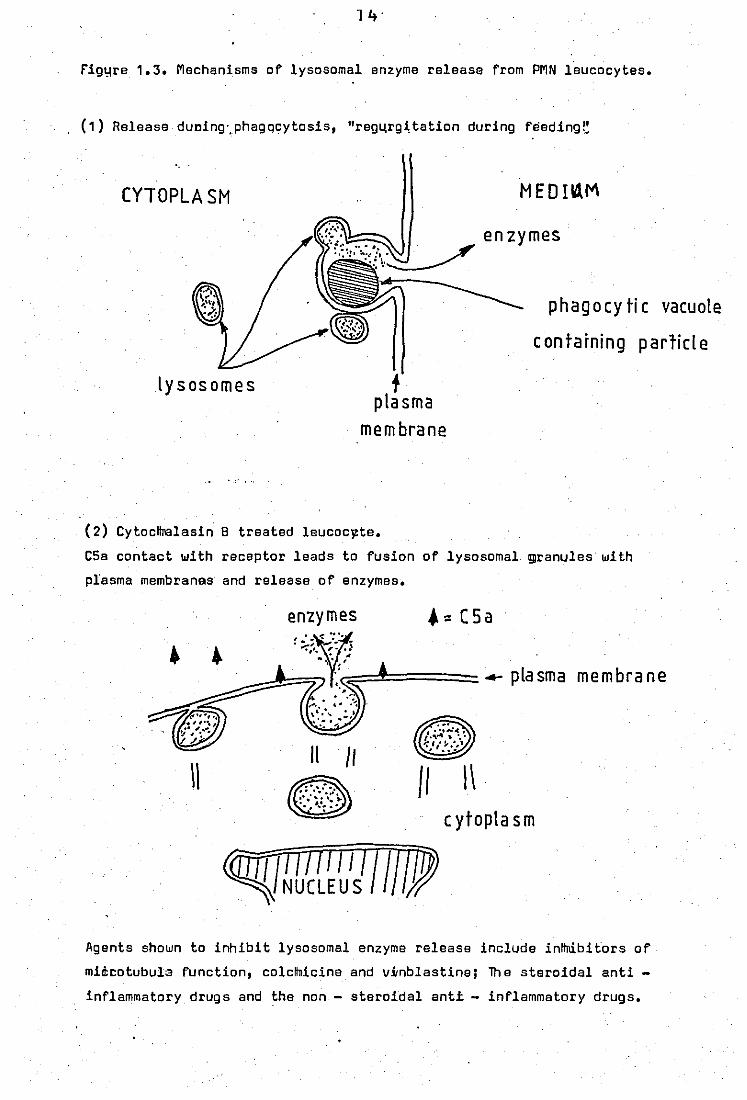
rig~re 1.3. Mechanisms of lysosomal enzyme release from PMN leucocytes.
, (1) Release dUDing',phagqcytosis, "reg4rg~tation during feeding~
CYTOPLASM
lysosomes f plasma
membrane
'.,'
MEDB1M
enzymes
phagocytic vacuole
containing p arlicl e
(2) Cytocltll3lasin 8 treated leucoclZte.
C5a contact with receptor leads to fusion of lysosomal ~ranules with
plasma membranes and release of enzymes.
1\
enzymes ~ .. ::; -: ':~" -
'-,'. '!' .• ]
", 'J(C' ;::=~.====== 4- P la sm a m e m bra ne ~~;.J:; .
. ' ...... , "
11 /1
8 /I \\ c ytopla s m
Agents shown to inhibit lysosomal enzyme release include inttnibitors of
mid:cotubula function, colcllnicine and vitnblastine; lh e steroidal anti -
inflammatory drugs and the non - steroida! anti - inflammatory drugs.
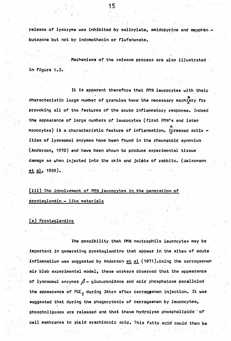
15
release of lysozyme was inhibited by salicylate, amidopyrine and a.Kyphan -
butazone but not by indomethacin or flufenamate.
Mechanisms of the release process are also illustrated
in figura 1.3.
It is apparent therefore that PMN leucocytes with their
h . Il
C aracteristic large numb~r of granules have the necessary mach~ery for
provoking all of the ,features of the acute inflammatory response. Indeed
the appoarance of large numbers of leucocytes (first PMN's and later n
monocytes) is a characteristic feature of inflammation. Icreased activ ,. ,
ities of lysosomal enzymes have been found in the rheumatoid synovium
'( Anderson, 1970) and have been shown to produce experimental tissue
damage as whim injected into the skin and joitlts of rabbits. (Weissmann
!i!!, 1969).
(iii) The involvnment of PMN leucocytes in the generation of
prostaglandin - like materials
(a) Prostaglandins
The possibility that PMN neutrophils leucocytes may be
important in generating prostaglandins that appear in the sites of acute
inflammation was suggested by Anderson!i!! (1971).Using the carrageenan
air bleb experimental model, ,these workers observed that the appearance
of lysosomal enzymes J - glucuronidase and acid phosphatase paralleled
the appearance of PGE 2 during 24hrs after carrag,enan injection. It was
suggested that during the phagocytosis of carrageenan by leucocytes,
phospholipases are released and that these hydrolyse phospholipids' 'of
cell membranes to yield arachidonic acid. This fatty acid could then be
16
converted to PGE 2 by freely aV,ailable tissue enzymes (prostaglandin
synthetase).
. Prostaglandins were shown to be released from rabbit
peritoneal PMN leucocytes during phagocytosis of bacteria (Higgs and
Voulten, 1972). The main prostaglandin released was PGE2
(56%), and
PGr2 (28%), with some additional compounds that were unidentified (16%).
Release of prostaglandins was greater in leucocytes undergoing phagocy -
tosis than in resting controls.
In a later report (Higgs,McCall and Voulten, 1975)
leucocytes undergoing phagocytosis were found to produce a chemotactic
su~stance which was absent if.indomethacin was included in the preparations.
One of the prostaglandins produced was identified as PGE, by thin -
layer chromatography and differential bioassay, and the chemotactic
property of PGE1 first shown by Kayley, and Wainer (1971) was' also con
firmed. Neither PGE 2 or PGr2~were found to be chemotactic for PMN
leucocytes. The production of PGE1 by stimulated PMNs·· is of particular
importance in inflammation as this would lead to furthe.r infiltration·
of leucocytes into the inflammatory site. 0' The rate et which phagocytosing
PMNs were found to produce prostaglandins could account for the levels
observed in inflammatory exudates. It also appeared that homogenates
from cells which had been pre - incubated with bacteria (ie phagocytosing
leucocytes) showed twice as much prostaglandin synthetase activity as
compared with homogenates from bacteria-free controls.
Similar release of prostaglandins has been demonstrated
from human PMNs exposed to zymosOan particles (Zurier and Sayadoff, 1975).
17
(b) Production of thromboxanes (TX) by PMN.leucocytes
Thromboxane production ~as first shown in sensitised
guinea - pig isolated perfused lungs when challenged with antigen (Piper
and Vane, 1969). An additional activity which was different from histamine,
SRS - A and prostaglandins was described in the perfusate. This caused
the contraction of a rabbit aorta strip, and ~as termed rabbit aorta
cQntracting substance (RCS). RCS was later identified to be a mixture wi~h. ""'''Llv "' ... " _to ., f
mainly consisting of thromboxane A2 (TXA2) '",;( prostaglandin endoperoxides
PGG2 and PGH2 (Hamberg, Swansson and Samuelsson, 1975).
TXA 2 - like activity is also produced by PMN leucocytes
(Higgs ll .. £!., 1976). When prostaglandin endoperoxides PGG 2 and PGH2~ere
incubated with homogenates of phagocytosing PMNs for as little as 2
minutes at O· e, there was an increase in the rabbit aorta contracting d
activity and the coelic artery contracting activity. This increase of . ~
biological activity ~as attributed to the production of TXA2 from
evaluation of its biological half life (Tt = 8 - 13 min at O·C and less
than 1 min at 37·C in aqueous solution), and in its greater potency
in contracting the rabbit aorta.
An increase in RCS was only observed with homogenates
of phagocytosing PMNs and not resting controls,' and the conversion to
TXA2 ~as prevented by boiling the preparations and by the agent benzy
damine, an inh~bitor of thromboxane synthetase. Approximately 25 30 %
of the endoperoxides were' estimated to be converted to TXA2 - like
activity, the remainder rearranging to prostaglandins which were
measured by the contraction of the rat colon.
Hj
, The generation of thromboxanes from PMNs has also been
shown by Goldstein ~~ (1978),using cytochalasin 8 treated leucocytes
exposed to opsonised zymosan particles. Thromboxane 82 (TX8 2) which is
the stable product of TXA2, was released in a time and concentration
dependant manner, with the earliest detection 1 - 2 mins after stimulation.
Th~ generation of TXAi may be important. It is known
to be e potent aggregator of platelets, and the ag9regation of platelets
leads to the release of more mediators into the system. It is now thought
that intermediates of prostaglandin biosynthesis (in particular PGG2
) ,
like TXA2, are the more important mediators as these have more potent
activity and shorter hal f lives than the more stable primary prostagl -
andins. further support arises from the st.udies of the compound MK -447.
This agent has anti - inflammatory properties like indomethacin but
actually increases prostaglandin synthesis (Kuehl ~~, 1977).In ,
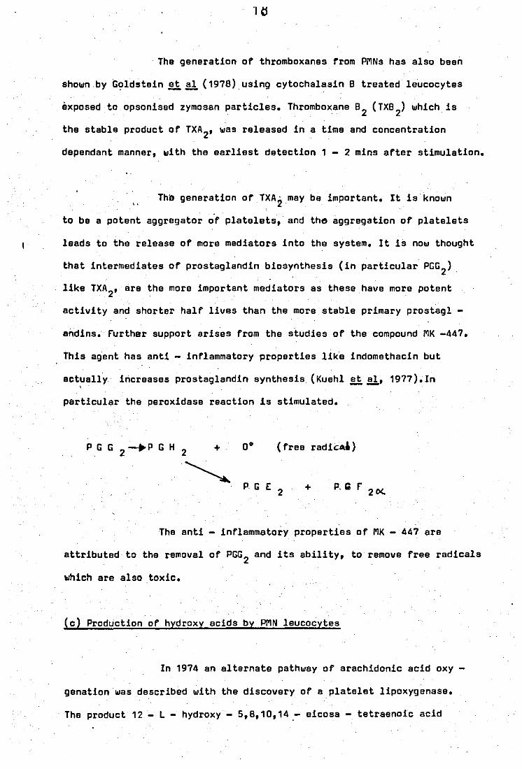
particular the peroxidase reaction is stimulated.
(free radic:~)
+ P,Gf 2 0<.
The anti - inflammatory properties of MK - 447 are
attributed to the removal of PGG 2 and its ability, to remove free radicals
which are also toxic.
(c) Production of hydroxy acids by PMN leucocytes
In 1974 an alternate pathway of arachidonic acid oxy
genationwas described with the discovery of a platelet lipoxygenase.
The product 12 - L - hydroxy - 5,8,10,14,- eicosa - tetraenoic acid
19
(HETE) is a potent chemotactic agent for PMN leucocytes and is therefore
of import~nce in inflammation. Lipoxygenase activity·has also been dete
cted in PMNs (Borgeat II aI, 1976) with oxyg~ation occuring at CS of
arachidonic acid and ca or dihomo - If - linolenic acid. The indomethacin
related drugs are not effactive against lipoxygenase·but BW 755C has been
shown to inhibit both lipoxygenase and cyclooxygenasa. This is proba~ly
why it has a significantly greater effect on leucocyte migratipn!!l ~
than indomethacin (Higgs II aI, 197B).
The ability of PMN laucocytes to produce the various
metabolites of arachidonic and dihomo -~ - linolenic acids suggests
th~t they may be the source of prostaglandins found in some forms of
inflammation, for example the carrageenan inducsd inflammatory response
in the rat. H.owever this may not be so in all cases. Glatt et al (1974) --using~.urate crystals to induce inf'lammation of ·intertarsal JOints of
chickens, found that the appearance of prosta9landins (PGE2
and PG~)
peaked at 1 - 2hrs along with maximum production of oedema and maximum
permeability of the vessels. However, PMNs were not found to arrive at
the site in significant· numbers until after 4hrs and reached a peak at 6hrs.
The appearence of the lysosomal enzyme/lysozyme,exactly paralleled PMN
infiltration.·The prostaglandin content of the eXUdate actually decreased
as numbers· of PMNs were .. increasing. It was suggested in this case that
other cells ( eg. platelets ) may be a more viable source of prostaglandins
in this model.
1.5 . Importanc.e of Phospholipase A2 in the Release of Prostaglandins.
Prosteglandins are not stored to any considerable extent .
in mammalian tissues and it is believed that any increase in their levels
20
brought about by ph,siological stimulation is through rapid biosynthesis.
The synthesis is therefore dep~ndant upon the availability of precursor
polyunsaturated fatty acids. The major portion of these fatty acids is
present in the·esterified form in tissue phospholipids which form a major
constituent of membranes. Release of arachidonic acid therefore, occurs
by the action of phospholipid splitting enzymes or phospholipases.
Phospholipases A, which are generally found in the
·lysosomes of most cells,are capable of bringing about the complete de -
acylation of phospholipids. Arachidonic acid is a polyunsaturated fatty
acid and generally occupies position 2 of phospholipids. Position 1 is
normally esterified with a saturated fatty acid (figure 1.4). Therefore
phospholipase A2 (phosphatide 2 - acyl hydrolase E.C. 3.1.1.4.) is the
important enzyme responsible for the release of arachidonic acid from
phospholipids. The other product of phospholipase A2 hydrolysis, lyso -
phospholipid, is also important because this is a .surface active agent
and also cytotoxic if allowed to accumulate.
Experimental evidence suggesting the importance of
phospholipase A2 in prostaglandin biosynthesis has now been obtained
in many tissues. A large proportion of the work was carried out on
isolated guinea-· pig per fused lungs although similar observations have
been found in guinea - pig spleen (flower and Blackwell, 1976), frog
intestine (Bartels ~ aI, 1970) and the thyroid gland (Haye~~, 1973).
The release of prostaglandin - like materials from,
sensitised, guinea - pig ~solated perfused lungs when challenged with
antigen has already been mentioned. This has also been shown by a
variety of other stimuli using unsensitised lungs. The list includes
brad~kinin, mechanical trauma, rabbit aorta contracting substance -
21
o 11
CH-O-C-R1· o 12 11
Rz-C-O-CH
. I H CH -O-P-O-X
I
°e
Phospholipid
1 Phospholipase A2
~ CH -0-C-R1 I
H O-C H
I ~ CH -O-P- O-X 2 . I
Oe
Lysophos phol ipid
+
o 11
R -C-OH 2
po Iyunsatura t ed
fatty acid
" . ".~ X :'b'ase moiety (clhioline, ethanolamine, serine, inosit,l etc.)
,.'
R 1: saturated fatty acid
R2 = unsaturated fatty acid.
I: - .. ,1', ,- •
~. ; ...
F'iqure 1.4 Action of phospholipase A2
•
22
releasing factor (Res - RF", thought to be a peptide) (Piper end Vane,
1969; Palmer ~!l, 1973),and SHT and histamine ~abastar and Bakhle,
·1970). F"urther· it has been shown that these stimuli cause the activation
or increase the activity of a phospholipase A2 which provides the fatty
acid necessary for the production of Res (Blackwell ~!l, 1978).
Infusion of erachidonic acid, thereby by - passing the phospholipase
step, elso leads to the release of ReS (Vargaftig· and Oao Hai, 1972).
Indeed inhibitors of phospholipase activity such as anti - inflammatory
steroids and mepacrine (see leter) were shown to block the effects of
these agents, again implying that they acted through effects on phos -
pholipase activity.
In guinea - pig isolated per fused lung:-
entigen challenge
mechanical trauma~ bradykinin
.. Res - RF"
SHT
histamine
stimulete
phospholipase
Arachidonic acid
cyclooxygenase
prostaglandins and
thromboxanes in perfusate
23
1.6 Phospholipases of PMN leucocytes
Phospholipase activity of rabbit peritoneal PMN leucocytes
was first described·by Elsbach and Rizack (1963) who measured the release
of fatty acids from dipalmitoyl phosphatidylcholine (PC) • The activity
was optimal in the acid pH range and found mainly in the granule fraction.
An acid active lipase activity was also datected. The granule free sup -
a. ernatllnt also contained an alkaline ly~ophospholipase activity.
"
In a later ,paper (Elsbach !i aI, 1965) use. was made of
\ substrates laballed with 32p. The accumulation of monoacyl (32p) phos _
phatidyl cholina (lysophosphatidyl choline) from diacyl (32p) phospha -
tidyl choline confirmed the presence of phospholipase A type activity. phos
Howaver the main product was glycerylphoryl choline indicating the pre -A . sence of lysophospholipase activity in the homogenate. The phospholipase
hydrolysed phosphati~yl ~thanolamine (PE) to the same extent as PC and
a direct transacylation activity, important for the synthesis of phos -
pholipids, was also functional at high lysophospholipidconcentration.
The difference in pH optima for the phospholipase A
and lysophospholipase activities led to the suggestion that accumulation
of lysophosph6Hpids may be important during phagocytosis and digestion
of bacteria by PMN leucocytes, as the pH of the phagosome is acid.
lysophospholipids being lytic:~gents may help in the fusion o~ lysosomes
with the phagocytic vacuole •.
In 1972 phospholipid metabolism was examined with the
use of leucocytes labelled during pre - incubation of the cells, for
1 hr at 37·C, with (32p) lysophosphatidyl choline (EIsbach !i~). The
gran~locytes lost less than 20% of incorporated radioactivity after 24hr.
24
at 37·C. This rata was not altarad during phagocytosis, and tha products
wara lysophosphatidyl choline, which was still associated with the cells,
and glycerophosphoryl choline, which was released into the medium.
When incubated with live E.coli for 15min in vitro,
leucocytes were capable of killing 99% of (1 - 14C) palmitic acid labelled
E.coli. However during this period only 30% of the phospholipids of E.coli
were degraded, suggesting that killing of bacteria was associated with
only a small degradatIon of phospholipids.
Certainly homogenates of leucocytes were found to readily
32 . \ degrade ( p) PE, which is the major phospholipid of E.coli. The pho~p -
d 2+ ,holipase A activity was maximal at pH 7.5, require Ca and was almost
all sedimentable at 8200g, indicating that it was present in the granules. . PLA,
Comparison of the leucocyte A with that from Crotal~s Adamenteus venom
demonstrated that the enzyme was of A2 specificity. This enzyme was then
shown to degrade all the phospholipids of E.coliequally well (Patriarca
et aI, 1972). --
A more detailed exemination of the phospholipase A and
its cellular localisation in PMN leucocytes was presented by. fransen
!i aI, 1974). Here extensive use of autoclaved E.coli which had been
grown on (1 - 14C) oleic acid was made as a substrate for phospholipase
A. The (1 - 14c) oleic acid was specifically incorporated in the 2 -
position of E.coli phospholipids. Leucocyte homogenates and isolated
phagosomes had a phospholipase A2 activity which was optimum at both
pH 5.5 and 7.5. The acid optima was inactive if ljposomal suspensions
2+ of PE were used. 80th activities required Ca ,a property which contrests
with lysosomal phospholipases of macrophages (fransen !i~, 1973).
The phospholipase activities were associ~ted with the. azurophilic and
25
specific granules and were membrane bound. The activities were susceptible
to product inhibition, and this inhibition was only-partially reversed
by the addition of albumin.
further study of this phospholipase A2 involved its .
purification (Weiss ~~, 1975) and its comparison with a permeability
increasing factor (PI) alsa located in the granules. PI is thought to
cause the bactericidal effect of leucocytes on E.coli. The purification
process which involved sulphuric acid extraction, dialysis and chromat -
ography on CM sephadex was unable to separate PI from phospholipase A2•
The preparation contained bactericidal activity but were clsarly seper -
ated from lysosomal enzymes such as lysozyme. Both phospholipase A2 and
PI are cationic and could possibly be a single protein. Howeve~ PI act
ivity was inhibited by Mg2+ or Ca2+, whereas phospholipase activity
i d C 2+ requ re a •
26
PART B THE ANTI - RHEUMATIC EFFECTS OF CHLOROQUINE
1.7 INTRODUCTION
Chloroquine was developed principally as an antimalarial
drug but has been used in the past 30 years in the treatment of Rheumatoid
Arthritis and related 'diseases. It is a beneficial drug and actually
slows down Joint erosion in 'contrast to the milder non - steroidal
anti - inflammatory drugs ~NSAIDS eg aspirin and indomathacin) which
although are very useful in relieving the symptoms, have no effect on
the destructive nature of the disease. Unfortunately chloroquine also
has many side effects, princip31 among which is retinopathy. This has
limited its use in the United Kingdom, but it is still widely used in
Sweden. Its anti - rheumatic effects are generally observed after 2
months treatment.
1.8 Tissue distribution and metabolism
A characteristic of chloroquine is its accumulation
in tissues and very high concentrations have been found in the adrenal
glands, liver, kidney, spleen, lungs and heart (Grundmann ~ aI, 1972).
Several reports have indicated that the, drug persists in tissue long
after discontinuation of therapy. Detectable amounts,of chloroquine
and metabolit;,e:s are found in plasma and urine 3.5 - 5 years after the
last administration (Rubin et aI, 1963). --
, Upon administration chloroquine is completely absorbed
in the gastro - intestinal tract and the drug is largely excreted
unchanged (70%) in the urine.(Frisk - Holmberg ~!l, 1979).The half
life measured from the plasma concentrations of a single oral dose
27
(O.31g chloroquine diphosphate) given to human patients was between
3 - 5 days. Various metabolites have been detected in the plasma and
urine and it appears that metabolism involves the gradual degradation
of the basic side chain of the chloroquine molecule so that eventually
tha 4 - amino - 7 chloroquinoline nucleus remains (Kuroda, 1962).
1.9 The anti - rheumatic mode of action of chloroquine
The·mode of action of chloroquine is largely unknown
but the drug also has many properties which do·seem of relevence to
its anti -rheumatic action. These include (a) affects on leucocyte
migration, (b) effects on cartilage and collegen degradation and
healing, and (c) lysosomotrophic activity and effects on lysosomal
function. These have been adequately reviewed by Bresloff (1977) and
only the actions of chloroquine important to the present work will
be discussed.
1.10 lysosomotrophic activity and effects on lysosome function
The relationship between chloroquine and the lysosomal
system is central in explaining many of its actions. lysosomal enzymas
from phagocy.tic cells are important mediators of inflammation and are
probably responsible for the connective tissua destruction in diseased
Joints. "It is thought that chloroquine might exert its therapeutic
effects· through Inhibition of lysosome function.
(i) Uptake into cells
Many cells exposed to chloroquine take it up avidly
and achieve much higher intracellular concentrations (up to 400 times)
2B
than in the surrounding medium. The selective uptake of a substance
into the lysosomes of a cell from the surrounding medium is called
lysosomotrophic activity (OeOuve ll.!!.!" 1974). This rapid uptake is
accompanied by a high degree of cytoplasmic vacuolation and evidence
suggests that these are lysosomal. This has been shown in many cell
types eg leucocytes of
quine (Fedorko, 1937),
patients with sarcoidoses treated with chloro )
pancreat1cr'cells of rats (Fedorko,1968), cultured
fibroblasts (Gaddioni ll'!!'!'; 1964), and mouse macrophages (Fedorko
ll.!!.!" 1968). Indeed lysosomotrophic activity is exhibited by most
cationic amphiphilic drugs.
Initial uptake of chloroquine, which is also rapid,
is known to be energy- independent and is followed by a slower energy
dependent phase. The initial phase also raises the overall pH of the
lysosomes (from pH 4.7 to 6.3 with 100 ~M chloroquine, Ohkuma and Poole,
1978). The uptake process is also illustrated in figure 1.5.
Chloroquine has 2 ionisation constants (PKa, = 10.2,
PKa2
= 8.1) and at physiological pH, 18% of the drug is in the mono -
protonated form which is soluble in lipid. This is able to pass through
plasma membranes into a slightly'more acid cytoplasm and to even more
acid.lysosomes, where the molecule becomes doubly protonated and incapable
of passing back. The protonation of chloroquine in lysosomes or diges -
+ tive vacuoles would depleta them of H and result in a reduction of the
aciditYf.,rut'th r uptake of chloroquine decreases unless more acid
containing vacuoles are formed. The concentration'. gradient is further
aided by the fact that chloroquine has been shown to bind to polar
lipids (Seydel and Wassarmann, 1976).
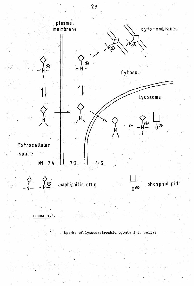
?<f> - N-
plasma me mbrane
. FIGURE 1.5.
29
Cytosol .
Uptake or lysosomotrophic agents into cells.
30
(ii) Effects on lysosomal enzymes
Chloroquine in concentrations that could be achieved
-4 intracellularly (~10 M) is able to inhibit a number of lysosomal
enzymes important in causing damage to connective tissue. These include:
chondromucoprotease of cartilage, cathepsin 8, cOllagenases (although
not lysosomal, therefore relevance not known) and neutral proteases
(Cowey and Whitehouse, 1966; Ali et aI, 1968; Mego and Chung, 1979).
This has also been achieved in whole cell systems; human fibroblasts
exposed to chloroquine (1 - 2 X 10⁻⁵M) in vitro are unable to digest
proteins and mucopolysaccharldes (Lie and Schofield, 1973), and macro
phages under the same conditions are also un5ble .. to degrade leucine
radiolabelled bacteria. Raising the pH ",of the medium to pH 8.0 has
been showm to have the same effect as chloroquine ie.inhibition of,
lysosomal enzyme activity and formation of vacuoles, on human fibro
blasts (Lie ~ aI, 1972) and also on the malarial parasite, Plasmodium
berghei, infected in erythrocytes (Homewood ~~, 1972), indicating
the importance of this property.
Inhibition or absence of lysosomal enzymes itself can
be a sufficient cause for formation of autophagic vacuoles. Grossly
"enlarged lysosomes 'are 'observed with the addition of spec! fic antibodies
to lysosomal enzymes and also in several' congenital storage diseases
where a lysosomal enzyme is lackin9 (Hers and Van Hoof, 1969).
(iii) Actions on lysosomal membranes
Chloroquine being, an amphiphilic molecule can be . ,-) .
predicted to interfere', with membrane properties as these are complex
struc~ures wbich have both lipophilic and hydrophilic constituents.
31
MaQi studies show stabilizing influences by chloroquine while others
show contrasting effects.
1n vitro studies on isolated lysosomes from liver
and leucocytes (also peritoneal neutrophils) have indicated that chlo
( . -5 -3) roquine 10 - 10 M and many other anti - inflammatory drugs inhi -
bited the leakage of lysosomal enzymes, suggesting stabilization of
the membranes (~g Weissmann; 1964; Ignarro, 1971).
Most anti - inflammatory drugs, whether steroidal
or non - steroidal, also inhibit release of lysosomal enzymes from
neutrophils and macrophages when exposed to various stimuli eg zymosan
particles, immune complexes. etc. However chloroquine does not possess
this property and if anything it has been shown to increase the . 4
. release of lysosomal enzymes at 10- M (Ringrose ~ aI, 1975; Perper
and Oronsky, 1974; Northover, 1977).
Chloroquine exposed to human fibroblasts not only
causes vacuolation but also release of lysosomal enzymes in a dose
dependant manner (Weissmann ~ aI, 1975). It also appears to inhibit
the uptake of lysosomal enzymes into fibroblasts that are genetically
deficient in these particular enzymes. This has been shown for aryl
sUlphatase (Weissmann et aI, 1975) and 0<- L - iduronidase (Sando et -- -
aI, 1979), and in both cases chloroquine interference with the specific
binding of the lysosomal enzyme with the plasma membrane has been
suggested.
32
(iv) Effects on lysosomal lipolytic processes
One of the unfortunate consequences of the lysosomo
trophic activity of chloroquine is lipidosis, and this is partly res
ponsible for the retinopathy seen in some patients on long term chloro
quine therapy. Lipidosis is a term used to describe a build up of
polar lipids in lysosomes. Amphiphilic cationic drugs are particularly
good lipidosis causing agents as they accumulate in lysosomes and bind
to polar lipids.The drug pol'ar Upid" complexes are thought not to be
, susceptible to enzymic attack by phospholipases and hence a build up
occurs. This, phenomenon can have grave consequences in tissues, such
as the retina, where a specially balanced lipid metabolism is essential
for normal function. In experimental rats chloroquine has been shown
to cause: lipidosis in the neuro~etinal cells of the retina (Drenckhahn
and 'Lullmann - Rauch,. 1979), in the cornea and in the lens which can
then develope a cataract '(Drenckhahn and Lullmann - Rauch, 1977).
Another important property of chloroquine which
contributes to retinopathy is its ability to bind strongly to the
pigment melanin. This binding can again lead to increased melanin con
centrations in the, retina which can have a toxic effect on the optic
nerve.
1.11 Effects of chloroguine and related drugs on lipolytic enzymes
Chloroquine ('1' t. 6 X'10-3M) along with quinine,
quinacrine, hydroxychloroquine end primaquine were shown to inhibit
lipolysis in rat epididymal fat pads by Markus and Ball (1969): The
drugs inhibited the:enzyme lipase whether it was activated by adrenaline"
released by hepirin or Just present in homogenates. Chloroquine has
33
also been shown to inhibit phospholipase A2 of PlasmOdium bergei, a
fact important as this enzyme provides fatty acids essential for the
malarial parasite to survive (Canedella ~!!, 1969).
Particularly important in inflammation is the study
of Vargaftig and Oao Hai (1972), who showed that mepacrine (quinacrine)
blocked the release of RCS from isolated guinea - pig per fused lungs
when injected with bradykinin but not when arachidonic acid was injected.
This indicated that the release of fatty acids. from phospholipids was
the site of action of mepacrine. Indeed bradykinin was later shown to
cause an increase in phospholipase A2 activity in this system (8lackwell
.~.!!, 1978).
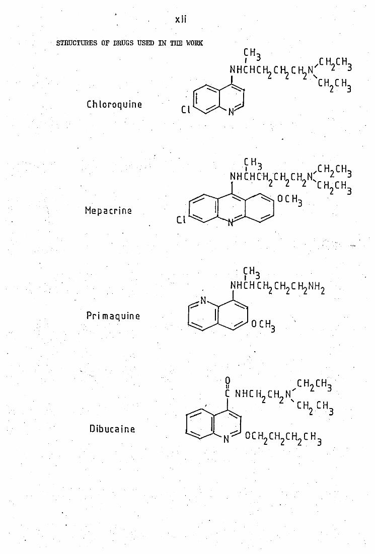
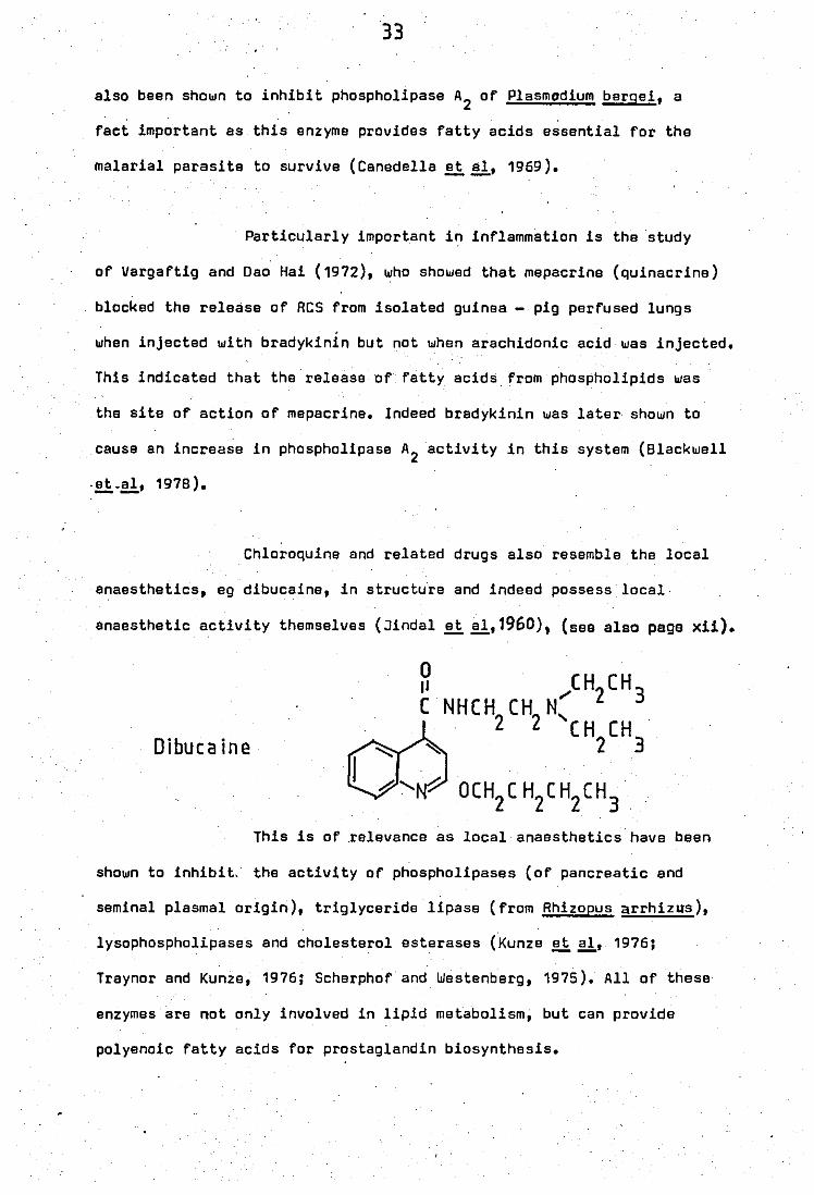
Chloroquine and related drugs also resemble the local
anaesthetics, eg dibucaine, in structure and indeed possess local·
anaesthetic activity themselves (Jindal ~!!,1960), (see also page xii).
Dibuca ine
This is of .relevance as local anaesthetics have been
shown to inhibit, the activity of phospholipases (of pancreatic and
seminal plasmal origin), triglyceride lipase (from Rhizopus arrhizus),
lysophospholipases and cholesterol esterases (Kunze ~ aI, 1976;
Traynor and Kunze, 1976; Scherphof and Westenberg, 1975). All of these
enzymes are not only involved in lipid metabolism, but can provide
polyenoic fatty acids for prostaglandin biosynthesis.
34
1.12 Aims of the project
In the light of the above' introduction PMN leucocytes
play an extremely important role, not only as phagocytic cells but
capable of providing prostaglandin related substances et sites of infla -
mmation. Phoepholipases ere therefore particularly important as they
can provide necessary prscursors ussd to synthesiseprostaglandin rslated
substancas.
The work intends to investigate the sbility of
peritonssl neutrophil lsucocytes, when stimulated, to contribute to any
phospholipese A ectivities present in cell - free inflammatory exudates.
The biochemical properties and possible control
mschanisms of any phospholipass A rsleased during stimulation will ba
examinsd using bactsrial membranse labelled with (1 - 14C) oleic acid,
as substrate, and compared with those of any phospholipase A present in
the cell - free inflammatory exudate.
The interactions of chloroquine - like agents with
these enzymes and other phospholipase A will be examined in an attempt
to gain more imformation on the mode of anti - rheumatic action of these
agents.
/
Chapter Two
MATERIALS AND
METHODS
35
SECTION I MATERIALS
1.1 Chemicals and Reagents.
All laboratory reagents and solvents_used were of analytical
grade and used without further purification.
Bovine serum albumin (BSA), zymosan A from S.cerevisiae
(yeast), N - 2 - hydroxyethyl piperazine - N'- 2 - ethanesulphonic acid
(HEPES), phenolphthalein mono -;.1- glucuronic acid sodium salt, 2-
amino - 2 - methyl - 1 - propanol, Micrococcus lysodeikticus (dried cells),
;.1- nicotinamide adenine dinucleotide reduced form and heparin (170 units
per milligram) were obtained from Sigma Chemical Company Limited (Poole,
U.K.).
Enzymes
Phospholipase A2 from" the snake venoms Crotalas adamenteus
(Eastern Oiamondback Rattlesnake) and Nala nalasputatrix (Malay Cobra)
were also from Sigma Chemical Company~ Poole. A partially purified
phospholipase A2 enzyme from pig pancreas was obtained from Bohringher
(Mannheim, GrR.).
PhOspholipaseA2
substrate
Escherichia coli (E. coli) strain W 8666. Thls was obtained
from the National Collection of Industrial Bacteria, Torry Research Sta-
tion, Aberdeen, Scotland.
. . 14 E. coli phospholipids were labelled using ( 1 - C) fatty acids.
36
(1 _14C) oleic acid (specific activity 57 mCi.mmol71), (1 _14C) linoleic
acid (specific activity 61 mCi.mmol:1 ), and (1 _14C) palmitic acid
(specific activity 57 mCi.mmol:1 ) were obtained from The Radiochemical
Cantre, Amersham, Buckinghamshire.
Lipid standards.
Olaic acid, L -0(- phosphatidyl ethanolamine dipalmitoyl,
L -~- phosphatidyl glycerol dipalmitoyl and L -~- lysophosphatidyl cho-
line were obtained from 5i9ma Chemical Company.
Drug substances
Chloroquine sulphate, mepacrine hydrochloride and sodium auro-
thiomalate were kind gifts from May and Baker Ltd. Chloroquine diphosphate,
D - penicillamine (free base) and primaquine diphosphate were obtained
from 5igma Chemical Company.
Liquid scintillation fluids.
Unisolve 1 and KL 372 (for radioactive counting of aqueous
samples) were purchased from Koch - Light Laboratories Ltd. Liquid scin-
tillation counting was carried out on a LKB 1215 Rackbeta Liquid Scintill-
ation Counter.
Thloglycollate mediu~
. ~ Thioglycollate medium (United states Pharmacopeeia, 18 revision
1970) was supplied by Oxoid Limited.
37
'1'.1 Media and Buffers.
(i) Triethanolamine medium.
E. coli for labelling were grown in a minimal media buffered
with triethanolamine. The composition of this medium was as follows.'
(NH4 )2 S04 2.0
feS04·7H2O 0.0005
KCl. 0.075
Triethanolamine 7.5
NaH2PO 4 ~ H2O 0.138
I'IgS04·7H2O 0.2
Sodium succinate 5.0
The pH was adjusted to 7.9 - B.O with dilute hydrochloric acid.
Sterilization of the media (less feS04• 7H20) was carried out
by autoclavin9 at 1200C and 2.7 kg/cm2 for 15 minutes. feS0
4.7H
20 was
sterilised by membrane filtration and added to the media prio.r to use.
(ii) Thioglycollate medium th (U. S. P. 1 B revision, 1970)
This was used to eUcit:' an inflammatory response in the peri-
toneal cavity of rabbits. The composition of this mediumis as shown
overleaf.
38
Yeast extract
Tryptone
Dextrose
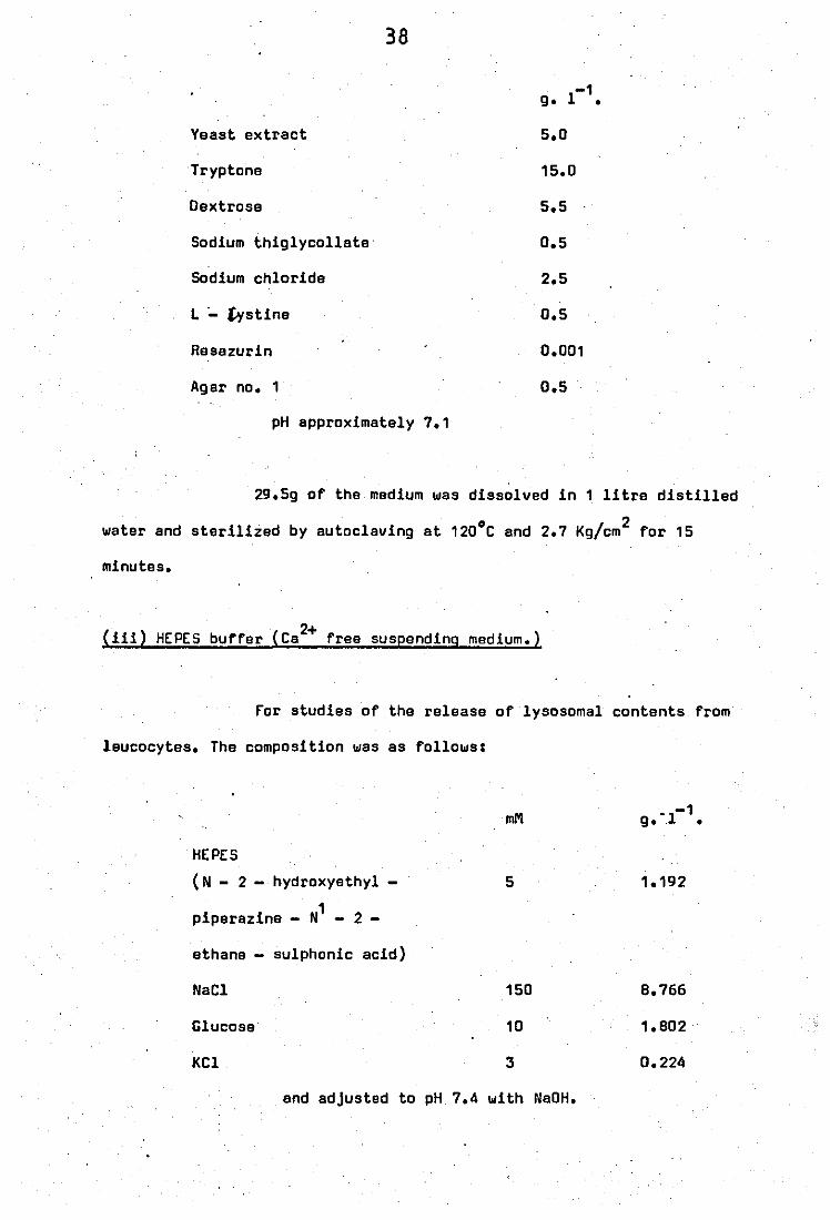
Sodium thiglycollate·
Sodium chloride
L ;.. £¥stine
Resazurin
Agar no. 1
pH approximately 7.1
-1 g. 1 •
5.0
15.0
5.5
0.5
2.5
0.5
0.001
0.5
29.5g of the medium was dissolved in 1 litre distilled
water and sterilized by autoclaving at 120°C and 2.7 Kg/cm2 for 15
minutes.
(iii) HEPE5 b ff (Ca 2+ f di di ) u er _ ree suseen n9 me um.
For studies of the release of lysosomal contents from
leucocytes. The composition was as follows:
mM -1 g. -.1 •
HEPES
(N - 2 - hydroxyethyl - 5 1.192
1 piperazine - N - 2 -
ethane - sulphonic acid)
NaCl 150 8.766
Glucose 10 1.802
KCl 3 0.224
and adjusted to pH 7.4 with NaOH.
39
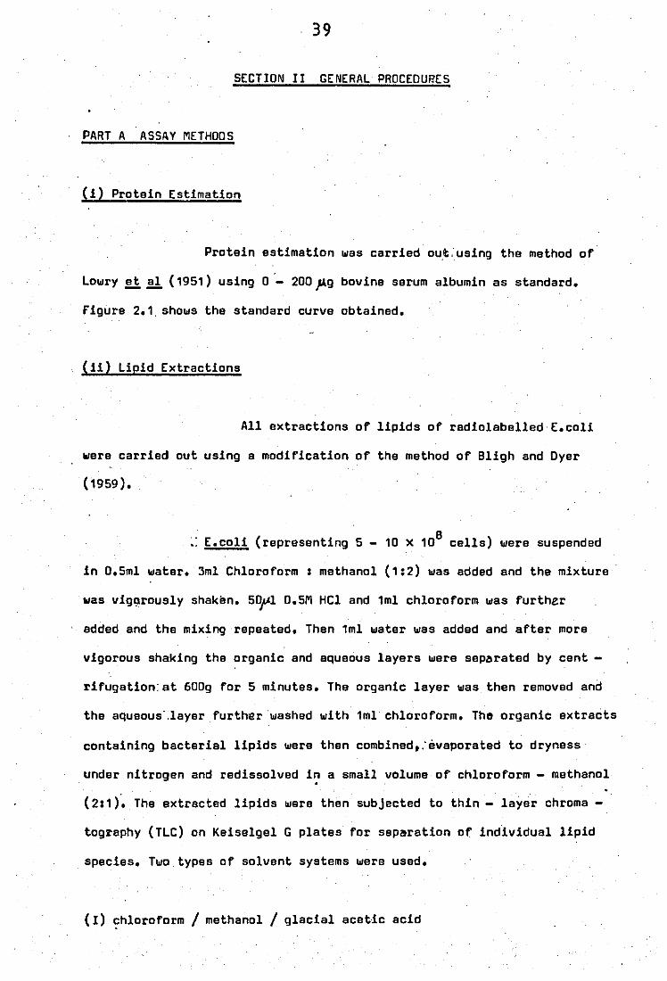
SECTION 11 GENERAL PROCEDU~ES
PART A ASSAY METHODS
(i) Protein Estimation
Protein estimation was carried out:using the method of
LowrY!l!l (1951) using 0 - 200~g bovine serum albumin as standard.
figure 2.1, shows the standard curve obtained.
(ii) Lipid Extractions
All extractions of lipids of radiolabelled E.coli
were carried out using a modification of the method of Bligh and Dyer
(1959).
~: E.coli (representing 5 - 10 x 10B cells) were suspended
in 0.5ml water. 3ml Chloroform: methanol (1:2) was added and the mixture
was viggrously shaken. 50~1 D.5M HCl and 1ml chloroform was further
added and the mixing repeated. Then 1ml water was added and after more
vigorous shaking tha organic and aquaous layers were separated by cent -
rifugation', at 600g for 5 minutes. The organic layer was then removed and
the aqueous' ,layer further washed with 1ml chloroform. The organic extracts
containing bacterial lipids were then combined,:evaporated to dryness
under nitrogen and redissolved i~ a small volume of chloroform - methanol
(2:1). The extracted lipids were then subjected to thin - layer chroma -
tography (TLC) on Keiselgel G plates for separation of individual lipid
species. Two types of solvent systems were used.
(1) chloroform / methanol/glacial acetic acid
.
40
65 , 25 6 v/v
(11) petroleum ether. (60 - 80) / diethyl ether / formic acid
60 I 40 I 1 v/v
System I was used for separation of phosphalipid species
and system II for sepa·ration of neutral lipid species •
,When ·radioactive samples were to be determined neutral
and phospholipid standards were also applied on the same plates as mar -
9 kers as the amounts of lipids present in 10 cells. of E.coli was not
detectable. The lipid species were visualised by exposure of the plates
to iodine vapour and after evaporation of the iodine the fractions were
scra~ed directly into counting vials for determination of radioactivity
by liquid scintillation counting. This was achieved in a medium af Unis -
olve 1 (7mls) plus water (3mls) which when mixed together formed a stable
gel in which the radioactive Keiselgel was counted at an efficiency of
83%.
(iii) Phospholipid determination
This was performed using the method of Raheja!i!l
(1973), which is a colourimetric method for the quantitative estimation
of phaspholipids without acid digestion. A calibration curve (fig~re
2.2) was constructed using,l - /)(. - phDsphatidyl choline dipalmitoyl
. " ) (0-800' .. g phospholipid representing 0 - 331Lg·P ior 0 - 1089 nmoles P • / r'. r- -
/' Phospholipid determination of radiolabelled E.coli was
9 carried out on 5 - 10 x 10 cells after extraction of lipids by the
method of 8ligh and Dyer (1959) as previously described. Estimation of
bacterial numbers was determined using Brown's opacity tubes
1
1
1
1
1
1
1
1
1
1
1
1
1
1
1
1
1
1
. 1
1
1
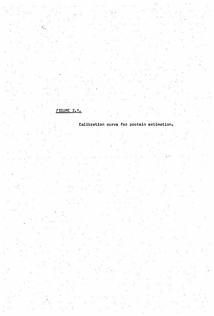
\:
fIGURE 2.1.
Calibration curve for protein estimation.
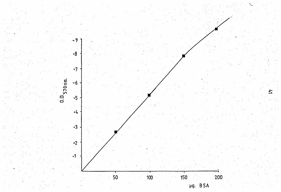
·8
E ,.6 'c <:> rLJ"'l
Cl o
'·5
·1
50 100 150 200
IJg. B SA

•
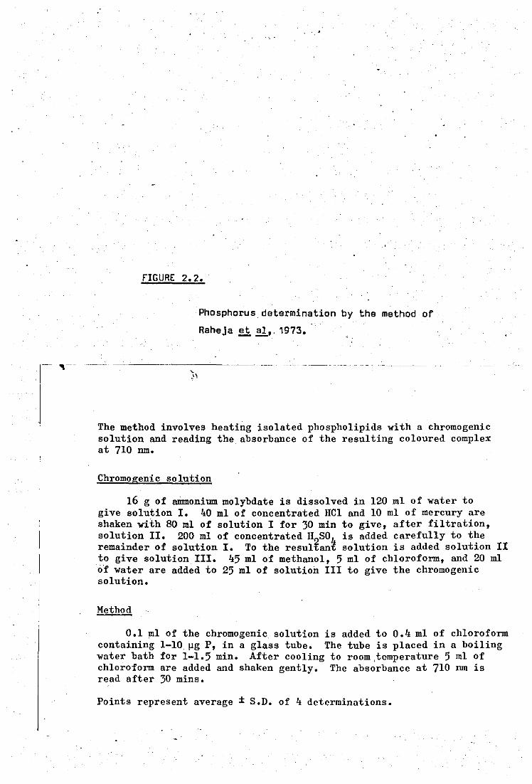
- "'--
fIGURE 2.2.
, -,'
Phosphorus determination by the method or
Raheja et aI" 1973.
The method involves heating isolated phospholipids with a chromogenic solution and reading the, absorbance of the resulting coloured complex at 710 nm.
Chromogenic solution
16 g of ammonium molybdate is dissolved in 120 ml of water to give solution 1. "0 ml of concentrated HCl and 10 ml of mercury are shaken with 80 ml of solution I for 30 min to give, after filtration, solution II. 200 ml of concentrated H SO is added carefully to the remainder of solution I. To the resultani solution is added solution II to give solution Ill. "5 ml of methanol, 5 ml of chloroform, and 20 ml o'f water are added to 25 ml of solution III to give the chromogenic solution.
Method
0.1 ml of the chromogenic solution is added to 0." ml of chloroform containing 1-10 ~g P, in a glass tube. The tube is placed in a boiling water bath for 1-1.5 min. After cooling to room ,temperature 5 ml of chloroform are added and shaken gently. The absorbance at 710 nm is read after 30 mins.
Points represent average ± S.D. of " determinations.
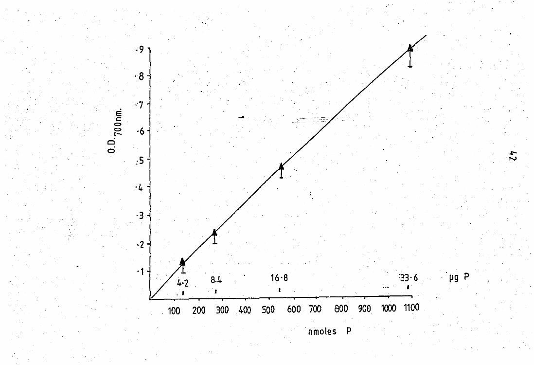
E c c:> .
c:> r-
ei o
··9
"7
·6
:s
·3
·2
-1
..
.~':-~- -- .-- .--
16·8 pg P . , .,
100 200 300 400 500 600 700 800 900 1000 1100
. nmoles P
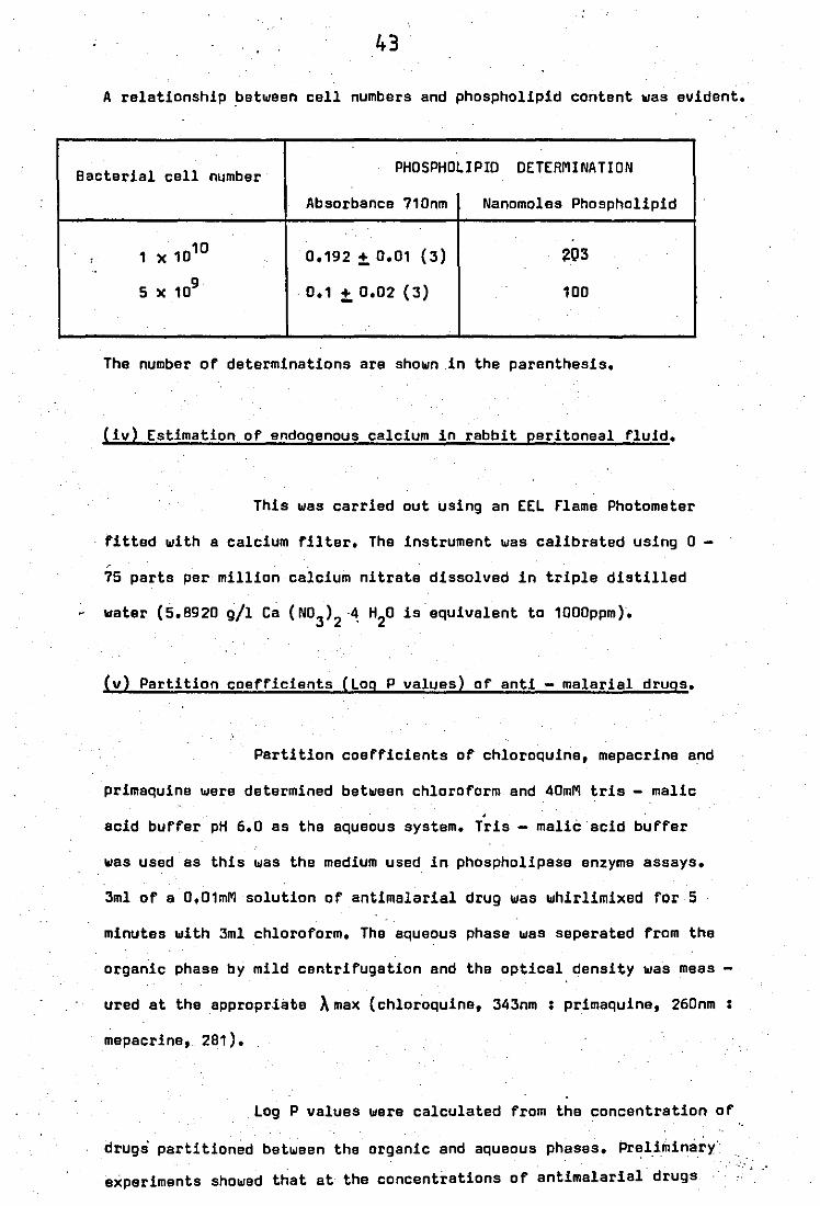
43
A reletionship between cell numbers and phospholipid content was evident.
Bacterial cell number PHOSPHOLIPID DETERMINATION
, "
Absorbance 710nm Nanomoles Phospholipid
1 X 10¹⁰ 0.192 + 0.01 (3) 2(13
5 X 10⁹ , 0.1 ±. 0.02 (3) 100
The number of determinations are shown in the parenthesis.
(iv) Estimation of endogenous calcium in rabbit peritoneal fluid.
This was carried out usin9 an EEL rlame Photometer
fitted with a calcium filter. The instrument was calibrated usin9 0 , 75 parts per million calcium nitrate dissolved in triple distilled
",ater (5.8920 g/l Ca (N03)2 A H20 is equivalent to 1000ppm).
(v) Partition coefficients (Log P values) of anti - malarial drugs.
Parti tion coefficients of chloroquine, mepacrine and
primaquine were determined between chloroform and 40mM tris - malic ,
acid buffer pH 6.0 as the aqueous system. Tris - malic acid buffer
",as used as this was the medium used in phospholipase enzyme assays.
3ml of a O,01mM solution of antimalarial drug was whirlimixed for 5
minutes with 3ml chloroform. The aqueous phase was seperated from the
organic phase by mild centrifugation and the optical density was meas
ured at the appropriate "max (chloroquine, 343nm : primaquine, 260nm :
mepacrine, 281).
log P values were calculated from the concentration of
drugs part! t!oned between the organic and aqueous phases. prelimina,ry' ... : ..
experiments showed that at the concentrations of antimalarial drugs
44
used there was a linear relationship between concentration of drug and
optical density at the appropriate "max.
Log, P = log 0.0. chloroform
0.0. aqueous.
PART B COLLECTION Of BIOLOGICAL SAMPLES
(i) Collection of polymorphonuclear leucocytes (neutrophils) from rabbits
female rabbits of the New Zealand white strain weighing
from 2 to 3kg were used. 100ml sterile thioglycollatemedium (USp) was
injected intraperitoneally through a 0.8mm external diameter stainless
steel needle while the rabbit was under light anaesthesia with ether.
After 17 to 22hr the rabbit was enaesthetised with 80mg/kg sodium pent -
obarbitone intraven ,ously and 200ml of sterile 0.15M sodium chloride
solution containing',heparin (4 in. /ml) was injected intraperitoneally.
After lightly massaging the abdomen the fluid was allowed to drain th -
rough a multi - perforated 2.5 inch 16 gauge plastic catheter.
8 9 The cells in the peritoneal fluid (10 - 10 ) were
sedimented by centrifugation in plas~ic tubes at 200g for 6 min at 22"C.
The sedimented cells were washed twice by resuspension and recentrifu -
gation at 200g for'6 min at 4"C in HEPES buffered medium. Any contamina -
ting.erythrocytes which were occasionally present in the peritoneal fluid
were lysed by resuspending the' cells in 0.9% ammonium chloride pH 7.2
for 5 minutes at 22"C (Henson, 1971) and then washing the remaining neu-
trophils in HEPES buffered medium. The resulting cells were 95% neutrophils
es determined by differential cell counts. The cell concentration wes
7 -1 edjusted to 1 x 10 cells ml •
45
(11) Collection of rabbit cell- free inflammatory peritoneal exudate
The inflammatory peritoneal fluid was collected, after
induction of a sterile peritonitis in female New Zealand white rabbits,
exactly as described ebove. The cells in the fluid (95% neutrophils)
were sedimented by centrifugation at 3000g for 6 minutes and the
supernatant was used as a source of phospholipase A activity without
further purification.
46
SECTION III EXPERIMENTAL METHODS
PART A PHOSPHOLIPASE SUBSTRATE
1.0 Preperation of labelled substrate for determination of phospholipase
~2 activity
Phospho1ipids of E.co1i were labelled with (1 _ 14C) by
oleic acid using a modification of the method described~Patriaca,
Beckerdite and E1sbach, (1972). E.co1i was grown in trie~ano1amine ~
medium overnight at 37·C. A1iquots of the overnight culture were
diluted ten - fold with fresh medium and further incubated with shaking
for 2.5 hrs at 37·C. The bacteria in 30mlsof culture (approximately
2 - 3 x:,10 10 cells) were sedimented by centrifugetion et 3000g and
resuspended in 10ml of fresh triethanolamine medium. To this was added
0.4ml of a 10% BSA solution in
xed 2.5~Ci of (1 - 14C) oleic
triethanolamine to which had been comple(0. 2."'MC>~)
acid by incubation with shaking at 37'C "
for 30 minutes. This mixture wes then incubated for 3hrs at 37'Cwith
shaking to allow the label to be taken up by the bacteria.
ro110wing this labelling period the cells were sedim -
ented by centrifugation at 3000g for 15 minutes, washed in fresh media,
recentrifuged and again resuspended in 10.4 ml triethanolamine media
containing 0.4ml 10% BSA, without any comp1exed fatty acid. This was
fUrther incubated for 1.75 to 2hrs to allow the label to become fully
incorporated into cellular phospho1ipids. After this period the cells
w,ere again collected, washed once with saline and then resuspended in
5m1 saline.
47
The rediolabelled E.coli were autoclaved for 15 minutes 2 .
at 120·C and 2.7 Kg/cm pressure. This procedure destroys bacterial pho -
spholipases but does not destroy the membrane phospholipids, and these
were then used as the substrate for phospholipase A2 assays.
Procedures using (1_14C) linoleic and (1_14C) palmitic
acids were also performed.
1.1 Determination of the position of the (1- '14C) oleic acid label in
E.coli phospholipids
This was carried out by the complete hydrolysis of E.coli
phospholipids using a specific phospholipase A2 present in thB snake
venom Crotalus adamenteus (van Deenan and De Haas, 1966). The assay ..
mixture contained 5 X 10B autoclaved radiolabelled E.coli (representing
10 nanomoles of phosphalipid), 5mM Ca2+ 40mM tris - Hel buffer, pH B.O
and O.Smg Crotalus adamenteus venom in a total volume of O.Sml. Tho
mixture was incubated at 2S·C with shaking for 2hrs. The reaction was
atopped by the addition of 3ml:, chloroform / methanol (1:2) and the lipids
were extracted by the method of Bligh and Dyer (19S9) and quantified as
previously described.
PART B STUOY Of INfLAMMATORY PHOSPHOLIPASES
2.1 Release and properties of a phospholipase A2 from rabbit
polymorphonuclear leucocytes (neutrophils)
The release of phospholipase from neutrophils was exa -
mine~ using two types of stimuli ; a phagocytosable particle zymosan
48
with bound complement (zymosan - complement) (Henson, 1971), and the
soluble stimulus calcium ions (Northover, 1977).
Neutrophils were collected from rabbits as described
earlier (page 44 ).
Zymosan'- complement (ZC) was prepared by incubating
zymosan previously washed ~ith'HEPES buffer, with rabbit serum (10mg/ml)
for 30 mins at 37"C in order to allow complement, in particular C3 to
fix (Henson; 1971). The particles were then washed ,extensively with
HEPES buffered medium at room tempreture.
7 Neutrophils (1 X 10 cells) in 1ml HEPES buffered medium • 0
were incubated with shakin9 at 37 C and either varying amounts of ZC
(0 - 5mg) or Ca2+ (0 - 5mM final concentration) were added in a volume
of 0.2ml. In control experiments 0.2ml HEPES buffered medium alone was
added to the neutrophils. Incubations were carried out for 60 mins except
when incubation. time -itself was veried, after which the tubes were cooled
in melting ice. The cells were sedimented and the supernatants removed
for esseys of'released contituents. The enzymes essayed were phospholipase_
A, the lysosomel enzymes ;3 - glucuronidase and lysozyme, and the cyto
plesmic enzyme marker lactate dehydrogenase. Total release of enzymes
were obtained by treatment of the cells with 0.2% Triton x -100 or by
sonicqtion (6JAmfor 3mins). Control experiments showed both treatments
to be equivalent. Both treatments were necessary as Triton x - 100 is
an inhibitor of phospholipase activity (rranson & al, 1974) •
. '
49
f3 - glucuronidase
lysozyme
.
~ctivity of sonicated preparation
compared with 0.2% Triton prep.
Triton preparation = 100%.
9B.2% to 4.0 (6)
95.5% + 5.0 (6)
Brackets indicate number of dste~minations •
. , In experiments using zymosan - complement the uptake
'of zymosan particles into the neutrophils was also determined using
phase - contrast microscopy. The percentage cells containing particles
end the number of particles per phagocytosis - positive cells, were dete -
rmined by observing 100 cells.
figure 2.$ summ~rises the procedure used.
Determination of enzyme activities
Phospholipase ~ activity in the supernatants was 14 .
sssayed using the E.coli labelled with (1 - C) oleic acid. The reaction
mixture contained 7.5 X 10B radiolabelled E.coli (representing approx
imately 15 nmoles of phospholipid), 40 mM tris - malic acid buffer pH 6.0
-3 2+ . 5 x 10 M Ca and an appropriate aUquot of the superna~nt. Total volume
was 0.5ml and incubations were cacried out for 60 mins at 37"C. Reactions
were stopped by the addition of 3ml. of chloroform / methanol (1:2) and
the lipids analysed as described earlier.
This enzyme was also characterised with respect to pH
p phosholipase
" A
FIGURE 2.3
so
. Peritoneal PMN
Leucocy tes
zymosan -complement or
calcium stimulation ,
assay released enzymes
lysozyme glucuronidase· lactate
dehydrogen ase
Outline of procedure for measurement of release
of neutrophil constituents during stimulation.
51
2+ profile and Ca requirement, such parameters were varied as indicattd.
;.1- Glucuronidase activity of the supernatants wes carried
out using the method of fishman (1955). Briefly this involved measure -
mentof the releese of phenolphthalein from phenolphthalein glucuronide
after 4hrs incubation at 56 "C.
Lysozyme was determined by the rate of lysis of
Micrococcus lysodeiktius measured by the decrease in absorbance at 510nm
as carried out by Smolelis and Hartsell (1949). Incubations ware carried
out for 20 mins at 25°C.
The cytoplasmic enzyme marker lactate dehydrogenase
was measured by the method of Wroblewski and La Oue (1955). Py~uvetecwes
converted to lactate in'the presence of the reduced form of ;S - lIicotin -
aminde adenine dineucleotide (fi- NAOH) and the enzyme assay was
monitored by the oxidation of NADH to NAO+ at 366nm.
All enzyme assays were normally carried out in triplicate
or at least in duplicate, and reactions were linear for the conditions
employed. No enzyme blanks were always included and appropriate corrections
made.
2.2 Properties of a phospholipase A present in the inflammed peritoneal
exudate of the rabbit
Pho~pholipase A activity of the rabbit inflammatory
peritoneal fluid (after removal of the cells by centrifugation, page45)
S2
was also characterised using radiolabelled E.coli. The incubation medium
contained, unless stated otherwise, 40mM tris - buffep, 5mM ca2+, 0 - 1mg B ..
enzyme protein and 5 x 1D autoclaved radiolabelled E.coli, in a total
volume of D.5ml. Incubations were normally carried out for 5 mins at
37·C. Reactions were stopped end hydrol;'tic activity was determined as
before (page 49). Various parameters were varied in order to characterise
the enzyme activity.
, . l:.Q. PART C ErrECTS or CHLOROQUINE - lIKE AGENTS ON INrlAMMATORY AND
OTHER PHOSPHOlIPASES.
The effects of chloroquine - like agents were examined
on phospholipases obtained from different sources. Preliminary work was
carried out with a cruda phospholipase A2 enzyme from Crotolas adamenteus
venom·and a purified enzyme from pig pancreas, against egg - yolk phos-
pholipids. Inflammatory phospholipases from rabbit peritoneal fluid and
14 from peritoneal neutrophils were tested using the (1 - C) oleic acid
labelled E.coli as substrate.
3.1 Effects of chloroquine - like agents on phospholipase A2 from
Crotolas adamenteus venom and pig pancreas
Enzyme activity in.these highly active sources was
assessed by direct titration of the fatty acids released from egg -
yolk phospholipids with time. The amount of alkali. needed to keep the
pH constant at B.O in the presence of phospholipase A2, was recorded
automatically using a Radiometer TTT - 1c Titrator equipped with a
5BU -1a syringe and a combined glass - calomel electrode. Rate of
S3
hydrol~sis was linear with time for at least 3 minutes after injection
of enzyme.
Procedure '.
\.
" \, One egg - yolk was st:"irred to homogeneity in 100ml
of distilled water for 10 minutes, and 20ml of a 160mM solution of ca
lcium chloride added. The emulsion was then freed from the yolk. bags
by filt 'ration through cheesecloth. This preparation was stable for up
to 3 days.
In a reaction vessel maintained at 25°C, were added
6ml of egg .,- yolk emulsion containing Ca2+, 1ml 20mM sodium deoxycholate
and the volume was made up to 20ml with distilled water. The pH was
brought up to B.O and the blank consumption of 10mM NaOH needed to .' ~
maintain this pH was recorded. Then 10,Ml of a 1mgjml solution of Crot -
alas adamenteus venom in water was added and the alkali consumption was
,again recorded. In this system all the fatty acids released were titrated
with 100% efficiency (Nieuwenhuizen, Kunze and De Haas, 1974), so the
number of micromoles of alkali used was equivalent to the number of
. micromoles of fatty acids released. The experiments were repeated in the
presence of varying concentrations of chloroquine diphosphate.
De terminations using the enzyme from pig pancreas were
carried out using an identical procedure but at 37°C. Drugs screened
against this enzyme were chloroquine sulphate, dibucaine hydrochloride,
cinchonine sulphate, salicylic acid and indomethacin.
Dose ~ response curves end E050 values were determined
using least squares analysis of data.
54
3.2 Effects of chloroouine - like aq_nts on the ectivity of peritoneal
fluid phospholipase A
Studies of the effects of the antimalarial drugs chlo
roquine, mepacrine and primaquine formed the.major part of'this work.
The drugs were first screened on the peritoneal fluid phospholipase
activity towards E.coli phospholipids since this was a more stable and a
more active 'sourca of enzyme activity than the neutrophil source. Other
age~ts tested were 0 - penicillamine, indomethacin, aspirin, salicylic
acid and benzydamine.
Oeterminations of phospholipase activity in the presence
and absence of drugs were carried out as those used to determine the
properties of the peritoneal fluid enzyme (paga51 ).
3.3 Effects of chloroquine - like aqents on the phospholipase activity
Of .PI'lN ieucocytes.
Studies of the effects of the antimalarial drugs chlo
roquine,. mepacrineand'primaquine on the phospholipase activities of
PI'lN neutrophils was tested on cell suspensions that had been sonicated
to release all of the cellular enzymes.
Leucocytes suspended in HEPES buffered medium at 4"C
at a concentration of 7 x 10⁶ cells ml-1 were sonicated at ~'for 2 X 2
mins with 2 minutes separating each soniacation period. The suspension
was then centrifugated at 3000g for 10 mins at 4'C and the supernatant
was used as a crude source of phospholipase activity. Incubations were
55
9 14 2+ carried out using 10 (1 - C) oleate labelled E.coli, 5mM Ca , 40mM
tris - malic acid buffer pH 6.5 and n.1mg 3000g neutrophil supernatent
for' 30 mins at·37·C. Termination of assays and analysis of lipids was
carriad out as before.
The effects of the antimalarial drugs were also tested
after partial purification of the neutrophil phospholipase. The purifi -
cation was carried out by the method of Weiss et al (1975) using sulph
uric. acid to extract the enzyme as outlined in fig 2.4. Washed neutrophUs
were resuspended in distilled water (4°C) at a concentration of 5 x 10⁷
cells/ml. Then ice - cold sulphuric acid was added to a final concentr -o .
ation of O.OBM and the suspension was kept at 4 C for 30 mins. After this
time a white precipitate was apparent and this was centrifuged at 23,000g
for 20 mins. The supernatant (desi9nated Sup I) was then; exhaustively
dialysed against 1mM tris - HCl bUffer1FH 7.4. : . The pellet (pellet
I) was resuspended in an equivalent volume of distilled water (aqual to
the supernatant removed). After dialysis of Sup I a further precipitate
. formed and was again separated by cent.rifugation at 23,000g for 20 mins.
The resulting supernatant (Sup 11) was used as the partially purified
preparation of phospholipase.
3.4 Int.eractions of heparin with 'antimalarial drugs
Interactions of heparin with the antimalarial drugs
chloroquine" mepacrine and primaquine were examined by measuring optical
densities at 700nm after mixing solutions of heparin (50 units/ml final
concentration) with varying amounts of antimalarial drugs (0 - 5mM),
in a total volume of 10ml' .• All three drugs showed very little absorbance
alone at 700nm (less than 0.02 0.0. units) in the concentration ranges
used.
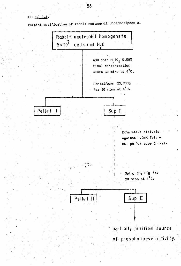
S6
fIGURE 2.4.
Partial purification of rabbit neutrophil phospholipase A.
Rabbit
S x 1 07
Pellet I
.'
neutrophil homogenate
cells I ml H20
"
I
Add cold H2S04 O.OBM
final concentration o
store 30 mins at.4 C.
Centrifuge; 23,0009 o
for 20 mins at 4 C.
Sup I
Exhaustive dialysis
against 1.0mM Tris -
HCI pH. 7.4 over 2 days.
Spin, 23,000g for o
20 .mins at 4 C.
I
Pellet II Sup II
partially purified source
of phospholipase activi ty.
57
3.5 Membrane actions of antimalarial drugs on guinea-pig red blood cells
The.effects of antimalarial drugs on the haemolysis of
guinea - pig red blood cells were examined. Such compounds are amphiphilic
molecules and thus may exert their effects on enzymes such as phospho1ip -
ases by membrana stabilizing or labelizing action.
firstly a 50%
fresh heparinised whole guinea - pi9!_ blood was used. using
lysis figure was obtained ,,0.05ml blood and 5ml of varying' ..
concentrations of sodium chloride. After exectly 30 mins incubation at
room temlllrd;ura the mixtures were centrifug ""ed at 1000g for 5 mins. The
haemoglobin content of the supernata~t was measured at 543nm on a Unicam
S.P. 500 spectrophotometer, and was used as a measure of cell lysis •
• Complete cell disruption was caused by incubation with distilled water.
50% Lysis was found at 0.425% NaCl. The experiments
were then repeated in the presence of varying concentrations of anti
malerial drugs dissolved in 0.425% NaCl." Mepacrine and primaquine both"
. " ,1 showed absorbances et 5~3nm, and s~ opticalmnsities of the haemoglobin
content in the preoonc'e of these drugs were corrected by the use of
eppropriate blanks.
•
58
4.0 PART D STUDIES or AN ENDOGENOUS INHIBITOR or PHOSPHOLIPASE A~
THE B2009 SUPERNATANT or NEUTROPHIL LEUCOCYTES
4.1 Preparation and determination of inhibitory activity in the B2009"
supernatant of neutrophil 1eucocytes
Neut~ophi1 1eucocytes were collected from New Zealand'
White rabbits as described earlier (page44). The 'washed 1eucocytes were
suspended in ice - cold 0.34M sucrose at a concentration of 1 X'10B'ce11s/m1
and stored at 4"C for 30mins. The cell suspension was then homogenised
with a Potter - E1vejham type homog~niser until cell disruption was
complete as monitored by phasecontrast microscopy. Tha homogenate was,
then subjected to centrifugation at 8,200g for 20 mins to separate the
nuclei, cellular membranes and lysosomal granules from the cytosol. The
8,200g pellet was used as the granule phospholipase preparation and the
,8,200g supernatant was used without further purification as the source
, of an endogenous inhibitor of phospholipase activity.
In this work the inhibitory activity towards the granule
phospholipase at acid pH was examined and the B,200g supernatant inhibitor
was screened against the phospholipase A present in the rabbit peritoneal
fluid exudate' (PE) after the neutrophils had been sedimented 1\" '~. Assays
were again carried out using (1 _ 14C) oleate labelled E.co1i as substrate
(1.5 X 10g
cells containing approximately
40mM tris buffer, 0.35mg peritoneal fluid
30nm of phospholipid), 5mM Ca2+, f""ohi"
or 0.5mg grenu1eAas the enzyme-
source, and varying amounts of B,200g supernatant as the inhibitor. (0 -
400pg protein) in:,a total volume of 0.5m1. Incubations were normally
carried out for either 5 or 10 mins depending upon the amounts of enzyme
S9
protein and inhibitor used.
A pH profile of the phospholipase inhibitory capacity
of the 8,200g supernat~nt on the peritoneal fluid enzyme was also deter -
mined. Tris - malic acid buffer was usad for pHs between 5.5 and 7.5 and
tris - HCl was used for .pHs between 7.5 and 9.0.
4.2 Effects of the PMN a,200g supernatant on the phospholipases from
pig pancreas, Crotalus adamenteus venom and Naja nalavenom
Inhibitory effects of the a,200g supernat6lnt fractions
were also tested against commercialt'y available phospholipases from
different sources. These sources were pig pancreas and the snake venom s
. Crotalus adamenteus and Nala nala.
Incubatiops were carried out as before for 10 mins and
at pH 7.5. Sufficient enzyme proteins were used to give approximately
20% hydrolysis of E.coli phopholipids as control values. Actual amounts
of enzyme used to achieve this was 0.05ng protein for the pig pancreas
enzyme, 11"g' :protein for the venom Crotalus adamanteus and 0.25.f'9.'
protein for the venom Nala nala.
The possibility that inhibition of phospholipase A2
activity by the a,200g supernatant could be due to a non - specific
protein effect was also examined as phenomena of this type had already
been shown in the case of phospholipase of the liver (Weglicki et aI,
. 1974). To examine this each of the 4 phospholipases examined were also
assayed in the presenc.B of an equivalent concentration of BSA.
•

Chapter Three
RESULTS
60
PART A PHOSPHOLIPASE A2 SUBSTRATE
1.0 Radiolabelling of E.coli with (1 14C) oleic acid
,ror adequate uptake of (1 - 14C) oleic acid by E.coli
very specific conditions were found to be necessary. These were:-
(i) The (1 _14C) fatty acid must be complexed with albumin prior
to contact with the E.coli otherwise the'radiolabel is not adequately
taken up, as shown by the values obtained below:-
(a) Without prior complexing ie (1 - 14C) oleic acid added tog
o ether with BSA and E.coli and shaken vigrously. , ~
Radioactivity (cpm) obtained per 5 x 10⁸ cells = 1441 ± 207 (5 determinations)
(b) With prior complexing ie BSA + (1 - 14C) oleic acid incubated
at 37"C for 30 mins before adding E.coli.
8 Radioactillity (cpm) per 5 x 10 cells = 16882 :!:",4634 (7 determinations)
(ii) The initial number of E.coli exposed to the radiolabel had an
effect on the usefulness of the preparation as a substrate for phosph -
, 10 oUpase A2 assays (see table below). An initial count of2 - 3 x 10
organisms in log phase of growth per 10.4ml incubation mixture was found
to be suitable as'this provided approximately 5 x 10¹⁰ cells (after
losses during washings in the.,procedure), with an adequate amount of
radioactivity and sufficient for approximately 50 enzyme assays.
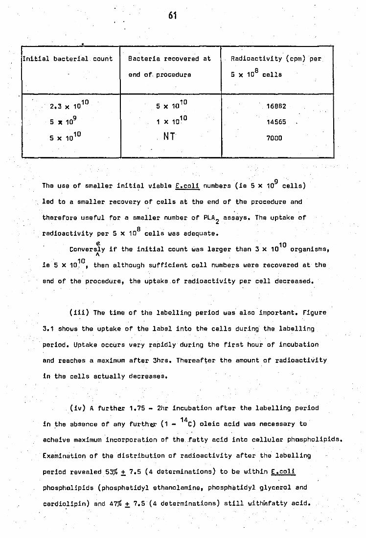
61
•
Initial bacterial count Bacteria recovered at Radioactivi ty (cpm) per
end of· procedure 8 5 X 10 cells
2.3 x 10¹⁰ 5 x 10¹⁰ 16882
5 X 10g
1 X 10¹⁰ 14565
5 X 10¹⁰ NT 7000
The use of smaller initial viable r.coli numbers (ie 5 X 10g
cells)
led to a smaller recovery of cells at the end of the procedure and
therefore useful for a smaller number of PLA 2 assays. The uptake of
8 . radioactivity per 5 X 10 cells was adequata.
e 10 Conversly if tha initial count was larger than 3 x 10 organisms,
1\
ie 5 X 10¹⁰, then elthough sufficient cell numbers were recovered at the
end of the procedure, the uptake of radioactivity per cell decreased.
(Hi) The time of the labelling period was· also important. Figure
3.1 shows the uptake of the label into the cells during the labelling
period. Uptake occurs very rapidly during the first hour of inCUbation
and reaches a maximum after 3hrs. Thereafter the amount of radioactivity
in the cells actually decreases.
(iv) A further 1.75 - 2hr inCUbation after the labelling period
in the absence of any further (1 - 14C) oleic acid was necessary to
acheive maximum incorporation of the .. fatty acid into cellular phospholipids.
Examination of the distribution of radioactivity after the labelling
period revealed 53% ± 7.5 (4 determinations) to be within E.coli
phospholipids (phosphatidyl ethanolamine, phosphatidyl glycerol and
cardio,lipin) and 47% ± 7.5 (4 de terminations) still withinfatty acid.

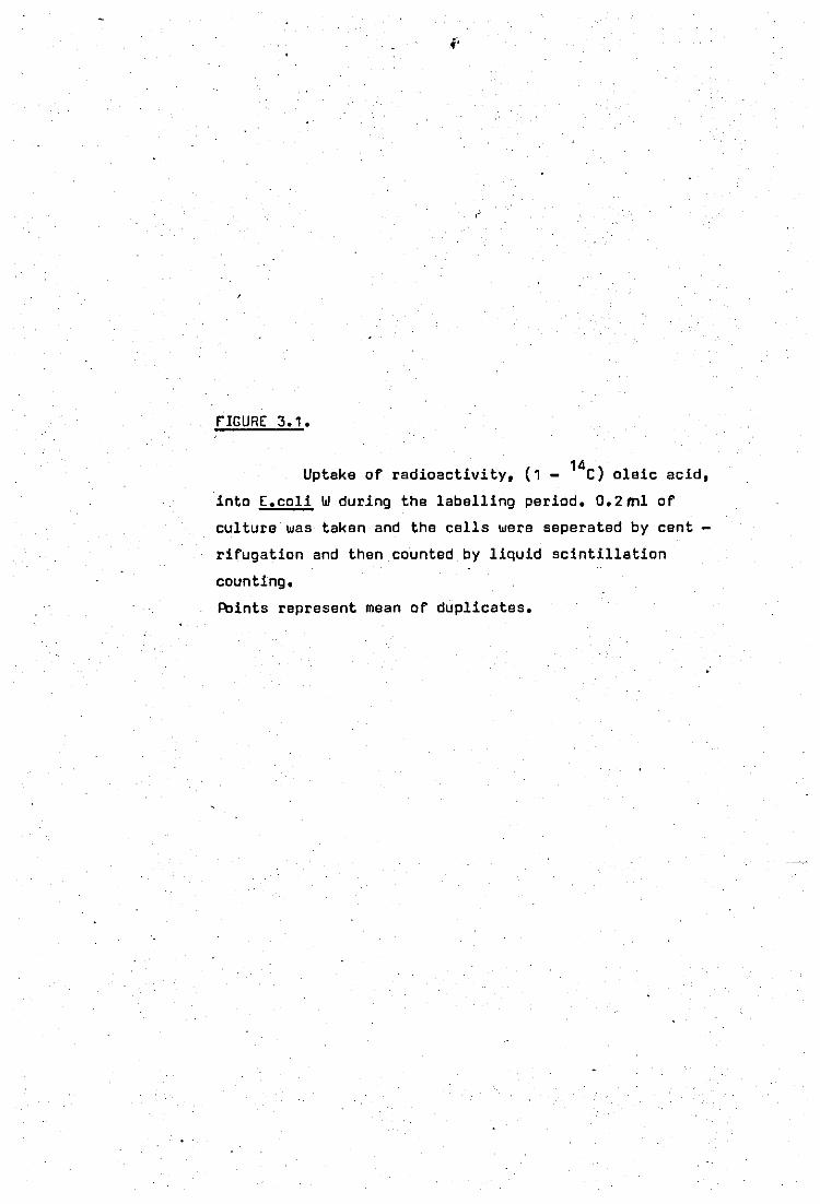
rIGURE 3.1.
14 Uptake of radioactivity, (1 - C) oleic acid,
·into E.coli W during the labelling period. O.2ml of
culture was taken and the cells were seperated by cent
rifugation and then counted by liquid scintillation
counting.
Points represent mean of duplicates.
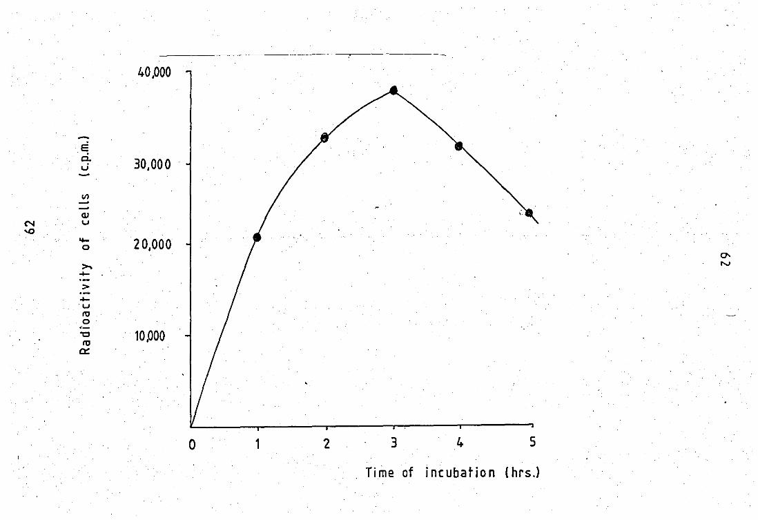
40,000
o 1 2 3 4 5
Time of incubation (hrs.l
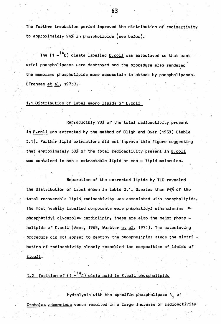
. 63
The further incubation period improved the distribution of radioactivity
to approximately 94% in phospholipids (see below).
14 . The (1 - C) oleate labelled E.coli was autoclaved so that bact
erial phospholipases were destroyed and the procedure also rendered
the membrane phospholipids more accessible to attack by phospholipases.
(fransen et aI, 1973).· --
1~1 Distribution of label among lipids of E.coli·
Reproducibly 70% of the total radioactivity present
in E.coli was extracted by the method of 8ligh and Oyer (1959) (table
3.1). further lipid extractions did not improve this figure suggesting
. that approximately 30% of the total radioactivity present in E.coli
was contained in non - extractable lipid or non - lipid molecules.
Se~ration of the extracted lipids by TLC revealed
the distribution of label shown in table 3.1. Greater than 94% of the
total recoverable lipid radioactivity was associated with phospholipids.
The most hea~ily labelled components were phophatidyl ethanolamine =-
phosph~tidyl glycerol=- cardiolipin, these are also the major phosp -
hol!pids of E.coli (Ames, 1968, Wurster II aI, 1971). The autocleving
procedure did not appear to destroy the phospholipids since the distri
bution of radioactivity closely resembled the composition of lipids of
E.coli.
1.2 Position of (1 _14C) oleic acid in E.coli phospholipids
Hydrolysis with the specific phospholipase·A2 of
Crotalas adamenteus venom resulted in a large increase of radioactivity
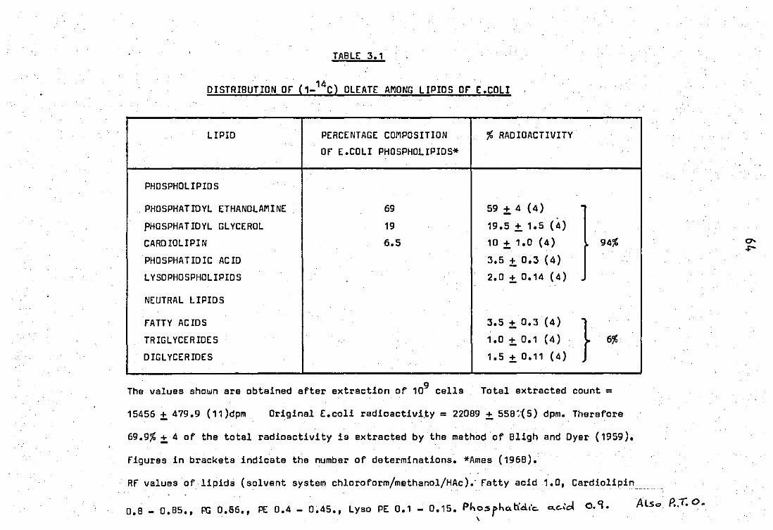
TABLE: 3.1
DISTRIBUTION Of (1_14C) OLEATE AMONG LIPIDS Of E: .COLI
LIPID PERCENTAGE COMPOSITION % RAOIOACTIVITY
Of E.COLI PHOSPHOLIPI05*
PH05PHOLIPID5
. PH05PHATIDYL ETHANOLAMINE 69 59+4(4) . PHO 5PHA TID YL GLYCEROL 19 19.5 ± 1.5 (4)
CARDIOLIPIN 6.5 10 ± 1.0 (4) 94%
PH05PHATIDIC ACID 3.5 + 0.3 (4)
LYSOPHOSPHOLIPIOS 2.0±0.14 (4)
NEUTRAL LIPID5
fATTY ACIDS 3.5 + 0.3 (4)
} TRIGL YCERIDE5 1.0 + 0.1 (4) 6%
OIGLYCERIDE5 1.5 ± 0.11 (4)
The values sho.wn are obtained after extraction of 109 cells Total extracted count =
15456 ± 479.9 (11)dpm Ori9inal E.coli radioactivi.ty = 22089 + 559:'(5) dpm. Therefore
69.9% + 4 of the total radioactivity is extracted by the method 'Of Bligh and Oyer (1959).
figures in brackets indicate the number of determinations. *Ames (1968).
Rf values of lipids (solvent system chloroform/methanol/HAc).' fatty acid 1.0, Cardiolipin
0.8 _ 0.95., PG 0.66., PE 0.4 - 0.45., Lyso PE 0.1 - 0.15. Phosrho.tid,'c- Q.e.,cl 0.9. \
Also. P.:r:. o.
R.f v...t..~ ~ solve.,J; ,'\':~sl-eM ret. etker / 04'e1t:J.e. dt..e.r I fl>Ywu'c <x(.;d,
'"fr.:jSce.r,cA L > 0.'7" !"",t;.I:...':l t>..c.-o'c!.S o· S " bij S.:.e.rid.t<> o,?oS.,1
rk..s.rk..Li(c\s O. o. '
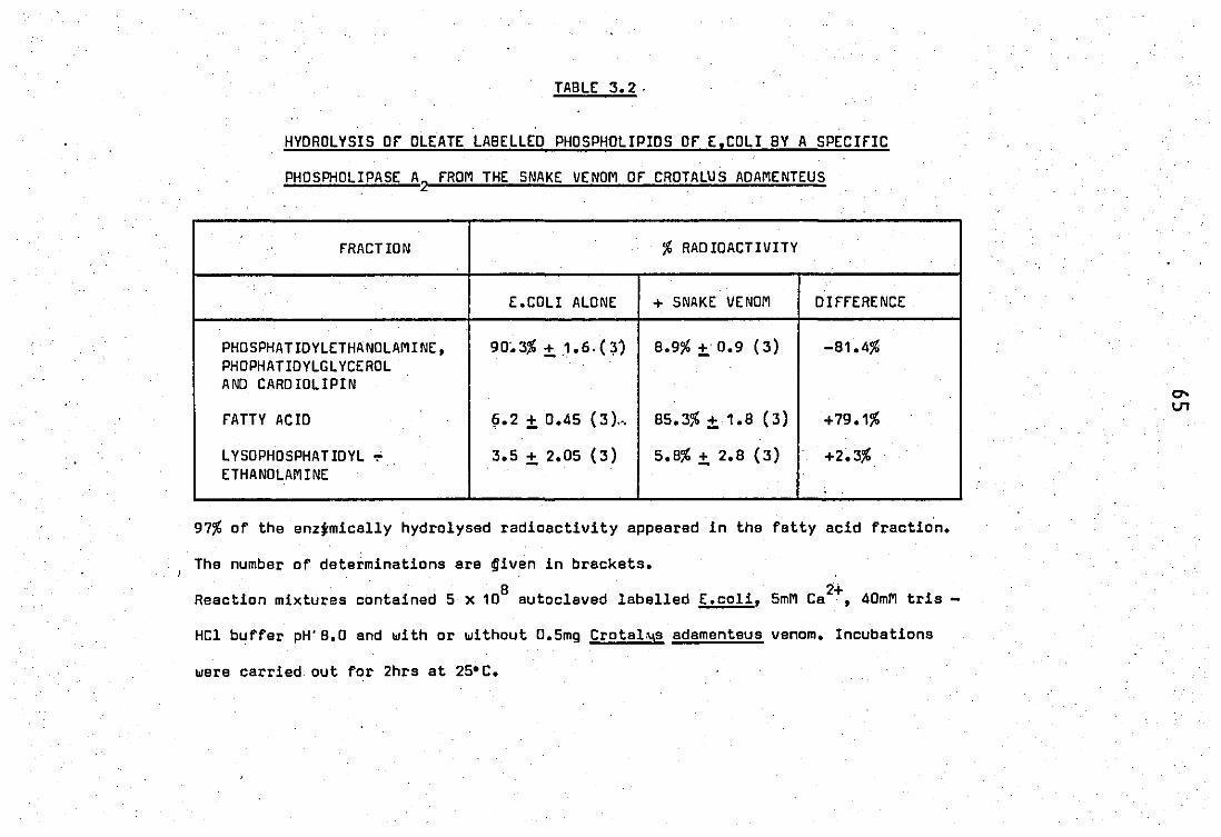
TABLE 3.2·
HYDROLYSIS or OLEATE LABELLED PHOSPHOLIPIDS Of E,COLI 8Y A SPECIfIC
PHOSPHOLIPASE A2 FROM THE SNAKE VENOM OF CROTALUS ADAMENTEUS
.
.
FRACTION % RAD IOACTIVITY
E.COLI ALONE + SNAKE VENOM DIffERENCE
PHOSPHATIDYLETHANOLAMINE. 90~3% ±. .1.6.0, B.9% ±.0.9 (3) -81.4% PHOPHATIDYLGLYCEROL AND CARDIOLIPIN
fATTY ACIO 1j.2 ±. 0.45 (3 ) .... 85.3% + 1.B (3) +79.1%
LYSOPHOSPHATIDYL ~ 3.5 ±. 2.0S (3) S.8% ±. 2.B (3) +2.3% ETHANOLAMINE
97% of the enzjmically hydrolysed radioactivity appeared in the fattyecid fraction.
The number of determinetions ara ~iven in brackats.
B U Reaction mixtures contained 5 x 10 autoclaved labelled E.coli. 5mM Ca • 40mM tris -
HCl buffar pH' B.O and with or without D.Smg Crotah\s adamenteus venom. Incubations
were carried out for 2hrs at 2S'C.
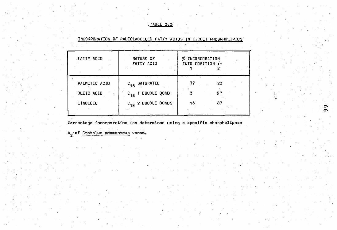
'. TABLE 3.3 .
INCORPORATION OF RADIOLABELLED FATTY ACIDS IN E.CDLI PHDSPHOLIPIDS
" .
fATTY ACID NATURE OF" % INCORPORATION FATTY ACID INTO POSITIDN :-, 2
PALMITIC ACID C16 SATURATED 77 23
OLEIC ACID C'8 1 DDUBLE BOND 3 97
LINOLEIC C18 2 DOUBLE BONDS 13 87
Percentage incorporation was determined using a specific phospholipase
A2·Of Crotalus adamenteus venom.
67
in the fatty acid fraction with only a small increase in the lysophos.
pholipid fraction (table 3.2). The results demonstrate that 97% of the
14 ) (1 - C oleic acid was in position 2 of the phosphol!pids.
14 Procedures using (1 - C) linoleic acid resulted in
less labelling of the 2 position (87% in pos~ 2 of phospholipids).
Use of (1 - 14C) palmitic acid resulted in 77% of the
fatty acid incorporated into position 1 and 23% in position 2 of the
bacterial phospholipids. Table 3.3 shows a comparison of these values.
Routinely (1 _14c) oleate labelled E.coli were used.
68
PART B STUDY OF INFLAMMATORY PHOSPHOLIPASES
2.0 Releese and properties of a phospholipase AZ from rabbit polymor -
phonuclear leucocytes (neutrophils)
2.1 Release of phospholipase A2 during phagocytosis of zymosan -
complement particles
Neutrophil leucocytes incubated with zymosan - comple -
ment (ze) particles, et 370 e over 60 mins, showed uptake of the particles
and release of granule enzymes known to be markers for lysosomes.
PhDspholipase A2 release (fig ... 3.2) was shown to be :-
(i) concentration dependant, (ii) linear up to 0.5mg ze, (iii) reach
a maximum of approximately 30% at 5mgZe and (iv) follow the release of
the lysosomal marker enzymesp - glucuronidase and lysozyme. The cytoplasmic
enzyme lactic dehydrogenase was released only to a minor extent during.
the reaction. This demonstrated that release of phospholipase along with
jJ - glucuronidase and 1 ysozyme was spec! fic and not dUB to cell break -
down as this would also release the enzyme lactate dehydrogenase to a
compareble extent.
The release. of phospholipase A2 activity was paralleled
by the uptake of zymosan - complement particles as shown in table 3.4.
As phospholipase activity was released, the percentage of cells conta -
ining zymosan particles increased and also the number of particles inge -
sted by each call increased.

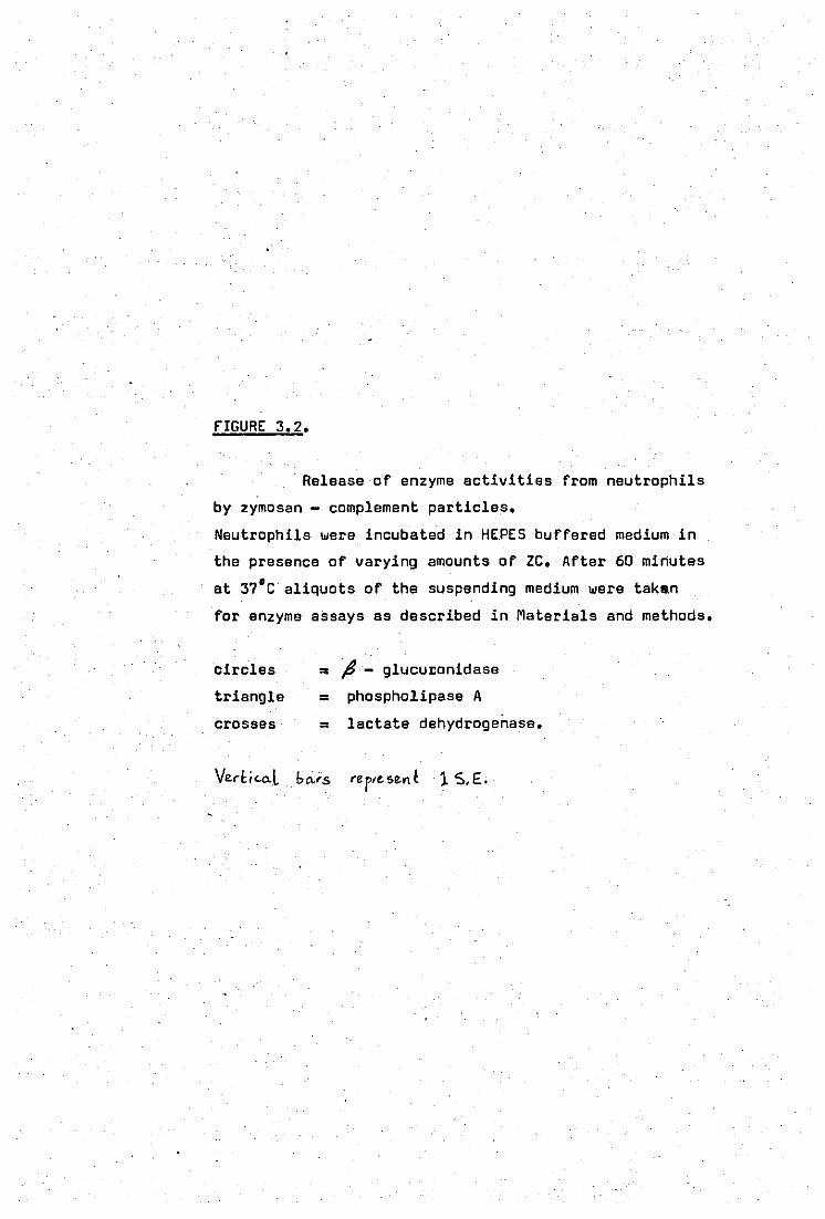
fIGURE 3.2.
Release of enzyme activities from neutrophils
by zymosan - complement particles.
Neutrophils were incubated in HEPES buffered medium in
the presence of varying amounts of ZC. After 60 minutes
at 37'C aliquots of the suspending medium were taken
for enzyme assays as described in Materials and methods.
circles ,. jJ - glucuI:onidase
triangle = phospholipase A
crosses = lactate dehydrogenase.
Vlt(t; <.o..l . bo.is re p,e. $en ~ 1 $,£.
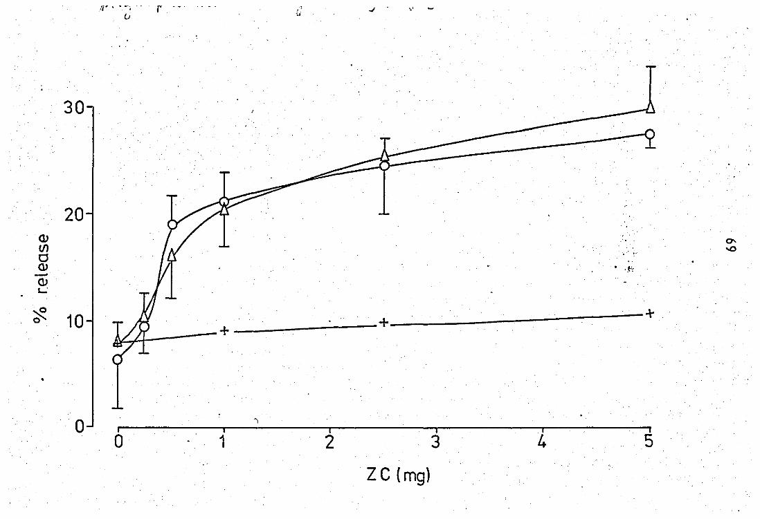
:1' •• " r - - -o ' ..
30
20 Q) 0-If)
", !" ..a
a ,-
Q) . ,~, -Q) '-'
~ 10 + +
•
o i i I I i I o 1 2 3 4 5
Z C (mg)
TA8LE 3.4
RELEASE OF' PHOSPHOLIPASE A2 AND PHAGOCYTOSIS OF'
lYMOSAM - COMPLEMENT PARTICLES (lC)
lC (mg) PLA 2 RELEASE (%) . PHAGOCYTOSIS - No. or PARTICLES POSITIVE CELLS (%) PER POSITIVE CELL
(%)
.
0 B.3 + 2.0 (3) 0 0
0.25 10.1 :t 2.8 (3) 53 1.4:t 0 • 5
0.5 16.0 :t 4.0 (3) 62 2.0 :t 1.2
1.0 20.3 :t 4.8 (3) 83 2.4 :t 1.7
2.5 25.0 :t 2.0 (3) 85 2.8 :t 1.2
5.0 30.0 :t 4.0 (3) 96 3.2 :t 1.5
' •... 8rackets indicate the number of determinations.
Phagocytosis- positive cells and no. of particles par positive cell
were determined by counting 100 cells.
Neutrophil leucocytes (1 x 10⁷ ) were incubated in the presence of
varying amounts CIf lC for1hr at 37°C. Phospholipase A activity at pH 6.0
was assayed as described in Materials and Methods.

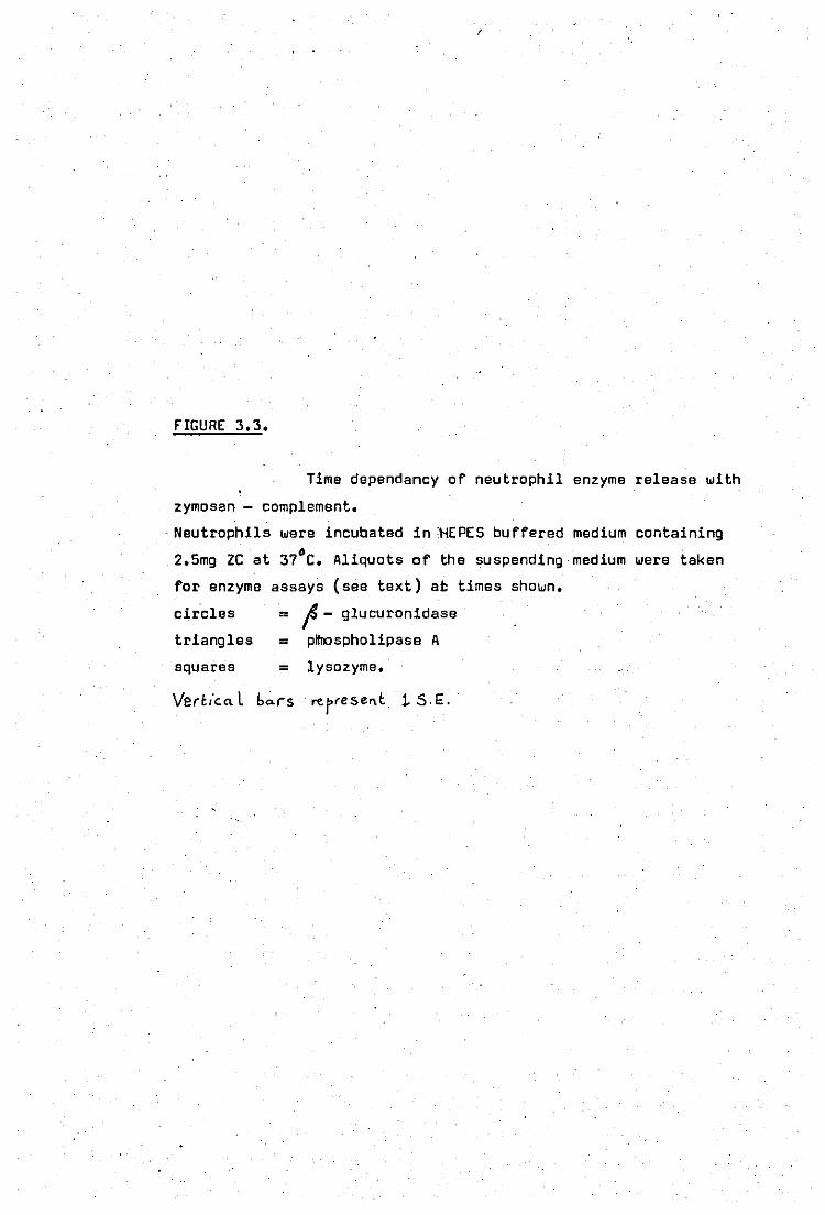
(
fIGURE 3.3.
Time dependancy of neutrophil enzyme release with
zymosan - complement •
. Neutrophils were incubated in ~EPES buffered medium containing 6 2.5mg ze at 37 e. Aliquots of the suspending medium were taken
for enzyme assays (see text) at times shown.
circles ,. f - glucuronidase
triangles ,. pltrospholipase A
squares = lysozyme.
Verb'c.o.l bo.r-s rerresent. 1. S.£.
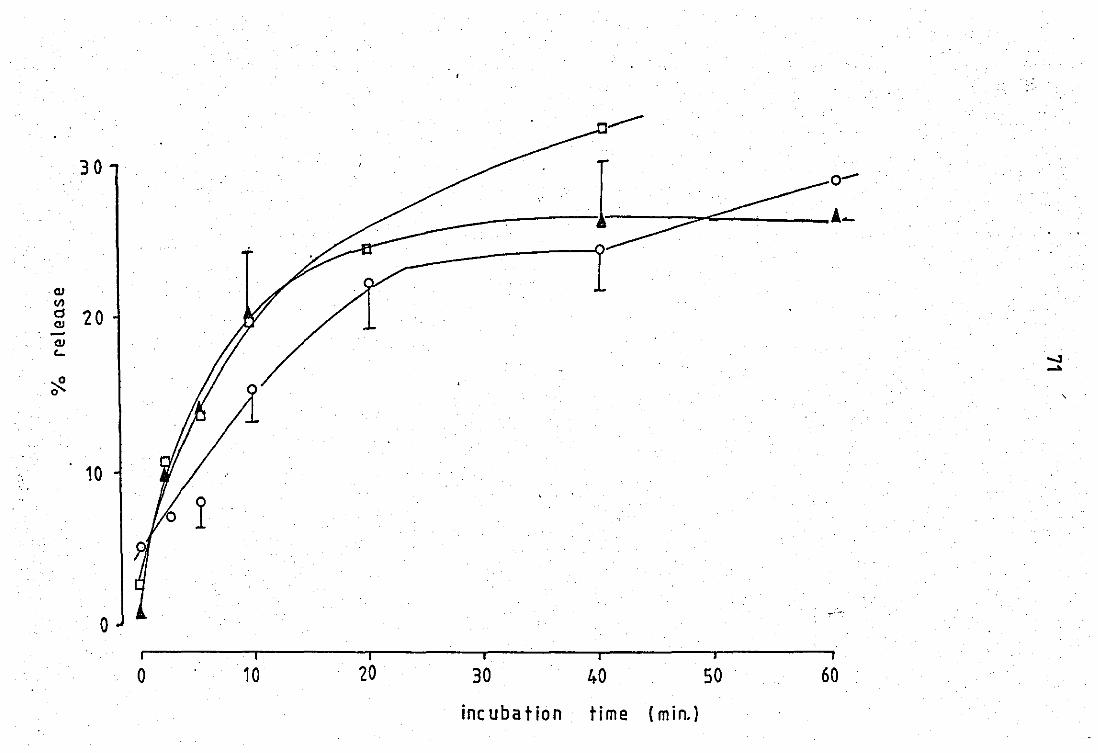
30
QJ f/)
d 20 C!J . -
QJ c..
~ 0
10
o
o 10 I
20
___ --I~~:
30 40 50 60
inc uba tion time (min.)
,.
,
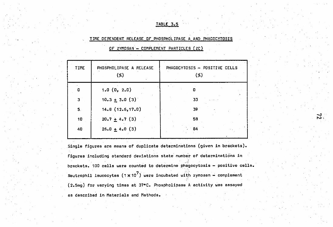
. "
TIME
0
3
5.
10
40
TABLE 3.5
TIME DEPENDENT RELEASE OF" PHOSPHOLIPASE A AND PHAGOCYTOSIS
OF" ZYMOSAN - COMPLEMENT PARTICLES (ZC)
PHOSPHOLIPASE A RELEASE PHAGOCYTOSIS - POSITIVE CELLS
(%) (%)
1.0 (0, 2.0) 0
10.3 :t 3.0 (3)· 33
14.8 (12.6,17.0) 39
20.7 :t 4.7 (3) 58
26.0 :t 4.0 (3) 84
Single figures ara means of duplicate de terminations (given in brackets).
figures including standard deviations state numbe"r of determinations in .. '"
brackets. 100 cells were counted to determine phagocytosis - positive calls • . :' ...•
Neutrophil leucocytes (1 x 10⁷ ) were incubated with zymosan - complement
(2.5mg) for varying times at 37·C. Phospholipase A activity was assayed
as described in Materials and Methods.

rrGURE 3.4.
Release of neutrophil enzymes in the presence
of Ca2+. Neutrophils were incubated in HEPES buffered medium in
the presence of varying amounts of Ca2+. After 60 mins •
. at 37°C aliquots of the supernatent were examined for
enzyme release as described in text.
circles = ~ - glucuronidasa
triangles = phospholipase A
squaras
crosses
= lysozyme
= lactate dehydrogenase.
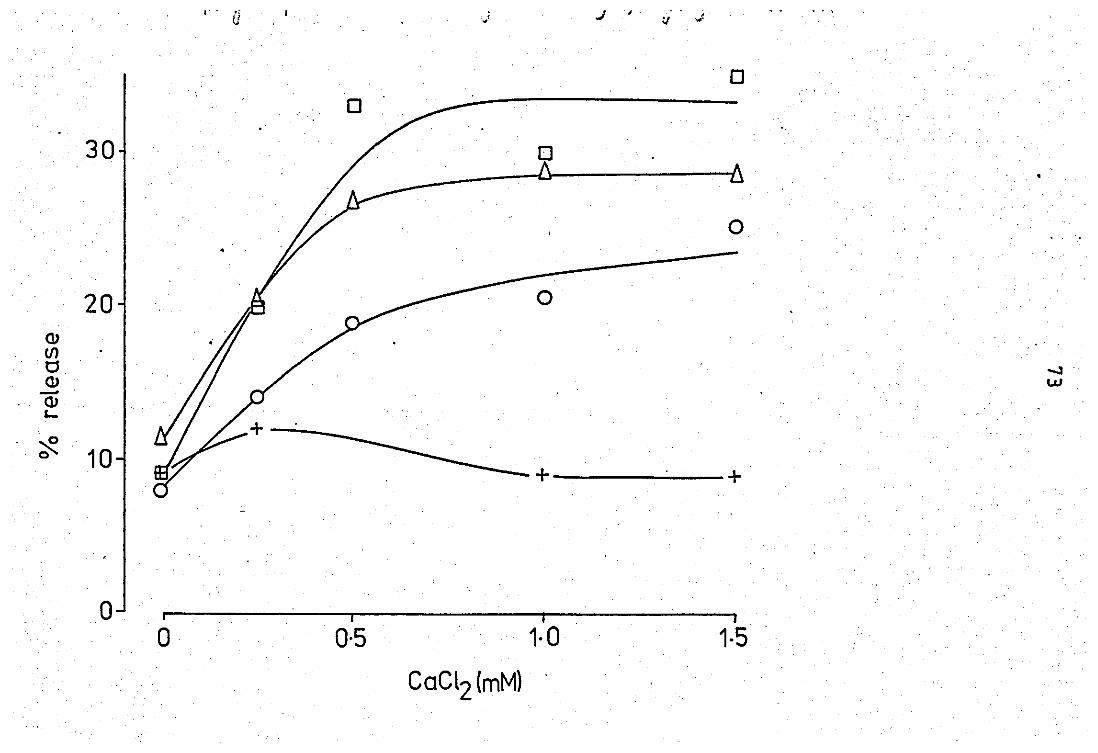
,- - ", !j ..J I· it ,,;
o
.. 30 o
o .' .
20 0
(J) (/)
0 -.l . (J) w -(J) '-
~ 10 ----0
-+ +
o I j j
o 0·5 1·0 1·5
CaCl2 (mM) .


fIGURE 3.5.
Time dependancy of neutrophil enzyme release 2+ with Ca •
Neutrophils were incubated in HEPES buffered medium 2+ • containing 2.5mM Ca at.37 C. Aliquots of the suspen-
ding medium were taken for enzyme assays' (see text) at
times shown.'
circles = ~ - glucuronidase
triangles = phospholipase A
squares = lysozyme.
\lerheat bo.rs ~erf'S.,~t
othu p.,,,,t!> cue ", ... ",$
.'.' : ... -: '.
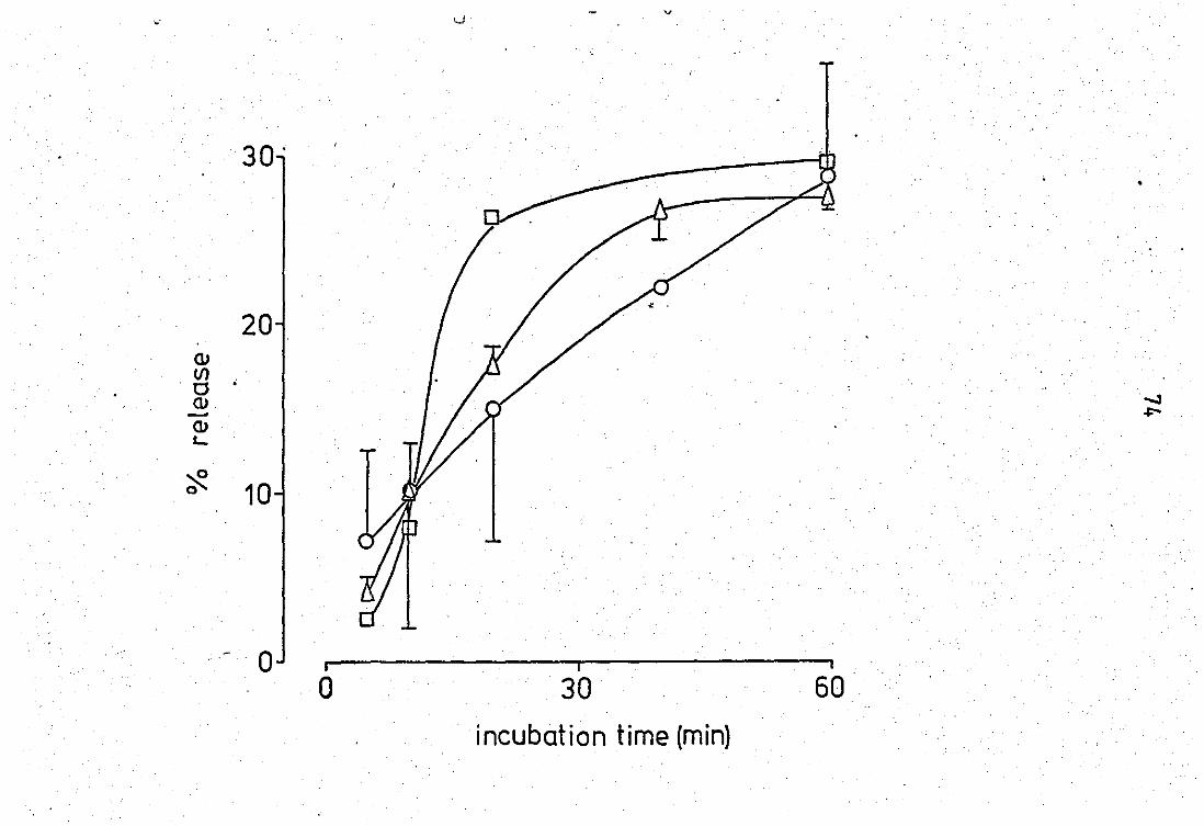
v ·.
30' •
20 Cl) I/)
·0 Q) -.J - ~ Cl) L..
~ 0 10
-·0 ~I--------------~I--------------~i o 30 60
incubation time (min)
75
The secretion of lysosomal constituents was also time
dependant. Release of enzymes was rapid during the first 10 mins and
then rose slowly reaching approximately 30% by 1 hour·(figure 3.3).
lysozyme andj3 - glucuronidase showed similar release patterns to phos
pholipase A.
The degree of phagocytosis (table 3.5) with time again.
showed a similar response demonstrating that release of phospholipase
activity occured during phagocytosis.'
2.2 Release of phospholipase by calcium ions
Addition of calcium ions to neutrophil leucocytes (which
were previously washed in a calcium free suspending medium) and incubation
at 37·C for 1 hour also c'aused the release of phospholipase A2 from
the cells. The release of this enzyme, (i) followed the release of fl -
glucuronidase and lysozyme but not of. lactate dehydrogenase, (ii) was
-3 2+ concentration dependant, 2.5 x 10 M Ca being sufficient to cause
maximal release of over 30% (figure 3.4) and (Ui) time 'dependant as
shown in figure 3.5.
2.3 Properties of the zymosan - complement released phospholipase A2-
of neutrophil leucocytes
The properties of the enzyme obtained from zymosan
complement release experiments were examined.
(i) Positional specificity
76
14 The enzyme caused release of (1 - C) oleic acid from
the 2 - position of membrane phospholipids of E.coli. All of the released
radioactivity was recovered in the fatty acid fraction and nonein the
lysophospholipids (figure 3.6). This suggested thut the enzyme was of
A2 specificity, These de terminations were carried out at pH 9.0.
furthur evidence for the A2 specificity of neutrophil
14 phospholipase was obtained using sonicated neutrophils and (1- C)
palmitic acid labelled E.coli.77% of the radiolabel .. io this case is
present in position 1 of E.coli phospholipids as determined by hydolysis
withLCrotelus adamenteus venom phospholipase A2 (page 66). The results
obtained are shown in table 3.6. The blank showed a non - enzymic hydr -
olysis of 2.4% over 1 hour. The sonicated enzyme preparation showed
S2.9% hydrolysis of which 20% of the radioactivity appeared in the
fatty acid fraction and 80% in the lysophospholipid fr£ction. As 77%
of the palmitic acid label was present in position 1 of E.coli phos -
pholipids then it appears that the neutrophil phospholipaseactiv!ty
is exclusively A2•
(ii) pH profile
A pH profile suggested the presence of two pH optima
as shown by figure 3.7. The enzyme had a broad acidic pH.optimum
covering.the range pH 6 -6.5 and an alkaline peak at pH 9.0. The
activity at pH 7.S .. was reproduc1bly lower than that obtained at pH,
6.0 or 9.0.
(iii) Calcium requirement
The released phospholipase activity had an absolute
77
requirement for calcium ions 'at both pH optima, reaching, maximal activity
2+ et 5mM (figures 3.8. and 3.~.). In the absence of added ca less than
half of the maximal activity was attained. This residual activity sugg -
ested the presence of endogenous calcium. Indeed addition of 5mM
ethylene diamine tetra acetic acid (EOTA) totally abolished all activity
(figure 3 •• ,,\).
(iv) Kinetics
rigure 3./0 and 3,'6 examine the effects of protein
and time respectively on the enzymic hydrolysis of E.coli phospholipids.
Enzymic activity was linear up to 64}'g protein and up to 40 minutes.
The effect of substrate was examined at concentrations
varying from 0 -20 I-J-M _" E.coli phospholipid. Using 96jJ-g enzyme protein
(figure 3.11) the hydrolysis reached a plateau at 20 "Mo",;, of phos -
pholipid. Lower amounts' of protein were generally used to allow
substrate concentrations to be et saturating levels, rigure 3.12 shows
the double reciprocal plot of data expressed in figure 3.11. The results
show an apparent Km value of 25 X 10-6 M phospholipid.
(v) 'Stability
Stability of the enzyme activity in HEPES buffered
medium was examined by storage at 4°C over 20 days (table 3~f). The
activity at pH 9.0 was relatively stable and lost approximately 20%
of ' its original activity over 20 days. Activity at pH 6.0 however was
much more-unstable and lost between 75 - 80% of its original activity
over the same time period.

"
FIGURE 3.6.
Hydrolysis of E.coli phospholipids with the
zymosan - complement released PLA of neutrophils.
Reaction mixtures contained 40 mM Tris pH 9.0, 96jU9
enzyme protein, 5mM Ca2+, 5 x 10⁸ autoclaved E.coli . . 14
labelled with:( 1 - C) oleic acid representing 10
nmoles phospholipid (PL).
Each point represents mean of duplicatedeterminations.
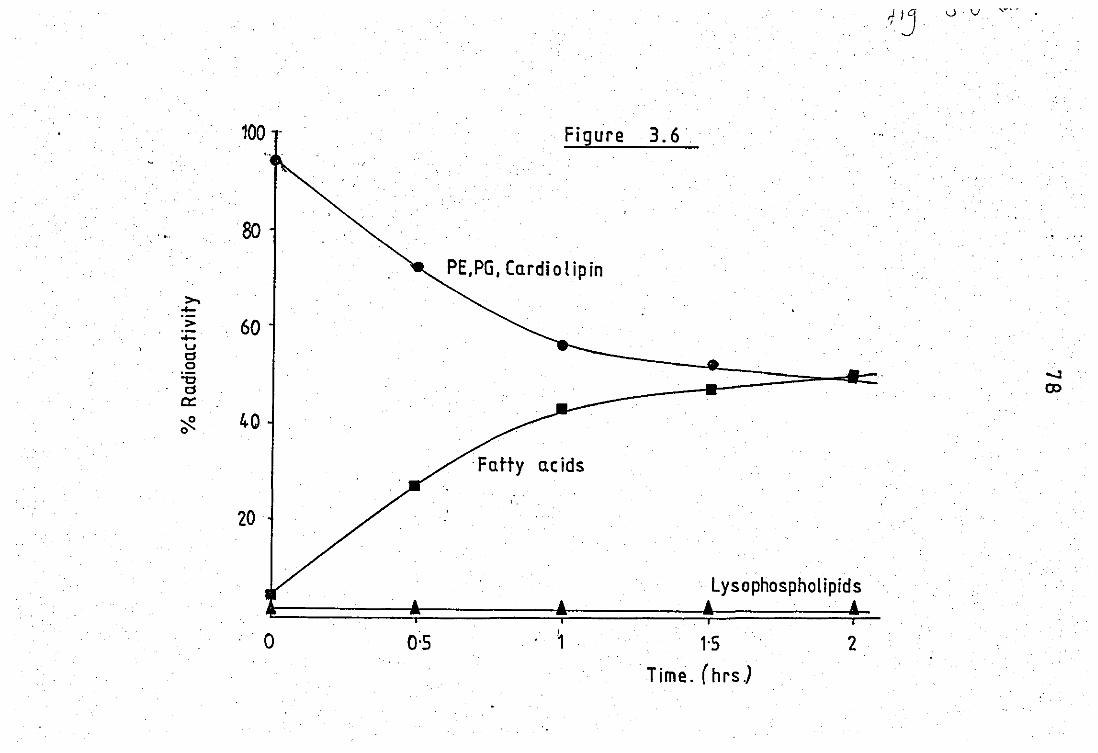
100 -
80
>. .0-
> 60 .0-U !=' 0
"C !='
c::: ~ 0
40
20
'0 0'5
Figure 3.6',
PE,PG, Cardiolipin
acids
. 1
Lysophospholipids
1'5
'Time. (hrs.)
2
.....:J co
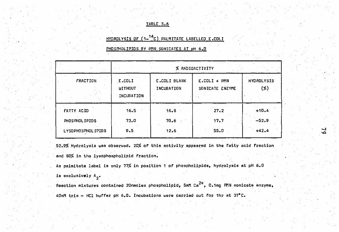
TABLE 3.6
HYDROLYSIS Of (1_14Cl PALMITATE LABELLED E.COLI
PHOSPHOLIPIDS BY PMN SONICATES AT pH 6.0
% RADIOACTIVITY
fRACTION E .COLI E.COLI BLANK E.COLI + PMN HYDROL YSIS
WITHOUT INCUBATION SONICATE ENZYME (%)
INCUBATION ..
fATTY ACID 16.5 15.8 27.2 +10.4
PHOSPHOL IPIDS 73.0 70.5 17.7 -52.9
LYSOPHOSPHOLIPIOS 9.5 12.6 55.0 +42.4 -
52.9% Hydrolysis was observed. 20% of this activity eppeared in the fatty acid fraction
and 80% in the lysophaspholipid fraction~
As palmitate label is only 77% in position 1 of phospholipids, hydrolysis et pH 5.0
is exclusively A2•
Reaction mixtures contained 20nmoles phospholipid, 5mM Ca2+, 0.1mg PMN sonicate enzyme,
40mM tris - HCl buffer pH 5.0. Incubations were carried out for 1hr at 37·C.

. fIGURE 3.7.
The effect of pH'on the activity of ZC -
released phospholipase A2 from neutrophil leucocytes. . . 2+
Reaction mixtures contained 60#9 protein, 5mM Ca ,
10 nmoles phospholipid and 40mMappropriate buffer
pH 4 - 6.0 sodium e'cetate, pH 5.5 - 7.5 Tris - malic;
pH 7.5 - 9.0 Tris - HCl. Incubations were for 1hr at 37°C.
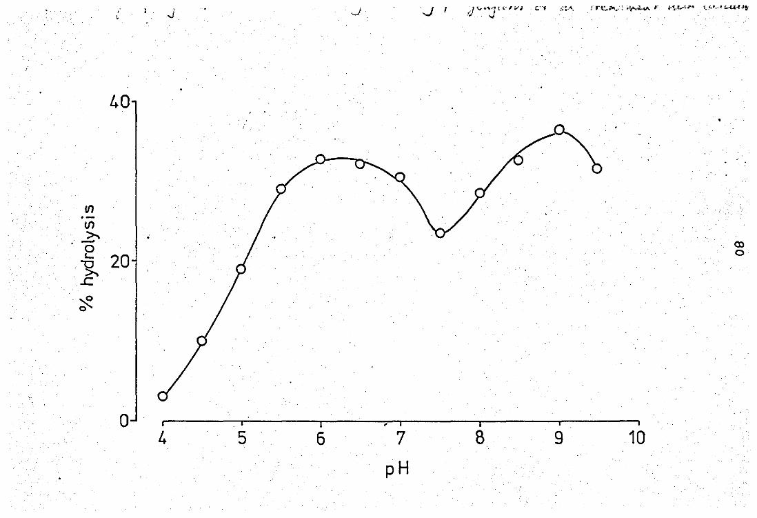
, , J
40
If) .-(/)
>-. , -0 ~ 20 'R
..c
~ 0
o 4
.J
. i
5 6 , i
7
pH
JI ;) ..... 1.>"' .. ; {;o., ..... J to'-' :;:"'''. rrt: ... ~~:: Hr.~~,"- r,,~.,A"· ~ .. (.," ... " ;:....u·..f..tt, . ...
•
CXl 0
I
8 9 10

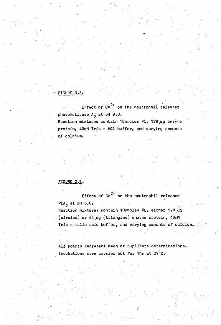
fIGURE 3.8.
Effect of Ca2+ on the neutrophil released
phospholipase A2 at pH 9.0.
Reaction mixtures contain 10nmoles PL, 128,1.19 enzyme
protein, 40mM Tris - HCI'buffer, and varying amounts
of calcium.
fIGURE 3.9.
Effect of Ca 2+ on the neutrophil released'
PLA 2 at pH 6.0.
Reaction mixtures contain 10nmoles PL, either 128,ug
'(circles) or 64,ug (triangles) enzyme protein, 40mM
Tris - malic acid buffer, and varying amounts of calcium.
All points represent mean of duplicate determinations.
Incubations were carried out for 1hr at 37·C.
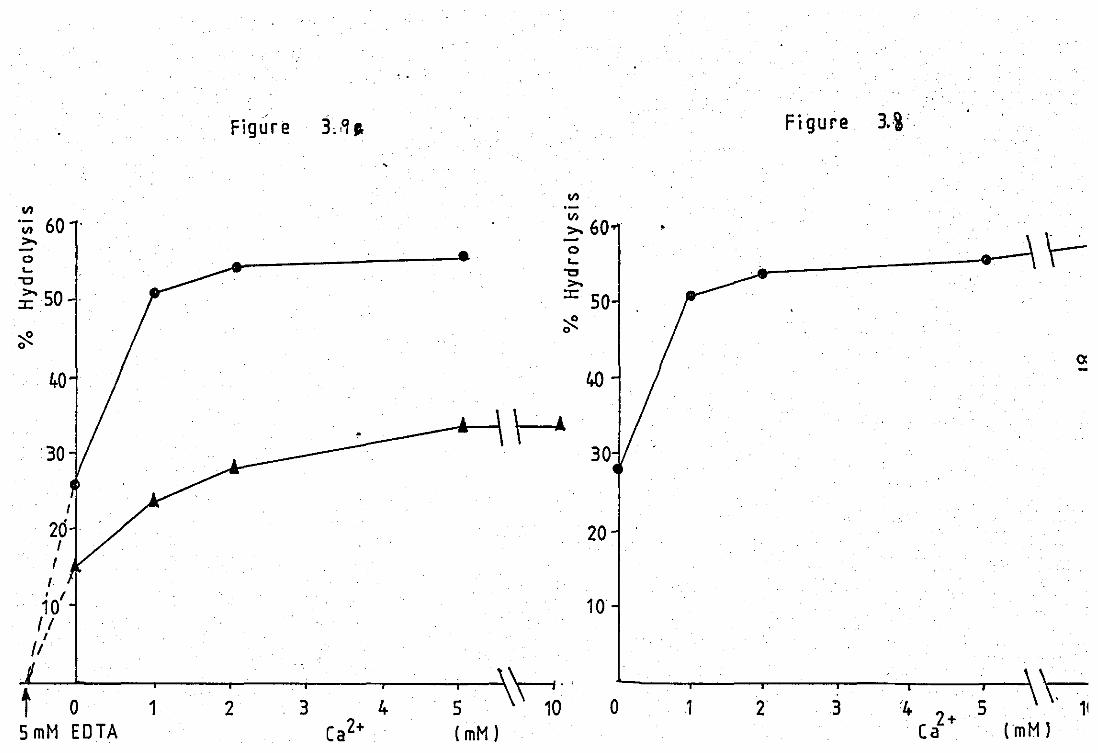
III .- 60 VI >. -0 '-"C
~50
~ 0
40
30
I
26
I
I , 10' (/ I
/1
..
Figur e 3,,If,.
III .-;;. 60 . -0 '-
't:J >.
:r:: 50 ~ 0
40
\-A
20
10
~t -O-'o.....--~1--2..---3-r----r4----;5----l,~O· , o
5 mM EOTA (mM)
Figure 3.i"
•
~
oc -
1 '4 2+.5 ~ Ca . (mM I
2 3

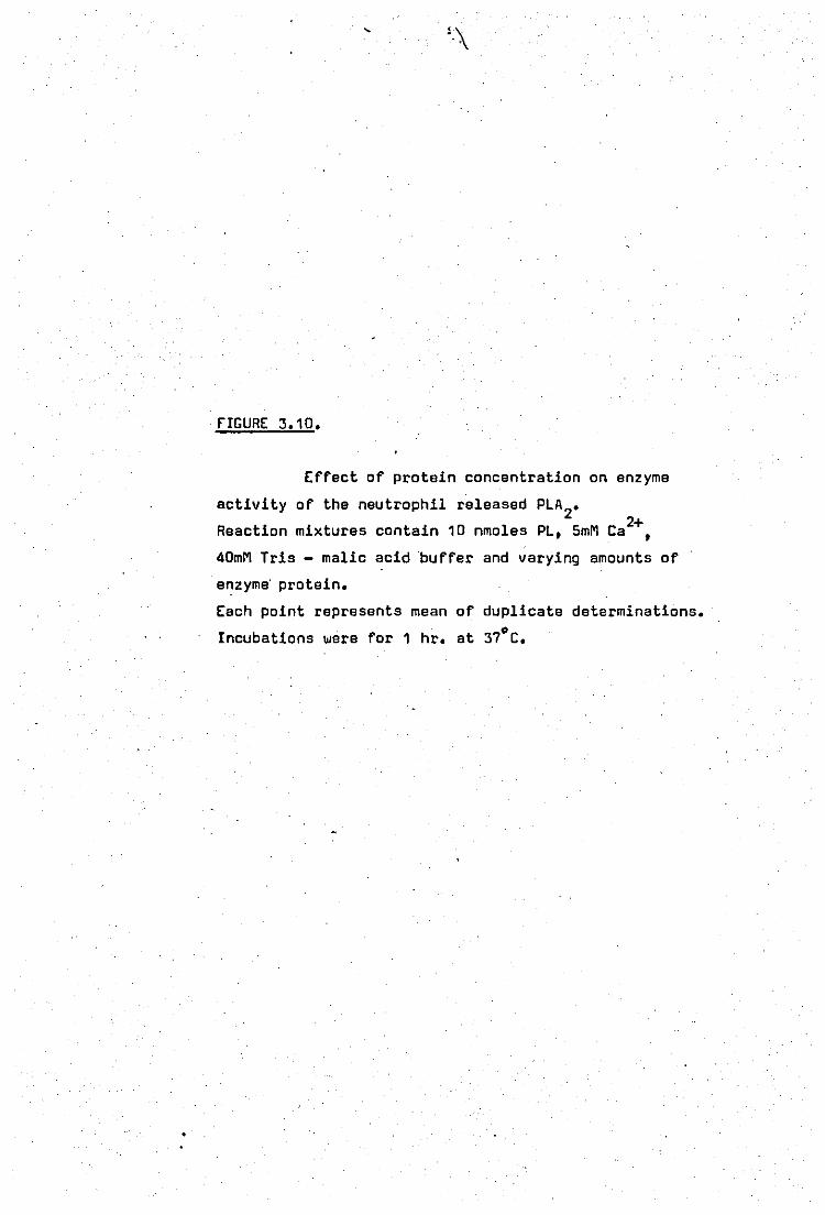
fIGURE 3.10.
Effect of protein concentration on enzyme
activity of the neutrophil released PLA 2•
Reaction mixtures contain 10 nmoles PL, 5mM Ca2+,
40mM Tris - malic acid 'buffer and varying amounts of
enzyme' protein.
Each point represents mean of duplicate determinations. '
Incubations were for 1 hr. at 37°C.
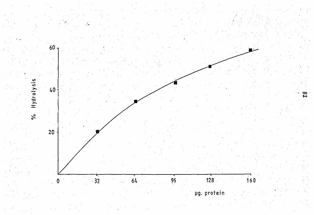
' ....
. . 60
VI .-VI >- 40 -0
. '. 00 . c... ,..,.
"C >-:c
~ 0
20
o ·32 6~ 128 160
pg. protein

j j j j j j j j j j j j j j j j j
J
j j j j j j j j j
j
, j
j j j
j
j j
j
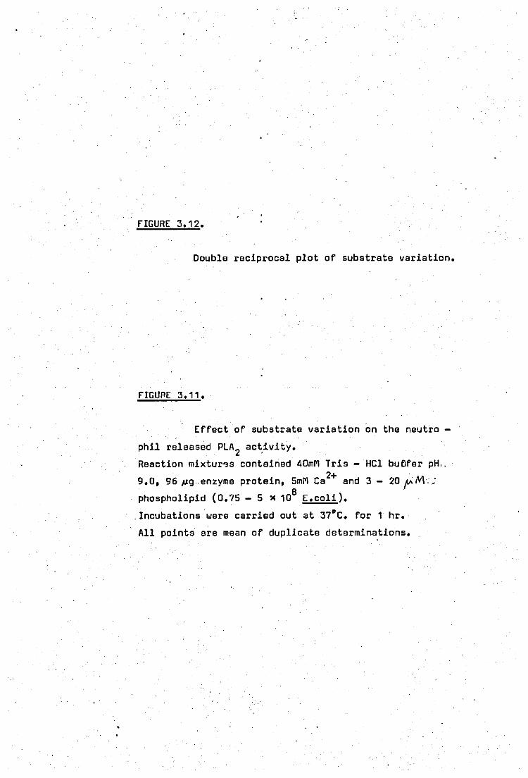
FIGURE 3.12.
Double reciprocal plot of substrate variation.
FIGURE 3.11.
Effect of substrate variation on the neutro -
phil released PLA2 act,ivity.
Reaction mixtur'9s contained 4DmM Tris - HCl butifer pH .. 2+
9.0, 96 P9 .. enzyme protein, 5mM Ca and 3 - 20 ~NL; . B phospholipid (0.75 - 5 x 10 E.coli) •
. Incubations were carried out at 37"C. for 1 hr.
All points are mean of duplicate determinations.
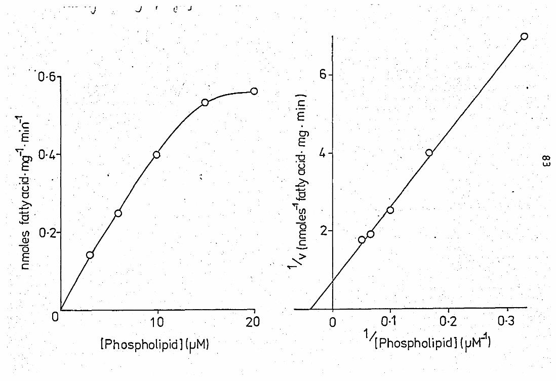
. .
"I c E . ~
~ •
:0 <J 0
..?:-S' C/l (I)
0 E c
0-6
0-4
0-2
J f (! J
.. ,
10
[Phospholipid] (~M)
20
6
-c ", .-
E • 0)
E . 4 :0 00 <J I..IJ
0
2:-...... J2
1 C/l (I) -0 E c -
'? ~
o 0-1 0-2 0-3 1 .' I( Phospholipid] (~M_1)
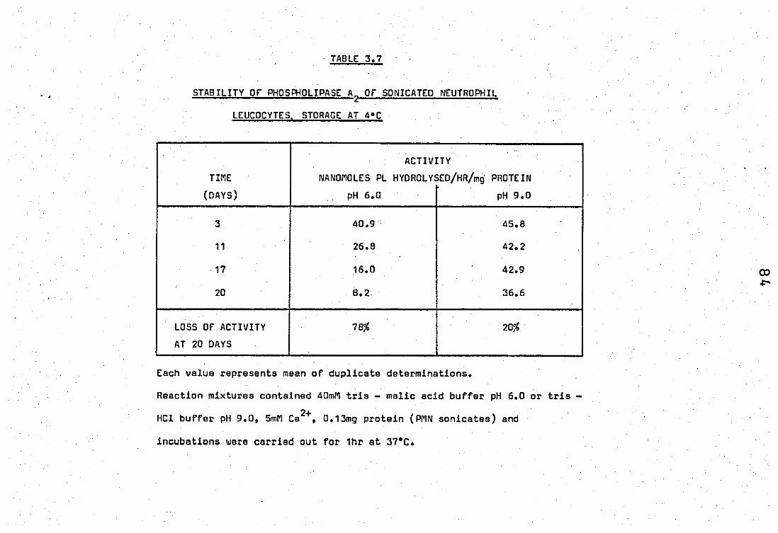
. .
TABLE 3.7
STABILITY or PHOSPHOLIPASE A2 or SONICATEO NEUTROPHIL
LEUCOCYTES. STORAGE AT 4°C
ACTIVITY
TIME NANOMOLES PL HYOROLYSEO/HR/mg PROTEIN
(OAYS) pH 6.0 pH 9.0
3 40.9' 45.8
11 26.8 42.2
17 16.0 42.9
20 8.2 36.6
LOSS OF ACTIVITY 78% 20%
AT 20 OAYS
Each value represents mean of duplicate determinations.
Reaction mixtures contained 40mM tris - malic acid buffer pH 6.0 or tris -
. 2+ HCl buffer pH 9.0, 5mM Ca ,O.13mg protein (PMN sonicates) and
incubations were carried out for 1hr at 37°C.
2.4 Properties of a phospholipase present in the rabbit inflammatory
PERITONEAL EXUDATE (PE)
A very active phospholipase activity was found in the
rabbit inflammatory peritoneal fluid supernatant after the invading
cells had been removed by centrifugation. The properties of this enzyme
were characterised.
(!) Effect oftims of incubation o"r .. ~ii~:ylne activity
The hydro~ysis,with time of 10 nmoles of (1 _14c) oleate
labelled E.coli phospholipid with 0.5mg enzyme pJ;:otein is shown in fig.
3.13. Enzyme activity was linear up to 10 minutes when 40% hydrolysis
r had occured, after which the activity levelled off reaching 53% by"30
1\
minutes. In subsequent experiments a 5 minute incubation period was used.
(ii) Effect of protein concentration on enzymic activity
figure 3.14 shows the enzyme',activity as a function
of protein concentration and as can be seen enzyme activity was linear
up to O.5mg'enzymeprotein under the conditions used.
(iii) Specificity
The enzymic hydrolysis again appeared to be of A2
specifidity as shown by figure 3.15. A decrease of radioactivity in
the phospholipid fraction was simultaneously recovered by an increase
in the fatty acid fractions but not in the lysophospholipid products.

rIGURE 3.13.
Variation of time on the peritoneal exudate
PLA2
activity.
Reaction mixtures contain 40mM Tris - HCl buffer pH .. B.O,
5 le 1 DB autoclaved labelled E. coli (10 nmoles PL),
0.5 mg enzyme protein, 5mM Ca2+; Incubations were at'
37°C for 5 mins.
rIGURE 3.14.
Effect of protein concentration; on the
peritoneal exudate PLA2
•
Reaction conditions as above except protein conce -
ntration was varied from 0 - .75 mg.
All points are mean of duplicate determinations.
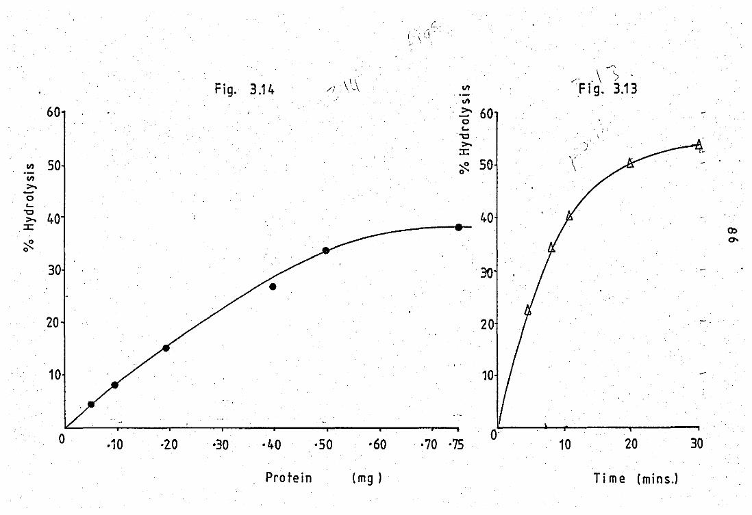
60
..., 50 '-VI >--0 L..
"'0 40 >-
:::c
~ 0
30
20
o
Fig. 3.14
·10 ·2.0 ·30 ·40
Protein
\ \ ' // '-''-\
·50 ·60
(mg )
VI
VI .. ~ 60
o L..
"'0 . >
:::c ~ 50 o
40
20
10
·70 ·75 o·
" ,\ '> Fig. 3.13
: , ',.
"; -' , , \
10 20 30
Ti me (mins.l

rrGURE 3.15.
Hydrolysis of'E.coli phosph~lipids by the
peritoneal exudate PLA2
•
Reaction, conditions are identiCal-, to figure 3.13.
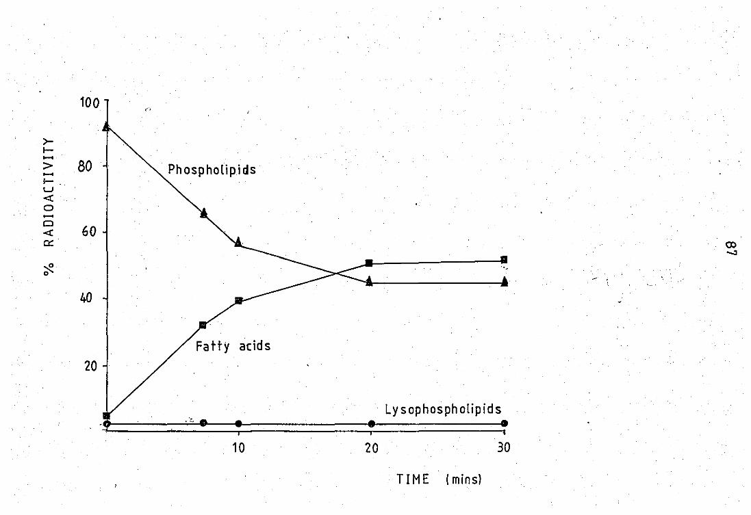
100 "
>-I-......
80 > Phospholipids ...... I-U 4: 0
. ...... Cl
60 4: ~ (X)
0000.3
~ 0
40
Fatty acids
20
Ly s op hos ph olip id s
10 20 30
TIME (mins)
86
(iv) pH ProFile
A pH proFile of the enzymic hydrolysis showed' two pH
optima at pH 8.0 and at pH 6.0 (figure 3.16). The acid peak was broad.
This pH profile was remarkably similar to that obtained with the neutr -
ophil released enzyme (figure 3.7) although the exact position of maxima
and minima of activity varied slightly.
"
(v) Calcium reguirement
figures 3.17 (a) and 3.17 (b) represent the calcium
requirement of the peritoneal Fluid phospholipase A2
• The enzymic activity
has an absolute requirement of Ca2+ as addition of greater than 150jAM
[OTA resulted in.a total loss of activity. Maximum hydrolysis occured
when ~mM Ca2+ was added to the reaction mixture. Larger amounts of
2+ added Ca caused slight inhibition of activity. The figure also shows
that on the absence of added calcium about 95% of the activity i~ still
2+ retained. This suggests the presence of sUFFicient endogenous Ca in
the peritoneal fluid to give almost total activity. This was supported
by the studies with [OTA which showed that increasing amounts of [OTA
caused phospholipase activity to decrease proportionately (figure 3.17
(b» •
As one [OTA molecule is able to bind to two divalent
cations, this suggests that the concentration of the latter was 300j'M
1n the reaction mixture. Flame photometer studies showed the calcium
concentration to be 200;.tM. This indicated that the peritoneal fluid
phospholipase .. ,ctivity rose very sharply with an increase of calcium
ions From 0- 0.2 mM, reached a maximum at 1.2 mM and thereaFter tne
activ~ty slightly decreased with large increases of calcium. These studies

fIGURE 3.16.
Effect of pH variation on the periton~al
PLA2•
Reaction mixtures contain 20 nmoles, 0.3mg enzyme
protein, 5mM ca2+ and 40 mM appropriate buffer.
pH 5.0
pH 7.5
7.5 Tris
9.0 Tris
Incubations were at
malic acid.
HC1.
37bC for 5 mins.
All points are mean of duplicate observations.
50
40
%Hydrolysis.
30
20
10
so ss 6·0 6'S 7·0 7'S 8·0 . 8'5 9·0 9-S
pH

fIGURE 3.17 (a) and (b).
2+ The effect of Ca. on the -peritoneal exudate
PLA2•
Reaction mixtures contain 23 nmoles PL, 40 mM Tris -
HCl buffer pH 6.0, 0.5 mg protein and either varying 2+ .
amounts of Ca or EOTA.
Incubations were carried out for 5 mins at 37°C.
All points are mean of duplicate determinations.
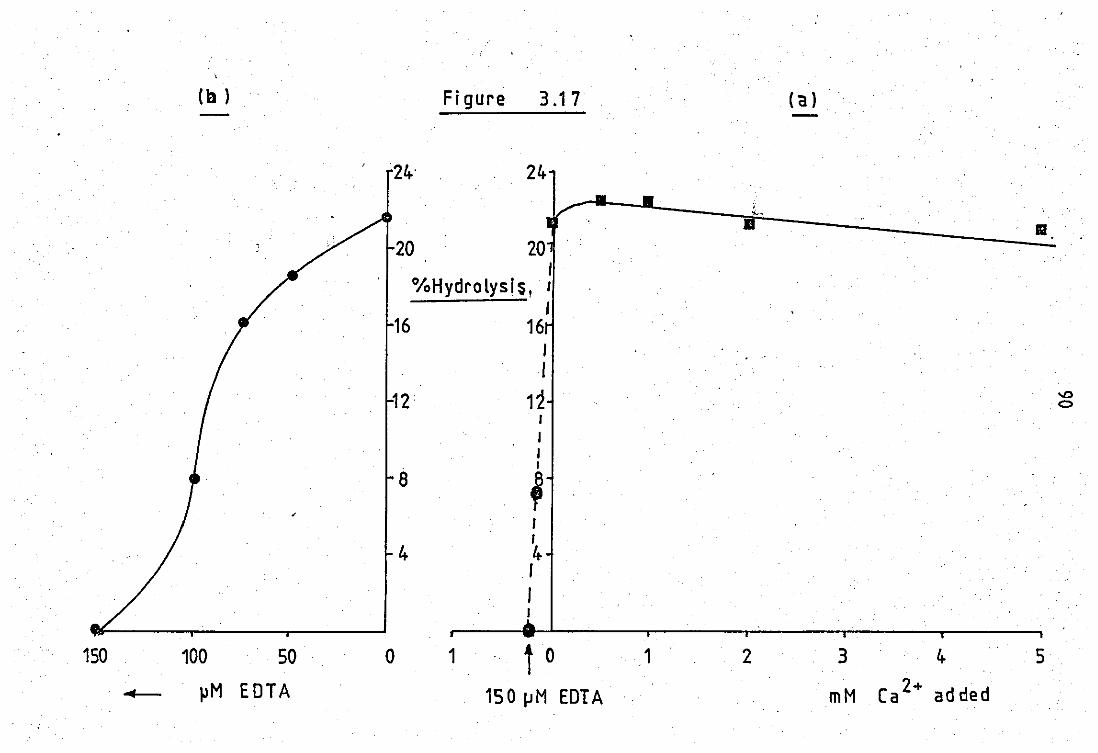
(ll ) Figure 3.17 (a)
24: 24-, .,;.-~
• 11 20 20 , %Hydrolysl~, '
J 16 16 ,
I
12 12 -.0 0
I , , I
8 , , I
4 4-I , I
150 100 50 0 1 t 0 1 2 3 4- 5
- ~M EDTA 150!JM EDTA mM Ca 2+ ad ded
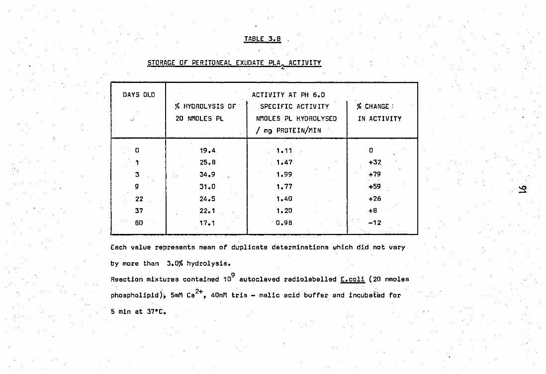
TABLE 3. B .
STORAGE or PERITONEAL EXUOATE PLA 2 ACTIVITY
DAYS OLD ACTIVITY AT PH 6.0
% HYDROLYSIS or SPECIfIC ACTIVITY % CHANGE:
o· 20 NMOLES PL NMoLES PL HYDROLYSED IN ACTIVITY
/ mg PRoTEIN/MIN .
0 19.4 1.11 0
1 25.B 1.47 +32
3 34.9 1.99 +79
9 31.0 1.77 +59
22 24.5 1.40 +26
37 22.1 1.20 +B
BD 17.1 . 0.98 -12
Each value represents mean of duplicate de terminations which did not vary
by more than 3.0% hydrolysis.
Reaction mixtures contained 109 autoclaved radiolabelled E.coli (20 nmoles
phospholipid), 5mM Ca2+, 40mM tris - malic acid buffer and incubated for
5 min at 37"C;
92
were carried out at pH 6.0.
(vi) Stability
o The effect of storage of the enzyme at 4 C is shown
in table 3.8. The enzyme activity at pH 6.0 was relatively stable over
a few months. However in the first few days an increase of specific
activity by as much as 79%. 'was observed at day 3. After this the enzyme
activity slowly decreased with tima.
"
9~
PART C EffECTS Of CHLOROQUINE - lIKE AGENTS ON INflAMMATORY AND
OTHER PHOSPHOlIPASES
3.1 Studies of effects of chloroquine - like agents on phospholipases
~2 from Crotalus adamenteus venom and porcine pancreas
(i) Optimal conditions for hydrolysis with Crotalus adamenteus venom
phospholipase A2
a) Calcium concentrations
figures 3.18 and 3.19 show the patterns of hydrolysis
obtained using this system and the optimal concentration of calcium
required for maximum enzymic activity. Reaction rates were linear in
the first 4 - 6 minutes and thereafter slowly levelled off. Initial
rates were taken and converted to micromoles fatty acid released per
minute per milligramme protein. Calcium ions were essential for enzyme
activity. Activity rose with an increase of Ca2+ concentration up to
6mM after which there was only a very slight increase of activity as
2+ shown in figure 3.19. Subsequently 8mM Ca was used in all reaction
mixtures.
b) Sodium deoxycholate
Approximately half of the total enzyme activity was
present in the absence of sodium dtoxycholate. Optimal activity was
obtained at a concentration of 1mM (figur~ 3.20). Higher concentrations

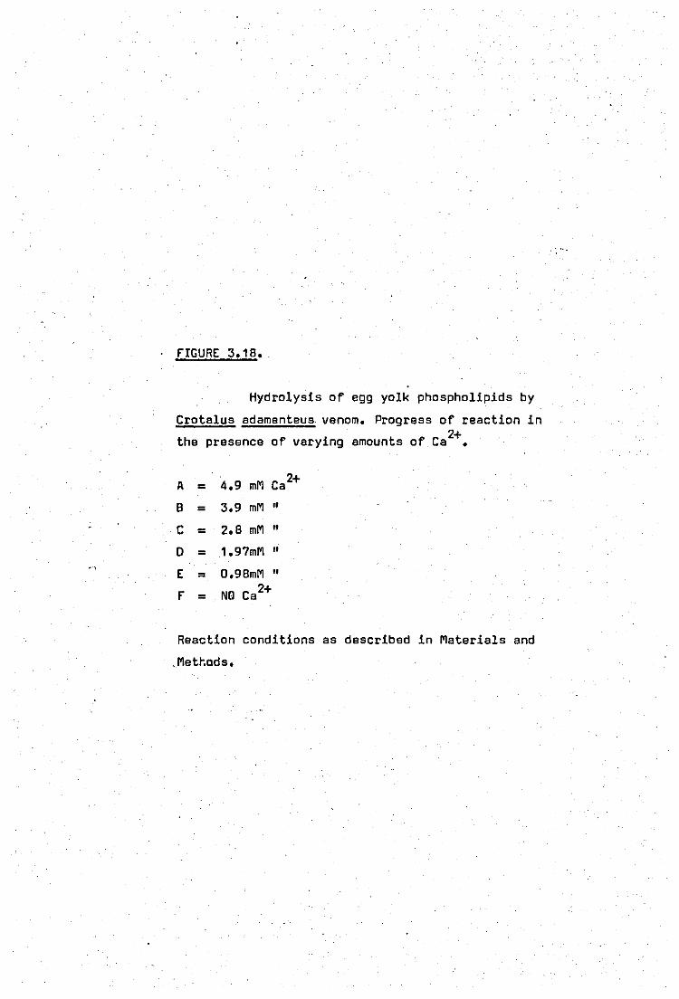
rIGURE 3.18 •.
Hydrolysis of egg yolk phospholipids by
Crotalus adamenteus. venom. Progress of reaction in 2+ the presence of varying amounts of Ca •
A = 4.9 mM Ca2+
B = 3.9 mM "
C = 2.8 mM "
0 = 1.97mM "
E = O.98mM "
r = NO Ca2+
Reaction conditions as described in Materials and
. Matt-.ads.
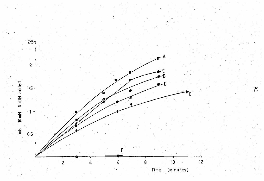
2-5
'2
"'C QJ.
"'C 1-5 -0 "'C +-,h"
tU E :c 0 tU z:
::E 1 E
C> .,....
vi -E 0-5
2 4 6 8 10 12
Time (m i nlltes J

rIGURE 3.19.
2+ . ;;E;f."ect o·f. Caveriation on the hydrolysis
of. egg yolk pho.spholipids by Crotalus adamenteus venom.
fIGURE 3.20 •.
Variation of deoxycholate on the· hydrolysis
. of egg yolk phospholipids by Crotalua adamenteus venom.
All points are means of duplicate determinations.
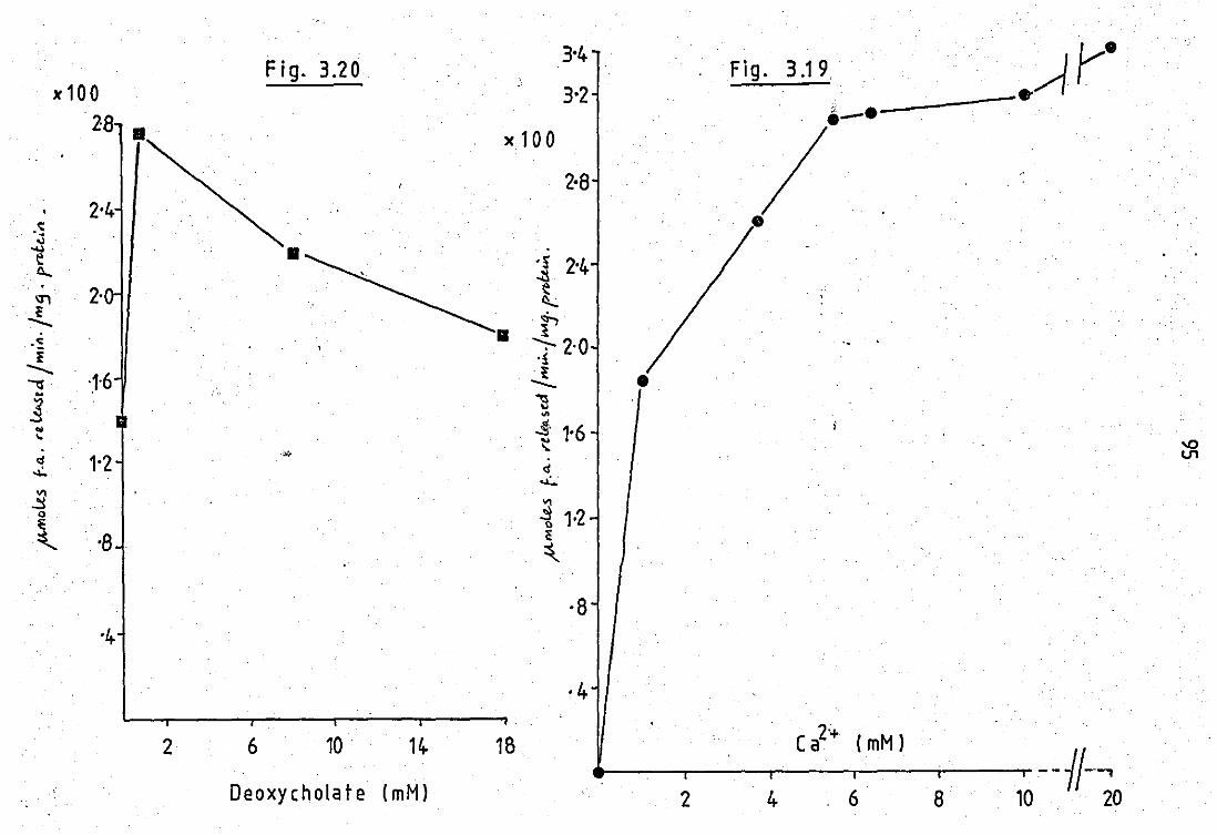
Fig. 3.20 3'4-
Fig. 3.19. ~r· ,,100 3,2 ... 28 i-e -
)(100
/ 2·8 2'4-
·s • " -u 11 e :1 2'4 tl....
IJ') 2· l ·i P --~~ . 11
~ 2·0 '. ~ '1 -...
'1'6 ~ e ~ --w ~ .§ " '" .. .:s' 1'6 '- t '" d 1'2
.", V1
..:. d ....:..
VI .!! VI
0 ~ 1·2 l: :$... '8 ~
~
·8 '4
·4
2'+ (mM) 2 6 10 14- 18 Ca --fl-~ .
Deoxycholate ( mM) 6 2 4 8 10 20

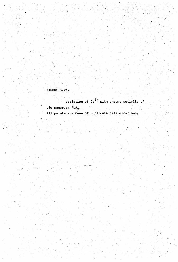
fIGURE 3.21.
2+ Variation of Ca with enzyme activity of
pig pancreas PLA2•
All points are mean of duplicate determinations.
. c: ill 4-0
x 103 L-
e. . Cl 1-8 E -c: .-E 1·6 -
"t:I QJ H. Vl
'" QJ --' QJ
1·2 '-
ro ....: Vl 1·0 QJ -0 E ~ '8
·6
·2
o 2
mM
6 8 10 12 14 16 . .
,.
. 2+ Ca .
18 20
I \
. I)
i
-.0 0-

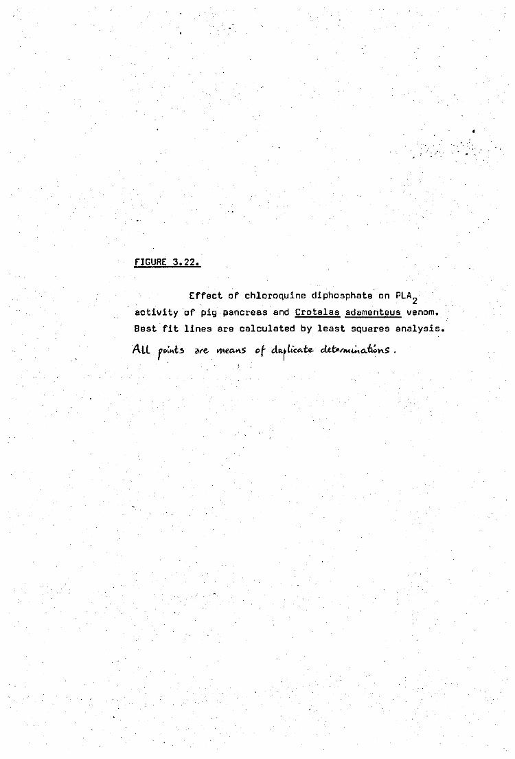
" .....
fIGURE 3.22.
Effect of chloroquine diphosphate on PLA2 activity of pig pancreas and Crotalas adamenteus venom.
Best fit lines are calculated by least squares analysis.
90
80
70
z 60 o
I--aJ 50 -::I: :z: - 4Q
30
20
10
•
o ,4 ",3 ... 2 -·1
•
Pancreatic
PLA 2
adamenteus PLAZ
o ·1 ·2 -3 '4 ·5. -6 -7.·-8
Log dose (mM)
98
actually caused a decrease in enzymechydrolysis and therefore a concen -
tration of 1mM was used in subsequent experiments.
(ii) Optimal conditions'for hydrolysis with pig pancreas phospholipase A2
Phospholipase activity from pig pancreas was analysed
at 37°C according to the method of Nieuwenhuizen, Kunze and Haas (1974).
Optimum calcium concentration was again 8mM (figure 3.21) and 1mM for
sodium deoxycholate.
(iii) Drug experiments
a) Inhibition of Crotalus adamenteus venom phospholipase A2 by
chloroquine diphosphate.
~hloroquine diphosphate showed a dose - related inhibition
of phospholipase A2 activity of Crotalus adamenteus venom over the range
o - 4mM as shown in figure 3.22. 50% Inhibition of enzyme activity was
echieved at 3.D',mM chloroquine.
b) Inhibition of pig pancreas phospholipase A2 by chloroquine
Inhibition of the purified pig pancreas enzyme was also
observed by chloroquine but at four fold lower doses (figure 3.22) .50%
Inhibition of activity as calculated from the best fit curve was obtained
at 0.7.imM.
c) Inhibition of pig pancreas phospholipase A2 by other agents
These were cinchonine sulphate and dibuceine hydrochloride
99
TABLE 3.9
ErrECT or INDOMETHACIN, SALICYLIC ACID, DIBUCAINE
AND CINCHONINE SULPHATE ON THE PHOSPHOLIPASE· rROM
PIG PANCREAS TOWARDS EGG YOLK EMULSIONS
INITIAL RATES % INHIBITION
(fL MOLES r.A. RELEASED
i MIN./mg. PROTEIN)
ACTUAL VALUE S MEAN
. CONTROL 6.06, 5.80 ~.
CINCHONINE SULPHATE 2.77, 2.63 hl.Q. 54%
(0.5mM)
DIBUCAINE ( 1mM) 4.23, 4.07 ~ 30%
CONTROL . 6.17, 6.37 ~ I NOOMETHAC I N (1mM) 5.36, 5.5 ~ 13%
(5mM) 5.23, 5.07 5.13 1B%
. SALICYLIC ACID 5.83, 5.57 hl 10% (1mM)
(5mM) 4.5, ,.4.23 ~ 30%
Phospholipase activities using egg yolk emulsions were determined as
described in Materials and Methods.
100
(closely related structures) and two anti - inflammatory agents; indo
methacin and salicylic acid. All four agents inhibited phospholipid
hydrolysis (table 3.9). Cinchonine'sulphate and dibucaine were more
effective than indomethacin and salicylic acid. Cinchonine sulphate
and dibucaine also caused a lag period of 3 minutes before maximum enzyme
activity was raached and th»s initial rates recorded were after this r
period. The order of potency of drugs acting on pancreatic phospholipasa
A2 was as follows:
. Cincbonine sulphate =- chloroquine diphosphata =- dibucaine hydrochloride
=- salicylic acid.::» indomethacin.
(d) Kinetics of inhibition of pig pancreas PLA2
by chloroquine
The nature of inhibition caused by chloroquine was
further investigated
(i) The variation of substrate and its effe'cts on the inhibition
of phospholipasa A2 activity by chloroquine is shown in figure 3.23 •
The doubla reciprocal plot shows tha two,curves run parallel to each e.
other indicating an uncomPAtitive inhibition with respect to phospholipid .' ,.
substrate.
". -' ".
(ii) It has been previously reported that calcium antagonises
the inhibitory' effects of local anaesthetics on phospholipase A2 of
human seminal plasma (Kunze ~,al, 1976). This finding was also invest -
igated using the present system. Figure 3.24 shows the double reciprocal
plot of the variation of calcium and its effeet on the inhibition of
pancreatic phospholipase A2 by O.75mM chlproquine and 1mM dibucaine.

fIGURE 3.23.
ad;vi~ Double reciprocal plot of e-nz..)"" ... -;.:.,. in the
presence and absence of D.75mM chloroquine.
Best fit lines are calculated by least squares analysis.
Poi ... ~$ _-: r~p'ts",,-t _ "'."'" or"t...pLi""'ft.. dw-,,,,,, t,:...s .
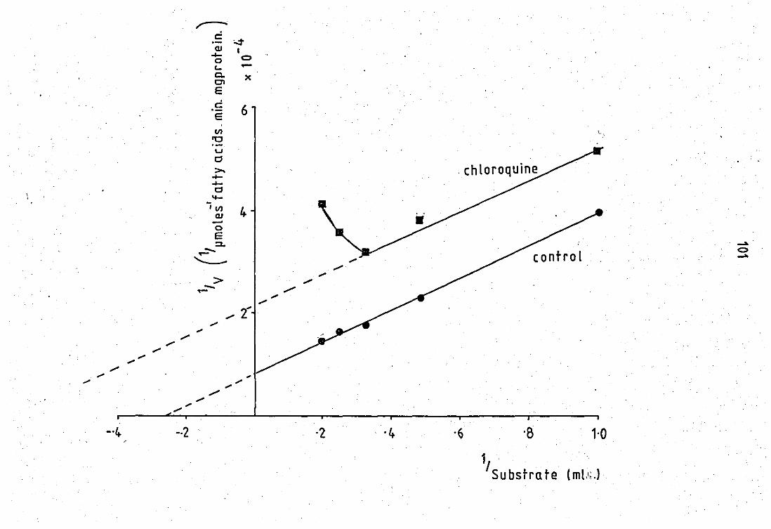
c:: cu ...... o <COl E
.~ E 11)
'0 u Cl >. ...... ...... Cl ....
ill) QJ -o E :::l.
~
o.:t I <:)
x
6
4
·2 ·4
c ..
control
·B 1·0
1, . Substra te (ml.'·;.)
..... <:) .....
. I
. I
I
I
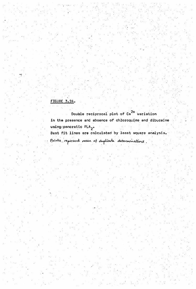
tIGURE 3.24.
2+ -Double reciprocal plot of Ca variation
in th& presence and absence of ch~oroquine and dibucaine
usinglpancratic PLA 2•
Best fit lines are calculated by least square analysis.
f'o,,,t,, .-r~re ~~"t. .... UVl. of clMj>t.c..fi.. el .. t:uv..,::.."ti .... s.
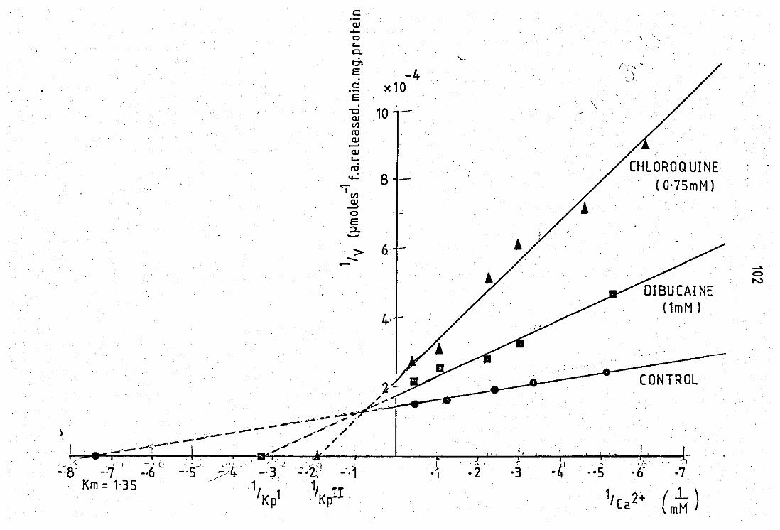
c: ,-aJ .... 0 \ 't-o. C'I (, , "
E -4 .. \
"'. I, c x10 E
\
'C 10 , aJ V)
ro aJ -aJ '-
CHLOROQ UINE n:i 'f- 8 (O'75mMl <-
'v) aJ -0 E :l..
> 6 --..... ->0 0 N
DIBUCAINE
4~ (1mM 1
f ,.' '~i .'
':'2,' -',1 'I '[,f " ' " . ,/'
·1 ·2 ·3 -4 . ·S .6' ·7 1, l! , Kp 1, Ca 2+ (m~ )
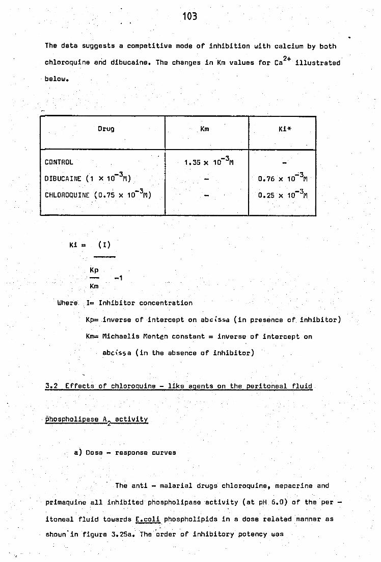
103
The data suggests a competitive mode of inhibition with calcium by both
chloroquine and dibucaine. The changes in Km values for CaZ+ illustrated
, balow.
Drug Km Ki*
CONTROL 1.35 x 10⁻³M -DIBUCAINE (1 X 10⁻³M) - 0.76 x 10";3M
CHLOROQUINE (0.75 X W-3M) - 0.25 X 10⁻³M
..
Ki = (I)
Kp -1
Km
Where 1= Inhibitor concentration
Kp=invarse of intercept on abcl$sd (in presence of inhibitor)
Km: Michaelis Menten constant = inversa of intarcept on
abci,ss a (in the absence of inhibitor)
3.2 Effects of chloroquine - like agents on the peritoneal fluid
phospholipase AZ activity
a) Dose - response curves
The anti - malarial drugs chloroquine, mepacrine and
primaquine all inhibited phospholipase activity (at pH 6.0) of the per -
itoneal fluid towards E.coli phospholipids in a dose related manner es
shown' in figure 3.25a.' 'The 'order of inhibitory potency was
•

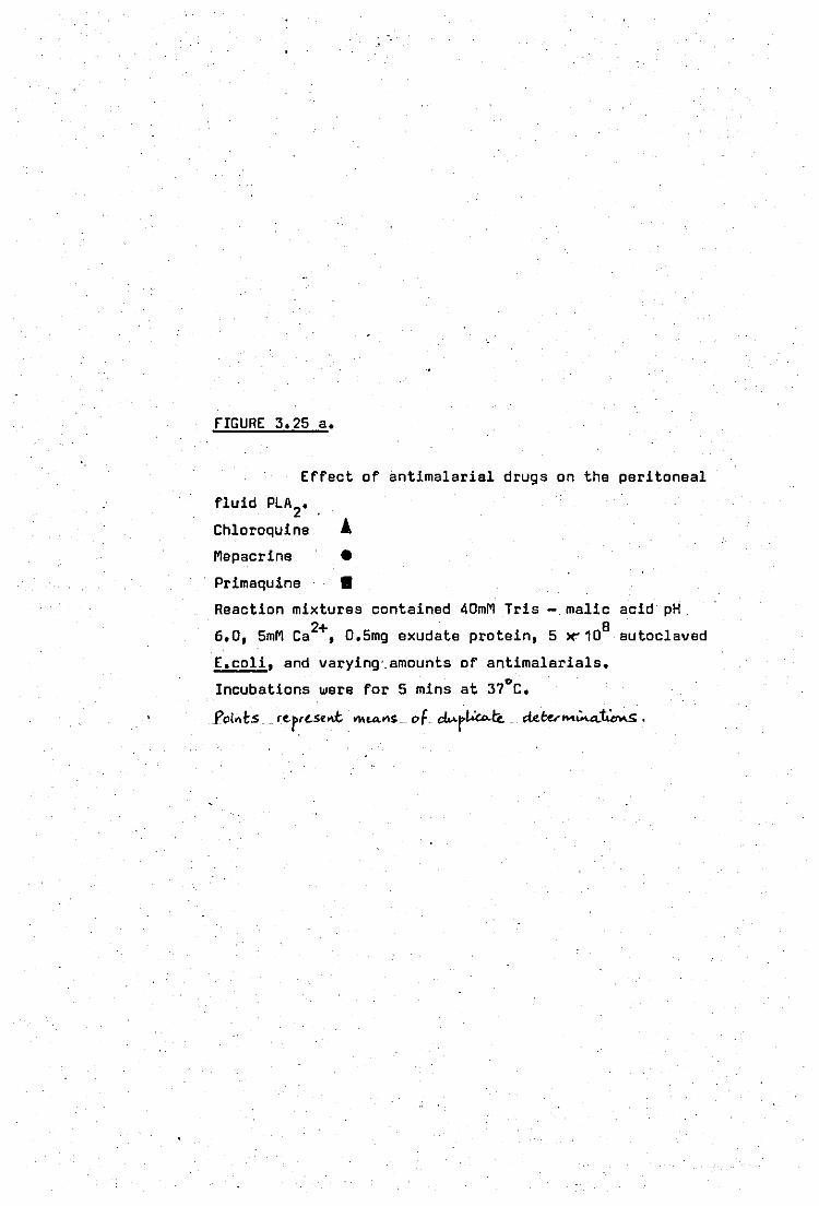
fIGURE 3.25 a.
Effect of antimalarial drugs on the peritoneal
fluid PLA 2•
Chloroquine A Mepacrine •
Primaquine •
Reaction mixtures contained 40mM Tris
6.0, 5mM Ca2+, 0.5mg exudate protein,
malic 8
5 le'10
E.coli, and varying'. amounts of antimalarials.
Incubations were for 5 mins at 37°C.
acid pH
autoclaved
J'oints .. rc.prc.se..t. ""U\."'~. of "{"'r/.."''' . .tetc.r .... .:...,,:t."'-s.
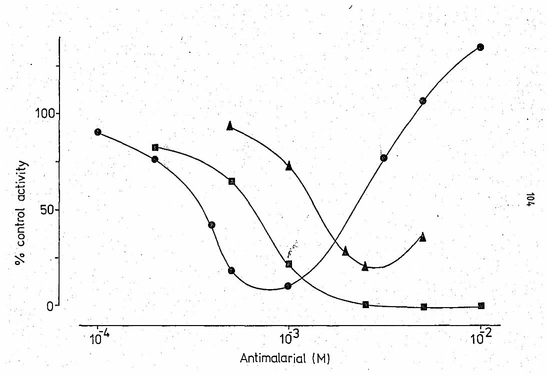
/
100 . .
>.. ..... . -> .-..... (J
-" 0 0
50 ""'" -e ..... c 0 (J
~ 0
o i
103
Antimalarial (M)
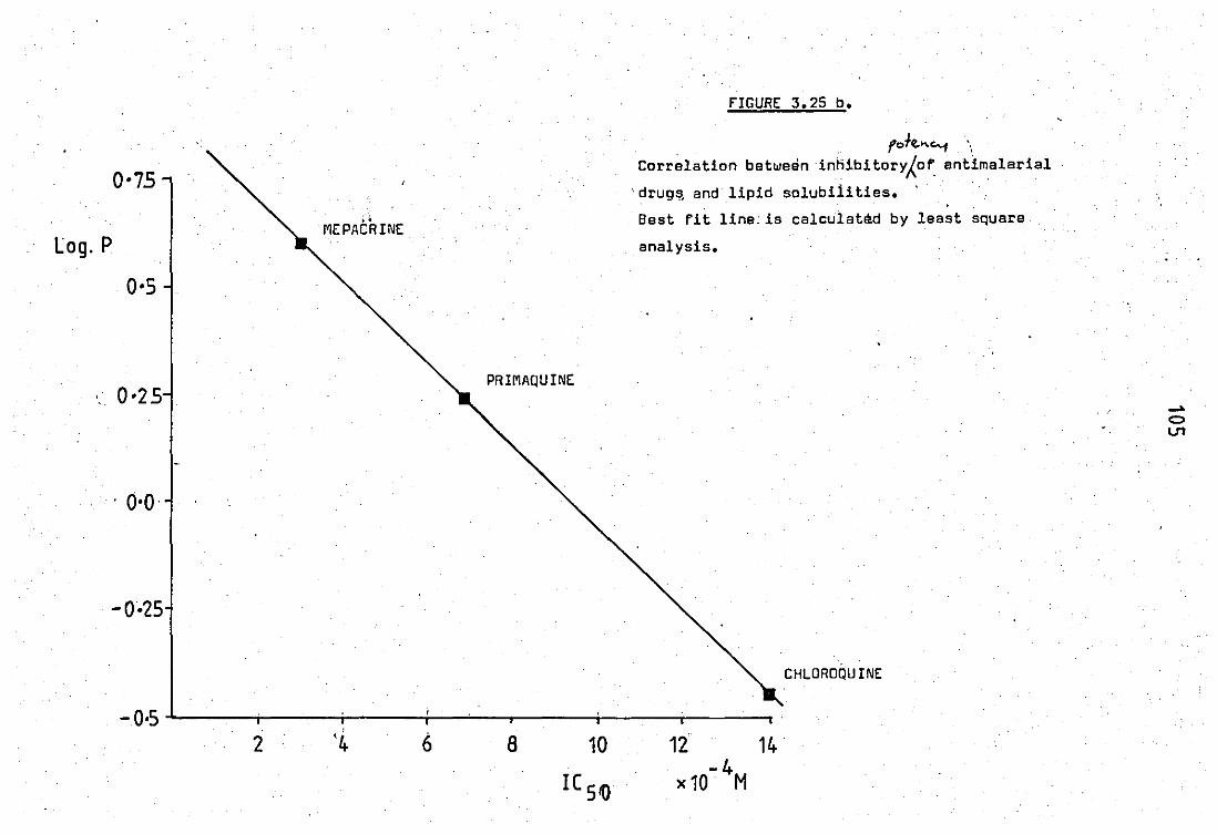
0·75
~ ~
MEPACRINE Log. P
0·5
' .. 0·25 PRIMAQUINE
FIGURE 3.25 b.
f oh.,,"'-1 \ Correlation betweeninhlbitoryAof. entimalarial
'drugs, and lipid solubiii ties. .
Best fit line: is calculatltd by least squara
analysis.
CHLOROQU INE
-0·5 ~---,--+---;---+---'--r-----+----i------t 2 '4 8
-" <=> 1ft
106
mepacrine ==- primaquine;.. chloroquine, and correlated well wi ththe lipid
Bolubilities of the drugs (figure 3.25b).
Inhibition of peritoneal fluid phospholpase A2 by antimalarial drugs
.
DRUG IC 50 Log P
'I'IE'PACR'I NE "3,"3 X '10-41'1 0.59
PRIMAQUINE 5 X 10-41'1 0.23
CHLOROQU INE 1.5 X 10⁻³¹'1 -0.45
.
Log P value represent the partition of 10jAM antimalarials between
chloroform and tris - malic acid buffer at pH 6.0.,
With mepacrine and chloroquine at doses larger than those causing
maximum inhibition, a reversal of the effect was seen. This even
resulted in stimulation of enzyma activity if high doses were used
( + 31% with 10mM mepacrine). This reversal of inhibition at higher
than inhibitory doses was not observed with primaquine.
"lb}.::Effecbof"pH Yen,.the inhibition of phospholipase A2 activity
by chloroquine
The degree of inhibition by chloroquine was dependant
on the pH. of: the reaction medium. Table 3.10 gives the results obtained
using 1mM chloroquine with the pH varied between pH 6.0 to pH 8.5. As
can be seen inhibition was found to increase with increasing pH. ThUs
least inhibition (16%) caused by 1mM chloroquine was found at pH 6.5
, .
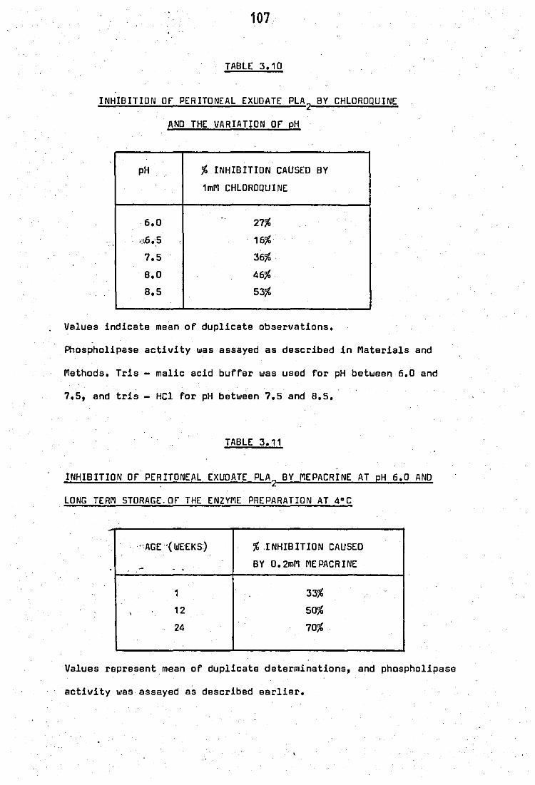
107
TABLE 3.10
INHIBITION OF PERITONEAL EXUOATE PLA 2 BY CHLOROQUINE
ANO THE VARIATION OF pH
pH % INHIBITION CAUSED BY
1mM CHLOROQUINE
6.0 '. 27%
. ~_t6.:5 16%
7.5 36%
B.D 46%
B.5 53%
Values indicate mean of duplicate observations.
Phospholipase activity was assayed as described in Materials and
Methods. Tris - malic acid buffer was used for pH between 6.0 and
7.5, and tris - HCl for pH between 7.5 and B.5.
TABLE 3.11
INHIBITION OF PERITONEAL EXUOATE PLA 2 BY MEPACRINE AT pH 6.0 AND
LONG TERM STORAGE, OF THE ENZYME PREPARATION AT 4°C
'AGE'( WEEKS) % INHIBITION CAUSED
~ BY 0.2mM MEPACRINE
, ,
1 33%
, 12 50%
24 70%
Values represent mean of duplicate determinations, and phospholipase
activity was assayed as described earlier.

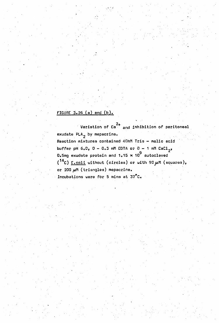
... >' ,:
FIGURE 3.26 (a) and (b).
2+ Variation of Ca and inhibition of peritoneal
exudate PLA 2 by mepacrine.
Reaction mixtures contained 40mM Tris - malic acid
buffer pH 6.0, 0 - 0.3 mM EOTA or 0 - 1 mM CaC1 2, 9 0.5mg exudate protein and 1.15 x 10 autoclaved
( 14C) E.coli without (circles) or with 90JAM (squares),
or 200~M (triangles) mepacrine.
Incubations were for 5 mins at 37·C.
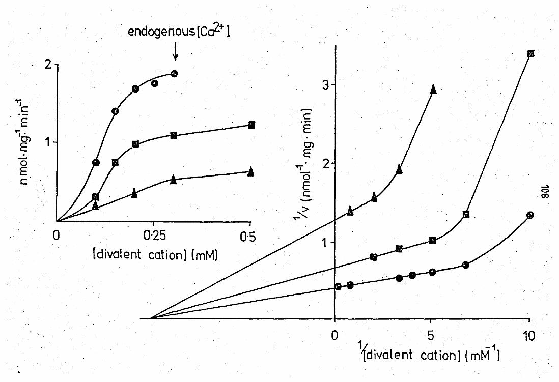
endogenous [ca2+ )
2 ~ 3
.,.., .
'e .-e E • .-
E '<j. . 0> 1 0> E E . - 2 0 ... E -0 e E ->. 0 e co _.
:<" """
0 0·25 0·5 [divalent cation] (mM)
o 5 Ydivalent cation] (mM"1)
10

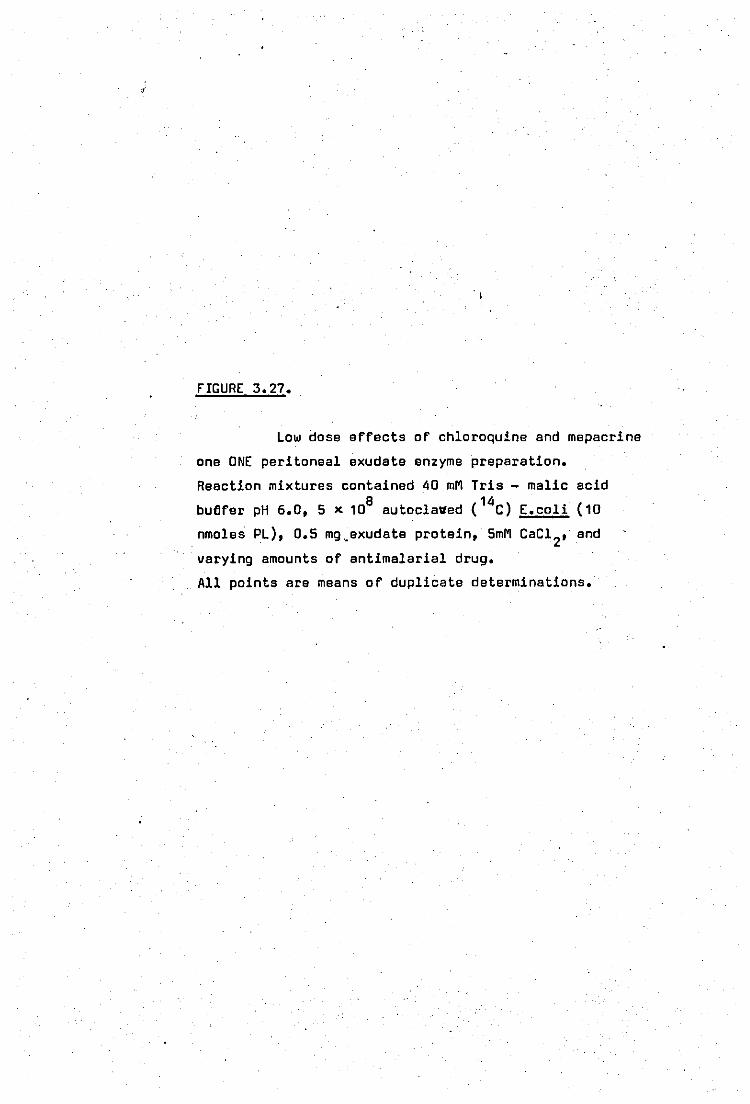
0,
FIGURE 3.27.
Low dose eFFects of chloroquine and mepacrine
one ONE peritoneal exudate enzyme preparation.
Reaction mixtures contained 40 mM Tris - malic acid
bu6Fer pH 6.0, 5 x 10⁸ autoclaved ( 14C) E.coli (10
nmoles PL), 0.5 mg.exudate protein, 5mM CaC1 2, and
varying amounts of antimalarial drug.
All points are means of duplicate determinations.
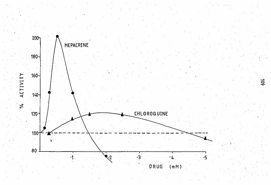
2 MEPACRINE
160
>- 160 I-, .... .... > 0
-0 .-I-u
1ltO «
~ 0
120 CHLOROQUINE
. 100 -
80~--~----~------~--------~--------~------~ -1 -3 -4- -5
DRUG (mM)
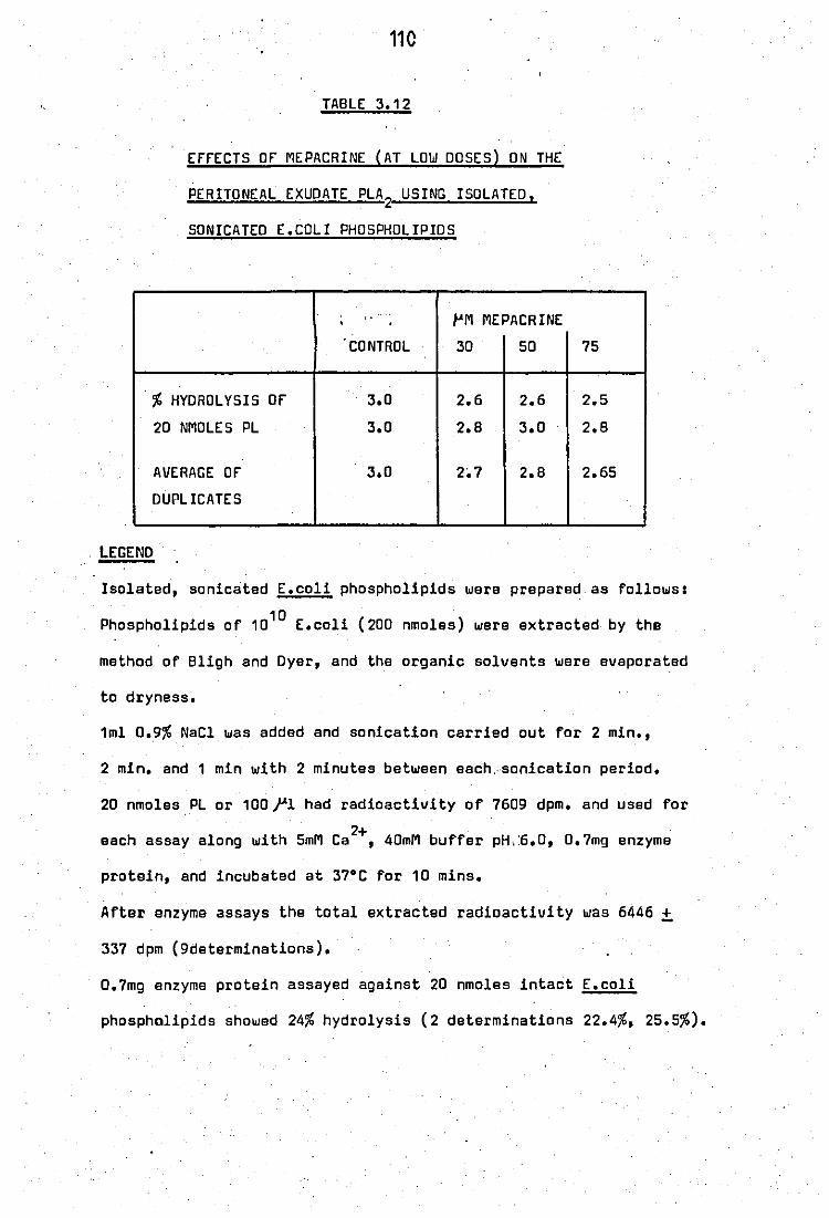
110
TABLE 3.12
ErrECTS or MEPACRINE (AT LOW DOSES) ON THE
PERITONEAL EXUDATE PLA2 USING ISOLATED.
SONICATED E.COLI PHOSPHOLIPIOS
, to ", • PM MEPACRINE
CONTROL 30 50
.
% HYDROLYSIS or 3.0 2.6 2.6
20 NMOLES PL 3.0 2.8 3.0
AVERAGE or 3.0 2~7 2.B
OUPLICATES
LEGEND
75
2.5
2.8
2.65
Isolated, sonicated E.coli phospholipids were prepared as follows:
Phospholipids of 1010 E.coli (200 nmoles) were extracted by the
method of Bligh and Oyer, and the organic solvents were evaporated
to dryness.
1ml D.9% NaCl was added and sonication carried out for 2 min.,
2 min. and 1 min with 2 minutes between each. sonication period.
20 nmoles PL or 100},1 had radioactivity of 7609 dpm. and used for
2+ each assay along with 5mM Ca ,40mM buffer pH. :6.0, 0.7mg enzyme
protein, and incubated at 37·C for 10 mins.
After enzyme assays the total extracted radioactivity was 6446 ±
337 dpm (9determinations).
0.7mg enzyme protein assayed against 20 nmoles intact E.coli
phospholipids showed 24% hydrolysis (2 determinations 22.4%, 25.5%).
111
whilst at pH 8.5 the same level of drug caused 53% inhibition.
c) Age of enzyme preparation
The age of the enzyme preparation was also found to
have a measurable effect on the inhibitory activity of mepacrine (table
3.11). Initially enzyme activity in the presence of 0.2mMmepacrine
was 67% of control values arid fell to 30% after 24 weeks.
d) The effect of variation of calcium on the inhibition of
'peritoneel fluid phosphblipase A2 by mepacrine
The Kinetics of calcium variation on the inhibitory
property of mepacrine on this system are shown in figures 3.26(a)and
M. Sigmoidal curves are obtained both in the presence and absence of
mepacrine (figure 3.26a). The data in the presence of less than 0.3mM
divalent cation concentration was obtained by adding varying concen -
trations of EOTA. With the data expressed as a double reciprocal
plot, it appeared that calcium interfered with the inhibition by
mepacrine in a non- competitive manner, and contrasted with the results
obtai:ned •. using .;panoreati:c,phosphol1pase A2 and egg - yolk substrates.
e) Low - dose stimulatory effects by chloroquine and mepacrine
On one peritoneal fluid enzyme preparation, at doses
lower than those causing inhibition, choloroquine (250~M) and mepacrine
(50~M) stimulated enzyme activity at pH 6.0 (figure 3.27). The greatest
effect was seen with mepacrine at 50~M where a 100% stimUlation of
enzyme activity was observed. Chloroquine produced a 20% stimulation
112
at 250;UM. This effect was ~eproducible only on this particular enzyme
preparation and could not be observed on other preparations. These
effects were reproducible up to 2 months storage of the enzyme prep
aration but were lost after this period.
Previous workers (Sherphof and Westenberg, 1975) have
observed similar inconsi~ences when working with pancreatic phospholi
pase A2 and labelled mitochondrial lipids. Such effects were explained
by the nature of theint<erfacl;<al<"c'substrat'e c<'8nd'we<renot,'obtained with
purified lipids. In our system using isolated E.coli phospholipids as
sonicated, micellar disparsions failed to reproduce the low dose
, stimulatory affects by mepacrine and chloroquine that was seen en one
peritoneal fluid enzyme preparation. Table 3.12 shows the data obtained
using ,the isolated E.coli phospholipids, and as can be seen low doses
of mepacrine does not have much effect. The results also show an 8 -
fold reduction of hydrolytic activity when micellar dispersions of
phospholipids are used as compared with intact membrane phospholipids.
f) Effects of other anti - inflammatory and anti - arthritic
<agents on the peritoneal fluid phospholipase A2
Table 3.13 shows the data obtained with a variety of drugs
-3 using 10 M as the drug concentration. The anti - arthritic drugs sodium
auroth!omalate and 0 - penicillamine were without effect but the
anti - inflammatory agents did show inhibitory effects. Indomethacin
(93%) and benzydamine (81%) were particularly effective.
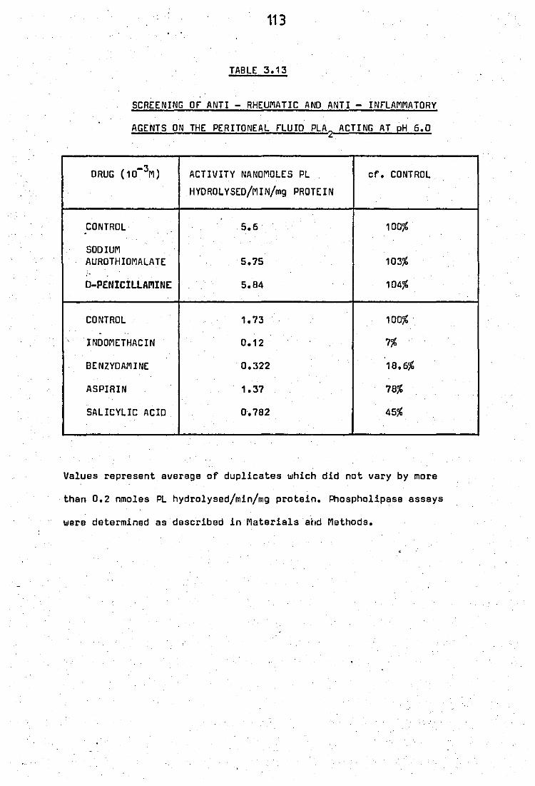
113
TABLE 3.13
SCREENING OF ANTI - RHEUMATIC AND ANTI - INFLAMMATORY
AGENTS ON THE PERITONEAL FLUID PLA2
ACTING AT pH 6.0
DRUG (10-3M) ACTIVITY NANOMOLES PL cf. CONTROL
HYDROLYSED/MIN/mg PROTEIN .
.
CONTRDL .5.6· 100% -SODIUM AUROTHIOMALATE 5.75 103%
D-PENICILLAMINE 5.84 104% . .
CONTROL 1.73 100% . -
INDOMETHACIN 0.12 7%
8E NZYDAM I NE 0.322 18.6%
ASPIRIN 1.37 78%
SALICYLIC ACID 0·.782 45%
Values represent average of duplicates which did not vary by more
than 0.2 nmoles PL hydrolysed/min/mg protein. Phospholipase essays
were determined as described in Materials aitd Methods.
.
114
3.3 Effects of chloroquine - like agents on the phospholipase activity
of polymorphonuclear leucocytes
a) Studies on sonicated neutrophil suspensions
The experiments were first carried out on neutrophil
suspensions that had been sonicated to release all the intracellular
enzymes. The effects of chloroquine",>,mapacl'ine'and primaquine on the
phospholipase A2 activity at pH 6.5 towards E.coli phospholipids are
shown in figure 3.28. Both chloroquine (0.25 - 10mM) and mepacrine
( 0.025 - 5mM) showed a dose - related stimulation of phospholipase
activity. Mepacrine was 10 times more active than chloroquine. No inhib -
ition of phospholipase activity was observed at any time of the conce -
ntrations studied.
Primaquine on the other hand showed a different res
ponse. At doses between 0.1 - 1.0mM a slight stimuletory effect was
recorded but, at higher levels a strong inhibition was observed.
Phospholipase A2 activity released from neutrophils
suspended in water also showed stimulatLon by.,O.5mM.mepacrine
(table 3.14).
b) Effect of mepacrine on the phospholipase activity of neutrophil
8200g granules
A purer preparation of neutrophil phospholipase A2
was obtained by centrifugation of a neutrophil homogenate in sucrose

FIGURE 3.28.
Effects of antimalarial drugs on the PLA
ectivity of sonicated neutrophils.
Reaction mixturas contained 0.1 mgl.'Q'1zyme protein,
109 autoclaved labelled E.coli (20 nmoles PL), 5mM 2+ Ca ,40mM Tris buffer pH 6.5, and varying,amounts
2
of antimalarial drugs. Incubations were for 30 mins
at 37·C.
All points are means of duplicate determinations.
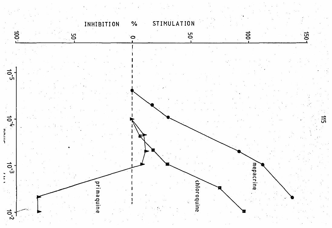
, , -,
.....
..... 0,
U'I
o o , .
U'I o ,
INHIBITION
o t
STIMULATION U'I o
..... o o
..... U'I o ,
-" -" U'I
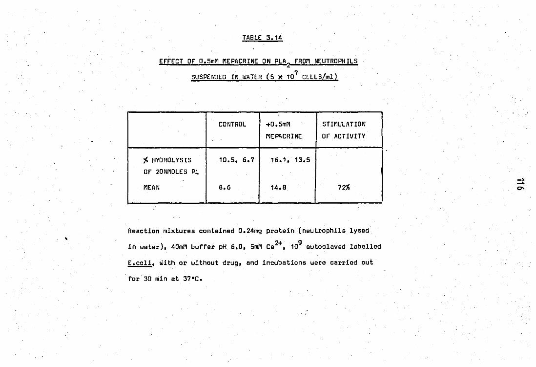
,
TABLE 3.14
ErrECT or 0.5mM MEPACRINE ON PLAZ
rROM NEUTROPHILS
SUSPENDED IN WATER (5 ~ 107 CELLS/ml)
CONTROL +0.5mM STIMULATION
MEPACRINE or ACTIVITY
% HYDROLYSIS 10.5, 6.7 16.1, 13.5
or 20NMOLES PL
MEAN B.6 14.B 72% .
Reection mixtures conteined 0.24mg protein (neutrophils lysed
2+ 9 in water), 40mM buffer pH 6.0, 5mM Ca ,10 autoclaved labelled
E.coli, with or without drug, and incubations were carried out
for 3D min at 37°C.
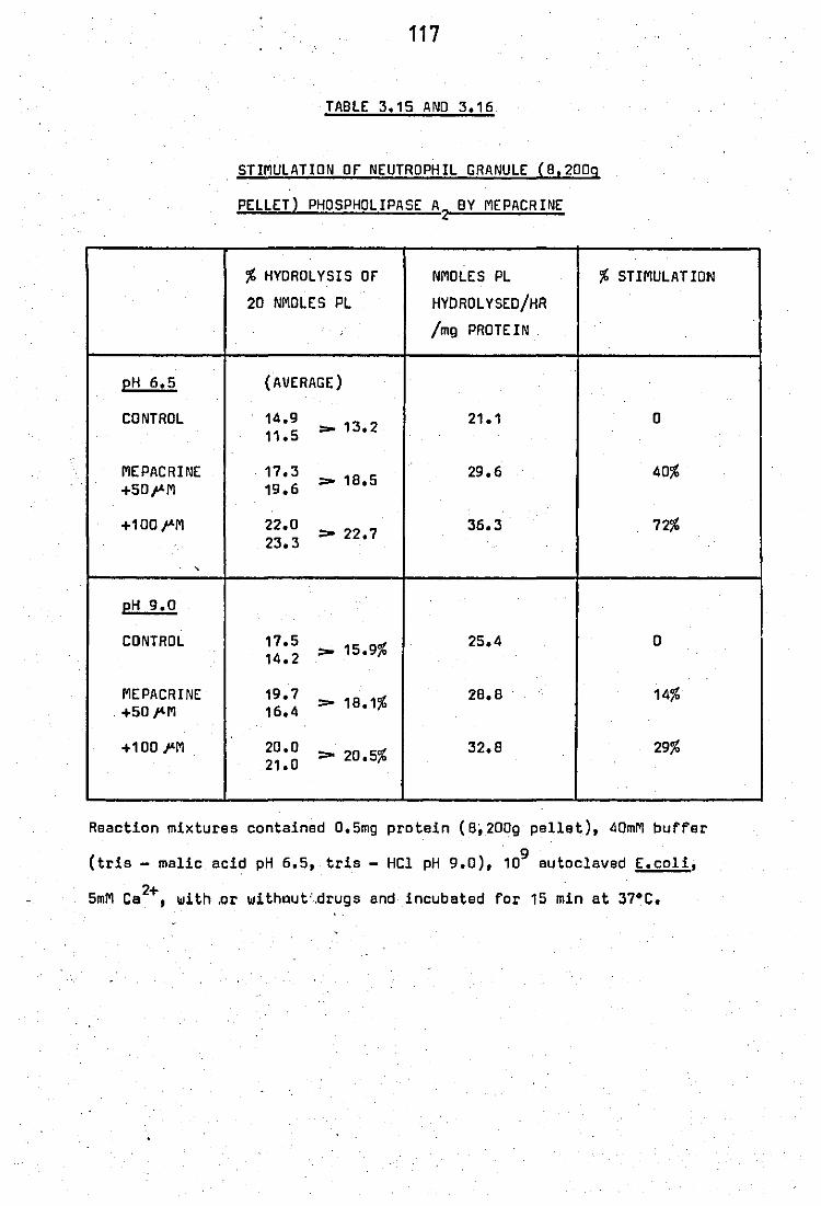
pH 6.5
CONTROL
MEPACRINE , '
+50 .... M
+100 .... M
,
pH 9.0
CONTROL
MEPACRINE , +50 I'M
+100 I'M
117
TABLE 3.15 AND 3.16
STIMULATION or NEUTROPHIL GRANULE (8,2009
PELLET) PHOSPHOLIPASE A2 BY MEPACRINE
% HYDROLYSIS or NMOLES PL
20 NMOLES PL HYDROL YSED/HR
/mg PROTEIN
(AVERAGE)
14.9 _ 13.2 21.1 11.5
17.3 > 18.5 29.6
19.6
22.0 > 22.7 36.3
23.3
17.5 _ 15.9% 25.4 14.2
19.7 > 18.1% 28.8
16.4
20.0 > 20.5% 32.8
21.0
% STIMULATION
0
40%
72%
0
14%
29%
Reaction mixtures contained 0.5mg protein (8,200g pellet), 40mM buffer , 9 '
(tris - malic acid pH 6.5, tris - Hel pH 9.0), 10 autoclaved E.coli,
5mM Ca2+, with.or withllut",drugs and incubated for 15 min at 37·C.

TABLE 3.17
STIMULATION or zc RELEASEO PLA2 rROM NEUTROPHIL LEUCOCYTES,
BY MEPACRINE AT pH 6.0
% HYOROLYSIS or NMOLES PL % STIMULATION . ,. 20 NMOLES PL HYOROLYSEO/HR
. /mg PROTEIN
CONTROL ~~90:> 10.5% 17.5 0
+ 0.5mM 16.9 :>17 0 2B.3 62% MEPACRINE 17.0 •
Reaction miXtures contained 40mM tris -. malic· .. 2+
buffer pH 6.0, 5mM Ca ,
• 9 < 0.12mg protein, 10 autoclaved radiolabelled E.coli, with or withbut
0.5mM mepacrine, and incubated for 1hr at 37"C.
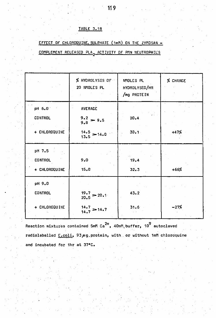
119
TABLE 3.18
EfrECT or CHLOROQUINE. SULPHATE (1mM) ON THE ZYMOSAN -
COMPLEMENT RELEASED PLA2
ACTIVITY or PMN NEUTROPHlt.:S
% HYDROLYSIS or NMoLES Pl
20 NMoLES Pl HYORol YSEO/HR
/m9 PROTEIN
pH 6.0 AVERAGE , .
CONTROL 9.2 9 5 9.8 - •
20.4
+ CHLORoQU INE 14.5 13.5 _14.0
30.1
pH 7.5
CONTROL 9.0 19.4
+ CHLOROQUINE 15.0 32.3
pH 9.0
CONTROL 19.7 0 20.5:>2 .1
43.2
+ CHlORoQU INE 14.7 14.7 >14.7
31.6
% CHItNGE
+47%
+66%
-27%
2+ 9 Reaction mixtures contained 5mM Ca ,40mM, buffer, 10 autoclaved
radiolaballed E.coli, 93p9.protein, with. or without 1mM chloroquine
and incubated for 1hr at 37°C.
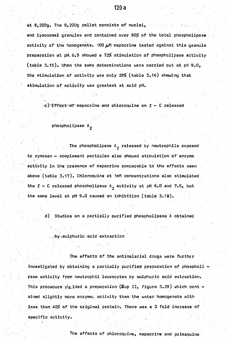
120 a
at 8,200g. The 8,200g pellet consists of nuclei,
and lysosomal granules and contained over 90% of the total phospholipase
activity of the homogenate. 100,uM mepacrine tested against this granule
preparation at pH. 6~5 showed a 72% stimulation of phospholipase activity
(table 3.15). When the same determinations were carried out at pH 9.0,
the stimulation of activity was only 29% (table 3.16) showing that
stimulation of activity was greatest at acid pH.
:c')"Ef'f"ectdO'f :mepacri:ne 'and chloroquine on Z - C released
phospholipase A2
The phospholipase A2 released by neutrophils exposed
to zymosan - complement particles also showed stimulation of enzyme
activity in the presence of mepacrine comparable to the effects seen
above (table 3.17). Chloroquine at 1mM concentrations also stimulated
the Z - C released phospholipase A2 activity at pH 6.0 and 7.5, but
the same level at pH 9.0 caused an inhibition (table 3.19).
d) Studies on a partially purified phospholipase A obtained
,by ,{sulphuric ,acid extraction
The effects of the antimalarial drugs were further
investigated by obtaining a partially purified preparation of phospholi
pase activity from neutrophil leucocytes by sulphuric acid extraction.
This procedure yie.,lded a preparation (Sup lI, figure 3.29) which cont
ained slightly more enzyme~ activity than the water homogenate with
less than, 40% of the original protein. There was a 3 fold increase of
specific activity.
The effects of chloroquine, mepacrine and primaquine
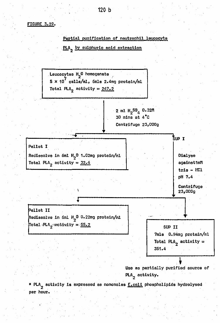
120 b
nCURE 3.29.
Pellet I
Partial purification of neutrophil 1aucocyte
~A2 by sUlphuric acid extraction
leucocytes H20 homogenate 7 !
5 X 10 cel1s/ml. 6mls 2.4mg protein/m1 Total PLA2 activity = 247.2
2 m1 H2S04 0.32M 30 mina at 4·C
Centrifuge 23.000g
. ·'·r:------------------.....,tup I
Redisso1vs in 6m1 H20 1.02mg protein/m1
Total PLA2 activity = ~
Dialyse against1mM
trie - HC1 pH 7.4
Pellet II Redisso1ve in 6m1 H
20 0.22mg protein/m!
. Total ",PLA2aa:ctlv.ity=~ SUP II
Centrifuge 23,OOOg
1 7m1s 0.94mg protein/m1
Total PLA2 activity = 351.4
.~ Use as partially purified source of PLA
2 activity.
* PLA2
activity is expressed as nonomo1es E.co1i phospho1ipids hydrolysed
per hour.
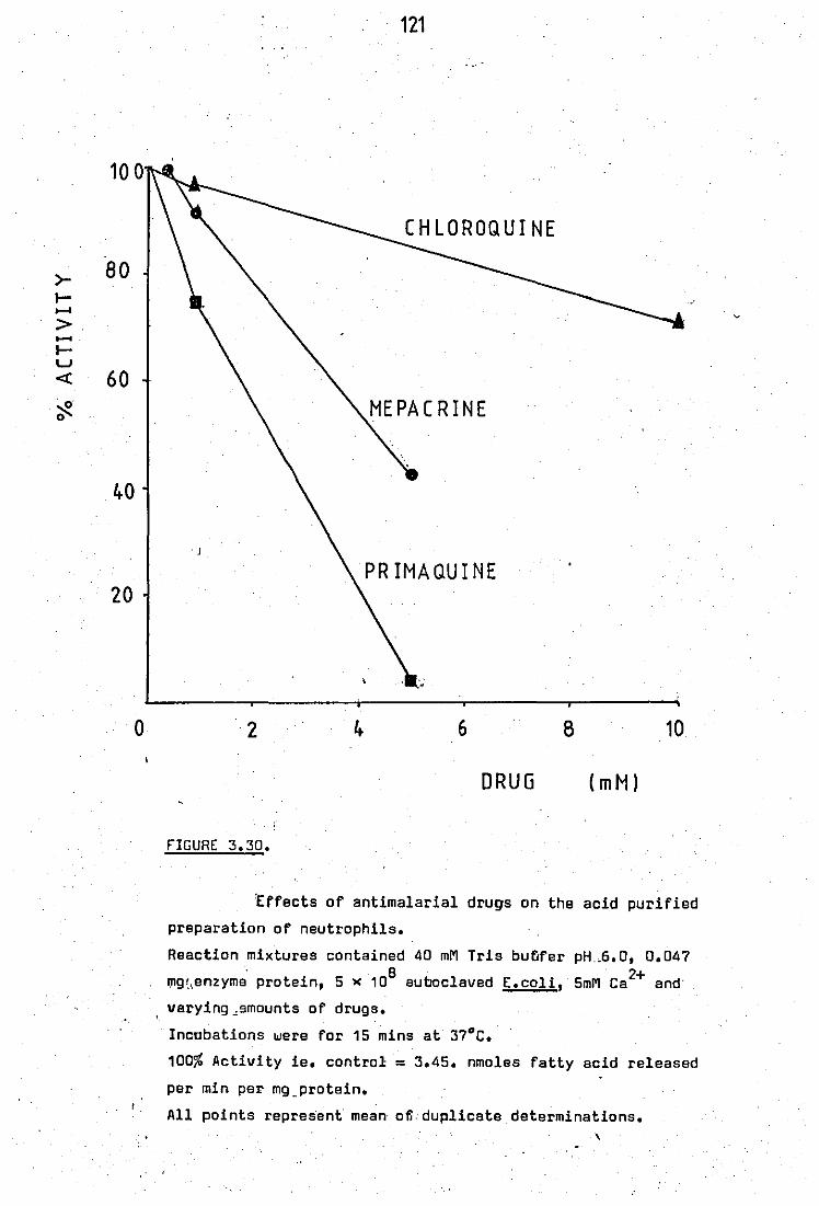
100
>- 80 I-...... > -I-w « 60 . o· ;;.:::
40
20
o
121
PRIMAQUINE
2 6 8 10
DRUG (mM)
FIGURE 3.30.
tffects of antimalarial drugs on the acid purified
preparation of neutrophils.
Reaction mixtures contained 40 mM Tris bufjfer pH.G.O, 0.047 . 8
mg'"enzyme protein, 5 >< 10 auboclaved
varying"smounts of drugs.
Incobations were for 15 mins at 37°C.
2+ E.coli, 5mM Ca and'
100% Activity ie. control = 3.45. nmoles fatty acid released
per min per mg_protein.
All points represent mean ofl. duplicate determinations. ,
122
on the partially purified enzyme preparation ere expressed in .figure
3.30. All these drugs showed inhibition of the purified phospholipase
ectivity with no evidence of stimulatory activity. Mepacrine and chlo
roquine at doses which showed strong stimulation of the.· enzy.me from
sonicated neutrophils, 8,200g granules and! - C released phospholipase
activities now showed inhibition of the acid purified enzyme from the
same source. The inhibitory response with primaquine was of the same
order and magnitude as that·obtained with the homogenate· enzyme
preparation.
3.4 Interactions of heparin with inflammatory phospholipases and with
antimalarial drugs
The antimalarial drugs are cationic amphiphilic agents.
Ttws the contrasting effects observed above may be due to alteration
of· phospholipase A2 activi ties by lnterie~~nce with endogenous anionic
ag~nts. Such as the glycosaminoglycans heparin or chondroitin sulphate.
These have been shown to be present in neutrophils in measurable quan -
tities (Olsson and Gardell, 1967., Olsson et al, 1968., Avila and Convit, -- .
1976).
a) Interaction of heparin with antimalarial drugs
An interaction between chloroquine and heparin was
immediately apparent when two solutions containing these compounds are
mixed togather.A visible complex·was formed which could be monitored
by optical density measurements at 700nm. The results obtained when
50 units/ml heparin was added to varying amounts of antimalarial drugs,
are expressed in figure 3.31. All three drugs alone or heparin alone

I
I
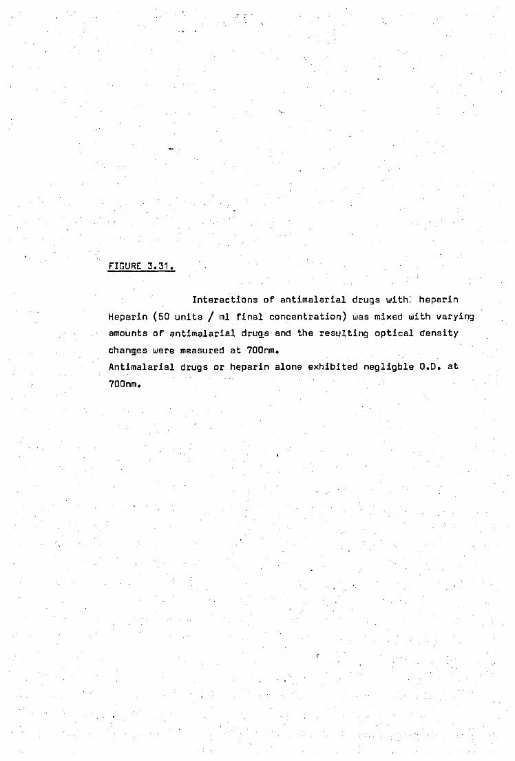
FIGURE 3.31.
Interactions of antimalarial drugs with: heparin
Heparin (50 units/ ml final concentration) was mixed with varying
amounts of antimalarial druqs and the resulting optical density
chan~es were measured at 700nm.
Antimalarial drugs or heparin alone exhibited negligble 0.0. at
700nm.
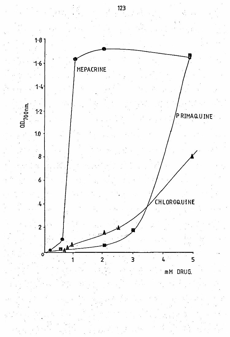
1·8
·1·6
1·4
e . . .; 1·2
o l
d CS
1.0
8
6
4
2
1
123
!, '
MEPACRINE
P RIMAQ.U INE·
2. 3 4 5
mM DRUG.
124
showed less than 0.02 optical density units at 700nm. The complexes
retained the original colours of the drug solutions; with mepacrine -
yellow, primaquine - orange, and chloroquina - white. On the basis of
optical density measurements mepacrine was the most active drug,
followed by primaquine and then chloroquine.
b) Effects of heparin on inflammatory phospholipase activities
Heparin affected the phospholipase 'activities of both
the peritoneal fluid and neutrophil leucocytes, although in opposite
ways.
Stimulation of the peritoneal fluid phospholipase A2
occured at physiological levels and was dose - depend@nt (figure 3.32).
The stimulatory effect was greater at pH 6.0 then at 7.5 (table 3.19).
The effect of heparin on the acid purified nautrophil
phospholipase was inhibitory. Table 3.20 shows the contrasting data
obtained using the acid purified neutrophil enzyme and indicating that
6 units/ml heparin was sufficient to cause almost total inhibition of
phospholipase activity.
Addition of mepacrine (5mM, which itself caused 52%
inhibition) and varying amounts of heparin (1 - 40 units/ml) to the
acid purified enzyme showed an additive inhibitory effect by the two
agents (table 3.21). No antagoni'stic effect between the cationic and
anionic agents was observed.

, ~. -
fIGURE 3.32.
(ffect of heparin on the peritoneal exudate PLA2
Reaction ,mixtures contained 40mM'Tris - malic acid buffer pH 6.0, B ' U
0.3mg enzyme protein, 15 ~ 10 autoclaved (.coli,5mM Ca and
varying amounts of heparin.
Incubations were for 10 mins at 37 C.
Control activities. 1.65 nmoles f.a. released / ~in / mg protein
• 100%.
1 S.E .. , ~
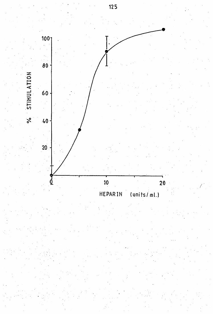
12S
100
80 z 0 ..... I-« -I 60 ;::)
4 I-
.Vl
·0· Cl' 40
20
10 20 (
HEPARIN (units! ml.)
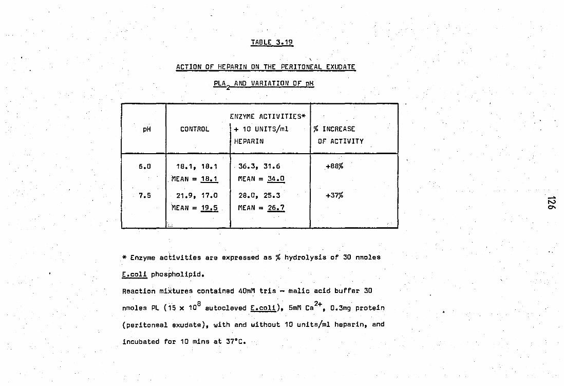
TABLE 3.19
, ACTION or HEPARIN ON THE PERITONEAL EXUDATE
PLA 2 AND VARIATION or pH
ENZYME ACTIVITIES*
pH CONTROL + 10 UNITS/ml % INCREASE
HEPARIN or ACTIVITY
6.0 18.1, 18.1 . 36.3, 31.6 +88%
MEAN = 1.!hl MEAN = ~ 7.5 21.9, 17.0 28.0, 25.3 +37%
'MEAN = ~ MEAN = 12.2
_.
* Enzyme activities are expressed as % hydrolysis of 30 nmoles
E.coli phospholipid.
Reaction mixtures contained 40mM tris - malic acid buffer 30
. 8 ) U nmoles PL (15 x 10 autoclaved E.coli , 5mM Ca ,0.3mg protein
(peritoneal exudate), with and without 10 units/ml heparin, and
incubated for 10 mins at 37°C.
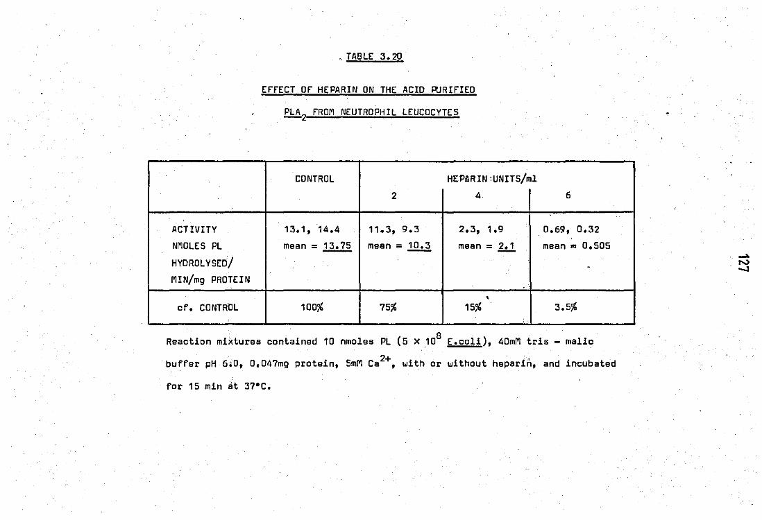
'. TABLE 3.20
EFFECT OF HEPARIN ON THE ACID PURIFIED
PLA 2 FROM NEUTROPHIL LEUCOCYTES
CONTROL HEP!RIN;UNITS/ml
2 4 6
ACTIVITY 13.1, 14.4 11.3, 9.3 2.3, 1.9 0.69, 0.32
NMOLES PL mean = 13.75 mean = ~ mean = hl mean = 0.505
HYDROLYSEO! . MIN/mg PROTEIN ..
, cf. CONTROL 100% 75% 15% 3.5%
Reaction mixtures contained 10 nmoles PL (5 X 10⁸ E.coli), 40mM tris - malic
buffar pH 6.0, O,047mg protein, 5mM Ca2+, with or without heparin, and incubated
for 15 min at 37"C.
TABLE 3.21
ErfECT Of MEPACRINE AND VARYING AMOUNTS Of HEPARIN ~. ~
TOGETHER.:ON THE ACID PURIfIED PLA2 fROM NEUTROPHIL LEUCOCYTES
" --. .-ENZYME + 5mM 5mM MEPACRINE + HEPARIN uNxf~/ml HEPARIN' ~ v/ml CONTROL MEPACRINE 1 5 r, 10. 20 40 WITHOUT
MEPACRINE . --- . .- ... -<'
% HYDROLYSIS 17.45 8.3 6.05 2.2 0.35 0.3 0.9 13.5
Of 5 X 10⁸
E .COLI
NANOMOLES PL 0.372 0.177 0.13 0.047 0.007 0.006 0.019 0.287
HYOROL YSEO/
MIN/mg PROTEIN
cf. CONTROL 100% 48% 35% 13% 2% 2% 5% 77%
% Hydrolysis values represent mean of duplicate determinations.
Reaction mixtures contained 5 X 10⁸ autoclaved radiolabelled E.coli,5mM Ca2+, 40mM tris -
malic buffer pH 6.0, 0.047mg protein, with or withbut drug mixtures, and incubated for
10 mins at 37°C.
129
3.5 Membrane actions ot: antimalarial drugs on guinea pig erythrocytes
Cationic amphiphilic drugs (eg antimalarial drugs)
being surface - active in nature are also considered to heve an effect
on membranes. In an ettempt to help explain the paradoxical inhibitory/
stimulatory effects on phQephmlipases, the membrane actions of the threa
antimalarials on the hypotonic haemolysis of guinea - pig red blood cells
were examined.
The results obtained are expressed in figure 3.33.
All three of the antimalarial compounds did indeed show paradoxical
dose - response curves.
At low doses all three drugs show stabilization of
the erythrocyte membrane to osmotic shock. With mepacrine and primaquine
maximum stabilization as measurad by a reduction in the relative
haemolysis was shown at 2 X 10⁻⁴M. Chloroquine showed maximum stabili
zation at 10-3M and was five times less active.
At higher doses all three drugs showed labilization
·of erythrocyte membranes (as measured by an increase in the relative
haemo·lysh).·.,to"osmotic ,shock. PrimElquine· showed maximum disruption. at
-3 -3 -3 2.5 X 10 M, mepacrine at 5 x 10 M and chloroquine at 10 M. At even , -2
higher doses ie above'10 M the destabilizing effect was again
reversed to stabilization again indicating the membranes having a
tri - phasic responsa to the drugs.

1
1
1
1
1
1
1
1
1
1
1
1
1
1
1
1
1
1
1
1
1
1
1
1
1
1
1
FIGURE 3.33.
Effects of antimalarial drugs on the hyptonic
haemo19sis of guinea - pig erythrocytes.
Reection mixtures contained 59)111 fresh heparinised blood in
0.425% NaGl and indicated amounts of antimalarial drugs.
mepacrine = •
primaquine = •
chloroquine = ! Incubations were performed for 30 mins at room temperature
0.425% NaGl alone caused 50% haemo19sis of g - p erythrocytes.
en en ~ o E (])
1·5
Cl 1.0 Co ..
(])
> .-
0·5
•
10-3
Antimalarial (M)
'-2 10
..... w o
131
"-PART 0 STUDIES OF ANENOOGENOUS INHIBITOR OF PHOSPHOLIPASE AZ'IN THE ..•.
8,2009 SUPERNATANT OF NEUTROPHIL LEUCOCYTES
4.1 Effect of the neutrophil 8,2009 supernatant on the peritoneal
fluid phospholipaseA2 activity
Figure 3.34- shows the effects of adding-.different
amounts of 8,200g supernatant protein to reaction mixtures containing
sufficient peritoneal fluid phospholipase A2 activity to causa approx
imately 20% h~drolysis at pH 9.0. Enzyme activity was inhibited in a
dose - depend~t manner and maximal inhibition of 64% was obtained with_
230 pg supernatant protein. Similar findings were obtained at pH 6.0,
and indeed as shown in figure 3.35, the degree of inhibition caused
by the neutrophil 8,200g supernatant on the peritoneal fluid enzyme
appeared to be independint of pH (between pH 5.5 and g.O).
However inhibition of phospholipase activity by the
8, 200g- :supernatant was not independent of pH if the neutrophil granule
(8,200g pellet) phospholipase activity was used. Indeed as shown by table
3.22 the 8,200g supernatant does not have very much effect on _the granular
enzyme activity at pH 6.5, although inhibition of enzyme activity has
been observed at pH7.5 (franson et aI, 1974).

F'IGURE 3.34.
Effect of 8,200g neutrophil leucocyte eupernat,art . ,
on the hydro19tic activity of peritoneal exudate PLA2•
Reaction mixtures contained 40mM Tris - HCl buffer pH 9.0, 5mM 2+ 8 . 14 ) Ca , 15 x 10 autoclaved E.coli ( C, either 0.3mg enzyme protein
end varying amounta of neutrophil 8,200g supernatant protein (circles)
or just verying amounts of 8,200g supernatant protein (triangles).
All points are mean of duplicate determinations.
•
. -(/) >.. ".-o
20
t. iJ . '>' '10 ..c'
Cl ' ~"
0
,
•
0
,.
I
. "
' .
•
•
j j i I
0·2 0·4 .. ..
8200g supernatant protein (mg)

.ICURE 3.35.
Effect of pH.on the activity of the peritoneal exudate
PLA2 in the presence and absence of neuorophil 8,200g supernatant
prot~in (0.42mg).
Incubations were as described· in Materials and Methods with 0.28mg
peritoneal exudate protein.
All points are means of duplicate determinations.
-0
• control
+ 8i2009 supernatent •
.:
.. . , ...
30
20 (fJ
. . -(1)
~ 0 ~
"0 >. .
.c ~ 0
10
0 I I
5 6 •
'" ' .... _ . ... " .,- ..
•
, .,
, '
i
7 .... . ,.
pH
•
§
. , .
•
i
9
.' .....
...... l..IJ l..IJ
•
TABLE 3.22 "
EffECT Of B,2009 SUPERNATANT ON THE NEUTROPHIL
GRANULE PHOSPHOLIPASE ACTIVITY AT pH 6.S
. " CONTROL + B,200g SUPERNATANT
GRANULES ALONE 105~g 230",g 355pg
. ACTIVITY % 9.B, 9.6 9.1, 9.6 B.O, 9.9 8.5, 7.5
HYDROLYSIS Of mean = 2.:1. mean = 2.:l§. mean = 9.0 mean = 8,9
20 NMOLES PL . .
cf. CONTROL 100% 96% 93% 92%
2+ 9 Reaction mixtures contained 4DmM tris - maleate buffer pH 6.5, 5mM Ca , 10 auto-
claved radiolabelled" E.coli, 0.5mg enzyma protein (8,200g pellet containing granules)
and with or withoUt varying amounts of 8,20Dg supernatant protein. IncGbations were
"carried out at 37'C for 15 mins.
135
4.2 Effect of the PMN 8,200g supernatant on the phospholipases from
pig pancreas and Crotalus adamenteus and Nala nala venoms
In an attempt to further investigate its selectivity
of action, the endogenous phospholipase inhibitor present in the
8,200g supernatant of neutrophil leucocytes was tested against phosp -
holipases from pig pancreas and venoms of Crotalus adamenteus and
Na la na la towards E. coli phbspholipids at pH 7.5. Taille· 3.23 shows the
results obtained.Jhe 8,200g supernatant displayed inhibitory effects'
of a similar potency with the enzymes from .rabbit peritoneal exudate,
the highly purified phospholipase A2 from pig pancreas and the crude
enzyme from Crotalus adamenteus venom (39%, 45% and 26% respectively , . with 420)"9 8,200g supernatant protein). However with Nala nala venom
a marked stimulation of activity was observed ( +58%).
To check whether these effects were due to non -
specific protein. actions, the experiments were repeated with a
similar concentration of 8SA (420~g) that itself exhibited no phos -
pholipase activity. In all cases .a marked stimulation of phospholipase
activity was observed ranging from +36% with Crotalus adamenteus venom
.to +63% with the enzyme from pig. pancreas.
All the phospholipases examined were calcium dependant
as addition of 1mM EO TA resulted in total loss of enzyme activities
(table 3.24). The inhibitory activity of the 8,200g supernatant was not
very stable to storage, and lost approximately one - half of its inhibitory
potency over 3 weeks storage at +4°C, as tested against the rabbit
peritoneal fluid phospholipase A2 (table 3.25).
TABLE 3.23
EffECT Of 8.2009 NEUTROPHIL LEUCOCYTE SUPERNATANT. ANO BOVINE
SERUM ALBUMIN (BSAl ON THE HYOROLYTIC ACTIVITY Of SOLUBLE
PHOSPHOLIPASES A2 TOWAROS (1_14Cl OLEATE LABELLEO ESCHERICHIA
COLI PHOSPHOLIPIOS
ENZYME SOURCE PHOSPHOLIPASE A2 ACTIVITY
(% CONTROL VALUES) IN THE
PRESENCE Of 8,200g SUPERNATANT .,' "
PERITONEAL EXUDATE 61
PIG PANCREAS 55
CROTALUS ADAMENTEUS VENOM 74
!:!.£a !:!.£a VE NOM 158 ;',
BSA("
CONTROL
VALUES)
141
163
136
138
Incubations were carried out as described in Materiels and Methodg at pH 7.5. in the
presence of 420}tg, 8,200g supernatant, or BSA protein. Enzyme protein (peritoneal
.exudate 0.35mg; pig pancreas 0.05ng; Crotelus adamenteus 1pg and Naja nala 0.5JA9)
was sufficient to produce approximately 20% hydrolysis of labelled bacterial
phbspholipids.
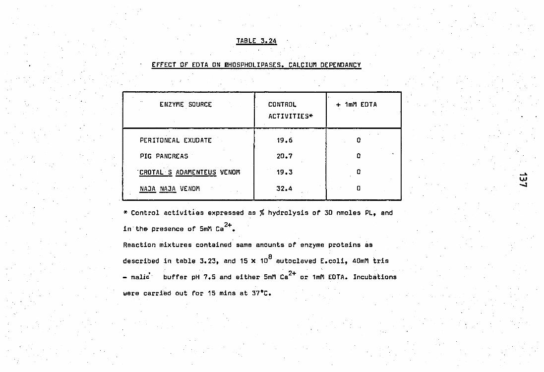
.
TABLE 3.24
EFFECT OF EDTA ON ~HOSPHOLIPASES. CALCIUM OEPENDANCY
ENZYME SOURCE CONTROL + 1mM EDTA
ACTIVITIES* .
PERITONEAL EXUDATE 19.6 0
PIG PANCREAS 20.7 0
'CROTALS ADAMENTEUS VENOM 19.3 0
NAJA ~ VENOM 32.4 0
* Control activities expressed as % hydrolysis of 30 nmoles PL, and
2+ in'th& presence of 5mM Ca •
Reaction mixtures contained same amounts of enzyme proteins as
B described in table 3.23, and 15 x 10autoclaved E.coli, 40mM tris
- malic' buffer pH 7.5 and either 5mM Ca2+ or 1mM EOTA. IncubaUons
were carried out for 15 mins at 37°C.
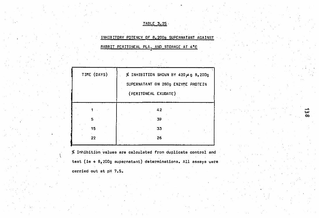
TABLE 3.25·
INHIBITORY POTENCY Ot B,2009 SUPERNATANT AGAINST
RABBIT PERITONEAL PLA 2 AND STORAGE AT 4'C
"
TIME (DAYS) % INHIB ITIDN SHOWN BY 420,u 9 B,200g
SUPERNATANT ON 280g ENZY~E PROTEIN
(PERITONEAL EXUDATE)
1 42
5 39
15 33
22 26
% Inhibition values are calculated from duplicate control and
test (ie + B,200g supernatant) determinations. All assays were
carried out at pH 7.5.
...... W CD

Chapter Four
o I S C U S S ION
139
DISCUSSION
PART A ESCHERICHIA COLI SUBSTRATE
The substrate used in this work for the determination
of phospholipase A activities was E.coli radiolabelled with (1 _ 14C)
oleic acid during growth, then autoclaved to destroy bacterial
phospholipases and to render the lipids more accessible to hydrolytic
·attack. Use of bacterial membranes as subtrates for phospholipases
was first described by Patriarca !!i al (1972) and subsequently by
many authors. It has the following advantages:
(a) The presentation of the lipids as part of a biological
'structure, although altered by the autoclave procedure, probably
provides conditions for the action ofPLA2
that are more physiological
than those that prevail when the substrate is in micellar form.
(b) This substrate is readily broken down under conditions
where micellar dispersions of isolated phospholipids are not (see later).
Uptake and distribution of (1 14C) oleic acid in E.coli
14 Adequate uptake of (1 - C) oleic acid by the
bacterium during growth did require specific conditions. These included
complexing of the fatty acid to albumin prior to contact with E.coli;
a suitable number of organisms in log - phase of growth; and an
optimal exposure time of the f.coli with the radiolabelled fatty acid
of 3hrs. A furth r incubation of 2hr to allow maximum incorporation of
the fatty acid into cellular phospholipids was also necessary.
almost all of the
140
Analysis of radiolabelled E.coli
14 (1 - C) oleic acid appeared in
revealed that
the phospholipids.
Indeed the distribution of radioactivity closely resembled the lipid
composition. The most heavily labelled compounds were phosphatidyl
ethanolamine (60%), phosphatidyl glycerol ('20%) and cardiolipin (10%) .:. I. I"
with very little radioactivity appearing in ' .. 1y.sQphos'pholipids and
neutral lipids. The presence of a small amount of radioactivity (approx.
4%) associated with free fatty acid may be due to adherence of fatty
acid. molecules to the organism; as this could be reduced to less than
1% by furth: 'r Idashings in saline. Therefore the substrate used for
most of the work was membrane - associated phosphatidyl ethanolamine.
14 . The (1 - C) oleic acid in E.coli phospholipids
.was exclusively in the 2 - position as determined using the specific
" phospholipase A2 present in.Crotalus adamenteus venom; This was to be
eXpected as oleic acid is an unsaturated long chain fatty acid and
such acids generally occupy the 2 - position of phospholipids
14 (rransen ~ aI, 1973). This was confirmed using (1 - C) linoleic
acid which was incorporated in the 2 - position to 87%, and the
unsaturated (1 - 14C) palmitic acid whic~ was incorporated preferentially
(77%) in the 1 - position. Attempts to increase this selectively
exclusive to the 1 - position failed even if cold oleic acid was
included in the procedures.
141
PART B STUDY Of INflAMMATORY PHOSPHOlIPASES
2.1 Release of phospholipase A from stimulated neutrophils
The results demonstrate" that stimulation of
rabbit peritoneal PMN leucocytes by zymosan - complement or exposure
of these cells to extracellular calcium ions led to a release of
phospholipase activity.
The release of the enzyme activity with ZC follows
phagocytosis of the particles in a concentration dependant manner,
and was not associated with cell lysis as only smal~ amounts of I·... ~
! v ,. " . •
the cytoplasmic enzyme, lactate dehydrogenase,~~(t-released. The
source of the released enzyme activity would appear to be the lysosomes
as f - glucuronidase and lysozyme> were released 10 similar extents
and in similar manner. The release of phospholipase activity was also
time dependant, an& follows the tima dependancy of phagocytosis ind -
icating a link b'etween the two.
The active principle in causing selective enzyme
release is the complement component, C3b, which becomes bound to
zymosan particles when incubated in serum. Zymosan particles alone
'have 'been shown 'riot to cause release of lysosomal enzymes from neutroph-
i1s (Henson, 1971). Indeed tha sarum treated with zymosan has also
been shown to contain a factor, C5a, which is able to induce release of
lysosomal enzymes from cytochalasin B treated neutrophils (Goldstein
.!!i &' 1975). Thus the release process is initiated by the' c'Jmbination
of ~ecific receptors on the leucocyte plasma membrane wi th activated
components of the complement pathway.In this system release of
lysosomal constituents occurs by a process called "regurgitation
during feeding" resulting from fusion of the lysosomes ,with incompletely
s
PH
closed phagosomes(figure 1.3).The pattern of selective release
occured in the absence of added calcium. Previous work by Henson
(1971) using peripheral leucocytes and ZC was carried out in the
presence of added calcium • . ' •
The present results also demonstrate that addition
of calcium ions to rabbit peritoneal neutrophils suspensions is a
suitable stimulus for lysosomal enzyme" release as measured by
release ofjJ- glucuronidase and lysozyme. Under these conditions
release to the suspending media of approximately 30% of the total , cellular phospholi'pase A2 activity, as assayed at pH 6.0 towards Lcoli
membrane phospholipids, occured in a concentration and time dependant
manner.
The release of pr- glucuronidase and lysozyme from
rabbit peritoneal PMN leucocytes has been previously reported by
Northover (1977). Approximately 15% selective release of both enzymes
2+ was recorded with 2.0mM Ca together with a very low, 2%, releese of
LDH.ln this study the amounts of enzymes released, both lysosomal and
cytoplasmic, were greater and is perhaps a reflection of the state of the
cells at the end of the expe·riment. The present observed release of
LDH{'fD%}'a'lso're'f'l'Bcts "90% 'ceTl viability as compared with 2% release
of LDH and 98% cell viability, obtained by Northover. However the
release of LDH was· ... still much lower than that of the lysosomal enzymes
j!J - glucuronidase and lysozyme and of phospholipase A2
•
Previous work on peripheral human PMN leucocytes has .
shown that Ca2+ alone only induces release of lysozyme and not
~- glucuronidase (Goldstein et al, 1975) thus implying its ability
to cau.se release of enzymes from the specific .; granules. The present
143
work on peritoneal polymorphs and in agreement with the findings of
Northover (1977) suggests that both types of granule enzymes may be
released. Thus peritoneal PMN leucocytes appear to show a greater
capability of lysosomal enzyme release than the peripheral counterparts.
However direct evidence, for or against this suggestion, requires
. further work on rabbit peripheral PMN's sinc~ the observed discrepan
cies may be species dependant. This difference in capability to lysoso
mal enzyme, release by the calcium may be due to the fact that peritoneal
PMN' s"ltr'l!"alrelldy"in 'a '''stimu'l'a'ted''O!tate as they have migrated to the
site of inflammation.
2.2 Properties of the released phospholipase A
These studies were carried out on the released
enzyme obtained from zymosan - complement treated neutrophils. As
calcium ions also caused a selective release of phospholipase A, the
properties were assumed to be the same.
The released phospholipase activity has an unusual
pH profile, having two pH optima, a broad peak in the acid region and
a sharper one at alkaline pH with.a consistent drop of activity between
the two·,peaks·'·at ·pH :/<5. 'Thi.s·may indicate the presence of two phospholipid
splitting activities, perhaps two isozymes, or a single protein may
. 2+ be responsible. Both activities show absolute requirements for Ca ,
. -3 2+ and maximal activity at S:x 10 M Ca • This property is in contrast
to the lysosomal phospho.!:ipases of alveolar macro phages which are
inhibited bYCa2+, but also have a lower pH optimum of 4.0. from
studies in·the literature so far, there appears to be two basic types
of lysosomal phospholipase activities:
144
1) Enzyme activity inhibited by Ca2+ and ~ active in the
pH range 3.3 - 4.5, for example, macrophages.
2+ . 2) Enzyme activity requiring Ca and active in the pH
range 5.0 - 7.5 (eg. 1eucocytes).
The activity is considered to be A2 specific as all
of the enzymically hydrolysed radioactivity, at both pH 9.0 and pH 6.0,
·"1·4 from (1 - C) oleic acid labelled E.coli, appeared in the fatty acid
fraction end none in the 1ysophospho1ipid fraction. furth 'r use of
(1 - 14C) palmitate labelled E.co1i, which is predominantly incorpor
ated in the 1 position, revealed the phospholipase activity at pH 6.0
of neutrophil homogenates to be A2 specific as the radioactive product
of hydrolysis was lysophospho1ipid and not fatty acid.
Kinetic studies examining the effects of time and
protein concentrations indicated that phos~ho1ipase activity was linear
for 40 minutes using up to 64~g protein if 10·nmo1es of substrate were . 8
used (5 x 10 autoc1aved E.co1i). The deviation from linearity after
these values may be due to inhibition by products. Certainly free
fatty acids and 1ysophospho1ipids have been demonstrated to inhibit
rabblt··'PMN leucocyte granule phospholipases (Franson et a1, 1974).
Kinetic studies involving substrate variation reve·a1ed an apparent Km . . -6
value for the enzyme of 25 x 10 M phospholipid.
storage of the phospholipase A2 activities of both
sonicated neu,trophil suspension and the released enzyme in HEPES
buffered medium exhibited a difference in the stability of the acid
and alkaline activitias. The activity at pH 6.0 being approximately
four times less stable than the activity at pH 9.0. This together with
145 '"
the fact that the phospholipase activity has two pH optima, is perhaps
a furthr Ir indication that two proteins may be responsible.
2.3 Subcellc lar localisation of phospholipase activities
Selective release of phospholipase A activity
without cell death and in conjunction with fi?- glucuronidase and
lysozyme suggests its association with the granules of neutrophil
leucocYtes.
Three types of granules have been identified in
rabbit PMN leucocytes by sucrose density "gradient centrifugation
and electron microscopy (8aggiolini ~ &, 1969 ... Wetzel ~ &;, 1967).
(1) The primary or azurophil granules which contain most of
the ~- glucuronidase, myeloperoxidase, approximately 33% of the ,
lysozyme and are the largest of the three types with.,.,a diameter of
0.5 - 0.8),1'1.
(2) The secondary or specific granules which are present in
the largest numbers, contain most of the alkaline phosphatase activity,
67% of ""the lysozyme "activity 'and" range in size from 0.3 - 0.5}A'1 in
diameter.
(3) Tertiary granules which are the smallest ( -05 - 0.2 )JPl),
contain the remainder of the acid hydrol~ses, are often rod shaped
and tend to occur in small clusters in the peripheral cytoplasm
(Wetzel et aI, 1967). --
rrom our findings it is not possible to say with
146
which granule (s) the released phospholipase A2 is associated. The
phospholipase A2 described in this study hes:.hof!!p,/er. marked similarities
to a membrane - bound phospholipase A2 associated mostly with the'
azurophil and specific granules with . some activity present in the
tertiary ill - defined granules (rransen ~ aI, 1974).This latter
enzyme also has two pH optima, at pH 5.5 and 7.5 with a drop in
activity at pH 7.0. It also requires ca2+, shows similar kinetics with
E.coli substrate, but differ in that the activity at both acid and
alkaline pH is stable.
A similar study on the cellular localisation of
phospholipase activities in human peripheral PMN leucocytes has also
been reported (rransen ~ aI, 1977). Human PMN PLA2
is also membrane
2+ - bound, Ca dependant, has one optimum peak at pH 7.0, and is associ 1" .
ated in part with the azurophilic and specific granules but a major
portion was found with an " empty vesicular· fraction" containing 85%
of the total alkaline phospholipase activity of whole homogenates.
The empty vesicular fraction are probably tertiary granules as they
sediment after specific granules from sucrose density zonal centrifu
gation procedures. Another difference:is that phospholipases from rabbit
PMNs and serum show 20 fold more activity than phospholipases from the
corresponding human sources (Weiss et 81,19'79 ; Kaplan - Harris ~ .!!!"
1980). Whether this has any evolutionary significance is not known.
2.4 The role of PMN lysosomal phospholipase A2 in the inflammatory
Response
PMN leucocytes play an extremely important role
in the infammatory process. In reponse to chemotactic stimuli they
are atlle to migrate into tissues in substantial numbers and perform
147
the task of phagocytosis and degradation of biological material. Neutrophils
are particularly important in defence ageinst microorganisme. and
lysosomal phospholipases participate in the intracellular killing and
breakdown of the organisms. Indeed when live E. coli labelled with
(1 - 14C) palmitic scid ere incubated with rabbit neutrophila, within
15 minutes 99% of the E. coli sre killed (non - viable) and 30% E'. coli
phospholipid breakdollln occurs (Elsbac!1.2i,!!!., 1972) •
. "During phagocytosis or stimulation of PI'IN leucocytes
by non .; phagocytosable stimuli such as immune complsxes bound to
basement membranee, there occurs release of hydrolytic enzymas IlIhich
are abls to impart considerable damage to the surrounding tissues.
Indsed in chronic inflammatory statessuch as rheumatoid arthritis much
of the tissue damage is thought to result from lysosomal enzyme activity
Cb/eissmann II al. 1972). Ths released phospholipase activity la able
to hydro1yss phospholipids of cell membranes to y;~ld polyenoic acids
and lysophospho1ipids. Lysophospholipids are surface active and cyto -
. toxic agents and will potsntiate the inflammation. Po1ysnoic fatty acids
can be converted to prostaglandin - like products by cyclooxygsnase
enzymes which are ubiquitous •
. "PI'lN"'J:eucocytssthemselves have been shollln ·to 'release
a varisty of prosteglandin - like products during phagocytosis, for
example PGE2
; PGF2
, (Higgs 11: Voulten, 1972), PGE1
(Higgs.2i a1, 1975),
TXA2 (Higgs.2i,!!!., 1976), TXB2 (Go1datsin ll,!!!.,197S), hydroxy acids
(Borgeat II aI, 1976) and Isucotrienea (Samue1ason ll,!!!., 1979).
The generation of all the prostaglandin - like
products is dependant upon a supply of po1yenoic fatty acids and as
the m~jor sourca of these are phoepho1ipids then the action of
148
phospholipase A2 is very important.
2.5 Phospholipase A activity of the call - frea peritoneal
inflammatory exudate
The peritoneal inflemmatory exudate obtained 17 hrs
after thioglycollate injection, was found to be a very potent eource
of phospholipid eplitting activity. Thie enzyme activity has properties
eome eimiler, eome different, to the enzyme released from the neutrophil
during phegocyto~ie of zymoean - complement particles. Using comparable
concentretions of eubstratas (10 - 20 nmoles), substantial enzyme activity
could be detected within 5 minutee incubation at 37°C. This preparation
also showad an acid and alkaline optima although the position of the
alkaline peak wae at pH 8.0 comppred with pH 9.0 for the neutrophil
released activity. Ths phospholipase activity was also A2 epacific.
Although this enzyme showed an'absolute requirement
for calcium ione, kinetic stUdies revealed different results from'
those obtained using the neutrophil released enzyme. Close examination
using varying emounts of EDTA showed that enzyme activity increased
in a eigmoid fashion, indicating allosteric behaviour. The optimum
concentratIon was 1. ~niM Ce,2+ '''',,:. i.:;': "'.- :>c.:: ~'_ ~;~ '. ,_ ,. ,- . '.' •..
>, ;?~"" ~ ., ,'. Thie implied that the eXUdate enzyme is very sensitive
to small changea in the calcium concentration~ The neutrophil releaaed
enzyme showed the normal hyperbolic curve of non - regulatory enzymes.
The peritoneal exudate PLA2 did show hyperbolic characteristics with
variation of autoclaved E.coli phospholipids, and an apparent Km approx. . -6 .
b70X 10 M. This value is aubstantially greater than the corresponding
Km value obtained with the neutrophil released enzyme.
~ee. . '1 po s il:e. fo, etA. te>..)
149
The data obtained on thestorsge of the peritonsal
exudste phospholipase ectivity is particularly interesting and obviously
contrasted with the corresponding data of the neutrophil enzyme. The
former activity at acid pH was found to be fairly etable to storags at
4°C and lost only 12% activity over 2.5 months. Particularly interesting,
wae the finding of an actual incrsase of specific ectivity, by 79%,
over the first few days (table 3.8), which then gradually returned to
be the original activity after approximately 1 month. One explanation
of thie finding may be that some of the enzymes exist in the form of
a zymogen, or proenzyme, which degrades to the active species during
the first few days storage, and hence shows an increase of specific
sctivity. Such bshsviour·of phospholipases hss been observed by other
workers. Etienns et sl, (1969s, 1969b) hsve shown thst storage of serum --or sddition of vsry high concentrations of trypsin to whole human snd'
rat serum increases the phospholipid - splitting activity many fold,
and concluded that the enzyme may exist as a zymogen. Indeed plasma
may indeed be a viable source of the phospholipase activity seen in
the inflam.sd peritonea, since serum derived factors also infiltrate
inflam· ed areas during the acute reaction. (f"ransan E!.!., 1978).
During the course of this work a report on theiso -
lation arid characterisation of a phospholiRase A2 from rabbit peritoneal
inflammatory exudate was published (f"ransan ~!.!.,1978). The inflammatory
reaction was elicted by the injection of glycogen in physiological
saline. The phospholipase activity described had two pH optima, at pH
2+ 6.0 and pH 7.5, was A2 specific, and also Ca dependant. A 300 fold
purification was carried out using carboxymethylsephadex columns to yr\ld
a eingle protein peak of 14800MW. Polyacrylamide gel electrophoresis
of this fraction revealed this to be a cationic protein more basic
than lysozyme (pI = 10.5). As this phospholipase has properties similar
150
to the grenule associated activity in PMN leucocytes, it was suggested
to be derived from this cell.
PART C ErrECTS Or CHlOROQUINE - LIKE AGENTS ON INrLAMMATORY AND
OTHER PHOSPHOLIPASES
3.1 Effoct of chloroquine on the phospholipase A2 activity of Crotalus
Adamenteus venom and of pig pancreas
Chloroquine diphosphate ahowed a dose - related inhibition
. of both phospholipase A2 activities as assayed against egg yolk substrates.
Egg yolk substrates consist primarily of phosphatidylcholins (79%) and
phosphatidylethanolamins (17%) (Raheja.\U. al, 1973). Inhibition towards
the mammalian enzyme was four fold· greater than towards Crotalus adamenteus
venom enzyme (E.050 values 0.73mM and 3.1mM respectively). However the
parallel nature of the inhibitory dose- response curves suggest a similar
mode of inhibition of the two enzymes.
Two chemically related structures, an alkaloid cinchonine
sulphate, and the local anaesthetic dibucaine, also exhibited inhibition
towards pig pancreas PLA2• The order of decreasing potsncy was cinchonine
sulphats>chToroquine ) dibucaine.
Kinetio studies involving the variation of substrate
in the pressnce and absence of chloroquine indicated an uncompetitive
2+ inhibition. Similar studies on the variation of Ca in the presence
and absence of either chloroquine or dibucaine indicated competition 2+ .
between Ca and the drugs. Thus inhibition of pancreatic PLA2 activity.
may be due to binding of the drug molecules to the eubstrate and thus
altering its physiochemical properties and/or competition with the function
151
2+ of Ca on the enzyme.
The effects of local anaesthetics such as dibucaine
on phospholipase activities has generated much interest. Indead local
anaesthetic potancy parallels tha ability to ~ffect phospholipase
activities (Kunze' et al,1976). Studies have shown the drugs to have --multiple effacts depending UpOA the nature and source of enzymes and
substrates used. Thus inhibition by local anaesthetics has been observed
with phospholipase A2 of pig pancrsas using egg yolk substrates (Scharphof
.!!l!!" 1972), and PLA2 0f human seminal plasma and Naja naja venom
towards sonicsted phosphatidylcholine (Kunze'.!!l!!" 1976), aithough with
the latter two enzymes stimulation of activity at low concentrations
was also observed. However the PLA2 from Crotalus adamenteus venom is \
only stimulated and shows no inhibitory effect using eithar sonicatad or
agg yolk substrates (Kunze .!!l!!" 1976).
Two hypothes.:.s hava baen forwardsd concerning the
mode pf inhibition of phospholipase activity. Firstly the anaesthetic
binds to the substrate thus modifying its physiochemical properties
and its susoeptibility to anzymic hydrolysis (Waite and Sisson, 1972;
Scherphof and Westenberg, 1975; Stewart Hendrickson and Van Dans - Mieras,
1976). Secondly the anaesthetic binds to the enzyme and displaces
. . 2+ ( calcium from tha enzyme - Ca complex Scherphof .!!l!!" 1976, Kunze
.!!l al, 1976). The results :. "'~,:':",,':: in this study indicate both a
binding of the dru9 to the substrate and competition with calcium. Indeed
recently cationic amphiphilic dru9s have also been shown to replace
45 2+ Ca from phospholipid monolayere in a dose - depend.,nt fashion •
(Lullmann .!!l!l,19BO). The potencies of the drugs were related to their
lipid eolubilities, although the charged form of the drug was also
necessary.
152
However it should be noted thst one should proceed
with caution when interpreting kinetic data using lipids. The substrates
used are not in true solution but dispersed as liposomee and micellar
dispersions. Therefore any conclusions derived using classical kinetics
from lipids should be interpreted as indicstions and not as stern facts.
It may be of importance to mention that in experiments
IlIi th cinchonine aulphateand _ .dibucaine "but not ,.p!tloroquina, there was a
leg phaee of 3 minutes before maximum enzymic hydrolysis was reached.
The reaeon for this is not certain but may be e phyeical effect euch as
an effect on substrate diepersion. Lag phase have been observed in a
previoua study by Rosenthal and Ching - Hsien Han (1970) where inhibition
of phospholipsse A2 by phosphinate analogues was reported using ,ethar
systems as the reaction medium.
3.2 Effect of chloroquine - like agents on the inflammatory peritoneal
exudate PLAZ activity
Phospholipase A2 activity of the rabbit peritoneal
(~ell - free) inflammatory exudate towards E.coli phospholipids was
inhibited by chloroquine, mepacrine and primaquine. The inhibitory log -
dose response curves were parallel indicating a similar mode of inhibition.
Mepacrine was almost twice as active as primaquine and five times more
active than chloroquine. The inhibitory potency (mepacrine> primaquine
;> chloroquine) was related to the lipid solubilities of these compounds
as shown by a correlation between the IC50 of the drugs on phospholipase
activity and the log P values determined between chloroform and water.
At doses above those causing maximum inhibition,
153 -------~-----" -------~-
chloroquine and mepacrine showed a reversal of the effect even leading to
stimulation of phospholipase activity at greater than 5mM mepacrine.
Primaquine however differed and showed no reversal of the inhibitory
effect at high doses. Thus chloroquine and mepacrine unlike primaquine
caused biphasic effects on the psritonesl sxudate phospholipase activity.
This difference between the drugs may be" due to structt{al differences
of thess drugs. Primaquine is an 8 - sminoquinoline whereas the other two
sre 4 - amino quinoline dsrivatives. This difference in ring structure
can relste to differences in the resonance of the charge (when protonated)
between the ring N and the secondary amino - N on ths side chain (see
figure 4.1). Thus with chloroquine. snd mepscrine thie resonance cen
occur with free movement of the proton bBt~een the nitrogens, but not
with primaquine. Thie factor together with .the difference in the terminal
.amino group may confer marksdly diffsrsnt properties on the primaquine
molecule. Indeed differences between the pharmacological actions of
chloroquine and primaquina have been reported. They sttack different
phases of the lifs cycle of the malarial parasite, after!!!..!!.!l!2.
administration chloroquine ie avidly bound by tissues whereas primaquins
is barely detectable after 24 hrs (Bowman and Rand, 1980) and primaquine,
but not chloroquine, is s potent inhibitor of alcohol dehydrogenass
(Li and Magnus, 1972).
The biphasic effects observed on the peritoneal exudate
phospholipase A2 also relate to paradoxical membrane active properties
of these drugs. All three drugs affected the hypotonic haemolysis of
guinea - pig erythrocytes in a similar manner. (figure 3.33).
Stabilization was caused at low doses, followed by labilization at high
doses, and a reversal again at even higher levels. Thue penetration of
low amounts of drug strsngthens the red cell membrane while too much
leads j;o rupture of the ordered structure~ A similar observation for.~
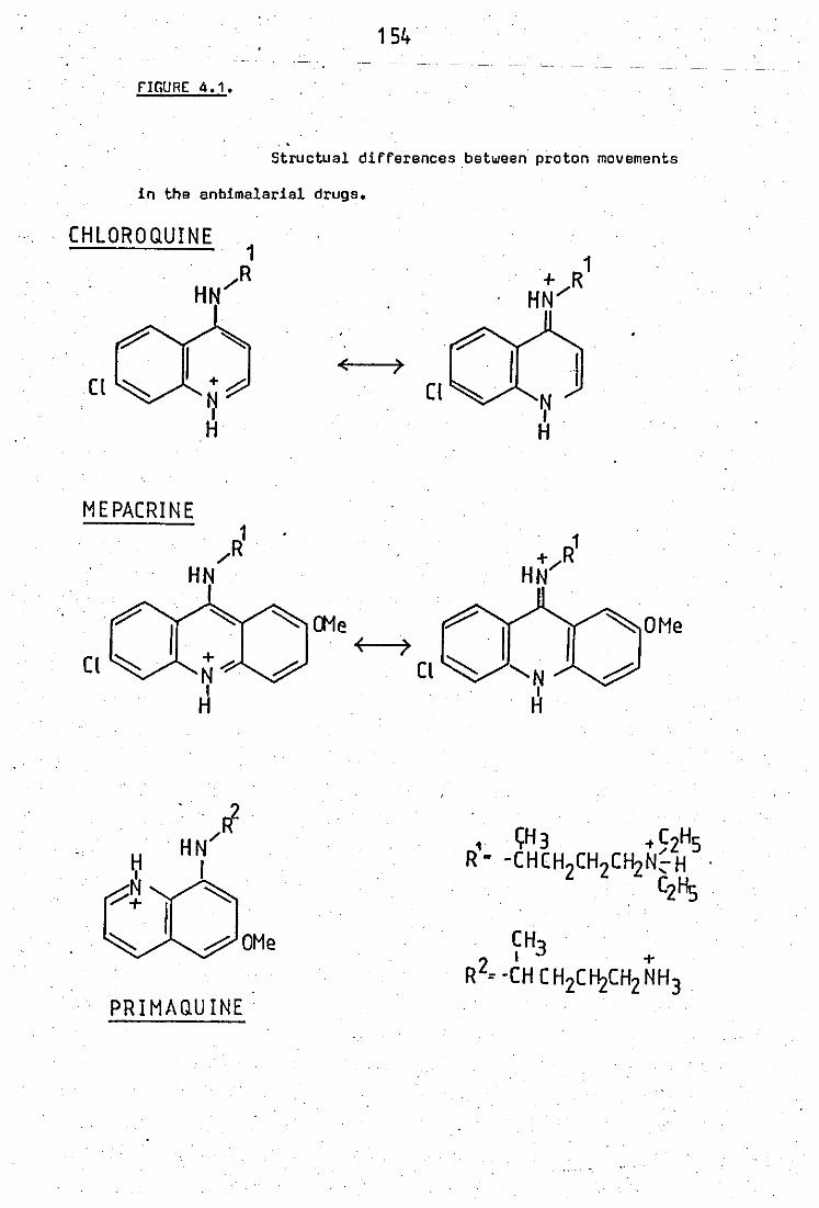
154
nGURE 4.1.
. . Structual differences.between proton movements
in the anbimalarial drugs.
CHLOROQUINE 1 . R
HN/
( )
Cl ~ Cl
MEPACRINE R1 . 1
/ +/R HN HN
7'" ~ (Ne
+~ N ,ij
(
~ I
H
CH3 2 I + R = -CH CH2C~CH2NH3
PRIMAQUINE
155
.,
chloroquine hes been obtained by Ing~ot and Wolna (1969) • . ,.
The degree of inhibition of phospholipase activity
also depended upon other factore. Variation of pH had a profound effect
on the inhibition. Studies with 1m~ chloroquine revealed the least
inhibitory effect at pH 6.5, at pH 7.5 the eame level of drug caused
twice as much inhibition and at pH 8.5 more than three times as much.
Thus inhibition increases with increasing pH, and reflects the importance
ef charge on the chloroquine molecule. Chloroquine has two pKa values
pKa1 = 8.06, pKa2 = 10.16 (Rosenberg and Schulman, 1978). At pH 7.5 an forms . .
equilibrium between the mono and diAexists and 18% of the drug is in·
ths monoprotonsted form. This amount dscreases at acid pH and increases
at alkaline pH, IUggesting that tha monoprotonated form is responsible
. for the inhibition. This may appear to contradict with the local anaesthatics
where greater inhibition of phospholipase activity is seen at acid rather
than alkaline pH (Scherphof and Westenberg, 1975). On the other hand
local anaesthetics such as dibucaine have essentially one ionisable group,·
the terminal N with pKa = 8.83 (Lullmann !i al, 1980). The aromatic N
will etay uncharged as its pKa is 2.2. Thus inhibition is observed at
acid pH where a greater proportion of the monoprotonated species
exists.
storage of this enzyme prsparation ovsr 3 months
caused a messurable increase in the inhibition caused by a fixed amount'
of antimalarial drug. The reasons for this are not known.
156
3.3 Kinetics of inhibition of peritoneal exudate PLA2 tOhlards E.co1i
phospho1ipids by mepacrine
Kinetic studies of the inhibition revealed results
which differed from thoee obtained hlith pancreatic phospholipase A2
activity tOhlards egg yolk substrates. Inhibition by mepacrine was non -
competitive with I;'espect to Ca2+ variation whilst an experiment in.
which substrate was varied did not provide any conclusive results.
These diffarences may reflect the fact that both' enzyme source and
phospholipid substrates are different. The fact that one substrate ie
present ae part of a biological membrane and the other as a micellar
dispersion may also be important.
It thersfore appeare that kinetic etudies using lipid
e.ystems revea1e differing reeults depending upon the eystem that is
being used. Table 4.1 eummariees the kinetic studies of inhibition of
phospho1ipases by cationic amphiphi1ic drugs.
3.4 Importent conseguences of inhibition of phospho1ipaee A2 activities
'bV ch10roguine
"Phospholipase A2 activities play a key role in the
generation of prostaglandin - like material a by their ability to provide
precursor fatty acids. Also agente that inhibit phospholipase A2 activities
have a greater potential therapsutic value in inflammatory conditions
than non - steroida1 anti - inflammatory drugs, as they inhibit products
generated by both 1ipoxygenase and.cyc10 - oxygenase enzyme syetems
(figure 4.2). The NSAIDS being only active in preventing generation of
cyclooxygenase products. Thus agents that inhibit phospholipase activity
can block generation of prostaglandin endoperoxides, thromboxanes, HPETE
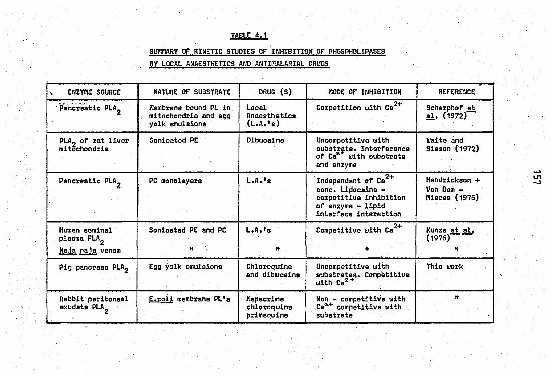
, ENZYME SOURCE .. ,.' ...... _0'
. Pancreatic PLA2
.
PLA2 of rat liver mitochondria
Pancreatic PLA2
Human eeminal . plasma PLA2
NaJa naJa venom
Pig pancreas PLA2
Rabbit peritoneal exudate PLA2
TABLE 4.1
SUMMARY or KINETIC STUDIES or INHIBITION or PHOSPHOLIPASES
BY LOCAL ANAESTHETICS AND ANTIMALARIAL DRUGS
NATURE or SUBSTRATE DRUG (s) MODE or INHIBITION
Membrane bound PL in . Local Competition with Ca2+ mitochondria and egg Anaasthetics yolk emulsions (L.A.'s)
.
Sonicated PE Dibucaine Uncompetitive with substI~te. Interference of Ca with substrate
. and enzyme
PC monolayers L.A.'s . 2+ Independant of Ca conc. Li~ocaine -competitive inhibition of enzyme - lipid interface interaction
Sonicated PE and PC L.A.f s Competitive with Ca 2+
" " n
Egg yolk emulsions Chloroquine Uncpmpetitive with and dibucaine substrates. Competitive
with Ca2. ... "
E.coii membrane PL's Mepacrine Non - competitive with chloroquine Ca~'" competitive with primaquine substrate
RErERENCE .
Scherphof et al, (1972)-..
Waite and Sisson (1972)
Hendrickson + Van Dam -Mieras (1976)
Kunze !!..!!!., (1976)
" Thie work
" .
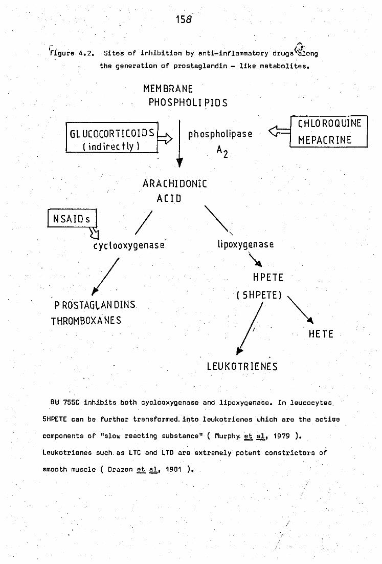
158
I" . rigure 4.2.
,... Sites of inhibition by anti-inflammatory drugs~ong
the generation of prostaglandin - like metabolites.
MEMBRANE PHOSPHOLI PlO S
GLUCOCORTICOIOS ( ind iree tly 1
phospholipase
A2
ARACHIDONIC
ACID
~ I eye I ooxygenas e
;.
..
P ROSTAG~AN DINS
THROMBOXANES
~. "-
lipoxygenase
~. HPETE
( SHPETE)
LEUKOTRIENES
CHLO ROQUINE
MEPACRINE
HETE
SW 755C inhibits both cyclooxygenase and lipoxygenase. In leucocytes
5HPETE can be further transformed. into leukotrieneswhich are the active
components of "slow reacting substance" ( Murphy. et &. 1979 ).
Leukotrienes such. as LTC and LTD are extremely potent constrictors of
smooth muscle ( Drazen~&. 1981 ).
!
;'
159
(which is pain producing), HETE which is chsmotsctic for polymorphs and
the newly discovered leucotrienes which are very potent constrictors of
smooth muscle (Samuelsson et aI, 1979; Drazen et aI, 1981). -- --
• The concentrations of antimalarial drugs required to
inhibit phospholipase activities as presented in thia etudy are higher
than the steady state therapeutic plesma levele achieved during treatment.
For mepacrine thie value is spproximately 10-7M and for chloroquine 10-6M '
(Wollhsim .2i aI, 1978) and for this reason the importance of inhibition
of phospholipase activities by these agents may bs qusstioned. However,
because of their lysosomotrophic nature, the levele achieved in various
tiesuee may exceed the plasma level.:by 400 - 700 fold (Berliner .2i ~,
1948; De Duve .2ial, 1974; Ohkuma and Poole, 1978). At thess levels
,inhibitory activity towards phospholipase A2 ,is observed. The fact that
chloroquine affects on inflammatory disorders are rarely eeen before two
monthe continoue therapy, is a reflection of the time required to build
'up sufficient quantities of the drug.
The ICSO of these agents on the rabbit inflamr ed
-4 -3 peritoneal exudate wsre 3.3 x 10 M for mepacrins and 1.S x 10 M for
chloroquine. On the guinea - pig isolated perfused lung preparation
mepacrine has been ahown to exhibit an ICSO = 20 pg/ml or approximately -S S ><.10 M (Blackwell .2i~, 1978), and thus showing a difference of
approximately '7 fold. Similarly:'chloroquine has been reported to inhibit
PLA2
activity of cultured inflamr,ed rat synovium at concentrations ae
low as 10 pg/ml or 2 x 10-SM (Floman, 1978), and a difference of approx -
imately 7S fold. Thus phospholipases of different tissues show different
suscsptibilities to inhibition by antimalarial drugs. These differences
may be partly related to the differing ability of the tissues to concsntrate,
these,compounde.
160
The inhibition of the generation of prostaglandin - lika
material a may in part axplain eome of the anti - rheumatic effects of the
drug. However inhibition of thie enzyme may also explain the phenomenon of
lipidosis which is a build up of polar lipids in lysosomes. (Drenchahn
and LlIllman - Rat.ch, 1976). The sccumulation of polar lipids can have
grave consequences in tissues such as the retina, where a specially
balanced lipid metsbolism ie essential for normal function. Lipidosis
is one of tha factors which leads to the drug - side effect, retinopathy.
3.5 Low dose stimuletion of phospholipase activity by chloroquine
and mepacrine
On one peritoneal axudate phospholipaee prepsration
both chloroquine and mepacrine at low doses (250~M and 50 ~M respectively)
caused a stimuletion of enzyme activity. These effects were striking
(figure 3.27), reproducible only on this preparation and were lost after
2 months storage at 4°C. They were not observed on approximately twelve
other preparations.
stimulation of phospholipase activity by low doses of
local anaesthetics has also been observed (Scherphof and Westenberg, 1975;
·Kunze!!:.':!jl' "1976,). "Sutthase effects are only observed on micellar
dispersions of phoepholipide and not when the eubstrata was present
in membranes. In this atudy, howaver even when extracted E.coli
phospholipids were used aa eonicated micellar diepersions, they failed to
show low dose stimulatory effects by mepacrine and chloroquina if othar
enzyme preparations were used (table 3.12). These results also highlighted
the differences in activity obtained when tha same substrata is present
as a micellar dispersion or bound in a biological membrane. Thus
phospholipase activity towards E.coli membrane phospholipids was 6 - fold
161
higher than if the eame phospholipids were present as eonicated diepersions.
3.6 Effects of other agents on the peritoneal exudate phospholipase A2
• . four other ~rugs associated with the treatment of
inflammatory disorders were also ecreened at 1mM concentrations. The
anti - arthritic drugs sodium aurothiomalate and 0 - penicillemine
ehowed no effect against the peritoneal exudate phospholipase A2
activity. However the anti - inflammatory drugs indomethacin and
benzydamine were particularly effective showing almoet total inhibition
at 1mM concentratione. furth .. r etudy evaluating IC50 values for the
latter two drugs may be worthwhile to compare their effectiveness against
their potencies towards the cyclooxygenase enzyme syetems. Recently low
concentrations of indomethacin have been reported to inhibit phospholipase
A2 of rabbit PI'IN leucocytes (Keplan .2.!:. .!!ob 1978). 1 /AM indomethacin
reproducibly caused 14% inhibition, and 50#1'1 cauaed 70% inhibition. These
findings are important as they indicate that inhibition of proataglandin
synthesis by indomethacin in leucocytes may be the result of an effect
of the drug on prostaglandin synthetase and phospholipase A2 activity.
3.7 Effects of chlorOquine - like agents on the phospholipase A2 activity
of PI'IN leucocytes
Chloroquine - like agents showed completely contrasting
effects when examined on the phospholipase A2 activity of PMN neutrophils
as compsred with effscts obtainsd on ths peritoneal eXUdate PlA2• Studies
conductsd on eonicated neutrophils, assaying PlA2activity at pH 6.5,
showed stimUlation of activity by chloroquine and mepacrine. This'effect
was dose - related and no biphasic effects, as observed with the peritoneal
162
exudate preparation, were eeen. Thie finding appeare to be unique
as mepacrine and chloroquine have alwaye been observed to inhibit
phospholipase activities (Vargaftig and Oao Hai,1972; Canedella ~ &,
1969; Blackwell et al, 1978). Mepacrine was again observed to be more --active than chloroquine and in this case the difference was 10 fold •
•
Primaquine behaved differently. Although exhibiting
a alight stimulation at low concentrations (at 0.5mM, 13%) a strong
inhibition at dosss greater than 1mM was observed.
The stimulatory action by chloroquine and mepacrine was
further shown on othsr enzyme preparations obtained from neutrophils
including (i) neutrophils suspended in water, (ii) 8,200g pellet granule
"preparations prepared in sucrose, and (iii) on phospholipase activity
released from neutrophils exposed to zymosan - complement particles.
Studies on the 8,2009 pellet showed that the atimulation of activity by
mepacrine was 2 to 3 fold greater at pH 6.5 than at pH 9.0 indicating
that the diprotonated form of the drug was more important for this effect
than the mono:",- charged species. This ia the reverse situation to the
inhibitory effects observed on the peritoneal fluid PLA2 where the effect
was greatest at alkaline pH's. Indeed at pH 9.0 chloroquine showed an
inhibition of the zymosan - cpmplement released "PLA2 activityfurth(;r
eupporting the inhibitory action of the monoprotonated epecies.
Studies usings partially purified PLA2
preparation
obtained by eulphuric acid extraction of neutrophils suspended in water •
revealed even more interesting results. Mepacrine and chloroquine no
longer showed etimulation of PLA2 activity at acid pH but only showed
inhibition. This effect wae not as strong ae that obtained with the peri -
toneal exudste preparation. Mepscrine as Bxpected was more potent than
163
chloroquine, but primaquine showed to be the most potent and its
inhibitory effect was of the same order and magnitude ae that obtained
with the neutrophil homogenate suspensions.
• One explanation of the fact that both chloroquine and
mepacrine etimulated PLA2 activity at acid pH with all the other preparations
except the sulphuric acid extracted enzyme, could be the involment of
another factor present in neutrophils which was lost during the purifi -
cation procedure. This of course assumes that the acid extracted PLA2 , is the ~ activity that is assayed in the homogenate. However the
possibility that different populations of the phospholipases may exist should
not be excluded, Thue in the presence of thie endogenous factor chloroquine
and mepacrine stimulated PLA2 activity while in its absence only inhibition
is observed. The unknown factor may· be an anionic agent eince it probebly
binds with the drugs which are cationic. The possibility that this could •
. be a glycosaminoglycan euch as heparin was investigated since heparin and
to a greater extent the related agent, chondroitin - 4 - sulphate have been
shown to bs present in leucocytes in measurable quantitiee (Olsson and
Gardell, 1976; Olsson ~ al,1958; Avila and Convit, 1976). The latter
authors also found that the glycosaminoglycans were able to inhibit 30
different lysosomal enzymes including PLA2, On s molsr basis the inhibitory
potency was heparin ) chondroitin sulphate > hyaluronic acid, with only
heparin showing activity against PLA2, Thus glycosaminoglycans present a
mode of control of leucocyte lysosomal enzyme activity.
Heparin is certainly abla to interact with all three
antimalarial drugs as clearly observed when solutions of the individual
drugs are added to heparin and the resulting 0.0. changes were measured
(figure 3,31). On a molar basis mepacrin~ again was the most active drug,
164
Heparin itself was found to inhibit PLA2 activity of
neutrophils at acid pH in agroement with the findings of other workers
(Avila an~ Convit, 1976; transon ~~, 1974). If however heparin wae
the endogenous factor· then mixtures of mepacrine and heparin added to -
gether ~o reaction mixtures containing the acid purified PLA2 enzyme,
should show eome antagonistic effects or even stimulation of activity.
Thie etatement again assumes that the acid purified enzyme preparation
ia the same as in the water homogenate. Howaver thie was not eo and tha
anionic and cationic agente together showed additiva inhibitory effects
on the acid purifiad preparation.
It may be fruitful in future Studiee to examine the
possibility that chondroitin sulphate may be involvad. Thia requiree
much higher concentrations than heparin to effect PLA2 activity and is
aleo present in larger quantities. The interaction between chloroquine
and heperin provide another factor for the lysosomotrophic effects of
this drug as ths glycosaminoglycans ie thought to be present in leucocyte
lyeosomes.
Alternatively the etimulation of PLA2 activity may be
due to another factor or even· another phenomenon. transon ~~, (1974)
found the major portion of PLA2 activity of rabbit neutrophils to be
aesociated with the granules and membrane bound. Association of the
enzyme with the membrane may be responsible for the etimulation of activity
observed as chloroquine ie a membrane - active agent. The purification
procedure certainly provides the enzyme in a solubilised state which may
account for the difference in response obtained. Thie however makes
interpretation of the atimulatory effects of chloroquine and mepacrine
165
on the zymosan - complement releaaed enzyme difficult unless one gets
the release of membrane fragments aswell. The peritoneal exudate PLA2 is also considered to be a soluble enzyme (transon ~~, 1978).
Heparin showed a completely contrasting effect on the
peritoneal exudate enzyme. At acid pH heparin showad stimulation of PLA2
activity at exactly the doses required to inhibit the neutrophil enzyme.
This effect was pH dependant as at pH 7.5 ths stimulatory activity shomn
by 10 units/ml heparin was lees than half of the effect at pH 6.0 (table
.3.19). Such differences in response of the two phospholipase activities
can only be explained by the suggestion the peritoneal exudate PLA2 is
a different enzyme and not derived from neutrophils but fro~~another source.
Recently rabbit serum too hae been shown to be a potent
source of PLA2 activity (Kaplan - Harris !l~, 1980). The described PLA2 2+ activity was meximally active at 2mM Ca and higher concentrations led
to a decrease of activi~y. This is very similar to the response obtained
in thie etudy, although no sigmoid behaviour was noted between 0 - 2mM Ca2+.
The stimulation of PLA2
activity of the peritoneal exudate by heparin is
shown primarily at pH 6.0 with a reduced effect at pH 7.5, thus the
physiological significance of this is not known. Heparin administration
to humans in large amounts (5000 units) leads to an increase in serum
PLA activity although thia has been associated with A1 specificity (Vogel'
and Bierman, 1967).
3.8 Consequences of a stimulatory effect of PLA2 of PMN leucocytes at
acid pH
Stimulation of PLA2 activity could be expected to lead
to an'increase in the release of polyenoic fatty acids and lysophospholipids.
166
Generetion of both producte leads to e potentiation of inflammation as
arachidonic acid is the precursor for the proetaglandin related products,
all of which are generated in etimulated neutrophile, and lysophospholipids are
cytotoxic. StimUlation of PLA2 activity by chloroquine at acid pH mey
be particularly important as (1) chloroquine accumuletes in the lysosomes
where the concentration achieved can be up to 700 times that in the medium
(2) the pH of the lysosomes is acid therefore an acid PLA2 activity ie
important and (3) the meJority of the PLA2 activity associated with
rabbit neutrophils is associated with the granules with an acid and an
alkaline pH optima.
Recently many etudiee have been published which relate
products of PLA2 hydrolysis and lysosomal enzyme rslsase from inflammatory
.celle (Naccache et al,1979; Smolen and Weissmann, 1980; Smith, 1978). In .--particular, many stUdies involve the use of chemotactic peptides formyl -
methionyl - leucyl - phanylalanine (FMLP) which also cause lysoaomal
enzyme releese. Thus if human PMN'e that have been treated with cyto -
chalaein B,are exposed to FMLP, then release of lyeoeomal ecid hydrolases
occure. Release of acid hydroleses could be inhibited by 50p M indomethacin,
12 pM 5, 8, 11, 14 eicosatetraynoic acid (ETYA, a structual analogue of
arachidonic acid) or 18)AM para - bromophenacyl bromide ( a PLA2 inhibitor).
Arachidonic acid itself has been sholln to cause degrannulation of rabbit
peritoneal PMN leucocytes although prior treatment with cytochalasin
B ie necessary (Naccache !i~, 1979). The other product of PLA2 action,
lysophospholipids have also been implicated in the release process. They
ere thsmselves msmbrene sctive and thus can promote fusion of granules
to phagoeomes or plasma membranee. They also produce intense inflammation
when applied locally (Thouveot !i~, 1974), intraperitoneally
(Westphal !i~, 1970) or subcutaneously (Philips !i al, 1965). They have
aleo been shown. to stimulate guanylate cyclsse (Shier!i~, 1976) thereby
167
increasing cGMP lsve1s which are known to elevate during release of
1yeosoma1 constitusnts.
Studiss of ths effects of chloroquine show that at
relatively high concentrations it increases the release of lysosomal
enzymes (Ringroee !!.!:. a1, 1975; Weissmann!!.!:..!!!" 1975; Riches'!!.!:..!!!" 1981).
Thie property may appear wrong for a drug that has been used to trsat
inflammatory disorders as most NSAID's and SAID's have generally been
found to dscrsass release of lysosomal enzymes. Csrtain1y if in PMN's
acidic phospho1ipases are important in 1ysoeoma1 enzyme release, then the
sffects of chloroquine observed in this study w&u1d explain the increase
of release of lysosomal enzymse observed by previous workers. The actions
observed by chloroquine occur at ralative1y high doses, and these levels
. can be achieved by virtue of its 1ysosomotrophic activity.
Stimulation of phospho1ipases and therefore of pros -
tag1andin biosynthesis may also have an anti - inflammatory action ae ve~
high levels of prostag1andins are known to have anti - inflammatory effects
euch as inhibition of rate of appearance of 1eucocytes and lysosomal
enzymes at sites of acute inflammation (Zurier et a1, 1973; 80nta and --Parnham, 1978). In 1eucocytes theae actions may arise through incraases
of cAMP levels. Thus the long 1ag - period required for the onset of
action of chloroquine in inflammatory disorders may reflect the time
required to build up sufficient concentrations of drugs to exert its -
effects. future studies·on the effects of chloroquine on prostaglandin
production of neutrophil 1eucocytes may be worthwhile to "evaluate this
point.
168
PART D STUDIES or AN ENDOGENOUS INHIBITOR or PLA2 IN RABBIT
PERITONEAL PMN'S
4.1 Cellular locelizetion end properties
When neutrophils are homogenised in sucrose and then
.centrifuged at B,200g, then the majority (90%) of the PLA2 activity was
. found to be associated with the pellet which conaist in the main of nuclei
and granules.
this was lost
The 8,200g supernatant contained little PLA2 activity and I> upon etorage at 4.C over five daye. On the other hand the
supernatant was also found to contain an inhibitory activity towards
PLA2 of the peritoneal sxudate. The inhibition was dose - depend,nt to
a maximum inhibition of 64% (figure 3.35). This inhibitory activity was
independant of pH and similar dose - response curves were obtained
between pH 5.5 and 9.0.
However inhibition was not indapend:,nt of pH if inhibitory
potency was assayed against PLA2 activity of neutrophil granules (8,200g
pellet). Indeed almost no inhibitory activity was present towards this
preparation at pH 6.5. Activity at pH 7.5 has been reportad (rranson ~!!,
1974) which was heat unstable ie. probably a protein. The different
inhibition patterns obtained indicated another difference between the
peritoneal exudate PLA2 and the neutrophil granule PLA2 activities. Indeed
it is somewhat of an oddity that the neutrophil cytOSOl should contain
an i~hibitor which was active only at neutral and alkaline pH towards the
PLA2 from neutrophil granules and active at" !loth acid and alkaline pH
towards the peritonsal sxudate enzyms.
Inhibition of PLA2 activity by the B,200g supernatant
was not confined to the abovs mentioned prsparations. Phoepholipase A2
169
of partially purified porcine pancreas and of crude Crotalus adamenteus
vanom ware also inhibited to similar extents (tabla 3.23), and therefore
appeared to ahow little selectivity of action. However the PLA2 from NaJa
naJa vanom was not.inhi~ited but a 58% stimulation was recorded. This
stimulation was probably a non - specific protein effect as equivalent
concentrations of BSA were able to stimulate the activities of all the
preparations examined. This is probably due to binding and removal: of
producte, ie. fatty acids and lysophospholipids, which era known to
cause product "inhlbit1on'(F"ranson .I!i!!., 1974).
Why the enzyme from Na.la na.!a venom wae not inhibited
by the neutrophil 8,200g supernatant is not certain. Since the substrate
wss the sams in all cases, it would thus be due to differences in the 2+ " enzymes. All the enzymes tested require;.Ca for activity as 1mM EOTA
was able to totally inhibit every enzyme preparation. This difference
msy be explained upon critical examination of the size and ionic nature
of the proteins used.
Moat venom phospholipases consist of diffarant numbars
of isozymas of alightl~ diffaring molecular waights and isoelectric points.
There is generally an abundance of Dna or a few particular isozymes and
thephospholipase'of"Ir'p-arti:cular venom maybe classified as 'a basic, neutral
or acidic protein with a particular isoelectric point and molecular
weight depanding upon the abundent species. The mora toxic venoms, as
measured by L050 values in mice, generally tend to have basic" cationic
PLA's than the less toxic venoms which contain neutral enzyms proteins
(Condrea .I!i!!.,1980). Reports in the litereture indicated that the PLA2 activities used in this study ie. rabbit peritoneal exudate, rabbit PMN
leucocyte, Crotalus adamenteus venom and pig pancreas ara basic. cationic
prote~ns (F"ranson .I!i!!.. 1978; blaiss .I!i!!.. 1975; Van Den Bosch, 1974;
170
De Haas .!!!.!!" 1970; respectively). But the enzyme from Naja naJa venom,
ie primarily an acidic enzyme with the most abundant isozyme hauing an
isoelectric point of 4.95 (Salach .!!!.&,1971). It is elso a larger protein
of MW = 20,000 while the ,rest are all below 15000MW. This may also indicate
that inhibitory activity present in the neutrophil cytosol ie an acidic
protein. furthr verification of this suggestion requires study with other
known acidic phospholipases.
Recently work by Hirata et el,(1980) hae also demonstreted --the existence of a phospholipase A2 inhibitory protein in the polymorphs
whose synthesis could be induced by glucocorticoids.PMN leucocytes treated
with glucocorticoids were shown to have a reduced response to the chemo -
tactic peptide f Met - Leu - Phe (fMLP) which wae related with an
inhibition of phospholipase activity. The inducad phospholipase inhibitory
protein wes'isolated from water lysed neutrophils which had previously
been incubated with flucinoline acetonide for 16 hours. The precipitate
arieing from water lysed neutrophils was solubilised and subsequent gel
filt -ration on sephadex G200 columns revealed a molecular weight of 40,000.
This protein exhibited inhibitory activity towards PMN and pancreatic
PLA2
• However there is one notable difference: the phospholipase inhibitory
protein as described by Hirata, was lost when the polymorphs were treated
with pronase. As pronase acts on the outer sllrface of the cells, the PLA2
inhibitory protein resides on the plasma membrane. franson .!!!.&' (1974)
'suggested that the PLA2 inhibitory protein is present in the soluble
portion of the cell as it wss alSG present in 100,000g supernatant
preparations of leucocyte homogsnates.
171
4.2 Endogenous inhibitor of PLA2 activity and the mode of action of
steroidel anti - inflammatory drugs
Much intersst has recently been shown in the inhibition
of proeteglandin biosynthesia by steroidal anti - inflammatory drugs (SAID),
which occurs only in intact cells. The fact that glucocorticoids do not
inhibit microsomal cyclooxygenasa (the site of action of NSAIDS) was
shown by Flower:;.!!:. al,(1972). In 1975 two hypothes: s were proposed~ Lawis
and Piper suggested that SAID's prevented the release of prostaglandins
from the cell by virtue of their membrane atabilizing effects and
Gryglewski !i~ suggeeted that glucocorticoids prevented the release
of polyenoic fatty acida from phospholipide, and hence to the 'microsomal
enzyme ayatem for proataglandin biosynthesie. Therefore glucocorticoida
were euggested to interfere with phospholipaee A2 activitiee.
Since then much evidence has been put forward subs tan -
tiaUflg the latter thaory. Danon and Aasouline (1978) showed that inhibition
of prostaglandin biosynthesie' in rat renal papillae by SAIDS involved RNA
and protein synthesis. Thus the glucocorticoids may work by the same
mechanism exhibited by the ateroid hormones on their target cells ie.
combination of the ateroid with a cytosolic receptor protein, translocation
of thia drug - receptor compl'ax to 'the nucleus, Eitimulationof transcrip -
tion and consequent biosynthesis of specific proteins. In 1979 Flower
and Blackwell, using the guinea - pig perfused lung preparation showad
that the glucocorticoids induced the aynthesis of a polypeptide or protein
that inhibited the releaee of arachidonic acid from phospholipids. Indeed
inhibitors of protein ayntheeis euch as cycloheximide were able to block
the effecte of the glucocorticoids. Potent inhibition of phoepholipasa
activity alao explainad why the glucocorticoids are more potant as anti -
inflammatory agenta than NSAIDS, as they prevent availability of subatrate
172
for both the cyclooxygenase and lipoxygenase enzyme systems whsreas
the NSAID's only block cyclooxygenass. Thsse sffscts have bsen furthl~r
demonstrated on rat peritoneal macro phages where incubation with the
drugs not only induced the biosynthesis of an inhibitor of PLA2, but Blso
released it into the medium (Carnuccio ~!!,1980), which wss then assayed
against prostaglandin production by lsucocytes undergoing phsgocytosis
of killsd bacteria. Evidsnce that this inhibitor was preformsd and stored
within macrophages and releassd upon incubation with hydrocortisons was
provided by Blackwell ~!!o (1980a) whose group showed that the inhibitory
factor, isolated from both guinea - pig lungs and rat peritoneal macrophages
was a polypeptide (designated "macroeortinn) of approximately 15,DOOMW.
An inhibitor of phospholipase activity in the cytosol of
rabbit PMN leucocytes was first observed by rranson ~al,(1974) and this.
observstion has been sxtended by this study showing that it was a~tive against
othsr basic cstionic phospholipases but not against the acidic, anionic
phospholipase from Naja na.la venom. Recsnt work by Dray ~!!,(1980)
suggestad;that the rat.leucocytes may be a cell type insensitive to the
actions of steroidal anti - inflammatory drugs. In their study rat peritoneal
PMN leucocytes were incubated with dexamethasone for 2 hrs at 37°C. Then
the cells were seperated from the medie and further incubated with killsd
Bortella pertussis in the presence of dexamethasone and the prostaglandin
levels at the end of this incubstion were measured by radioimmunoassay.
-4 Cells treated with dexamethasone up to 10 M showed no reduction in the
amount of prostaglandins produced. ,
Thsse observstions were furthur substantiatedby work in
our own laboratory (Traynor, Solanky and Authi, unpublished observations).
Rat peritoneal PMN leucocytes were incubated with and without hydrocortisone
. for. 2 ·hrs at 37·C. The phospholipase A2 inhibitory activity present in the
173
8,200g supernatant of leucocyte homogenates towards rat peritoneal exudate
PLA2 was aesayed using (1 - 14C) oleic acid labelled E.coli. Hydrocortisone
-5 up to 10 M showed no increase in the PLA2 inhibitory activity of the
8,200g supernatant preparations.
However work by Hirata !i~,(1980) showed that rebbit
PMNt s do contain an inhibitor of phospholipase activity whose synthesis
could be induced by glucocorticoids but at least 5 - 10 hours of preincubation
with the drugs was neceesary. The phospholipasa inhibitory protein in. this
case had a molecular weight of 40,000. Thus information obtained so far
reveals the PMN leucocytes to differ from macro phages in that longer
incubatione with the eteroids were necessary to ahow an effect and the
resultant inhibitory protein has a higher molecular weight.
Recently the importance of this PMN phospholipase
inhibitory protein has been furth,.r highlighted by the finding that some
. patients suffering from chronic inflammatory conditions such as systemic
lupus erythematosis or rheumatoid arthritis contain in their plasma an
autoantibody towards the'phoepholipase inhibitory protein (Hirata!i al,
1981). Thue in such patients thie autoantibody allows increased formation alld
of arachidonic acid subsequently the formation of inflammatory metabolites. A .
Tha enti - inflammatory steroids have ale. been shown
to enhance the ability .f tissues to breakdown prostaglandins (Moore
and Hoult, 1980).
174
CON C L U 5 ION 5
Phospholipass A2 enzymea are belived to play an
important role in inflammation by their role in providing polysnoic
fatty acids which are the precursors for prostaglandin - like compounds.
The present research shows that one cell type known to accumulate at
. damaged tissues, namely the polymorphonuclear leucocyte, doee release
a phoepholipass A2 enzyme under various conditions of atimulation.
Indesd the degree of phagocytosie by the calls correlates with the degree
of rslease of phospholipase A2•
The released phospholipase A2 activity has been
. characterised and compared with a phospholipase A2 activity found in
the peritoneal inflammatory exudate. Close examination of the properties
end responsee to specific pharmacological agents reveals striking differences
betwsen the two enzymes. Table 4.2 summarises the described properties
of the two enzymes.
In agreement with the findings of other workers using
other tissues, chloroquine and mepacrine were found to inhibit phos -
pholipase A2 of the peritoneal exudate. Inhibition of PLA2 activity may
explain eome of the anti - rheumatic effects of chloroquine as thie
prevents the generation of proetaglandin - like mediators. It may also
explain the major eide effect, lipidosie leading to retinopathy, which
is a build up of polar lipids within lysosomee.
However the effect of chloroquine onphospholipases
is not simply inhibition ae described in the literature, but biphasic
175
TABLE 4.2
COMPARISON OF PROPERTIES OF THE RABBIT NEUTROPHIL RELEASED
PHOSPHOLIPASE A2 AND THE RABBIT PERITONEAL EXUDATE PLA 2•
ENZYME CHARACTERISTICS AND EFFECTS OF PHARMACOLOGICAL AGENTS.
. ENZYME CHARACTERISTICS
PH OPTIMA
Ca2+ REQUIREMENT
Km
STABILITY AT 4°C
MOLECULAR WEIGHTS
(FRANSEN ET AL,1974)
EFFECTS OF PH~RMACO -
LOG ICAL AGENTS
CHLOROQUINE
ANO MEPACRINE
PRIMAQUINE
HEPARIN
NEUTROPHIL B,200g
SUPERNATANT
PMN PLA2
fi. 0 ... 9;0
OPTIMUM = SmM Ca2+
NORMAL HYPERBOLIC
CHARACTERISTICS
~ 25 X 1O-6M
ACID ACTIVITY MORE
UNSTABLE THAN
ALKALINE
15,000
STIMULATION AT ACID
Pill! (INHIBITION ON
ACID - RJRIFIED
PREPARATION)
STIMULATION AT LOW
CONC. STRONG. INHIBITION
AT HIIGH CONe.
INK-IBITION AT ACID
PH.'
INHIBITION AT NEUTRAL
PH(rRANSON ET AL,1974)
PERITONEAL EXUDATE
PLA 2
6.0, 8.0
OPTIMUM = 1. 2mM
SIGNOID KINETICS ANO
GREATER THAN 2mM
INHIBIT S
':!: b70 X 10-6M
POSSIBILITY THAT
ENZYME EXISTS AS A
ZYMOGEN
14,BOO
INHIBITION, BIPHASIC
EFFECTS
INHIBITION
STIMULATION AT ACID
PH
INHIBITION INOEPEND -
ANT OF PH
176
dose - reeponee curves were obssrved. These effscts were furth'. t' repeated o
whon physical effects on membrane stability such. as hyp~onic haemolysis
of erythrocytas were examinad. Indeed chloroquine and mepacrine were
obaerved to stimulate PLA2 activity of PMN leucocytes. This finding may • be very important as it explains the observed increase of lysosomal
enzymas by chloroquine from PMN leucocytes and macrophages. F'urth:.,t'
atudies extending this to proataglandin synthesis may be worthwhile to
elucidate thie point.
Recently much interest has been shown in endogenous
inhibitors of phospholipase activity. Within the lysosomes the
glucoeaminoglycan;: heparin may provide regulation of enzyme activity at
acid pH. However at neutral pH the major control of phospholipase A2
activity is by a phospholipase inhibitory protein whose biosyntheeis
can be enhanced by glucocorticoids. This protein appears to have little
eelectivity of action towards phospholipasee from different sources
except perhaps those that are acidic proteins. The phospholipase inhibitory
protein or the lack of its activity may be of great importance in
inflammatory conditions as recently an autoantibody to this protein has
been found in some patients with rheumatoid arthritis and systemic lupus
erythrematoSis. Thus the presence of this auto antibody may lead to inadequate
regulation of· phospholipase A2 activities in these diseased states.

BIBLIOGRAPHY
177
BIBLIOGRAPHY
ALABASTER, V.A., & BAKHLE Y.S. (1970). The release of biologically
active substances from isolated lungs by 5HT and tryptamine.
Br. J. Pharmec. ~, 582 - 5B3P.
ALl, S.Y., EVANS, L., STAINTHORPE, E., LACK, C.H.,(1967).
Characterisation of cathepsins in cartilage.
Biochem. J.105, 549 - 557.
AMES, G.A. (196B). Lipids of salmonella typhimurium and Escherichia
~ : structure and metabolism.
J. Bacteriology. 95, B33 - B43.
ANOERSON, A.J. (1970). Lysosomal enzyme activity in rats with
adjuvant induced arthritis.
Ann. Rheum. Dis., 29, 307 - 313.
ANDERSON, A.J., BROCKLEHURST, W.E., & WILLIS, A.L. (1971).
Evidence for the role of the lysosomes in the formation of
prostaglandins during carrageenan - induced inflammation in the
rat.
Pharmacol. ~. Commun. 1, 13 - 19.
AVILA, J. L. & CONVIT, J. (1975). Inhibition of leucocytic lysosomal
enzymes by glycosamino glycans in vitro.
Biochem. ~. 152, 57 - 64.
AVILA, J.L., & CONVIT, J. (1976). Physiochemical characteristics of
the glycosaminoglycan - lysosomal enzymes interaction ~ vitro.
Biochem. J. (1976) 160 , 129- 136.
BAGGIOLINI, M., HIRSCH, J.G., & OE DUVE, C. (1969). Resolution of
granules from rabbit heterophil leucocytes into distinct
populations by zonal' sedimentation. J. Cell BioI. ~, 529 - 541
BAINTON, D.F., & FARQUHAR, M.G. (1966). Origin of granules in PMN
leucocytes.
'J. cell. BioI. £.!!., 277 - 301.
178
BARTElS, J., KUNZE, H., VOGT, W., WIllE, G. (1970). Prostaglandin I
liberation from and formation in perfused frog intestine.
Naunyn - Schmiedebergs Arch. ~. ~. Pharmak. 266, 199 - 207.
B~RGSTROM, 5., & SJOVAll, J. (1960). The isolation of prostaglandin
E from sheep prostrate glands.
~chem. Scand.~, 1701.
BERLINER, R.W., EARlE, O.P., TAGGART, J.V., ZU8ROO, C.G., WELCH,
W.J., CONRAN, N.J., ~AUMAN, E., STUOOER, 5. T., & SHANNON,
.J..A •. (,1948)..,,studies on ·the .chemotherapy of the human
malarias. (iv).
,;;t. £!!.!!.. Invest.ll, Symposium on malaria 98 - 107.
BlACKWEL.l, G.J., flOWER, R.J., NIJKAMP, f.P., & VANE J.R. (1978).
Phospholipase A2 activity of guinea - pi9 isolated perfused
lungs: stimulation and inhibition by anti - inflammatory
steroids.
!!£. •.. ,;;t. fharmac. g, 79 - 89.
BlACKWEll, G.J., CARNUCCIO, R., 01 ROSA, M., flOWER, R.J.,& PARENTE,
l. (1980)a. Storage and steroid - induced release from
rat leucoc¥tes of a phospholipase inhibitor.
!!£.. ,;;t. Pharmac. 11, 136P - 137P.
BlACKWEll-, G.J., CARNUCCIO, R., 01 ROSA, M., flOWER, R.J., & PARENTE,
l.,PERSICO, P. (1980)b. Macrocortin : a polypeptide causing
the anti - PlA effect of glucocorticoids.
·Nature,'2,!!1., '147 - '149.
BlIGH, E.G., & DYER, W. J. (1959). A ra~id method of
and purification.
~. ,;;t. 8iochem. fhysiol. lI, 911 - 917.
1;r;4 total"extraction
80NTA, I. l.,& PARNHAM, M.J. (1978). Prostaglandins and chronic
inflammation.
8iochem. fharmac. ll, 1611 - 1623.
80RGEAT, P., ~AM8ERG, M., & SAMUElSSON, 8. (1976). Transformation
. of arachidonic acid and homo - r - linolenic acid by PMN's •
. J R,_, _. rlQJ.~ Chem. ~, 7816 - 7820.
179·
BOWMAN, W.C., & RAND; M.J. (1980). Textbook of pharmacol09Y, 2nd edn.
p.36.6 - 36.8. Black well scientific, Oxford.
BRE5LOFF, P. (1977). Miscellaneous anti - rheumatic drugs and their
possible modes of action.
Advences in Drug Research. 11, 1 - 21.
CARNUCCIO, R., Dl ROSA, M., & PER51CO, P. (1980). Hydrocortisone
induced inhibitor of prosteglandin biosynthesis in rat leucocytes.
!!.t. l. Pharmac. £!!., 14 - 16.
CENEDELLA, R. J., JARRELL, J; 'J., & SAXE, L. H. (1969). Plasmodium
berghei : Production in vitro of Free Fatty Acids. '
Experimental Parasitology. 1!, 130 - 136.
CONDREA, E., YANG, C.C., & R05ENBERG, P. (1960). Composition of a
relatively toxic PLA2
fron Naja Nigricollis venom with a
relatively non - toxic PLA2
from Hemachatus hemachatus venom.
I Enzymatic activity on free and membrane bound substrates.
!l.!.!l. Pharmac. ,£2., (11) 1555 - 1564.
COWEY, F. K., & WHITEHOUSE, M.W.(1966). Biochemical properties
of anti - inflammatorY,drugs - VII.
Biochemical Pharmacology. 21, 1071 - 10B4.
DANON, A.,' & ASSO(LINE, G. (1978). Inhibition of prostaglandin
biosynthesis by corticosteroids requires RNA and protein
synthesis.
Nature. 112, 552 - 554.
DE DUVE, C., DE BARSV, T., POOLE, B., TRDUET, A., TULKENS, P., &
VAN HOOF, F. (1974). Lysosomotrophic agents.
Biochem. Pharmac. ~ 2495 - 2531.
, DE HAAS, G. H. J. A ~ 5LOTBOOM, P.P.M. BANSER & LLM VAN DEENAN.
(1970). Studies on PLA and its zymogen from porcine pancreas.
The comp~ete a.a. sequence.
Biochim. Biophys. ~. £1l, 31 - 53.
180
01 ROSA, M., GIROUD, J. P., WILLOUGHBY, D. A. (1971 la. Studies of
the mediators of the acute inflammatory response induced in
rats in different sites by carrageenan and turpentine.
:1.. Pathol. 1Q!, 15 - 29.
01 ROSA, M., PAPADIMITRIO , J.M., WILLOUGHBY, O.A. (1971)b.
A histopathological and pharmacolog~cal analysis of the mode
of action of non - steroidal anti - inflammatory drugs.
:1.. Pathol. Bacteriol. ~, 239 - 256.
01 ROSA, M., & PERSICO, P. (1979). Mechanism of inhibition of
prostaglandin biosynthesis by hydrocortisone in rat leucocytes.
Br. :1.. Pharmac, ~, 161 - 163.
ORAY, f., McCALL, E., & YOULTEN, L.J.f. (19BO). failure of anti
inflammatory steroids to inhibit prostaglandin production
by rat PMN leucocytes.
~. ~ Pharmac. ~, 199 - 205.
DRAZEN, J. M., LEWIS, R.A., AUSTEN, K.f., TODA, M., BRION, f.,
MARfAT, A., & COREY, E. J. (19B1). Contractile activities
of structural analog5.of leukotrienes C and 0 : Necessity
of a hydrophobic region.
£!:.2.£. ~. ~. Sci. ~. 1!!., 3195 - 319B.
DRENCKHAHN, D., & LULLMANN - RAUCH, R. (1977). Lens opacities
associated with lipidosis - like ultrastructural alterations
in rats treated with chloroquine, chlorphentermine or Iprindole.
f!e.. Eye.Res. li, 621 - 632.
DRENCHAHN, D., & LULLMANN .,. RAUCH, R. (1978). Drug induced retinal
lipidosis: Differential susceptibilities of pigment epithelium
and neuroretina toward several amphiphilic cationic drugs.
Experimental and Molecular Pathology ~, 360 - 371.
ELIASSON, R. (1959). Studies on prostaglandins - Occurence, function
and biol09ical actions.
~. Physiol. Scand. ~, 1 - 73 (suppl. 158).

181
ELSBACH, P., GOLOMAN, J., & PATRIARCA, P. (1972). Phospholipid
metabolism by phagocytic cells VI.
Biochim. Biophys. ~. ~, 33 - 44.
ELSBACH, P., & MARTIN A., RIZACK. (1963). Acid lipase and phospho
'lipase activity in homogenates of rabbit polymorphonuclear
leukocytes. " ,'"
~. l. of Physiology. 205, 1154 - 115B.
ELSBACH, P., VAN OEN BERG, 'J.W.O., VAN OEN BORSCH, H., VAN OEENAN,
L.L .M. (1965). Metabolism of phospholipUs by PMN leu~ocytes.
Biochim. Biopl-I'S. ~. 106, 33B - 347.
ETIENNE, J., PAYSANT, M., GRUBER, A., & POLONOVSKI, J. (1969)a.
Presence d'un precurseur de phospholipase dans le serum
humain normal.
fu!ll. ~. ~. !U&!.. §l, 709 - 716.
ETIENNE, J., PAYSANT, M., GRUBER, A., & POLONOVSKI, J. (1969)b.
Phospholipase A du plasma saguin de ret. Cinetique et
proprietes. Existence d'un precurseur inactif.
fu!ll. ~. ~. !ll£!.. 21, B63 - B73 •
., fEOORKO, M.E. (1967). Effect of chloroquine on morphology of
cytoplasmic granules in maturing human leucocytes - an
ultrastructural study.
l. ~. Invest. ~, 1932 - 1942.
'fEOORKO,'M. E. (1968). Effect of chloroquine on morphology of
leucocytes and pancreatic exocrine cells from the rat.
Laboratory Investigation .1!!., .27 - 37.
fEOORKO, I"i. E., HIRSCH, J.G., &,COHN, Z.A. (1968). Autophagic
vacuoles produced ~ vitro.
l. ~. BioI. 2!!, 377 - 402.
fERREIRA, S.H., MONCAOA, 5., & VANE, J.R. (1971). Indomethacin
a,.d aspirin abolish prostaglandin release from the spleen.
Nature.,lli!.!!!.!ll£!.. 231, 237 - 239.
182
FERREIRA, S.H., & VANE, J.R. (1974). New aspects of the mode of
action of non - steroidal anti - inflammatory drugs.
~. ~. Pha~mac. li, 57 -73.
FISHMAN, W.H. (1955). Beta - glucuronidase.
Ml!. • .!n Enzymol., .ll, 361 - 409.
FLOMAN, Y. (1976). Prostaglandin E production by cultured inf!aml'.ed
rat synovium. Stimulation by colchicine and inhibition by
chloroquine.
Prostaglandins and Medicine • 1, 39 - 48
FLOWER, R.J. & BLACKWELL, G.J. (1976). The importance of phospholipase'
A2 in prostglandin biosynthesis.
Biochem. ·Pharmac. 25, 285 - 291.
FLOWER, R. J., & BLACKWELL, G. J. (1979). Anti - inflammatory'
steroids induce biosynthesis of a phospholipase A2 inhibitor
which prevents p~ostaglandin generation.
Nature. ~, 456 - 459.
FLOWER, R., GRYGLEWSKI, R., HERBAC ZYNSKA - CEDRO,K., & VANE, J.R.
(1972). Effects of anti - inflammatory drugs on prostaglandin
biosynthesis.
Nature • .!:!.!!.!!!. BioI. 238, 104.
FRANS N, R., BECKERDITE, S., WANG, P., WAITE, M., & ELSBACH, P.
(1973). Some properties of phospholipases of alveolar macro
phages.
Biochim. Biophys. ~. ~, 365 - 373.
FRANSDN, R., DDBRDW, R., WEISS, J., ELSBACH, P., & WEGLICKI, W. B.
(1976). Isolation and characterisation of phospholipase A . 2 from an inflammatory exudate •
.;1.. Lipid ~ • .12., (1) 18 - 23.
FRANSON, R., PATRIARCA, P., & ELSBACH, P.(1974). Phospholipid
metabolism by phagocytic cells. Phospholipases A2 associated
with rabbit PMN leucocyte granules •
. J. Lipid Research • .1§., 380 - 388.
183
fRANSON, R., WEISS, J., MARTIN, L., SPITNAGEL, J. K., & ELSBACH, P.
(1977). Phospholipase A activity associated with: the membranes
of human PMN leucocytes.
Biochem • .;1.. ill, B39 - B41.
fRISK - HOLMBERG, M., BERGKVIST, Y., DOMEIJ - NYBERG, B., HELLSTROM,
& JANSSON, f. (1979). Chloroquine serum concentration and
side effects; Evidence for dose - dependant kinetics.
~. Pharm. and Therpeutics. ~, 345 - 350.
GADDIONI, G., CARRARO, POoR., CAPIJANI, G. (1964).
Archivio Italiano El Dermatologia, Venerologia & Sessilogia.
33, 397. The. .effec.t or c-I.lo ... "''t> ..... ''e \>" fibyo bl",~ t.,. G-«L.4uecl "'" ~. PIU!;~. Eff"c.~ 0'" eellu.l"-,, Y\'\ovr"ol"j~.
GOLDSTEIN , I. M., HOffSTEIN, S. T., WEISSMANN, G. (1975).
Influence of divalent cations upon complement mediated enzyme
release from human polymorphonuclear leucocytes •
.;1.. Immun. 01. ~, 665 - 670.
GOLDSTEIN, I. M., MALMSTEN, C.L., KINDAH, I., KAPLAN, H. B., RADMARK,
0., SAMUELSSON, B., & WEISSMANN, G. (197B). Thromboxane
generation by human peripheral blood PMN leucocytes •
.;1.. ~. ~. ~, 7B7 - 792.
GLATT, M., PESKAR, B., & BRU~E, K. (1974). Leucocytes and prosta -
glandin in acute inflammation.
Experientia. 30 (11), 1257 - 1259.
GOLDBLATT, M. W. (1933). A depressor sUbstance in seminal fluid •
.;1.. ~. Cheril. l!!!!. g, 1056
GRUNDMANN, M., VRUBLOVSKY, P., DEMKOVA, V,., MIKULIKOVA, I., &
PEGRIMOVA, E. (1972). Tissua distribution and urinary excretion
of chloroquine in rats.
Arzneimittel - forschung, 11, B2 - BB.
GRYGLEWSKI, R. J., PANCZENKO, B., KDRBUT, R., GRADZINSKA, L., &
OCETKIEWICZ, A. (1975).,Corticosteroids inhibit prostaglandin
release ,from perfused mesenteric blood vessels of rabbit and
. from perfused lungs of sensitised guinea - pigs.
,Prostaglandins. !Q.. 343 _ 355.
1S4
HAMBERG, M., SVENSSON, J., & SAMUElSSON, B. (1975~. Thromboxanes,
A new group of biologically active compounds derived from
prostaglandin endoperoxides.
£!:£E.. ~. ~. 2£!.. ill· 11" 2994 - 2998.
HAYE, B., CHAMPION,S.; & JACQUEMIN, C. (1973). Control by TSH of
a phospholipase A2 activity, a limiting factor in the bio -
synthesis of prostaglandins'in the thyroid. ~. Letters ~, 253-260.
HENORICKSON, H.S., & VAN'. DAM - MIERAS, M.C. (1976). Local
enaesthetic inhibition of pancreatic phospholipase A2 action on
lecithin monolayer.
:!. Lipid .!l2 • .11, 393 - 398.
HENSON, P. M. (1971). The lmmunologic release of constituents
from neutrophU·leuk"ocytes.
Journal EL Immunology. 2Ql (6), 1535 - 1557.
HERS, H. G., & VAN HOOt, t. (1969). Genetic abnormalities of lysosomes.,
from, "Lysosomes in Biology and Pathology", edited by Oingle,
J. T. and Fell, H. B. 2, 19 - 40. (North. Holland, Amsterdam.)
HIGGS, G. A., BUNTING,S., MONCAOA, 5., & VANE, J. R. (1976). R'lN
leucocytes produce thromboxane A2 ~ like activity during pha90cy -
tosis.
Prostaglandins. 11 (5), 749 - 757.
HIGGS, G. A., McCALL, Eo, & YOULTEN, L. J. t. (1975). A chemotactic
role for prostaglandins released from PMN leucocytes during
phagocytosis •
.[!:. :!. Pharmac. §1" 539 - 546.
HIGGS, G. A., & YOULTEN, L. J. t. (1972). Prostaglandin production by
rabbit peritoneal PMN leucocytes in vitro.
~. :!. Pharmac. 44, 330p.
/
1'95
HIRATA, r., CARMINE, R.D., NELSON, C. A., A'XE.IROD, J., SCHlrrMANN, E.,
WARABI, A., DE BLAS, A. L., NIRENBERG, M., MANGANIELLD, V.,
VAUGHAN, M., KUMAGAI, 5., GREEN, I., DECKER, J.L., & STEINBERG,
A.D. (19B1). Presence of autoantibody for phospholipase inhibitory
protein, lipomodulin, in patients wi~h rheumatic diseases.
~. Nat!. ~. 2&. !!2E.. 1..[, 3190 - 3194 •.
HIRATA, r., SCHlrrMANN, E., VENKATASUBRAMANIAN, K., SALOMON, D., & AXELROD, J. (1980). A phospholipase A2 inhibitory protein in
rabbit neutrophils induced by glucocorticoids •
. ~.!!.ll!:.:8£2&.Sci. !!2E.. 11., 2533 - 2536.
HOMEWOOO, C. A., WARHURST, D.C., PETERS, W., & BAGGALEY, V.C. (1972).
Lysosomes, pH and the antimalarial action of chloroquine.
Nature. ~, 50 - 52.
IGNARRO, L. J. (1971). Dissimilar effects of anti - inflammatory drugs
on stability of lysosomes from peritoneal and circulating
leucocytes and liver.
Biochem. Pharmac. 1£, 2861 - 2870.
INGLOT, A.D., & WOLNA, E. (1968). Reactions of non - steroidal anti -
inflammatory drugs with the erythrocyte'membrane.
Biochem. Pharmacol. 11, 269.- 279.
JlNOAL, M.N., PATEL, M.A., & JOSEPH, A.D. (1960). Local anaesthetic
action of antimalarials. (chloroquine and amodiaquine).
B£Eh. lni. Pharmacodyn. 127, 132 - 139.
KAPLAN, L., WEISS, J., & ELSBACH, P. (1978). Low concentrations of
indomethacin inhibit phospholipase A2 of rabbit polymorphonuc -
lear leucocytes.
Proc. Natl. ~. 2&. USA. li, 2955 - 2958.
KAPLAN - HARRIS, lo, WEISS, J., MOONEY, C., BECKERDITE - QUAGLIATA, 5.,
& ELSBACH,. P. (1980). The action of human and rabbit serum
phospholipase A2 on E.coli phospholipids.
~. Lipid ~. 11, 617 - 624.
186
KAYLEY, G., & WEINER, R. (1971). Effect of PGE, on leucocyte
migration.
Nature ~~. 234, 114 - 115.
KUEHL, F.A., HUMES, J.L., EGAN, R.W., HAN, E.A., BEVERIDGE, G.C., &
VAN ARMAN, C.G. (1977). Role of prostaglandin endoperoxide PGG2
in the inflammatory respo~se.
Nature ~, 170 - 173.
KUNZE, H., NAHAS, N., TRAYNOR, J.R., & WURL, M. (1976). Effect of
local anaesthetics on phospholipases. '8.iocliM ",i" ~;op~~ kl;" ",' 441, 93 - 102.
KURODA,K. (1962). Detection and distribution of chloroquine metabol -
ites in human tissues.
1. Pharmacol. ~. Therapeutics. 121, 156 - 161.
KURZROCK, R., & LIEB, C.C. (1930). Biochemical studies of human semen
11. The action of semen on the human uterus.
£!:2£. ~~. BioI. f!.2£. 2§., 26B - 272.
LEWIS, G.P., & PIPER, P.J. (1975). Inhibition of release of prostagl -
andins as an explanation of some of the actions of anti -
inflammatory steroids.
Nature. London. 254, 30B - 311.
LI, T.K., & MAGNES, L.J. (1972). Inhibitio~ of liver alcohol dehydroge
nase by primaquine and 8 - amino - 6 - methoxyquinoline compounds.
Biochem. Pharmac • .£1., '17- '25.
LIE, 5.0., McKUSIK, V.A., & NEUFIELD, E.F. (1972). Stimulation of genetic
mucopolysaccharidosis in normal human fibroblasts by alteration
of the pH of· the medium.
E!2£. ~. ~. Sc!. !:l2.a. 69, 2361 - 2363.
LIE, 5.0. & SCHOFIELD, B. (1973). Inactivation of lysosomal function in
normal cultured human fibroblasts by chloroquine.
Biochem. Pharmacal. 11, 3109 - 3114.
1 ?l7
LOWRY, O.H., ROSEBROUGH, N.J., fARR, A.L., & RANOALL, R.J. (1951).
Protein measurement with the folinphenol reagent.
1. Biol Chem. ill, 265 - 275 •
. LULLMANN, H., LULLMANN - RAtlCH, R., & WASSERMANN, O. (1975). Drug
induced lipiosis.
£!ii. ~. Toxicol. i, 185 - 218. Also in :
Biochem. Rlarmacol. 27, 1103 - 1106.(1976). (commentary).
LULLMANN, H., LULLMANN - RAVCH, R., & WASSERMANN, O. (1976). Lipidosis
induced by amphiphilic cationic drugs.
Biochem. Pharmacol. 27, 1103 - 1106.
2+ LULLMANN, H., POLSCH, H., & ZIEGLER, A. (1960). Ca Replacement by
cationic amphiphilicdrugs from lipid monolayers.
Biochem. Pharm. 12., 2969 - 2974.
MARKUS, H.6., & 6ALL, E.G. (1969). Inhibition of lipolytic processes
in rat edopose tissue by antimalarial drugs.
6iochim. 6iophys. ~. 
188
NIEUWENHUIZEN, W., KUNZE, H., & DE HAAS, G.H. (1974).Methods in
Enzymology vol. ~, Biomembranes part B, edited by S. rleischer
andl. Packer. p. 147 - 1S4.
NDRTHOVER, B.J. (1977). Effects of indomethacin and related drugs 2+ on the Ca dependant secretion of lysosomal and other, enzymes
by neutrophil polymorphonuclear leucocytes !n vitro.
Br. J. Pharmac. 253 - 259.
OHKUMA, S., & POOLE, B. (1978). rluorescent probe measurement of the
intralysosomal,pH in living cells and the perturbation of pH
by various agents.
£!:2£. !:illll. ~ • .§£!.. ~. 12" 3327 - 3331.
OLSSON. I., & GARDElL, S. (1967). Isolation and characterisation of
glycosaminoglycans from human leucocytes and platelets.
Biochim. Biophys. ~ • .!!lo 348 - 357.
OlSSON, I., & GARDELL, S. (1968). Biosynthesis of glycosaminoglycans
(mucopolysaccharides)in human leucocytes.
Biochim. Biophys. ~. ~, 309 - 323.
PAlMER, M., PIPER, P.J., & VANE, J.R. (1973). Release of rabbit aorta
contracting substance (RCS) and prostaglandins induced by chsmical
or mecbanical stimulation of guinea - pig ,lungs.
!!!:.. 1,. Pharmac. ~, 226 - 242.
PATRIACA, P., BECKERDITE, S., & ELSBACH, P. (1972). Phospholipases
'and 'phosphol'1pidturnover in E.coli spheroplast.
Iliochim.· B'iciphys •. ,Acta •. 260,.593 .. ,600;: .... ~'" .. "~' .. , , .
PERPER, R.J., & ORONSKY, A.l. (1974). Enzyme release from human leucocytes
and degradation of cartilage matrix.
Arthritis ~ Rheum. 11, 47 - 55.
PHIlIPS, G.B., BACHNER, P., McKAY, D.C. (1965). Tissue effects of
lysolicithin injected subsequently in mice, includinglyso -
phospholipids as cytotoxic agents.
£!:2£ • .§£!:.. ill· Biol. !!2!!.. lli.,. 846 - 850.
189
PIPER, P.J., & VANE, J.R. (1969). Releese of additional factors.in
anaphylaxis and its antagonism by anti - inflammatory drugs.
Nature. (Lond.) m, 29 - 35.
RAHEJA, R.K., KAUR, c., SINGH, A., & BHATIA, 1.5. (1973). A new
colorimetric method for the quantitative estimation of
phospholipids without acid digestion.
1. Lipid~. li, 695 - 697.
RICHES, D.W.H., MORRIS, C.J., & STANWORTH, D.R. (19B1). Induction of
selective acid hydrolase release from mouse macro phages during
exposure to chloroquine and quinine.
Biochem. Pharmac. 2Q, 629 - 634.
RINGRDSE, P.S., PARR, M.A., & McLAREN, M. (1975). Effects of anti -
~ inflammatory: aad -other: ,cgmpouQd 9 on the ·.bel~ase_'Qf 1 yeosorhal:'u
; .. ~nzymes- t:r:om -macrophages.
Biochemical Pharmacol. £i, 607.
ROSENBERG, L.S., & SCHULMAN, S.G. (197B). Tantomerism of singly
protonated chloroquine and quinacrine.
1. Pharmaceutical Sciences. 67, (12) 1770 - 1772.
RDSENTHAL, A.f., & CHING - HSIEN HAN, S. (1970). A study of phospholi
pase A inhibition by g~ycerophosph.atide analogQes in various
systems.
Biochim. Biophys. ~. ~, 213 - 220.
RUBIN, M., BERSTEIN, H.N., & ZVAIfLER, N.J. (1963). Studies on the
i»larma!;,O'log,. of cht.oroquide.
Archives of Dpthalmology. 70, 474 - 481.
SALACH, J.I., TURINI, P., SENG, R., HAUBER, J., & SINGEq, T.P. (1971).
Phospholipase A of snake venoms.
1. ~. ~. 245, 331 - 339.
SAMUELSSON, B., BDRGEAT, P., HAMMARSTROM, S., & MURPHY', R.C •. (1979).
Introduction of a nomenclature; Leukotrienes.
Prostaglandins. 11, 785 - 787.
,190
SANDO, G.N., TITUS - OILLON, P., HALL, C.W., & NEUfELD, E.f. (1979).
Inhibition of receptor - mediated uptake of a lysosomal enzyme
into fibroblasts by chloroquine, procaine and ammonia.
Experimental ~ Research. ~, 359 - 364.
SCHERPHOr., G.' L., SCARP, A., & VAN .TOURENENBERGEN, A. (1972). The
effect of local anaethetics on the hydrolysis of free and
membrane bound phospholipids catalysed by various phospholipases.
SCHERPHOf, G., & WESTENBERG, H. (1975). Stimuletion and inhibition
of pancreatic phospholipase A2 by local anaesthetics as a
result of their intraction with.the substrate.
Biochim. Biophys. ~. ~, 442 - 451.
SEyDEL, J.K., & WASSERMANN, O. (1976). NMR studies of the molecular
basis of drug - induced phospholipidosis.
Biochem. Pharmac. ~, 2357 - 2364.
SHIER, W.T., BALDWIN, J.H., NILSEN - HAMILTON, M., H~ILTDN, R.T.,
& THANASSI, N.N. (1976). Regulation of guanylate and adenylate
cyclase activities by lysolecithi~ •
.I:!2!l. ~. ~ • .§£l. ~. 11, 15B6 - 1590.
SMITH, J.8., & WILLIS, A.L. (1971). Aspirin selectively inhibits
prosteglandin production in human platelets.
Nature New!li£!.. m, 235 - 237.
SMITH, R.J. (1978). Non steroid anti - inflammatory .agents : Regulators
of the phagocytic secretion of lysosomal enzymes from Guinea -pig
Neutrophils.
;L. Pharmac. ~. ~. 207, 61B - 629.·
SMOLELIS, A.N., & HARTSELL, S.E. (1949). The determination of lysozyme.
;L' Bact. 58, 731 - 736.
191"
SMOLEN, J. E., &'WEISSMANN, G. (1980). Effects of indomethacin, 5,8,11,
14. eicosatetraynoic acid, ETYA and p - bromophenacyl bromide
(BPB) on lysosomal enzyme release and superoxide an generation
by human polymorphonuclear leukocytes.
,Biochem. Pharmac. 12., 533 - 538.
THOUVENOT, J.P., DURAUT, 5., & DOUSTE - BLAZY, L. (1974). Activite
phospho1!pasique a dans les li,quides d' epanchement au cours
des pancreatites aigues.
&£!!.. .E!:.. t!.&. ill!!?. Q.!.g,. 63, 479 - 483.
TOMLINSON, R.V., RINGOLD, H.J., QURESHI, M.C., & rORCHEILLI, E. (1972).
Relationship between inhibition of prostaglandin synthesis and
drug' efficacy : support for the current theory on modes of
action of aspirin - like drugs.
Biochim. 8iophys. ~. Commun. 46, 552 - 559.
TRAYNOR, J.R., & KUNZE, Ho' (1976). Effects of local anaesthe.tics on
the, lipase ,of flhizopus Anhizus.
Biochim. Biophys. ~. ~, 246 - 252.
VAN DEENAN, L.L.M., & DE HAAS, G.H. (1966). Phosphoglycerides and
phospholipases.
Ann. Rev. 8iochem. 35, 157 - 194. -,- -
VAN OEN BOSCH, Hi,' (1974). Phosphoglyceride metabolism (review).
&m!!..,~. 8iochem. 43, 243 - 277.
VANE, J.R.(1971). Inhibition ofprostagl,andin synthesis as a mechanism
of action for aspirin - like drugs.
Nature ~ §l2!. 231, 232- 235.
VARGAfTIG" B.B., & DAO HIU, N. (1972). Release of rabbit aorta contr -
acting substance and PG's from perfused lung tissue by pho.spholi -
pases.
1. ~. Pharmac. li, 158 - 161.
VOGEL, W.C., &.:.: BIERMAN.E.(1967). Post - heparin serum lecithinase in
man and its positional spec!ficity •
. 2,. Lipid fu!.!!.:.. ,ll.,' 46 -53.
192
VON EUlER, U. s. (1936). On the specific vasodilating and plain muscle
stimulating substances from accessory genital glands in man and.
certain animals (prostaglandin and vesiglandin).
~,I Physiol (lond) .§.!l,i. 213 - 234.
WAITE, M., GRIffIN, H., & fRANSON, R. (1975). The phospholipases A of
lysosomes. from frontiers of Biology Ed. A. Neuberger & E. L.
Tatum.
lysosomes in Biology & Pathology, ~, 255 - 305.
Ed. J.T. Oingle & R.T. Dean.
WAITE, M., & SISSON, P. (1972). Effect of local anaesthetics on phos -
pholipases from mitochondria and lysosomes. A probe 'into the
role of calcium ion in phospholipid hydrolysis.
Biochemistry 11, 30B9 -3105.
WEGLICKI, W.G., RUTH, R.C., OWENS, K., GRIffIN, H., & WAITE, B.M. (1974).
Changes in lipid composition of triton - filled lysosomes during
lysis.
Biochim. Biophys. ~. 2lI, 145 - 152.
WEISS,'J., fRANSON, R., BECKEROITE, 5., SCHNEIDlER, K., & ElSBACH, P.
(1975). Partial characterisation & purification of a rabbit
Granulocyte factor that increases permeability of E.coli.
~. Clin. ~. Invest. 55, 33 - 42.
WEISS, J., BECKERDITE ,- QUAGLIATA, 5., & ElSBACH, P. (1979).
Oeterminants of the action of phospholipase A on the envelope
phospholipids of E.coli. '
;!. BioI. £!!.!!.!!!.. 11, 11010 - 11014.
WEISSMANN, G. (1964). labilization and stabilization of lysosomes.
federation Proceedings. 11, 1038 - 1044.
WEISSMANN, G., SPIlBERG, I., & KRAKANER, K. (1969). Arthritis induced in'
rabbits by lysates of granulocyte lysosomes.
Arthritis Rheum. 1£, 103 - 116.
193
WEISSMANN, G., ZURIER, R.B., & HDFFSTEIN, S. (1972). Leukocytic
proteases and the. immunologic release of lysosomal enzymes.
~. ~. Pathol. ~, 539 - 564.
WESTPHAL, D., fISHER, H., & MUNDER, P.G. (1970). Lysophospholipids and
inflammation.
~.' Bth Int. Congr. Biochemistry, Interlaken. 319.
WETZEL, B.K., HORN, R.G.,.& SPICER, S.S. (1967). Fine structural
studies on the developement of heterophil, eosinophil and basophil
granulocytes in rabbits.
• ~. Invest • .1.§" 349 382.
WEISSMANN, U.N., 01 DDNATO, S., & HERSCH KDWITZ, N.N. (1975). Effect of
chloroquine on cultured fibroblasts: Release of lysosomal
hydrolases and inhibition of their uptake.
Biochemical & Biophysical Research Communications. 66, /4. 133B - 1343
WILLIS, A.L. (1969). Release of'histamine, kinin, and prostaglandin
during carrageenan induced inflammation in the rat.
Prostaglandins, Peptides and Amines (P. Mantegazza and E.W. Horton,
eds). p. 31 - 3B, Academic Press, London.
WIBO, M., & POOLE, B. (1974). Protein degl;'adation in cultured cells
II. The uptake of ch',o.roquine by rat fibroblasts and inhibition
of cellular protein degradation and cathepsin B1
•
~. Cell. BioI. ,§l, 430 - 440.
WDLLHEIM, F.A., HANSON, A., & LAURELL, C.B. (197B). Chloroquine
treatment in Rheumatoid Arthritis. ,'.l • ~."
Scand. ~. Rheumat. 1, 171 - 176.
WROBLEWSKI, F., & LA DUE, J.S. (1955). Lactic dehydrog~nase activity
in blood.
Proc. Soc. exp.~. ~. 2.2" 210 - 213.
194
WURSTER, N., ELSBACH, P., RAND, J., & SIMON, E.J. (1971). Effects of
levorphanol on phospholpid metabolism and cnmposition in E.coli.
Biochim. Biophys. Acta. ill, 2B2 - 292.
ZURIER, R.B., HOrrSTEIN, 5., &.WEISSMANN, G. (1973)a. Supression of
acute and chronic inflammation in adrenalectonised rats by
pharmacologlc amounts of prostaglandins.
Arthritis Rheum. 1.6, 606 - 618.
ZURIER, R.B., HOrrSTEIN, 5., & WEISSMANN, G.(1973)b. Mechanisms of
lysosomal enzyme release from, human ,leukocytes. I. Effect of
cyclic nucleotides and colchicine •
.;t. £!!ll. !!.!El. ~~ 27.
ZURIER, R.B., &.SAYADOrr, D.M. (1975). Release of prostaglandins
from human PMN leukocytes.
Inflammation. 1,. C,' 93 - 101.

· . I
I
I

Biochimica et Biophysica Acta, 665 (1981) 571-577 Elsevier/North-HoUand Biomedical Press
BBA 57884
571
PHOSPHOLIPASE A, ACTIVITY OF LYSOSOMAL ORIGIN SECRETED BY POLYMORPHONUCLEAR LEUCOCYTES DURING PHAGOCYTOSIS OR ON TREATMENT WIm CALCIUM
JOHN R. TRAYNOR and KALWANT S. AUTHI
Biochemical Pharmacology Laboratory, Department of Chemistry. University of Technology, Loughborough. Leicestershire, LEll 3TU (U.K.)
(Received March 13th, 1981)
Key words: Lysosomal enzyme; Phospholipase A2,' Ca2+ treatment; Phagocytosis; (Neutrophil, Leucocyte)
I. Peritoneal neutrophil leucocytes, derived from the rabbit, release phospholipase A (EC 3.1.1.4) activity during phagocytosis of complement-coated zymosan particles, or during treatment with Ca". This enzyme is able to
, release [1.14C]oleate from the membrane phospholipids of Escherichia coli. 2. The release of phospholipase A paralleled that of the known lysosomal marker enzymes Il-glucuronidase and lysozyme. The phospholipase A would thns appear to be derived from the lysosomal granules of the cells. 3. The released enzyme is of A, spec· ificity, has an absolute requirement for divalent cations, and is active over a broad pH range (pH 6-9) .
Introduction
The importance of prostaglandins and related metabolites of arachidonic acid in inflammatory reac· tions is well documented, as indeed is the role of phospholipase A, in freeing arachidonic acid from phospholipids [1,2]. The appearance of prostaglan' dins in some inflammatory exudates is paralleled by infiltration of the inflamed area with polymorpho· nuclear leucocytes. A close correlation between the release of lysosomal enzymes from these cells and the appearance of prostaglandins led to the suggestion that lysosomal phospholipases A" derived from these cells, cause the release of precursor fatty acids for prostaglandin production [3].
A variety of physiological and pharmacological stimuli are able to cause the selective release of lysosomal contents from neutrophils. We have inves· tigated the ability of two such stimuli to cause the release of phospholipase A, activity from rabbit peritoneal polymorphonuclear leucocytes. The stimuli we have used are Ca'· [4] and the phago·
Abbreviation: Hepes, N-2 ,-hydroxycthylpiperazine-N' -2-ethanesulphonic acid.
.!
cytosis of zymosan particles coated with complement [5]. This current paper presents the results of these findings and a discussion of the properties of the released enzyme.
A preliminary account of parts of this work has' been presented [6].
Materials and Methods
All reagents and solvents used were of analytical grade.
Neutrophil suspension Female rabbits of the New Zealand White strain,
weighing 2-3 kg, were lightly anaesthetised with diethyl ether and injected intraperitoneally with 100 ml sterile thioglycollate medium (U .S. Pharmacopea), supplied by Oxoid Ltd. (Basingstoke, U.K.). After 19-22 h each rabbit was anaesthetised by intrave· nous injection of 80 mg . kg-I sodium pentobarbital,' then given 150 ml sterile isotonic saline, containing 4 I.U./ml heparin, into the peritoneal cavity. The abdomen was lightly massaged and the peritoneal fluid allowed to drain from the cavity via a 16 gauge perforated plastic catheter. The cells in the fluid were collected by centrifugation at room temperature for
0005·2760/81/0000-0000/$02.50 © 1981 Elsevier/North·Holland Biomedical Press
S72
4 min, at 200 Xg. The cell pellet was resuspended in 0.83% NH4C1 in water (pH 7.2) to lyse contaminating erythrocytes. After 5 min, the cells were coUected and washed by resuspension and centrifugation in a buffered medium containing: 150 mM NaCI; 3 mM KCI, 10 mM glucose, 5 mM Hepes, adjusted to pH 7.4 with NaOH. Microscopic exainination of the final suspension showed that neutrophils accounted for more than 95% of the total leucocyte population.
Release experiments Aliquots of the leucocyte suspension, containing
approximately I . 107 cells in I ml Hepes buffered medium were incubated in the presence of varying amounts of complement-coated zymosan particles (0-5 mg) prepared as described by Hemen [5], or calcium (0-5 mM) added in 0.2 m1 buffered medium. Incubations were performed with shaking at 37°C for 60 min, unless stated otherwise. After incubation the tubes were cooled in melting ice and the cells removed by centrifugation at 3 000 Xg for 5 min. An aliquot of the supernatant was taken for subsequent assay of released enzymes. Protein in the samples was determined by the method of. Lowry et al. [7] using bovine serum albumin as standard. In experiments using complement-coated zymosan particles the uptake of particles by the neutrophils was determined by phase-contrast microscopy" counting at least 100 cells.
Enzyme assays The marker enzymes /l·glucuronidase, lysozyme
and lactate dehydrogenase were assayed by methods described by others [4]. The results of all assays were expressed as a percentage of the activity released in the presence of Triton X·100 at a fmal concentration of 0.2%. It was assumed that Triton released aJi leucocyte . enzyme activity into the suspending medium.
Phospholipase A. activity was determined by the ability to remove [1.l4Cjoleic acid (The Radio·
. chemical Centre, Amersham, U.K.) from the membrane phospholipids of E. coli prepared as described by Patriarca et al. [8]. More than 95% of the incor· porated 1_14C label was in the 2-position of the membrane phospholipids, as shown' by treatment with the phospholipase A. of crude Crotalus adamanteus venom.
Enzyme assays were performed using an incubation medium consisting of, unless otherwise stated, 7.5 . 10' autoclaved labelled bacteria, containing approximately 15 nmol of phospholipid (determined as described in Ref. 9). 40 mM Tris-malic acid buffer, pH 6.0, 5 mM CaCI. and an appropriate aliquot of the supernatant, in a total volume of 0.5 ml. Blanks without enzyme were included in all assays. Incubations were carried out for 60 min at 37°C in a shaking water bath, then stopped and hydrolytic activity determined as described previously [10]. Radioactivity in substrates and products was counted in a medium containing 3 ml water and 7 m1 Unisolve I (Koch-Light, Colnbrook, U.K.). Enzyme activities were corrected for nonenzymic hydrolysis and expressed as initial velocities or, in release experiments, as a percentage of the total phospholipase A. activity released after sonication (6 /Lm, 3 min). This was preferred to Triton X-lOO treatment since detergents are known to interfere with the activity of phospholipase A. Control experiments with the marker enzymes showed that there was no difference between the total amounts of enzyme released by the two treatments. Phospholipase A, activity due to the complement~ coated zymosan particles preparation was I % or less of total cellular enzyme.
Enzyme activity was also determined against E. coli labelled with [1-14C]palmitic acid (The Radiochemical Centre, Amersham, U.K.) of which 77% was incorporated into the I-position of membrane phospholipids as determined by treatment with the phospholipase A, of C. adamanteus venom.
Results
Release a/phospholipase A during phagocytosis Neutrophil leucocytes incubated with serum
treated zymosan, and therefore particles coated with complement, at 37°C over 60 min demonstrated the ingestion of particles and the release of phos'pholipase A activity. The release was concentrationdependent, being linear up to 0.5 mg coated zymosan particles and continuing up to a maximum release at 5 mg zymosan. By this method a maximum of 30% of total cell phospholipase A (activity released by sonication) was obtained. The release of phospholipase A activity was paraJieled by the uptake of
• (
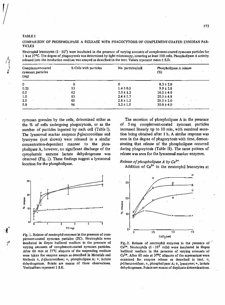
" I'
," ,I
573
TABLE I
COMPARISON OF PHOSPHOLIPASE A RELEASE WITH PHAGOCYTOSIS OF COMPLEMENT-COATED ZYMOSAN PARTICLES
Neutrophilleucocytes (1 . 107) Were incubated in the presence of varying amounts of complement-coated zymosan particles for 1 h at 37°C. The degree of phagocytosis was determined by light microscopy, counting at least 100 cells. Phospholipase A activity released into the incubation medium was assayed as described in the text. Values represent mean ± S.O.
Complement-coated zymosan particles (mg)
o 0,25 0,5 1.0 2.5 5.0
% Cells with particles
o 53 62 83 85 96
zymosan granules by the cells, determined either as the % of cells undergoing phagocytosis, or as the number of particles ingested by each cell (Table I), The lysosomal marker enzymes Il-glucuronidase and lysozyme (not shown) were released in a similar concentration-<iependent manner to the phospholipase A, however, no significant discharge of the cytoplasmic enzyme lactate dehydrogenase was observed (Fig, 1). These findings suggest a lysosomal location for the phospholipase,
30
2D
~ '$ 10 •
0 0 2
ze (mgl
Fig. 1. Release of neutrophil enzymes in the presence of complement-coated zymosan particles (Ze). Neutrophils were incubated - in Hepes buffered medium in the presence of varying amounts of complement-coated zymosan particles. After 60 min at 37°C aliquots of the suspending medium were taken for enzyme assays as described in Materials and Methods 0, f3-g1ucuronidase; .0., phospholipase A; +, lactate dehydrogenase. Points are means of three observations. Vertical bars represent 1 S.E.
No. particles/cell
o 1.4 ± 0.5 2.0± 1.2 2.4 ± 1.7 2.8 ± 1.2 3.2 ± 1.5
Phospholipase A release (%)
8.3 ± 2.0 9.9 ± 2.8
16.0 ± 4.0 20.3 ± 4.8 25.0 ± 2.0 30.0±4.0
The secretion of phospholipase A in the presence of 5 mg complement-coated zymosan particles increased linearly up to 10 min, with maximal secretion being obtained after 1 h, A similar response was seen in the degree of phagocytosis with time, demonstrating that release of the phospholipase occurred during phagocytosis (Table II), The Same pattern of release was seen for the lysosomal marker enzymes_
Release a/phospholipase A by Ca'· Addition of Ca'· to the neutrophilleucocytes at
o o
30
o
20 0
• • 0 • 1!
" 10 •
o o 05
CaCI2(mM]
Fig.2. Release of neutrophil enzymes in the presence of Ca2+. Neutrophils (1 .107 cells) were incubated in Hepes buffered medium in the presence of varying amounts ·of Ca2+. After 60 min at 37°C aliquots of the supernatantwere examined for enzyme release as described in text. 0,
f3-Glucuronidase;.o., phospholipase A; 0, lysozyme; +, lactate dehydrogenase. Points are means of duplicate determinations.
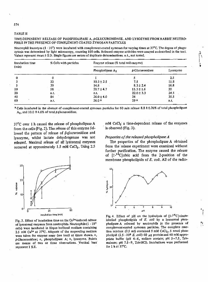
574
TABLE Il
TIME-DEPENDENT RELEASE OF PHOSPHOLIPASE A. ~-GLUCURONIDASE. AND LYSOZYME FROM RABBIT NEUTROPHILS IN THE PRESENCE OF COMPLEMENT-COATED ZYMOSAN PARTICLES
Neutrophil leucotyes (1 . 107) were incubated with complement-coated zymosan for varying times at 37°C. The degree of phagocytosis was determined by light microscopy. counting 100 cells. Released enzyme activities were assayed as described in the text. Values represent mean ± S.D. Single figures are means of duplicate determinations. n.t., not tested.
Incubation time % Cells with particles Enzyme release (% total cell enzyme) (min)
Phospholipase A2 p-G!ucuronidase Lysozyme
0 0 I 5 2.5 3 33 10.3± 2.5 7.5 11.8 5 39 14.8 8.3 ± 2.4 16.8
10 58 20.7±4.7 15.3 ± 1.6 20 20 n.t. n.t. 22.0 ± 3.3 24.5 40 84 26.0±4.0 24 35.5 60 n.t. 26.0 a 29 a n.t.
a Cells incubated in the absence of complement-coated zymosan particles for 60 min release 8.8 ± 0.26% of total phospholipase A2• and 10.0 ± 4.6% of total s3-glucuronidase.
37'C over I h caused the release of phospholipase A from the cells (Fig. 2). The release of this enzyme followed the pattern of release of ~-g1ucuronidase and lysozyme. whilst lactate dehydrogenase was not released. Maximal release of all lysosomal enzymes occurred at approximately 1.5 mM CaCI,. Using 2.5
30
,0
~ 10
o 6 3'0
incubation time (min)
GO
Fig. 3. Effect of incubation time on the Ca2+·induced release of lysosomal enzymes from neutrophils. Neutrophils (1 . 107
cells) were incubated in Hepes buffered medium containing 2.5 mM Ca2+ at 37°C. Aliquots of the suspending medium were taken for enzyme assay (see text) at times shown. 0,
p-GIUCUlonidase; 6, phospholipase A; 0, lysozyme. Points are means of two or three observations. Vertical bars represent 1 S.E.
mM CaCl,' a time-dependent release of the enzymes . is observed (Fig. 3).
Properties of the released phospholipase A The properties of the phospholipase A obtained
from the release experiment were examined without further purification. The enzyme caused the release of [1-14C]oleic acid from the 2-position of the membrane phospholipids of E. coli. All of the radio-
40
o , 9 10
pH
Fig. 4. Effect of pH on the hydrolysis of [l_1 4C]oIcate-labelled phospholipids of E. coli by a lysosomal phospholipase A released by neutrophils in the presence of complement~oated zymosan particles. The complete reaction mixture (0.5 ml) contained 5 mM CaC1z, 5 nmol phospholipid (2.5 ·10' E. coil) 60 "g protein and 40 mM appropriate buffer (pH 4-6. sodium acetate; pH 5 -7.5. Trismaleate; pH 7.5 -9, Tris·HCI). Incubations were performed for 1 h at 37°C.
"
f
"i ; ,
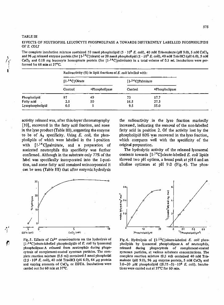
/:' I
575
TABLE III
EFFECTS OF NEUTROPHIL LEUCOCYTE PHOSPHOLIPASE A TOWARDS DIFFERENTLY LABELLED PHOSPHOLIPIDS OF E. COLI
The complete incubation mixture contained 10 nmol phospholipid (5 . 108 E. com. 40 mM Tris-maleate (pH 9.0), 5 mM CaC1l and 96 p.g released enzyme protein (for [I·14Cloleate) or 20 nmol phospholipid (1 . 109 E. coil), 40 mM Tris-HCl (pH 6.0), 5 mM Ca02 and 0.18 mg leucocyte homogenate protein (for [1-14CJpalmitate) in a total volume of 0.5 ml. Incubations were performed for 60 min at 37°C.
Radioactivity (%) in lipid fractions of E. coli labelled with:
[I-14C]Oleate
Control +Phospholipase
Phospholipid 97 49 Patty acid 2.5 50 Lysophospholipid 0.5 1
activity released was, after thin-layer'chromatography [10], recovered in the fatty acid fraction, and none in the lyso product (Table Ill), suggesting the enzyme to be of A2 specificity _ Using E. coli, the phospholipids of which were labelled in the I-position with [I_14C)paJrnitate, and a preparation of sonicated neutrophils this specificity was further confirmed. Although in the substrate only 77% of the label was specifically incorporated into the I-position, and some fatty acid remained unincorporated it can be seen (Table Ill) that after enzymic hydrolysis
• 20
~
f ;l. 10
I --,-- 0~------------~5'-------------~'0
EOTA (mM) CaCl2 (mM)
Fig. 5. Effects of Ca2+ concentrations on the hydrolysis of [1.14CJoleate4abelled phospholipids of E. coli by lysosomal phospholipase A released from neutrophils during phagocytosis of complement~oated· zymosan particles. The complete leaction mixture (05 m!) contained 5 nmol phospholid (2.5 . 10' E. coil), 40 mM Tris+lCl (pH 6.0), 64 pg protein and varying amounts of CaC12 or EDTA. Incubations were carried out for 60 min at 37OC.
[ 1-14C]Palmitate
Control +Phospholipase
73 17_7 16.5 27.3
9.5 55.0
the radioactivity in the lyso fraction markedly increased, indicating the removal of the non-labelled fatty acid in position 2_ Of the activity lost by the phospholipid 80% 'l'as recovered in the lyso fraction, which compares well with the specificity of the original preparation_
The hydrolytic activity of the released lysosomal contents towards [1-14Cloleate-labelled E_ coli lipids showed two pH optirila, a broad peak at pH 6 and an alkaline optimum at pH 9.0 (Fig. 4). The phos-
0·, ,
10 20 o Q.1 0-2: 0·3 (Phospholipid) [I'Ml ''\PhOSPhOliPidllpMJ
)
Fig. 6. Hydrolysis of [1.i4C]oleate-labelled E. coli phospholipids by lysosomal phospholipase A of neutrophils, released during phagocytosis of complement~oated
zymosan particles. at various substrate concentrations. The complete reaction mixture (0.5 ml) contained 40 mM Tris~ maleate (pH 9.0), 96 pg enzyme protein, 5 mM CaCl2 and 3.0-20 pM phospholipid «0.75-5) ·10' E. coli). Incubations were carried out at 370C for 60 min.
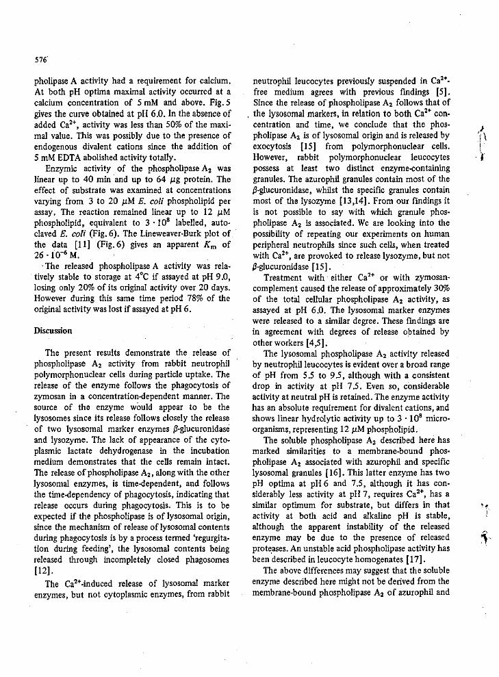
576'
pholipase A activity had a requirement for calcium. At both pH optima maximal activity occurred at a calcium concentration of 5 mM and above. Fig. 5 gives the curve obtained at pH 6.0. In the absence of added Ca'+, activity was less than 50% of the maximal value. This was possibly due to the presence of endogenous divalent cations since the addition of 5 mM EDTA abolished activity totaIly.
Enzymic activity of the phospholipase A, was linear up to 40 min and up to 64 JoLg protein. The effect of substrate was examined at concentrations varying from 3 to 20 JoLM E. coli phospholipid per assay. The reaction remained linear up to 12 JoLM phospholipid, equivalent to 3·!O8 labelled, auto· claved E. coli (Fig. 6). The Lineweaver·Burk plot of the data [11] (Fig.6) gives an apparent Km of 26 '10.6 M.
. The released phospholipase A activity was relatively stable to storage at 4°C if assayed at pH 9.0, losing only 20% of its original activity over 20 days. However during this same time period 78% of the original activity was lost if assayed at pH 6.
Discussion
The present results demonstrate the release of phospholipase A, activity from rabbit neutrophil polymorphonuclear cells during particle uptake. The release of the enzyme follows the phagocytosis of zymosan in a concentration-dependent manner. The source of the enzyme would appear to be the Iysosomes since its release follows closely the release of two lysosomal marker enzymes /l-glucuronidase and lysozyme. The lack of appearance of the cytoplasmic lactate dehydrogenase in the incubation medium demonstrates that the cells remain intact_ The release of phospholipase A" along with the other lysosomal enzymes, is time-dependent, and follows the time-dependency of phagocytosis, indicating that release occurs during phagocytosis. This is to be expected if the phospholipase is of lysosomal origin, since the mechanism of release oflysosomal contents during phagocytosis is by a process termed 'regurgitation during feeding', the lysosomal contents being released through incompletely closed phagosomes [12].
The Ca"-induced release of lysosomal marker enzymes, but not cytoplasmic enzymes, from rabbit
neutrophil leucocytes previously suspended in Ca'+free medium agrees with previous rmdings [5]. Since the release of phospholipase A, follows that of
, the lysosomal markers, in relation to both Ca'+ concentration and time, we conclude that the phospholipase A, is of lysosomal origin and is released by exocytosis [15] from polymorphonuclear cells. However, rabbit polymorphonuclear leucocytes possess at least two distinct enzyme-containing granules. The azurophil granules contain most of the /l-glucuronidase, whilst the specific granules contain most of the lysozyme [13,14]. From our findings it is not possible to say with which granule phospholipase A2 is associated. We are looking into the possibility of repeating our experiments on human peripheral neutrophils since such cells, when treated with Ca2+, are provoked to release lysozyme, but not /l-glucuronidase (15].
Treatment with either Ca2+ or with zymosancomplement caused the release of approximately 30% of the total cellular phospholipase A, activity, as assayed at pH 6.0. The lysosomal marker enzymes were released to a similar degree_ These rmdings are in agreement with degrees of release obtained by other workers [4,5].
The lysosomal phospholipase A, activity released by neutrophilleucocytes is evident over a broad range of pH from 5.5 to 9.5, although with a consistent drop in activity at pH 7.5. Even so, considerable activity at neutral pH is retained_ The enzyme activity has an absolute requirement for divalent cations, and shows linear hydrolytic activity up to 3 . !O' microorganisms, representing 12 JoLM phospholipid_
The soluble phospholipase A, described here has marked similarities to a membrane-bound phospholipase A2 associated with azurophil and specific lysosomal granules (16]- This latter enzyme has two pH optima at pH 6 and 7.5, although it has considerably less activity at pH 7, requires Ca2+, has a similar optimum for substrate, but differs in that activity at both acid and alkaline pH is stable, although the apparent instability of the released enzyme may be due to the presence of released prote,ases. An unstable acid phospholipase activity has been described in leucocyte homogenates [17].
The above differences may suggest that the soluble enzyme described here might not be derived from the membrane-bound phospholipase A2 of azurophil and
'. , ..
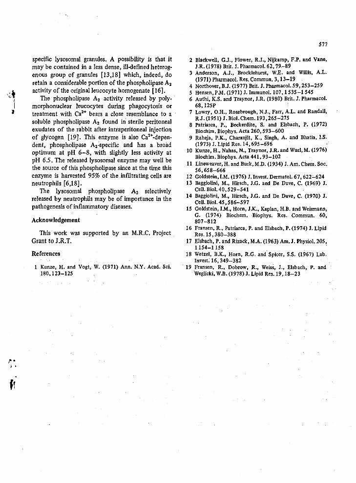
" . j •
specific lysosomal granules. A possibility is that it may be contained in a less dense, ill·defined heterog· enous group of granules [13,18] which, indeed, do retain a considerable portion of the phospholipase A. activity of the original leucocyte homogenate [16].
The phospholipase A. activity released by poly. morphonuclear leucocytes during phagocytosis or treatment with Ca'+ bears a close resemblance to a soluble phospholipase A, found iri sterile peritoneal exudates of the rabbit after intraperitoneal injection of glycogen [19]. This enzyme is also Ca'+.depen. dent, phospholipase A,·specific and has a broad optimum at pH 6-8, with slightly less activity at pH 6.5. The released lysosomal enzyme may well be the source of this phospholipase since at the time this enzyme is harvested 95% of the infiltrating cells are neutrophils [6,18].
The lysosomal phospholipase A. selectively released by neutrophils may be of importance in the pathogenesis of inflammatory diseases.
Acknowledgement
This work waS supported by an M.R.C. Project Grant to J.R.T.
References
1 Kunze, H. and Vogt, W. (1971) Ann. N.Y. Acad. Sci. 180,123-125
577
2 Blackwell, G.J., Flower, R.J., Nijkamp, F.P. and Vane, J.R. (1978) Brit. J. Pharmacol. 62, 79.-89
3 Anderson, A.J., BrocklehuIst, W.E'. and Willis, A.L. (1971) Pharmacol. Res. Commun. 3, 13-19
4 Northover, B.J. (1977) Brit. J. Pharmacol. 59, 253-259 5 Hensen,PM. (1971) J. Immunol.107,1535-1545 6 Authi, K.S. and Traynor, J.R. (1980) Brit. J. Pharmacol.
68,I25P 7 Lowry, O.H., Rosebrough. N.J., Fau, A.L. and RandaU,
R.J. (1951) J. BioI. Chem.193, 265 -275 8 Patriarca, P., Beckerdite, S. and EIsbach, P. (1972)
Biochim. Biophys. Acta 260,593-600 9 Raheja, P.K., Charanjit, K., Singh, A. and Bhatia, I's.
(1973) J. Lipid Res. 14, 695-696 10 Kunze, H .• Nahas, N., Traynor, J.R.and Wurl,M. (1976)
Biochim. Biophys. Acta 441, 93-102 11 Lineweaver, H. and Burk, M.D. (1934) J. Am. Chem. Soc.
56,658-666 12 Goldstein, I.M. (1976) J. Invest. Dermatol. 67,622-624 13 Baggiolini, M., Hirsch, J.G. and De Duve, C. (1969) J.
Cell. BioI. 40, 529-541 14 Baggiolini, M., Hirsch, J.G. and De Duve, C. (1970) J.
Cell. BioI. 45,586-597 15 Goldstein, I.M., Horn, J.K., Kaplan. H.B. and Weismann,
G. (1974) Biochem. ·Biophys. Res. Commun. 60, 807-812
16 Fransen, R., Patriarca, P. and Elsbach, P. (1974) J. Lipid Res. 15,380-388
17 Elsbach, P. and Rizack, M.A. (1963) Am. J. Physiol. 205, 1154-1158 .
18 Wetzel, B.K., Horn, R.G. and Spicer, S.S. (1967) Lab. Invest. 16,349-382
. 19 Fransen, R., Dobrow, R., Weiss, J., Elsbach, P. and 'Weglicki, W.B. (1978) J. Lipid Res. 19,18-23
n n
,
• I
•
..
i .' i I I I
I !
• •
PROCEEDINGS OF THE 8.P.s., 12th-14th SEPTEMBER, 1979 Il5P
Release of a lysosomal phospholipase A from rabbit neutrophilleucocytes
K.s. AUTHI & J.R. TRAYNOR
B;odwmi('(JI Pharm"colofj')' Laboratory. Department of Clwmisfr),. Ulliversify of Technology. Loughborough, u'k,·stt'rsJiirt· LE, I JrU
The appearance of prostaglandins in some inftammatory exudates is paralleled by infiltration of the inftamed area wilh polymorphonuclear leucocytes (PMN). A close correlation between the release of lysosomal enzymes from these cells, and the appearance of prostaglandins led to the suggestion that lysosomal phospholipases A cause the release of precursor fatty acid, for prostaglandin production (Anderson, Brocklehurst & Willis, 1971). The importance of phospholipase A, in prostaglandin biosynthesis has been confirmed (Blackwell. Flower. Nijkamp & Vane. 1978~ This communication describes the release and
properties 01 a phospholipase A liberated from PMN leuc'Ocyles during phagocytosis' of zymosan-complement (ZC) (Hen,on, 1971), and by calcium (North-over, 1977t . . PMN', were obtained from female White New Zea
land r<lbbits (2.5 kg) 18 h after intraperitoneal injeclion 01 sterile thioglycollate medium, US.P. (100 ml) <IS described by . Northov.r . (l977~ Neutrophils. (I x I(}') were incubated at137° ill medium containing (mM) NaCI 150. Kc;I 3, glucose 10, Hepes 5, pH 7.4, with varying amounts of ze or calcium. Aliquots of the medium were incubated with [1-'4C)-oleate labelled E. coli for 60 min' to assay phospholipase A aClivity (Authi & Traynor. 1979~ p-Glucuronidase. Iyso,yme and lactate dehydrogenase activities were also delermined (Norlhover, 197n Results of enzyme assays are expressed as percentages or the activities released after sonication (6 iUR, 3 minI of the cells (for phospholipase aClivitY1 or trealment with Trilon X-lOO (0.2%~
ZC and calcium caused a time and concentration dependant increase in the release of phospholipase A aclivily. Maximal release 01 approximalely 30% of the tOlal aclivily was obtained at 2.5 mM calcium, or 2 ms/ml Zc. The release of phospholipase A fol-10wFd that of p-glucuronidase and Iysosyme, confirm-
ing a lysosomal lOCation lor Ihe enzyme. No such release of Ihe cyloplasmic marker laclale dehydrogen-ase was observed. .
The released phospholipase' A has two pH oplima at pH 6 (broad) and pH 9 and a requirement for calcium. The enzyme appears to be of A, specifieity since 95% of the label incorporaled inlo phospholipid was in the 2~position and no radioact.ivity was re:.
. covered in the Iyso product. A phospholipase A with similar properties has been lound in rabbit periloneal exudates ufler injection of glycogen (Frunsen, ft .1., 1978~
The lysosomal phospholipase A deScribed m~y be of importance in the observed production of proslaglandin. by PMN leucocytes during phagocylosis (Higgs, McCall & Youlten, 1975) and in Ihe pathogen.esis or infta~matory diseases generally.
We thank the MRC for supporL
References
ANOr:ItSON. A~J.. BIH)("KL~iHURST,. W.E. &. WILlIS.- A.L. (1971). Evidence for the role oflysosomes in the forma~ tion of prostaglandins during carrageenin·induced in. ftammulion in the rut. Phurm. Rt's.. CommulI., 3, 13-19.
AUntl, K. S. & TRAYNOR, J.R. (1979). Effects or anlimalar-' . ial drugs on phospholipase A2 • 8r. J. Pharmac., 66, 496P.
BLACKWliLl .. O.J .• Fl.OwvR, R.J., NIJK.AMI', F.P. & V"NE. J.R. (1978). Phospholipase A2 activity or guinea-pig iso~ tated perrused lungs: stimulation. and inhibition by anti-inftammatory steroids. Br. J. Pharmac .• 61:. 79-89 .
FR"NSEN. R .• D08Row. R .• Wmss. J., ELS8A.CIt. P. & Wt;C1~ LlCKI. W.B. (l978). Isolation and characterisation of a phospholipase A2 rrom an inflammatory exudate. J. Lipid Res" 19. 18-23. .
HEN SON, P.M. (l97H Immunological release of constitu~ cnts from neutrophil leucocytes. J. ImmrH1ol .. 107, 1535-1545. .
HIOGs, G.A., McCALL. E. & YouLnN, L.I.F. (l975~ A chemotactic role ror prostaglandins released from poly ... morphonuclear leucocytes during phagocytosis. Br. J. Pharmac" 53. 539-546. .
NORTHOVrR, B.l (1977~ Effect of indomethecin and related drugs on the calcium jon~dependant secretion of Iysoso- . mal and other enzymes by neutrophil polymorphonuOo lear leucocytes' in vitro. 8r. J. Pharmac .• 59, 253-259.
>: "
;. I
i i
i I ! , ,
\
'. '.
, 1
.. '
496P PROCEEDINGS OF THE B.P.S., 411.-6th APRIL. 1979
ElI'ects of antimalarial drugs on phospholipase A,
K.S. AUTHI & J.R. TRA YNOR · B/nrht'mi('QI Phurmlu:oM"y Luht"ulory. DfptJrlmC'", of'
ChtmlslrY. Unloml,y 0/ T."hntlIOflY, Loughhoro.~h. L.lr~ LEII JTU
'. Antimalarial drugs or th. chloroquin.·type Inhibit · lipolytic processes in rat tissue I. vitro (Marku. & · Bal~ 1969) and phospholipase activity in various tissu •• (Blackw.l~ Flow.r. Nijkamp & Vant, 1978) including th. malarial parasi t. (Cened.11a, Jarr.D & S .... 1969~ Several or th.se drugs have been used in th. tr.atment of rh.umatoid conditions. W. r.port th. effects or three antimalarial drugs, chloroquine, m.pacrin. and primaquin •• on th. activity or a crud. phospholipas. A.' enzyme obtain.d rrom an inftam· matory peritoneal exudat. (Fransen. Dobrow. W.iSL Elsbach & W.glicki, 1978~
Phospholipase A. activity was assayed against E .. coli labelled with [I."C)-oleat •. Great.r than 95% or the incorporated label was in the 2-position or m.mbran. phospholipid .. Radiolabelled E. con were . autoclaved ror 15 min at 2.7 k8lcrn' to inactivat. endogenous bact.rial phospholipases and r.nd.r th. m.mbran. more susceptible to enzymic attack. Assays .wereperrormed at pH 6.0 in tris buffer 4)( 10-' 11 containing calcium S )( 10-' 11 at 37"C for S min. Lipid products w.r •• xtract.d. separated by nc and
· areas or plates containing radioactiv. lipid. were . scraped off and radioactivity d.termined by scintilla· tion counting. , '. .,..'
All three drugs inhibited th. enzymlc hydrolysis or E. coli phosphofipid.. IC '0' ror m.pacrln. was
,33 )( 10- M. primaquine was twice and chloroquin. five times I.ss active. In addition the drugs chloro··
" quin. and m.pacrine showed a stimulation of hy·
-.,,-
. '
-;-.' ' .. ,
'1
..
I . i
/
drolysis at approximately 2O-rold lower doses. whilst at doses above those causing maximum inhibition a rev.rsal or the inhibitory effects was seen.
Th. antimalarial drugs examined are amphiphilic cationic drugs. Other drug. or this general type in· hibit phospholipase enzyme .. possibly by effects 011 the substrat. (Kunz., Naha .. Traynor & Wure, 1976). In r.lation to this we have studied the effe.is or the drugs on th. stability or guinea.pig red blood cell. to hypotonic haemolysis. All three' drugs again show.d paradoxical dose.d.pendent inhibitory and stimulatory .ffect •.
Th. findings described may be or r.levance to the use or th. drugs in Ihe tr.atmenl of malaria and rheu· matoid disease. and in th.ir effects in causing drug·
. induced lipidoses. .
W. thank Ih. M.R.C. ror lupport.
Rd.r_
Bur.wELI. O.J.: FLOW,,". R.J .. Nu ...... F.P, &; V ANI· .. J.R. (t978~ Phospholipase A. activily of guine"pi, iso- ,. laled perrused lungs: stimulation and inhibition by . anti-innHmmalory slerolds. Br, J. Phan,,"f' •• 62. 78-7'1.
Cu.u>uu. R.J .. b ... LI. I.J. &; S .... I..M. (l969~ Piu .• , I', . moJlum ""'gh,'/: produdion'in lIitro or free fatly acids. . Ex,...I"".,,'o/ par.,iIO/og)·, 24, 1»-136. "
fUNS>N. R .• Don.ow. R .. Wl1SL I .• E'''Aru. P. &; W ... • I UC.I, W.B. (l978~ Ioolallon and characlerisation or • .1' phospholipase A, from an innammalory exudale: 'J. Lipid R .... 19, 18-23.
KUNZ •. H .• NAHA" N .. TRAYNOR: J.R. &; W"RI. M. (l976~' ElfeclS of local ana.slhelics on phmpholipapses. BInchin;. Bloph)'& Acta • ...... 93-102.
. MAR.US, H.B. &; BALL. E.O. (1969~ Inhibition or lipolylic processes in rat :adipose tissue by antimalarial drup. Blochlm. Blophy •• Ar,a. 181,486:-491. '
• •
• . :. ,-,'
'" ."
..... . ".
r ...
, ,. . ,
..
, •
, . 1.,.,
' .. . .