Embed Size (px)
Citation preview
REVIEW
Potential viral pathogenic mechanism in human type 1 diabetes
Darius A. Schneider & Matthias G. von Herrath
Received: 29 January 2014 /Accepted: 9 June 2014# The Author(s) 2014. This article is published with open access at Springerlink.com
Abstract In type 1 diabetes, as a result of as yet unknowntriggering events, auto-aggressive CD8+ T cells, together witha significant number of other inflammatory cells, includingCD8+ T lymphocytes with unknown specificity, infiltrate thepancreas, leading to insulitis and destruction of the insulin-producing beta cells. Type 1 diabetes is a multifactorial dis-ease caused by an interactive combination of genetic andenvironmental factors. Viruses are major environmental can-didates with known potential effects on specific key points inthe pathogenesis of type 1 diabetes and recent findings seemto confirm this presumption. However, we still lack well-grounded mechanistic explanations for how exactly virusesmay influence type 1 diabetes aetiology. In this review weprovide a summary of experimentally defined viral mecha-nisms potentially involved in the ontology of type 1 diabetesand discuss some novel hypotheses of how viruses may affectthe initiation and natural history of the disease.
Keywords Autoimmune . Enterovirus . Environmentalfactors . Humans . Insulitis . Type 1 diabetes . Viruses
AbbreviationsCVB Group B coxsackievirusHERV Human endogenous retrovirusnPOD Network of Pancreatic Organ Donors
PBMC Peripheral blood mononuclear cellPLN Pancreatic lymph node
Introduction
While there is undoubtedly evidence for a genetic basis oftype 1 diabetes, especially with regard to permissive HLAclass II genotypes, many features of this disease have to beattributed to environmental factors, specifically, (1) the annualincrease in type 1 diabetes incidence, currently estimated to be3% [1]; (2) the strong heterogeneity of its geographical distri-bution, which is subject to considerable regional gradients [2];(3) the fact that the incidence rate in first-generation offspringof immigrants is the same as that in the new home country[3, 4].
In animal models of diabetes the established role of innateinflammation in the insulitic process [5–7] and the increasingevidence supporting the contribution of viral infections to aproinflammatory islet milieu [8–10] strongly suggest thatviruses may contribute to beta cell damage and dysfunction.The evidence for the presence of similar mechanisms inhumans is still circumstantial [11–13]; however, the insightsgained from animal studies imply that innate immunity is animportant component of the pathogenesis of type 1 diabetes.Lately, novel developments in analysis techniques, as well asaccess to organ libraries, such as the Network of PancreaticOrgan Donors (nPOD, www.jdrfnpod.org) in the USA or thecollection of Foulis et al [14] in the UKwill be instrumental inallowing us to link the presence of these environmentaldeterminants to the highly complex histopathologicalfeatures of type 1 diabetes.
In this review we will analyse how viral infections canaccount for these highly complex scenarios.
D. A. Schneider :M. G. von Herrath (*)La Jolla Institute for Allergy and Immunology, 9420 Athena Circle,La Jolla, CA 92037, USAe-mail: [email protected]
D. A. SchneiderDepartment of Medicine, UC San Diego, La Jolla, CA, USA
M. G. von HerrathNovo Nordisk Type 1 Diabetes Research Center,Seattle, WA 98109, USA
DiabetologiaDOI 10.1007/s00125-014-3340-7
Hallmarks of type 1 diabetes pathogenesis
Type 1 diabetes results from the selective and progressivedestruction of insulin-producing cells by autoreactive CD8+
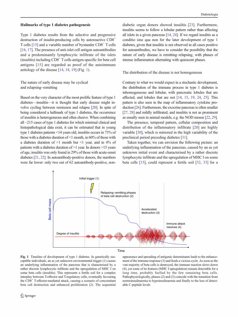
T cells [15] and a variable number of bystander CD8+ T cells[16, 17]. The presence of anti-islet cell antigen autoantibodiesand a predominantly lymphocytic infiltrate of the islets(insulitis) including CD8+ Tcells antigen-specific for beta cellantigens [15] are regarded as proof of the autoimmuneaetiology of the disease [14, 18, 19] (Fig. 1).
The nature of early disease may be cyclicaland relapsing–remitting
Based on the very character of the most prolific feature of type 1diabetes—insulitis—it is thought that early disease might in-volve cycling between remission and relapse [20]. In spite ofbeing considered a hallmark of type 1 diabetes, the occurrenceof insulitis is heterogeneous and often elusive.When combiningall ~215 cases of type 1 diabetes for which minimal clinical andhistopathological data exist, it can be estimated that in youngtype 1 diabetes patients <14 years old, insulitis occurs in 73% ofthosewith a diabetes duration of <1month, in 60% of thosewitha diabetes duration of >1 month but <1 year, and in 4% ofpatients with a diabetes duration of >1 year. In donors >15 yearsof age, insulitis was only found in 29% of those with acute-onsetdiabetes [21, 22]. In autoantibody-positive donors, the numberswere far lower: only two out of 62 autoantibody-positive, non-
diabetic organ donors showed insulitis [23]. Furthermore,insulitis seems to follow a lobular pattern rather than affectingall islets in a given pancreas [14, 24]. If we regard insulitis as aconditio sine qua non for the later development of type 1diabetes, given that insulitis is not observed in all cases positivefor autoantibodies, we have to consider the possibility that thenature of early disease is remitting–relapsing, with phases ofintense inflammation alternating with quiescent phases.
The distribution of the disease is not homogeneous
Contrary to what we would expect in a stochastic development,the distribution of the immune process in type 1 diabetes isinhomogeneous and lobular, with pancreatic lobules that areaffected, and lobules that are not [14, 15, 19, 24, 25]. Thispattern is also seen in the map of inflammatory cytokine pro-duction [26]. Furthermore, the exocrine pancreas is often smaller[27, 28] and mildly infiltrated, and insulitis is not as prominentas usually seen in animal models, e.g. the NODmouse [22, 29].
The presence, temporal pattern, cellular composition anddistribution of the inflammatory infiltrate [20] are highlyvariable [30], which is mirrored in the high variability of thepreclinical period preceding diabetes [31].
Taken together, we can envision the following picture: anunderlying inflammation of the pancreas, caused by an as yetunknown initial event and characterised by a rather discretelymphocytic infiltrate and the upregulation ofMHC I on somebeta cells [15], could represent a fertile soil [32, 33] for a
Initial trigger (1)
Relapsing–remitting phasesof beta cell destruction (2)
Accelerateddestruction (3)
Immune attackresolves (4)
Degree of insulitis
Bet
a ce
ll m
ass
Time
Fig. 1 Timeline of development of type 1 diabetes. In genetically sus-ceptible individuals, an as yet unknown environmental trigger (1) causesan underlying inflammation of the pancreas that is characterised by arather discrete lymphocytic infiltrate and the upregulation of MHC I onsome beta cells (insulitis). This represents a fertile soil for a complexinterplay between T-effector and T-regulatory cells, eventually favouringthe CD8+ T-effector-mediated attack, causing a scenario of concomitantbeta cell destruction and enhanced proliferation (2). The sequential
appearance and spreading of antigenic determinants leads to the enhance-ment of the immune response (3) and feeds a vicious cycle. As soon as thevast majority of beta cells is destroyed, the immune reaction slows down(4), yet some of its features (MHC I upregulation) remain detectable for along time, probably fuelled by the few remaining beta cells.Pathophysiologically, phases (2) and (3) coincide with the transition fromnormoinsulinaemia to hypoinsulinaemia and finally to the loss of detect-able C-peptide levels
Diabetologia
complex interplay between effector and regulatory T cells,eventually favouring CD8+ effector T cells that attack the betacells. This leads to a scenario of concomitant beta cell destruc-tion and enhanced proliferation, with the sequential appear-ance and spreading of antigenic determinants and the en-hancement of the immune response [34]. As recent workssuggest [35], the underlying inflammatory state of the pancre-as might initially involve the exocrine pancreas, prior to theinduction of autoimmunity. As soon as the latter is triggered, avicious cycle is born.
How can the histopathological hallmarks of diabetesbe explained by viral infection?
In spite of a plethora of studies (see Table 1 for an overview ofimportant studies), we are still lacking a clearly established,causal link between viral infection or presence and the devel-opment of autoimmunity or progression to diabetes, yet thehistopathological hallmarks of diabetes can be elegantlyexplained as effects of viral interference:
(1) The heterogeneous, possibly relapsing–remitting insulitis,with upregulation of MHC class I molecules on mostislets, which persists for years irrespective of the function-al status of the islets, can be caused by interferons or otherfactors secreted from cells infected by virus.
(2) Enteroviruses have been shown to have a strongpancreotropism—severe islet damage has been demon-strated in fatal group B coxsackievirus (CVB) infectioncases [36], human islets show strong expression of thecoxsackie virus and adenovirus receptor (CAR) [37] andbeta cells are permissive for enterovirus in vitro [38].Also, human peripheral blood mononuclear cells(PBMCs) experimentally infected with CVB4 showenhanced production of proinflammatory cytokines suchas TNF-α and IL-6 [39].
(3) It has recently been shown that the inflammatory state ofthe pancreas can be explained by direct or indirect viraleffects [40, 41].
Mechanisms of viral involvement in type 1 diabetesaetiology
When we discuss viral infections, we have to consider viralinfections of the pancreatic beta cells as well as of those cellsadjacent to the beta cells (acinar cells, endothelial cells, neu-rons) or of cells at a remote location, such as dendritic cells,leading to presentation of cross-reacting epitopes, or of gutcells, leading to increased gut permeability and the
presentation of cross-reacting antigens in the pancreatic lymphnodes, where pancreatic and gut lymphatic drainage intersect[42]. From a chronological point of view, viral infections canbe either (1) acute, (2) exacerbated chronic infections, (3)reactivated persistent, quiescent viruses (e.g. Herpesviridae),or (4) represent the restoration of pathogenicity of viral seg-ments that had long been integrated in the human genome,such as human retroviruses.
Infection of beta cells
Infection of beta cells with subsequent damage and release ofantigens, as well as release of interferons and involvement ofboth innate and adaptive immune system, is the mechanism bywhich enteroviruses are thought to be involved in the patho-genesis of type 1 diabetes. This is, by far, the mechanism thathas been investigated to the largest extent and has dominatedscientific debate on this topic for many years.
Acute infection Acute infection is linked with severe damageof the beta cells and rapid progression towards fulminant dia-betes. This is the case in fulminant type 1 diabetes, where anassociation with direct enteroviral infection has been discussedin case reports [43] and where autoimmunity is not primarilyinvolved. Here we may also add examples of enterovirus-induced diabetes in animals: cattle after infection with the footand mouth virus [44], voles after infection with Ljungan virus[45], rats after infection with the Kilham virus [46], as well ashigher non-human primates infected with CVB4 virus [47].Especially in the case of CVB4, we suspect considerable dif-ferences in viral strain-specific pancreotropism.
Chronic infection Chronic, rather slow and persistent type ofinfection with stimulation of resting beta-cell-antigen-specific Tlymphocytes is thought to be the link between CVB infectionsand the induction of autoimmunity and/or progression to diabe-tes and leads to subtle changes in the beta cells. These changesmay involve the induction of endoplasmic reticulum stress [48]or mutations in tyrosine kinase 2 (TYK2) or similar proteinsleading to beta-cell-specific suppression of cytokine responsesincluding interferon [49], which leads to high sensitivity toCVB4 infection [50] or other changes in beta cell metabolismwhich, in turn, lead to local inflammation and tissue damagewith the slow release of sequestered islet antigen and stimulationof quiescent, beta-cell-antigen-specific T (memory) cells.
Infection of cells adjacent to beta cells
Exocrine pancreatic cells The exocrine pancreas is targetedby a myriad of different viruses, and many of them have beenassociated with type 1 diabetes either in humans or in animalmodels: measles virus, congenital rubella virus, mumps virus,cytomegalovirus [51], or even viruses with high penetration of
Diabetologia
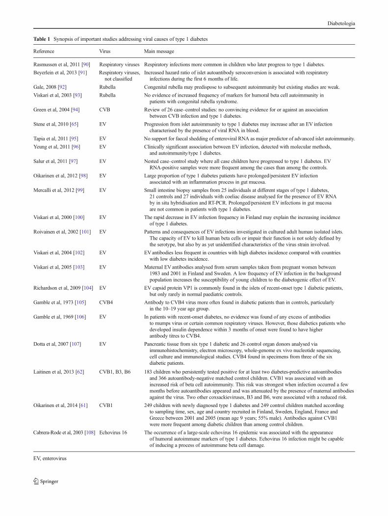
Table 1 Synopsis of important studies addressing viral causes of type 1 diabetes
Reference Virus Main message
Rasmussen et al, 2011 [90] Respiratory viruses Respiratory infections more common in children who later progress to type 1 diabetes.
Beyerlein et al, 2013 [91] Respiratory viruses,not classified
Increased hazard ratio of islet autoantibody seroconversion is associated with respiratoryinfections during the first 6 months of life.
Gale, 2008 [92] Rubella Congenital rubella may predispose to subsequent autoimmunity but existing studies are weak.
Viskari et al, 2003 [93] Rubella No evidence of increased frequency of markers for humoral beta cell autoimmunity inpatients with congenital rubella syndrome.
Green et al, 2004 [94] CVB Review of 26 case–control studies: no convincing evidence for or against an associationbetween CVB infection and type 1 diabetes.
Stene et al, 2010 [65] EV Progression from islet autoimmunity to type 1 diabetes may increase after an EV infectioncharacterised by the presence of viral RNA in blood.
Tapia et al, 2011 [95] EV No support for faecal shedding of enteroviral RNA as major predictor of advanced islet autoimmunity.
Yeung et al, 2011 [96] EV Clinically significant association between EV infection, detected with molecular methods,and autoimmunity/type 1 diabetes.
Salur et al, 2011 [97] EV Nested case–control study where all case children have progressed to type 1 diabetes. EVRNA-positive samples were more frequent among the cases than among the controls.
Oikarinen et al, 2012 [98] EV Large proportion of type 1 diabetes patients have prolonged/persistent EV infectionassociated with an inflammation process in gut mucosa.
Mercalli et al, 2012 [99] EV Small intestine biopsy samples from 25 individuals at different stages of type 1 diabetes,21 controls and 27 individuals with coeliac disease analysed for the presence of EV RNAby in situ hybridisation and RT-PCR. Prolonged/persistent EV infections in gut mucosaare not common in patients with type 1 diabetes.
Viskari et al, 2000 [100] EV The rapid decrease in EV infection frequency in Finland may explain the increasing incidenceof type 1 diabetes.
Roivainen et al, 2002 [101] EV Patterns and consequences of EV infections investigated in cultured adult human isolated islets.The capacity of EV to kill human beta cells or impair their function is not solely defined bythe serotype, but also by as yet unidentified characteristics of the virus strain involved.
Viskari et al, 2004 [102] EV EVantibodies less frequent in countries with high diabetes incidence compared with countrieswith low diabetes incidence.
Viskari et al, 2005 [103] EV Maternal EV antibodies analysed from serum samples taken from pregnant women between1983 and 2001 in Finland and Sweden. A low frequency of EV infection in the backgroundpopulation increases the susceptibility of young children to the diabetogenic effect of EV.
Richardson et al, 2009 [104] EV EV capsid protein VP1 is commonly found in the islets of recent-onset type 1 diabetic patients,but only rarely in normal paediatric controls.
Gamble et al, 1973 [105] CVB4 Antibody to CVB4 virus more often found in diabetic patients than in controls, particularlyin the 10–19 year age group.
Gamble et al, 1969 [106] EV In patients with recent-onset diabetes, no evidence was found of any excess of antibodiesto mumps virus or certain common respiratory viruses. However, those diabetics patients whodeveloped insulin dependence within 3 months of onset were found to have higherantibody titres to CVB4.
Dotta et al, 2007 [107] EV Pancreatic tissue from six type 1 diabetic and 26 control organ donors analysed viaimmunohistochemistry, electron microscopy, whole-genome ex vivo nucleotide sequencing,cell culture and immunological studies. CVB4 found in specimens from three of the sixdiabetic patients.
Laitinen et al, 2013 [62] CVB1, B3, B6 183 children who persistently tested positive for at least two diabetes-predictive autoantibodiesand 366 autoantibody-negative matched control children. CVB1 was associated with anincreased risk of beta cell autoimmunity. This risk was strongest when infection occurred a fewmonths before autoantibodies appeared and was attenuated by the presence of maternal antibodiesagainst the virus. Two other coxsackieviruses, B3 and B6, were associated with a reduced risk.
Oikarinen et al, 2014 [61] CVB1 249 children with newly diagnosed type 1 diabetes and 249 control children matched accordingto sampling time, sex, age and country recruited in Finland, Sweden, England, France andGreece between 2001 and 2005 (mean age 9 years; 55% male). Antibodies against CVB1were more frequent among diabetic children than among control children.
Cabrera-Rode et al, 2003 [108] Echovirus 16 The occurrence of a large-scale echovirus 16 epidemic was associated with the appearanceof humoral autoimmune markers of type 1 diabetes. Echovirus 16 infection might be capableof inducing a process of autoimmune beta cell damage.
EV, enterovirus
Diabetologia
the population, examples of which include influenza A [52]and B [53]. To date, however, we lack a direct causative linkbetween viraemia and the development of autoantibodies orthe transition from autoantibody positivity to diabetes, as arecent analysis of cases from The Environmental Determi-nants of Diabetes in the Young (TEDDY) study has shown[54]. We can envision that a viral infection of acinar cells andthe subsequent activation of the innate immune response maybe responsible for the inflammatory state that we and othershave noticed in the pancreas, which sets the stage for thedevelopment of autoimmunity (self meets inflammation). Ifcorrect, this might support the application of anti-inflammatory therapies at onset of diabetes (in conjunctionwith other tolerogenic approaches).
Neuronal cells adjacent to beta cells The pancreatic islet is ahighly innervated and vascularised mini organ, and viral in-fections can occur in any of the cell types surrounding theactual beta cell. The recent observation in the mouse that thefirst structures to be prone to the autoimmune attack do notnecessarily have to be the beta cells themselves, but can besensory neuronal cells innervating the islet [55], prompted usto investigate the role of neurotropic viruses in the inductionof autoimmunity in human samples. Reoviridae (like rotavi-rus) have long been found to induce autoimmune biliaryatresia and to infect the plexus myentericus and the nervusvagus and persist in neuroenteric structures, and ciclosporinwas shown to inhibit rotavirus replication and to restore inter-feron beta signalling pathway both in vitro and in vivo [56].This hypothesis would elegantly account for the lobularspreading and the temporal pattern of diabetes, and also forthe methodical difficulties encountered in identifying viraltraces in islets, since neurotropic viruses would reside indorsal root ganglia when quiescent. This concept, corroborat-ed by the fact that remote infections of neurons innervating themyenteric plexus with human herpesvirus 6 (HHV6)may leadto increased gut inflammation and, subsequently, permeability[57] offers a new avenue for research.
Infection of cells at remote locations
Infection of dendritic cells by viruses carrying cross-reactiveepitopes: the concept of molecular mimicry This mechanismhas been extensively discussed elsewhere [58, 59] and isbased on the observation that a single T cell receptor canrecognise quite distinct but structurally related peptides frommultiple pathogens [60]. Directly linked to type 1 diabetestriggering was the observation that one amino acid sequencefrom GAD65 (PEVKEK) is highly conserved in CVB4isolates as well as in different viruses of the subgroup ofCVB-like enteroviruses.
Coxsackie viruses have long been suspected to be the mainculprits in the induction of autoimmunity in the aetiology of
type 1 diabetes, and large studies of young children havesubstantiated these hypotheses: antibodies against CVB1 aremore frequent among diabetic children than in control chil-dren, while other CVB types do not differ between the groups[61], and CVB1 is associated with an increased risk of betacell autoimmunity [62]. This risk is strongest when infectionoccurs a few months before the appearance of autoantibodiesand is attenuated by the presence of maternal antibodiesagainst the virus. Two other CVB types, B3 and B6, areassociated with a reduced risk, with an interaction patternsuggesting immunological cross-protection against CVB1[62].
Besides enterovirus, other viruses known to contain cross-reactive epitopes (rubellavirus, rotavirus) have often beenidentified during the onset of diabetes [63–65] or have beenshown to facilitate the appearance of autoantibodies, as in thecase of certain strains of echovirus [66].
Pivotal players in these scenarios are the dendritic cells,which are known to initiate the immune response by potentexpression of co-stimulatory molecules. The observation thatreduced early virus replication blunted CD8+ T cell primingand prevented the onset of diabetes in a model of virus-induced diabetes led to the discovery that early virus replica-tion in dendritic cells is essential to disrupt immune toleranceand that this process is dependent on expression of ubiquitinspecific peptidase 18 (USP18), an inhibitor of the IFNγ path-way [67]. In this study, early viral replication was onlypossible in dendritic cells, in which the IFN pathway wasdownregulated, which is also an important prerequisite forthe induction of autoimmunity.
Infection of gut cells and changes in gut permeability This is aspeculative thought based on the observation that many viralinfections of the gut lead to increased gut permeability, withnovel antigens being presented in draining lymph nodes.Bearing in mind that the lower gut and the pancreatic lym-phatic drainage intersect in the pancreatic lymph nodes(PLNs) [42], one can easily envision a scenario in whichauto-reactive CD8+ cells are primed with cross-reactive epi-topes occurring in the PLNs as the result of increased gutpermeability. Another possibility is that changes in themicrobiome are induced by viral infections. For example, itis known that, in particular, bacteriophage, i.e. viruses thatinfect bacteria of the human gut microbiome, show highvariation as a result of changes in diet, hormonal balance oreven climate changes [68]—much more than the infectedbacteria themselves. Recent findings suggest that alteringcertain bacterial populations present in the gut can lead to aninflammatory state associated with Th1/Th17 polarisationand, thus, to autoimmunity [69]. Therefore, since bacterio-phage seem to be important determinants of the gutmicrobiome, the differences in diabetes incidence betweenmonozygotic twins, or between inhabitants, different regions
Diabetologia
[2], or between immigrants and the first-generation ofoffsprings from those immigrants [3, 4] can be explained bydifferences in the gut virome.
Other mechanisms potentially involved in virallyfacilitated initiation or acceleration of autoimmunity
Reactivation of endogenous HERV
Human endogenous retroviruses (HERVs) [70] are fossilviruses that began to be integrated into the human genomesome 30–40 million years ago and now make up 8% of thegenome. HERVs may be triggers of autoimmune disease byprovision of a source of novel viral genes for recombinationwith exogenous viruses, by immune dysregulation or bysuper-antigen motifs. HERV infection has been shown totrigger autoimmune rheumatic disease, and the resultantinflammation observed could lead to elevated HERV expres-sion [70]. The potential role of HERV in diabetes has not beendeciphered yet, but we will likely see more work done in thisfield in the upcoming years.
Activation of polyclonal T cells
Recent observations in a virus-based diabetes models of themouse [17, 71, 72] have taught us that beta-cell-antigen-specific T cells can recruit a high number of non-beta-cell-antigen-specific bystander Tcells that add to the destruction ofbeta cells (more than 98% of infiltrating CD8 cells in the ratinsulin promoter–lymphocytic choriomeningitis virus(RIP-LCMV)model are not viral-antigen-specific [17]). Fromthis point of view, any viral infection of pancreatic structurescan lead to an accumulation of activated T cells in the imme-diate vicinity of beta cells, which might significantly affecttheir health and function.
Viral transformation of autoreactive B cells
Polyreactivity can arise as a result of random rearrangement ofIg genes during B cell development [73], yet most of theautoreactive B cells are eliminated via clonal deletion, anergyor receptor editing [74]. However, cross-reactivity is a com-mon serological feature of certain viral infections in humans(HIV [75], Epstein–Barr virus [76], hepatitis A virus [77],hepatitis C virus [78]) and persisting viral infection have longbeen shown to lead to polyclonal B cell activation [79]. Wehave yet to find an explanation of how these phenomena areaccompanied by a mechanism for affinity maturation of theseclonal products, which would be the crucial prerequisite forautoimmunity, but recent observations have provided new,exciting insights [80, 81].
Can viral infections afford protection against diabetes?
In the NOD mouse, we and others have shown that infectionwith CVB can abrogate the development of type 1 diabetes[82] when given at a very early time point. Mechanisticexplanations comprise an upregulation of programmed deathligand 1 (PD-L1) and TNF-α production, as well as a by-stander activation of protective regulatory T cells. Further-more, transferring a small number of regulatory T cells (thatwould not normally be sufficient to afford protection) from aNOD mouse that has previously been infected with CVB3 toanother NOD mouse will protect the latter from developingtype 1 diabetes; thus, the viral infection may invigorate theregulatory T cell compartment [82]. These enhancing effectsupon polyclonal Tregs are mainly elicited through TLR2 [83].
Conclusive information in humans is lacking. A prerequi-site for viral infection-associated induction of autoimmunity isthe ability of the viral strain to damage islet cells and to induceproinflammatory innate immune responses within the infectedislets. Thus, the presence of certain viral strains that lack thisability (as is the case with CVB6 or echovirus E4 [66]) mayprotect the host from infections with their beta cytotoxiccounterparts. Hence, there might be a protective mechanismat play, similar to the one facilitated by the commensal bacte-ria present in the gut, and further work is necessary to ascertainthis.
Besides strain specificities, infection dose and viral repli-cation rate may also determine whether a given infection isprotective or promoting with regard to the initiation of auto-immunity [84]. These differential effects may be explained bydifferences in the virus-mediated upregulation of inflammato-ry cytokine production, since the potentiation of cytokineproduction in infected human PBMCs has been shown to beassociated to CVB4 infectivity. Hence, a vaccine covering allmajor CVB strains, for example, might lower the severity ofinfections and transform a promoting into a protective infec-tion type.
Conclusions
When cumulative environmental determinants facilitate thedevelopment of autoimmunity, virus infections may serve asone of many risk factors. While many associations have beenfound between type 1 diabetes and viruses, mostly enterovi-ruses, we do not have a clear picture overall. It remains to bedetermined how often viruses induce autoimmunity or betacell destruction and how often they accelerate the progressionfrom autoimmunity to disease.
Attempts to directly demonstrate the presence of viralpeptides within beta cells of diabetic patients have led tocontroversial results. In spite of several successful reports[85], a recently published work by Korsgren and colleagues
Diabetologia
[86] has reported that the particular mouse monoclonal anti-body (clone 5D8/1) used to detect the viral capsid protein VP1strongly cross-reacts with human mitochondrial peptides, es-pecially in situations of mitochondrial stress. Thus, in additionto the detection of viruses by antibodies, we will need todemonstrate the presence of their genome by in situ hybridi-sation and, in the ideal case, associate these findings with localpathology present in the human pancreas, such as the lobularMHC class I upregulation in whole islets—indeed, such effortis already being carried out by the nPOD viral consortium(nPOD-V) [87] and should provide more conclusive resultswithin the next year. The recently founded nPOD-V grouprepresents an unparalleled collaborative setting, highly com-mitted to answer the question of viral implication in diabetesand we are looking forward to the results.
From a mechanistic point of view, viral infections can verywell explain many of the hallmark features of early diabetes,the difficult task is now to focus on novel methodologies thatare capable of dissecting the few existing specimens in depthand establish conclusive associations between pathology andviral presence or traces thereof. The breakdown of tolerancetowards autoantigens could indeed represent a derailment ofphysiological autoreactivity and may thus be a secondaryphenomenon caused by chronic stimuli. One of these stimulimight be chronic viral infections, but we have to bear in mindthat there are other chronic states, such as the recently de-scribed regurgitation of duodenal bacteria into the commonpancreatic duct [88] or various forms of stress [89], that canserve as stimuli in this very context.
For the future, we need considerably more studies onhuman specimens to address crucial questions in the questfor the causes of type 1 diabetes: What is the exact phenotypeand antigen specificity of immune cells infiltrating the islets?What is the exact involvement of the innate immune systemsin human type 1 diabetes? What is the exact time point for theoccurrence of insulitis? What methods are most suitable todetect viral causes of autoimmunity and how can we avoidcausality traps in complex biological systems? What changeswithin the islets may lead to their own demise even beforeautoimmunity is involved?
Acknowledgements We thank M. C. Nussenzweig (Howard HughesMedical Institute, New York, NY, USA) for insightful discussions. Weacknowledge Englue (http://www.englue.com) for donating artificialintelligence-based technology to data-mine articles contained in PubMedCentral.
Duality of interest statement The authors declare that there is noduality of interest associated with the manuscript.
Contribution statement Both authors were responsible for the con-ception and design of the manuscript as well as for revising it critically forimportant intellectual content. DAS drafted and wrote the manuscript.Both authors approved the final version of the manuscript.
Open Access This article is distributed under the terms of the CreativeCommons Attribution License which permits any use, distribution, andreproduction in any medium, provided the original author(s) and thesource are credited.
References
1. KarvonenM, Viik-KajanderM,Moltchanova E, Libman I, LaPorte R,Tuomilehto J (2000) Incidence of childhood type 1 diabetes world-wide. DiabetesMondiale (DiaMond) Project Group. Diabetes Care 23:1516–1526
2. Kondrashova A, Reunanen A, Romanov A et al (2005) A six-foldgradient in the incidence of type 1 diabetes at the eastern border ofFinland. Ann Med 37:67–72
3. Bodansky HJ, Staines A, Stephenson C, Haigh D, Cartwright R(1992) Evidence for an environmental effect in the aetiology ofinsulin dependent diabetes in a transmigratory population. BMJ304:1020–1022
4. Delli AJ, Lindblad B, Carlsson A et al (2010) Type 1 diabetespatients born to immigrants to Sweden increase their native diabetesrisk and differ from Swedish patients in HLA types and isletautoantibodies. Pediatr Diabetes 11:513–520
5. Lang KS, Recher M, Junt T et al (2005) Toll-like receptor engage-ment converts T-cell autoreactivity into overt autoimmune disease.Nat Med 11:138–145
6. Rhode A, Pauza ME, Barral AM et al (2005) Islet-specific expres-sion of CXCL10 causes spontaneous islet infiltration and acceler-ates diabetes development. J Immunol 175:3516–3524
7. Frigerio S, Junt T, Lu B et al (2002) Beta cells are responsible forCXCR3-mediated T-cell infiltration in insulitis. Nat Med 8:1414–1420
8. von Herrath M, Holz A (1997) Pathological changes in the isletmilieu precede infiltration of islets and destruction of beta-cells byautoreactive lymphocytes in a transgenic model of virus-inducedIDDM. J Autoimmun 10:231–238
9. Wolter TR, Wong R, Sarkar SA, Zipris D (2009) DNA microarrayanalysis for the identification of innate immune pathways implicat-ed in virus-induced autoimmune diabetes. Clin Immunol 132:103–115
10. Nair A, Wolter TR, Meyers AJ, Zipris D (2008) Innate immunepathways in virus-induced autoimmune diabetes. AnnNYAcad Sci1150:139–142
11. Foulis AK (2008) Pancreatic pathology in type 1 diabetes in human.Novartis Found Symp 292:2–13, discussion 13–18, 122–129, 202–203
12. Roep BO, Kleijwegt FS, van Halteren AG et al (2010) Islet inflamma-tion and CXCL10 in recent-onset type 1 diabetes. Clin Exp Immunol159:338–343
13. Kallionpää H, Elo LL, Laajala E et al (2014) Innate immune activityis detected prior to seroconversion in children with HLA-conferredtype 1 diabetes susceptibility. Diabetes 63:2402–2414
14. Foulis AK, Liddle CN, Farquharson MA, Richmond JA, Weir RS(1986) The histopathology of the pancreas in type 1 (insulin-dependent) diabetes mellitus: a 25-year review of deaths in patientsunder 20 years of age in the United Kingdom. Diabetologia 29:267–274
15. Coppieters KT, Dotta F, Amirian N et al (2012) Demonstration ofislet-autoreactive CD8 T cells in insulitic lesions from recent onsetand long-term type 1 diabetes patients. J Exp Med 209:51–60
16. Savinov AY, Wong FS, Stonebraker AC, Chervonsky AV (2003)Presentation of antigen by endothelial cells and chemoattraction arerequired for homing of insulin-specific CD8+ T cells. J Exp Med197:643–656
Diabetologia
17. Chabot S, Fakhfakh A, Beland K et al (2012) Mouse liver-specific CD8+ T-cells encounter their cognate antigen andacquire capacity to destroy target hepatocytes. J Autoimmun42:19–28
18. Gepts W (1965) Pathologic anatomy of the pancreas in juvenilediabetes mellitus. Diabetes 14:619–633
19. Eisenbarth GS (1986) Type I diabetes mellitus. A chronic autoim-mune disease. N Engl J Med 314:1360–1368
20. Campbell-Thompson ML, Atkinson MA, Butler AE et al (2013)The diagnosis of insulitis in human type 1 diabetes. Diabetologia56:2541–2543
21. Pipeleers D, Ling Z (1992) Pancreatic beta cells in insulin-dependent diabetes. Diabetes Metab Rev 8:209–227
22. In’t Veld P (2011) Insulitis in human type 1 diabetes: the quest for anelusive lesion. Islets 3:131–138
23. In’t Veld P, Lievens D, De Grijse J et al (2007) Screening forinsulitis in adult autoantibody-positive organ donors. Diabetes 56:2400–2404
24. Gepts W, De Mey J (1978) Islet cell survival determined by mor-phology. An immunocytochemical study of the islets of Langerhansin juvenile diabetes mellitus. Diabetes 27(Suppl 1):251–261
25. Gianani R, Campbell-Thompson M, Sarkar SA et al (2010)Dimorphic histopathology of long-standing childhood-onset diabe-tes. Diabetologia 53:690–698
26. Schneider D, Von Herrath M (2014) Studies of the cytokine milieuof pancreatic islets and its role in the ontology of immune mediateddiabetes. In: Annual Conference of the Network for PancreaticOrgan Donors, nPOD, Jacksonville (Abstract)
27. Fonseca V, Berger LA, Beckett AG, Dandona P (1985) Sizeof pancreas in diabetes mellitus: a study based on ultrasound.Br Med J (Clin Res Ed) 291:1240–1241
28. Campbell-ThompsonM,Wasserfall C,Montgomery EL,AtkinsonMA,Kaddis JS (2012) Pancreas organ weight in individuals withdisease-associated autoantibodies at risk for type 1 diabetes. JAMA308:2337–2339
29. Willcox A, Richardson SJ, Bone AJ, Foulis AK, Morgan NG(2009) Analysis of islet inflammation in human type 1 diabetes.Clin Exp Immunol 155:173–181
30. AtkinsonMA, Eisenbarth GS (2001) Type 1 diabetes: new perspec-tives on disease pathogenesis and treatment. Lancet 358:221–229
31. Ziegler AG, Nepom GT (2010) Prediction and pathogenesis intype 1 diabetes. Immunity 32:468–478
32. von Herrath MG, Fujinami RS, Whitton JL (2003) Microorganismsand autoimmunity: making the barren field fertile? Nat RevMicrobiol1:151–157
33. von Herrath M, Sanda S, Herold K (2007) Type 1 diabetes as arelapsing-remitting disease? Nat Rev Immunol 7:988–994
34. Fujinami RS, von Herrath MG, Christen U, Whitton JL(2006) Molecular mimicry, bystander activation, or viral per-sistence: infections and autoimmune disease. Clin MicrobiolRev 19:80–94
35. Skog O, Korsgren S, Melhus A, Korsgren O (2013) Revisiting thenotion of type 1 diabetes being a T-cell-mediated autoimmunedisease. Curr Opin Endocrinol Diabetes Obes 20:118–123
36. Jenson AB, Rosenberg HS, Notkins AL (1980) Pancreatic islet-celldamage in children with fatal viral infections. Lancet 2:354–358
37. Oikarinen M, Tauriainen S, Honkanen T et al (2008) Analysis ofpancreas tissue in a child positive for islet cell antibodies.Diabetologia 51:1796–1802
38. Skog O, Korsgren O, Frisk G (2011) Modulation of innate immu-nity in human pancreatic islets infected with enterovirus in vitro.J Med Virol 83:658–664
39. Alidjinou EK, Sané F, Engelmann I, Hober D (2013) Serum-dependent enhancement of coxsackievirus B4-induced productionof IFNα, IL-6 and TNFα by peripheral blood mononuclear cells.J Mol Biol 425:5020–5031
40. Parks GD, Alexander-Miller MA Paramyxovirus activation andinhibition of innate immune responses. J Mol Biol 425: 4872-4892
41. Koma T, Huang C, Kolokoltsova OA, Brasier AR, Paessler S(2013) Innate immune response to arenaviral infection: a focus onthe highly pathogenic New World hemorrhagic arenaviruses.J Mol Biol 425:4893–4903
42. Turley SJ, Lee JW, Dutton-Swain N, Mathis D, Benoist C (2005)Endocrine self and gut non-self intersect in the pancreatic lymphnodes. Proc Natl Acad Sci U S A 102:17729–17733
43. Akatsuka H, Yano Y, Gabazza EC et al (2009) A case of fulminanttype 1 diabeteswith coxsackie B4 virus infection diagnosed by elevatedserum levels of neutralizing antibody. Diabetes Res Clin Pract 84:e50–e52
44. Barboni E, Manocchio I, Asdrubali G (1966) Observationson diabetes in cattle due to experimental epizootic aphthae(Preliminary note). Nuovi Ann Ig Microbiol 17:223–226[Article in Italian]
45. Niklasson B, Heller KE, Schonecker B et al (2003)Development of type 1 diabetes in wild bank voles associ-ated with islet autoantibodies and the novel Ljungan virus.Int J Exp Diabesity Res 4:35–44
46. Ellerman KE, Richards CA, Guberski DL, Shek WR, Like AA(1996) Kilham rat triggers T-cell-dependent autoimmune diabetesin multiple strains of rat. Diabetes 45:557–562
47. Yoon JW, London WT, Curfman BL, Brown RL, Notkins AL(1986) Coxsackie virus B4 produces transient diabetes in nonhu-man primates. Diabetes 35:712–716
48. Yang C, Diiorio P, Jurczyk A, O’Sullivan-Murphy B, Urano F,Bortell R (2013) Pathological endoplasmic reticulum stress mediat-ed by the IRE1 pathway contributes to pre-insulitic beta cell apo-ptosis in a virus-induced rat model of type 1 diabetes. Diabetologia56:2638–2646
49. ChehadehW, Kerr-Conte J, Pattou F et al (2000) Persistent infectionof human pancreatic islets by coxsackievirus B is associated withalpha interferon synthesis in beta cells. J Virol 74:10153–10164
50. Prchal-Murphy M, Semper C, Lassnig C et al (2012) TYK2 kinaseactivity is required for functional type I interferon responses in vivo.PLoS One 7:e39141
51. Richer MJ, Horwitz MS (2008) Viral infections in the pathogenesisof autoimmune diseases: focus on type 1 diabetes. Front Biosci 13:4241–4257
52. Capua I, Mercalli A, Pizzuto MS et al (2012) Influenza A virusesgrow in human pancreatic cells and cause pancreatitis and diabetesin an animal model. J Virol 87:597–610
53. Sano H, Terasaki J, Tsutsumi C, Imagawa A, Hanafusa T (2008) Acase of fulminant type 1 diabetes mellitus after influenza B infec-tion. Diabetes Res Clin Pract 79:e8–e9
54. Bonifacio E, Krumsiek J,Winkler C, Theis FJ, Ziegler AG (2013) Astrategy to find gene combinations that identify children who prog-ress rapidly to type 1 diabetes after islet autoantibody seroconver-sion. Acta Diabetol 51:403–411
55. Razavi R, Chan Y, Afifiyan FN et al (2006) TRPV1+ sensoryneurons control beta cell stress and islet inflammation in autoim-mune diabetes. Cell 127:1123–1135
56. Shen Z, He H, Wu Y, Li J (2013) Cyclosporin a inhibits rotavirusreplication and restores interferon-beta signaling pathway in vitroand in vivo. PLoS One 8:e71815
57. Lakhan SE, Kirchgessner A (2010) Gut inflammation in chronicfatigue syndrome. Nutr Metab (Lond) 7:79
58. Coppieters KT, von Herrath MG (2010) Viruses and cytotoxicT lymphocytes in type 1 diabetes. Clin Rev Allergy Immunol 41:169–178
59. Vreugdenhil GR, Geluk A, Ottenhoff TH, Melchers WJ, Roep BO,Galama JM (1998) Molecular mimicry in diabetes mellitus: thehomologous domain in coxsackie B virus protein 2C and isletautoantigen GAD65 is highly conserved in the coxsackie B-like
Diabetologia
enteroviruses and binds to the diabetes associated HLA-DR3molecule. Diabetologia 41:40–46
60. Wucherpfennig KW, Strominger JL (1995) Molecular mimicry inT cell-mediated autoimmunity: viral peptides activate human T cellclones specific for myelin basic protein. Cell 80:695–705
61. Oikarinen S, Tauriainen S, Hober D et al (2014) Virus antibodysurvey in different european populations indicates risk associationbetween coxsackievirus B1 and type 1 diabetes. Diabetes 63:655–662
62. Laitinen OH, Honkanen H, Pakkanen O et al (2014) CoxsackievirusB1 is associated with induction of beta-cell autoimmunity thatportends type 1 diabetes. Diabetes 63:446–455
63. Yoon JW, Austin M, Onodera T, Notkins AL (1979) Isolationof a virus from the pancreas of a child with diabetic ketoacidosis.N Engl J Med 300:1173–1179
64. Ramondetti F, Sacco S, Comelli M et al (2011) Type 1diabetes and measles, mumps and rubella childhood infec-tions within the Italian Insulin-dependent Diabetes Registry.Diabet Med 29:761–766
65. Stene LC, Oikarinen S, Hyoty H et al (2010) Enterovirus infectionand progression from islet autoimmunity to type 1 diabetes: theDiabetes andAutoimmunity Study in the Young (DAISY). Diabetes59:3174–3180
66. Sarmiento L, Frisk G, Anagandula M, Cabrera-Rode E,Roivainen M, Cilio CM (2013) Expression of innate immu-nity genes and damage of primary human pancreatic isletsby epidemic strains of echovirus: implication for post-virusislet autoimmunity. PLoS One 8:e77850
67. Honke N, Shaabani N, ZhangDE et al (2013) Usp18 driven enforcedviral replication in dendritic cells contributes to break of immuno-logical tolerance in autoimmune diabetes. PLoS Pathog 9:e1003650
68. Minot S, Bryson A, Chehoud C, Wu GD, Lewis JD, Bushman FD(2013) Rapid evolution of the human gut virome. Proc Natl Acad SciU S A 110:12450–12455
69. Mazmanian SK, Round JL, Kasper DL (2008) A microbial symbi-osis factor prevents intestinal inflammatory disease. Nature 453:620–625
70. Balada E, Vilardell-Tarres M, Ordi-Ros J (2010) Implication ofhuman endogenous retroviruses in the development of autoimmunediseases. Int Rev Immunol 29:351–370
71. Schneider D, Chodaczek G, Coppieters K, Von Herrath M (2013)Antigen-specificity requirements for T cell invasion of pancreaticislets in a virus-induced autoimmune diabetes model. In: AnnualConference of the Federation of Clinical Immunology Societies,Boston, No. F.38 (Abstract)
72. Pane JA, Webster NL, Graham KL, Holloway G, Zufferey C,Coulson BS (2012) Rotavirus acceleration ofmurine type 1 diabetesis associated with a T helper 1-dependent specific serum antibodyresponse and virus effects in regional lymph nodes. Diabetologia56:573–582
73. Tonegawa S (1983) Somatic generation of antibody diversity.Nature 302:575–581
74. Goodnow CC, Sprent J, Fazekas de St Groth B, Vinuesa CG (2005)Cellular and genetic mechanisms of self tolerance and autoimmunity.Nature 435:590–597
75. Zandman-Goddard G, Shoenfeld Y (2002) HIVand autoimmunity.Autoimmun Rev 1:329–337
76. Niller HH, Wolf H, Minarovits J (2008) Regulation anddysregulation of Epstein–Barr virus latency: implications forthe development of autoimmune diseases. Autoimmunity 41:298–328
77. Vento S, McFarlane BM, McSorley CG et al (1988) Liverautoreactivity in acute virus A, B and non-A, non-B hepatitis.J Clin Lab Immunol 25:1–7
78. Cassani F, Cataleta M, Valentini P et al (1997) Serum auto-antibodies in chronic hepatit is C: comparison with
autoimmune hepatitis and impact on the disease profile.Hepatology 26:561–566
79. Ahmed R, Byrne JA, Oldstone MB (1984) Virus specificity ofcytotoxic T lymphocytes generated during acute lymphocyticchoriomeningitis virus infection: role of the H-2 region in determin-ing cross-reactivity for different lymphocytic choriomeningitis virusstrains. J Virol 51:34–41
80. Hunziker L, Recher M, Macpherson AJ et al (2003)Hypergammaglobulinemia and autoantibody induction mecha-nisms in viral infections. Nat Immunol 4:343–349
81. Mouquet H, Nussenzweig MC (2011) Polyreactive antibodies inadaptive immune responses to viruses. Cell Mol Life Sci 69:1435–1445
82. Filippi CM, Estes EA, Oldham JE, von Herrath MG (2009)Immunoregulatory mechanisms triggered by viral infections protectfrom type 1 diabetes in mice. J Clin Invest 119:1515–1523
83. Filippi CM, Ehrhardt K, Estes EA, Larsson P, Oldham JE,von Herrath MG (2011) TLR2 signaling improves immunoregu-lation to prevent type 1 diabetes. Eur J Immunol 41:1399–1409
84. Kanno T, Kim K, Kono K, Drescher KM, Chapman NM, Tracy S(2006) Group B coxsackievirus diabetogenic phenotype correlateswith replication efficiency. J Virol 80:5637–5643
85. WillcoxA, Richardson SJ, BoneAJ, Foulis AK,Morgan NG (2011)Immunohistochemical analysis of the relationship between islet cellproliferation and the production of the enteroviral capsid protein,VP1, in the islets of patients with recent-onset type 1 diabetes.Diabetologia 54:2417–2420
86. Hansson SF, Korsgren S, Ponten F, Korsgren O (2013)Enteroviruses and the pathogenesis of type 1 diabetes revisited:cross-reactivity of enterovirus capsid protein (VP1) antibodies withhuman mitochondrial proteins. J Pathol 229:719–728
87. Pugliese A, Yang M, Kusmarteva I et al (2014) The JuvenileDiabetes Research Foundation Network for Pancreatic OrganDonors with Diabetes (nPOD) Program: goals, operational modeland emerging findings. Pediatr Diabetes 15:1–9
88. Korsgren S,Molin Y, Salmela K, Lundgren T,Melhus A, Korsgren O(2012) On the etiology of type 1 diabetes: a new animal modelsignifying a decisive role for bacteria eliciting an adverse innateimmunity response. Am J Pathol 181:1735–1748
89. Temajo NO, Howard N (2014) The mosaic of environmentinvolvement in autoimmunity: the abrogation of viral latencyby stress, a non-infectious environmental agent, is an intrin-sic prerequisite prelude before viruses can rank as infectiousenvironmental agents that trigger autoimmune diseases.Autoimmun Rev 13:653–658
90. Rasmussen T, Witso E, Tapia G, Stene LC, Ronningen KS(2011) Self-reported lower respiratory tract infections and develop-ment of islet autoimmunity in children with the type 1 diabetes high-risk HLA genotype: the MIDIA study. Diabetes Metab Res Rev 27:834–837
91. Beyerlein A, Wehweck F, Ziegler AG, Pflueger M (2013)Respiratory infections in early life and the development of isletautoimmunity in children at increased type 1 diabetes risk: evidencefrom the BABYDIET study. JAMA Pediatr 167:800–807
92. Gale EA (2008) Congenital rubella: citation virus or viral cause oftype 1 diabetes? Diabetologia 51:1559–1566
93. Viskari H, Paronen J, Keskinen P et al (2003) Humoral beta-cellautoimmunity is rare in patients with the congenital rubella syn-drome. Clin Exp Immunol 133:378–383
94. Green J, Casabonne D, Newton R (2004) Coxsackie B virus serol-ogy and type 1 diabetes mellitus: a systematic review of publishedcase-control studies. Diabet Med 21:507–514
95. Tapia G, Cinek O, Rasmussen T et al (2010) Human enterovirusRNA in monthly fecal samples and islet autoimmunity inNorwegian children with high genetic risk for type 1 diabetes: theMIDIA study. Diabetes Care 34:151–155
Diabetologia
96. YeungWC, RawlinsonWD, CraigME (2011) Enterovirus infectionand type 1 diabetes mellitus: systematic review andmeta-analysis ofobservational molecular studies. BMJ 342:d35
97. Salur L, Oikarinen S, Tauriainen S, Mandel M, Hyoty H, Uibo R(2011) Enterovirus infections in young infants: are children stillprotected by maternal antibodies? Hum Vaccin 7:966–971
98. OikarinenM, Tauriainen S, Oikarinen S et al (2012) Type 1 diabetesis associated with enterovirus infection in gut mucosa. Diabetes 61:687–691
99. Mercalli A, Lampasona V, Klingel K et al (2012) No evidence ofenteroviruses in the intestine of patients with type 1 diabetes.Diabetologia 55:2479–2488
100. Viskari HR, Koskela P, Lonnrot M et al (2000) Can enterovirusinfections explain the increasing incidence of type 1 diabetes?Diabetes Care 23:414–416
101. Roivainen M, Ylipaasto P, Savolainen C, Galama J, Hovi T,Otonkoski T (2002) Functional impairment and killing of humanbeta cells by enteroviruses: the capacity is shared by a wide range ofserotypes, but the extent is a characteristic of individual virusstrains. Diabetologia 45:693–702
102. Viskari H, Ludvigsson J, Uibo R et al (2004) Relationship betweenthe incidence of type 1 diabetes and enterovirus infections in
different European populations: results from the EPIVIR project.J Med Virol 72:610–617
103. Viskari H, Ludvigsson J, Uibo R et al (2005) Relationshipbetween the incidence of type 1 diabetes and maternal en-terovirus antibodies: time trends and geographical variation.Diabetologia 48:1280–1287
104. Richardson SJ,WillcoxA, Bone AJ, Foulis AK,Morgan NG (2009)The prevalence of enteroviral capsid protein vp1 immunostaining inpancreatic islets in human type 1 diabetes. Diabetologia 52:1143–1151
105. Gamble DR, Taylor KW, CummingH (1973) Coxsackie viruses anddiabetes mellitus. Br Med J 4:260–262
106. Gamble DR, Kinsley ML, FitzGerald MG, Bolton R, Taylor KW(1969) Viral antibodies in diabetes mellitus. Br Med J 3:627–630
107. Dotta F, Censini S, van Halteren AG et al (2007) Coxsackie B4virus infection of beta cells and natural killer cell insulitis in recent-onset type 1 diabetic patients. Proc Natl Acad Sci U S A 104:5115–5120
108. Cabrera-Rode E, Sarmiento L, Tiberti C et al (2003) Type 1 diabetesislet associated antibodies in subjects infected by echovirus 16.Diabetologia 46:1348–1353
Diabetologia