Embed Size (px)
Citation preview

PowerPoint® Lecture Presentations prepared by Bradley W. Christian, McLennan Community College
C H A P T E R
© 2016 Pearson Education, Inc.
Disorders Associated with the Immune System
19
© 2016 Pearson Education, Inc.
© 2016 Pearson Education, Inc.
Disorders of the Immune System
© 2016 Pearson Education, Inc.
Disorders of the Immune System
Not necessarily infectious diseases
Compromises response to infectious disease
© 2016 Pearson Education, Inc.
Disorders of the Immune System
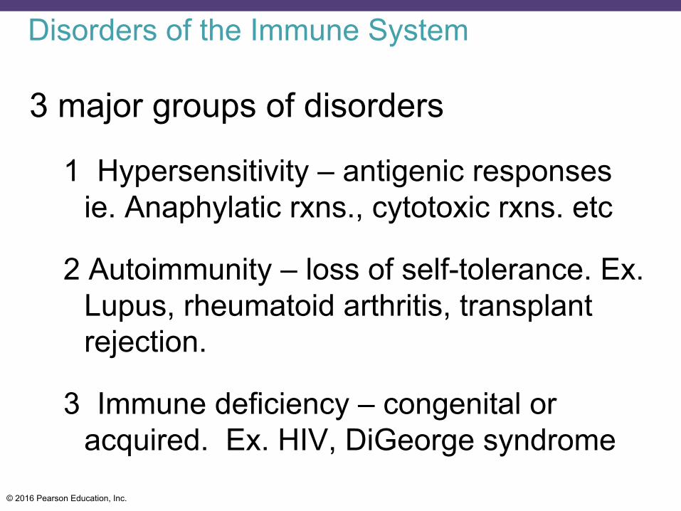
3 major groups of disorders
1 Hypersensitivity – antigenic responses ie. Anaphylatic rxns., cytotoxic rxns. etc
2 Autoimmunity – loss of self-tolerance. Ex. Lupus, rheumatoid arthritis, transplant rejection.
3 Immune deficiency – congenital or acquired. Ex. HIV, DiGeorge syndrome
© 2016 Pearson Education, Inc.
Hypersenitivity
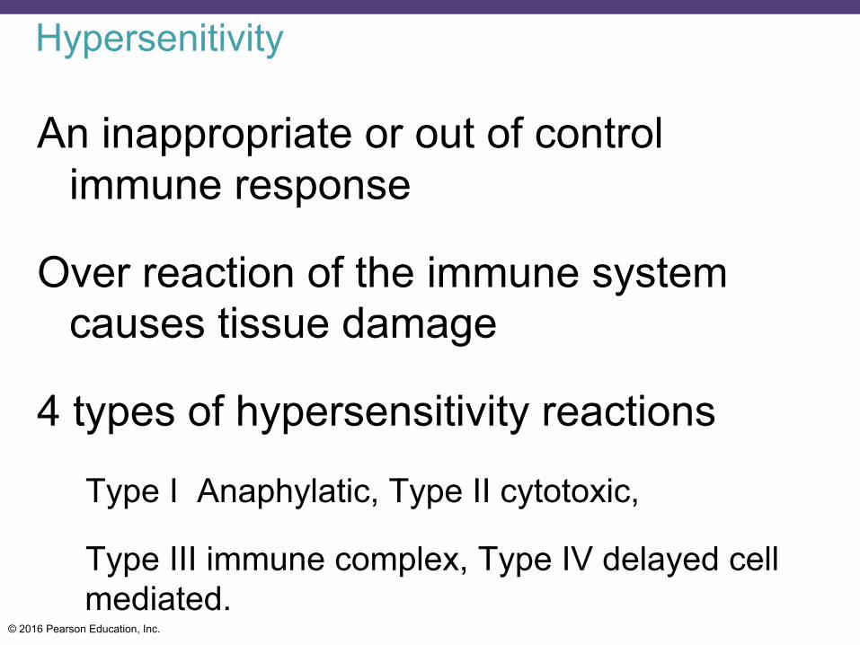
An inappropriate or out of control immune response
Over reaction of the immune system causes tissue damage
4 types of hypersensitivity reactions
Type I Anaphylatic, Type II cytotoxic,
Type III immune complex, Type IV delayed cell mediated.
© 2016 Pearson Education, Inc.
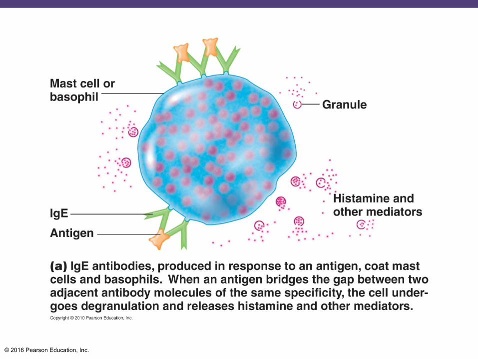
Type I Anaphylatic
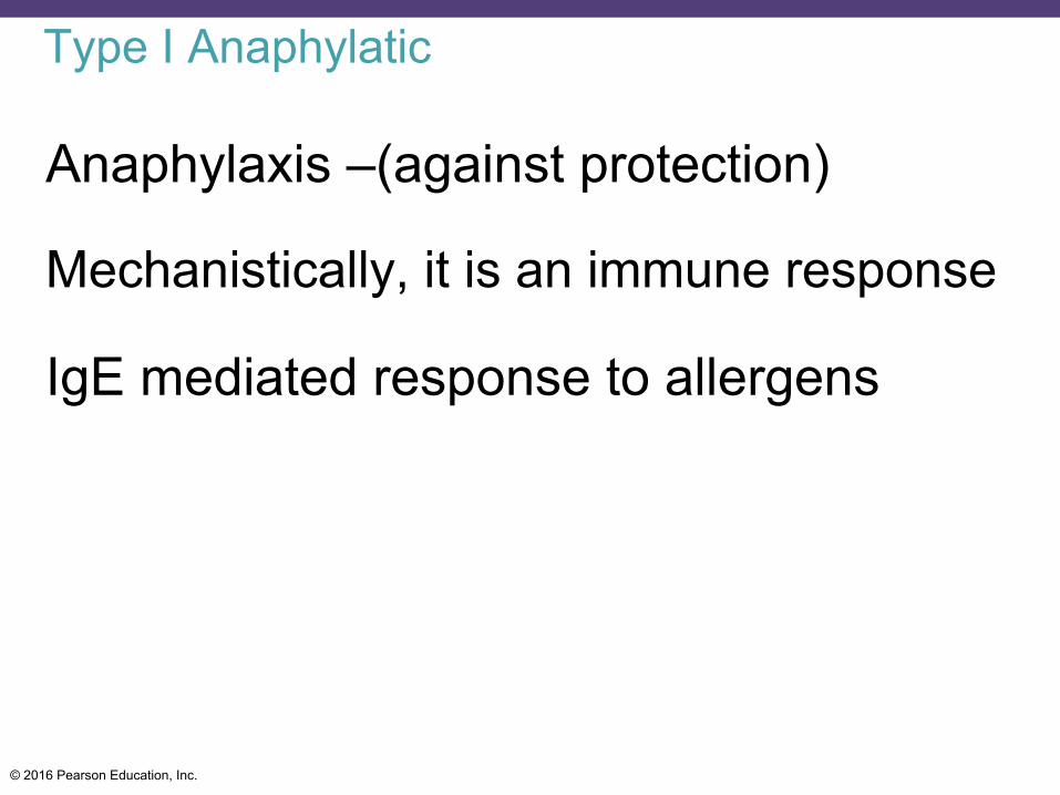
Anaphylaxis –(against protection)
Mechanistically, it is an immune response
IgE mediated response to allergens
© 2016 Pearson Education, Inc.
Type I Anaphylatic
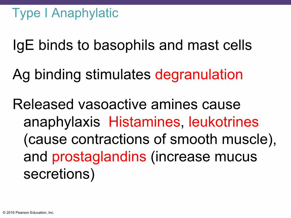
IgE binds to basophils and mast cells
Ag binding stimulates degranulation
Released vasoactive amines cause anaphylaxis Histamines, leukotrines (cause contractions of smooth muscle), and prostaglandins (increase mucus secretions)
© 2016 Pearson Education, Inc.
© 2016 Pearson Education, Inc.
© 2016 Pearson Education, Inc.
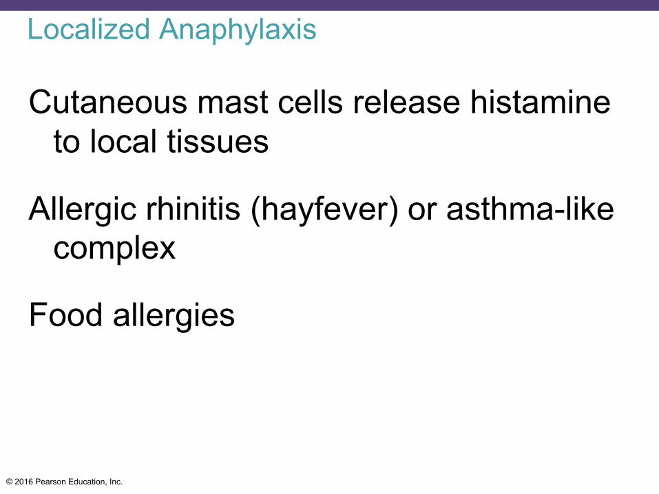
Localized Anaphylaxis
Cutaneous mast cells release histamine to local tissues
Allergic rhinitis (hayfever) or asthma-like complex
Food allergies
© 2016 Pearson Education, Inc.
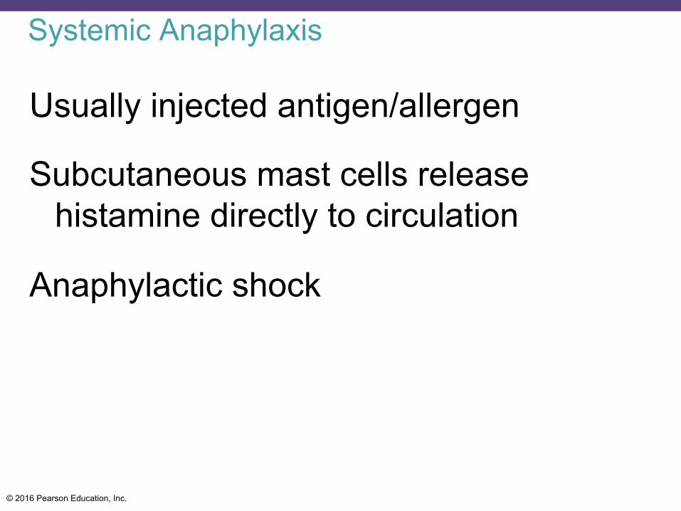
Systemic Anaphylaxis
Usually injected antigen/allergen
Subcutaneous mast cells release histamine directly to circulation
Anaphylactic shock
© 2016 Pearson Education, Inc.
Type II Cytotoxic
Cytotoxic reactions
Abs bind to cells and activate Complement or cytotoxic cells
© 2016 Pearson Education, Inc.
Type II Cytotoxic
Transfusion reactions
Hemolytic disease of the newborn
© 2016 Pearson Education, Inc.
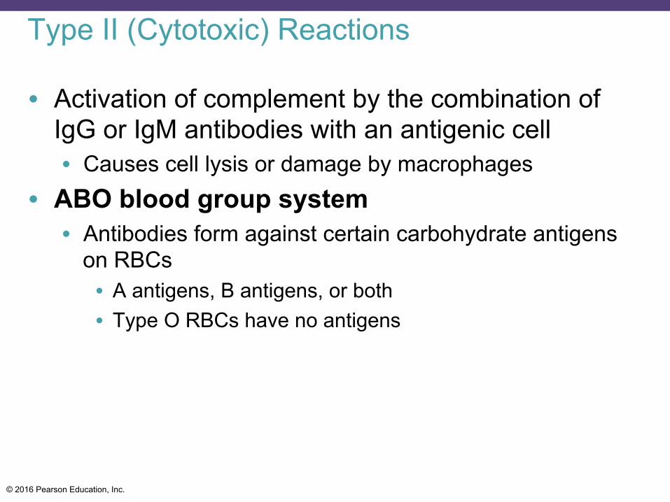
Type II (Cytotoxic) Reactions
• Activation of complement by the combination of IgG or IgM antibodies with an antigenic cell • Causes cell lysis or damage by macrophages
• ABO blood group system • Antibodies form against certain carbohydrate antigens
on RBCs • A antigens, B antigens, or both • Type O RBCs have no antigens
© 2016 Pearson Education, Inc.
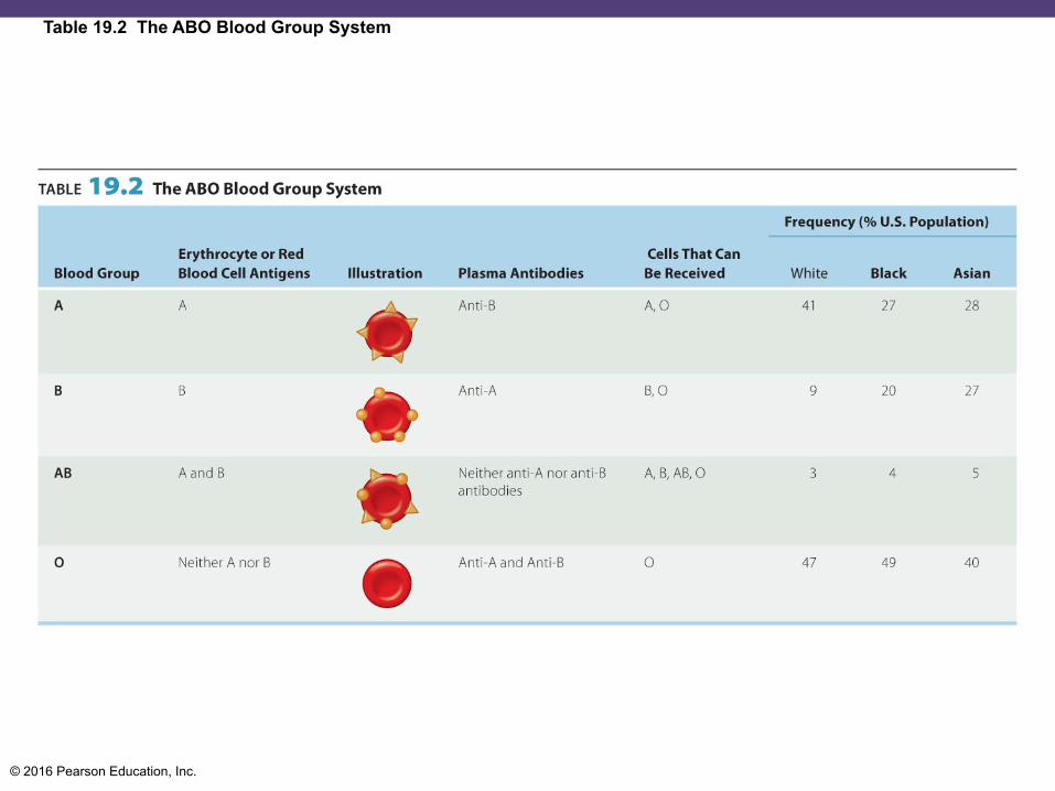
Table 19.2 The ABO Blood Group System
© 2016 Pearson Education, Inc.
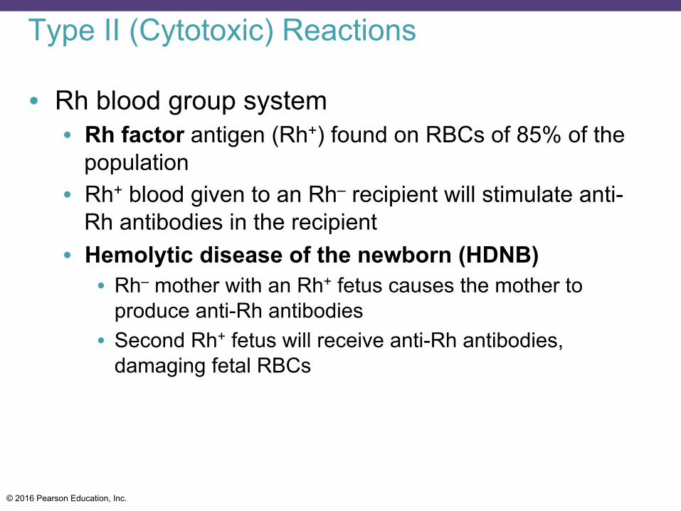
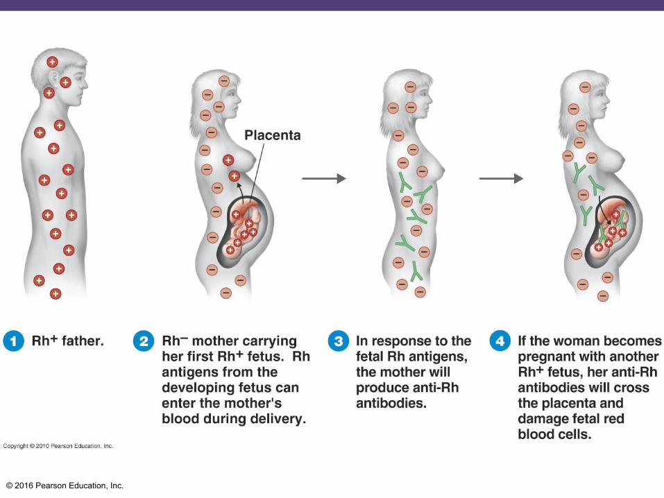
Type II (Cytotoxic) Reactions
• Rh blood group system • Rh factor antigen (Rh+) found on RBCs of 85% of the
population • Rh+ blood given to an Rh– recipient will stimulate anti-
Rh antibodies in the recipient • Hemolytic disease of the newborn (HDNB)
• Rh– mother with an Rh+ fetus causes the mother to produce anti-Rh antibodies
• Second Rh+ fetus will receive anti-Rh antibodies, damaging fetal RBCs
© 2016 Pearson Education, Inc.
© 2016 Pearson Education, Inc.
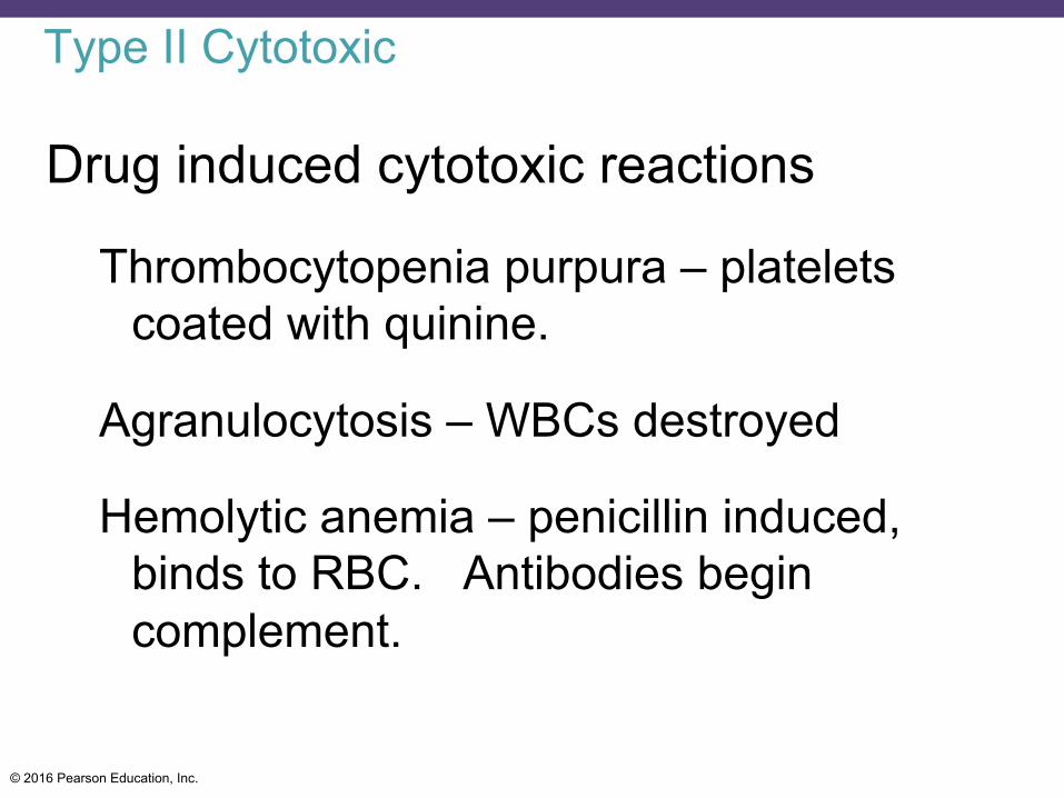
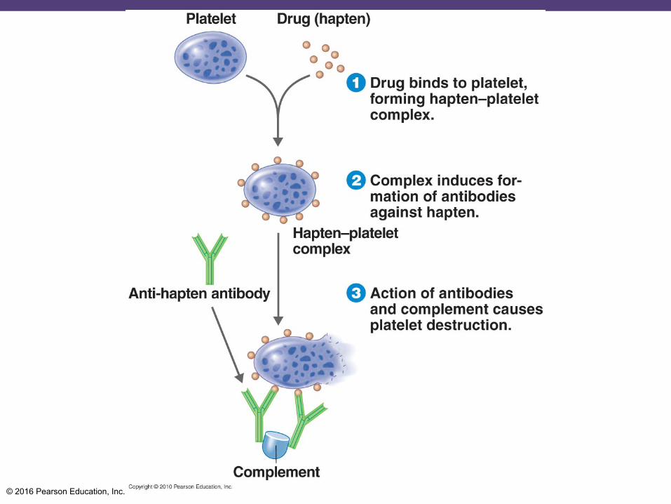
Type II Cytotoxic
Drug induced cytotoxic reactions
Thrombocytopenia purpura – platelets coated with quinine.
Agranulocytosis – WBCs destroyed
Hemolytic anemia – penicillin induced, binds to RBC. Antibodies begin complement.
© 2016 Pearson Education, Inc.
© 2016 Pearson Education, Inc.
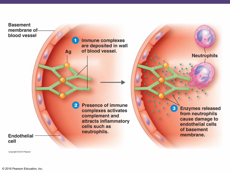
Type III Immune Complex
Mediated by immune complexes
Soluble Ag-Ab complexes
Relative concentrations of Ag and Ab determine fate of complex
Ag and Abs in equal amounts with slight excess of Ag leads to complex becoming trapped in the basement layer beneath endothelial cells.
© 2016 Pearson Education, Inc.
© 2016 Pearson Education, Inc.
Type III Hypersensitivity
Ag excess
Smaller complexes stay soluble longer Filtered at kidney
Complex activates C’ at site
Glomerulonephritis –inflammatory damage to kidney glomeruli.
© 2016 Pearson Education, Inc.
Type IV Delayed type hypersensitivity (DTH) Delayed type hypersensitivity (DTH)
Mediated by activated TH1 cells Released lymphokines attract and activate
macrophages which cause tissue damage
Delayed because mediated by T-helper cells which then become T-memory cells.
Ex. TB test. T-memory cells will proliferate.

© 2016 Pearson Education, Inc.
Allergic contact dermatitis
© 2016 Pearson Education, Inc.
Autoimmune Diseases
Self-reactivity due to a loss of immunological tolerance
Mechanisms somewhat parallel to hypersensitivity
© 2016 Pearson Education, Inc.
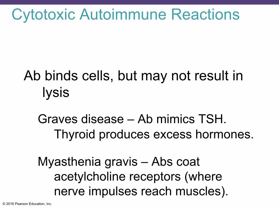
Cytotoxic Autoimmune Reactions
Ab binds cells, but may not result in lysis
Graves disease – Ab mimics TSH. Thyroid produces excess hormones.
Myasthenia gravis – Abs coat acetylcholine receptors (where nerve impulses reach muscles).
© 2016 Pearson Education, Inc.
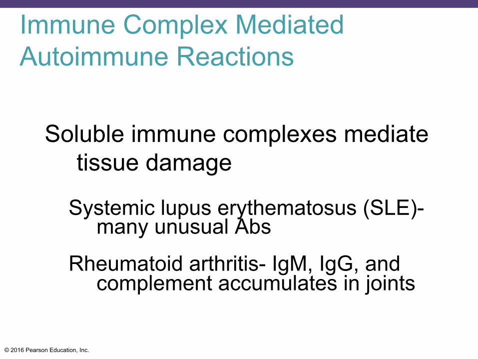
Immune Complex Mediated Autoimmune Reactions
Soluble immune complexes mediate tissue damage
Systemic lupus erythematosus (SLE)- many unusual Abs
Rheumatoid arthritis- IgM, IgG, and complement accumulates in joints
© 2016 Pearson Education, Inc.
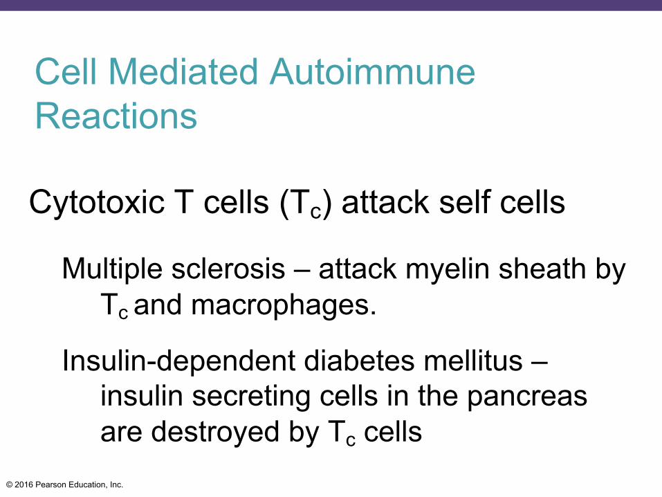
Cell Mediated Autoimmune Reactions
Cytotoxic T cells (Tc) attack self cells
Multiple sclerosis – attack myelin sheath by Tc and macrophages.
Insulin-dependent diabetes mellitus – insulin secreting cells in the pancreas are destroyed by Tc cells
© 2016 Pearson Education, Inc.
Transplantation Reactions
Major histocompatibility complex also called human leukocyte antigen (HLA)
A gene complex that codes for tissue specific antigens. There are four types.
MHC Ag’s are recognized by T cells in transplantation reactions
Probably resemble altered self – immune systems thinks foreign tissue is actually damanged tissue.
© 2016 Pearson Education, Inc.
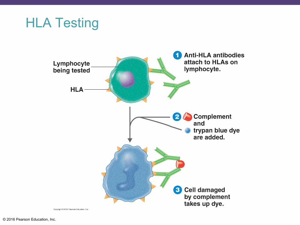
HLA Testing
© 2016 Pearson Education, Inc.
Immune Deficiency
Impaired ability or inability to make immune responses
Congenital (primary)- genetic based
Acquired (secondary)- many specific and non-specific causes
© 2016 Pearson Education, Inc.
Congenital Immune Deficiency
Deficiencies affecting T cells result in a marked increase in viral and parasitic infections, and cancer
© 2016 Pearson Education, Inc.
T Cell Deficiencies
DiGeorge Syndrome – no thymus gland therefore no cell-mediated immunity.
© 2016 Pearson Education, Inc.
Acquired Immune Deficiency
May be non-specific- Radiation or chemotherapy, nutritional, etc.
May be infectious- Epstein-Barr Virus, cytomegalovirus (CMV), HIV, and others

© 2016 Pearson Education, Inc.
Acquired Immunodeficiency Syndrome (AIDS)
• 1981: in the United States, a cluster of cases of Pneumocystis pneumonia, Kaposi's sarcoma, and loss of immune function are discovered in young homosexual men
• 1983: the discovery of a virus causing the loss of immune function (HIV) • Selectively infects T helper cells (CD4+)
© 2016 Pearson Education, Inc.
The Origin of AIDS
• SIV crossed over into the human population in west and central Africa from chimpanzees (around 1908, from bushmeat)
• Spread throughout Africa as a result of urbanization and increased sexual promiscuity
• Patient who died in 1959 in the Congo is the oldest known case
• Norwegian sailor who died in 1976 is the first known case in Western world
© 2016 Pearson Education, Inc.
The Structure of HIV
• Genus Lentivirus • Retrovirus • Two identical strands of RNA, reverse
transcriptase enzyme, phospholipid envelope • gp120 glycoprotein spikes • Death of CD4 cells results in loss of regulation of
the immune system
© 2016 Pearson Education, Inc.
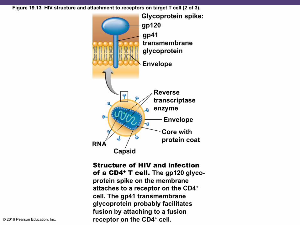
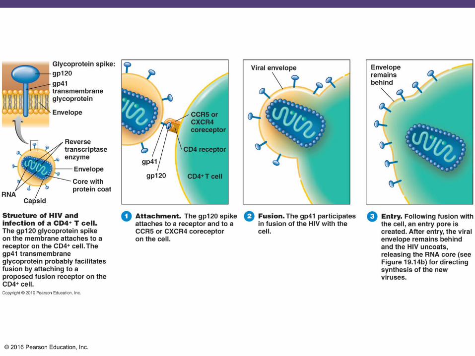
Glycoprotein spike: gp120 gp41 transmembrane glycoprotein
Envelope
Reverse transcriptase enzyme
Envelope
RNA Capsid
Core with protein coat
Structure of HIV and infection of a CD4+ T cell. The gp120 glyco- protein spike on the membrane attaches to a receptor on the CD4+
cell. The gp41 transmembrane glycoprotein probably facilitates fusion by attaching to a fusion receptor on the CD4+ cell.
Figure 19.13 HIV structure and attachment to receptors on target T cell (2 of 3).
© 2016 Pearson Education, Inc.
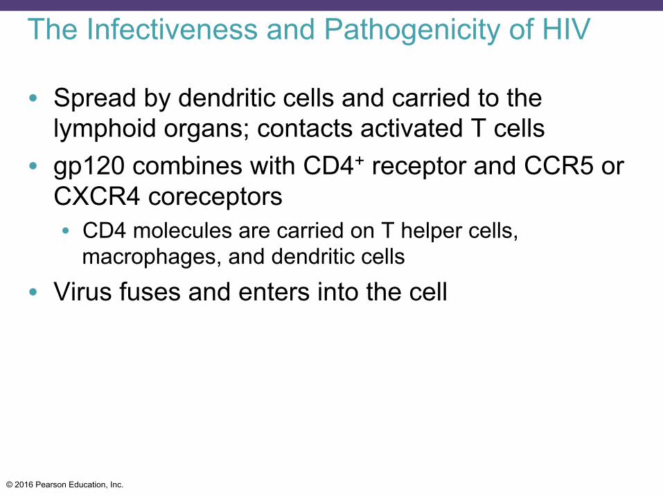
The Infectiveness and Pathogenicity of HIV
• Spread by dendritic cells and carried to the lymphoid organs; contacts activated T cells
• gp120 combines with CD4+ receptor and CCR5 or CXCR4 coreceptors • CD4 molecules are carried on T helper cells,
macrophages, and dendritic cells • Virus fuses and enters into the cell
© 2016 Pearson Education, Inc.
© 2016 Pearson Education, Inc.
© 2016 Pearson Education, Inc.
© 2016 Pearson Education, Inc.
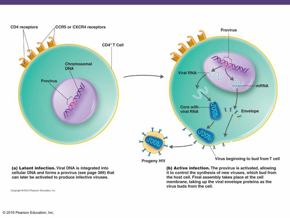
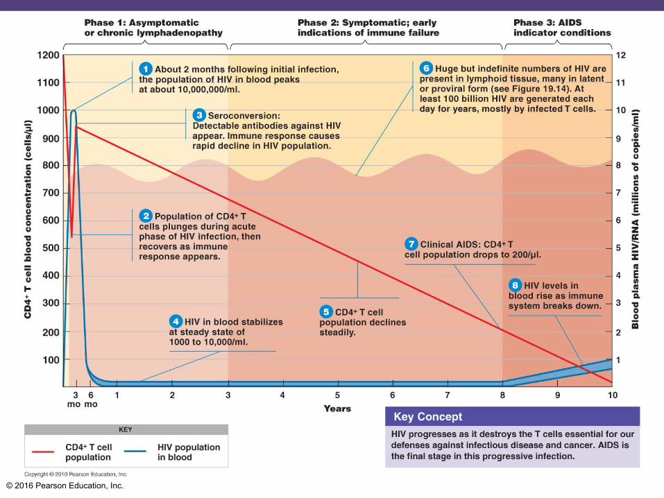
AIDS
Terminal stage of HIV infection
Incubation time ~10 years
© 2016 Pearson Education, Inc.
Diagnostic Methods
• Seroconversion is the period of time between infection and the appearance of antibodies • Takes up to 3 months
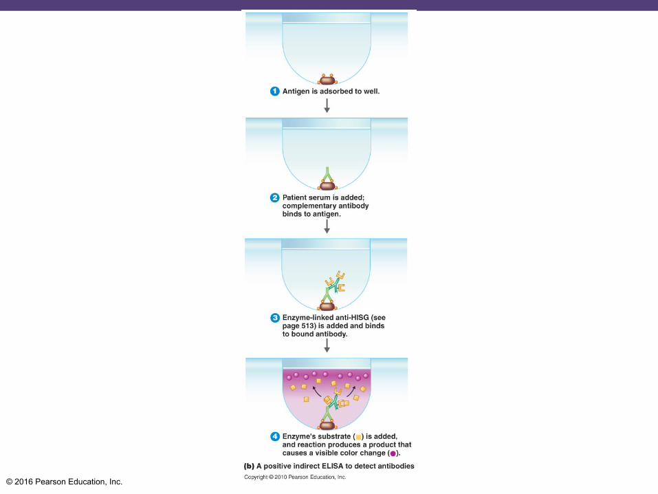
• HIV antibodies detected by ELISA • Viruses detected by Western blotting or APTIMA
(RNA testing) • Plasma viral load (PVL) is determined by PCR or
nucleic acid hybridization
© 2016 Pearson Education, Inc.
HIV Transmission
• HIV survives 6 hours outside a cell • HIV can survive more than 1.5 days inside a cell • Routes of transmission: intimate sexual contact,
breast milk, transplacental infection, contaminated needles, organ transplants, and blood transfusion • Anal-receptive intercourse is the most dangerous form
of sexual contact
© 2016 Pearson Education, Inc.

![Antigens and antibodies interact… They associate: [Ag] + [Ab] 6 [AgCAb] They dissociate: [AgCAb] 6 [Ag] + [Ab] The velocity of the association: v 1 %[Ag]](https://img.pdfslide.net/doc/110x75/56649d995503460f94a83ec3/antigens-and-antibodies-interact-they-associate-ag-ab-6-agcab-they.jpg)