Embed Size (px)
Citation preview
8/1/2016
1
Center for Computational Imaging and Personalized Diagnostics
Pallavi Tiwari, Ph.D. Assistant Professor, Biomedical Engineering
Director, Brain Image Computing Laboratory
Case Western Reserve University
RADIOMICS, RADIO-GENOMICS, RADIO-PATHOMICS:
CONNECTING THE DOTS TOWARDS PERSONALIZED MEDICINE
IN BRAIN TUMORS
Center for Computational Imaging and Personalized Diagnostics
Brain Tumors– Prevalence and Incidences
18,600 brain tumor deaths/year in the US.
GBM most aggressive. Median survival of 12
months [21.3 years of life lost].
Current treatment: maximal surgical resection,
radiotherapy, and concomitant and adjuvant
chemotherapy with temozolomide.
Poorly understood complex micro-enviornmnent
(hanahan and Weinberg 2000).
Precision Medicine
An emerging approach for disease treatment and prevention that takes into account
individual variability in genes, environment, and lifestyle for each person.
8/1/2016
2
FUNDAMENTAL QUESTIONS
WHO TO TREAT?
HOW TO TREAT?
WILL THE TREATMENT WORK?
6
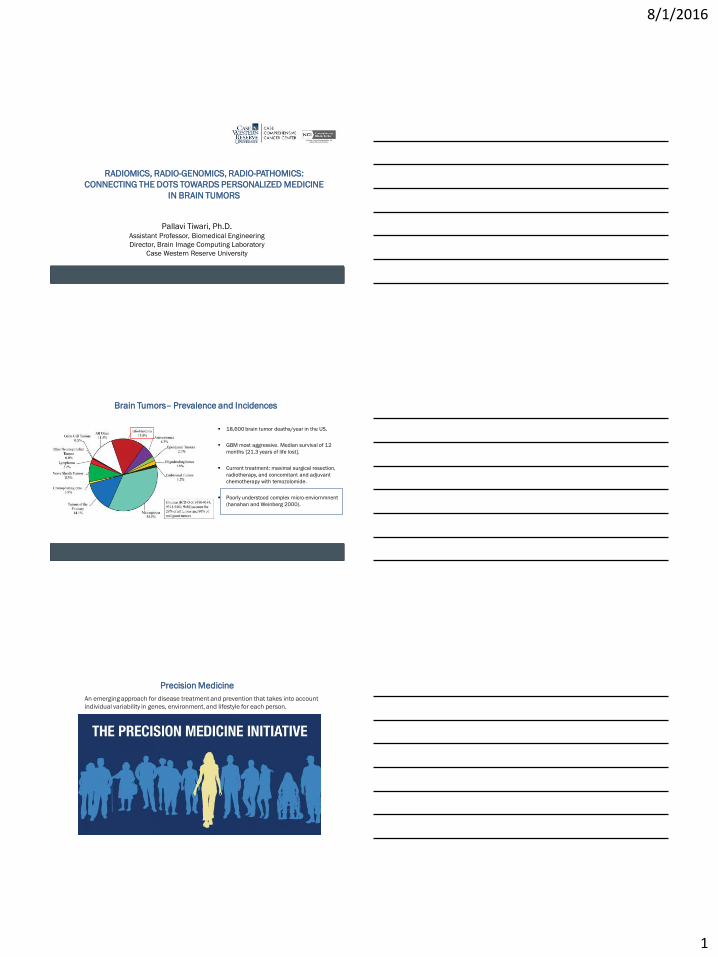
Intra-tumoral heterogeneity captures tumor behavior and individual patient’s response to treatment
o Assessment of severity/aggressiveness o Prediction of response to therapy. o Changes in response to therapy over time.
85% probability of recurrence
20% probability of recurrence
8/1/2016
3
Center for Computational Imaging and Personalized Diagnostics
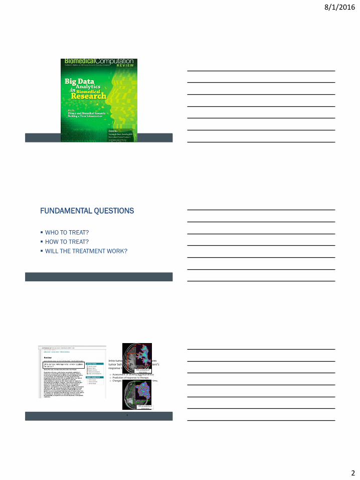
Radiomics: Study of capturing subtle differences in imaging that are
not visually appreciable
- Local Texture-based Radiomics Features
- Global Structural Radiomics Features
Center for Computational Imaging and Personalized Diagnostics
Apophenia: Tendency to “unlock” hidden meaningful patterns
Center for Computational Imaging and Personalized Diagnostics
Radiomics: Local Image Feature Representations
What is texture? Capturing local intensity statistics
within small neighborhoods,
quantify smoothness,
heterogeneous appearance etc.
Feature types: 1st order Statistical: Mean, range
2nd order Statistical: Statistics
based on co-occurring intensities
Laws features: characterize edges,
waves, ripples
Gabor: Multi-scale, multi-orientation
filter responses
J.C. Russ. The Image Processing Handbook. CRC Press, 5th edition, 2007.
Haralick features
Laws Energy features
Gabor features
8/1/2016
4
Center for Computational Imaging and Personalized Diagnostics
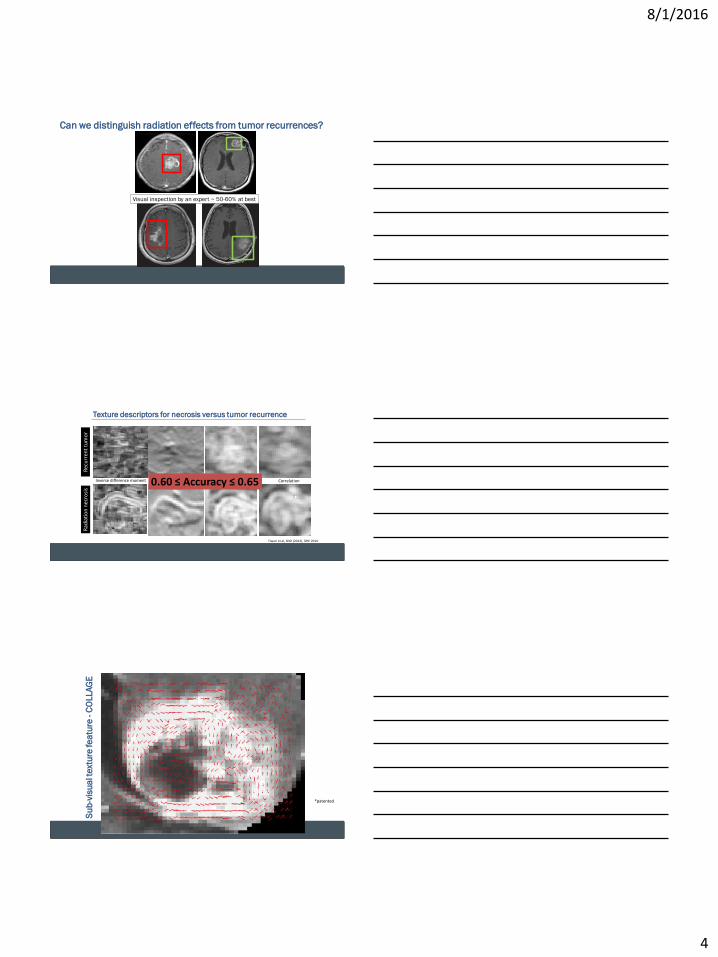
Can we distinguish radiation effects from tumor recurrences?
Visual inspection by an expert ~ 50-60% at best
Center for Computational Imaging and Personalized Diagnostics
Tiwari et al, SNO (2013), SPIE 2014
Laws (E5L5)
Rad
iati
on
nec
rosi
s
Inverse difference moment Difference Variance(Laplace) Correlation
Rec
urr
ent
tum
or
0.60 ≤ Accuracy ≤ 0.65
Texture descriptors for necrosis versus tumor recurrence
Center for Computational Imaging and Personalized Diagnostics
Su
b-v
isu
al t
ext
ure
fe
atu
re - C
OLLA
GE
*patented
8/1/2016
5
Center for Computational Imaging and Personalized Diagnostics
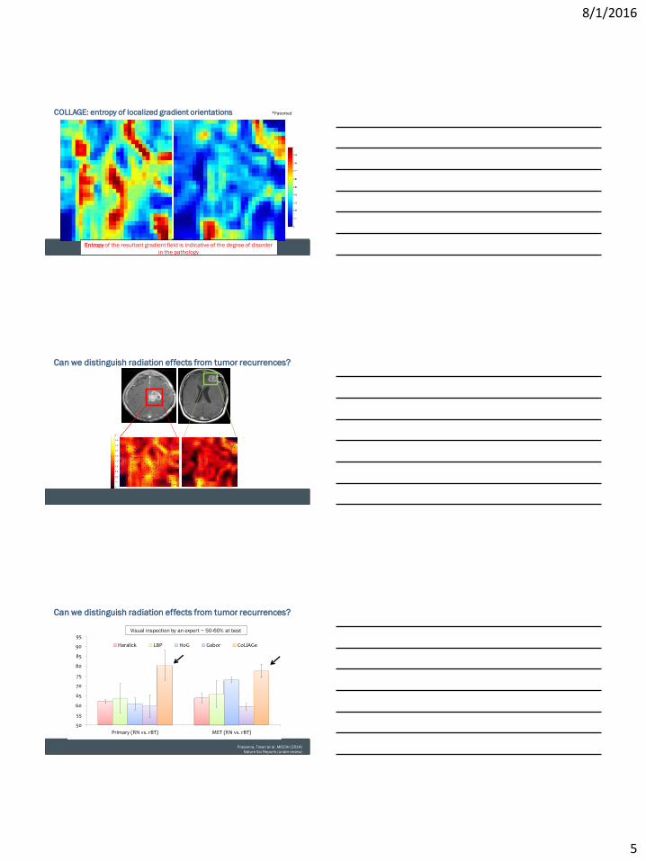
*Patented COLLAGE: entropy of localized gradient orientations
Entropy of the resultant gradient field is indicative of the degree of disorder
in the pathology
Center for Computational Imaging and Personalized Diagnostics
Can we distinguish radiation effects from tumor recurrences?
Center for Computational Imaging and Personalized Diagnostics Prasanna, Tiwari et al. MICCAI (2014)
Nature Sci Reports (under review)
50
55
60
65
70
75
80
85
90
95
Primary (RN vs. rBT) MET (RN vs. rBT)
Haralick LBP HoG Gabor CoLlAGe
Visual inspection by an expert ~ 50-60% at best
Can we distinguish radiation effects from tumor recurrences?

8/1/2016
6
Center for Computational Imaging and Personalized Diagnostics
“pure” RN Predominant RN Predominant tumor “pure” tumor
Prasanna et al. ISMRM (2015)
Quantifying grades of cerebral radiation necrosis vs. Tumor
0.1
0.2
0.3
0.4
0.5
0.6
0.7
0.8
0.9
1.0
Prasanna et al. ISMRM, SNO (2015)
Center for Computational Imaging and Personalized Diagnostics
100% CRN >80% CRN <20% CRN >0% CRN
Co
LlA
Ge
en
tro
py
COLLAGE discriminate necrosis from recurrent cancer
At a normalized threshold of 0.8, 100% tumor
patients and 67% CRN patients were
correctly identified using CoLlAGe values
Pure RN
>80% RN
<20% RN
Treatment naïve GBM
Pathology CoLlAGe entropy
‘pure’ CRN 0.74 +/- 0.11
‘predominant’ CRN 0.84 +/- 0.10
‘predominant’ tumor 0.91 +/- 0.06
Treatment-naïve GBM 0.88 +/- 0.03
Prasanna et al. ISMRM (2015)
Risk Score
Center for Computational Imaging and Personalized Diagnostics
Human-machine comparison for radiation necrosis vs. recurrent tumors
Prasanna et al. RSNA (2016)
8/1/2016
7
Center for Computational Imaging and Personalized Diagnostics
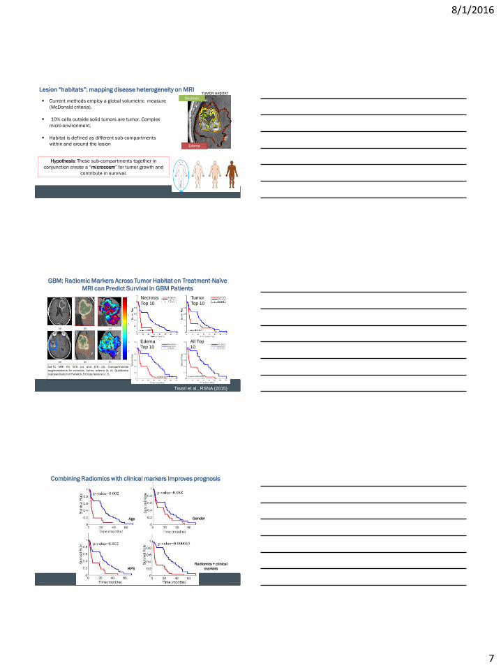
Lesion “habitats”: mapping disease heterogeneity on MRI
Current methods employ a global volumetric measure
(McDonald criteria).
10% cells outside solid tumors are tumor. Complex
micro-environment.
Habitat is defined as different sub-compartments
within and around the lesion
Hypothesis: These sub-compartments together in
conjunction create a “microcosm” for tumor growth and
contribute in survival.
Necrosis
TUMOR HABITAT
Edema
21
Center for Computational Imaging and Personalized Diagnostics
GBM: Radiomic Markers Across Tumor Habitat on Treatment-Naïve
MRI can Predict Survival in GBM Patients
(f) (e) (d)
Gd-T1 MRI for STS (a) and LTS (d). Compartmental
segmentations for necrosis, tumor, edema (b, e). Qualitative
representation of Haralick Entropy feature (c, f).
(a) (b) (c)
Necrosis
Top 10
All Top
10
Edema
Top 10
Tumor
Top 10
Tiwari et al., RSNA (2015)
Center for Computational Imaging and Personalized Diagnostics
Age Gender
KPS
Radiomics + clinical
markers
Combining Radiomics with clinical markers improves prognosis
8/1/2016
8
Center for Computational Imaging and Personalized Diagnostics
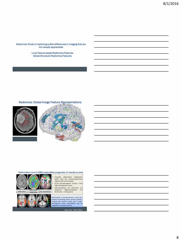
Radiomics: Study of capturing subtle differences in imaging that are
not visually appreciable
- Local Texture-based Radiomics Features
- Global Structural Radiomics Features
Center for Computational Imaging and Personalized Diagnostics
Radiomics: Global Image Feature Representations
Center for Computational Imaging and Personalized Diagnostics
Deformation due to GBM mass-effect prognostic of overall survival
GBM subject Deformation
Magnitudes
Deformation in somato-sensory, visual and
sematic processing areas, primary auditory
cortices and memory areas due to right-
hemispheric tumor mass-effects may be
prognostic of overall survival.
• Correlate deformation distribution
within each AAL cortical/subcortical
regions with survival (days).
• N=30 left-hemispheric tumors, n=24
right-hemispheric tumors.
• Left-hemispheric (14 short-term, 12
long-term), Right-hemispheric (12
short-term, 12 long-term)
AAL parcellations
Mitra et al., SNO (2016)
8/1/2016
9
Center for Computational Imaging and Personalized Diagnostics
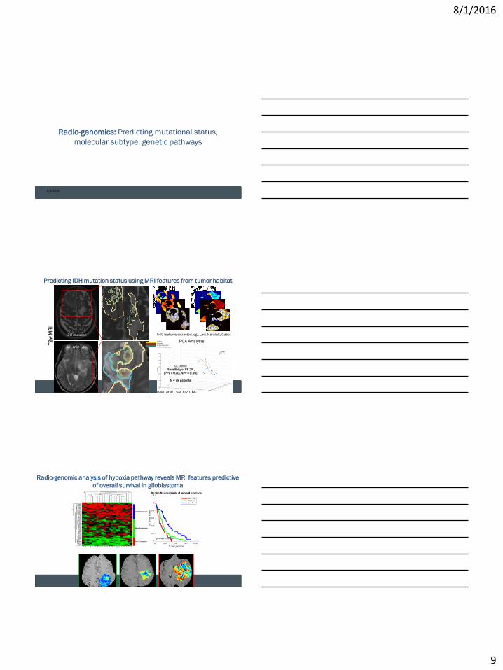
Radio-genomics: Predicting mutational status,
molecular subtype, genetic pathways
8/1/2016
27
Center for Computational Imaging and Personalized Diagnostics
Predicting IDH mutation status using MRI features from tumor habitat
T2
w M
RI
IDH mutation
IDH Wild Type
IDH-WT
IDH mut
Edema
Necrosis
Enhancing tumor
Non enhancing tumor
540 features extracted. eg., Law, Haralick, Gabor
PCA Analysis
T2, Edema:
Sensitivity of 88.2%
(PPV = 0.83, NPV = 0.90)
N = 78 patients
Beig et al., SNO (2016)
Center for Computational Imaging and Personalized Diagnostics
Radio-genomic analysis of hypoxia pathway reveals MRI features predictive
of overall survival in glioblastoma

8/1/2016
10
Center for Computational Imaging and Personalized Diagnostics
Radio-pathomics: Understanding the biological
underpinning of tumor on imaging
8/1/2016
30
Center for Computational Imaging and Personalized Diagnostics
Radio-pathomics for radiation necrosis vs. recurrent tumors –
Correlating COLLAGE to known pathological processes
Prasanna et al., RSNA (2016)
Center for Computational Imaging and Personalized Diagnostics
CONCLUDING REMARKS
Radiomics have the potential to complement personalized prognosis, and treatment
evaluation to address questions such as:
o Who to treat?
o How to treat?
o When to treat?
o Did the treatment work?
Radio-genomics, radio-pathomics allow interactions across length scales and provide
mechanisms to identify “image biomarkers” for prognosis and treatment evaluation.
Radiomic techniques not only allow for bench-to-bedside personalized medicine
solutions, but also provide reliable and reproducible tools for feature discovery.
8/1/2016
11
Center for Computational Imaging and Personalized Diagnostics
Collaborators:
Dr. Anant Madabhushi (Director, CCIPD)
Dr. Lisa Rogers (UH)
Dr. Leo Wolansky (UH)
Dr. Mark Cohen (UH)
Dr. Marta Couce (UH)
Dr. Andrew Sloan (UH)
Dr. Shabbar Danish (UMDNJ, Rutgers)
Dr. Raymond Huang (BWH, Boston)
Dr. Marco Pinho (UT Southwestern)
Dr. T.C. Lam (Tuen Mun Hospital, China)
Dr. Susann Brady-Kalnay (CWRU)
Acknowledgements
Funding:
Case Comprehensive Cancer Center
Coulter Translational Phase I, II, III Award
Ohio Third Frontier
NSF i-corps
http://bric-lab.com
http://ccipd.case.edu
Center for Computational Imaging and Personalized Diagnostics
Center for Computational Imaging and Personalized Diagnostics
THANK YOU FOR YOUR ATTENTION