Embed Size (px)
Citation preview

Pre-AP Biology Bellwork September 24th
1. What is a cell?
2. What is “cell theory”
3. Why are cells considered the building blocks of all living things?

Definition of Cell
A cell is the smallest unit that is capable of performing life functions.
It is the basic building block of all living things.

Cell Theory
• All living things are made up of one or more cells.
• Cells are the smallest working units of all living things.
• All cells come from preexisting cells through cell division.

Why is it a “building block”?
• Organism – group of organ systems functioning together.
• Organ System – group of organs functioning together.
• Organ – group of tissues functioning together.
• Tissue – group of cells functioning together.
• Cell – the foundation of all of the above! IE a “building block”
REVIEW

The Microscopic World of Cells
– Organisms are either:• Single-celled, such as most bacteria and protists• Multicelled, such as plants, animals, and most fungi


History of the Cell

Figure 4.3

Microscopes as a Window on the World of Cells
– The light microscope is used by many scientists.• Light passes through the specimen.• Lenses enlarge, or magnify, the image.• Magnification
– Is an increase in the specimen’s apparent size.
• Resolving power– Is the ability of an optical
instrument to show two objects as being separate.
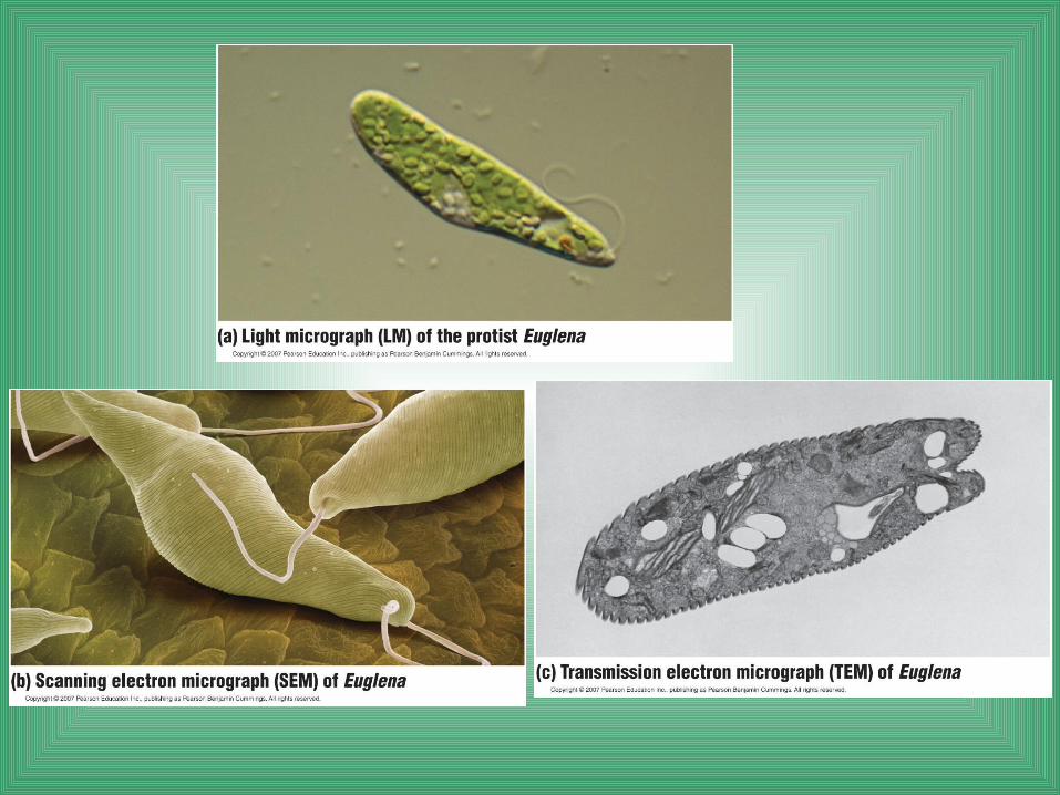
Euglena

1. The electron microscope (EM) uses a beam of electrons.• It has a higher resolving power than the light microscope.• The electron microscope can magnify up to 100,000X.
– Such power reveals the diverse parts within a cell.
2. The scanning electron microscope (SEM) is used to study the detailed architecture of the surface of a cell.
3. The transmission electron microscope (TEM) is useful for exploring the internal structure of a cell.

Microscopes and Cells
• 1600’s.–Anton van Leeuwenhoek first described living cells as seen through a simple microscope.

Microscopes and Cells– In 1665 Robert Hooke used the first compound microscope to view thinly sliced cork cells.
•Compound scopes use a series of lenses to magnify in steps.•Hooke was the first to use the term “cell”.

Microscopes and Cells• 1830’s.
–Mathias Schleiden identified the first plant cells and concluded that all plants
made of cells.
-Thomas Schwann made the same conclusion about animal cells.

Examples of Cells
Amoeba Proteus
Plant Stem
Red Blood Cell
Nerve Cell
Bacteria
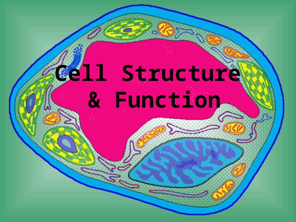
Cell Structure & Function

Two Types of Cells
1. Prokaryotic
2. Eukaryotic

Prokaryotic• Do not have structures surrounded by
membranes
• Do not have structures surrounded by membranes
• NO TRUE NUCLEUS
• Few internal structures
• Examples: unicellular organisms, archaebacteria & eubacteria

Eukaryotic• Contain organelles surrounded by membranes• Most living organisms• Contains a true nucleus• Unicellular or multicellular• Examples: Protista, Fungi, Plants, Animals
Plant Animal
In your notes…
• Compare and contrast a prokaryotic cell and a eukaryotic cell

Two Basic Cell Types• Prokaryotes
– Small– no not contain any
membrane-bound organelles
– No true nucleus– Bacteria
• Eukaryotes– Do contain membrane-
bound organelles– Most multicellular (also
amoebas, some algae)– Contain a Nucleus (Robert
Brown and Rudolf Virchow)

Figure 4.4
Parts of the Cell

I. Surrounding the CellCell Boundries

1. Cell Membrane
• The outer layer of animal cells, found inside cell walls (in plants)
• Double layer (bi-lipid layer)
• Function: Controls what goes in and out of a cell.

The Cell MembraneThe Plasma Mosaic Membrane Model

Membrane Structure– The plasma membrane separates the living cell from its nonliving surroundings.– The membranes of cells are composed mostly of:
• Lipids• Proteins

Why Cells Must Control Materials
Cells need nutrients (glucose, AA, lipids etc)• Plasma Membrane controls what goes in and what
goes out• Waste removed through membrane• Selectively Permeable
– Allows some things through, – keeps others out– Water – osmosis– Ca+2, Na+ - only certain times

Let’s Draw It Together!

Study!
STUDY THE DIAGRAM YOU JUST DID ON THE CELL MEMBRANE. THERE WILL BE A QUIZ THE NEXT CLASS WHERE YOU WILL DRAW, LABEL AND EXPLAIN THE FUNCTION OF THE PARTS OF THE PLASMA MOSAIC MEMBRANE

A Better Rendition…

Phospholipid Bilayer• Lipids – nonpolar
– Makes it difficult for water to get through
– HYDROPHOBIC
• Phosphate head – polar– Face inside and outside of cell– Work well with water – water
soluble barrier– HYDROPHILIC
• Fluid Mosaic Model– Phospholipids move within
membrane– Proteins create mosaic
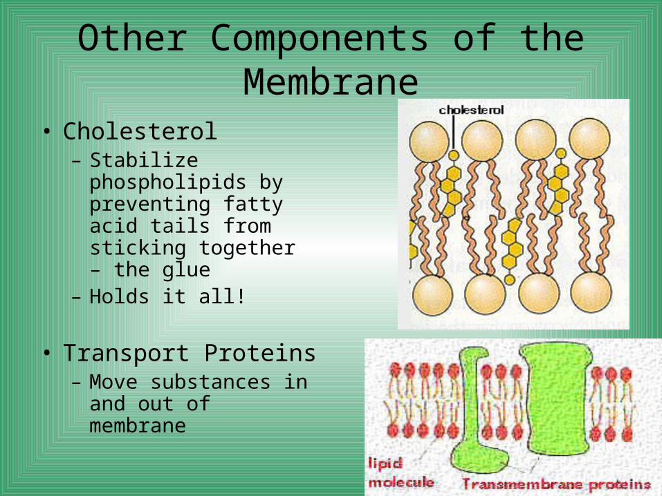
Other Components of the Membrane
• Cholesterol– Stabilize phospholipids
by preventing fatty acid tails from sticking together – the glue
– Holds it all!
• Transport Proteins– Move substances in
and out of membrane

Membrane Proteins - Examples

Evolution Connection:The Origin of Membranes
– Phospholipids were probably among the organic molecules on the early Earth.
– When mixed with water, phospholipids spontaneously form membranes.

Cell Surfaces– Most cells secrete materials for coats of one
kind or another• That are external to the plasma membrane.
– These extracellular coats help protect and support cells
• And facilitate interactions between cellular neighbors in tissues.

– Plant cells have cell walls,• Which help protect the cells, maintain their shape,
and keep the cells from absorbing too much water.
– Animal cells have an extracellular matrix,• Which helps hold cells together in tissues and
protects and supports them.

2. Cell Wall
• Most commonly found in plant cells & bacteria
• NOT FOUND IN ANIMAL CELLS!
• Rigid layer of non living material
• Function: Protection & Support

II. Inside the Cell

3. Cytoplasm / Cytosol
• Gel-like mixture– water (70%)– Proteins, fats,
carbohydrartes, nucleic acids, ion (30%)
• Surrounded by cell membrane

4. Nucleus
• The boss!– The nucleus is the manager of the cell.
• Genes in the nucleus store information necessary to
produce proteins.
• Directs cell activities• Contains genetic material
– DNA– The nucleus is bordered by a double
membrane called the nuclear envelope.• It contains chromatin.• It contains a nucleolus.

5. Nuclear Envelope
• Also known as the nuclear mebrane
• Surrounds nucleus• Made of two layers• Openings allow
material to enter and leave nucleus through large pores

Figure 4.8

6. Chromosomes
• The blueprints!• In nucleus• Made of DNA• Contain instructions
for traits & characteristics

7. Nucleolus
• The “secretary” • Contains RNA copy of
the DNA to build proteins
• Inside nucleus• Responsible for
ribosome production

8. Ribosomes
• Protein factory• Responsible for
protein synthesis• Each cell contains
thousands• Make proteins• Found on
endoplasmic reticulum & floating throughout the cell

How DNA Controls the Cell
– DNA controls the cell by transferring its coded information into RNA.
• The information in the RNA is used to make proteins.

Figure 4.9

The Endomembrane System: Manufacturing and Distributing
Cellular Products
– Many of the membranous organelles in the cell belong to the endomembrane system.

9. Endoplasmic Reticulum• Produces an enormous variety of molecules.• Moves materials such as protein around in cell• Smooth type: lacks ribosomes• Rough type: ribosomes embedded in surface

Figure 4.10

Rough ER– The “roughness” of the rough ER is due to
ribosomes that stud the outside of the ER membrane.
– The functions of the rough ER include:• Producing two types of membrane proteins• Producing new membrane
– After the rough ER synthesizes a molecule, it packages the molecule into transport vesicles.

Figure 4.11

Smooth ER
– The smooth ER lacks the surface ribosomes of ER and produces lipids, including steroids.

10. Golgi Body (Golgi Apparatus)• Works with ER• Protein 'packaging plant‘• Packages proteins with
lipids– Refines, stores, and
distributes the chemical products of cells.
• A series of flattened sacs where newly made lipids and proteins from the E.R. are repackaged and shipped to the plasma membrane.

Figure 4.12

11. Lysosome• A lysosome is a membrane-
enclosed sac.• Garbage bag of the cell!• Digestive 'plant' for proteins, fats,
and carbohydrates– Contains digestive enzymes– These enzymes break down
macromolecules• Lysosomes have several types of
digestive functions– They fuse with food vacuoles to digest
the food.– Transports undigested material to cell
membrane for removal
•More numerous in ANIMAL cells•Contain enzymes that function in digestion of food and dead cell parts•Also known as “suicide sacs” because they can destroy the whole cell: Cell breaks down if lysosome explodes
Lysosome Formation

Figure 4.13a
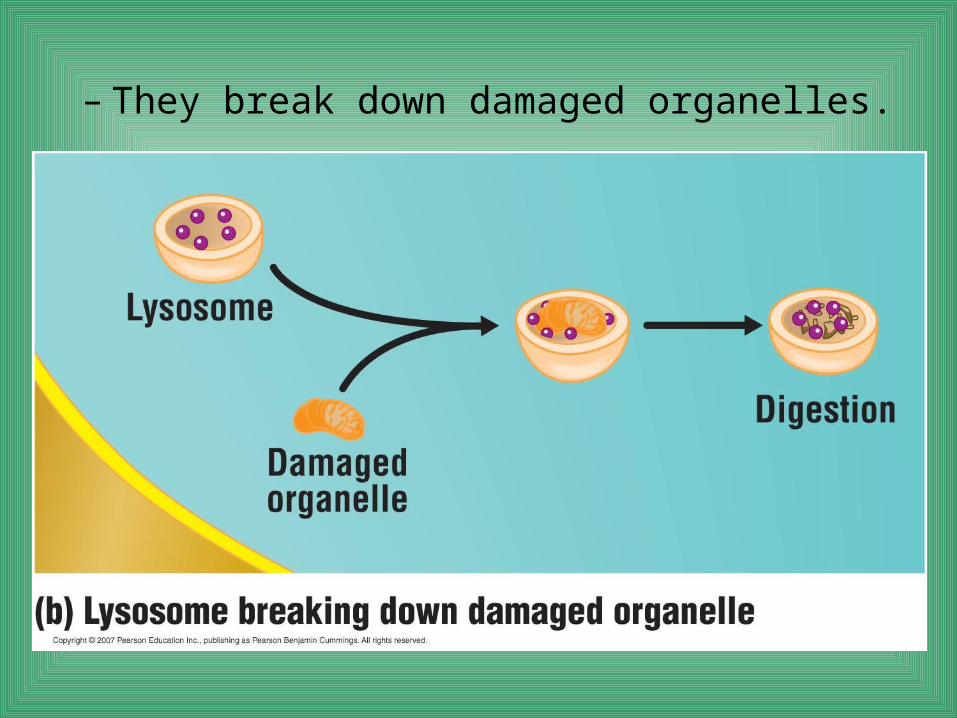
– They break down damaged organelles.

12. Vacuoles
• Vacuoles are membranous sacs.Contains water solution
• Two types are the contractile vacuoles of protists and the central vacuoles of plants.– MUCH larger in plants– Small in animal cells
• FUNCTION: Membrane-bound sacs for storage of food and water, digestion, and waste removal. Help plants maintain shape (support)
Paramecium Vacuole

Figure 4.14

– A review of the endomembrane system
Chloroplasts and Mitochondria: Energy Conversion
– Cells require a constant energy supply to do all the work of life.

13. Chloroplast
• Site of photosynthesis• Usually found in plant
cells• NOT FOUND IN ANIMAL
CELLS!• Contains green
chlorophyll in the thylakoid membrane
• Turn the Sun’s energy into food through photosynthesis– They do not make energy,
they convert it

Figure 4.16

14. Mitochondria• Mitochondria are the sites of cellular
respiration, which involves the production of ATP from food molecules.
• The “powerhouse” of the cell• Produces energy in the form of ATP
through chemical reactions – breaking down fats & carbohydrates
• Controls level of water and other materials in cell
• Recycles and decomposes proteins, fats, and carbohydrates
• Has a highly folded inner membrane (cristae).

Figure 4.17

– Mitochondria and chloroplasts share another feature unique among eukaryotic organelles.
• They contain their own DNA.
– The existence of separate “mini-genomes” is believed to be evidence that
• Mitochondria and chloroplasts evolved from free-living prokaryotes in the distant past.

15. Cytoskeleton
•A network of thin, fibrous materials that act as a scaffold and support the organelles.•Microtubules – hollow filaments of protein. Produced in centrosome•Microfilaments – solid filaments of protein.
The cytoskeleton is an infrastructure of the cell consisting of a network of fibers.

Maintaining Cell Shape– One function of the cytoskeleton
• Is to provide mechanical support to the cell and maintain its shape.

– The cytoskeleton can change the shape of a cell.
• This allows cells like amoebae to move.

16. Cilia• Short, numerous,
hair-like projections from the plasma membrane.
• Move with a coordinated beating action.

Figure 4.19c

17. FlagellaLonger, less numerous projections from the plasma membrane.
Move with a whiplike action.

Cilia and Flagella– Cilia and flagella are motile appendages– Flagella propel the cell in a whiplike motion.– Cilia move in a coordinated back-and-forth motion.– Some cilia or flagella extend from nonmoving cells.
• The human windpipe is lined with cilia.
Paramecium CiliaCilia and Flagella

18. Centrosome
• Microtubles are produced here
• The centrosome contains a pair of small organelles, the centrioles in animal cells ONLY
• Plant cells have centrosomes BUT no CENTRIOLES!

19. CentrioleMade of protein.Play a role in the splitting of the cell into two cells (cell division).Found in animal and fungi cells. NEVER IN PLANTS!Made of microtubules

Draw a Generalized CellPlease use all 19 parts mentioned we just went over

Study!
STUDY THE DIAGRAM YOU JUST DID OF A GENERALIZED CELL. THERE WILL BE A QUIZ THE NEXT CLASS WHERE YOU WILL DRAW, LABEL AND EXPLAIN ALL OF THE PARTS OF A GENERALIZED CELL.

Composite Animal Cell
...

Composite PLANT Cell

Cell Project!
Please see the rubric handed out in class for specifics on this project.

Liver Cell

Lily Parenchyma Cell

Specialized Cells
• Muscle Cells–Long, slippery and
elastic so that they can slide past one another upon expansion and contraction

Specialized Cells
• Red Blood Cell–Has no nucleus so that
it can carry more oxygen

Specialized Cells
• Nerve cells–Can be several feet
long –pass chemical and
electrical signals throughout the body

Cellular Transport

Cellular Transport

• Osmosis – diffusion of water across a selectively permeable membrane
• Plasma membrane does NOT limit water movement
Osmosis: Review

OsmosisIf two solutions are separated by a selectively
permeable membrane….
Only water can flow from side to side to make the concentrations equal
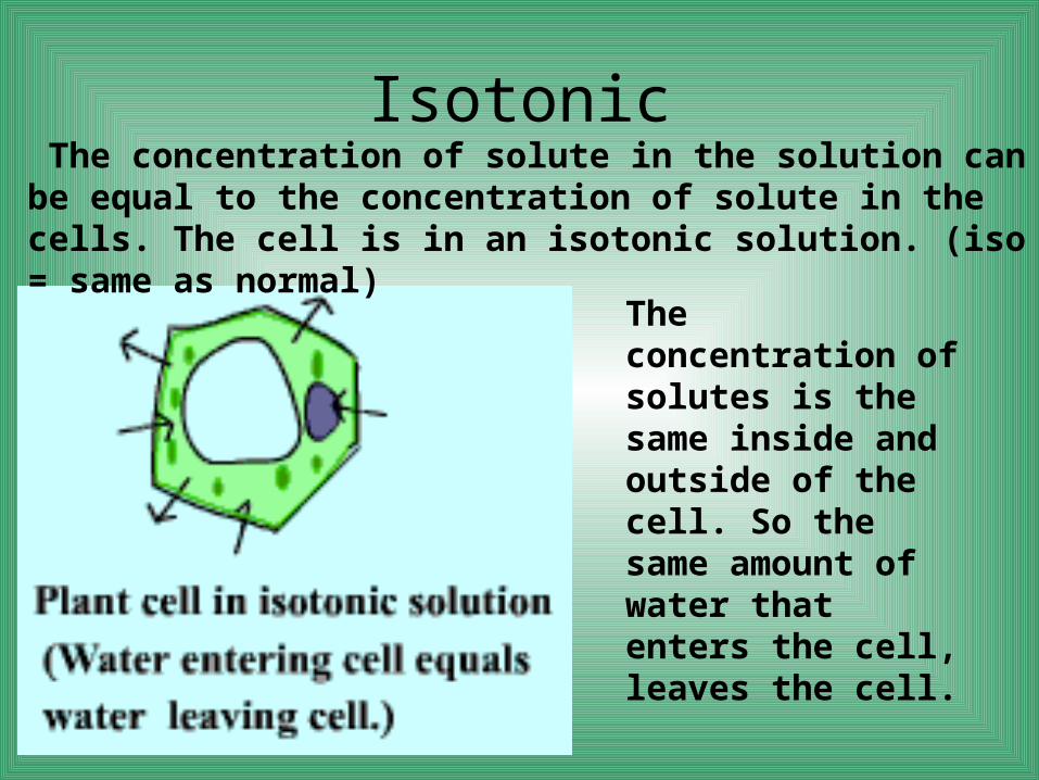
Isotonic The concentration of solute in the solution can be equal to the concentration of solute in the cells. The cell is in an isotonic solution. (iso = same as normal)
The concentration of solutes is the same inside and outside of the cell. So the same amount of water that enters the cell, leaves the cell.
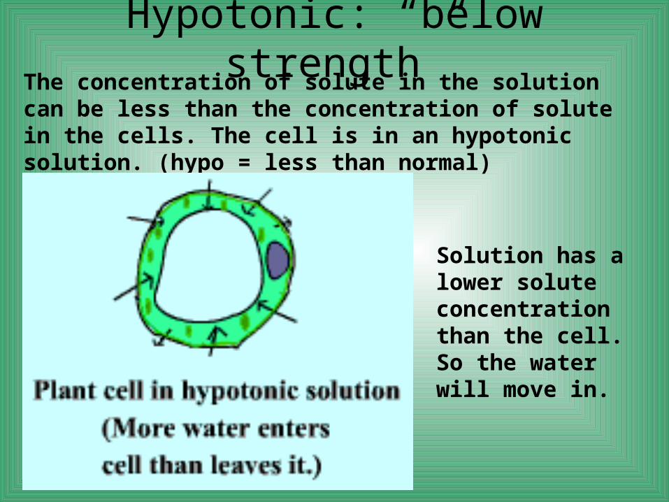
Hypotonic: “below strength”The concentration of solute in the solution can be less than the concentration of solute in the cells. The cell is in an hypotonic solution. (hypo = less than normal)
Solution has a lower solute concentration than the cell. So the water will move in.

Hypertonic: “above strength”The concentration of solute in the solution can be greater than the concentration of solute in the cells. The cell is in an hypertonic solution. (hyper = more than normal)
Solution has a higher solute concentration than the cell. So water will leave the cell.

Osmotic Relationships in Blood Cells

Summary
• Isotonic Solution– Concentration of dissolved substances in solution is
same as dissolved substances inside cell
• Hypotonic Solution– Concentration of substances is lower in solution– Water flows out of solution into cell to lower concentration
in cell
• Hypertonic– Concentration of substances is higher in solution– Water flows into solution and out of cell to raise
concentration in cell

Passive Transport
• Cells don’t use energy to move things across the plasma membrane
• Facilitated diffusion– Passive transport using proteins– Channel proteins– Carrier Proteins– With the concentration gradient

Active Transport
• Moving things from lower to higher concentration
• Against diffusion• Carrier Proteins

Transport of Large Particles
• Endocytosis– Cell surrounds and
takes in material– phagocytosis: solid
material is taken in– Pinocytosis: liquid is
taken in
• Exocytosis– Expulsion or secretion
of materials

Transport of Large Particles
Endocytosis
Exocytosis