Embed Size (px)
Citation preview
Pre-Validation of the Aromatase Assay using Human and Bovine Placental, and Human
Recombinant Microsomes
Endocrine Disruptor Methods Validation Subcommittee (EDMVS) Plenary Session
June 5, 2003
Research Triangle Park, North Carolina
•RTI International is a tradename of Research Triangle Institute.
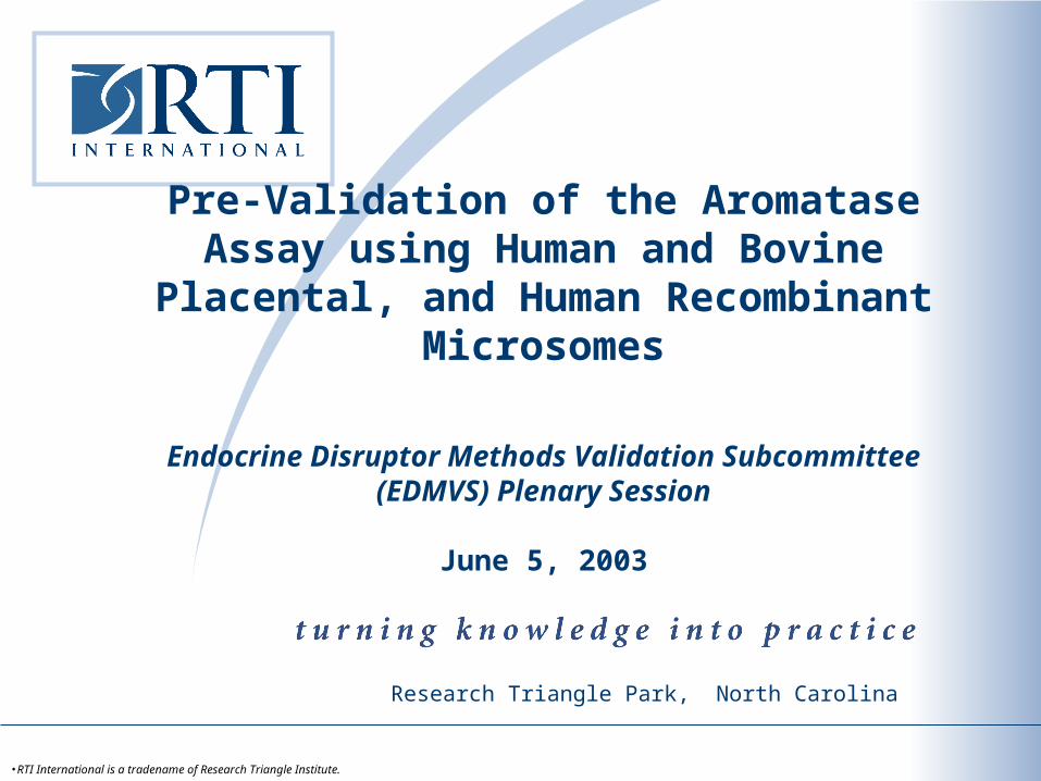
Overview
Background: Aromatase (CYP19)
Study Goals
Substrate characterization
Placenta tissues – human, bovine, porcine
Methods
Results
Protein yield
P450 Spectra
Aromatase activities
Conclusions
Future studies
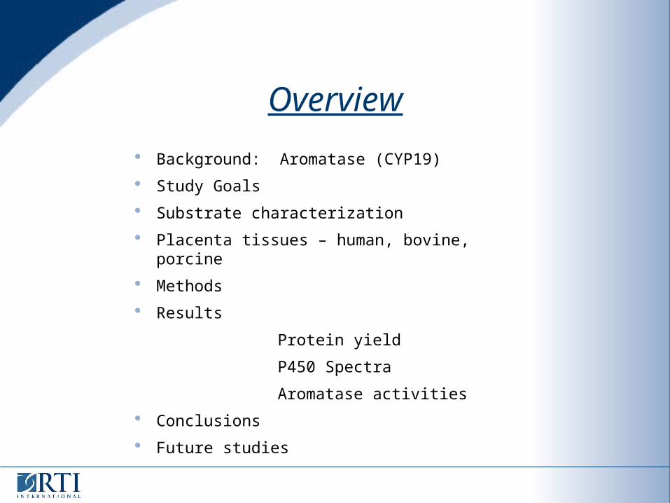
Aromatase
Cytochrome P450 enzyme – CYP19
Present in the gonads and placenta
Responsible for the biosynthesis of estrogen steroid hormones
Can be inhibited at the level of gene expression (e.g., ethylhexylphthalate), or directly at enzyme (e.g. azoles)
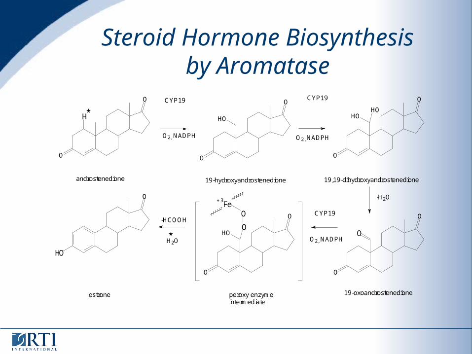
Steroid Hormone Biosynthesis by Aromatase
O
O
H
O2, NADPH
CYP19
O
O
HOHO
O2, NADPH
CYP19
androstenedione 19-hydroxyandrostenedione
O
O
HO
19,19-dihydroxyandrostenedione
O
HO
estrone
-HCOOH
H2O
O
O
O
HOO
O
O
O
Fe3+
CYP19
O2, NADPH
peroxy enzymeintermediate
19-oxoandrostenedione
-H2O
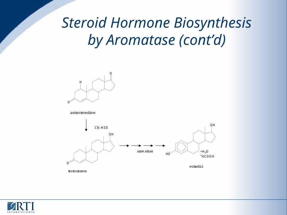
Steroid Hormone Biosynthesis by Aromatase (cont’d)
O
H
O
O
OH
androstenedione
17 -HSD
testosterone
aromataseHO
OH
estradiol
+H2O+HCOOH
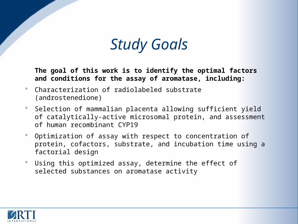
Study Goals
The goal of this work is to identify the optimal factors and conditions for the assay of aromatase, including:
Characterization of radiolabeled substrate (androstenedione)
Selection of mammalian placenta allowing sufficient yield of catalytically-active microsomal protein, and assessment of human recombinant CYP19
Optimization of assay with respect to concentration of protein, cofactors, substrate, and incubation time using a factorial design
Using this optimized assay, determine the effect of selected substances on aromatase activity
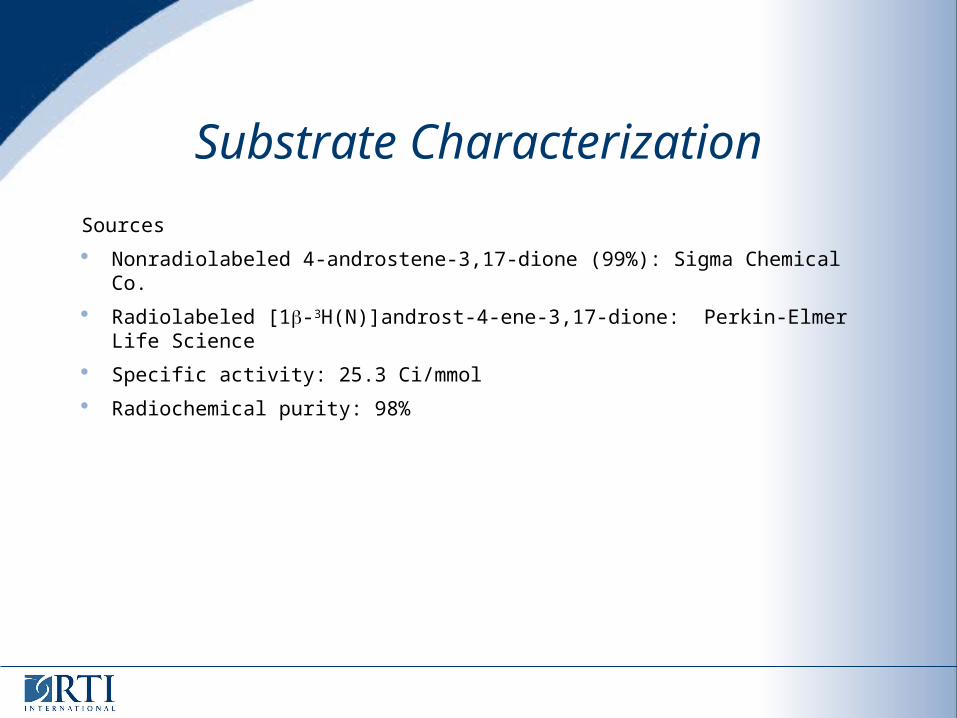
Substrate Characterization
Sources
Nonradiolabeled 4-androstene-3,17-dione (99%): Sigma Chemical Co.
Radiolabeled [1-3H(N)]androst-4-ene-3,17-dione: Perkin-Elmer Life Science
Specific activity: 25.3 Ci/mmol
Radiochemical purity: 98%
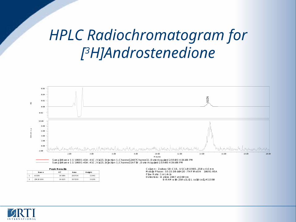
HPLC Radiochromatogram for[3H]Androstenedione
SampleName 1:1 10691-43A: 41C; Vial 5; Injection 1; Channel 2487Channel 1; Date Acquired 2/19/03 4:36:08 PMSampleName 1:1 10691-43A: 41C; Vial 5; Injection 1; Channel SATIN ; Date Acquired 2/19/03 4:36:08 PM
-0.02
0.00
0.02
0.04
0.06
-2.00
0.00
2.00
4.00
6.00
8.00
10.00
Minutes
2.00 4.00 6.00 8.00 10.00 12.00 14.00 16.00 18.00 20.00
Column: Zorbax SB-C18, USCL011903, 250 x 4.6 mmMobile Phase: 55:15:30 ddH2O: THF:MeOH 10691-95AFlow Rate: 1 mL/minDetectors: Waters 2487 at 240 nm B-RAM with 250 ul LiGL solid cell, #11590
1
2
Name RT Area Height
ASDN
[3H]ASDN
14.506
14.625
293714
337219
11441
11235
Peak Results
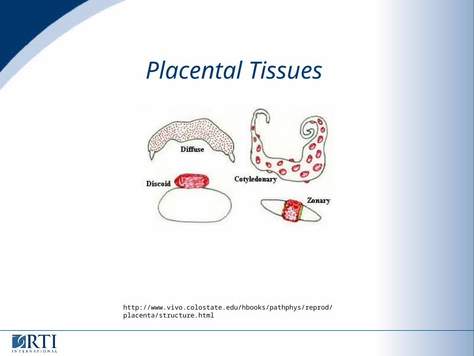
Placental Tissues
http://www.vivo.colostate.edu/hbooks/pathphys/reprod/placenta/structure.html
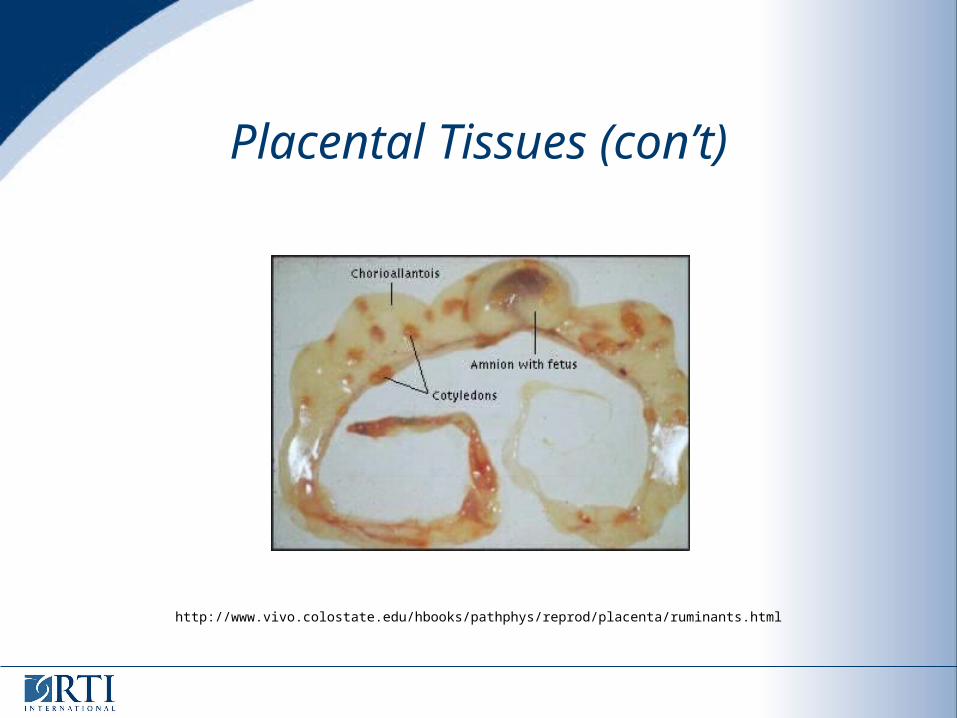
Placental Tissues (con’t)
http://www.vivo.colostate.edu/hbooks/pathphys/reprod/placenta/ruminants.html
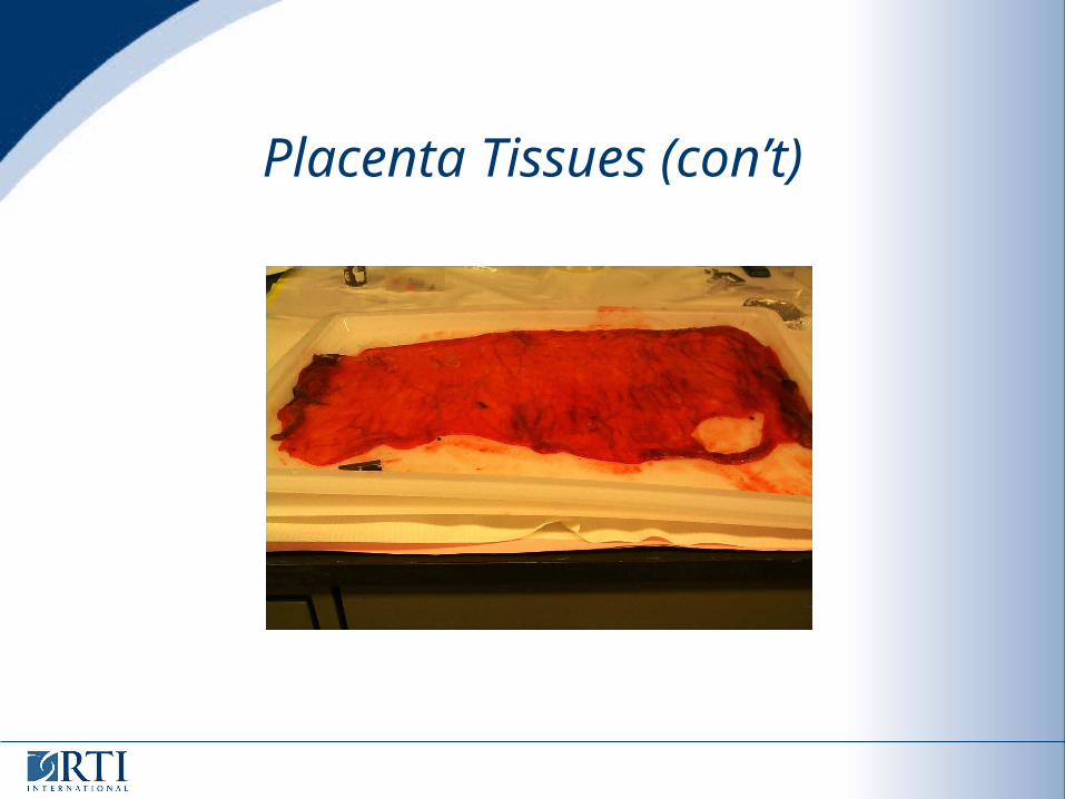
Placenta Tissues (con’t)
Methods
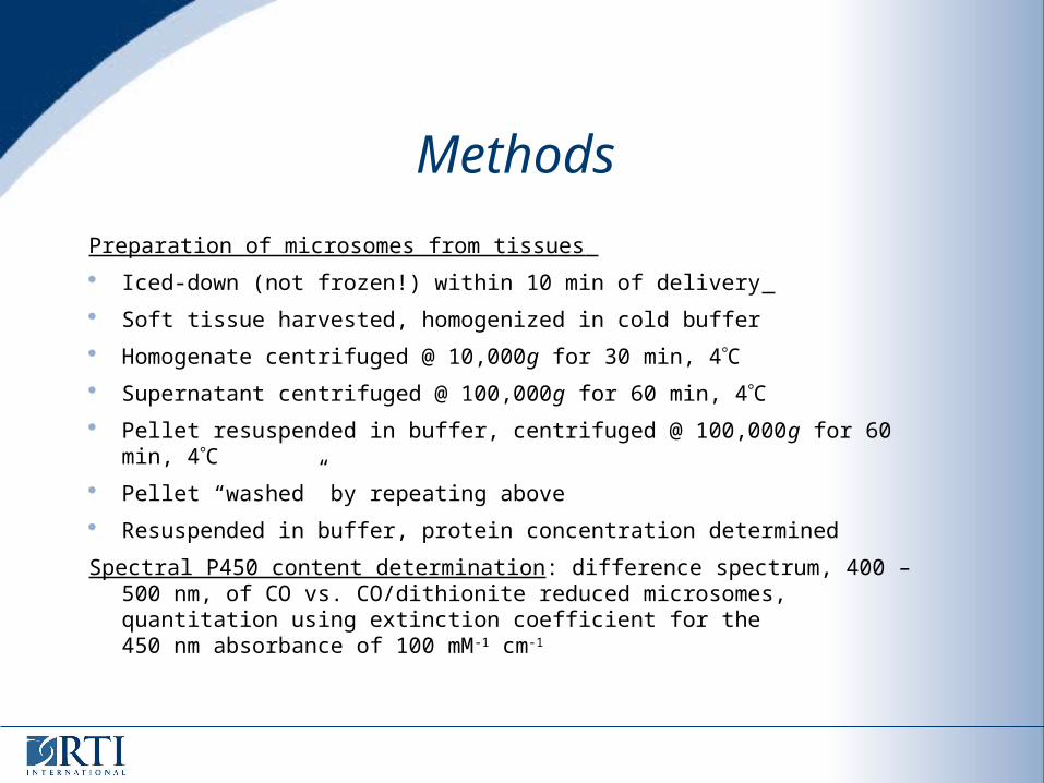
Preparation of microsomes from tissues
Iced-down (not frozen!) within 10 min of delivery
Soft tissue harvested, homogenized in cold buffer
Homogenate centrifuged @ 10,000g for 30 min, 4C Supernatant centrifuged @ 100,000g for 60 min, 4C
Pellet resuspended in buffer, centrifuged @ 100,000g for 60 min, 4C
Pellet “washed” by repeating above
Resuspended in buffer, protein concentration determined
Spectral P450 content determination: difference spectrum, 400 – 500 nm, of CO vs. CO/dithionite reduced microsomes, quantitation using extinction coefficient for the450 nm absorbance of 100 mM-1 cm-1
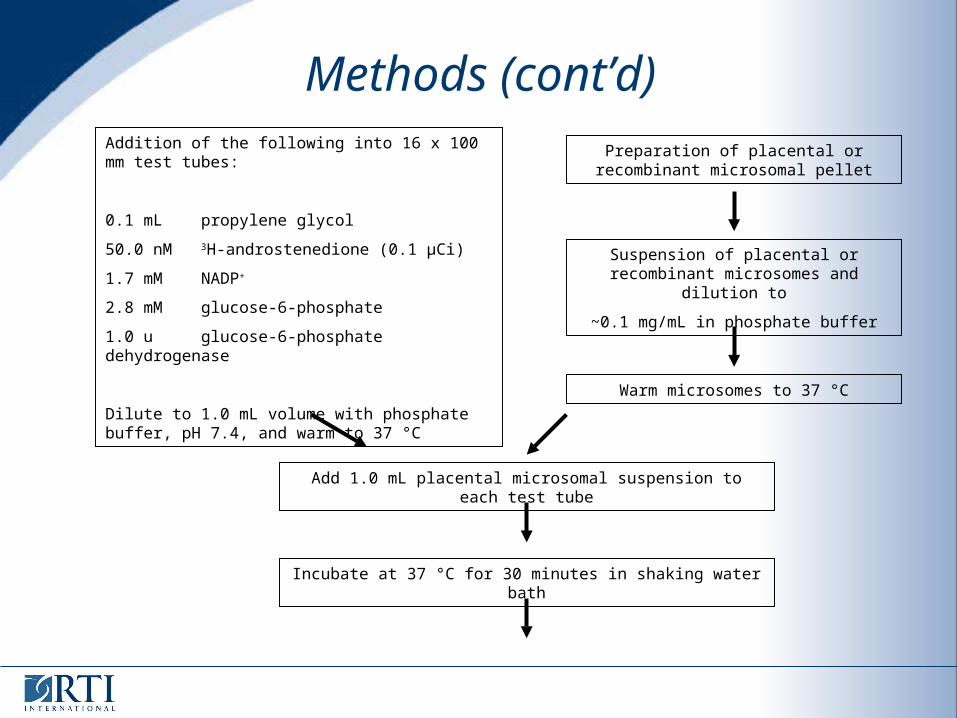
Addition of the following into 16 x 100 mm test tubes:
0.1 mL propylene glycol
50.0 nM 3H-androstenedione (0.1 μCi)
1.7 mM NADP+
2.8 mM glucose-6-phosphate
1.0 u glucose-6-phosphate dehydrogenase
Dilute to 1.0 mL volume with phosphate buffer, pH 7.4, and warm to 37 °C
Preparation of placental or recombinant microsomal pellet
Suspension of placental or recombinant microsomes and dilution to
~0.1 mg/mL in phosphate buffer
Warm microsomes to 37 °C
Add 1.0 mL placental microsomal suspension to each test tube
Incubate at 37 °C for 30 minutes in shaking water bath
Methods (cont’d)
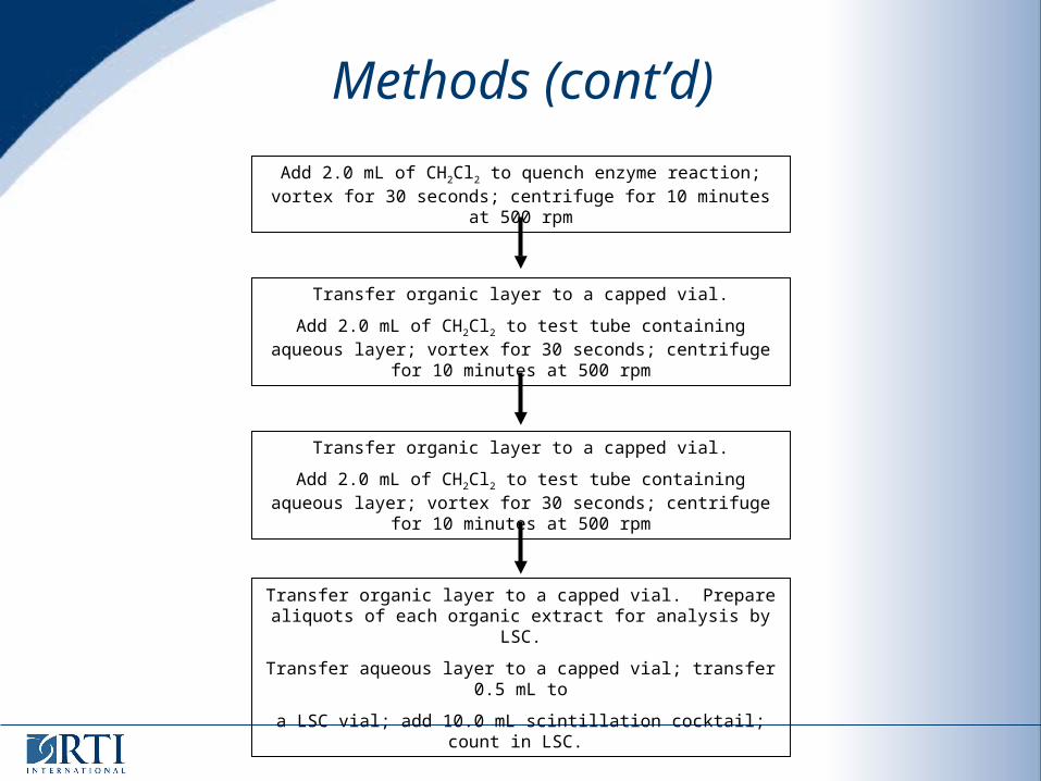
Add 2.0 mL of CH2Cl2 to quench enzyme reaction; vortex for 30 seconds; centrifuge for 10 minutes at 500 rpm
Transfer organic layer to a capped vial.
Add 2.0 mL of CH2Cl2 to test tube containing aqueous layer; vortex for 30 seconds; centrifuge for 10 minutes at 500 rpm
Transfer organic layer to a capped vial.
Add 2.0 mL of CH2Cl2 to test tube containing aqueous layer; vortex for 30 seconds; centrifuge for 10 minutes at 500 rpm
Transfer organic layer to a capped vial. Prepare aliquots of each organic extract for analysis by LSC.
Transfer aqueous layer to a capped vial; transfer 0.5 mL to
a LSC vial; add 10.0 mL scintillation cocktail; count in LSC.
Methods (cont’d)
Tissue Procurement Issues
Human placenta
Caesarian section allows for 1) timed delivery and optimal collection conditions, and 2) less chance of disease transmission
Bovine and porcine
Requires farms close to laboratories, assistance of farm staff
Deliveries seasonal, and any time of day
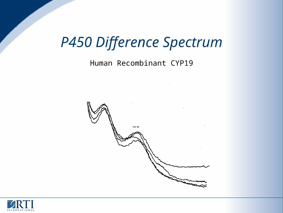
P450 Difference SpectrumHuman Recombinant CYP19
Results (cont’d)
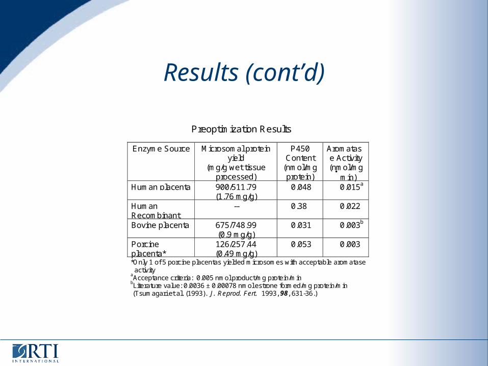
Preoptimization Results
Enzyme Source Microsomal proteinyield
(mg/g wet tissueprocessed)
P450Content
(nmol/mgprotein)
Aromatase Activity(nmol/mg
min)
Human placenta 900/511.79(1.76 mg/g)
0.048 0.015a
HumanRecombinant
-- 0.38 0.022
Bovine placenta 675/748.99(0.9 mg/g)
0.031 0.003b
Porcineplacenta*
126/257.44(0.49 mg/g)
0.053 0.003
*Only 1 of 5 porcine placentas yielded microsomes with acceptable aromatase activityaAcceptance criteria: 0.005 nmol product/mg protein/minbLiterature value: 0.0036 ± 0.00078 nmol estrone formed/mg protein/min (Tsumagari et al. (1993). J. Reprod. Fert. 1993, 98, 631-36.)

Conclusions
Collection conditions used for placentas are crucial to activity
Human Placentas
easiest to collect under optimal conditions
well-defined morphology and good yield of microsomal protein
high activity
Human recombinant, comparable activity with best placental preparations
However,
SOPs must be in place for handing potentially infectious materials
Although Caesarian vs. birth canal delivery minimizes infection of the placental tissue, information regarding screening for HIV, hepatitis, etc. should be obtained if available.
Future Studies
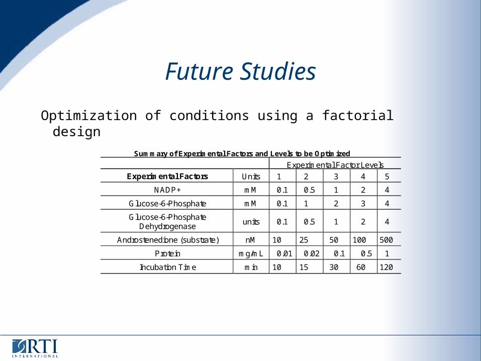
Optimization of conditions using a factorial design
Summary of Experimental Factors and Levels to be Optimized
Experimental Factor Levels
Experimental Factors Units 1 2 3 4 5
NADP+ mM 0.1 0.5 1 2 4
Glucose-6-Phosphate mM 0.1 1 2 3 4
Glucose-6-PhosphateDehydrogenase
units 0.1 0.5 1 2 4
Androstenedione (substrate) nM 10 25 50 100 500
Protein mg/mL 0.01 0.02 0.1 0.5 1
Incubation Time min 10 15 30 60 120
Future Studies
Determination of variance of the optimized assay Using the optimized conditions determined for each preparation,
three technicians independently conduct the assay on three separate days
The results are assessed for technician-to-technician and day-to-day variance.
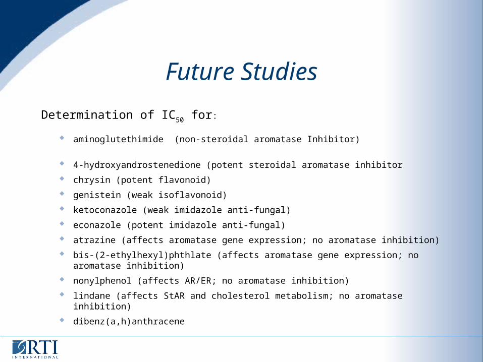
Future Studies
Determination of IC50 for:
aminoglutethimide (non-steroidal aromatase Inhibitor)
4-hydroxyandrostenedione (potent steroidal aromatase inhibitor
chrysin (potent flavonoid)
genistein (weak isoflavonoid)
ketoconazole (weak imidazole anti-fungal)
econazole (potent imidazole anti-fungal)
atrazine (affects aromatase gene expression; no aromatase inhibition)
bis-(2-ethylhexyl)phthlate (affects aromatase gene expression; no aromatase inhibition)
nonylphenol (affects AR/ER; no aromatase inhibition)
lindane (affects StAR and cholesterol metabolism; no aromatase inhibition)
dibenz(a,h)anthracene
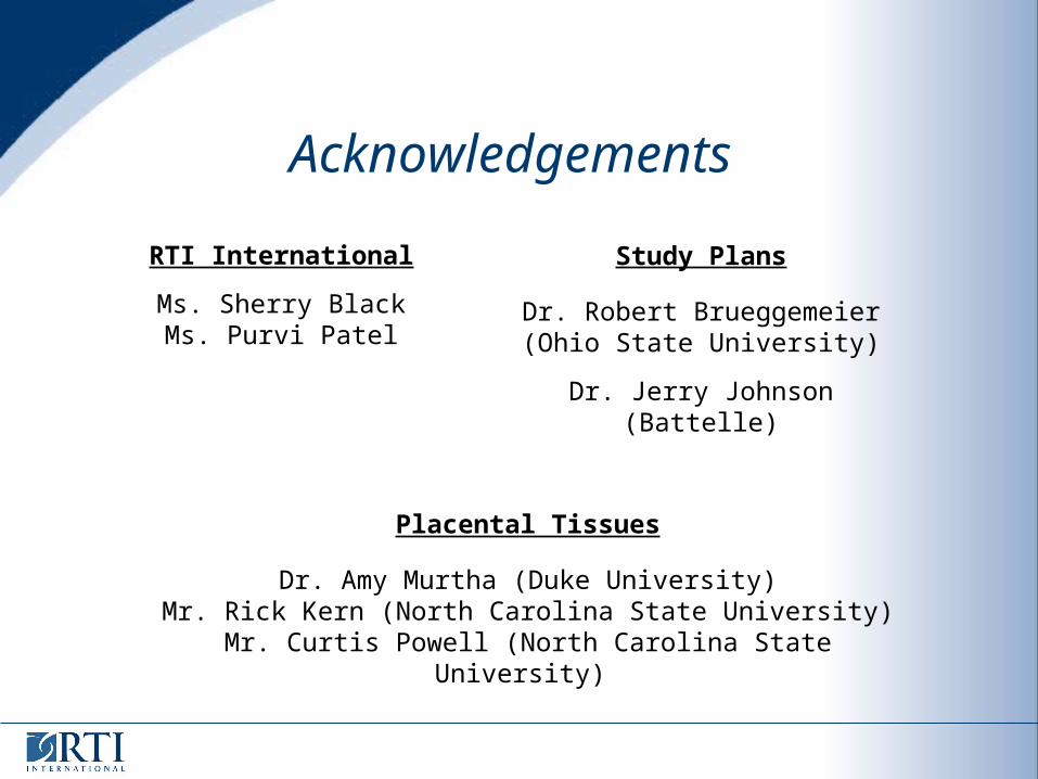
Acknowledgements
Study Plans
Dr. Robert Brueggemeier(Ohio State University)
Dr. Jerry Johnson (Battelle)
Placental Tissues
Dr. Amy Murtha (Duke University)Mr. Rick Kern (North Carolina State University)
Mr. Curtis Powell (North Carolina State University)
RTI International
Ms. Sherry BlackMs. Purvi Patel


![Peranan Aromatase Inhibitor dalam Induksi Ovulasi …pustaka.unpad.ac.id/wp-content/uploads/2016/02/Peranan-Aromatase... · infertilitas yang disebabkan oleh keadaan anovulasi[2]](https://img.pdfslide.net/doc/110x75/5b93db7609d3f2bd1e8c37c1/peranan-aromatase-inhibitor-dalam-induksi-ovulasi-infertilitas-yang-disebabkan.jpg)