Embed Size (px)
Citation preview
Therapies associated with potential benefit or lack of benefit, as indicated above, are based on biomarker results provided in this report and are based on published medical evidence. Thisevidence may have been obtained from studies performed in the cancer type present in the tested patient’s sample or derived from another tumor type. The selection of any, all, or noneof the matched therapies resides solely with the discretion of the treating physician. Decisions on patient care and treatment must be based on the independent medical judgment ofthe treating physician, taking into consideration all available information in addition to this report concerning the patient’s condition in accordance with the applicable standard of care.
4610 South 44th Place, Suite 100 • Phoenix, AZ 85040 • (888) 979-8669 • Fax: (866) 479-4925CLIA 03D1019490 • CAP 7195577 • ISO 15189:2012 - 3531.01 • Zoran Gatalica, MD, DSc, Medical Director • ©2016 Caris Life Sciences. All rights reserved. Page 1 of 11
FINAL REPORT
P A T I E N T S P E C I M E N I N F O R M A T I O N O R D E R E D B Y
Name: Patient, TestDate of Birth: XX-Mon-19XXSex: MaleCase Number: TN16-XXXXXXDiagnosis: Adenocarcinoma, NOS
Primary Tumor Site: Body of pancreasSpecimen Site: Pancreas, NOSSpecimen ID: ABC-1234-XYSpecimen Collected: XX-Mon-2016Testing Completed: XX-Mon-2016
Ordering Physician, MDCancer Center123 Main Street Springfield, XY 12345(123) 456-7890
Bold Therapies = On NCCN Compendium® Therapies
THERAPIES WITH POTENTIAL BENEFIT (PAGE 4)
capecitabine, fluorouracil
TS✮
cisplatin, oxaliplatin
ERCC1
gemcitabine RRM1✮
carboplatin ERCC1
pemetrexed TS✮
✮ Indicates Clinical Trial Opportunity • 252 Chemotherapy Trials • 43 Targeted Therapy Trials (See Clinical Trials ConnectorTM on page 7 for details.)
THERAPIES WITH POTENTIAL LACK OF BENEFIT (PAGE 5)
docetaxel, nab-paclitaxel
TUBB3 dabrafenib,vemurafenib
BRAF paclitaxel TUBB3
THERAPIES WITH INDETERMINATE BENEFIT (PAGE 6)
irinotecan✝
everolimus, temsirolimus
imatinib topotecan✝
✝Association to Benefit was not indicated due to assay failure.
SAMPLE R
EPORT. FOR IL
LUSTRATIV
E PURPOSES O
NLY. N
OT FOR CLIN
ICAL U
SE.

Biomarker Results continued on the next page. >
PATIENT: Patient, Test (XX-Mon-19XX) TN16-XXXXXX PHYSICIAN: Ordering Physician, MD
4610 South 44th Place, Suite 100 • Phoenix, AZ 85040 • (888) 979-8669 • Fax: (866) 479-4925CLIA 03D1019490 • CAP 7195577 • ISO 15189:2012 - 3531.01 • Zoran Gatalica, MD, DSc, Medical Director • ©2016 Caris Life Sciences. All rights reserved. Page 2 of 11
SUMMARY OF RESULTS (SEE APPENDIX FOR FULL DETAILS)
Assay Result
Total Mutational Load Low | 9 Mutations / Megabase
Biomarker Method Result
AKT2 NGS Amplification Not Detected
ALK NGS Amplification Not Detected
ARID1A NGS Amplification Not Detected
ATM NGS Mutation Not Detected
AURKB NGS Amplification Not Detected
BRAF NGS Mutation Not Detected
BRCA1 NGS Mutation Not Detected
BRCA2 NGS Mutation Not Detected
c-KIT NGS Mutation Not Detected
CCND1 NGS Amplification Not Detected
CCND3 NGS Amplification Not Detected
CCNE1 NGS Amplification Not Detected
CDK4 NGS Amplification Not Detected
CDK6 NGS Amplification Not Detected
CDK8 NGS Amplification Not Detected
CDKN2A NGS Amplification Not Detected
NGS Amplification Not DetectedcMET
NGS Mutation Not Detected
CREBBP NGS Amplification Not Detected
CRKL NGS Amplification Not Detected
NGS Amplification Not DetectedEGFR
NGS Mutation Not Detected
EP300 NGS Amplification Not Detected
ERCC1 IHC Negative | 0, 100%
EZH2 NGS Amplification Not Detected
FGF10 NGS Amplification Not Detected
FGF3 NGS Amplification Not Detected
FGF4 NGS Amplification Not Detected
FGFR1 NGS Amplification Not Detected
FGFR2 NGS Amplification Not Detected
FGFR3 NGS Amplification Not Detected
Biomarker Method Result
GATA3 NGS Amplification Not Detected
Her2/Neu NGS Amplification Not Detected
Her2/Neu (ERBB2) NGS Mutation Not Detected
IDH1 NGS Mutation Not Detected
KDR (VEGFR2) NGS Amplification Not Detected
Mutated, PathogenicKRAS NGS
Exon 2 | G12D
MCL1 NGS Amplification Not Detected
MDM2 NGS Amplification Not Detected
MEK1 NGS Amplification Not Detected
MLH1 IHC Positive | 1+, 80%
MSH2 IHC Positive | 1+, 100%
MSH6 IHC Positive | 1+, 50%
MYC NGS Amplification Not Detected
NF2 NGS Amplification Not Detected
NFKBIA NGS Amplification Not Detected
NRAS NGS Mutation Not Detected
NTRK1 NGS Amplification Not Detected
PD-1 IHC Negative | 0/HPF
PD-L1 IHC Negative | 2+, 1%
PDGFRA NGS Mutation Not Detected
PIK3CA NGS Mutation Not Detected
PMS2 IHC Positive | 1+, 5%
NGS Amplification Not Detected
Mutated, PathogenicRB1NGS
Exon 19 | c.1960+1G>T
RET NGS Mutation Not Detected
RICTOR NGS Amplification Not Detected
ROS1 NGS Amplification Not Detected
RRM1 IHC Negative | 2+, 10%
TOP1 NGS Amplification Not Detected
IHC: Immunohistochemistry NGS: Next-Generation Sequencing
SAMPLE R
EPORT. FOR IL
LUSTRATIV
E PURPOSES O
NLY. N
OT FOR CLIN
ICAL U
SE.
PATIENT: Patient, Test (XX-Mon-19XX) TN16-XXXXXX PHYSICIAN: Ordering Physician, MD
4610 South 44th Place, Suite 100 • Phoenix, AZ 85040 • (888) 979-8669 • Fax: (866) 479-4925CLIA 03D1019490 • CAP 7195577 • ISO 15189:2012 - 3531.01 • Zoran Gatalica, MD, DSc, Medical Director • ©2016 Caris Life Sciences. All rights reserved. Page 3 of 11
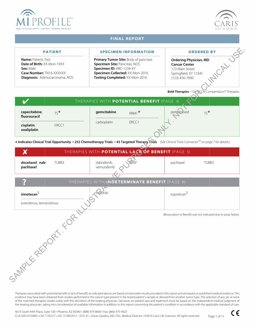
SUMMARY OF RESULTS (SEE APPENDIX FOR FULL DETAILS)
Biomarker Method Result
TOPO1 IHC Test Not Performed
Mutated, PathogenicTP53 NGS
Exon 7 | Y234C
TrkA/B/C IHC Negative | 0, 100%
Biomarker Method Result
TS IHC Negative | 0, 100%
TUBB3 IHC Positive | 2+, 100%
WT1 NGS Amplification Not Detected
IHC: Immunohistochemistry NGS: Next-Generation Sequencing
The Next-Generation Sequencing results above include only the genes most commonly associated with cancer. See summary below and forfull Next-Generation Sequencing results, see Appendix page 1.Genes tested: 592 | Genes with actionable mutations: 3 | Genes with unclassified mutations: 17 | Genes with no mutations detected: 557
See the Appendix section for a detailed overview of the biomarker test results for each technology.
SAMPLE R
EPORT. FOR IL
LUSTRATIV
E PURPOSES O
NLY. N
OT FOR CLIN
ICAL U
SE.

PATIENT: Patient, Test (XX-Mon-19XX) TN16-XXXXXX PHYSICIAN: Ordering Physician, MD
4610 South 44th Place, Suite 100 • Phoenix, AZ 85040 • (888) 979-8669 • Fax: (866) 479-4925CLIA 03D1019490 • CAP 7195577 • ISO 15189:2012 - 3531.01 • Zoran Gatalica, MD, DSc, Medical Director • ©2016 Caris Life Sciences. All rights reserved. Page 4 of 11
Clinical Association
Therapies Test Method Result Value✝
PotentialBenefit
DecreasedPotential
Benefit
Lack ofPotential
Benefit
HighestLevel of
Evidence*Reference
capecitabine,fluorouracil,pemetrexed
TS IHC Negative 0+ 100% ✔ I / Good 1, 2, 3
ATM NGSMutation Not
DetectedII-2 / Good 13, 14, 15
BRCA1 NGSMutation Not
DetectedII-2 / Good
9, 10,
11#, 12
BRCA2 NGSMutation Not
DetectedII-2 / Good 9, 10, 11#
carboplatin,cisplatin,
oxaliplatin
ERCC1 IHC Negative 0+ 100% ✔ II-2 / Good 4, 5, 6, 7, 8
gemcitabine RRM1 IHC Negative 2+ 10% ✔ I / Good 27
* The level of evidence for all references is assigned according to the Literature Level of Evidence Framework consistent with the US Preventive Services Task Forcedescribed further in the Appendix of this report. The data level of each biomarker-drug interaction is the highest level of evidence based on the body of evidence, overallclinical utility, competing biomarker interactions and tumor type from which the evidence was gathered.
# Evidence reference includes data from the same lineage as the tested specimen.
✝Refer to Appendix for detailed Result and Value information for each biomarker, including appropriate cutoffs, unit of measure, etc.
SAMPLE R
EPORT. FOR IL
LUSTRATIV
E PURPOSES O
NLY. N
OT FOR CLIN
ICAL U
SE.
PATIENT: Patient, Test (XX-Mon-19XX) TN16-XXXXXX PHYSICIAN: Ordering Physician, MD
4610 South 44th Place, Suite 100 • Phoenix, AZ 85040 • (888) 979-8669 • Fax: (866) 479-4925CLIA 03D1019490 • CAP 7195577 • ISO 15189:2012 - 3531.01 • Zoran Gatalica, MD, DSc, Medical Director • ©2016 Caris Life Sciences. All rights reserved. Page 5 of 11
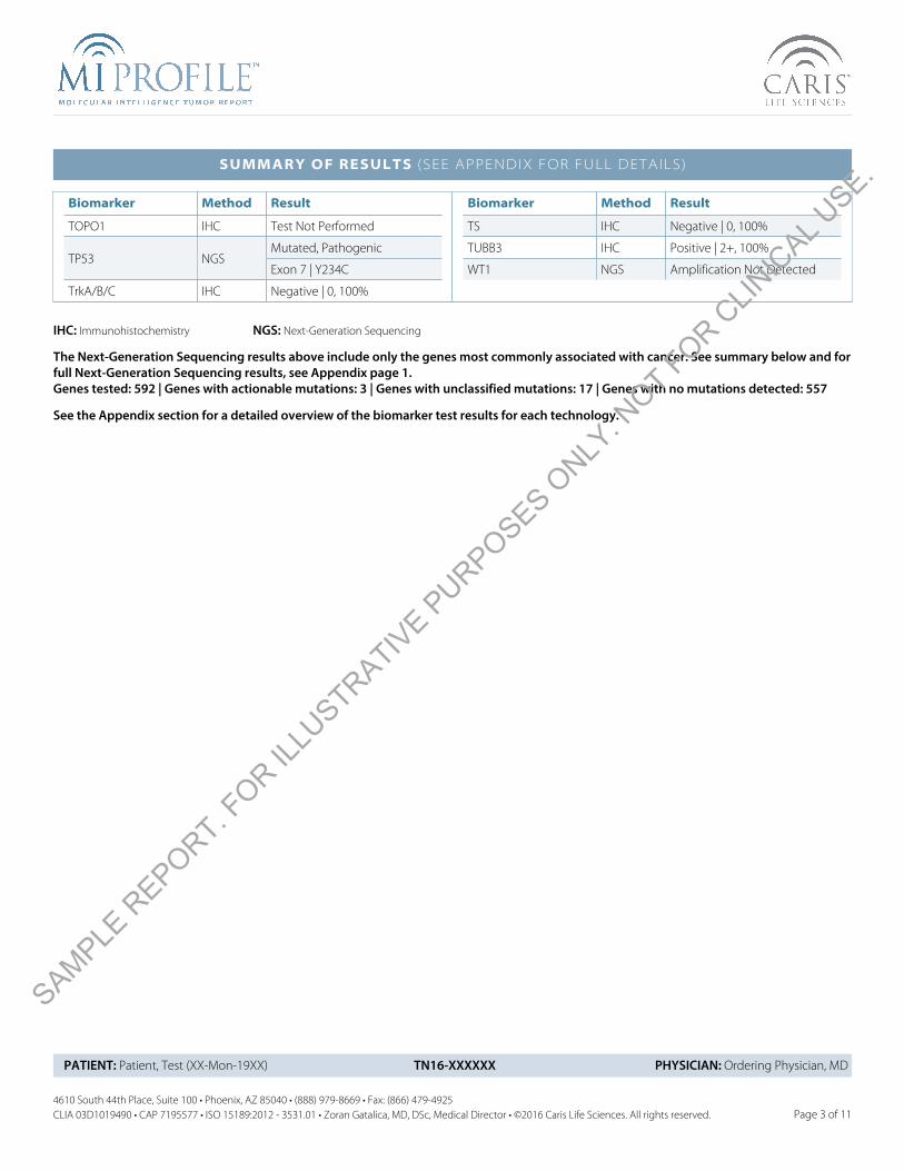
Clinical Association
Therapies Test Method Result Value✝
PotentialBenefit
DecreasedPotential
Benefit
Lack ofPotential
Benefit
HighestLevel of
Evidence*Reference
dabrafenib,vemurafenib BRAF NGS
Mutation NotDetected ✔ I / Good
16, 17,18, 19
docetaxel,nab-paclitaxel,
paclitaxelTUBB3 IHC Positive 2+ 100% ✔ I / Good
20, 21,22, 23
* The level of evidence for all references is assigned according to the Literature Level of Evidence Framework consistent with the US Preventive Services Task Forcedescribed further in the Appendix of this report. The data level of each biomarker-drug interaction is the highest level of evidence based on the body of evidence, overallclinical utility, competing biomarker interactions and tumor type from which the evidence was gathered.
✝Refer to Appendix for detailed Result and Value information for each biomarker, including appropriate cutoffs, unit of measure, etc.
SAMPLE R
EPORT. FOR IL
LUSTRATIV
E PURPOSES O
NLY. N
OT FOR CLIN
ICAL U
SE.

PATIENT: Patient, Test (XX-Mon-19XX) TN16-XXXXXX PHYSICIAN: Ordering Physician, MD
4610 South 44th Place, Suite 100 • Phoenix, AZ 85040 • (888) 979-8669 • Fax: (866) 479-4925CLIA 03D1019490 • CAP 7195577 • ISO 15189:2012 - 3531.01 • Zoran Gatalica, MD, DSc, Medical Director • ©2016 Caris Life Sciences. All rights reserved. Page 6 of 11
Clinical Association
Therapies Test Method Result Value✝
PotentialBenefit
DecreasedPotential
Benefit
Lack ofPotential
Benefit
HighestLevel of
Evidence*Reference
everolimus,temsirolimus PIK3CA NGS
Mutation NotDetected ✔ II-2 / Good 24, 25, 26
c-KIT NGSMutation Not
Detected ✔ II-2 / Good 31, 32
imatinib
PDGFRA NGSMutation Not
Detected ✔ II-3 / Good 28, 29, 30
irinotecan,topotecan TOPO1 IHC
TechnicalIssues
Technical Issues
* The level of evidence for all references is assigned according to the Literature Level of Evidence Framework consistent with the US Preventive Services Task Forcedescribed further in the Appendix of this report. The data level of each biomarker-drug interaction is the highest level of evidence based on the body of evidence, overallclinical utility, competing biomarker interactions and tumor type from which the evidence was gathered.
✝Refer to Appendix for detailed Result and Value information for each biomarker, including appropriate cutoffs, unit of measure, etc.
SAMPLE R
EPORT. FOR IL
LUSTRATIV
E PURPOSES O
NLY. N
OT FOR CLIN
ICAL U
SE.
PATIENT: Patient, Test (XX-Mon-19XX) TN16-XXXXXX PHYSICIAN: Ordering Physician, MD
4610 South 44th Place, Suite 100 • Phoenix, AZ 85040 • (888) 979-8669 • Fax: (866) 479-4925CLIA 03D1019490 • CAP 7195577 • ISO 15189:2012 - 3531.01 • Zoran Gatalica, MD, DSc, Medical Director • ©2016 Caris Life Sciences. All rights reserved. Page 7 of 11
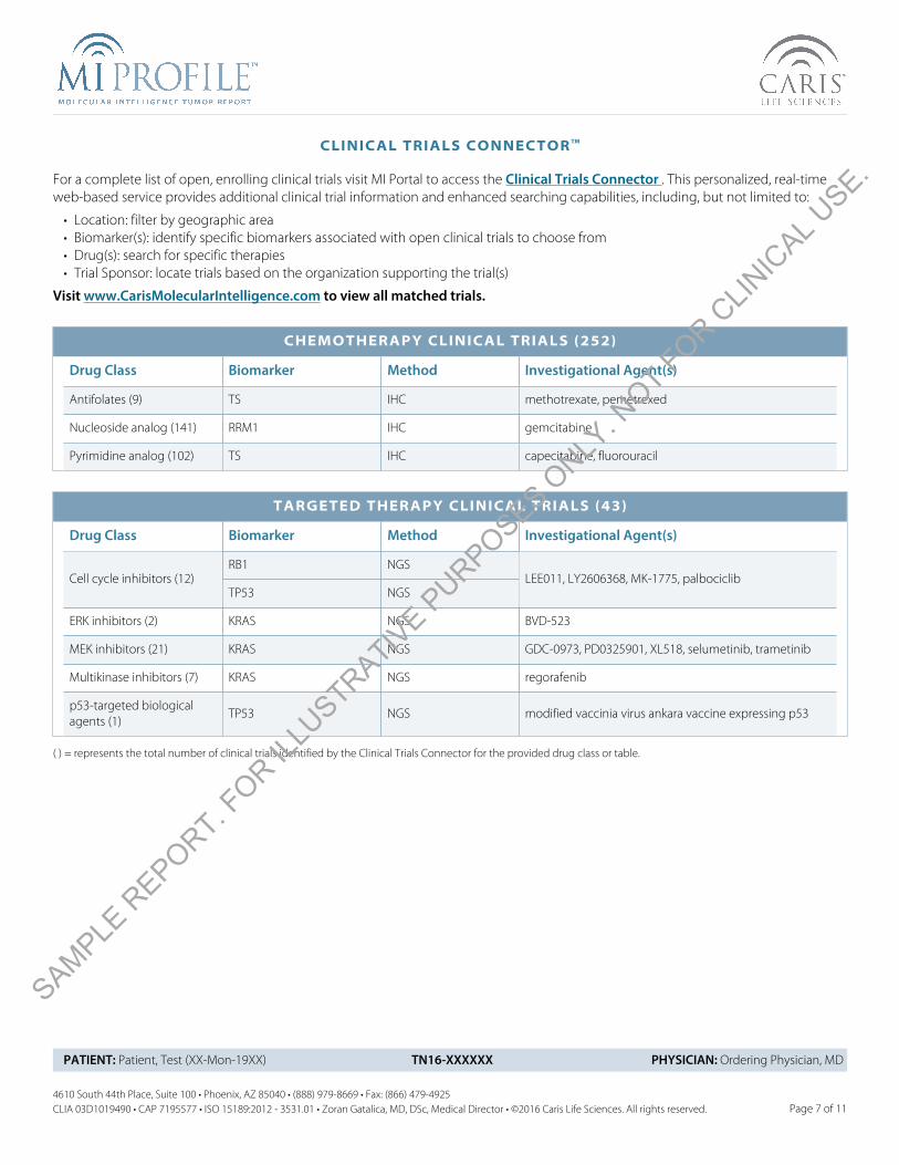
CLINICAL TRIALS CONNECTOR TM
For a complete list of open, enrolling clinical trials visit MI Portal to access the Clinical Trials Connector . This personalized, real-time web-based service provides additional clinical trial information and enhanced searching capabilities, including, but not limited to:
• Location: filter by geographic area• Biomarker(s): identify specific biomarkers associated with open clinical trials to choose from• Drug(s): search for specific therapies• Trial Sponsor: locate trials based on the organization supporting the trial(s)
Visit www.CarisMolecularIntelligence.com to view all matched trials.
CHEMOTHERAPY CLINICAL TRIALS (252)
Drug Class Biomarker Method Investigational Agent(s)
Antifolates (9) TS IHC methotrexate, pemetrexed
Nucleoside analog (141) RRM1 IHC gemcitabine
Pyrimidine analog (102) TS IHC capecitabine, fluorouracil
TARGETED THERAPY CLINICAL TRIALS (43)
Drug Class Biomarker Method Investigational Agent(s)
RB1 NGSCell cycle inhibitors (12)
TP53 NGSLEE011, LY2606368, MK-1775, palbociclib
ERK inhibitors (2) KRAS NGS BVD-523
MEK inhibitors (21) KRAS NGS GDC-0973, PD0325901, XL518, selumetinib, trametinib
Multikinase inhibitors (7) KRAS NGS regorafenib
p53-targeted biologicalagents (1)
TP53 NGS modified vaccinia virus ankara vaccine expressing p53
( ) = represents the total number of clinical trials identified by the Clinical Trials Connector for the provided drug class or table.
SAMPLE R
EPORT. FOR IL
LUSTRATIV
E PURPOSES O
NLY. N
OT FOR CLIN
ICAL U
SE.

* See Appendix page 6 for Level of Evidence description.
PATIENT: Patient, Test (XX-Mon-19XX) TN16-XXXXXX PHYSICIAN: Ordering Physician, MD
4610 South 44th Place, Suite 100 • Phoenix, AZ 85040 • (888) 979-8669 • Fax: (866) 479-4925CLIA 03D1019490 • CAP 7195577 • ISO 15189:2012 - 3531.01 • Zoran Gatalica, MD, DSc, Medical Director • ©2016 Caris Life Sciences. All rights reserved. Page 8 of 11
REFERENCES
SOURCE LEVEL OFEVIDENCE*
1. Chen, C.-Y., P.-C. Yang, et al. (2011). "Thymidylate synthase and dihydrofolate reductase expression in non-small cell lung carcinoma:The association with treatment efficacy of pemetrexed." Lung Cancer 74(1): 132-138. View Citation Online
II-1 / Good
2. Qiu, L.X., M.H. Zheng, et. al. (2008). "Predictive value of thymidylate synthase expression in advanced colorectal cancer patientsreceiving fluoropyrimidine-based chemotherapy: Evidence from 24 studies." Int. J. Cancer: 123, 2384-2389. View Citation Online
I / Good
3. Lee, S.J., Y.H. Im, et. al. (2010). "Thymidylate synthase and thymidine phosphorylase as predictive markers of capecitabinemonotherapy in patients with anthracycline- and taxane-pretreated metastatic breast cancer." Cancer Chemother. Pharmacol. DOI10.1007/s00280-010-1545-0. View Citation Online
II-3 / Good
4. Scheil-Bertram, S., A. Fisseler-Eckhoff, et. al. (2010). "Excision repair cross-complementation group 1 protein overexpression as apredictor of poor survival for high-grade serous ovarian adenocarcinoma." Gynecologic Oncology. 119, 325-331. View Citation Online
II-3 / Good
5. De Dosso, S., E. Zanellato, et al.(2013). "ERCC1 predicts outcome in patients with gastric cancer treated with adjuvant cisplatin-basedchemotherapy". Cancer Chemotherapy and Pharmacology. 72:159-165. View Citation Online
II-3 / Good
6. Li P., Y-J. Fang, et al. (2013). "ERCC1, defective mismatch repair status as predictive biomarkers of survival for stage III colon cancerpatients receiving oxaliplatin-based adjuvant chemotherapy". British Journal of Cancer. 108:1238-1244. View Citation Online
II-2 / Good
7. Steffensen, K.D., A. Jakobsen, et al. (2009). "The Relationship of Platinum Resistance and ERCC1 Protein Expression in Epithelial OvarianCancer." Int. J. Gynecol. Cancer 19: 820-825. View Citation Online
II-3 / Good
8. Kaira, K., M. Serizawa, et al. (2011). "Expression of Excision Repair Cross-Complementation Group 1, Breast Cancer Susceptibility 1, andBeta-III-Tubulin in Thymic Epithelial Tumors". Journal of Thoracic Oncology. 6(3): 606-613. View Citation Online
II-3 / Good
9. Tan, D.S.P., M.E. Gore, et. Al. (2008) ""BRCAness" syndrome in ovarian cancer: a case-control study describing the clinical features andoutcome of patients with epithelial ovarian cancer associated with BRCA1 and BRCA2 mutations." J Clin Oncol. 26(34):5530-6 ViewCitation Online
II-2 / Good
10. Hennessy, B.T., G.B. Mills, et al. (2010) "Somatic mutations in BRCA1 and BRCA2 could expand the number of patients that benefit frompoly (ADP ribose) polymerase inhibitors in ovarian cancer" J Clin Oncol. 28(22):3570-6 View Citation Online
II-3 / Good
11. Lowery, M.A., E.M. O'Reilly, et.al. (2011) "An emerging entity: pancreatic adenocarcinoma associated with a known BRCA mutation:clinical descriptors, treatment implications, and future directions." Oncologist. 16(10):1397-402. View Citation Online
II-3 / Fair
12. Byrski, T., S. Narod, et. Al. (2009) "Pathologic complete response rates in young women with BRCA1-positive breast cancers afterneoadjuvant chemotherapy." J Clin Oncol. 28(3):275-9. View Citation Online
II-3 / Good
13. Bambury, R.M., J.E. Rosenberg, et al. (2015). "Association of somatic mutations in DNA damage repair (DDR) genes with efficacy ofplatinum-based chemotherapy in advanced urothelial carcinoma". J Clin Oncol. 33, (suppl; abstr 4532).
III / Good
14. Pennington, K.P., E.M. Swisher, et al. (2014). "Germline and somatic mutations in homologous recombination genes predict platinumresponse and survival in ovarian, fallopian tube, and peritoneal carcinomas". Clin Cancer Res. 20(3):764-775.
II-3 / Good
15. Plimack, E.R., E.A. Ross, et al. (2015). "Defects in DNA repair genes predict response to neoadjuvant cisplatin-based chemotherapy inmuscle-invasive bladder cancer". Eur Urol. 68:959-967.
II-2 / Good
16. Flaherty, K.T., P.B. Chapman, et al. (2010). "Inhibition of Mutated, Activated BRAF in Metastatic Melanoma." N Engl J Med 363:809-819. View Citation Online
II-2 / Good
17. Hauschild, A., P.B. Chapman, et al. (2012). "Dabrafenib in BRAF-mutated metastatic melanoma: a multicentre, open-label, phase 3randomised controlled trial." Lancet 358-365. View Citation Online
I / Good
18. Chapman, P.B., G.A. McArthur, et. al. (2011). "Improved survival with vemurafenib in melanoma with BRAF V600E mutation." N. Engl. J.Med. This article (10.1056/NEJMoa1103782) was published on June 5, 2011, at nejm.org. View Citation Online
I / Good
SAMPLE R
EPORT. FOR IL
LUSTRATIV
E PURPOSES O
NLY. N
OT FOR CLIN
ICAL U
SE.

* See Appendix page 6 for Level of Evidence description.
PATIENT: Patient, Test (XX-Mon-19XX) TN16-XXXXXX PHYSICIAN: Ordering Physician, MD
4610 South 44th Place, Suite 100 • Phoenix, AZ 85040 • (888) 979-8669 • Fax: (866) 479-4925CLIA 03D1019490 • CAP 7195577 • ISO 15189:2012 - 3531.01 • Zoran Gatalica, MD, DSc, Medical Director • ©2016 Caris Life Sciences. All rights reserved. Page 9 of 11
REFERENCES
SOURCE LEVEL OFEVIDENCE*
19. Falchook, G.S., R. F. Kefford, et al. (2012). "Dabrafenib in patients with melanoma, untreated brain metastases, and other solid tumours:a phase I dose-escalation trial." Lancet 379:1893-901. View Citation Online
II-2 / Good
20. Ploussard, G., A. de la Taille, et al. (2010). "Class III β-Tubulin Expression Predicts Prostate Tumor Aggressiveness and Patient Responseto Docetaxel-Based Chemotherapy." Clin Cancer Res 70(22): 9253-9264. View Citation Online
II-3 / Good
21. Gao, S., J. Gao, et al. (2012). "Clinical implications of REST and TUBB3 in ovarian cancer and its relationship to paclitaxel resistance."Tumor Biol 33:1759-1765. View Citation Online
II-3 / Good
22. Zhang, H.-L., X.-W. Zhou, et al. (2012). "Association between class III β-tubulin expression and response to paclitaxel/vinorelbine-basedchemotherapy for non-small cell lung cancer: A meta-analysis." Lung Cancer 77: 9-15. View Citation Online
I / Good
23. Seve, P., C. Dumontet, et al. (2005). "Class III β-tubulin expression in tumor cells predicts response and outcome in patients with non-small cell lung cancer receiving paclitaxel." Mol Cancer Ther 4(12): 2001-2007. View Citation Online
II-3 / Good
24. Moroney, J.W., R. Kurzrock, et. al. (2011). "A phase I trial of liposomal doxorubicin, bevacizumab, and temsirolimus in patients withadvanced gynecologic and breast malignancies." Clin. Cancer Res. 17:6840-6846. View Citation Online
II-3 / Fair
25. Janku, F., R. Kurzrock, et. al. (2012) "PIK3CA Mutation H1047R Is Associated with Response to PI3K/AKT/mTOR Signaling PathwayInhibitors in Early-Phase Clinical Trials", Cancer Res; 73(1); 276-84. View Citation Online
II-2 / Good
26. Janku, F., R. Kurzrock, et. al. (2012). "PI3K/Akt/mTOR inhibitors in patients with breast and gynecologic malignancies harboring PIK3CAmutations." Journal of Clinical Oncology. DOI: 10.1200/JCO.2011.36.1196. View Citation Online
II-3 / Good
27. Gong, W., J. Dong, et. al. (2012). "RRM1 expression and clinical outcome of gemcitabine-containing chemotherapy for advanced non-small-cell lung cancer: A meta-analysis." Lung Cancer. 75:374-380. View Citation Online
I / Good
28. Cassier, P.A., P. Hohenberger, et al. (2012). "Outcome of Patients with Platelet-Derived Growth Factor Receptor Alpha-MutatedGastrointestinal Stromal Tumors in the Tyrosine Kinase Inhibitor Era." Clin Cancer Res 18:4458-4464. View Citation Online
II-3 / Good
29. Debiec-Rychter, M., I. Judson, et al. (2006). "KIT mutations and dose selection for imatinib in patients with advanced gastrointestinalstromal tumours." Eur J Cancer 42:1093-1103. View Citation Online
II-3 / Good
30. Heinrich, M.C., J.A. Fletcher, et. al. (2008). "Correlation of kinase genotype and clinical outcome in North American Intergroup phaseIII trial of imatinib mesylate for treatment of advanced gastrointestinal stromal tumor: CALGB 150105 study by Cancer and LeukemiaGroup B and Southwest Oncology Group." J Clin Oncol 26(33):5360-5367. View Citation Online
II-3 / Good
31. Guo, J., S. Qin, et. al. (2011). "Phase II, open-label, single-arm trial of imatinib mesylate in patients with metastatic melanoma harboringc-Kit mutation or amplification." J. Clin. Oncol. 29:2904-2909. View Citation Online
II-2 / Good
32. Carvajal, R.D., G.K. Schwartz, et. al. (2011). "KIT as a therapeutic target in metastatic melanoma." JAMA. 305(22):2327-2334. View CitationOnline
II-2 / Good
33. Wells, S.A., M.J. Schlumberger, et al. (2012). "Vandetanib in Patients with Locally Advanced or Metastatic Medullary Thyroid Cancer: ARandomized, Double-Blind Phase III Trial." J Clin Oncol 30: 134-141. View Citation Online
I / Good
34. Oza, A.M., M. Friedlander, et.al. (2015). "Olaparib combined with chemotherapy for recurrent platinum-sensitive ovarian cancer: arandomised phase 2 trial." Lancet Oncol. 16:87-97 View Citation Online
I / Good
35. Ledermann, J., U. Matulonis, et.al. (2014). "Olaparib maintenance therapy in patients with platinum-sensitive relapsed serous ovariancancer: a preplanned retrospective analysis of outcomes by BRCA status in a randomised phase 2 trial." Lancet Oncol. 15(8):852-61. View Citation Online
I / Good
36. Kaufman, B., S.M. Domcheck, et al. (2015). "Olaparib monotherapy in patients with advanced cancer and a germline BRCA1/2mutation". J Clin Oncol. 33(3): 244-250. View Citation Online
II-1 / Good
SAMPLE R
EPORT. FOR IL
LUSTRATIV
E PURPOSES O
NLY. N
OT FOR CLIN
ICAL U
SE.

* See Appendix page 6 for Level of Evidence description.
PATIENT: Patient, Test (XX-Mon-19XX) TN16-XXXXXX PHYSICIAN: Ordering Physician, MD
4610 South 44th Place, Suite 100 • Phoenix, AZ 85040 • (888) 979-8669 • Fax: (866) 479-4925CLIA 03D1019490 • CAP 7195577 • ISO 15189:2012 - 3531.01 • Zoran Gatalica, MD, DSc, Medical Director • ©2016 Caris Life Sciences. All rights reserved. Page 10 of 11
REFERENCES
SOURCE LEVEL OFEVIDENCE*
37. Mateo, J., J.S. de Bono, et al. (2015). "DNA-repair defects and olaparib in metastatic prostate cancer". N Engl J Med. 373(18): 1697-1708. View Citation Online
II-1 / Good
38. Bang, Y-J., Y-K. Kang, et. al. (2010). "Trastuzumab in combination with chemotherapy versus chemotherapy alone for treatment ofHER2-positive advanced gastric or gastro-oesophageal junction cancer (ToGA): a phase 3, open-label, randomised controlled trial."Lancet. 376:687-97. View Citation Online
I / Good
39. Baselga, J., S.M. Swain, et. al. (2012). "Pertuzumab plus trastumab plus docetaxel for metastatic breast cancer". N. Engl. J. Med.36:109-119. View Citation Online
I / Good
40. Yin, W., J. Lu, et. al. (2011). "Trastuzumab in adjuvant treatment HER2-positive early breast cancer patients: A meta-analysis of publishedrandomized controlled trials." PLoS ONE 6(6): e21030. doi:10.1371/journal.pone.0021030. View Citation Online
I / Good
41. Cortes, J., J. Baselga, et. al. (2012). "Pertuzumab monotherapy after trastuzumab-based treatment and subsequent reintroduction oftrastuzumab: activity and tolerability in patients with advanced human epidermal growth factor receptor-2-positive breast cancer." J.Clin. Oncol. 30. DOI: 10.1200/JCO.2011.37.4207. View Citation Online
II-1 / Good
42. Hurvitz, S.A., E.A. Perez, et. al. (2013) "Phase II randomized study of trastuzumab emtansine versus trastuzumab plus docetaxel inpatients with human epidermal growth factor receptor 2-positive metastatic breast cancer." J Clin Oncol.31(9):1157-63 View CitationOnline
I / Good
43. Bartlett, J.M.S., K. Miller, et. al. (2011). "A UK NEQAS ISH multicenter ring study using the Ventana HER2 dual-color ISH assay." Am. J. Clin.Pathol. 135:157-162. View Citation Online
II-3 / Good
44. Slamon, D., M. Buyse, et. al. (2011). "Adjuvant trastuzumab in HER2-positive breast cancer." N. Engl. J. Med. 365:1273-83. View CitationOnline
I / Good
45. Verma, S., K. Blackwell, et. al. (2012) "Trastuzumab Emtansine for HER2-Positive Advanced Breast Cancer" N Engl J Med. 367(19):1783-91.View Citation Online
I / Good
46. Amir, E. et. al. (2010). "Lapatinib and HER2 status: results of a meta-analysis of randomized phase III trials in metastatic breast cancer."Cancer Treatment Reviews. 36:410-415. View Citation Online
I / Good
47. Johnston, S., Pegram M., et. al. (2009). "Lapatinib combined with letrozole versus letrozole and placebo as first-line therapy forpostmenopausal hormone receptor-positive metastatic breast cancer. Journal of Clinical Oncology. Published ahead of print onSeptember 28, 2009 as 10.1200/JCO.2009.23.3734. View Citation Online
I / Good
48. Press, M. F., R. S. Finn, et al. (2008). "HER-2 gene amplification, HER-2 and epidermal growth factor receptor mRNA and proteinexpression, and lapatinib efficacy in women with metastatic breast cancer." Clin Cancer Res 14(23): 7861-70. View Citation Online
I / Good
SAMPLE R
EPORT. FOR IL
LUSTRATIV
E PURPOSES O
NLY. N
OT FOR CLIN
ICAL U
SE.

PATIENT: Patient, Test (XX-Mon-19XX) TN16-XXXXXX PHYSICIAN: Ordering Physician, MD
4610 South 44th Place, Suite 100 • Phoenix, AZ 85040 • (888) 979-8669 • Fax: (866) 479-4925CLIA 03D1019490 • CAP 7195577 • ISO 15189:2012 - 3531.01 • Zoran Gatalica, MD, DSc, Medical Director • ©2016 Caris Life Sciences. All rights reserved. Page 11 of 11
SPECIMEN INFORMATION
Specimen ID: ABC-1234-XY Specimen Collected: XX-Mon-2016
Specimen Received: XX-Mon-2016 Testing Initiated: XX-Mon-2016
Gross description: 1 (A) Paraffin Block - Client ID(ABC-1234-XY) from Springfield, XY, with the corresponding surgical pathology report labeled "ABC-123-XY.”
Pathologic Diagnosis: Body and tail of pancreas, pancreatectomy with splenectomy: Ductal adenocarcinoma, G2.
DisclaimerAll of the individual assays that are available through Caris Molecular Intelligence™ were developed and validated by Caris MPI, Inc. d/b/a Caris Life Sciences® and their test per-formance characteristics were determined and validated by Caris Life Sciences pursuant to the Clinical Laboratory Improvements Amendments and accompanying regulations(“CLIA”). Some of the assays that are part of Caris Molecular Intelligence have been approved by the U.S. Food and Drug Administration (FDA). For any remaining assays, Caris MPI,Inc. is certified under CLIA to perform high complexity testing, including all of the assays that comprise the Caris Molecular Intelligence.
The CLIA certification number of Caris MPI, Inc. laboratory performing testing in connection with Caris Molecular Intelligence can be found at the bottom of each page. This reportincludes information about therapies that appear to be associated with clinical benefit based on NCCN Compendium® guidelines, relevance of tumor lineage, level of publishedevidence and strength of biomarker results. This report, neither ranks biomarkers listed nor therapies associated with such biomarkers, in order of potential or predicted efficacy, andsuch therapies may or may not be suitable for administration to a particular patient. A determination of biomarker results do not necessarily indicate pharmacologic effectivenessor lack thereof. This report does not guarantee or suggest that any particular agent will be effective with the treatment of any particular condition. Caris Life Sciences expresslydisclaims and makes no representation or warranty whatsoever relating, directly or indirectly, to review of identified scientific literature, the conclusions drawn from such review orany of the information set forth in this report that is derived from such review, including information and conclusions relating to therapies that are included or omitted from thisreport.
Decisions regarding care and treatment should not be based on a single test such as this test or the information contained in this report. The decision to select any, all or none ofthe listed therapies resides solely within the discretion of the treating physician. Decisions on patient care and treatment must be based on the independent medical judgmentof the treating physician, taking into consideration all applicable information concerning the patient’s condition, including but not limited to, patient and family history, physicalexaminations, information from other diagnostic tests, and patient preferences, in accordance with the applicable standard of care.
The information presented in the Clinical Trials Connector™ section of this report (if applicable) is compiled from sources believed to be reliable and current. We have used our bestefforts to make this information as accurate as possible. However, the accuracy and completeness of this information cannot be guaranteed. The contents are to be used for clinicaltrial guidance and may not include all relevant trials. Current enrollment status for these trials is unknown. The clinical trials information present in the biomarker description wascompiled from www.clinicaltrials.gov. The contents are to be used only as a guide, and health care providers should employ their judgment in interpreting this information for aparticular patient. Specific eligibility criteria for each clinical trial should be reviewed as additional inclusion criteria may apply. Caris Life Sciences makes no promises or guaranteesthat a healthcare provider, insurer or other third party private or government payor, will provide reimbursement for any of the tests performed.
The next-generation sequencing assay performed by Caris Life Sciences examines nucleic acids obtained from tumor tissue only and does not examine normal tissue such as tumoradjacent tissue or whole or peripheral blood. As such, the origin of any mutation detected may be a somatic mutation (not inherited) or a germline mutation (inherited) and will not be distinguishable by this assay. It is recommended that results be considered within the patient’s clinical and health history. If a germline inheritance pattern is suspected thencounseling by a board certified genetic counselor is recommended.
Molecular testing of this specimen was performed after harvesting of targeted tissues with an approved manual microdissection technique. Candidateslides were examined under a microscope and areas containing tumor cells (and separately normal cells, when necessary for testing) were circled. Alaboratory technician harvested targeted tissues for extraction from the marked areas using a dissection microscope. The areas marked and extractedwere microscopically reexamined on post-microdissected slides and adequacy of microdissection was verified by a board certified Pathologist.
Electronic Signature
SAMPLE R
EPORT. FOR IL
LUSTRATIV
E PURPOSES O
NLY. N
OT FOR CLIN
ICAL U
SE.

Additional Next-Generation Sequencing results continued on the next page. >
PATIENT: Patient, Test (XX-Mon-19XX) TN16-XXXXXX PHYSICIAN: Ordering Physician, MD
4610 South 44th Place, Suite 100 • Phoenix, AZ 85040 • (888) 979-8669 • Fax: (866) 479-4925CLIA 03D1019490 • CAP 7195577 • ISO 15189:2012 - 3531.01 • Zoran Gatalica, MD, DSc, Medical Director • ©2016 Caris Life Sciences. All rights reserved. Appendix 1 of 6
MUTATIONAL ANALYSIS BY NEXT-GENERATION SEQUENCING (NGS)
T O T A L M U T A T I O N A L L O A D
Result Mutations / Megabase (Mb) Threshold
Low 9 ≥ 17 Mutations per Mb
Interpretation: Total Mutational Load is calculated using only missense mutations that have not previously been reported as germline alterations. Incolorectal cancer, all samples tested by our laboratory that exhibited microsatellite instability (MSI-H) had a Total Mutational Load of ≥ 17 mutationsper megabase sequenced. Samples with 17 or more mutations may be hypermutated, which is a potential indicator of immunotherapy response. (Leet al., NEJM, 2015; Rizvi et al., Science, 2015; Rosenberg et al., Lancet, 2016; Snyder et al., NEJM, 2014)
G E N E S T E S T E D W I T H A L T E R A T I O N S
Gene Alteration Frequency (%) Exon Result
KRAS G12D 19 2 Mutated, Pathogenic
Interpretation: A pathogenic mutation was detected in KRAS
KRAS or V-Ki-ras2 Kirsten rat sarcoma viral oncogene homolog encodes a signaling intermediate involved in many signaling cascades including theEGFR pathway. KRAS somatic mutations have been found in pancreatic (57%), colon (35%), lung (16%), biliary tract (28%), and endometrial (15%)cancers. Several germline mutations of KRAS (V14I, T58I, and D153V amino acid substitutions) are associated with Noonan syndrome.
RB1 c.1960+1G>T 39 19 Mutated, Pathogenic
Interpretation: A pathogenic mutation that disrupts an intron splice site was detected in RB1
RB1 or retinoblastoma-1 is a tumor suppressor gene whose protein regulates the cell cycle by interacting with various transcription factors, includingthe E2F family (which controls the expression of genes involved in the transition of cell cycle checkpoints). Besides ocular cancer, RB1 mutations havealso been detected in other malignancies, such as ovarian (10%), bladder (41%), prostate (8%), breast (6%), brain (6%), colon (5%), and renal (2%) cancers.RB1 status, along with other mitotic checkpoints, has been associated with the prognosis of GIST patients. Germline mutations of RB1 are associatedwith the pediatric tumor, retinoblastoma. Inherited retinoblastoma is usually bilateral. Studies indicate patients with a history of retinoblastoma areat increased risk for secondary malignancies.
TP53 Y234C 29 7 Mutated, Pathogenic
Interpretation: A mutation, Y234C, was detected in TP53. This mutation has been reported previously in numerous publications. In biochemicalstudies, it has been shown to cause the TP53 protein to become unstable at physiological temperatures, disrupting normal signaling (Dearth 2007Carcinogenesis 28:289). Y234C has also been reported as a germline mutation, causal for Li-Fraumeni syndrome (Monti 2011 Mol Cancer Res 9:271).
TP53, or p53, plays a central role in modulating response to cellular stress through transcriptional regulation of genes involved in cell-cycle arrest, DNArepair, apoptosis, and senescence. Inactivation of the p53 pathway is essential for the formation of the majority of human tumors. Mutation in p53 (TP53)remains one of the most commonly described genetic events in human neoplasia, estimated to occur in 30-50% of all cancers. Generally, presenceof a disruptive p53 mutation is associated with a poor prognosis in all types of cancers, and diminished sensitivity to radiation and chemotherapy.Germline p53 mutations are associated with the Li-Fraumeni syndrome (LFS) which may lead to early-onset of several forms of cancer currently knownto occur in the syndrome, including sarcomas of the bone and soft tissues, carcinomas of the breast and adrenal cortex (hereditary adrenocorticalcarcinoma), brain tumors and acute leukemias.
G E N E S T E S T E D W I T H N O M U T A T I O N S D E T E C T E D
ATMBRAFBRCA1
BRCA2c-KITcMET
EGFRHer2/Neu (ERBB2)IDH1
NRASPDGFRAPIK3CA
RET
SAMPLE R
EPORT. FOR IL
LUSTRATIV
E PURPOSES O
NLY. N
OT FOR CLIN
ICAL U
SE.

PATIENT: Patient, Test (XX-Mon-19XX) TN16-XXXXXX PHYSICIAN: Ordering Physician, MD
4610 South 44th Place, Suite 100 • Phoenix, AZ 85040 • (888) 979-8669 • Fax: (866) 479-4925CLIA 03D1019490 • CAP 7195577 • ISO 15189:2012 - 3531.01 • Zoran Gatalica, MD, DSc, Medical Director • ©2016 Caris Life Sciences. All rights reserved. Appendix 2 of 6
MUTATIONAL ANALYSIS BY NEXT-GENERATION SEQUENCING (NGS)
The mutations reported in the section below have not been analyzed to determine clinical significance and are being reported for informationalpurposes. In addition, the origin of the reported mutations (germline or somatic) has not been determined. All variants listed in the dbSNP 137common list have been excluded from this table, as well as non-coding variants that are not part of the conserved splice sequence (+/- 1 or 2 bases).All excluded variants and regions are available upon request. Certain gene regions were also excluded due to high homology with other loci in thegenome.
G E N E S T E S T E D W I T H U N C L A S S I F I E D M U T A T I O N S
Gene Alteration
AFF1 K20R
BARD1 V507M
BCL9 P375R
CCDC6 P446L
ECT2L V570A
Gene Alteration
ERCC4 I73V
IGF1R R437H
IL21R R297Q
KIAA1549 G1709D
KMT2D R4162Q
Gene Alteration
MTOR A329T
PCM1 N159S
POLE T1429S
TCF3 G431S
TLX1 V134M
Gene Alteration
TRIP11 P1877T
I67 _R68delinsMWUSP6I276L
G E N E S T E S T E D W I T H I N D E T E R M I N A T E R E S U L T S
ATRXBIRC3CASC5
CD79AELLKDM5C
MED12MNX1MSN
NOTCH2NUTM2BPCSK7
PMS2STAT5BVEGFB
For Next-Generation Sequencing, a total of 592 genes were analyzed. The results above include genes most commonly associated withcancer and any additional mutations identified. No alterations were identified in 557 genes. For a complete list of genes tested, visitwww.CarisMolecularIntelligence.com/profilemenu.
NGS Methods
Next Generation Sequencing: Direct sequence analysis was performed on genomic DNA isolated from a formalin-fixed paraffin-embedded tumorsample using the Illumina NextSeq platform. An Agilent custom-designed SureSelect XT assay was used to enrich 592 whole-gene targets. The genesand amino acids evaluated in this report can be found at www.carislifesciences.com. All variants reported by this assay are detected with > 99 %confidence based on the frequency of the mutation present and the amplicon coverage. This test is not designed to distinguish between germ lineinheritance of a variant or acquired somatic mutation. This test has a sensitivity to detect as low as approximately 10 % population of cells containinga mutation. This test has not been cleared or approved by the United States Food and Drug Administration (FDA) as such approval is not necessary. Allperformance characteristics were determined by Caris Life Sciences. Benign and non-coding variants are not included in this report but are availableupon request.
SAMPLE R
EPORT. FOR IL
LUSTRATIV
E PURPOSES O
NLY. N
OT FOR CLIN
ICAL U
SE.

PATIENT: Patient, Test (XX-Mon-19XX) TN16-XXXXXX PHYSICIAN: Ordering Physician, MD
4610 South 44th Place, Suite 100 • Phoenix, AZ 85040 • (888) 979-8669 • Fax: (866) 479-4925CLIA 03D1019490 • CAP 7195577 • ISO 15189:2012 - 3531.01 • Zoran Gatalica, MD, DSc, Medical Director • ©2016 Caris Life Sciences. All rights reserved. Appendix 3 of 6
COPY NUMBER VARIATIONS BY NEXT-GENERATION SEQUENCING (NGS)
G E N E S T E S T E D W I T H N O A M P L I F I C A T I O N D E T E C T E D
AKT2ALKARID1AAURKBCCND1CCND3CCNE1
CDK4CDK6CDK8CDKN2AcMETCREBBPCRKL
EGFREP300EZH2FGF10FGF3FGF4FGFR1
FGFR2FGFR3GATA3Her2/NeuKDRMCL1MDM2
MEK1MYCNF2NFKBIANTRK1RB1RICTOR
ROS1TOP1WT1
CNV Methods
Copy number variation was determined by comparing the depth of sequencing of genomic loci to a diploid control as well as the known performanceof these genomic loci. Copy number gains ≥8 copies can be detected by this assay with >95% sensitivity. Please note: high levels of polyploidy orscant tumor cells may prevent the detection of copy number changes.
SAMPLE R
EPORT. FOR IL
LUSTRATIV
E PURPOSES O
NLY. N
OT FOR CLIN
ICAL U
SE.

Additional IHC results continued on the next page. >
PATIENT: Patient, Test (XX-Mon-19XX) TN16-XXXXXX PHYSICIAN: Ordering Physician, MD
4610 South 44th Place, Suite 100 • Phoenix, AZ 85040 • (888) 979-8669 • Fax: (866) 479-4925CLIA 03D1019490 • CAP 7195577 • ISO 15189:2012 - 3531.01 • Zoran Gatalica, MD, DSc, Medical Director • ©2016 Caris Life Sciences. All rights reserved. Appendix 4 of 6
PROTEIN EXPRESSION BY IMMUNOHISTOCHEMISTRY (IHC)
Patient Tumor Thresholds*Biomarker Staining Intensity
(0, 1+, 2+, 3+) Percent of cells Result Conditions for a Positive Result:
ERCC1 0 100 NegativeIntensity of ≥3+ with ≥10% or≥2+ with ≥50% of cells stained
MLH1 1 + 80 Positive Intensity ≥1+ and ≥1% of cells stained
MSH2 1 + 100 Positive Intensity ≥1+ and ≥1% of cells stained
MSH6 1 + 50 Positive Intensity ≥1+ and ≥1% of cells stained
PD-L1 2 + 1 Negative Intensity ≥2+ and ≥5% of cells stained
PMS2 1 + 5 Positive Intensity ≥1+ and ≥1% of cells stained
RRM1 2 + 10 Negative Intensity ≥2+ and ≥50% of cells stained
TOPO1 Technical Issues Technical Issues Technical Issues Intensity ≥2+ and ≥30% of cells stained
TrkA/B/C 0 100 Negative Intensity ≥1+ and ≥1% of cells stained
TS 0 100 Negative Intensity ≥1+ and ≥10% of cells stained
TUBB3 2 + 100 Positive Intensity ≥2+ and ≥30% of cells stained
Electronic Signature
IHC Methods
These tests were developed and their performance characteristics determined by Caris MPI, Inc. d/b/a Caris Life Sciences®
* Caris Life Sciences has defined threshold levels of reactivity of IHC to establish cutoff points based on published evidence. Polymer detection systemsare used for each IHC.
* HER2 results and interpretation follow the ASCO/CAP scoring criteria. See reference section for more information.
* Please note that PD-L1 staining is read from the membrane staining of cancer cells.
Clones used: TOPO1(1D6), TUBB3(Polyclonal), MLH1(M1), MSH2(G219-1129), MSH6(44), PMS2(EPR3947), ERCC1(8F1), PD-L1(SP142), TrkA/B/C(EPR17341),RRM1(Polyclonal), TS(TS106/4H4B1).
Biomarker TIL Count/HPF w/40X Objective ResultThreshold
(Condition for a Positive Result)
PD-1 0/HPF Negative Intensity ≥1+
Electronic Signature
IHC Methods
These tests were developed and their performance characteristics determined by Caris MPI, Inc. d/b/a Caris Life Sciences®
* Please note that PD1 staining is read from the tumor infiltrating lymphocytes (TIL).Clones used: PD-1(MRQ-22).
Clones used: PD-1(MRQ-22).SAMPLE R
EPORT. FOR IL
LUSTRATIV
E PURPOSES O
NLY. N
OT FOR CLIN
ICAL U
SE.
PATIENT: Patient, Test (XX-Mon-19XX) TN16-XXXXXX PHYSICIAN: Ordering Physician, MD
4610 South 44th Place, Suite 100 • Phoenix, AZ 85040 • (888) 979-8669 • Fax: (866) 479-4925CLIA 03D1019490 • CAP 7195577 • ISO 15189:2012 - 3531.01 • Zoran Gatalica, MD, DSc, Medical Director • ©2016 Caris Life Sciences. All rights reserved. Appendix 5 of 6
PROTEIN EXPRESSION BY IMMUNOHISTOCHEMISTRY (IHC)
Comments on IHC AnalysisAppropriate staining for TOPO1 was not achieved, thus no result is reported for this biomarker.
SAMPLE R
EPORT. FOR IL
LUSTRATIV
E PURPOSES O
NLY. N
OT FOR CLIN
ICAL U
SE.
PATIENT: Patient, Test (XX-Mon-19XX) TN16-XXXXXX PHYSICIAN: Ordering Physician, MD
4610 South 44th Place, Suite 100 • Phoenix, AZ 85040 • (888) 979-8669 • Fax: (866) 479-4925CLIA 03D1019490 • CAP 7195577 • ISO 15189:2012 - 3531.01 • Zoran Gatalica, MD, DSc, Medical Director • ©2016 Caris Life Sciences. All rights reserved. Appendix 6 of 6
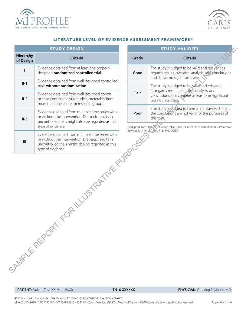
LITERATURE LEVEL OF EVIDENCE ASSESSMENT FRAMEWORK*
STUDY DESIGN
Hierarchyof Design Criteria
I Evidence obtained from at least one properlydesigned randomized controlled trial.
II-1 Evidence obtained from well-designed controlledtrials without randomization.
II-2Evidence obtained from well-designed cohortor case-control analytic studies, preferably frommore than one center or research group.
II-3
Evidence obtained from multiple time series withor without the intervention. Dramatic results inuncontrolled trials might also be regarded as thistype of evidence.
III
Evidence obtained from multiple time series withor without the intervention. Dramatic results inuncontrolled trials might also be regarded as thistype of evidence.
STUDY VALIDITY
Grade Criteria
GoodThe study is judged to be valid and relevant asregards results, statistical analysis, and conclusionsand shows no significant flaws.
Fair
The study is judged to be valid and relevantas regards results, statistical analysis, andconclusions, but contains at least one significantbut not fatal flaw.
PoorThe study is judged to have a fatal flaw such thatthe conclusions are not valid for the purposes ofthis test.
* Adapted from Harris, T., D. Atkins, et al. (2001). "Current Methods of the U.S. PreventiveServices Task Force." Am J Prev Med 20(3S)
SAMPLE R
EPORT. FOR IL
LUSTRATIV
E PURPOSES O
NLY. N
OT FOR CLIN
ICAL U
SE.


![Theranostics · Web viewThus, several molecular markers, such as TP53 mutations [9, 10], P53/P16 immunohistochemistry [11], HPV genotyping [12, 13], gene expression [14], and loss](https://img.pdfslide.net/doc/110x75/60e49b4976d2144a4809da27/theranostics-web-view-thus-several-molecular-markers-such-as-tp53-mutations-9.jpg)