Embed Size (px)
Citation preview
Preclinical Safety and Efficacy of in Situ REIC/Dkk-3 Gene Therapy for Prostate Cancer
Keiichiro Kawauchia, Masami Watanabea,b,c*, Haruki Kakua,b,c, Peng Huangb, Kasumi Sasakia, Masakiyo Sakaguchid, Kazuhiko Ochiaic, Nam-ho Huhd,
Yasutomo Nasua,c, and Hiromi Kumona,b,c
aDepartment of Urology, bCenter for Gene and Cell Therapy, cInnovation Center Okayama for Nanobio-Targeted Therapy, and dCell Biology, Okayama University Graduate School of Medicine, Dentistry and Pharmaceutical Sciences, Okayama 700-8558, Japan
The preclinical safety and therapeutic efficacy of adenoviral vectors that express the REIC/Dkk-3 tumor suppressor gene (Ad-REIC) was examined for use in prostate cancer gene therapy. The Ad-human (h) and mouse (m) REIC were previously demonstrated to induce strong anti-cancer effects in vitro and in vivo, and we herein report the results of two in vivo studies. First, intra-tumor Ad-hREIC administration was examined for toxicity and therapeutic effects in a subcutaneous tumor model using the PC3 prostate cancer cell line. Second, intra-prostatic Ad-mREIC administration was tested for toxicity in normal mice. The whole-body and spleen weights, hematological and serum chemistry parameters, and histological evaluation of tissues from throughout the body were analyzed. Both experiments indicated that there was no significant difference in the examined parameters between the Ad-REIC-treated group and the control (PBS- or Ad-LacZ-treated) group. In the in vitro analysis using PC3 cells, a significant apoptotic effect was observed after Ad-hREIC treatment. Confirming this observation, the robust anti-tumor efficacy of Ad-hREIC was demonstrated in the in vivo subcutaneous prostate cancer model. Based on the results of these preclinical experiments, we consider the adenovirus-mediated REIC/Dkk-3 in situ gene therapy to be safe and useful for the clini-cal treatment of prostate cancer.
Key words: REIC, Dickkopf-3, gene therapy, prostate cancer, preclinical study
rostate cancer is a common and significant dis-ease in the male population of the United States
and many other developed countries [1]. Cancer progression accelerates death in cancer patients, and is often accompanied by a decrease in quality of life. The current therapeutic strategies, such as androgen deprivation therapy, radiation, and cytotoxic chemo-therapy, often fail to stop the disease progression.
Therefore, novel and effective therapies against pros-tate cancer are urgently needed. Cancer progression is often accompanied by the down-regulation of apoptosis [2] and the increased invasive and motile activity of the cancer cells [3]. Cancer cells modulate the apoptotic and metastatic processes by producing both positive and negative effectors [4, 5]. REIC/Dkk-3, a member of the Dickkopf (Dkk) gene family known to interfere with Wnt signaling via Wnt receptors [6, 7], was previ-ously reported to play a distinct role in the induction of apoptosis and inhibition of metastasis [8, 9].
P
Acta Med. Okayama, 2012Vol. 66, No. 1, pp. 7ン16CopyrightⒸ 2012 by Okayama University Medical School.
Original Article http ://escholarship.lib.okayama-u.ac.jp/amo/
Received June 10, 2011 ; accepted September 9, 2011.*Corresponding author. Phone : +81ン86ン235ン7287; Fax : +81ン86ン231ン3986E-mail : [email protected] (M. Watanabe)
REIC/Dkk-3 induced robust anti-tumor effects in vivo and cancer-specific apoptosis in a variety of cancer types when expressed by adenovirus-mediated gene transfer [8-13]. Moreover, our recent study dis-closed that the REIC/Dkk-3 protein also has a role in monocyte differentiation and tumor regression [14]. Intratumoral administration of the REIC/Dkk-3 pro-tein significantly suppressed tumor growth, with CD11c+ and CD8+ (dendritic and killer T cell marker, respectively) cell accumulation, and enhanced the anti-cancer cytolytic activity of splenocytes. These data indicated a cytokine-like role of the REIC/Dkk-3 protein in monocyte differentiation that might be exploited therapeutically. Gene therapy has been applied to treat human dis-eases in clinical trials and is considered to have thera-peutic potential. Our group has previously demon-strated the feasibility of using adenoviral vectors for the treatment of prostate cancer in the clinic [15]. Based on the background information noted above, we are promoting the translation of in situ REIC/Dkk-3 gene therapy from the lab to the clinic for patients with prostate cancer. To perform the necessary pre-clinical study as part of the progression to clinical use of the strategy, we herein examined the toxicity and therapeutic effects of the adenoviral vectors encoding the novel tumor suppressor REIC/Dkk-3.
Materials and Methods
Experimental animals. We used adult male mice (C57BL/6: 30-39g, BALB/C nu/nu: 24-31g) to evaluate the toxicity and therapeutic efficacy of the
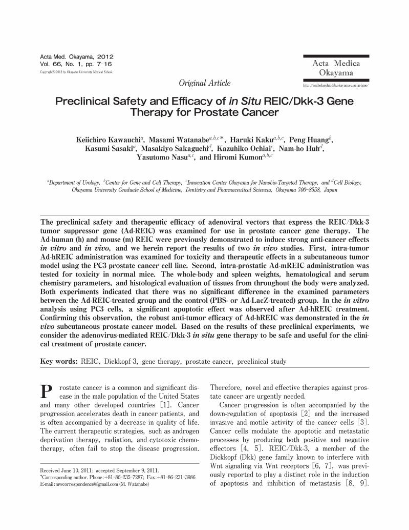
treatment strategy of using adenoviral vectors for the treatment of prostate cancer. The mice were purchased from Japan SLC, Inc. (Hamamatsu, Japan) and main-tained in a specific pathogen-free environment with free access to food and water at the laboratory animal center of Okayama University. They were allowed to adapt to their environment for more than one week before beginning the experiments. The animals were housed and handled in accordance with the Okayama University Animal Research Committee Guidelines. Adenovirus vector carrying REIC/Dkk-3 (Ad-REIC). The full-length cDNA of mouse or human REIC/Dkk-3 was integrated into the cosmid vector, pAxCAwt, and then transferred into an aden-oviral vector by the COS-TPC method (Takara Bio, Shiga, Japan) [8]. An adenoviral vector carrying the LacZ gene (Ad-LacZ) was used as a control. The adenoviral vectors are a replication-defective adenovi-rus of serotype 5 that contain the indicated gene under the control of the CMV early enhancer/chicken β actin (CAG) promoter. For the Ad-human REIC, in vivo transduction ability after intratumoral vector injection was confirmed by detecting the REIC protein expression [14]. Treatment protocol. The experimental design and doses of virus administered are shown in Fig. 1. The first experiment was done using the prostate tumor mouse model. To generate this model, PC3 human prostate cancer cells (2.5×10⁶ in 50µl phos-phate buffered saline (PBS)) were subcutaneously injected into the right thigh of BALB/C nu/nu mice. The tumors were permitted to grow to approximately 5mm in diameter. The mice were then randomly
8 Acta Med. Okayama Vol. 66, No. 1Kawauchi et al.
PBSAd-LacZAd-mREIC
(No tumor)0 21
vectorharvest
C57BL/6 5×10¹⁰ Intra-prostate
No.2 Dose
(viral particle)Mouse Treatmentgroup
Time frame(Days)
Injectionsite
Experiment
Dose(viral particle)Mouse Treatment
group
PBSAd-LacZAd-hREIC
No.1
PC3, subcutaneoustumor 0 32
vector harvest
-14
Time frame(Days)
5×10¹⁰
PC3
Injectionsite
Intra-tumor
Experiment
BALB/C-nu/nu
Fig. 1 The experimental design of the preclinical study is shown. Toxicological analysis of mice administered Ad-hREIC and Ad-mREIC was performed in both experiment 1 and 2 using the indicated mouse models. A study of the efficacy of intratumoral administration of Ad-hREIC in a subcutaneous PC3 prostate cancer model was performed in experiment 1.
assigned into the treatment group with (1) intratu-moral injection of Ad-LacZ, (2) intratumoral injection of Ad-human REIC (Ad-hREIC) at the dose of 5×10¹⁰ viral particles/tumor in 50µl buffer, or (3) intra-tumoral injection of 50µl PBS. The selection of the dose was based on previous clinical studies of prostate cancer gene therapy in which adenovirus-mediated gene delivery was performed using a dose between 1010 and 1011 viral particles/prostate [16-18]. The injections were targeted to the center and periphery of each mass to deliver the agent diffusely. The size of the tumor was measured with vernier calipers until 25 days after each treatment. Tumor volume was calcu-lated using the following formula: 1/2× (the shortest diameter)2× (the longest diameter). In the second experiment, the same number of Ad-mouse REIC (Ad-mREIC) viral particles in 50µl buffer was inocu-lated into the bilateral lobes of the dorsolateral pros-tate in normal C57BL/6 mice without any tumors. The body weight of each mouse was monitored using a sensitive balance during the entire study period. All the animals were observed daily for physical appearances, activity levels, and mortality patterns. The first day of vector administration was taken as Day 0, while the day of sacrifice was desig-nated as Day 32 and Day 21 in the first and second experiments, respectively. In both experiments, the spleen weight was measured at the time of sacrifice. For the histological analysis of the potential side effects of the Ad-REIC treatment, the dorsolateral prostate (the injection site) and selected organs (brain, lungs, heart, liver, stomach, spleen, kidneys, blad-der, and rectum) were dissected at the sacrifice of animals in the second experiment. The tissues were then fixed in formalin and embedded in paraffin sec-tions. The sections (5µm) were stained with hema-toxylin and eosin and were examined for histopatho-logical and cytotoxic changes. Blood parameters. Blood was drawn from the inferior vena cava of each animal under deep anesthe-sia with sodium pentobarbital. The blood samples were collected in a plastic tube with ethylenedi-aminetetra-acetic acid (EDTA). The measurements of white blood cells (WBC), hemoglobin, hematocrit, and platelets were carried out with the EDTA-treated anti-coagulated blood using a hematological instrument (MEK-6358, Celltac-alpha, Nihon Kohden, Tokyo, Japan) within hours of collection. To analyze the
biochemical parameters, we centrifuged the anti-coagulated blood at 3,000 rpm for 10min, and blood plasma was obtained and stored at -80℃. Albumin concentrations were determined by the Bromo Cresol Green (BCG) assay, using a commercially available kit (Wako Pure Chemical Industries Osaka, Japan) and following the manufacturerʼs instructions. The plasma levels of alanine aminotransferase (ALT), aspartate aminotransferase (AST), alkaline phosphatase (ALP), and lactate dehydrogenase (LDH) were deter-mined by the JSCC transferable method by employing the standard ready-to-use kits (Wako Pure Chemical Industries). Total bilirubin was evaluated by the azo-bilirubin method, using a commercial kit (Daiichi Pure Chemicals Co. Tokyo, Japan). Western blotting analysis. The PC3 cells were washed twice with PBS and then lysed with lysis buffer (50mM HEPES, pH7.4, 250mM NaCl, 1mM EDTA, 1オ NP-40, 1mM DTT, 1mM PMSF, 5µg/ml leupeptin, 5µg/ml aprotinin, 2mM Na3VO4, 1mM NaF, 10mM ß-GP), and the proteins were extracted. After centrifugation, the supernatants were adjusted to achieve equal protein concentrations in each sample and then diluted with the same volume of 2×SDS sample buffer and heated for 5min at 95℃. Samples (10µg of protein) were separated on 7.5オ SDS-PAGE gels and electroblotted onto a polyvinylidene fluoride (PVDF) membrane. The blots were blocked for 1h with 10オ nonfat milk powder, 6オ Glycine, and 0.1オ Tween-20 in Tris buffered saline (TBS) at room temperature. The proteins were then identified with the use of the primary antibody, a rabbit anti-human REIC/Dkk-3 polyclonal antibody raised in our laboratory [8] (1 : 1,000 dilution). After extensive washing with 0.1オ Tween-20 in TBS (T-TBS), the blots were exposed to the appropriate horseradish peroxidase-conjugated secondary antibody. After another round of extensive washing with T-TBS, membranes were developed using the enhanced chemi-luminescence detection method (ECL kit, Amersham Pharmacia Biotech, Chandler, AZ, USA). Apoptosis assay. To examine in vitro apoptosis induction after the treatments, we seeded cells in flat-bottom 6-well plates and incubated them for 24h. The cells were then treated with Ad-LacZ and Ad-REIC at the indicated multiplicity of infection (MOI) in serum-free medium (500µl) for 2h, and the medium was exchanged for fresh complete medium
9in Situ REIC/Dkk-3 Gene TherapyFebruary 2012
(2ml). After an additional 48h of incubation, Hoechst 33342 stock solution was added into the medium at a 2µg/ml concentration, and the cells were incubated in the dark for 10min. Hoechst 33342 is an intercalating dye that allows the determination of variations in the total chromatin quantity and the degree of chromatin condensation [11]. Using fluorescence microscopy, we identified apoptotic cells by the presence of highly condensed or fragmented nuclei. Apoptotic cells were counted at 5 different fields under microscopic obser-vation. One hundred cells were judged under each field. Statistical analysis. The data are expressed as the means ± standard error (S.E.). An unpaired Studentʼs t-test was performed for the statistical analysis between the 2 groups. The differences were considered statistically significant at p<0.05.
Results
In the first experiment, we examined the in vivo toxicity and efficacy of the Ad-hREIC vector using the subcutaneous human prostate tumor mouse xenograft model. The experiment was designed as a preclinical study to support the future clinical use of Ad-hREIC, and we therefore evaluated its toxicological profile and anti-tumor effects. We also conducted a second experi-ment in non-tumor-bearing mice to examine the safety of Ad-mREIC, to ensure that specifically overex-pressing REIC/Dkk3 via intra-prostatic injection does not result in toxicity. None of the mice died during the first experiment in any of the treatment groups (n=5). In the second experiment, 1 or 2 mice died in each group during the experiment [PBS (n=15): one mouse died on day 18; Ad-LacZ (n=16): 1 mouse each died on day 7 and day 18; Ad-mREIC (n=16): 1 mouse died on day 18 and one on day 19]. At necropsy, there was no obvious cause of death such as extensive inflammation, abnormal bladder distension, trauma of various organs, or abundant ascites. In both the first and second experiments, there was no statistically significant difference in the survival between the Ad-REIC-treated group and the control groups by Mantel-Cox log-rank analysis (data not shown). The physical appearances and activities were observed carefully, and there were no definite differ-ences among the groups in either experiment.
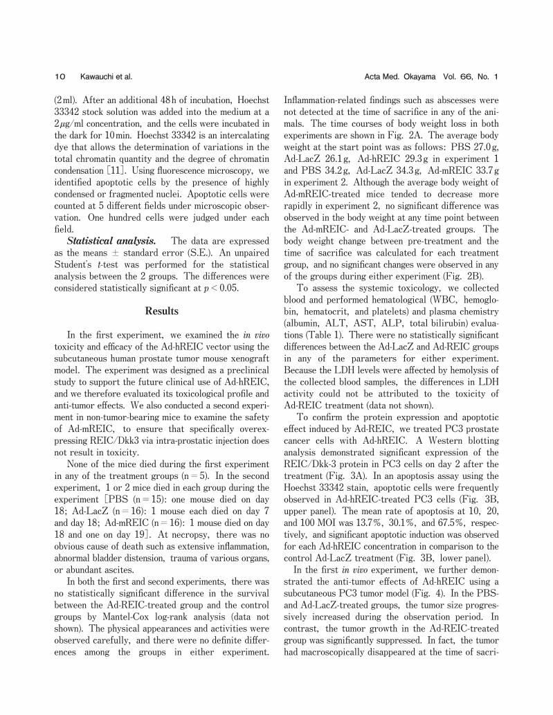
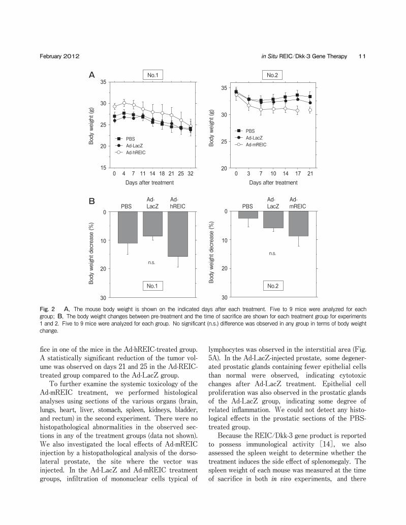
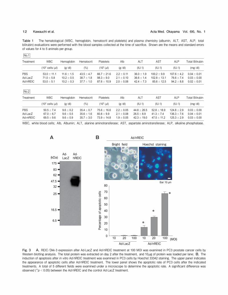
Inflammation-related findings such as abscesses were not detected at the time of sacrifice in any of the ani-mals. The time courses of body weight loss in both experiments are shown in Fig. 2A. The average body weight at the start point was as follows: PBS 27.0g, Ad-LacZ 26.1g, Ad-hREIC 29.3g in experiment 1 and PBS 34.2g, Ad-LacZ 34.3g, Ad-mREIC 33.7g in experiment 2. Although the average body weight of Ad-mREIC-treated mice tended to decrease more rapidly in experiment 2, no significant difference was observed in the body weight at any time point between the Ad-mREIC- and Ad-LacZ-treated groups. The body weight change between pre-treatment and the time of sacrifice was calculated for each treatment group, and no significant changes were observed in any of the groups during either experiment (Fig. 2B). To assess the systemic toxicology, we collected blood and performed hematological (WBC, hemoglo-bin, hematocrit, and platelets) and plasma chemistry (albumin, ALT, AST, ALP, total bilirubin) evalua-tions (Table 1). There were no statistically significant differences between the Ad-LacZ and Ad-REIC groups in any of the parameters for either experiment. Because the LDH levels were affected by hemolysis of the collected blood samples, the differences in LDH activity could not be attributed to the toxicity of Ad-REIC treatment (data not shown). To confirm the protein expression and apoptotic effect induced by Ad-REIC, we treated PC3 prostate cancer cells with Ad-hREIC. A Western blotting analysis demonstrated significant expression of the REIC/Dkk-3 protein in PC3 cells on day 2 after the treatment (Fig. 3A). In an apoptosis assay using the Hoechst 33342 stain, apoptotic cells were frequently observed in Ad-hREIC-treated PC3 cells (Fig. 3B, upper panel). The mean rate of apoptosis at 10, 20, and 100 MOI was 13.7オ, 30.1オ, and 67.5オ, respec-tively, and significant apoptotic induction was observed for each Ad-hREIC concentration in comparison to the control Ad-LacZ treatment (Fig. 3B, lower panel). In the first in vivo experiment, we further demon-strated the anti-tumor effects of Ad-hREIC using a subcutaneous PC3 tumor model (Fig. 4). In the PBS- and Ad-LacZ-treated groups, the tumor size progres-sively increased during the observation period. In contrast, the tumor growth in the Ad-REIC-treated group was significantly suppressed. In fact, the tumor had macroscopically disappeared at the time of sacri-
10 Acta Med. Okayama Vol. 66, No. 1Kawauchi et al.
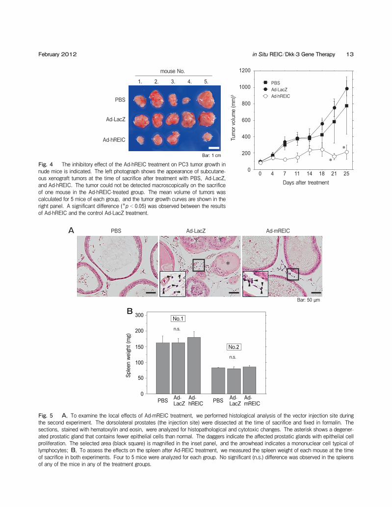
fice in one of the mice in the Ad-hREIC-treated group. A statistically significant reduction of the tumor vol-ume was observed on days 21 and 25 in the Ad-REIC-treated group compared to the Ad-LacZ group. To further examine the systemic toxicology of the Ad-mREIC treatment, we performed histological analyses using sections of the various organs (brain, lungs, heart, liver, stomach, spleen, kidneys, bladder, and rectum) in the second experiment. There were no histopathological abnormalities in the observed sec-tions in any of the treatment groups (data not shown). We also investigated the local effects of Ad-mREIC injection by a histopathological analysis of the dorso-lateral prostate, the site where the vector was injected. In the Ad-LacZ and Ad-mREIC treatment groups, infiltration of mononuclear cells typical of
lymphocytes was observed in the interstitial area (Fig. 5A). In the Ad-LacZ-injected prostate, some degener-ated prostatic glands containing fewer epithelial cells than normal were observed, indicating cytotoxic changes after Ad-LacZ treatment. Epithelial cell proliferation was also observed in the prostatic glands of the Ad-LacZ group, indicating some degree of related inflammation. We could not detect any histo-logical effects in the prostatic sections of the PBS-treated group. Because the REIC/Dkk-3 gene product is reported to possess immunological activity [14], we also assessed the spleen weight to determine whether the treatment induces the side effect of splenomegaly. The spleen weight of each mouse was measured at the time of sacrifice in both in vivo experiments, and there
11in Situ REIC/Dkk-3 Gene TherapyFebruary 2012
A
15
20
25
30
35
Ad-hREICAd-LacZPBS
Days after treatment
Body weight (g)
0 4 7 11 14 18 21 25 32
No.1
20
25
30
35
Ad-mREICAd-LacZPBS
0 3 7 10 14 17 21Days after treatment
Body weight (g)
No.2
B
Body weight decrease (%)
30
20
10
0
Ad-hREIC
Ad-LacZPBS
No.1
n.s.
Ad-mREIC
Ad-LacZPBS
Body weight decrease (%)
30
20
10
0
n.s.
No.2
Fig. 2 A, The mouse body weight is shown on the indicated days after each treatment. Five to 9 mice were analyzed for each group; B, The body weight changes between pre-treatment and the time of sacrifice are shown for each treatment group for experiments 1 and 2. Five to 9 mice were analyzed for each group. No significant (n.s.) difference was observed in any group in terms of body weight change.
12 Acta Med. Okayama Vol. 66, No. 1Kawauchi et al.
A
Ad-LacZ
Ad-hREIC
175
83
62
47.532
25
16.5
6.5
(kDa)
B
0
10
20
30
40
50
60
70
80
Percentage of apoptotic cells
10 20 100 10 20 100 (MOI)Ad-LacZ Ad-hREIC
Ad-hREIC
Bar: 10 µm
Hoechst stainingBright field
*
*
*
Fig. 3 A, REIC/Dkk-3 expression after Ad-LacZ and Ad-hREIC treatment at 100 MOI was examined in PC3 prostate cancer cells by Western blotting analysis. The total protein was extracted on day 2 after the treatment, and 10µg of protein was loaded per lane; B, The induction of apoptosis after in vitro Ad-hREIC treatment was examined in PC3 cells by Hoechst 33342 staining. The upper panel indicates the appearance of apoptotic cells after Ad-hREIC treatment. The lower panel shows the apoptotic rate of PC3 cells after the indicated treatments. A total of 5 different fields were examined under a microscope to determine the apoptotic rate. A significant difference was observed (*p<0.05) between the Ad-hREIC and the control Ad-LacZ treatment.
Table 1 The hematological (WBC, hemoglobin, hematocrit and platelets) and plasma chemistry (albumin, ALT, AST, ALP, total bilirubin) evaluations were performed with the blood samples collected at the time of sacrifice. Shown are the means and standard errors of values for 4 to 5 animals per group.
No.1
Treatment WBC Hemoglobin Hematocrit Platelets Alb ALT AST ALP Total Bilirubin
(102 cells/µl) (g/dl) (%) (104/µl) (g/dl) (IU/l) (IU/l) (IU/l) (mg/dl)
PBS 53.0±11.1 11.6±1.5 43.5±4.7 66.7±21.6 2.2±0.11 36.0±1.9 100.2±9.9 107.6±4.2 0.04±0.01Ad-LacZ 71.0±5.8 10.2±0.5 38.7±1.8 98.3±9.0 2.1±0.10 36.6±1.4 102.8±13.1 76.6±7.4 0.03±0.00Ad-hREIC 53.0±5.1 10.2±0.3 37.7±1.0 87.6±10.9 2.0±0.08 42.4±7.3 85.6±12.5 94.2±8.8 0.02±0.01
No.2
Treatment WBC Hemoglobin Hematocrit Platelets Alb ALT AST ALP Total Bilirubin
(102 cells/µl) (g/dl) (%) (104/µl) (g/dl) (IU/l) (IU/l) (IU/l) (mg/dl)
PBS 50.5±7.4 9.6±0.2 35.4±0.7 75.6±16.6 2.2±0.05 44.8±26.5 52.8±18.9 124.8±2.9 0.03±0.00Ad-LacZ 67.0±6.7 9.6±0.5 35.6±1.6 60.8±9.9 2.1±0.08 26.5±8.9 41.3±7.4 136.3±7.6 0.04±0.01Ad-mREIC 69.5±9.6 9.6±0.9 35.7±3.0 73.9±14.8 1.9±0.05 42.3±19.0 47.5±11.2 125.3±2.9 0.03±0.00
WBC, white blood cells; Alb, Albumin; ALT, alanine aminotransferase; AST, aspartate aminotransferase; ALP, alkaline phosphatase.
13in Situ REIC/Dkk-3 Gene TherapyFebruary 2012
Ad-hREIC
Ad-LacZ
PBS
1. 2. 3. 4. 5.
mouse No.
Bar: 1cm
0
200
400
600
800
1000
1200
Ad-hREICAd-LacZPBS
Tumor volume (mm)3
Days after treatment0 4 7 11 14 18 21 25
*
*
Ad-LacZ Ad-mREICPBS† †
*
Bar: 50 µm
A
BNo.1
No.2
Ad-mREIC
Ad-LacZPBSAd-
hREICAd-LacZPBS
0
50
100
150
200
300
Spleen weight (mg)
n.s.
n.s.
Fig. 5 A, To examine the local effects of Ad-mREIC treatment, we performed histological analysis of the vector injection site during the second experiment. The dorsolateral prostates (the injection site) were dissected at the time of sacrifice and fixed in formalin. The sections, stained with hematoxylin and eosin, were analyzed for histopathological and cytotoxic changes. The asterisk shows a degener-ated prostatic gland that contains fewer epithelial cells than normal. The daggers indicate the affected prostatic glands with epithelial cell proliferation. The selected area (black square) is magnified in the inset panel, and the arrowhead indicates a mononuclear cell typical of lymphocytes; B, To assess the effects on the spleen after Ad-REIC treatment, we measured the spleen weight of each mouse at the time of sacrifice in both experiments. Four to 5 mice were analyzed for each group. No significant (n.s.) difference was observed in the spleens of any of the mice in any of the treatment groups.
Fig. 4 The inhibitory effect of the Ad-hREIC treatment on PC3 tumor growth in nude mice is indicated. The left photograph shows the appearance of subcutane-ous xenograft tumors at the time of sacrifice after treatment with PBS, Ad-LacZ, and Ad-hREIC. The tumor could not be detected macroscopically on the sacrifice of one mouse in the Ad-hREIC-treated group. The mean volume of tumors was calculated for 5 mice of each group, and the tumor growth curves are shown in the right panel. A significant difference (*p<0.05) was observed between the results of Ad-hREIC and the control Ad-LacZ treatment.
were no statistically significant differences in spleen weight following treatment in either treatment group (Fig. 5B).
Discussion
We previously cloned the REIC (Reduced Expression in Immortalized Cells) gene and reported that its expression is down-regulated in a variety of cancer cell lines and tumors [8, 10-12, 19-21]. The REIC gene is identical to the Dickkopf-3 (Dkk-3) gene [20] and REIC/Dkk-3 seems to be one of the most innovative tumor suppressor genes used for cancer gene therapy [8, 9, 12, 21]. We have extensively utilized an adenoviral vector agent, Ad-hREIC, con-taining the human REIC/Dkk-3 gene under the control of the CAG promoter in preclinical cancer models in mice. The adenoviral vector carrying human REIC/Dkk-3 selectively induced apoptosis in prostate cancer and malignant mesothelioma cells through the activa-tion of c-Jun-NH2-kinase (JNK) and c-Jun [8, 12]. The adenovirus-mediated expression of the REIC/Dkk-3 gene efficiantly induces endoplasmic reticulum (ER) stress-mediated apoptosis in a cancer cell-spe-cific manner [12, 13]. The current study also demon-strated that the Ad-hREIC treatment induced signifi-cant apoptosis in a prostate cancer cell line. We further demonstrated the robust therapeutic utility of Ad-hREIC in a subcutaneous prostate cancer model. Thus, the adenoviral agent appears to be effective against human prostate tumors in terms of the cancer-specific targeting and a more radical response to expression of the gene. The therapeutic effects of Ad-REIC were previ-ously observed in a variety of in vivo tumor models generated with prostate, breast, and mesothelioma cancer cells [8, 9, 11, 12], but the toxicity of the Ad-REIC treatment remained unknown. Thus, we currently focused on the safety of local injection of Ad-REIC for the treatment of prostate cancer. Non-human primates have been used in some studies of adenoviral vector toxicity [22, 23], and mice have many advantages for preclinical evaluations prior to these primate studies [24]. To evaluate the toxicity of the Ad-REIC vector, we first analyzed the lethality and survival in 2 experiments. There was no toxicity when tumor-bearing mice were administered the human-REIC vector, although some lethality was observed in
the mice during the second experiment. However, since the survival was not statistically different between the groups (1 or 2 mice died in both the con-trol and treatment groups), it is likely that the Ad-REIC treatment itself did not specifically induce the deaths. The most conceivable reason for the lethality is that the mice in the second experiment died due to post-operative complications of the open sur-gery for the prostatic injections. The other possibility is that adenovirus toxicity may be the cause of the deaths. Although definite histological damage was not observed in the examined mice, we cannot exclude this possibility. To confirm the reproducibility of the deaths and to exclude the adenovirus-related death, we did similar additional experiments using C57BL/6 mice in which there was no death during the experi-mental schedule until day 28. Thus, it seems that the death in experiment 2 was not reproducible and not adenovirus-related. Based on these results, another possibility is that the mice deaths may have been procedure-related. We next investigated the systemic toxicity of the Ad-REIC treatment. The liver has been widely recog-nized as the primary target organ of adenoviral vec-tors after an intravenous adenoviral administration [25]. Although the Ad-REIC was locally adminis-tered by intratumoral or intra-prostate injection in our experiments, the possibility could not be excluded that the viral particles might disseminate into the general circulation and affect the liver functions. However, in the current studies using Ad-hREIC and Ad-mREIC, we did not detect any differences in the liver enzymes or other biochemical parameters in any of the 3 treat-ment groups. None of the mice showed histopatho-logical liver abnormalities such as cytotoxicity or inflammation in the PBS-, Ad-LacZ-, or Ad-mREIC-treated groups during the second experiment (data not shown), indicating the consistency of the results of biochemical evaluations. As for other distant organs, no histological abnormalities were observed (data not shown) in the screening, although there were some histological changes at the injection site, the dorsolat-eral prostate. In addition, there was no effect of Ad-REIC treatment on hematological parameters or spleen weight. It therefore seems that Ad-REIC and the gene product, secreted REIC protein, do not induce serious systemic toxicity or non-specific inflam-mation under the indicated conditions.
14 Acta Med. Okayama Vol. 66, No. 1Kawauchi et al.
The second step of our work was to analyze the local side effects associated with an intra-prostatic delivery of the Ad-REIC vector by direct injection. To investigate the probable local toxicity due to the adenovirus or the gene products, including the secreted REIC protein, we examined the histopatho-logical changes of the dorsolateral prostate after the Ad-mREIC injection. We could not detect any histo-logical damage to the prostate gland after PBS or Ad-REIC treatment. In the Ad-LacZ group, there was some damage to the prostate gland, which may have been due to the toxicological profile of the adenoviral vectors [26]. The lack of local toxicity in the Ad-mREIC-injected prostate glands was consistent with the previous reports showing that Ad-hREIC induces cytotoxic or apoptotic effects only in cancer cells and not in normal cells [8, 11]. As for the lymphocyte infiltration in the prostatic interstitial space, the phenomenon was observed in both the Ad-LacZ and Ad-mREIC treatment groups. It is likely that the adenoviral vector may have caused the inflammatory reaction. In addition, a recent study reported that the REIC protein induced a chemokine-like action that caused the accumulation of immune cells when directly injected into subcutaneous tumors in C57BL/6 mice [14]. Thus, the other possibility is that the gene product, the REIC protein, might have been involved in lymphocyte infiltration in the Ad-mREIC-treated group. Further studies will clarify the mechanisms of the Ad-REIC-induced immunomod-ulatory effects in locally injected normal and tumor tissues. Our results show that the adenoviral vector encod-ing the REIC/Dkk-3 gene and the CAG promoter has no significant toxicity and is therapeutically effective when directly injected into the tumors of mice with experimental prostate cancer or into the prostates of normal mice at the indicated doses. We believe that the current study provides valuable information for future clinical trials of Ad-REIC-mediated gene ther-apy against human prostate cancer.
Acknowledgments. This work was supported by a grant from the Ministry of Education, Culture, Sports, Science and Technologyʼs FY2006 “Creation of Innovation Centers for Advanced Interdisciplinary Research Areas” Scheme in Japan. We thank Hideo Ueki and Shun-Ai Li (Okayama University) for their valuable assistance.
References
1. Jemal A, Murray T, Ward E, Samuels A, Tiwari RC, Ghafoor A, Feuer EJ and Thun MJ: Cancer statistics, 2005. CA Cancer J Clin (2005) 55: 10-30.
2. Rittmaster RS, Thomas LN, Wright AS, Murray SK, Carlson K, Douglas RC, Yung J, Messieh M, Bell D and Lazier CB: The util-ity of tissue transglutaminase as a marker of apoptosis during treatment and progression of prostate cancer. J Urol (1999) 162: 2165-2169.
3. Sehgal I, Baley PA and Thompson TC: Transforming growth factor beta1 stimulates contrasting responses in metastatic versus pri-mary mouse prostate cancer-derived cell lines in vitro. Cancer Res (1996) 56: 3359-3365.
4. Ferreira CG, Epping M, Kruyt FA and Giaccone G: Apoptosis: target of cancer therapy. Clin Cancer Res (2002) 8: 2024-2034.
5 Curran S and Murray GI: Matrix metalloproteinases in tumour inva-sion and metastasis. J Pathol (1999) 189: 300-308.
6. Krupnik VE, Sharp JD, Jiang C, Robison K, Chickering TW, Amaravadi L, Brown DE, Guyot D, Mays G, Leiby K, Chang B, Duong T, Goodearl AD, Gearing DP, Sokol SY and McCarthy SA: Functional and structural diversity of the human Dickkopf gene family. Gene (1999) 238: 301-313.
7. Mao B, Wu W, Davidson G, Marhold J, Li M, Mechler BM, Delius H, Hoppe D, Stannek P, Walter C, Glinka A and Niehrs C: Kremen proteins are Dickkopf receptors that regulate Wnt/beta-catenin signalling. Nature (2002) 417: 664-647.
8. Abarzua F, Sakaguchi M, Takaishi M, Nasu Y, Kurose K, Ebara S, Miyazaki M, Namba M, Kumon H and Huh NH: Adenovirus-mediated overexpression of REIC/Dkk-3 selectively Induces apop-tosis in human prostate cancer cells through activation of c-Jun-NH2-kinase. Cancer Res (2005) 65: 9617-9622.
9. Edamura K, Nasu Y, Takaishi M, Kobayashi T, Abarzua F, Sakaguchi M, Kashiwakura Y, Ebara S, Saika T, Watanabe M, Huh NH and Kumon H: Adenovirus-mediated REIC/Dkk-3 gene transfer inhibits tumor growth and metastasis in an orthotopic pros-tate cancer model. Cancer Gene Ther (2007) 14: 765-772.
10. Abarzua F, Sakaguchi M, Tanimoto R, Sonegawa H, Li DW, Edamura K, Kobayashi T, Watanabe M, Kashiwakura Y, Kaku H, Saika T, Nakamura K, Nasu Y, Kumon H and Huh NH: Heat shock proteins play a crucial role in tumor-specific apoptosis by REIC/Dkk-3. Int J Mol Med (2007) 20: 37-43.
11. Kawasaki K, Watanabe M, Sakaguchi M, Ogasawara Y, Ochiai K, Nasu Y, Doihara H, Kashiwakura Y, Huh NH and Kumon H: Date H. REIC/Dkk-3 overexpression downregulates P-glycoprotein in multidrug-resistant MCF7/ADR cells and induces apoptosis in breast cancer. Cancer Gene Ther (2009) 16: 65-72.
12. Kashiwakura Y, Ochiai K, Watanabe M, Abarzua F, Sakaguchi M, Takaoka M, Tanimoto R, Nasu Y, Huh NH and Kumon H: Down-regulation of inhibition of differentiation-1 via activation of activat-ing transcription factor 3 and Smad regulates REIC/Dickkopf-3-induced apoptosis. Cancer Res (2008) 68: 8333-8341.
13. Abarzua F, Kashiwakura Y, Takaoka M, Watanabe M, Ochiai K, Sakaguchi M, Iwawaki T, Tanimoto R, Nasu Y, Huh NH and Kumon H: An N-terminal 78 amino acid truncation of REIC/Dkk-3 effectively induces apoptosis. Biochem Biophys Res Commun (2008) 375: 614-618.
14. Watanabe M, Kashiwakura Y, Huang P, Ochiai K, Futami J, Li SA, Takaoka M, Nasu Y, Sakaguchi M, Huh NH and Kumon H: Immunological aspects of REIC/Dkk-3 in monocyte differentiation and tumor regression. Int J Oncol (2009) 34: 657-663.
15in Situ REIC/Dkk-3 Gene TherapyFebruary 2012
15. Nasu Y, Saika T, Ebara S, Kusaka N, Kaku H, Abarzua F, Manabe D, Thompson TC and Kumon H: Suicide gene therapy with adenoviral delivery of HSV-tK gene for patients with local recurrence of prostate cancer after hormonal therapy. Mol Ther (2007) 15: 834-840.
16. Freytag SO, Khil M, Stricker H, Peabody J, Menon M, DePeralta-Venturina M, Nafziger D, Pegg J, Paielli D, Brown S, Barton K, Lu M, Aguilar-Cordova E and Kim JH: Phase I study of replication-competent adenovirus-mediated double suicide gene therapy for the treatment of locally recurrent prostate cancer. Cancer Res (2002) 62: 4968-4976.
17. Kubo H, Gardner TA, Wada Y, Koeneman KS, Gotoh A, Yang L, Kao C, Lim SD, Amin MB, Yang H, Black ME, Matsubara S, Nakagawa M, Gillenwater JY, Zhau HE and Chung LW: Phase I dose escalation clinical trial of adenovirus vector carrying osteo-calcin promoter-driven herpes simplex virus thymidine kinase in localized and metastatic hormone-refractory prostate cancer. Hum Gene Ther (2003) 14: 227-241.
18. van der Linden RR, Haagmans BL, Mongiat-Artus P, van Doornum GJ, Kraaij R, Kadmon D, Aguilar-Cordova E, Osterhaus AD, van der Kwast TH and Bangma CH: Virus specific immune responses after human neoadjuvant adenovirus-mediated suicide gene therapy for prostate cancer. Eur Urol (2005) 48: 153-161.
19. Tsuji T, Nozaki I, Miyazaki M, Sakaguchi M, Pu H, Hamazaki Y, Iijima O and Namba M: Antiproliferative activity of REIC/Dkk-3 and its significant down-regulation in non-small-cell lung carcino-mas. Biochem Biophys Res Commun (2001) 289: 257-263.
20. Tsuji T, Miyazaki M, Sakaguchi M, Inoue Y and Namba M: A REIC gene shows down-regulation in human immortalized cells
and human tumor-derived cell lines. Biochem Biophys Res Commun (2000) 268: 20-24.
21. Hsieh SY, Hsieh PS, Chiu CT and Chen WY: Dickkopf-3/REIC functions as a suppressor gene of tumor growth. Oncogene (2004) 23: 9183-9189.
22. Driesse MJ, Vincent AJ, Sillevis Smitt PA, Kros JM, Hoogerbrugge PM, Avezaat CJ, Valerio D and Bout A: Intracerebral injection of adenovirus harboring the HSVtk gene combined with ganciclovir administration: toxicity study in nonhuman primates. Gene Ther (1998) 5: 1122-1129.
23. Morral N, O'Neal WK, Rice K, Leland MM, Piedra PA, Aguilar-Córdova E, Carey KD, Beaudet AL and Langston C: Lethal toxic-ity, severe endothelial injury, and a threshold effect with high doses of an adenoviral vector in baboons. Hum Gene Ther (2002) 13: 143-154.
24. Su C, Cao H, Tan S, Huang Y, Jia X, Jiang L, Wang K, Chen Y, Long J, Liu X, Wu M, Wu X and Qian Q: Toxicology profiles of a novel p53-armed replication-competent oncolytic adenovirus in rodents, felids, and nonhuman primates. Toxicol Sci (2008) 106: 242-250.
25. Muruve DA, Barnes MJ, Stillman IE and Libermann TA: Adenoviral gene therapy leads to rapid induction of multiple chemokines and acute neutrophil-dependent hepatic injury in vivo. Hum Gene Ther (1999) 10: 965-976.
26. Teramoto S, Johnson LG, Huang W, Leigh MW and Boucher RC: Effect of adenoviral vector infection on cell proliferation in cultured primary human airway epithelial cells. Hum Gene Ther (1995) 6: 1045-1053.
16 Acta Med. Okayama Vol. 66, No. 1Kawauchi et al.


![REIC Professional Capabilities [Low-Res]](https://img.pdfslide.net/doc/110x75/54e5ea9f4a7959bd3e8b456f/reic-professional-capabilities-low-res.jpg)