Embed Size (px)
Citation preview
Prediction of the 3D Structure and Dynamics of Human DPG-Protein Coupled Receptor Bound to an Agonist and an
Antagonist
Youyong Li,† Fangqiang Zhu,† Nagarajan Vaidehi,*,†,‡ William A. Goddard, III,*,†
Felix Sheinerman,§ Stephan Reiling,§ Isabelle Morize,§ Lan Mu,§ Keith Harris,§
Ali Ardati,§ and Abdelazize Laoui§
Contribution from the Materials and Process Simulation Center (MC 139-74), CaliforniaInstitute of Technology, Pasadena, California 91125, and Sanofi-AVentis Pharma,
Bridgewater, New Jersey 08807-0800
Received February 13, 2007; E-mail: [email protected]; [email protected]
Abstract: Prostanoids play important physiological roles in the cardiovascular and immune systems andin pain sensation in peripheral systems through their interactions with eight G-protein coupled receptors.These receptors are important drug targets, but development of subtype specific agonists and antagonistshas been hampered by the lack of 3D structures for these receptors. We report here the 3D structure forthe human DP G-protein coupled receptor (GPCR) predicted by the MembStruk computational method.To validate this structure, we use the HierDock computational method to predict the binding mode for theendogenous agonist (PGD2) to DP. Based on our structure, we predicted the binding of different antagonistsand optimized them. We find that PGD2 binds vertically to DP in the TM1237 region with the R chaintoward the extracellular (EC) region and the ω chain toward the middle of the membrane. This structureexplains the selectivity of the DP receptor and the residues involved in the predicted binding site correlatevery well with available mutation experiments on DP, IP, TP, FP, and EP subtypes. We report moleculardynamics of DP in explicit lipid and water and find that the binding of the PGD2 agonist leads to correlatedrotations of helices of TM3 and TM7, whereas binding of antagonist leads to no such rotations. Thus,these motions may be related to the mechanism of activation.
1. Introduction
Prostanoids (prostaglandins (PG) and thromboxanes (TX),both metabolites of arachidonic acid)1,2 play important physi-ological roles in the cardiovascular and immune systems andin pain sensation in peripheral systems. They exert a variety ofactions in the body through binding to specific cell surfaceprostanoid receptors. The eight subtypes of prostanoid receptorsall belong to the family A of G-protein coupled receptors(GPCRs). Various prostanoids exert their activity preferentiallythrough prostanoid receptors. For example, the DP receptorpreferentially binds prostaglandin PGD2 with substantially moreaffinity than any of the other prostanoid ligands, and PGD2shows quite a preference to DP receptor with diminished activityon EP3 and no activity on the other prostanoid receptors.1 Thisextensive heterogeneity and preference of the prostanoid recep-tors is reflected by the remarkable diversity of physiologicaleffects that can be elicited by prostanoids.
Perhaps the most well-known of these effects are those thatproduce pain, fever, and inflammation, which can be relieved
through the inhibition of COX-1 or COX-2 by aspirin andnonsteroidal anti-inflammatory drugs (NSAIDs).3,4 Other im-portant effects of the prostanoids involve the vascular, reproduc-tive, bone, and immune systems.1 Specifically, PGD(2) functionsas a mast cell-derived mediator to trigger asthmatic responses.5
The development of subtype-specific agonists and antagonistshas been hampered by the lack of 3D structures for prostanoidreceptors. As a first step in providing a structural basis forunderstanding the activity and selectivity of these receptors, weused the MembStruk computational procedure6-14 to predict the
† California Institute of Technology.‡ Present address: Division of Immunology, Beckman Research Institute
of City of Hope, 1500, Duarte Road, Duarte, CA 91010.§ Sanofi-Aventis Pharma.
(1) Narumiya, S.; Sugimoto, Y.; Ushikubi, F.Physiol. ReV. 1999, 79, 1193.(2) Breyer, R. M.; Bagdassarian, C. K.; Myers, S. A.; Breyer, M. D.Annu.
ReV. Pharmacol. Toxicol.2001, 41, 661.
(3) Marnett, L. J.; DuBois, R. N.Annu. ReV. Pharmacol. Toxicol.2002, 42,55.
(4) Subbaramaiah, K.; Dannenberg, A. J.Trends Pharmacol. Sci.2003, 24,96.
(5) Matsuoka, T.; Hirata, M.; Tanaka, H.; et al.Science2000, 287, 2013.(6) Hall, S. E.; Floriano, W. B.; Vaidehi, N.; Goddard, W. A., III.Chem. Sens.
2004, 29, 595.(7) Floriano, W. B.; Vaidehi, N.; Goddard, W. A., III.Chem. Sens.2004, 29,
269.(8) Kalani, M. Y.; Vaidehi, N.; Hall, S. E.; Trabanino, R.; Freddolino, P.;
Kalani, M. A.; Floriano, W. B.; Kam, V.; Goddard, W. A., III.Proc. Natl.Acad. Sci.2004, 101, 3815.
(9) Freddolino, P.; Kalani, M. Y.; Vaidehi, N.; Floriano, W.; Hall, S. E.;Trabanino, R.; Kam, V. W. T.; Goddard, W. A.Proc. Natl. Acad. Sci.2004, 101, 2736.
(10) Trabanino, R.; Hall, S. E.; Vaidehi, N.; Floriano, W.; Goddard, W. A.Biophys. J.2004, 86, 1904.
(11) Hummel, P.; Vaidehi, N.; Floriano, W. B.; Hall, S. E.; Goddard, W. A.,III. Protein Sci.2005, 14, 703.
(12) Peng, J.; Vaidehi, N.; Hall, S.; Goddard, W. A., III.Chem. Med. Chem.2006, 1, 878.
Published on Web 08/11/2007
10720 9 J. AM. CHEM. SOC. 2007 , 129, 10720-10731 10.1021/ja070865d CCC: $37.00 © 2007 American Chemical Society
structure for the human DP receptor starting from its aminoacid sequence. Then, we used the HierDock computationalprocedure15-20 to predict the binding site for endogenous agonistPGD2 with human DP receptor. The details for these predictionsare in section 2. Then, we carry out molecular dynamics inexplicit lipid and water to check the binding effect on the proteinstructure. Finally, we report docking results of antagonist basedon our structure.
2. Methods
We predicted the three-dimensional structure of DP using Memb-Struk4.1 computational method summarized here.
2.1. Prediction of the TM Regions and Hydrophobic Centers.The TM regions were predicted using TM2ndS method described inref 10. The input to TM2ndS method was the 43 sequences ofprostanoid receptors from various species. Multiple sequence alignmentof the 43 sequences was performed using clustalW. Using the multiplesequence alignment as input, the TM regions were predicted usingTM2ndS procedure.10 The hydrophobic maximum was chosen as thecentral residue (referred to as the centroid) for each helix that dividesthe area under the hydrophobicity curve equally. The centroid for eachhelix is positioned to be in the samexy plane (the midpoint of thelipid).2.2. Prediction of the 3D Structure.On the basis of the predictedTM regions and the TM centroids, the MembStruk program was usedto build and optimize the 3D structure for the human DP receptor. Thesteps of MembStruk and the predicted structure are described below.
2.2.1. Helix Packing.First, canonicalR-helices were built for eachTM domain. TheseR-helix structures were then bundled together asfollows. The predicted helix centroid is placed on thexy plane usingx,y coordinates on the basis of the low-resolution (7.5 Å) electrondensity map of frog rhodopsin. The orientation of each helix about itsz-axis (theø angle) is chosen so that its helical face with the maximumhydrophobic moment points outward to contact the lipid. In this
analysis, we calculate the hydrophobic moment over the full helix butinclude only the half of the residues that would face outward. Then,each helix is tilted about the point at which the central axis intersectsthe xy plane to match the tilt angles (θ,æ) from frog rhodopsin.
2.2.2. Helix Bending.Next, molecular dynamics (MD) simulationswere performed (200 ps) for each individual helix, allowing the helixto attain its equilibrium structure (in some cases it bends or kinks).Then, we chose the structure with the lowest potential energy for eachhelix and assembled it back into the bundle so that the average axiscoincides with the original axis. The side chains were then optimizedusing SCWRL,21,22 and the total energy was minimized (conjugategradients).
2.2.3. RotMin. This initial packed structure was minimized, andthen we allowed the individual packing interactions to optimize asfollows. Each helix was independently rotated (ø) by +5° and -5°,the side chains were repositioned using SCWRL, and then all atomsof the bundle were optimized. If either new angle was lower, it wasselected.
2.2.4. Lipid Insertion. At this point, we inserted the seven-helixbundle into a lipid framework ending up with 48 lipids moleculesarranged as a bilayer. These lipid molecules were optimized using rigidbody dynamics.
2.2.5. RotScan.Starting from the final RotMin structure, weperformed a full 360-degree rotational scan (ø) on each of the helicesin 5° increments. For each angle, the side chains were reassigned withSCWRL, and the full bundle was reminimized. Multiple minima basedon energy and interhelical hydrogen bonds were chosen for each helix.Combination of multiple minima for each helix leads to an ensembleof conformations which were then sorted by the number of interhelicalhydrogen bonds and then by total energy.
2.3. Prediction of the Extracelluar (EC) and Intracellular (IC)Loop Structure. We took the best structure from the previous stepand added the three EC and IC loops. We expect the three EC and thethree IC loops of human DP to be quite flexible and strongly affectedby the solvent, which is treated only implicitly in MembStruk. Thus,to provide initial loop structures for our MD studies of the DP receptor,we used the alignment of DP with bovine rhodopsin and then homologythreaded the DP loops to the crystal structure (1L9H.pdb). Then, wecarried out minimization and dynamics on the loops with fixed helixbundle atoms.
In the crystal structure of bovine rhodopsin, the ECII loop (con-necting TM4 and TM5) is closed over the 7-TM barrel, contributingto the binding of 11-cis-retinal. This ECII loop has a disulfide bond toTM3 (C105-C183), which is highly conserved among the rhodopsinsuperfamily of GPCRs. Thus, we include this disulfide bond in ourloop structures. It is generally believed that the disulfide bond playscritical role in the folding of seven helices and in the closing of theECII loop over the 7-TM barrel.23 Since the rhodopsin in the crystalstudy is in the inactive form, it is possible that substantial changesoccur in ECII and in other loops upon activation.
2.4. Molecular Dynamics Simulation.Since the description of lipidand water in MembStruk is implicit with a skimpy layer of lipid bilayer,we performed molecular dynamics (MD) simulations of the predictedstructure of DP receptor with and without ligand for 1∼2 ns in explicitlipid bilayer and water. We carried out MD simulations using NAMDincluding explicit water and a periodically infinite lipid to determinethe interactions of the protein with lipid and water.14
We started with the predicted hDP structure, stripped away the lipidmolecules, and inserted it in a periodic structure of 1-palmytoil-2-oleoyl-sn-glycero-3-phosphatidylcholine (POPC). In this process, we elimi-nated lipid molecules within 5 Å of theprotein. Then, we inserted this
(13) Vaidehi, N.; Schlyer, S.; Trabanino, R.; Kochanny, M.; Abrol, R.; Koovakat,S.; Dunning, L.; Liang, M.; Sharma, S.; Fox, J. M.; Floriano, W. B.;Mendonca, F. L. d.; Pease, J. E.; Goddard, W. A., III; Horuk, R.J. Biol.Chem.2006, 281, 27613.
(14) Spijker, P.; Vaidehi, N.; Freddolino, P.; Hilbers, P.; Goddard, W.Proc.Natl. Acad. Sci.2006, 103, 4882.
(15) Datta, D.; Vaidehi, N.; Floriano, W. B.; Kim, K. S.; Prasadarao, N. V.;Goddard, W. A., III.Proteins: Struct., Funct., Genet.2003, 50, 213.
(16) Floriano, W. B.; Vaidehi, N.; Zamanakos, G.; Goddard, W. A., III.J. Med.Chem.2004, 47, 56.
(17) Datta, D.; Vaidehi, N.; Zhang, D.; Goddard, W. A., III.Protein Sci.2004,13, 2693.
(18) Cho, A.; Wendel, J. A.; Vaidehi, N.; Kekenes-Huskey, P. M.; Floriano,W. B.; Maiti, P. K.; Goddard, W. A., III.J. Comp. Chem.2005, 26, 48.
(19) McClendon, C.; Vaidehi, N.; Kam, V.; Zhang, D.; Goddard, W.ProteinEng. Des. Selection2006, 19, 195.
(20) Gama, C.; Tully, S.; Sotogaku, N.; Clark, P.; Rawat, M.; Vaidehi, N.;Goddard, W.; Nishi, A.; Hsieh-Wilson, L. C.Nat. Chem. Biol.2006, 2,467.
(21) Canutescu, A.; Shelenkov, A.; Dunbrack, R.Protein Sci.2003, 12, 2001.(22) Altschul, S. F.; Gish, W.; Miller, W.; Myers, E. W.; Lipman, D. J.J. Mol.
Biol. 1990, 215, 403.(23) Palczewski, K.; Kumasaka, T.; Hori, T.; Behnke, C.; Motoshima, H.; Fox,
B.; Trong, I.; Teller, D.; Okada, T.; Stenkamp, R.; Yamamoto, M.; Miyano,M. Science2000, 289, 739.
Figure 1. The TM regions and EC, IC loops of human DP receptorpredicted using TMPred.
Dynamics of Human DP G-Protein Coupled Receptor A R T I C L E S
J. AM. CHEM. SOC. 9 VOL. 129, NO. 35, 2007 10721

in a box of water molecules and eliminated waters within 5 Å of thelipid and protein. Then, keeping the protein fixed, we allowed the lipidand water to relax using minimization. Then, we minimized the wholesystem before doing dynamics. The full system (Figure 4) containsthe hDP protein, 100 lipid molecules, 6617 water molecules, and 15chlorine ions for a total of 33 347 atoms per periodic cell. The boxsize is 66 Å by 66 Å by 72 Å. We then used the NAMD program tocarry out 1∼2 ns of NPT MD with a bath temperature of 300 K.
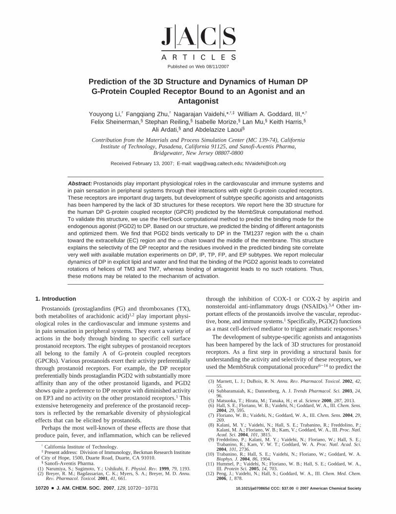
2.5. HierDock Method: Scan the Entire DP Receptor for BindingSites. We used HierDock approach to predict the binding mode ofligand to DP receptor. The first step is to scan all void regions (shownas magenta dots in Figure 8) in the entire DP receptor structure to locateputative binding regions for PGD2. The void region in the entirereceptor structure was partitioned into 27 regions, and the HierDockmethod was used to dock the cyclopentane ring of PGD2 in each box.Here, we examined the best binding sites that have at least 80% buriedsurface area. This leads to the TM1237 region shown in Figure 8.Subsequently, we docked the entire PGD2 molecule in this putativebinding region using the HierDock2.0 method. The large cavity in theTM1237 region arises from the presence of prolines on TM2 and TM7.The proline on TM7 is 100% conserved among the rhodopsinsuperfamily. The proline on TM2 is highly conserved among prostanoidfamily except TP and FP, which have a glycine residue before thatposition. Rhodopsin has two glycines around that position, and thebending angle of TM2 helix is in a shape that makes TM2 helix packtightly with TM1,3,7 helices.
3. Results and Discussion
3.1. Description of the Predicted Human DP Structure.The DP receptor sequence lacks some of the well-conservedmotifs present in class A GPCRs. For example, the DRY motifon TM3 is ECW, the well-conserved Trp on TM4 becomes Leu,the WXP motif on TM6 becomes SXP, and the NPXXY motifon TM7 is a DPWXF in the DP receptor. Thus, we can expectthat the DP receptor might have a different set of stabilizinginterhelical hydrogen bonds from rhodopsin. The predicted 3Dstructure of human apo-DP receptor is shown in Figure 2, andthe residues forming interhelical H-bonds are highlighted.
We find an interhelical hydrogen bond between N34(1) andD72(2). N34(1) and D72(2) are conserved in the rhodopsinfamily A including DP, but the conserved Asn of the NPXXYmotif in TM7 is a DPWXF motif in the DP receptor. S316(7),which is not a conserved residue, makes a hydrogen bond withthe N34(1) and D72(2). D319(7) makes a hydrogen bond with
S119(3). D72(2) also forms a strong salt bridge with the K76-(2) on the same helix. K76(2) is a conservative replacement inother prostaglandin receptors except for thromboxane receptors.We also find a hydrogen bond between R310(7) and Y87(2),where R310(7) is conserved across all prostaglandin receptorswhile Y87 is present only in DP receptors.
3.2. Molecular Dynamics Study of the Predicted HumanDP Structure with Lipid and Water. After predicting humanapo-DP structure, we performed 1 ns of MD simulations onthe apo protein structure.
Figure 3 shows the 3D structure of human DP receptor after1 ns MD with lipid and water. We find that the protein remainsstable during the simulations. Thus, Figure 5 shows that thefive important interhelical hydrogen bonds (HB) are all main-tained during the 1 ns MD.
Of particular interest is that five water molecules diffuse intobinding pocket within the TM regions (Figure 6a), formingwater-mediated hydrogen bonds with the buried polar residues.For example, three water molecules cluster around D319(7) withone water molecule around D72(2) and one around S119(3).Thus, the interhelical hydrogen bond between N34(1) and D72-(2) becomes water mediated. Figure 6b shows the time evolutionof the distance of these water molecules to the polar residues.The rhodopsin X-ray structure also showed some water mol-
Figure 2. Predicted 3D structure of human DP receptor from MembStruk.(Residues forming interhelical H-bonds are highlighted here.)
Figure 3. 3D structure of human apo-DP receptor after 1 ns MD withlipid and water (residues forming interhelical H-bonds are highlighted andthey are stable during 1 ns MD).
Figure 4. The molecular dynamics simulation box of hDP with lipid andwater. Structure after 1 ns of simulation. The EC region is at the top.
A R T I C L E S Li et al.
10722 J. AM. CHEM. SOC. 9 VOL. 129, NO. 35, 2007
ecules sufficiently strongly bound to be observed,24 which wereobserved in the vicinity of highly conserved residues and havebeen suggested to regulate the activity of rhodopsin-like GPCRs.
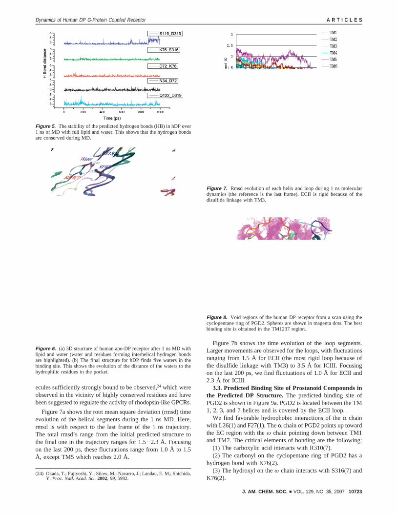
Figure 7a shows the root mean square deviation (rmsd) timeevolution of the helical segments during the 1 ns MD. Here,rmsd is with respect to the last frame of the 1 ns trajectory.The total rmsd’s range from the initial predicted structure tothe final one in the trajectory ranges for 1.5-2.3 Å. Focusingon the last 200 ps, these fluctuations range from 1.0 Å to 1.5Å, except TM5 which reaches 2.0 Å.
Figure 7b shows the time evolution of the loop segments.Larger movements are observed for the loops, with fluctuationsranging from 1.5 Å for ECII (the most rigid loop because ofthe disulfide linkage with TM3) to 3.5 Å for ICIII. Focusingon the last 200 ps, we find fluctuations of 1.0 Å for ECII and2.3 Å for ICIII.
3.3. Predicted Binding Site of Prostanoid Compounds inthe Predicted DP Structure. The predicted binding site ofPGD2 is shown in Figure 9a. PGD2 is located between the TM1, 2, 3, and 7 helices and is covered by the ECII loop.
We find favorable hydrophobic interactions of theR chainwith L26(1) and F27(1). TheR chain of PGD2 points up towardthe EC region with theω chain pointing down between TM1and TM7. The critical elements of bonding are the following:
(1) The carboxylic acid interacts with R310(7).(2) The carbonyl on the cyclopentane ring of PGD2 has a
hydrogen bond with K76(2).(3) The hydroxyl on theω chain interacts with S316(7) and
K76(2).(24) Okada, T.; Fujiyoshi, Y.; Silow, M.; Navarro, J.; Landau, E. M.; Shichida,
Y. Proc. Natl. Acad. Sci.2002, 99, 5982.
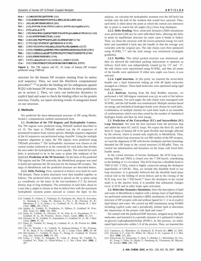
Figure 5. The stability of the predicted hydrogen bonds (HB) in hDP over1 ns of MD with full lipid and water. This shows that the hydrogen bondsare conserved during MD.
Figure 6. (a) 3D structure of human apo-DP receptor after 1 ns MD withlipid and water (water and residues forming interhelical hydrogen bondsare highlighted). (b) The final structure for hDP finds five waters in thebinding site. This shows the evolution of the distance of the waters to thehydrophilic residues in the pocket.
Figure 7. Rmsd evolution of each helix and loop during 1 ns moleculardynamics (the reference is the last frame). ECII is rigid because of thedisulfide linkage with TM3.
Figure 8. Void regions of the human DP receptor from a scan using thecyclopentane ring of PGD2. Spheres are shown in magenta dots. The bestbinding site is obtained in the TM1237 region.
Dynamics of Human DP G-Protein Coupled Receptor A R T I C L E S
J. AM. CHEM. SOC. 9 VOL. 129, NO. 35, 2007 10723
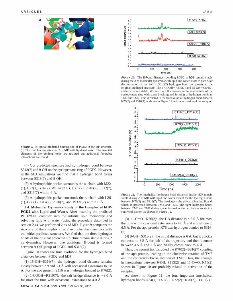
(4) Our predicted structure had no hydrogen bond betweenS313(7) and 9-OH on the cyclopentane ring of PGD2. However,in the MD simulations we find that a hydrogen bond formsbetween S313(7) and 9-OH.
(5) A hydrophobic pocket surrounds theR chain with M22-(1), G23(1), Y87(2), W182(ECII), L309(7), R310(7), L312(7),and S313(7) within 6 Å.
(6) A hydrophobic pocket surrounds theω chain with L26-(1), G30(1), I317(7), P320(7), and W321(7) within 6 Å.
3.4. Molecular Dynamics Study of the Complex of hDP-PGD2 with Lipid and Water. After inserting the predictedPGD2/hDP complex into the infinite lipid membrane andsolvating fully with water (using the procedure described insection 2.4), we performed 2 ns of MD. Figure 9 compares thestructure of the complex after 2 ns molecular dynamics withthe initial predicted structure. We find that the three hydrogenbonds of the original predicted structure remain stable during 2ns dynamics. However, one additional H-bond is formedbetween 9-OH group of PGD2 and S313(7).
Figure 10 shows the time evolution for the hydrogen bonddistances between PGD2 and hDP.
(1) 15-OH-S316(7): the hydrogen bond distance remainsmostly between 2.9 and 3.1 Å with occasional extensions to 3.8Å. For the apo protein, S316 was hydrogen bonded to K76(2).
(2) 1-COOH-R310(7): the salt bridge distance is∼3.0 Åfor most the time with occasional extensions to 4.0 Å.
(3) 11-CdO-K76(2): the HB distance is∼3.5 Å for mostthe time with occasional extensions to 4.0 Å and a brief one to6.5 Å. For the apo protein, K76 was hydrogen bonded to S316-(7)
(4) 9-OH-S313(2): the initial distance is 6 Å, but it quicklycontracts to 3.5 Å for half of the trajectory and then bouncesbetween 4.5 Å and 7 Å and finally comes back to 4 Å.
Thus, the agonist has disrupted the K76(2)-S316(7) couplingof the apo protein, leading to the clockwise rotation of TM3and the counterclockwise rotation of TM7. Thus, the changesin interactions between 9-OH, S313(2) and 11-CdO, K76(2)shown in Figure 10 are probably related to activation of thereceptor.
As shown in Figure 11, the four important interhelicalhydrogen bonds N34(1)-D72(2), D72(2)-K76(2), D319(7)-
Figure 9. (a) Initial predicted binding site of PGD2 in the DP structure.(b) The final binding site after 2 ns MD with lipid and water. The essentialelements of the binding mode are retained but additional favorableinteractions are found.
Figure 10. The H-bond distances bonding PGD2 to hDP remain stableduring the 2 ns molecular dynamics with lipid and water. Note in particularthe formation of the 9-OH-S313(7) hydrogen bond not present in theoriginal predicted structure. The 1-COOH-R310(7) and 15-OH-S316(7)anchors remain stable. We see more fluctuations in the interactions of thecyclopentane ring with some breaking and forming of hydrogen bonds toTM2 and TM7. This is related to the fluctuation of hydrogen bond betweenK76(2) and S316(7) as shown in Figure 11 and the activation of the receptor.
Figure 11. The interhelical hydrogen bond distances inside hDP remainstable during 2 ns MD with lipid and water except for the hydrogen bondbetween K76(2) and S316(7). The breakage is the effect of binding ligand,which is presented between TM2 and TM7. The tight hydrogen bondsbetween TM3 and TM7 during dynamics makes the two helices rotate in acogwheel pattern as shown in Figure 12.
A R T I C L E S Li et al.
10724 J. AM. CHEM. SOC. 9 VOL. 129, NO. 35, 2007
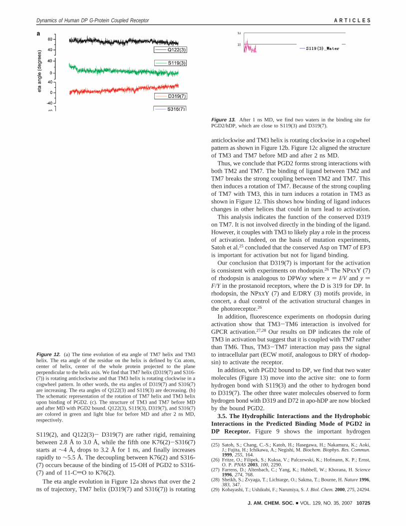
S119(2), and Q122(3)- D319(7) are rather rigid, remainingbetween 2.8 Å to 3.0 Å, while the fifth one K76(2)-S316(7)starts at∼4 Å, drops to 3.2 Å for 1 ns, and finally increasesrapidly to∼5.5 Å. The decoupling between K76(2) and S316-(7) occurs because of the binding of 15-OH of PGD2 to S316-(7) and of 11-CdO to K76(2).
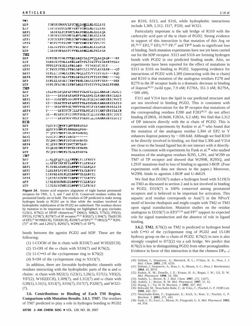
The eta angle evolution in Figure 12a shows that over the 2ns of trajectory, TM7 helix (D319(7) and S316(7)) is rotating
anticlockwise and TM3 helix is rotating clockwise in a cogwheelpattern as shown in Figure 12b. Figure 12c aligned the structureof TM3 and TM7 before MD and after 2 ns MD.
Thus, we conclude that PGD2 forms strong interactions withboth TM2 and TM7. The binding of ligand between TM2 andTM7 breaks the strong coupling between TM2 and TM7. Thisthen induces a rotation of TM7. Because of the strong couplingof TM7 with TM3, this in turn induces a rotation in TM3 asshown in Figure 12. This shows how binding of ligand induceschanges in other helices that could in turn lead to activation.
This analysis indicates the function of the conserved D319on TM7. It is not involved directly in the binding of the ligand.However, it couples with TM3 to likely play a role in the processof activation. Indeed, on the basis of mutation experiments,Satoh et al.25 concluded that the conserved Asp on TM7 of EP3is important for activation but not for ligand binding.
Our conclusion that D319(7) is important for the activationis consistent with experiments on rhodopsin.26 The NPxxY (7)of rhodopsin is analogous to DPWxy wherex ) I/V andy )F/Y in the prostanoid receptors, where the D is 319 for DP. Inrhodopsin, the NPxxY (7) and E/DRY (3) motifs provide, inconcert, a dual control of the activation structural changes inthe photoreceptor.26
In addition, fluorescence experiments on rhodopsin duringactivation show that TM3-TM6 interaction is involved forGPCR activation.27,28 Our results on DP indicates the role ofTM3 in activation but suggest that it is coupled with TM7 ratherthan TM6. Thus, TM3-TM7 interaction may pass the signalto intracellular part (ECW motif, analogous to DRY of rhodop-sin) to activate the receptor.
In addition, with PGD2 bound to DP, we find that two watermolecules (Figure 13) move into the active site: one to formhydrogen bond with S119(3) and the other to hydrogen bondto D319(7). The other three water molecules observed to formhydrogen bond with D319 and D72 in apo-hDP are now blockedby the bound PGD2.
3.5. The Hydrophilic Interactions and the HydrophobicInteractions in the Predicted Binding Mode of PGD2 inDP Receptor. Figure 9 shows the important hydrogen
(25) Satoh, S.; Chang, C.-S.; Katoh, H.; Hasegawa, H.; Nakamura, K.; Aoki,J.; Fujita, H.; Ichikawa, A.; Negishi, M.Biochem. Biophys. Res. Commun.1999, 255, 164.
(26) Fritze, O.; Filipek, S.; Kuksa, V.; Palczewski, K.; Hofmann, K. P.; Ernst,O. P.PNAS2003, 100, 2290.
(27) Farrens, D.; Altenbach, C.; Yang, K.; Hubbell, W.; Khorana, H.Science1996, 274, 768.
(28) Sheikh, S.; Zvyaga, T.; Lichtarge, O.; Sakma, T.; Bourne, H.Nature1996,383, 347.
(29) Kobayashi, T.; Ushikubi, F.; Narumiya, S.J. Biol. Chem.2000, 275, 24294.
Figure 12. (a) The time evolution of eta angle of TM7 helix and TM3helix. The eta angle of the residue on the helix is defined by CR atom,center of helix, center of the whole protein projected to the planeperpendicular to the helix axis. We find that TM7 helix (D319(7) and S316-(7)) is rotating anticlockwise and that TM3 helix is rotating clockwise in acogwheel pattern. In other words, the eta angles of D319(7) and S316(7)are increasing. The eta angles of Q122(3) and S119(3) are decreasing. (b)The schematic representation of the rotation of TM7 helix and TM3 helixupon binding of PGD2. (c). The structure of TM3 and TM7 before MDand after MD with PGD2 bound. Q122(3), S119(3), D319(7), and S316(7)are colored in green and light blue for before MD and after 2 ns MD,respectively.
Figure 13. After 1 ns MD, we find two waters in the binding site forPGD2/hDP, which are close to S119(3) and D319(7).
Dynamics of Human DP G-Protein Coupled Receptor A R T I C L E S
J. AM. CHEM. SOC. 9 VOL. 129, NO. 35, 2007 10725
bonds between the agonist PGD2 and hDP. These are thefollowing:
(1) 1-COOH of theR chain with R310(7) and W182(ECII).(2) 15-OH of theω chain with S316(7) and K76(2).(3) 11-CdO of the cyclopentane ring to K76(2)(4) 9-OH of the cyclopentane ring to S313(7).In addition, there are favorable hydrophobic channels with
residues interacting with the hydrophobic parts of theR andωchains: R chain with M22(1), G23(1), L26(1), F27(1), V83(2),Y87(2), W182(ECII), L309(7), and L312(7) andω chain withG30(1), L31(1), S313(7), S316(7), I317(7), P320(7), and W321-(7).
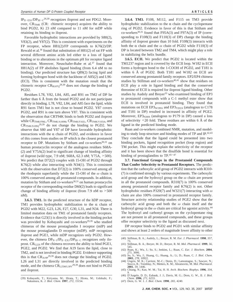
3.6. Contributions to Binding of Each TM Region.Comparison with Mutation Results. 3.6.1. TM7.The residuesof TM7 predicted to play a role in hydrogen bonding to PGD2
are R310, S313, and S316, while hydrophobic interactionsinclude L309, L312, I317, P320, and W321.
Particularly important is the salt bridge of R310 with thecarboxylic acid part of theR chain of PGD2. Strong evidencein support of this interaction is that mutation of this Arg onIP,30,31EP2,32 EP3,34,35FP,37 and TP38 leads to significant lossof binding. Such mutation experiments have not yet been carriedout for the hDP receptor. S313 and S316 are forming hydrogenbonds with PGD2 in our predicted binding mode. Also, noexperiments have been reported for the effect of mutations inS313 or S316 on binding to PGD2. Supporting evidence forinteractions of PGD2 with L309 (interacting with theR chain)and R310 is that mutation of the analogous residues F278 andR279 in the IP receptor leads to a dramatic decrease in bindingof iloprost30,31 (wild type, 7.9 nM; F278A, 351.3 nM; R279A,>500 nM).
F311 and V314 face the lipid in our predicted structure andare not involved in binding PGD2. This is consistent withexperimental observations for the IP receptor that mutations ofthe corresponding residues F280 and F28330,31 do not affectbinding (F280A, 10.8nM; F283A, 6.2 nM). We find that L312of DP interacts directly with theR chain of PGD2. This isconsistent with experiments by Kedzie et al.32 who found thatthe mutation of the analogous residue L304 of EP2 to Yenhances iloprost potency by∼100-fold. Although we find R310to be directly involved in binding, we find that L306 and V314are close to the bound ligand but do not interact with it directly.This is consistent with experiments by Funk et al.38 who studiedmutation of the analogous residues R295, L291, and W299 onTM7 of TP receptor and showed that W299R, R295Q, andL291F mutations lead to loss of binding to agonist I-BOP. (Fourexperiments with data not shown in the paper.) Moreover,W299L binds to agonists I-BOP and U-46619.
We find that D319(7) makes a hydrogen bond with S119(3)on TM3 as discussed in section 2 and is not involved in bindingto PGD2. D319(7) is 100% conserved among prostanoidreceptors and is believed to be important for activation. Thisaspartic acid residue corresponds to Asn(7) in the NPxxYmotif of bovine rhodopsin and might couple with TM2 or TM3upon signal transduction. Mutation studies on the residueanalogous to D319(7) in EP325,34and FP37 support its expectedrole for signal transduction and the absence of role in ligandbinding.
3.6.2. TM2. K76(2) on TM2 is predicted to hydrogen bondwith CdO of the cyclopentane ring of PGD2 and 15-OHhydroxy group on theω chain of PGD2. K76(2) in turn is alsostrongly coupled to D72(2) via a salt bridge. We predict thatK76(2) is key in distinguishing PGD2 from other prostaglandins.Evidence in favor of this interaction is that the chimera DPN-I/
(30) Stitham, J.; Stojanovic, A.; Merenick, B. L.; O’Hara, K. A.; Hwa, J.J.Biol. Chem.2003, 278, 4250.
(31) Stitham, J.; Stojanovic, A.; Ross, L. A.; Blount, A. C.; Hwa, J.Biochemistry2004, 43, 8974.
(32) Kedzie, K. M.; Donello, J. E.; Krauss, H. A.; Regan, J. W.; Gil, D. W.Mol. Pharmacol.1998, 54, 584.
(33) Audoly, L.; Breyer, R.J. Biol. Chem.1997, 272, 13475.(34) Audoly, L.; Breyer, R. M.Mol. Pharmacol.1997, 51, 61.(35) Huang, C.; Tai, H. H.Biochem. J.1995, 307, 493.(36) Rehwald, M.; Neuschafer-Rube, F.; de Vries, C.; Puschel, G. P.FEBS Lett.
1999, 443, 357.(37) Neuschafer-Rube, F.; Engemaier, E.; Koch, S.; Roer, U.; Puschel, G. P.
Biochem. J.2003, 371, 443.(38) Funk, C. D.; Furci, L.; Moran, N.; Fitzgerald, G. A.Mol. Pharmacol.1993,
44, 934.
Figure 14. Amino acid sequence alignment of eight human prostanoidreceptors for TM1, 2, 3, and 7 and ECII. Conserved residues within theprostanoid family are in bold. The residues of DP predicted to make stronghydrogen bonds to PGD2 are in blue while the residues involved inhydrophobic stabilization of the PGD2 are underlined. The residues shownby mutation to be important to binding are highlighted in gray (includesG23(1), K76(2) of DP/IP chimericas;29 D60(2), S68(2), Y75(2), F95(3),F97(3), F278(7), R279(7) of IP receptor;30,31R302(7), L304(7), Thr(ECII)of EP2;32 W199(ECII), T202(ECII), R329(7) of EP3;33-35 H81(2);36,37R291-(7)37 of FP; and L291(7), R295(7), W299(7) of TP38).
A R T I C L E S Li et al.
10726 J. AM. CHEM. SOC. 9 VOL. 129, NO. 35, 2007
IPII-EX1/DPIII -C29,39 recognizes iloprost and not PDG2. More-
over, CRT94K (CR: chimeric receptor) acquires the ability tobind PGD2, Ki 23 nM compared to 11 nM for mDP whileretaining its binding to iloprost.
Favorable hydrophobic interactions are provided by S80(2),V83(2), and Y87(2). This is consistent with experiments on theFP receptor, where H81(2)/FP corresponds to K76(2)/DP.Rewald et al.36 found that substitution of H81(2) of rat FP withseveral different amino acids led either to a loss of ligandbinding or to alterations in the optimum pH for receptor ligandinteraction. Moreover, Neuschafer-Rube et al.37 found thatH81A(2) of FP abolishes ligand binding (from 6.4 nM to nobinding). Our predicted structure has Q89(2) facing lipid andforming hydrogen bond with the backbone of A85(2) and L96-(ECI). This is consistent with the mutation result that thechimeric receptor CRR107Q
29,39 does not change the binding ofPGD2.
Residues L78, V82, L84, A85, and R91 on TM2 of DP liefarther than 6 Å from the bound PGD2 and do not participatedirectly in binding. L78, V82, L84, and A85 face the lipid, whileR91 faces TM1 but is not close to bound PGD2. Y87 coversPGD2, and R91 is one turn above Y87. This is consistent withthe observation that CRT94K binds to both PGD2 and iloprostwhile CRT94K/F96L, CRT94K/A100M, CRT94K/F102L, CRT94K/V103A, andCRT94K/S109Q
29,39 do not change the binding to PGD2. Weobserve that S80 and Y87 of DP have favorable hydrophobicinteractions with theR chain of PGD2, and evidence in favorof this comes from studies on IP which is the closest prostanoidreceptor to DP. Mutations by Stitham and co-workers30,31 onhuman prostacyclin receptor of the analogous residues S68A-(2) and Y75A(2) lead to significant change of binding affinityof iloprost (wild type, 7.9 nM; S68A, 62.3 nM; Y75A,>500).We predict that D72(2) couples with 15-OH of PGD2 throughK76(2) while also interacting with N34(1). This is consistentwith the observation that D72(2) is 100% conserved among allthe rhodopsin superfamily while the 15-OH of theω chain is100% conserved among all prostanoid compounds. In addition,mutation by Stitham and co-workers30,31on human prostacyclinreceptor of the corresponding residue D60(2) leads to significantchange of binding affinity of iloprost (from 7.9 nM to>500nM).
3.6.3. TM1. In the predicted structure of the hDP receptor,TM1 provides hydrophobic stabilization to theR chain ofPGD2 with M22, G23, L26, F27, G30, L31, and N34. There islimited mutation data on TM1 of prostanoid family receptors.Evidence that G23(1) is directly involved in the binding pocketwas provided by Kobayashi and co-workers29,39 who studiedchimeras of the mouse prostaglandin I receptor (mIP) andthe mouse prostaglandin D receptor (mDP). mIP recognizesiloprost and PGE1, while mDP recognizes only PGD2. How-ever, the chimera DPN-I/IPII-EX1/DPIII -C recognizes only ilo-prost. CRG22Sof the chimera recovers the ability to bind PGE1,PGE2, and PGD2. We find that A19 faces the lipid, close toTM2, and is not involved in binding PGD2. Evidence supportingthis is that CRA19P
29,39 does not change the binding of PGD2.L26 and L31 are directly involved in the predicted bindingmode, and the chimera CRL25M/L30V
29,39does not bind to PGD2and iloprost.
3.6.4. TM3. F108, M112, and F115 on TM3 providehydrophobic stabilization to theR chain and the cyclopentanering of PGD2. Evidence in favor of this is that Stitham andco-workers30,31 found that F95A(3) and F97A(3) of IP (corre-sponding to F108(3) and F110(3) of DP) change the bindingaffinity of iloprost greater than 10 fold. F108(3) interacts withboth theR chain and theω chain of PGD2 while F110(3) ofDP is located between TM2 and TM4, which might play a rolein stabilizing the helix bundle.
3.6.5. ECII. We predict that PGD2 is located within theTM1237 region and is covered by the ECII loop. W182 in ECIIforms a hydrogen bond to the 1-COOH of PGD2, and T181 iswithin 6 Å of PGD2. Both T181 and W182 on ECII areconserved among prostanoid family receptors. EP2/EP4 chimerastudies by Stillman and co-workers40,41 show that residues onECII play a role in ligand binding and that the conservedthreonine of ECII is required for iloprost ligand binding. Otherstudies by Audoly and Breyer33 who examined binding of EP3to prostanoid compounds with a C-1 methyl ester show thatECII is involved in prostanoid binding. They found thatmutations on ECII EP3W199A and EP3T202A (analogous to C178and T181 in DP) resulted in increased in affinity of PGE2.Moreover, EP3P200S(analogous to P179 in DP) caused a lossof selectivity <20 fold. These residues are within 6 Å of theligand in the predicted binding mode.
Ruan and co-workers combined NMR, mutation, and model-ing to study loop structure and binding modes of TP and IP.42,43
They conclude that the ligand could be presented into twobinding pockets, ligand recognition pocket (loop region) andTM pocket. This might explain the selectivity of the receptorand it has been shown that the disulfide bond is essential forbinding of prostaglandins to TP.45-47
3.7. Functional Groups in the Prostanoid CompoundsThat Confer Selectivity to Prostanoid Receptors.The predic-tion that the carboxylic acid group of PGD2 interacts with R310-(7) is confirmed strongly by various experiments. The carboxylicacid group and the hydroxyl group on theω chain are presentin all the prostanoid compounds. R310(7) is 100% conservedamong prostanoid receptor family and K76(2) is not. Otherhydrophobic residues P320(7) and W321(7) interacting withωchain are also 100% conserved in prostanoid receptor family.Structure activity relationship studies of PGE2 show that thecarboxylic acid group and both theω chain itself and thehydroxyl group in theω chain are critical for agonist activity.44
The hydroxyl and carbonyl groups on the cyclopentane ringare not present in all prostanoid compounds, and these groupsoffer receptor selectivity to the ligand as discussed next.
DP receptor binds to PGD2 and PGD1 with similar affinityand shows at least 2 orders of magnitude lower affinity to other
(39) Kobayashi, T.; Kiriyama, M.; Hirata, T.; Hirata, M.; Ushikubi, F.;Nakamura, K.J. Biol. Chem.1997, 272, 15154.
(40) Stillman, B. A.; Audoly, L.; Breyer, R. M.Eur. J. Pharmacol.1998, 357,73.
(41) Stillman, B. A.; Breyer, M. D.; Breyer, R. M.Mol. Pharmacol.1999, 56,545.
(42) Ruan, K.; Wu, J.; So, S.; Jenkins, L.; Ruan, C.Eur. J. Biochem.2004,271, 3006.
(43) So, S.; Wu, J.; Huang, G.; Huang, A.; Li, D.; Ruan, C.J. Biol. Chem.2003, 278, 10922.
(44) Ungrin, M. K.; Carriere, M.-C.; Denis, D.; Lamontagne, S.; Sawyer, N.;Stocco, R.; Tremblay, N.; Metters, K. M.; Abramovitz, M.Mol. Pharmacol.2001, 59, 1446.
(45) Chiang, N.; Kan, W. M.; Tai, H. H.Arch. Biochem. Biophys.1996, 334,9.
(46) D’Angelo, D. D.; Eubank, J. J.; Davis, M. G.; Dorn, G. W., II.J. Biol.Chem.1996, 271, 6233.
(47) Dorn, G. W. I.J. Biol. Chem.1990, 265, 4240.
Dynamics of Human DP G-Protein Coupled Receptor A R T I C L E S
J. AM. CHEM. SOC. 9 VOL. 129, NO. 35, 2007 10727
prostanoid compounds. However, the IP receptor binds to PGE1and PGI analogues (iloprost), but it does not bind PGE2.Assuming that these other prostanoid compounds bind to thehDP receptor in similar binding mode as PGD2, we can explainhow the DP receptor prefers PGD2 to other prostanoidcompounds like PGF2R, PGE2, and iloprost.
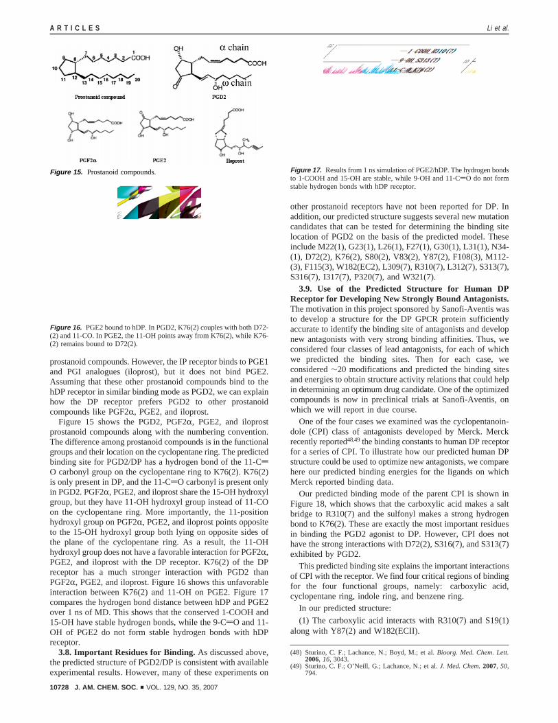
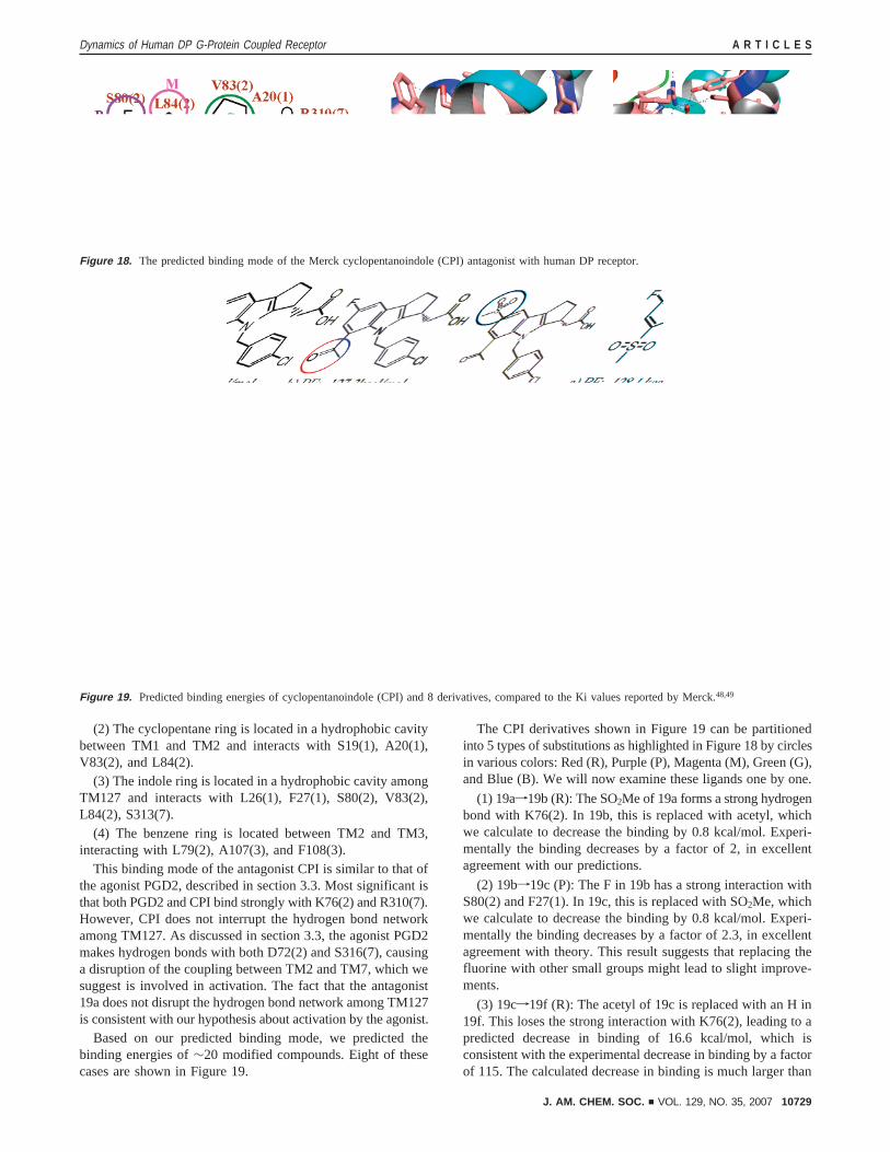
Figure 15 shows the PGD2, PGF2R, PGE2, and iloprostprostanoid compounds along with the numbering convention.The difference among prostanoid compounds is in the functionalgroups and their location on the cyclopentane ring. The predictedbinding site for PGD2/DP has a hydrogen bond of the 11-CdO carbonyl group on the cyclopentane ring to K76(2). K76(2)is only present in DP, and the 11-CdO carbonyl is present onlyin PGD2. PGF2R, PGE2, and iloprost share the 15-OH hydroxylgroup, but they have 11-OH hydroxyl group instead of 11-COon the cyclopentane ring. More importantly, the 11-positionhydroxyl group on PGF2R, PGE2, and iloprost points oppositeto the 15-OH hydroxyl group both lying on opposite sides ofthe plane of the cyclopentane ring. As a result, the 11-OHhydroxyl group does not have a favorable interaction for PGF2R,PGE2, and iloprost with the DP receptor. K76(2) of the DPreceptor has a much stronger interaction with PGD2 thanPGF2R, PGE2, and iloprost. Figure 16 shows this unfavorableinteraction between K76(2) and 11-OH on PGE2. Figure 17compares the hydrogen bond distance between hDP and PGE2over 1 ns of MD. This shows that the conserved 1-COOH and15-OH have stable hydrogen bonds, while the 9-CdO and 11-OH of PGE2 do not form stable hydrogen bonds with hDPreceptor.
3.8. Important Residues for Binding.As discussed above,the predicted structure of PGD2/DP is consistent with availableexperimental results. However, many of these experiments on
other prostanoid receptors have not been reported for DP. Inaddition, our predicted structure suggests several new mutationcandidates that can be tested for determining the binding sitelocation of PGD2 on the basis of the predicted model. Theseinclude M22(1), G23(1), L26(1), F27(1), G30(1), L31(1), N34-(1), D72(2), K76(2), S80(2), V83(2), Y87(2), F108(3), M112-(3), F115(3), W182(EC2), L309(7), R310(7), L312(7), S313(7),S316(7), I317(7), P320(7), and W321(7).
3.9. Use of the Predicted Structure for Human DPReceptor for Developing New Strongly Bound Antagonists.The motivation in this project sponsored by Sanofi-Aventis wasto develop a structure for the DP GPCR protein sufficientlyaccurate to identify the binding site of antagonists and developnew antagonists with very strong binding affinities. Thus, weconsidered four classes of lead antagonists, for each of whichwe predicted the binding sites. Then for each case, weconsidered∼20 modifications and predicted the binding sitesand energies to obtain structure activity relations that could helpin determining an optimum drug candidate. One of the optimizedcompounds is now in preclinical trials at Sanofi-Aventis, onwhich we will report in due course.
One of the four cases we examined was the cyclopentanoin-dole (CPI) class of antagonists developed by Merck. Merckrecently reported48,49the binding constants to human DP receptorfor a series of CPI. To illustrate how our predicted human DPstructure could be used to optimize new antagonists, we comparehere our predicted binding energies for the ligands on whichMerck reported binding data.
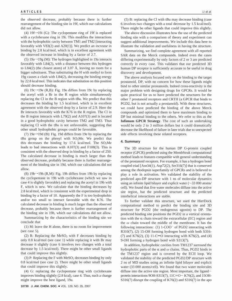
Our predicted binding mode of the parent CPI is shown inFigure 18, which shows that the carboxylic acid makes a saltbridge to R310(7) and the sulfonyl makes a strong hydrogenbond to K76(2). These are exactly the most important residuesin binding the PGD2 agonist to DP. However, CPI does nothave the strong interactions with D72(2), S316(7), and S313(7)exhibited by PGD2.
This predicted binding site explains the important interactionsof CPI with the receptor. We find four critical regions of bindingfor the four functional groups, namely: carboxylic acid,cyclopentane ring, indole ring, and benzene ring.
In our predicted structure:
(1) The carboxylic acid interacts with R310(7) and S19(1)along with Y87(2) and W182(ECII).
(48) Sturino, C. F.; Lachance, N.; Boyd, M.; et al.Bioorg. Med. Chem. Lett.2006, 16, 3043.
(49) Sturino, C. F.; O’Neill, G.; Lachance, N.; et al.J. Med. Chem.2007, 50,794.
Figure 15. Prostanoid compounds.
Figure 16. PGE2 bound to hDP. In PGD2, K76(2) couples with both D72-(2) and 11-CO. In PGE2, the 11-OH points away from K76(2), while K76-(2) remains bound to D72(2).
Figure 17. Results from 1 ns simulation of PGE2/hDP. The hydrogen bondsto 1-COOH and 15-OH are stable, while 9-OH and 11-CdO do not formstable hydrogen bonds with hDP receptor.
A R T I C L E S Li et al.
10728 J. AM. CHEM. SOC. 9 VOL. 129, NO. 35, 2007
(2) The cyclopentane ring is located in a hydrophobic cavitybetween TM1 and TM2 and interacts with S19(1), A20(1),V83(2), and L84(2).
(3) The indole ring is located in a hydrophobic cavity amongTM127 and interacts with L26(1), F27(1), S80(2), V83(2),L84(2), S313(7).
(4) The benzene ring is located between TM2 and TM3,interacting with L79(2), A107(3), and F108(3).
This binding mode of the antagonist CPI is similar to that ofthe agonist PGD2, described in section 3.3. Most significant isthat both PGD2 and CPI bind strongly with K76(2) and R310(7).However, CPI does not interrupt the hydrogen bond networkamong TM127. As discussed in section 3.3, the agonist PGD2makes hydrogen bonds with both D72(2) and S316(7), causinga disruption of the coupling between TM2 and TM7, which wesuggest is involved in activation. The fact that the antagonist19a does not disrupt the hydrogen bond network among TM127is consistent with our hypothesis about activation by the agonist.
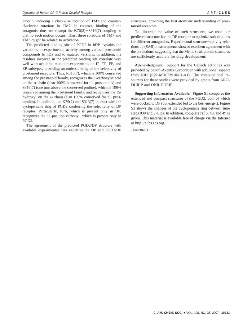
Based on our predicted binding mode, we predicted thebinding energies of∼20 modified compounds. Eight of thesecases are shown in Figure 19.
The CPI derivatives shown in Figure 19 can be partitionedinto 5 types of substitutions as highlighted in Figure 18 by circlesin various colors: Red (R), Purple (P), Magenta (M), Green (G),and Blue (B). We will now examine these ligands one by one.
(1) 19af19b (R): The SO2Me of 19a forms a strong hydrogenbond with K76(2). In 19b, this is replaced with acetyl, whichwe calculate to decrease the binding by 0.8 kcal/mol. Experi-mentally the binding decreases by a factor of 2, in excellentagreement with our predictions.
(2) 19bf19c (P): The F in 19b has a strong interaction withS80(2) and F27(1). In 19c, this is replaced with SO2Me, whichwe calculate to decrease the binding by 0.8 kcal/mol. Experi-mentally the binding decreases by a factor of 2.3, in excellentagreement with theory. This result suggests that replacing thefluorine with other small groups might lead to slight improve-ments.
(3) 19cf19f (R): The acetyl of 19c is replaced with an H in19f. This loses the strong interaction with K76(2), leading to apredicted decrease in binding of 16.6 kcal/mol, which isconsistent with the experimental decrease in binding by a factorof 115. The calculated decrease in binding is much larger than
Figure 18. The predicted binding mode of the Merck cyclopentanoindole (CPI) antagonist with human DP receptor.
Figure 19. Predicted binding energies of cyclopentanoindole (CPI) and 8 derivatives, compared to the Ki values reported by Merck.48,49
Dynamics of Human DP G-Protein Coupled Receptor A R T I C L E S
J. AM. CHEM. SOC. 9 VOL. 129, NO. 35, 2007 10729
the observed decrease, probably because there is furtherrearrangement of the binding site in 19f, which our calculationsdid not allow.
(4) 19ff19i (G): The cyclopentane ring of 19f is replacedwith a cyclohexane ring in 19i. This modifies the interactionswith the hydrophobic cavity between TM1 and TM2 [interactingfavorably with V83(2) and A20(1)]. We predict anincreaseinbinding by 2.8 kcal/mol, which is in excellent agreement withthe observed increase in binding by a factor of 2.7.
(5) 19af19g (M): The hydrogen highlighted in 19a interactsfavorably with L84(2), with a distance between this hydrogento L84(2) (the closest atom) of 3.07 Å, leaving no room for abigger substituent. Thus substituting the H with methyl to form19g causes a clash with L84(2), decreasing the binding energyby 22.8 kcal/mol. This indicates that substitution on this positionshould decrease binding.
(6) 19cf19e (R,B): Fig. 19e differs from 19c by replacingthe acetyl with a Br in the R region while simultaneouslyreplacing the Cl in the B region with tBu. We predict that thisdecreases the binding by 1.5 kcal/mol, which is in excellentagreement with the observed drop by a factor of 2.9. Here theBr interacts favorably with the K76 in the R region. The Cl inthe B region interacts with L79(2) and A107(3) and is locatedin a good hydrophobic cavity between TM2 and TM3. Thusreplacing Cl with the tBu is not unfavorable, suggesting thatother small hydrophobic groups could be favorable.
(7) 19ef19d (B): Fig. 19d differs from 19e by replacing thetBu group on the phenyl with SO2Me. We predict thatthis decreases the binding by 17.6 kcal/mol. The SO2Meleads to bad interactions with A107(3) and F108(3). This isconsistent with the observed drop in binding by a factor of 250.The calculated decrease in binding is much larger than theobserved decrease, probably because there is further rearrange-ment of the binding site in 19d, which our calculations did notallow.
(8) 19bf19h (R,M): Fig. 19h differs from 19b by replacingthe cyclopentane in 19b with cyclohexane (which we saw incase 4 is slightly favorable) and replacing the acetyl in 19b withF, which is new. We calculate that the binding decreases by2.8 kcal/mol, which is consistent with the experimental drop inbinding by a factor of 10. Apparently the F is too hydrophobicand/or too small to interact favorable with the K76. Thecalculated decrease in binding is much larger than the observeddecrease, probably because there is further rearrangement ofthe binding site in 19h, which our calculations did not allow.
Summarizing by the characteristics of the binding site weconclude that
(1) M: leave the H alone, there is no room for improvement(see case 5).
(2) R: Replacing the MeSO2 with F decreases binding byonly 0.8 kcal/mol (see case 1) while replacing it with Br maydecrease it slightly (case 6 involves two changes with a totaldecrease by 1.5 kcal/mol). There might be other small ligandsthat could improve this slightly.
(3) P: Replacing the F with MeSO2 decreases binding by only0.8 kcal/mol (see case 2). There might be other small ligandsthat could improve this slightly.
(4) G: replacing the cyclopentane ring with cyclohexaneimproves binding slightly (2.8 kcal), case 4. Thus, such a changemight improve the best ligand, 19a.
(5) B: replacing the Cl with tBu may decrease binding (case6 involves two changes with a total decrease by 1.5 kcal/mol).There might be other ligands that could improve this slightly.
The above discussion illustrates how the use of the predictedbinding site with a comparison of theory and experiment cansuggest additional improvements. We include this data here toillustrate the validation and usefulness in having the structure.
Summarizing, we find complete agreement with all reportedSAR data on the Merck compounds. Indeed even the casesdiffering experimentally by only factors of 2 or 3 are predictedcorrectly in every case. This validates that our predicted 3Dhuman DP receptor is sufficiently accurate to be useful in drugdiscovery and development.
The above analysis focused only on the binding to the targetprostanoid, DP, with no concern for how these ligands mightbind to other similar prostanoids. Indeed cross-reactivity is themajor problem with designing drugs for GPCRs. It would bequite practical for us to have predicted the structures for theother 7 prostanoid receptors and CRTH (a GPCR that binds toPGD2, but is not actually a prostanoid). With these structures,we could have predicted the binding of the above Merckcompounds and optimized them to have maximum binding toDP but minimal binding to the others. We refer to this as theInfimum GPCR Strategy. The cost of such an undertakingwould be only 2 to 3 million dollars but it could dramaticallydecrease the likelihood of failure in later trials due to unexpectedside effects involving these related receptors.
4. Summary
The 3D structure for the human DP G-protein coupledreceptor (GPCR) predicted using the MembStruk computationalmethod leads to features compatible with general understandingof the prostanoid receptors. For example, it has a hydrogen bondcoupled triad [Asn34(1), Asp72(2), Ser316(7)] that is conservedamong the rhodopsin superfamily of GPCRs and is believed toplay a role in activation. We validated the stability of thepredicted apo-DP structure with 1 ns of molecular dynamicsusing an infinite lipid bilayer and explicit water (∼33 000 atoms/cell). We found that five water molecules diffuse into the activesite region, but the predicted structure and the predictedinterhelical interactions are stable.
To further validate this structure, we used the HierDockcomputational method to predict the binding site and 3Dstructure for PGD2 (the endogenous agonist) to DP. Thepredicted binding site positions the PGD2 in a vertical orienta-tion with theR chain toward the extracellular (EC) region andthe ω chain toward the middle of the membrane. It has thefollowing interactions: (1) 1-COO- of PGD2 interacting withR310(7), (2) 15-OH forming hydrogen bond with both S316-(7) and K76(2), (3) 11-CdO interacting with K76(2), and (4)9-OH forming a hydrogen bond with S313(7).
In addition, hydrophobic cavities from TM1237 surround thehydrophobic parts of theR andω chains. Thus, PGD2 binds inthe TM1237 region and is covered by the ECII loop. Wevalidated the stability of the predicted PGD2/DP structure with2 ns of MD studies using an infinite lipid bilayer and explicitwater (33 000 atoms/cell). We found that two water moleculesdiffuse into the active site region. Most important, the ligand-protein interactions 9OH-S313(7), 11C)O- K76(2), and 15OH-S316(7) disrupt the coupling of K76(2) and S316(7) in the apo
A R T I C L E S Li et al.
10730 J. AM. CHEM. SOC. 9 VOL. 129, NO. 35, 2007
protein, inducing a clockwise rotation of TM3 and counter-clockwise rotations in TM7. In contrast, binding of theantagonist does not disrupt the K76(2)-S316(7) coupling sothat no such motion occurs. Thus, these rotations of TM7 andTM3 might be related to activation.
The predicted binding site of PGD2 in hDP explains thevariations in experimental activity among various prostanoidcompounds to hDP and to mutated versions. In addition, theresidues involved in the predicted binding site correlate verywell with available mutation experiments on IP, TP, FP, andEP subtypes, providing an understanding of the selectivity ofprostanoid receptors. Thus, R310(7), which is 100% conservedamong the prostanoid family, recognizes the 1-carboxylic acidon theR chain (also 100% conserved for all prostanoids) andS316(7) (one turn above the conserved proline), which is 100%conserved among the prostanoid family, and recognizes the 15-hydroxyl on theω chain (also 100% conserved for all pros-tanoids). In addition, the K76(2) and S313(7) interact with thecyclopentane ring of PGD2 conferring the selectivity of DPreceptor. Particularly, K76, which is present only in DP,recognizes the 11-position carbonyl, which is present only inPGD2.
The agreement of the predicted PGD2/DP structure withavailable experimental data validates the DP and PGD2/DP
structures, providing the first atomistic understanding of pros-tanoid receptors.
To illustrate the value of such structures, we used ourpredicted structure for the DP receptor to optimize substitutionsfor different antagonists. Experimental structure-activity rela-tionship (SAR) measurements showed excellent agreement withthe predictions, suggesting that the MembStruk protein structuresare sufficiently accurate for drug development.
Acknowledgment. Support for the Caltech activities wasprovided by Sanofi-Aventis Corporation with additional supportfrom NIH (R21-MH073910-01-A1). The computational re-sources for these studies were provided by grants from ARO-DURIP and ONR-DURIP.
Supporting Information Available: Figure S1 compares theextended and compact structures of the PGD2, both of whichwere docked to DP (but extended led to the best energy.). FigureS2 shows the changes of the cyclopentane ring between timesteps 830 and 870 ps. In addition, complete ref 5, 48, and 49 isgiven. This material is available free of charge via the Internetat http://pubs.acs.org.
JA070865D
Dynamics of Human DP G-Protein Coupled Receptor A R T I C L E S
J. AM. CHEM. SOC. 9 VOL. 129, NO. 35, 2007 10731