Embed Size (px)
Citation preview
ences 271 (2008) 68–74www.elsevier.com/locate/jns
Journal of the Neurological Sci
Prefrontal hypoperfusion and cognitive dysfunction correlates inspinocerebellar ataxia type 6
Y. Kawai a, M. Suenaga a, H. Watanabe a, M. Ito a, K. Kato b, T. Kato c,K. Ito c, F. Tanaka a, G. Sobue a,⁎
a Department of Neurology, Nagoya University Graduate School of Medicine, Nagoya 466-8550, Japanb Department of Radiology, Nagoya University Graduate School of Medicine, Nagoya 466-8550, Japan
c Department of Radiology, National Hospital for Geriatric Medicine, Aichi, Japan
Received 30 October 2007; received in revised form 18 March 2008; accepted 24 March 2008Available online 7 May 2008
Abstract
Objective: The aim of this study is to evaluate the correlation between brain perfusion and cognitive dysfunction in spinocerebellar ataxiatype 6 (SCA6) patients.Methods: Thirteen genetically confirmed SCA6 patients and 21 age- and education-matched control subjects were subjected to single photonemission computed tomography (SPECT) and neuropsychological tests. Brain perfusion was examined with SPECT analysis, while generalcognition, verbal and visual memory, attention, visuospatial ability, language, executive function, depression, and anxiety were examinedwith the neuropsychological tests.Results: SCA6 patients showed prefrontal hypoperfusion, and impairments of visual memory, verbal fluency, and executive functioncompared to control subjects. These neuropsychological impairments in SCA6 patients were significantly correlated with a decrease inprefrontal perfusion. This relation was not correlated to other factors, such as age, education and severity of cerebellar ataxia, which arepossible relevant factors associated with cognitive performance.Conclusions: SCA6 patients have mild cognitive impairment, and correlating prefrontal hypoperfusion. These results indicate cognitiveimpairment in SCA6 patients resulting from prefrontal hypoperfusion.Crown Copyright © 2008 Published by Elsevier B.V. All rights reserved.
Keywords: SPECT; Spinocerebellar ataxia type 6; Neuropsychological tests; Higher nervous activity; Cerebral blood flow; Cerebellum; Prefrontal cortex
1. Introduction
Spinocerebellar ataxia type 6 (SCA6) is an autosomaldominant cerebellar atrophy caused by an unstable CAGtrinucleotide repeat expansion present in a gene onchromosome 19p13 that encodes the voltage-dependentα1A-subunit of the calcium channel CACNA1A [1]. Thischannel is highly expressed in cerebellar Purkinje cells and is
⁎ Corresponding author. Department of Neurology, Nagoya UniversityGraduate School of Medicine, 65 Tsurumai-Cho, Showa-Ku, Nagoya 466-8550, Japan. Tel.: +81 52 744 2385; fax: +81 52 744 2384.
E-mail address: [email protected] (G. Sobue).
0022-510X/$ - see front matter. Crown Copyright © 2008 Published by Elsevierdoi:10.1016/j.jns.2008.03.018
of critical importance for the function and the developmentof cerebellar Purkinje cells.
Ataxia, gait disturbance, and dysarthria develop slowly inmost SCA6 patients. Neurologic signs often associated withother spinocerebellar degeneration (SCD) disorders areseldom associated with SCA6, and so SCA6 is characterizedas pure cerebellar ataxia [2].
Recently, there has been increasing evidence of thenonmotor role of the cerebellum [3,4], and clinical reports ofpatients with cerebellar lesions have implicated the cerebel-lum as having a role in cognitive functions [5,6]. Many ofthese clinical and imaging studies in cerebellar patientssuffer from methodological shortcomings concerning patient
B.V. All rights reserved.
69Y. Kawai et al. / Journal of the Neurological Sciences 271 (2008) 68–74
selection, such as the inclusion of patients with additionalextracerebellar damage. Although regional cerebral bloodflow (rCBF) and cognitive function in spinocerebellar ataxiapatients have been examined before genetic analysis becameavailable [7,8], these studies were controversial since theyincluded many types of spinocerebellar ataxia with addi-tional extracerebellar damage.
Neuropathologically, SCA6 is characterized by almostexclusive cerebellar involvement particularly selective lossof the cerebellar Purkinje cells [9]. Degeneration is mostlyrestricted to the cerebellum in SCA6 patients while corticalstructures and basal ganglia are spared. SCA6 thereforerepresents an excellent model for investigating the cerebellarcontribution to cognition. So far, however, only a few studieshave examined the rCBF of SCA6 patients [10]. Globas et al.failed to provide clear evidence for cognitive deficits inSCA6 patients [11]. However, we recently revealedsignificant impaired visual memory and verbal fluency ingenetically confirmed SCA6 patients [12], suggesting thatSCA6 patients have prefrontal dysfunction. Previous studieshave indicated hypoperfusion restricted to the cerebellum[10], however these results may be attributable to the smallsample size of the SCA6 patients used. In previous studies,VOI analysis methods was used [10], and they may be notentirely objective because we could not examine theperfusion of whole brain by VOI analysis methods.
The aims of this study were to clarify the rCBF and toevaluate the relationships between rCBF and cognitivedysfunction in genetically confirmed SCA6 patients.
2. Methods
2.1. Subjects (Table 1)
Thirteen genetically confirmed SCA6 patients fromthirteen families and 21 control subjects were enrolled inthis study (Table 1). These 13 SCA6 patients were from thesame pool of 18 patients who were examined in our previousreport [12]. All were native Japanese speakers. Severity ofataxia was rated on the International Cooperative AtaxiaRating Scale (ICARS) [13]. Genomic DNA was extracted
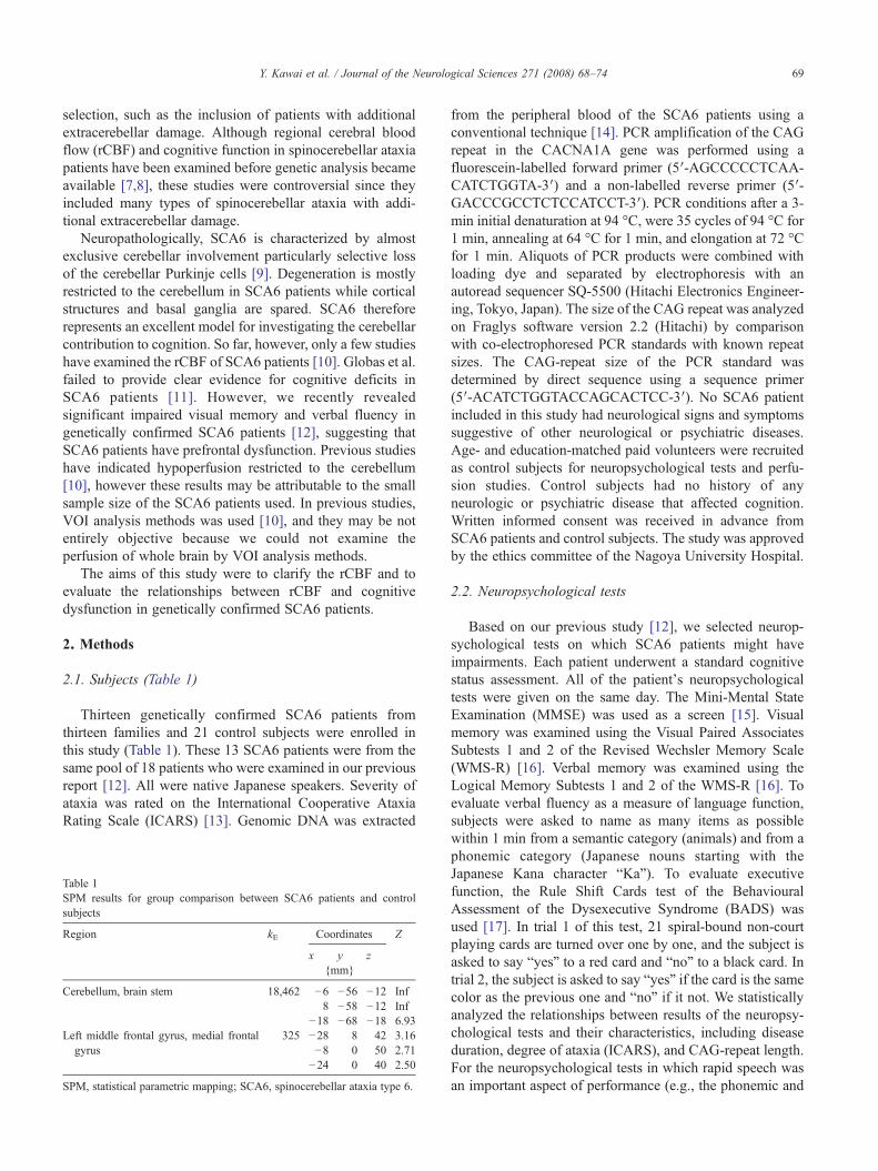
Table 1SPM results for group comparison between SCA6 patients and controlsubjects
Region kE Coordinates Z
x y z{mm}
Cerebellum, brain stem 18,462 −6 −56 −12 Inf8 −58 −12 Inf
−18 −68 −18 6.93Left middle frontal gyrus, medial frontal
gyrus325 −28 8 42 3.16
−8 0 50 2.71−24 0 40 2.50
SPM, statistical parametric mapping; SCA6, spinocerebellar ataxia type 6.
from the peripheral blood of the SCA6 patients using aconventional technique [14]. PCR amplification of the CAGrepeat in the CACNA1A gene was performed using afluorescein-labelled forward primer (5′-AGCCCCCTCAA-CATCTGGTA-3′) and a non-labelled reverse primer (5′-GACCCGCCTCTCCATCCT-3′). PCR conditions after a 3-min initial denaturation at 94 °C, were 35 cycles of 94 °C for1 min, annealing at 64 °C for 1 min, and elongation at 72 °Cfor 1 min. Aliquots of PCR products were combined withloading dye and separated by electrophoresis with anautoread sequencer SQ-5500 (Hitachi Electronics Engineer-ing, Tokyo, Japan). The size of the CAG repeat was analyzedon Fraglys software version 2.2 (Hitachi) by comparisonwith co-electrophoresed PCR standards with known repeatsizes. The CAG-repeat size of the PCR standard wasdetermined by direct sequence using a sequence primer(5′-ACATCTGGTACCAGCACTCC-3′). No SCA6 patientincluded in this study had neurological signs and symptomssuggestive of other neurological or psychiatric diseases.Age- and education-matched paid volunteers were recruitedas control subjects for neuropsychological tests and perfu-sion studies. Control subjects had no history of anyneurologic or psychiatric disease that affected cognition.Written informed consent was received in advance fromSCA6 patients and control subjects. The study was approvedby the ethics committee of the Nagoya University Hospital.
2.2. Neuropsychological tests
Based on our previous study [12], we selected neurop-sychological tests on which SCA6 patients might haveimpairments. Each patient underwent a standard cognitivestatus assessment. All of the patient's neuropsychologicaltests were given on the same day. The Mini-Mental StateExamination (MMSE) was used as a screen [15]. Visualmemory was examined using the Visual Paired AssociatesSubtests 1 and 2 of the Revised Wechsler Memory Scale(WMS-R) [16]. Verbal memory was examined using theLogical Memory Subtests 1 and 2 of the WMS-R [16]. Toevaluate verbal fluency as a measure of language function,subjects were asked to name as many items as possiblewithin 1 min from a semantic category (animals) and from aphonemic category (Japanese nouns starting with theJapanese Kana character “Ka”). To evaluate executivefunction, the Rule Shift Cards test of the BehaviouralAssessment of the Dysexecutive Syndrome (BADS) wasused [17]. In trial 1 of this test, 21 spiral-bound non-courtplaying cards are turned over one by one, and the subject isasked to say “yes” to a red card and “no” to a black card. Intrial 2, the subject is asked to say “yes” if the card is the samecolor as the previous one and “no” if it not. We statisticallyanalyzed the relationships between results of the neuropsy-chological tests and their characteristics, including diseaseduration, degree of ataxia (ICARS), and CAG-repeat length.For the neuropsychological tests in which rapid speech wasan important aspect of performance (e.g., the phonemic and
70 Y. Kawai et al. / Journal of the Neurological Sciences 271 (2008) 68–74
semantic fluency task), we analyzed the correlations to thedysarthria subscore of the ICARS.
2.3. Assessment of regional brain perfusion with SPECT
600 MBq of 99mTc-Ethylcysteine dimer (ECD) (Neuro-lite, Daiichi Radioisotope Laboratories, Ltd, Tokyo, Japan)was injected intravenously while the subjects were in asupine position with eyes closed in a quiet dimly lit room.SPECT scanning was carried out between 5 and 30 min afterinjection using a triple-head GCA 9300A gamma camera(Toshiba, Tokyo, Japan) equipped with low energy, superhigh resolution, fan-beam collimators. The data wereacquired in a 128×128 matrix through a 120° rotation atan angle interval of 4°. The projection data were prefilteredthrough a Butterworth filter, and reconstructed using filteredbackprojection with a Ramp filter. No attenuation correctionwas performed. The in-plane spatial resolution was 8 mm infull width at half maximum (FWHM). The final image sliceswere set up parallel to the orbitomeatal line and wereobtained at an interval of 6.9 mm through the entire brain.
2.4. Data analysis
For the statistical analysis of the neuropsychological tests,SPSS version 11.0 for Windows (SPSS Japan, Tokyo, Japan)was used. The Shapiro–Wilk test was used to assess thenormality of continuous variables. For comparison betweenthe SCA6 group and the control group, we performed anunpaired t test for normally distributed data or a non-parametric Mann–Whitney U test for non-normally dis-tributed data. Statistical significance was chosen at a P valueof 0.05, with the correction for multiple comparisons usingHolm–Sidak method. For correlation studies we used thePearson product moment correlation test for normallydistributed variables or the Spearman rank correlation testfor non-normally distributed variables.
The SPECT data were analyzed using Statistical Para-metric Mapping version 2 (SPM2; Wellcome Department ofCognitive Neurology, Institute of Neurology, London, UK)implemented in MATLAB version 6.5.1 (Math Works,Sherborn, MA, USA). In a pre-processing step, datasets were
Fig. 1. SPMmaps comparing brain perfusion between SCA6 patients and control suand transverse (right column) views of the standard brain.
spatially normalized to a standard stereotactic three dimen-sional space and smoothed with an isotropic Gaussian kernelof FWHM 12 mm. All of the images resulting from thenormalization procedure were visually acceptable. In thefollowing analyses, proportional scaling was applied toadjust the mean whole brain activity to 50 ml/100 g/min toavoid inter-individual variation in global cerebral blood flow.The grey matter threshold was 0.8. The normalized imagesof the SCA6 patients and control subjects were comparedwith voxel by voxel t statistics. Confounding effects mayarise from age or education differences, which couldinfluence regional cerebral blood flow change in SCA6.Therefore, age and education were inserted as covariates inthe statistical parametric mapping (SPM) analyses. Inaddition, we compared the images of the SCA6 patients todetect voxels in which the rCBF was significantly correlatedwith the scores of neuropsychological tests on which SCA6patients showed impairment. Each value of the neuropsy-chological tests was used as a covariate of interest, and thevalues of the global rCBF, age, education and ICARS, whichare the relevant factors associated with cognitive perfor-mance, were excluded as nuisance variables [18]. Regionswere reported as significant if they contained voxels with avalue of at least Pb0.01, uncorrected for multiple compar-isons, with cluster extent threshold (kE)=100.
3. Results
3.1. Neuropsychological features
We assessed the neuropsychological features of 13patients out of 18 patients who were examined in ourprevious study [12], since these patients could receive bothneuropsychological tests and SPECT. The test results weresimilar to our previous report [12]. SCA6 patients did notdiffer significantly from controls with regard to MMSE, butshowed significant cognitive impairment in several othertasks (Supplemental Table 1). Performance on the VisualPaired Associates Subtest 1 was significantly reduced inSCA6 patients compared with controls (P=0.003), whileperformance on the Visual Paired Associates Subtest 2 wasnot. Verbal fluency tasks in semantic and phonemic
bjects (uncorrected Pb0.01). Sagittal (left column), coronal (middle column)
71Y. Kawai et al. / Journal of the Neurological Sciences 271 (2008) 68–74
categories were remarkably impaired in SCA6 patients(P=0.006, P=0.006, respectively). The result of the RuleShift Cards test tasks showed a tendency to be impaired inSCA6 patients compared with controls, although notsignificant. These neuropsychological impairments inSCA6 patients did not show any significant correlation toCAG-repeat length and disease duration, nor to ICARSdysarthria subscores. No significant differences wereobserved between SCA6 patients and control subjects inverbal memory function as tested using Logical MemorySubtests 1 and 2 of the WMS-R.
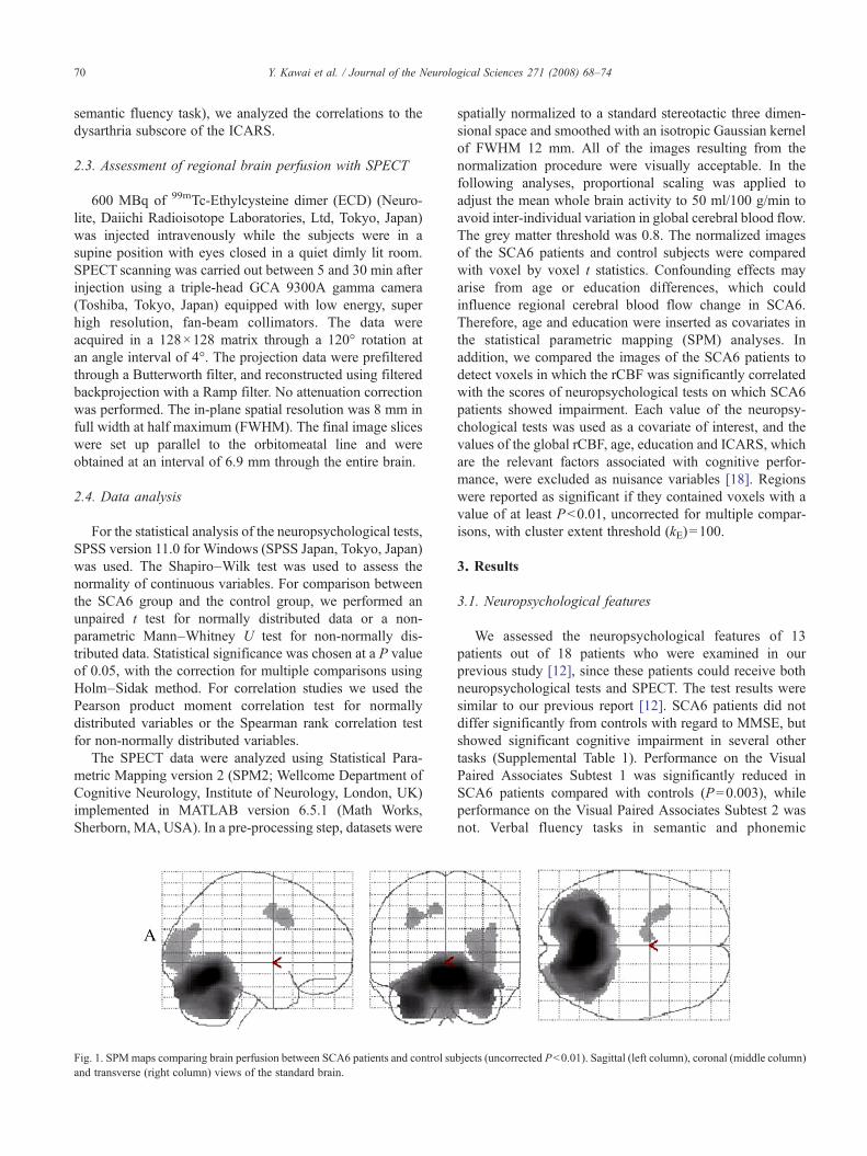
Fig. 2. SPM maps of the correlation between brain perfusion in SCA6 patients and(middle column) and transverse (right column) views of the standard brain. A: VisuaD: Rule Shift Cards test.
3.2. Correlation between rCBF and cognitive dysfunction inSCA6 patients
According to SPM analyses, SCA6 patients showedreduced brain perfusion in the left middle frontal and medialfrontal cortices, as well as in the cerebellum and brain stem,compared to control subjects (uncorrected Pb0.01; Fig. 1and Table 1).
We examined the correlation between rCBF and cognitivedysfunction after age, education, and severity of cerebellarataxia were excluded statistically (Fig. 2 and Table 2). There
cognitive performance (uncorrected Pb0.01). Sagittal (left column), coronall Paired Associates 1 test, B: phonemic fluency test, C: semantic fluency test,
Table 2SPM results of correlations between the results of the neuropsychologicaltests and brain perfusion in SCA6 patients
Region kE Coordinates Z
x y z{mm}
Perfusion positively correlated with the score of the Visual Paired Associates 1Right inferior frontal gyrus 163 56 2 30 2.94
60 10 28 2.79Right superior temporal gyrus 143 62 8 −2 2.78
64 −4 6 2.65
Perfusion positively correlated with the score of the phonemic fluencyRight superior temporal gyrus 270 60 2 4 3.91Cingulate gyrus 521 −4 14 38 3.11
4 12 40 2.91−10 24 34 2.91
Medial frontal gyrus 115 −2 8 68 2.96−2 18 68 2.698 14 66 2.46
Perfusion positively correlated with the score of the semantic fluencyRight middle frontal gyrus 110 32 16 48 3.80
30 24 40 2.34Right superior frontal gyrus 168 32 20 68 3.39
22 28 66 2.8222 26 74 2.44
Left superior frontal gyrus 113 −22 6 78 3.19−32 20 68 3.14−24 16 72 2.88
Left middle frontal gyrus 103 −30 40 26 3.05−36 22 36 2.49−34 30 34 2.45
Perfusion positively correlated with the score of the Rule Shift Cards testRight superior frontal gyrus 130 52 12 52 3.67
36 18 48 2.7338 18 56 2.70
Left superior temporal gyrus 126 −58 6 −20 2.98−60 10 −10 2.73
SPM, statistical parametric mapping; SCA6, spinocerebellar ataxia type 6.
72 Y. Kawai et al. / Journal of the Neurological Sciences 271 (2008) 68–74
was a positive correlation between the score of the VisualPaired Associates 1 test and the perfusion in the right inferiorfrontal and right superior temporal gyri in SCA6 patients. InSCA6 patients, there was a positive correlation between thephonemic fluency score and the perfusion in the medialfrontal and superior temporal gyri, between the semanticfluency score and the perfusion in the bilateral superior andmiddle frontal gyri, and between the Rule Shift Cards testscore and the perfusion in the right middle frontal andsuperior temporal gyri.
4. Discussion
In this study, we demonstrate that SCA6 patients have areduction of rCBF in not only the cerebellum but alsoprefrontal cortices. In our previous study, we documentedthat visual memory, verbal fluency, and executive functionwere significantly impaired in SCA6 patients [12]. In the
present study, we show that the rCBF in prefrontal cortices issignificantly correlated to these neuropsychological impair-ments in SCA6 patients.
rCBF had been examined in patients with various types ofspinocerebellar ataxia before we could confirm our diagnosisof spinocerebellar ataxias genetically [7,8]. However severaltypes of diseases might be included in these studies, and theresults were not consistent. Recently, one study examinedrCBFs in genetically confirmed SCA6 patients and describedonly cerebellar hypoperfusion [10]. These results agree withresults of previous neuropathologic studies showing thatlesions of SCA6 are restricted to the cerebellum, whereabundant expression of P/Q-type calcium channels has beenfound [19]. Magnetic resonance imaging analysis of SCA6patients demonstrated no abnormalities in the centralnervous system except for cerebellar atrophy [20]. However,one study showed that crossed cerebello-cerebral diaschisis(CCCD) was derived from the functional deactivation of thecerebello-ponto-thalamo-cerebral pathways [21]. Unilateralimpairment of the cerebellum would lead to reducedradioisotope uptake in the contralateral cerebral hemisphere,and CCCD has been established by PET and SPECT studiesin various cerebellar diseases [8]. Therefore, CCCDmight bealso found in SCA6 patients. In previous studies, astatistically significant difference of prefrontal perfusioncould not be established between SCA6 patients andcontrols, which may be due to the small sample size ofpatients [10]. Recently, new SPECTanalysis techniques suchas SPM have been developed that allow brain functionalimages to be studied more easily and accurately than before.It is entirely automated and objective, and completely over-comes the disadvantage of earlier VOI analysis methods.Although imaging studies using SPM have been performedon various diseases recently, there has been only one studyexamining SCA6 patients by SPM, revealing the reductionof metabolism in frontal and prefrontal cortices [22]. In thepresent study, we examined the perfusion of whole brain inSCA6 patients using SPM, and revealed hypoperfusion inprefrontal cortices as well as cerebellum. We speculate thatthe mechanism of rCBF reduction in prefrontal cortices ofSCA6 patients is the functional deactivation of the cerebello-ponto-thalamo-cerebral pathways.
In this study, we reveal visual memory deficits, impair-ments of verbal fluency, and executive dysfunction in SCA6patients, which are similar to those described in patients withspinocerebellar ataxia type 1 [23], type 2 [24,25], andMachado–Joseph disease [26,27]. Cognitive dysfunction inthese diseases is considered to be derived from the disruptionof the cortico-cerebellar loop [23,24,26,27]. However, thesediseases have extracerebellar involvement with degenerationof frontal lobes, thalamus, brainstem or basal ganglia,[28,29]. Cognitive impairment is therefore likely to resultfrom additional extracerebellar damage as well as cerebellardegeneration itself in these disorders. Because lesions inSCA6 patients are restricted to the cerebellum, we suggestthat the cognitive dysfunction in SCA6 patients derives from
73Y. Kawai et al. / Journal of the Neurological Sciences 271 (2008) 68–74
the disruption of the cortico-cerebellar loop. There are a fewstudies that address cognitive function in SCA6 patients, anda previous study could not disclose a significant impairmentof attention, working memory, verbal and visuospatialmemory, or fronto-executive functions [11]. However, thelack of statistical significance in their study may be due to aninsufficient sample size of SCA6 patients or to othermethodological issues such as different neuropsychologicaltests.
We found correlations between the score on the VisualPaired Associates 1 test and the perfusion in the right inferiorfrontal and right superior temporal gyri, between the score onthe phonemic fluency test and the perfusion in the medialfrontal and superior temporal gyri, between the score on thesemantic fluency test and the perfusion in the bilateralsuperior and middle frontal gyri, and between the score onthe Rule Shift Cards test and the perfusion in the right middlefrontal and superior temporal gyri in SCA6 patients. Theselesions were mostly consistent with the prefrontal hypoper-fusion in SCA6 patients. Previous studies demonstrated thatfrontal lobe function is associated with visual memory task[30,31]. In addition, impairments of verbal fluency havebeen considered to reflect frontal lobe damage[32]. The RuleShift Cards test is used for measuring the executivedysfunction, including frontal lobe dysfunction. Takentogether, the cognitive dysfunctions seen in SCA6 patientsmay suggest prefrontal involvement, although the cerebralcortex is well preserved [9].
In summary, SCA6 patients have mild cognitive impair-ment, and prefrontal hypoperfusion, and these results arerelated to each other. These data indicate that cognitiveimpairment in SCA6 patients may result from prefrontalhypoperfusion.
Acknowledgement
This study was supported by grants from the Ministry ofHealth, Labour, and Welfare of Japan.
Appendix A. Supplementary data
Supplementary data associated with this article can befound, in the online version, at doi:10.1016/j.jns.2008.03.018.
References
[1] Zhucheko O, Bailey J, Bonnen P, et al. Autosomal dominant cerebellarataxia (SCA6) associated with small polyglutamine expansions inthe alpha 1A-voltage-dependent calcium channel. Nat Genet 1997;15:62–9.
[2] Matsumura R, Futamura N, Fujimoto Y, et al. Spinocerebellar ataxiatype 6. Molecular and clinical features of 35 Japanese patientsincluding one homozygous for the CAG repeat expansion. Neurology1997;49:1238–43.
[3] Grafman J, Litvan I, Massaquoi S, et al. Cognitive planning deficit inpatients with cerebellar atrophy. Neurology 1992;42:1493–6.
[4] Akshoomoff NA, Courchesne E. A new role for the cerebellum incognitive operations. Behav Neurosci 1992;106:731–8.
[5] Schmahmann JD, Sherman JC. The cerebellar cognitive affectivesyndrome. Brain 1998;121:561–79.
[6] Paulus KS, Magnano I, Conti M, et al. Pure post-stroke cerebellarcognitive affective syndrome: a case report. Neurol Sci 2004;25:220–4.
[7] Gliman S, St Laurent RT, Koeppe RA, Junck L, Kluin KJ, Lohman M.A comparison of cerebral blood flow and glucose metabolism inolivopontocerebellar atrophy using PET. Neurology 1995;45:1345–52.
[8] Botez MI, Leveille J, Lambert R, Botez T. Single photon emissioncomputed tomography (SPECT) in cerebellar disease: cerebello-cerebral diaschisis. Eur Neurol 1991;31:405–12.
[9] Ishikawa K, Watanabe M, Yoshizawa K, et al. Clinical, neuropatho-logical, and molecular study in two families with spinocerebellar ataxiatype 6 (SCA6). J Neurol Neurosurg Psychiatry 1999;67:86–9.
[10] Honjo K, Ohshita T, Kawakami H, et al. Quantitative assessment ofcerebral blood flow in genetically confirmed spinocerebellar ataxiatype 6. Arch Neurol 2004;61:933–7.
[11] Globas C, Bosch S, Zuhlke CH. The cerebellum and cognition. J Neurol2003;250:1482–7.
[12] Suenaga M, Kawai Y, Watanabe H, et al. Cognitive impairment inspinocerebellar ataxia type 6. J Neurol Neurosurg Psychiatry Aug 6 2007Electronic publication.
[13] Trouillas P, Takayanagi T, Hallett M, et al. International CooperativeAtaxia Rating Scale for pharmacological assessment of the cerebellarsyndrome. J Neurol Sci 1997;145:205–11.
[14] Watanabe H, Tanaka F, Matsumoto M, et al. Frequency analysis ofautosomal dominant cerebellar ataxias in Japanese patients andclinical characterization of spinocerebellar ataxia type 6. Clin Genet1998;53:13–9.
[15] Folstein MF, Folstein SE, McHugh PR. Mini-Mental State: a practicalmethod for grading the cognitive state of patients for the clinician.J Pschiatr Res 1975;12:189–98.
[16] D. Wechsler, Wechsler Memory Scale—Revised. San Antonio, Tex:
Psychological Corporation; 1981.[17] Wilson BA, Alderman N, Burgess P, et al. Behavioural assessment ofthe dysexecutive syndrome (BADS). Bury St Edmunds: ThamesValley Test Company; 1996.
[18] Y Kawai, M Suenaga, A Takeda, et al., Cognitive impairments inmultiple system atrophy: MSA-C vs. MSA-P. Neurology (in press).
[19] Hillman D, Chen S, Aung TT, Cherksey B, Sugimori M, Llinas RR.Localization of P-type calcium channels in the central nervous system.Proc Natl Acad Sci U S A 1991;88:7076–80.
[20] Murata Y, Kawakami H, Yamaguchi S, et al. Characteristic magneticresonance imaging findings in spinocerebellar ataxia 6. Arch Neurol1998;55:1348–52.
[21] Sasaki K, Kawaguchi S, Oka H, Sakai M, Mizuno N. Electrophysio-logical studies on the cerebellocerebral projections in monkeys. ExpBrain Res 1976;24:495–507.
[22] Wang PS, Liu RS, Yang BH, Soong BW. Regional patterns of cerebralglucose metabolism in spinocerebellar ataxia type 2, 3 and 6 : a voxel-based FDG-positron emission tomography analysis. J Neurol 2007;254:838–45.
[23] Burk K, Bosch S, Globas C, et al. Executive dysfunction inspinocerebellar ataxia 1. Eur Neurol 2001;46:43–8.
[24] Burk K, Globas C, Bosch S, et al. Cognitive deficits in spinocerebellarataxia 2. Brain 1999;122:769–77.
[25] Le Pira F, Giuffrida S, Maci T, Marturano L, Tarantello R, Zappalà G,Nicoletti A, Zappia M. Dissociation between motor and cognitiveimpairments in SCA2: evidence from a follow-up study. J Neurol2007;254:1455–6.
[26] Maruff P, Tyler P, Burt T, et al. Cognitive deficits in Machado–Josephdisease. Ann Neurol 1996;40:421–7.
[27] Kawai Y, Takeda A, Abe Y, Washimi Y, Tanaka F, Sobue G. Cognitiveimpairments inMachado–Joseph disease. ArchNeurol 2004;61:1757–60.
[28] Durr A, Smadja D, Cancel G, et al. Autosomal dominant cerebellarataxia type I in Martinique (French West Indies). Clinical andneuropathological analysis of 53 patients from three unrelated SCA2families. Brain 1995;118:1573–81.

74 Y. Kawai et al. / Journal of the Neurological Sciences 271 (2008) 68–74
[29] Genis D, Matilla T, Volpini V, et al. Clinical, neuropathologic, andgenetic studies of a large spinocerebellar ataxia type 1 (SCA1) kindred:(CAG)n expansion and early premonitory signs and symptoms.Neurology 1995;45:24–30.
[30] McCarthy G, Puce A, Constable RT, et al. Activation of humanprefrontal cortex during spatial and nonspatial working memory tasksmeasured by functional MRI. Cereb Cortex 1996;6:600–11.
[31] Ungerleider LG, Courtney SM, Haxby JV. A neural system for humanvisual working memory. Proc Natl Acad Sci U S A 1998;95:883–90.
[32] Benton A. Differential behavior effects in frontal lobe disease.Neuropshychologia 1968;6:53–60.