Embed Size (px)
Citation preview

Premalignant and Malignant
Epithelial Neoplasms
Lynne J. Goldberg, MD Jag Bhawan Professor of Dermatology and Pathology & Laboratory Medicine Boston University School of Medicine
AAD 2018
Essential Dermatopathology
February 18, 2018

DISCLOSURES
None
Lynne J. Goldberg, MD
Essential Dermatopathology
Premalignant and Malignant Epithelial Neoplasms

library.med.utah.edu
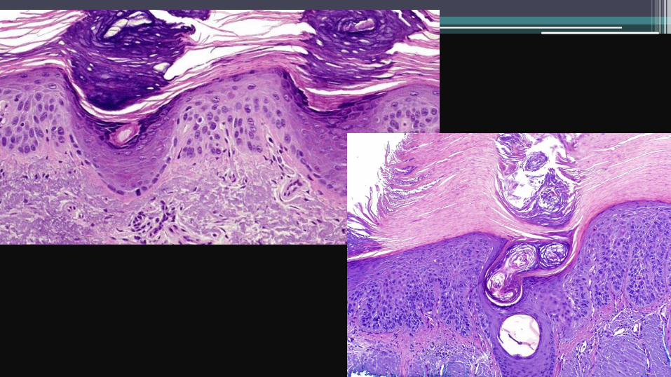
Actinic keratosis
• Usually multiple
• Covered with adherent scale
• No induration
• Histologic variants
• Hypertrophic
• Atrophic
• Bowenoid
• Acantholytic
• Pigmented


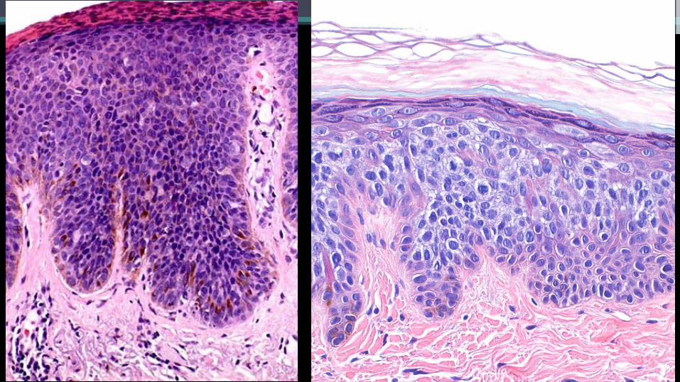
Bowen’s disease
• Usually solitary, larger than
AK
• Can occur on sun exposed or
sun protected skin
• Can occur on the penis
(erythroplasia of Queyrat)
• Progression to invasive SCC
3-5% to 11%


The eyeliner sign
www.hercampus.com

Squamous Cell Carcinoma • Sun damaged skin
• Secondarily in
• Scars (burns, Marjolin’s ulcer)
• Radiation sites
• Inflammatory dermatoses
• Lichen planus
• Lichen sclerosus
• Higher incidence in immunosuppressed patients
• Variants – classic, adenoid, mucin producing, spindle cell,
verrucous carcinoma, clear cell, basaloid, etc.

Risk stratification of cutaneous SCC
• 180,000 to 520,000 tumors/yr in the US
• 2-5% metastasis rate, typically preceded by local
recurrence and regional spread
• Brigham and Women’s Hospital high risk factors
• Depth into subcutis
• Perineural invasion of nerves > 0.1 mm
• Poor differentiation
• Other factors
Baum, et al. JAAD 2018


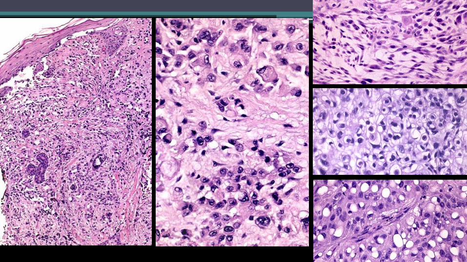


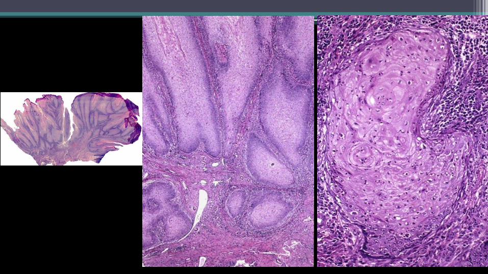
Verrucous Carcinoma
• Low grade squamous cell carcinoma
• First described in the oral cavity
• Highly differentiated, ultimately can invade deeply
• Regional metastases late, if at all

Keratoacanthoma (KA) • Solitary
• Separated from SCC in 1950
• Most on sun exposed areas
• Period of rapid growth, then involution
• Increased in immunosuppressed, Muir-Torre syndrome
• Variants – Giant KA, KA centrifugum marginatum, subungual KA
• Multiple
• Ferguson Smith (multiple self healing)
• Childhood or adolescence
• Grzybowski (eruptive)
• Adults, can involve mucosa


Differential diagnosis of SCC
• Pseudocarcinomatous hyperplasia
• Deep fungal infections
• Bromoderma
• Pyoderma vegetans
• Edges of ulcers of various causes
• Granular cell tumor
• Gout

www.the-dermatologist.com

Basal Cell Carcinoma
• 5 major variants
• Superficial
• Nodular
• Micronodular
• Infiltrating
• Fibroepithelioma of Pinkus
• Others – pigmented, keratinizing, cystic, morpheaform,
metatypical, basosquamous, etc.

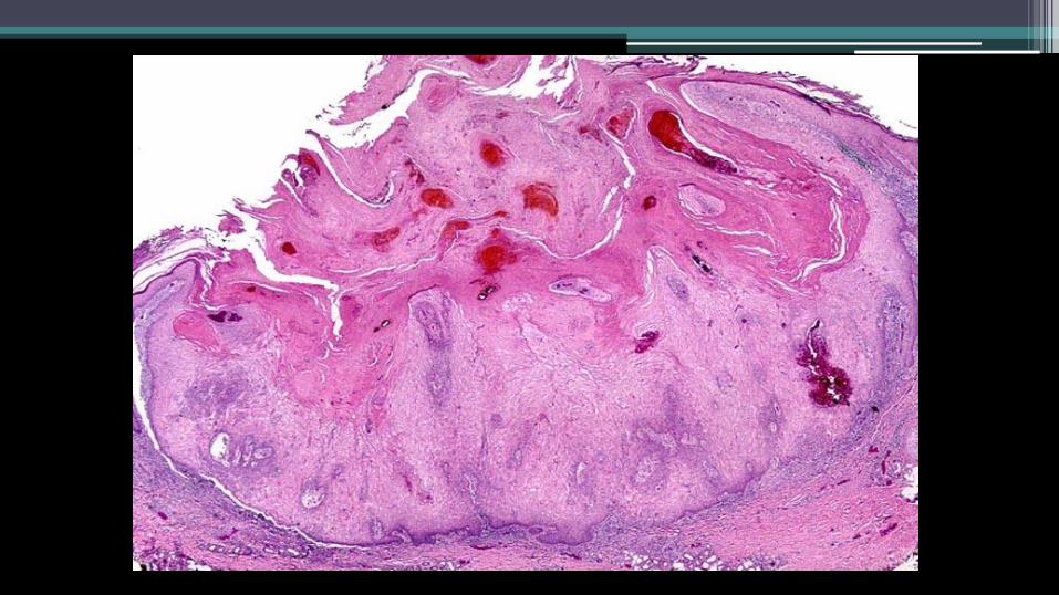
Superficial BCC
Tandon and Brodell Derm Online J 2012;18(9):1
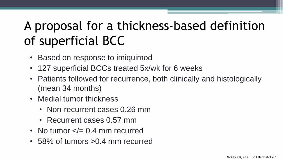
A proposal for a thickness-based definition
of superficial BCC
• Based on response to imiquimod
• 127 superficial BCCs treated 5x/wk for 6 weeks
• Patients followed for recurrence, both clinically and histologically
(mean 34 months)
• Medial tumor thickness
• Non-recurrent cases 0.26 mm
• Recurrent cases 0.57 mm
• No tumor </= 0.4 mm recurred
• 58% of tumors >0.4 mm recurred
McKay KM, et al. Br J Dermatol 2013

Nodular BCC
en.wikipedia.org

Micronodular BCC
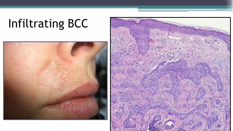
Infiltrating BCC
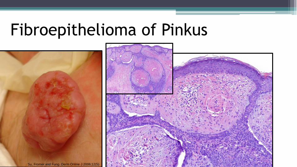
Fibroepithelioma of Pinkus
Su, Fromer and Fung. Derm Online J 2006;12(5)
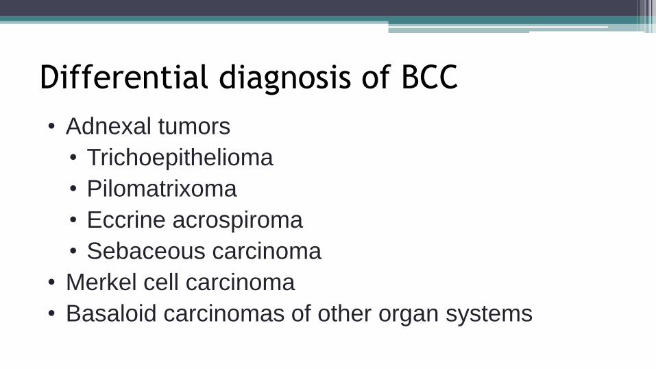
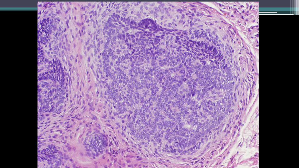
Differential diagnosis of BCC
• Adnexal tumors
• Trichoepithelioma
• Pilomatrixoma
• Eccrine acrospiroma
• Sebaceous carcinoma
• Merkel cell carcinoma
• Basaloid carcinomas of other organ systems
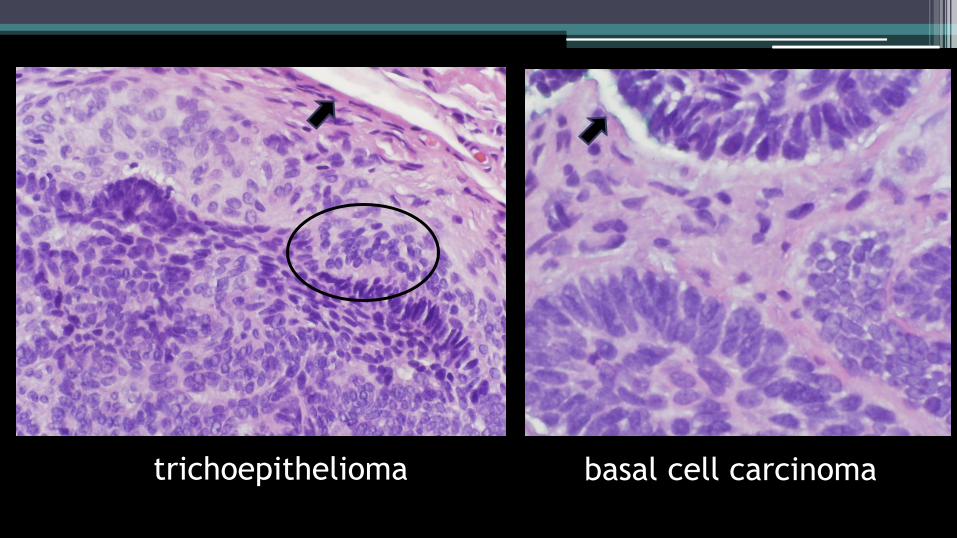
trichoepithelioma basal cell carcinoma

desmoplastic
trichoepithelioma infiltrative
basal cell carcinoma


Thank you for your attention!
Thank You! www.washingtonpost.com
Photo by Cynthia Dial

![Surgical Management of Primary Cutaneous Mucinous Carcinoma · represents 0.005% of all malignant epithelial neoplasms [1]. These adnexal tumours have been thought to be of eccrine](https://img.pdfslide.net/doc/110x75/5f0b6f0f7e708231d4307f6a/surgical-management-of-primary-cutaneous-mucinous-represents-0005-of-all-malignant.jpg)