Embed Size (px)
Citation preview
PRENATAL DIAGNOSISPrenat Diagn 2008; 28: 964–966.Published online in Wiley InterScience(www.interscience.wiley.com) DOI: 10.1002/pd.2074
RESEARCH LETTER
Prenatal sonographic diagnosis of Nager acrofacial dysostosiswith unilateral upper limb involvement
Carrie A. Couyoumjian1*, Marjorie C. Treadwell1 and Mason Barr2
1Division of Maternal-Fetal Medicine, Department of Obstetrics and Gynecology, Perinatal Assessment Center, University ofMichigan, Ann Arbor, Michigan, USA2Teratology Unit, Departments of Pediatrics, Pathology, and Obstetrics and Gynecology, University of Michigan, Ann Arbor,Michigan, USA
KEY WORDS: micrognathia; mandibulofacial; micromelia; single gene disorders; fetal and placental pathology;genetic counseling
Nager acrofacial dysostosis is a rare form of acrofa-cial dysostosis first described by Nager and de Reynier,1948. It is characterized by mandibulofacial dysosto-sis and limb anomalies. Typical craniofacial findingsinclude down-slanting palpebral fissures, malar hypopla-sia, micrognathia, abnormalities of the palate includinghigh-arched hard palate, cleft palate or bifid uvula, small,low-set and posteriorly rotated ears, and abnormalities ofthe external and middle ear. Limb malformations associ-ated with Nager acrofacial dysostosis most often involvethe radial aspect of the upper limbs and may result inthumb hypoplasia or aplasia and/or aplasia/hypoplasiaof the radius. Findings in a single affected individualare often not symmetrical but both upper limbs are usu-ally involved. Severity can range from mild hypoplasiaof the thumb to phocomelia. Radio-ulnar synostosis andabnormalities of the lower limbs have also been reported.Intelligence is typically normal. Congenital bilateralconductive hearing loss, speech difficulties and upperairway obstruction are common secondary to otologicand oral/mandibular abnormalities. A review of reportedcases indicates wide variability in manifestations andseverity of craniofacial and limb findings (Halal et al.,1983; Hall, 1989; Le Merrer et al., 1989; Danziger et al.,1990; Aylsworth et al., 1991). Cases of Nager acrofa-cial dysostosis are most often sporadic; however, sev-eral cases of autosomal dominant inheritance have beenreported (Hall, 1989; Aylsworth et al., 1991). There arealso reports of recurrence in children of unaffected par-ents, suggesting either autosomal recessive inheritance,possible germ line or somatic mosaicism, and, therefore,genetic heterogeneity (Chemke et al., 1988). Diagnosisdepends on recognition of the phenotype, most oftenwithout a family history.
A 23-year-old G2P0010 female presented for sec-ond trimester ultrasound at 20 weeks gestation based
*Correspondence to: Carrie A. Couyoumjian, Division ofMaternal-Fetal Medicine, Department of Obstetrics and Gynecol-ogy, University of Michigan, Room F4811 Mott, 1500 E. MedicalCenter Dr. Ann Arbor, Michigan 48109-0264, USA.E-mail: [email protected]
on first trimester ultrasound dating. She underwent akidney transplant 6 years prior to this pregnancy follow-ing trauma and subsequently developed type I diabetes.Hemoglobin A1C taken 1 month prior to conception waselevated at 7.2%. The patient took the following medi-cations during pregnancy: CellCept, Cleocin, Humalog,Lantus, Prednisone, Prevacid, Prograf and prenatal vita-mins. Significant family history included a brother ofthe patient who died at 18 months of age due to com-plications from severe hypoxia at birth. The remainderof the patient’s and her partner’s family history wasunremarkable.
Prenatal ultrasound findings included the following:single umbilical artery; marked micrognathia; abnormalears, which were thought to be low-set and posteriorlyrotated; and short left humerus with more pronouncedshortening of the left radius and ulna. The left handappeared anomalous with abnormal-appearing thumbsuspicious for syndactyly of the thumb and index finger.Overall long bone measurements for the right arm wereconsistent with dating; the right hand was not visualizeddue to fetal position. Fetal echocardiography revealednormal cardiac structures.
The patient and her partner were informed of theultrasound findings and were counseled regarding themost likely diagnosis of Nager acrofacial dysostosis aswell as differential diagnoses, which included TreacherCollins syndrome, Mohr syndrome, hemifacial microso-mia, or, less likely, a chromosome abnormality such astrisomy 18. Treacher Collins syndrome, another formof acrofacial dysostosis, does not exhibit associatedlimb findings. There is a form of hemifacial micro-somia that has been described with associated radialanomalies; however, the facial findings are usually notsymmetrical. Findings associated with Mohr syndromeinclude cleft palate and micrognathia; however, there isoften polysyndactyly of both the hands and feet. Whilefetuses with trisomy 18 may have severe micrognathiaand radial anomalies, there are often other associatedfindings such as heart defects, growth restriction androcker-bottom feet. After counseling, amniocentesis wasdeclined. The couple elected to undergo early induction
Copyright 2008 John Wiley & Sons, Ltd. Received: 11 March 2008Revised: 3 July 2008
Accepted: 6 July 2008
NAGER ACROFACIAL DYSOSTOSIS WITH UNILATERAL LIMB INVOLVEMENT 965
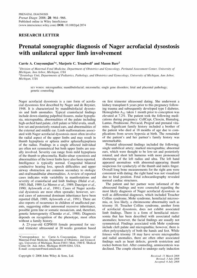
Figure 1—Postabortal photographs illustrating severe micrognathia,hypoplastic left thumb and type III microtia
of labor for termination of pregnancy with subsequentpostabortal autopsy examination. The patient underwentinduction of labor at 20 weeks and 4 days of gesta-tion and delivered a 306 g female (41.68 percentile)(Figure 1).
Anatomic pathology exam revealed down-slantingpalpebral fissures and shallow orbits. The ocular globeswere unremarkable. The nasal root was wide and themouth was microstomic with severe micrognathia. Thepalate was intact through the uvula, but forwardly dis-placed by the tongue, which was herniated into thenasopharynx and folded over the epiglottis. There wastype III microtia with external ears consisting of 2 ×1 mm double hillocks. The external auditory canals wereabsent. The temporal bone morphology was unremark-able as viewed from the interior of the skull. The leftarm was hypoplastic with flexion contractures at theshoulder and the elbow. The fingers of the left hand,especially the thumb, were short. By radiograph, theright humerus measured 35 mm, the radius 25 mm andthe ulna 25 mm. The left humerus measured 27 mm, theleft radius 15 mm and the left ulna 15 mm. The femursmeasured 30 mm each. The first left rib was hypoplastic(no sternal articulation) and the sternal-rib articulationswere asymmetrical. The lungs were hypoplastic, weigh-ing 4.29 g (0.4 percentile, −2.65 SD). The right kidneywas rotated 90◦ (hilum ventral) with a circumferentialgroove. Aside from absence of the left umbilical artery,there were no cardiovascular or other visceral anoma-lies. Brain morphology was appropriate for gestationalage. Karyotype was not obtained.
To our knowledge, this is the third reported caseof Nager acrofacial dysostosis diagnosed with prenatal
ultrasound in the absence of a positive family his-tory (Benson et al., 1988; Paladini et al., 2003). Theprior reported cases involved bilateral limb findings,unlike the unilateral findings seen in this case. Postna-tal diagnosis includes a predominance of bilateral upperlimb anomalies although unilateral findings have beendescribed. A review of the available English-languageliterature reveals that of the 80 reported cases of Nageracrofacial dysostosis, which indicated laterality of upperlimb findings, 77 (96%) were bilateral.
There is a known group of acrofacial dysostoses thatinclude a spectrum of findings involving mandibulofa-cial dysostosis with or without limb anomalies. Animalmodels of these conditions, which include Nager andMiller acrofacial dysostoses and Treacher Collins syn-drome, have suggested that the associated craniofacialanomalies are a result of cell death in the proximal aspectof the maxillary and mandibular prominences of the firstvisceral arch and in the case of Nager and Miller acrofa-cial dysostoses in the apical ectodermal ridge of the limbbud (Sulik et al., 1989). Mutations in the TCOF geneon chromosome 5 are responsible for the majority ofcases of Treacher Collins syndrome; however, the spe-cific gene mutation(s) responsible for Nager acrofacialdysostosis has not been identified. In the present case,gene testing to rule out Treacher Collins syndrome wasnot performed since physical findings associated withTreacher Collins syndrome are specifically exclusive ofthe limbs.
Since varying degrees of severity in affected individ-uals from the same family have been observed, clinicalgenetics evaluation of parents of apparently sporadiccases is necessary to provide accurate recurrence riskinformation. In our case, both parents were evaluated inan adult genetics clinic and were not found to exhibit anycharacteristics suggestive of Nager acrofacial dysostosis.
This case reinforces the spectrum of findings seenwith Nager acrofacial dysostosis. Recognition of theunilateral nature of a portion of cases may improve theability to accurately diagnose affected fetuses prenatally.Increased willingness of patients to undergo genetictesting prenatally or postnatally could certainly expandour understanding of this spectrum of diseases andultimately improve our ability to counsel patients.
REFERENCES
Aylsworth AS, Lin AE, Friedman PA. 1991. Nager AcrofacialDysostosis: Male-to-male transmission in 2 families. Am J MedGenet 41: 83–88.
Benson CB, Pober BR, Hirsh MP, Doubilet PM. 1988. Sonography ofNager acrofacial dysostosis syndrome in utero. J Ultrasound Med7: 163–167.
Chemke J, Mogilner BM, Ben-Itzhak I, Zurkowski L, Ophir D. 1988.Autosomal recessive inheritance of Nager acrofacial dysostosis.J Med Genet 25: 230–232.
Danziger I, Brodsky L, Perry R, Nusbaum S, Bernat J, Robinson L.1990. Nager’s acrofacial dysostosis. Case report and review of theliterature. Int J Pediatr Otorhinolaryngol 20: 225–240.
Halal F, Herrmann J, Pallister PD, Opitz JM, Desgranges MF,Grenier G. 1983. Differential Diagnosis of Nager AcrofacialDysostosis Syndrome: Report of four patients with Nager syndromeand discussion of other related syndromes. Am J Med Genet 14:209–224.
Copyright 2008 John Wiley & Sons, Ltd. Prenat Diagn 2008; 28: 964–966.DOI: 10.1002/pd
966 C. A. COUYOUMJIAN ET AL.
Hall BD. 1989. Nager acrofacial dysostosis; Autosomal dominantinheritance in mild to moderately affected mother and lethallyaffected phocomelic son. Am J Med Genet 33: 394–397.
Le Merrer M, Cikuli M, Ribier J, Briar ML. 1989. Acrofacialdysostoses. Am J Med Genet 33: 318–322.
Nager FR, de Reynier JP. 1948. Das Gehororgan bei den angeborenenKopfmissbildungen. Pract Otorhinolaryngol 10: 1–128.
Paladini D, Tartaglione A, Lamberti A, Lapadula C, Martinelli P.2003. Prenatal ultrasound diagnosis of Nager syndrome. UltrasoundObstet Gynecol 21: 195–197.
Sulik K, Smiley S, Turvey T, Speight H, Johnston M. 1989.Pathogenesis of cleft palate in Treacher Collins, Nager, and Millersyndromes. Cleft Palate J 26: 209–216.
Copyright 2008 John Wiley & Sons, Ltd. Prenat Diagn 2008; 28: 964–966.DOI: 10.1002/pd