Embed Size (px)
Citation preview
Received 01/27/2017 Review began 02/07/2017 Review ended 02/14/2017 Published 02/16/2017
© Copyright 2017Haider et al. This is an open accessarticle distributed under the terms ofthe Creative Commons AttributionLicense CC-BY 3.0., which permitsunrestricted use, distribution, andreproduction in any medium,provided the original author andsource are credited.
Spontaneous Intracranial HypotensionPresenting as a "Pseudo-Chiari 1"Ali S. Haider , Suraj Sulhan , Ian T. Watson , Dean Leonard , Eliel N. Arrey , Umair Khan , Phu Nguyen , Kennith F. Layton
1. Texas A&M College of Medicine 2. Houston Methodist Neurological Institute, Houston MethodistHospital, Houston, TX 3. School of Medicine, St. Georges University 4. Department of Radiology, BaylorUniversity Medical Center
Corresponding author: Umair Khan, [email protected] Disclosures can be found in Additional Information at the end of the article
AbstractSpontaneous intracranial hypotension (SIH) is classified as a decrease in cerebrospinal fluid(CSF) pressure secondary to a CSF leakage and consequent descent of the brain into theforamen magnum. Diagnosing SIH can be difficult due to its overlapping findings with Arnold-Chiari type 1 Malformation (CM1) where the cerebellar tonsils herniate into the foramenmagnum. The similarity of both conditions calls for a more reliable imaging technique tolocalize the CSF leak which could narrow the differential diagnosis and aid in choosing thecorrect treatment. Here, we present a case of a 28-year-old female, ten weeks post-partum withsymptoms similar to SIH. MRI of the brain was remarkable for tonsillar herniation below theforamen magnum. Literature was reviewed for additional neuroradiology techniques that wouldaid in narrowing our differential diagnosis. Interestingly, computed tomography-, digitalsubtraction-, and magnetic resonance myelography with intrathecal gadolinium are thepreferred techniques for diagnosis of high flow and low flow CSF leaks, respectively. Thesemodalities further aid in choosing the correct treatment while avoiding complications.Literature suggests that treatment for CM1 involves posterior fossa decompression, whereas themainstay of treatment for SIH involves an epidural blood patch (EBP). Thus, our patient wastreated with an EBP and recovered without complication.
Categories: Neurology, NeurosurgeryKeywords: arnold – chiari type 1 malformation, epidural blood patch, posterior fossa decompression,spontaneous intracranial hypotension
IntroductionSpontaneous intracranial hypotension (SIH) is a neurological condition characterized bycerebral spinal fluid (CSF) pressure below 60 mmH2O with descent of the brain into theforamen magnum [1]. Recent neuroimaging studies have shown CSF leakage to be the probableetiology [2]. Diagnosis of SIH is supported by magnetic resonance imaging (MRI), indicating thedisplacement of the brainstem and causing occipital headache and meningism. Thesesymptoms and findings also overlap with Arnold-Chiari type 1 malformation (CM1), anothercondition characterized by displacement of the cerebellar tonsils through the foramenmagnum. We present a rare and interesting case of occipital headaches with cerebellardisplacement into the foramen magnum and discuss the importance of additional imaging tonarrow the differential diagnosis. Informed consent was obtained from the patient for thisstudy.
1 1 1 1 2 3
1 4
Open Access CaseReport DOI: 10.7759/cureus.1034
How to cite this articleHaider A S, Sulhan S, Watson I T, et al. (February 16, 2017) Spontaneous Intracranial HypotensionPresenting as a "Pseudo-Chiari 1". Cureus 9(2): e1034. DOI 10.7759/cureus.1034
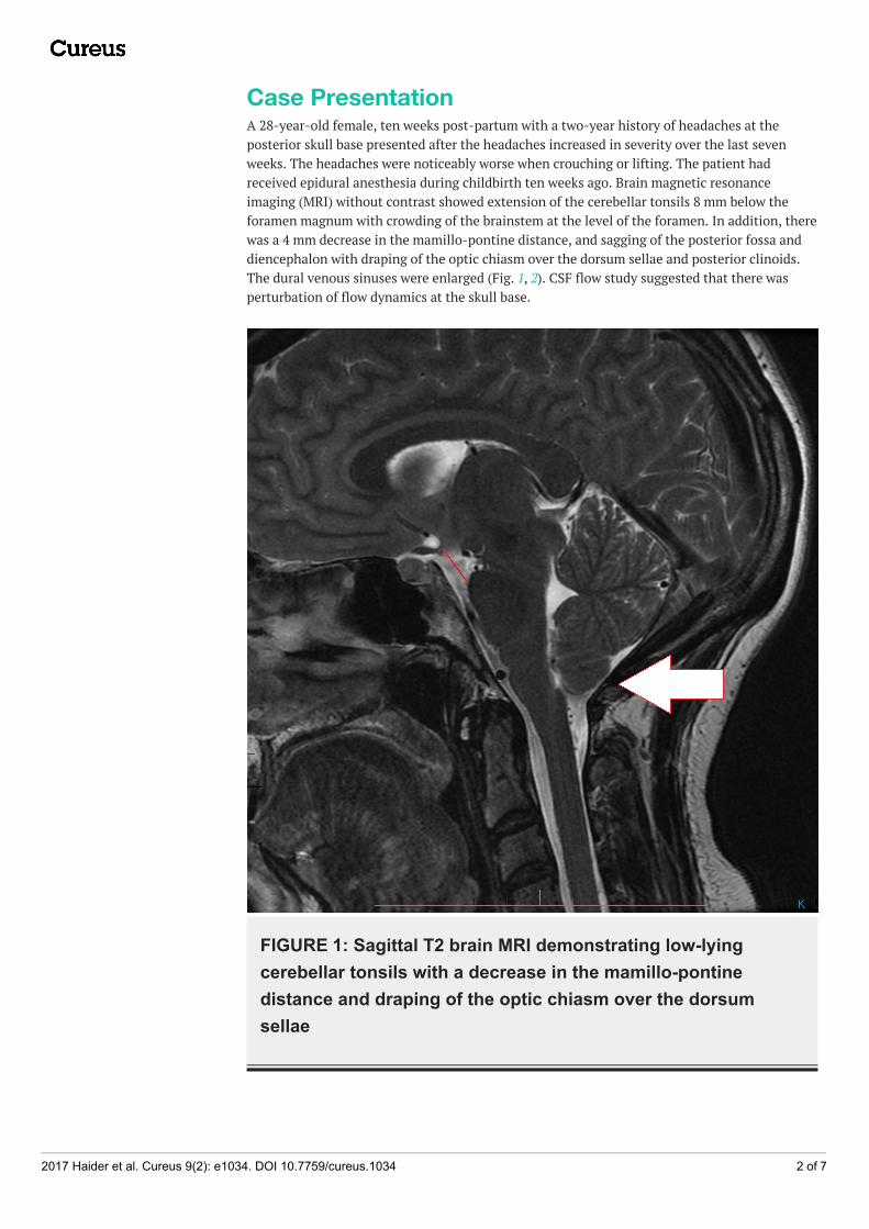
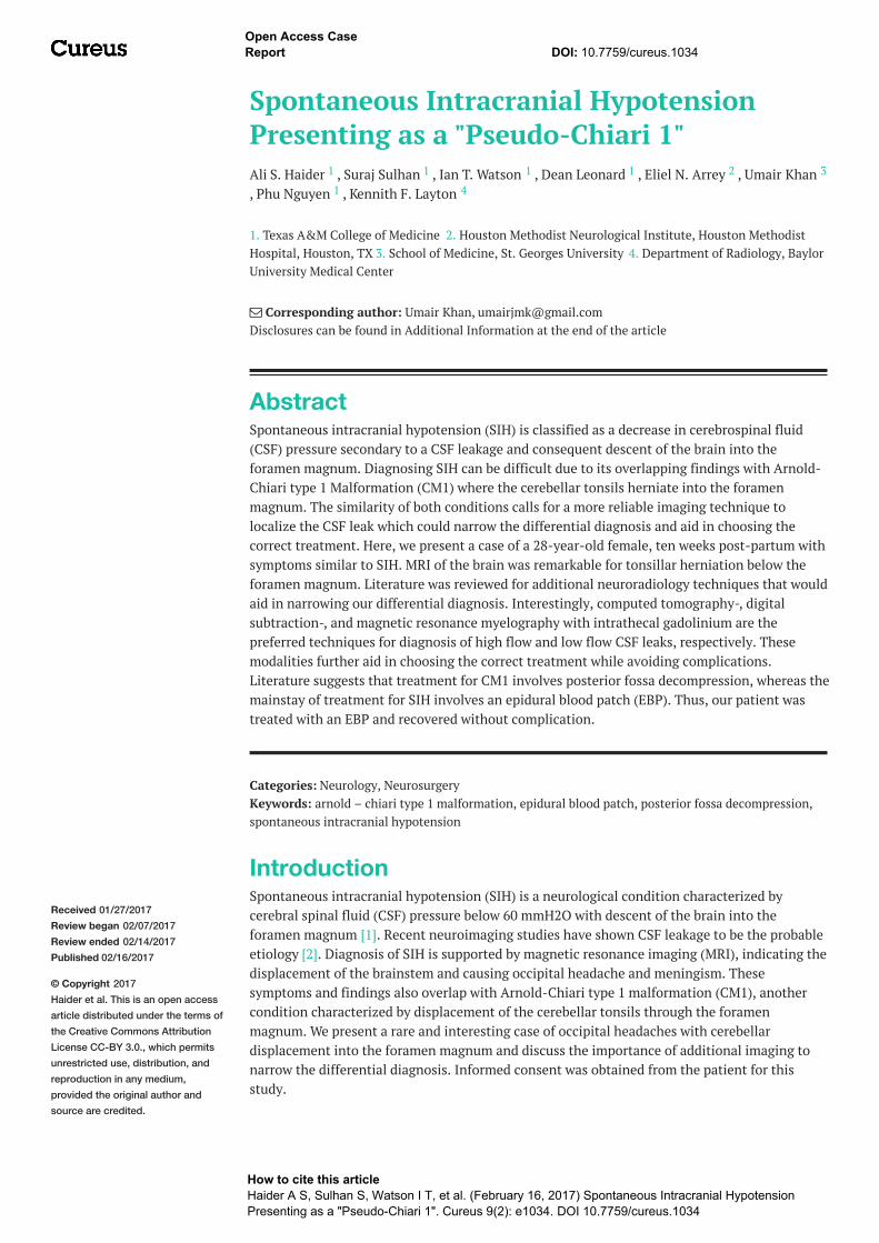
Case PresentationA 28-year-old female, ten weeks post-partum with a two-year history of headaches at theposterior skull base presented after the headaches increased in severity over the last sevenweeks. The headaches were noticeably worse when crouching or lifting. The patient hadreceived epidural anesthesia during childbirth ten weeks ago. Brain magnetic resonanceimaging (MRI) without contrast showed extension of the cerebellar tonsils 8 mm below theforamen magnum with crowding of the brainstem at the level of the foramen. In addition, therewas a 4 mm decrease in the mamillo-pontine distance, and sagging of the posterior fossa anddiencephalon with draping of the optic chiasm over the dorsum sellae and posterior clinoids.The dural venous sinuses were enlarged (Fig. 1, 2). CSF flow study suggested that there wasperturbation of flow dynamics at the skull base.
FIGURE 1: Sagittal T2 brain MRI demonstrating low-lyingcerebellar tonsils with a decrease in the mamillo-pontinedistance and draping of the optic chiasm over the dorsumsellae
2017 Haider et al. Cureus 9(2): e1034. DOI 10.7759/cureus.1034 2 of 7
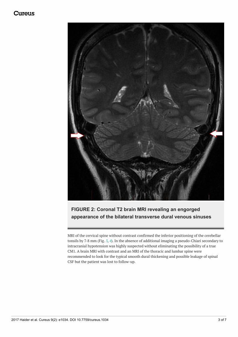
FIGURE 2: Coronal T2 brain MRI revealing an engorgedappearance of the bilateral transverse dural venous sinuses
MRI of the cervical spine without contrast confirmed the inferior positioning of the cerebellartonsils by 7-8 mm (Fig. 3, 4). In the absence of additional imaging a pseudo-Chiari secondary tointracranial hypotension was highly suspected without eliminating the possibility of a trueCM1. A brain MRI with contrast and an MRI of the thoracic and lumbar spine wererecommended to look for the typical smooth dural thickening and possible leakage of spinalCSF but the patient was lost to follow-up.
2017 Haider et al. Cureus 9(2): e1034. DOI 10.7759/cureus.1034 3 of 7
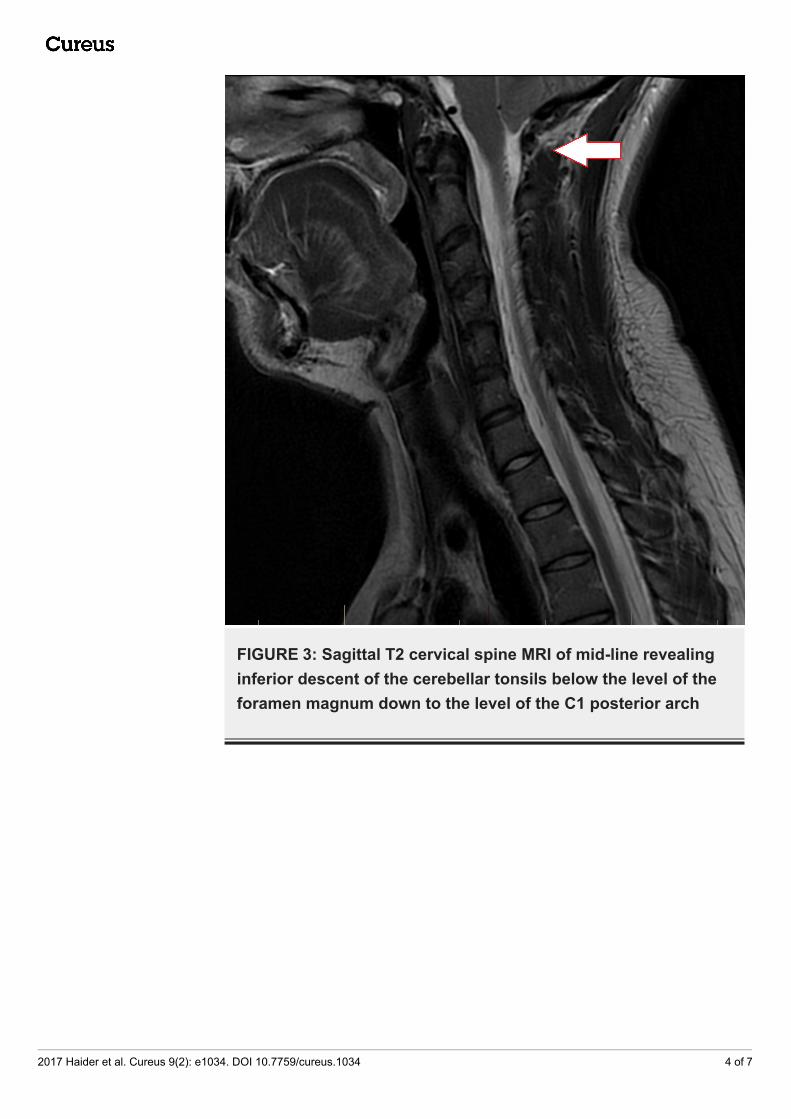
FIGURE 3: Sagittal T2 cervical spine MRI of mid-line revealinginferior descent of the cerebellar tonsils below the level of theforamen magnum down to the level of the C1 posterior arch
2017 Haider et al. Cureus 9(2): e1034. DOI 10.7759/cureus.1034 4 of 7
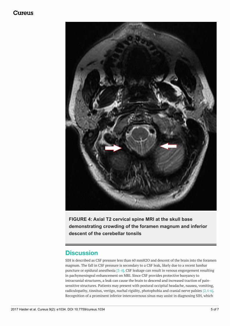
FIGURE 4: Axial T2 cervical spine MRI at the skull basedemonstrating crowding of the foramen magnum and inferiordescent of the cerebellar tonsils
DiscussionSIH is described as CSF pressure less than 60 mmH2O and descent of the brain into the foramenmagnum. The fall in CSF pressure is secondary to a CSF leak, likely due to a recent lumbarpuncture or epidural anesthesia [3-4]. CSF leakage can result in venous engorgement resultingin pachymeningeal enhancement on MRI. Since CSF provides protective buoyancy tointracranial structures, a leak can cause the brain to descend and increased traction of pain-sensitive structures. Patients may present with postural occipital headache, nausea, vomiting,radiculopathy, tinnitus, vertigo, nuchal rigidity, photophobia and cranial nerve palsies [2,4-6].Recognition of a prominent inferior intercavernous sinus may assist in diagnosing SIH, which
2017 Haider et al. Cureus 9(2): e1034. DOI 10.7759/cureus.1034 5 of 7
presents as a rounded structure at the floor of the sellae seen in 50% of patients with SIH [6].Difficulty in diagnosing SIH is due to overlapping findings with CM1, a congenital syndromecharacterized by herniation of the cerebellar tonsils into the foramen magnum withoutbrainstem involvement. The pathogenesis remains unclear, but herniation of the cerebellartonsils can lead to pain, weakness, dysphagia and sensory disturbances. An occipital headacheis one of the most common presenting symptoms occurring in 15%-98% of patients, commonlyaccentuated by postural change or exertion [3,7]. Puget, et al., however, described a pseudo-Chiari tonsillar herniation in SIH due to a CSF leak with no association to syringomyelia, butrather, pachymeningeal enhancement [4]. This highlights a key difference in the twosyndromes. The overlap of symptomology between SIH and CM1 calls for the reliance onadditional imaging to localize a CSF leak as seen in SIH. Computed tomography (CT)myelography is the preferred diagnostic modality to detect initial CSF leaks followed bydynamic CT myelography to differentiate high-flow from low-flow leaks [8]. CT myelography ordigital subtraction myelography are specific for high-flow leaks, whereas magnetic resonancemyelography with intrathecal gadolinium is preferred for low-flow leaks [8]. Treatment for SIHinvolves rest, caffeine, fluid supplementation, or an epidural blood patch (EBP). EBP is thecurrent mainstay of treatment and can be targeted to the specific site of a CSF leak on imagingor delivered blindly into the lumbar region [6]. One retrospective, non-randomized seriesshowed that 87% of patients who received a single targeted EBP experienced a benefit and100% after receiving two EBP procedures [4,6-9].
ConclusionsThough patients with SIH present with findings similar to those seen in a CM1, advances inneuroradiology and myelographic techniques can narrow the differential diagnosis andultimately support the correct treatment choice for the patient.
Additional InformationDisclosuresHuman subjects: Consent was obtained by all participants in this study. Our institution doesnot require IRB approval for our single case report. . Conflicts of interest: In compliance withthe ICMJE uniform disclosure form, all authors declare the following: Payment/services info:All authors have declared that no financial support was received from any organization for thesubmitted work. Financial relationships: All authors have declared that they have nofinancial relationships at present or within the previous three years with any organizations thatmight have an interest in the submitted work. Other relationships: All authors have declaredthat there are no other relationships or activities that could appear to have influenced thesubmitted work.
References1. Alcaide-Leon P, Lopez-Rueda A, Coblentz A, Kucharczyk W, Bharatha A, de Tilly LN:
Prominent inferior intercavernous sinus on sagittal T1-weighted images: a sign of intracranialhypotension. AJR Am J Roentgenol. 2016, 206(4):817–822. 10.2214/ajr.15.14872
2. Lagrand TJ, Beukers R: Sagging brain causing postural loss of consciousness: a case of severespontaneous intracranial hypotension. Pract Neurol. 2015, 15(6):471–473.10.1136/practneurol-2015-001183
3. Mea E, Chiapparini L, Leone M, Franzini A, Messina G, Bussone G: Chronic daily headache inthe adults: differential diagnosis between symptomatic Chiari I malformation andspontaneous intracranial hypotension. Neurol Sci. 2011, 32(3):291–294. 10.1007/s10072-011-0698-x
4. Puget S, Kondageski C, Wray A, Boddaert N, Roujeau T, Di Rocco F, Zerah M, Sainte-Rose C:Chiari-like tonsillar herniation associated with intracranial hypotension in Marfan syndrome.Case report. J Neurosurg Pediatr. 2007, 106(1):48–52. 10.3171/ped.2007.106.1.48
2017 Haider et al. Cureus 9(2): e1034. DOI 10.7759/cureus.1034 6 of 7

5. Santillan A, Aamodt W, Bhavaraju-Sanka R: Pearls & oy-sters: spontaneous intracranialhypotension and posterior reversible encephalopathy syndrome. Neurology. 2016, 86(6):55–57. 10.1212/wnl.0000000000002349
6. Smith KA: Spontaneous intracranial hypotension: targeted or blind blood patch . J ClinNeurosci. 2016, 25:10–12. 10.1016/j.jocn.2015.07.009
7. Zhao JL, Li MH, Wang CL, Meng W: A systematic review of Chiari I malformation: techniquesand outcomes. World Neurosurg. 2016, 88:7–14. 10.1016/j.wneu.2015.11.087
8. Kranz PG, Luetmer PH, Diehn FE, Amrhein TJ, Tanpitukpongse TP, Gray L: Myelographictechniques for the detection of spinal CSF leaks in spontaneous intracranial hypotension. AJRAm J Roentgenol. 2016, 206(1):8–19. 10.2214/ajr.15.14884
9. Kumar Y, Hooda K, Li S, Karol I, Muro GJ: A case of spontaneous intracranial hypotension:the role of dynamic CT myelography and epidural blood patch in diagnosis and treatment.Connecticut medicine. 2015, 79(9):547–549.
2017 Haider et al. Cureus 9(2): e1034. DOI 10.7759/cureus.1034 7 of 7